Embed Size (px)
Citation preview

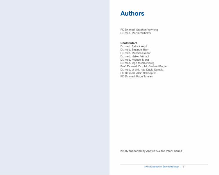
Swiss Essentials in Gastroenterology I 3
PD Dr. med. Stephan VavrickaDr. med. Martin Wilhelmi
Contributors Dr. med. Patrick AepliDr. med. Emanuel BurriDr. med. Mathias DolderDr. med. Heiko FrühaufDr. med. Michael ManzDr. med. Ingo Mecklenburg Prof. Dr. med. Dr. phil. Gerhard RoglerDr. med. et phil. nat. David SemelaPD Dr. med. Alain SchoepferPD Dr. med. Radu Tutuian
Kindly supported by AbbVie AG and Vifor Pharma
Authors
4 I Swiss Essentials in Gastroenterology Swiss Essentials in Gastroenterology I 5
Contents
Foreword 6Endoscopy in general Gastrointestinal Endoscopy 8Esophagus Reflux 12Barrett esophagus 13Eosinophilic esophagitis 14Achalasia 16Varices 20Sengstaken – Tube 23Linton – Tube 23Esophageal cancer 24Corrosive injury 31Motility of the esophagus 34Stomach Dyspepsia 42Metaplasia 43Gastritis 44Gastric ulcers 49Upper Gl bleeding 50Functional Gl disorders 54 Intestine Operation Techniques 58Chronic diarrhea 61Bacterial overgrowth 70IBD Therapy 72Clostridium difficile therapy 96Celiac disease 97Constipation therapy 100Mid Gl bleeding 104Lower Gl bleeding 105Polyps 106CRC 110HNPCC 112
High risk colorectal cancer conditions 114Ogilvie syndrome 118Diverticulitis 120Malabsorption 122Lactose intolerance 126Small intestine bacterial overgrowth 127Proctology hemorrhoids 128Proctology fissures 130Hepatology Liver segments 132Hepatopathy 134Ascites-SBP-HRS 139Child Pugh score 143HCC Hepatocellular carcinoma 144Portal vein thrombosis (PVT) 146TIPS 148MELD score 150Acute liver failure 152Hemochromatosis 153Wilson Disease 155Hepatitis B 156Hepatitis C 165Autoimmune hepatitis 173Liver and pregnancy 176Liver nodule 182Alcoholic hepatitis 184 Biliary Diseases Gallbladder polyps 186Choledocholithiasis 188Choledochal cyst 189Pancreas Pancreas cyst 190Acute pancreatitis 192Increase of Amylase and Lipase 195
6 I Swiss Essentials in Gastroenterology Swiss Essentials in Gastroenterology I 7
Foreword
Another gastroenterology handbook? During the daily clinical routine as well as during the preparation for specialist exams or student courses, it became evident that a suitable, compact reference book comprising relevant decision-making gastroenterological facts was lacking. Most of the ever-changing data usually needs to be sourced from original publications, current websites of the various gastroenterological associations (eg, SGGSSG, AASLD, AGA, DGVS), or diverse textbooks. Our aim was to speed up and simplify this time-consuming process with «Swiss Essentials in Gastroenterology».
During two meetings held in Berne (11.05.2012) and Zurich (27.06.2012), the authors came together to develop a basic structure of such a quick reference book. The outcome of our work is currently only available in this print version, as we believe in the effectiveness of the «white coat pocket guide book» even in the era of «apps» and the Internet. This actual publication cannot and should not replace any specialty text book. In addition, no responsibility can be taken for its completeness. The original source data are referenced in the current tables and figures.
Any additional and critical feedback is very welcome and should be addressed to Stephan Vavricka ([email protected]). We will try our best to incorporate your comments into a planned updated version.
In the name of all authors
S. Vavricka M. WilhelmiZurich, July 2012
Disclaimer: The publisher and authors of «Swiss Essentials in Gastroenterology» assume no obligation to update the publication and no responsibility or warranty with respect to the timeliness, accuracy, and completeness of the information, and rejects any responsibilities relating to this matter. No liability is accepted for any damage or loss arising from the use of this information. This also applies for indirect incidental or consequential damages that have occurred as a result of the use of this publication.
This book has been critically reviewed by:Dr. Melissa Wilhelmi, PhD. We thank the reviewer for her invaluable feedback and input.
8 I Swiss Essentials in Gastroenterology Swiss Essentials in Gastroenterology I 9
Endoscopy in general
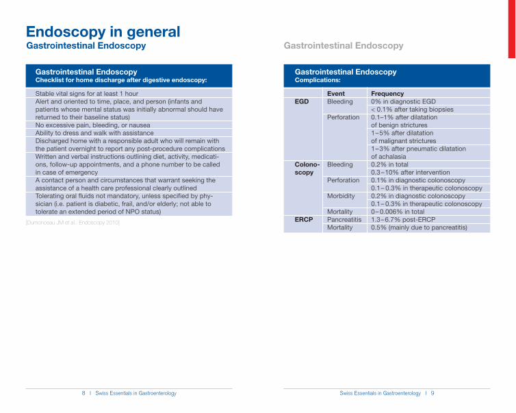
Gastrointestinal EndoscopyComplications:
EGD
Colono-scopy
ERCP
EventBleeding
Perforation
Bleeding
Perforation
Morbidity
MortalityPancreatitisMortality
Frequency0% in diagnostic EGD< 0.1% after taking biopsies0.1–1% after dilatation of benign strictures1 – 5% after dilatation of malignant strictures1 – 3% after pneumatic dilatation of achalasia0.2% in total 0.3 – 10% after intervention0.1% in diagnostic colonoscopy0.1 – 0.3% in therapeutic colonoscopy0.2% in diagnostic colonoscopy0.1 – 0.3% in therapeutic colonoscopy0 – 0.006% in total1.3 – 6.7% post-ERCP0.5% (mainly due to pancreatitis)
[Dumonceau JM et al.: Endoscopy 2010]
Gastrointestinal EndoscopyChecklist for home discharge after digestive endoscopy:
Stable vital signs for at least 1 hourAlert and oriented to time, place, and person (infants and patients whose mental status was initially abnormal should have returned to their baseline status)No excessive pain, bleeding, or nausea Ability to dress and walk with assistanceDischarged home with a responsible adult who will remain with the patient overnight to report any post-procedure complications Written and verbal instructions outlining diet, activity, medicati-ons, follow-up appointments, and a phone number to be called in case of emergencyA contact person and circumstances that warrant seeking the assistance of a health care professional clearly outlinedTolerating oral fluids not mandatory, unless specified by phy-sician (i.e. patient is diabetic, frail, and/or elderly; not able to tolerate an extended period of NPO status)
Gastrointestinal Endoscopy Gastrointestinal Endoscopy
10 I Swiss Essentials in Gastroenterology Swiss Essentials in Gastroenterology I 11
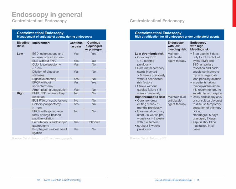
Gastrointestinal EndoscopyManagement of antiplatelet agents during endoscopy:
[Boustiere C et al.: Endoscopy 2011 and www.sggssg.ch]
Bleeding Risk:
Low
High
Continue clopidogrel or prasugrel
Yes
YesNo
No
NoYes
NoNo
NoNo
No
Unknown
No
Intervention:
EGD, colonoscopy and enteroscopy ± biopsiesEUS without FNAColonic polypectomy < 1 cmDilation of digestive stenosesDigestive stentingERCP without sphincterotomyArgon plasma coagulationEMR, ESD, or ampullary resectionEUS FNA of cystic lesionsColonic polypectomy > 1 cmERCP with sphinctero-tomy or large-balloon papillary dilationPercutaneous endoscopic gastrostomyEsophageal variceal band ligation
Continue aspirin
Yes
YesYes
Yes
YesYes
YesNo
NoYes
No
Yes
Yes
Endoscopy in general
Gastrointestinal EndoscopyRisk stratification for GI endoscopy under antiplatelet agents:
Endoscopy with high bleeding risk: Stop aspirin 5 days
only for EUS-FNA of cysts, EMR and ESD, ampullary resection and endo- scopic sphincteroto- my with large-bal- loon papillary dilation In patients taking
thienopyridine alone, it is recommended to substitute with aspirin Delay endoscopy and/
or consult cardiologist to discuss temporary cessation of thienopy- ridine: clopidogrel, 5 days prasugrel, 7 days Aspirin should be
maintained in all cases
Low thrombotic risk: Coronary DES
> 12 months previously Bare metal coronary
stents inserted > 6 weeks previously without associated risk factors Stroke without
cardiac failure > 6 weeks previouslyHigh thrombotic risk: Coronary drug
eluting stent ) 12 months previously Bare metal coronary
stent ) 6 weeks pre- viously or > 6 weeks with risk factors stroke ) 6 weeks
previously
Endoscopy with low bleeding risk:Maintain antiplatelet agent therapy
Maintain dual antiplatelet agent therapy
[Boustiere C et al.: Endoscopy 2011]
Gastrointestinal Endoscopy Gastrointestinal Endoscopy
12 I Swiss Essentials in Gastroenterology Swiss Essentials in Gastroenterology I 13
Esophagus
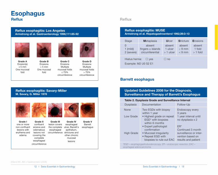
Reflux esophagitis: Los AngelesArmstrong et al. Gastroenterology 1996;111:85-92
Grade AErosion(s) n 5 mm
One mucosal fold
Grade CErosions Multiple
mucosal folds n 75%
circumference
Grade BErosions> 5 mm
One mucosal fold
Grade DErosions Multiple
mucosal folds> 75%
circumference
[Wand KK. AM J Gastroenterol 2008]
Reflux
Updated Guidelines 2008 for the Diagnosis, Surveillance and Therapy of Barrett’s Esophagus
Endoscopy every 3 years1 year interval until no dysplasia x 2
ER*Continued 3 month surveillance or inter-vention based on results and patient
None
Low Grade
High Grade
Two EGDs with biopsy within 1 year
Highest grade on repeat EGD* with biopsies within 6 months
Expert pathologist confirmation
Mucosal irregularityRepeat EGD with
biopsies to rule out EAC
Table 2. Dysplasia Grade and Surveillance IntervalDysplasia Documentation Follow-Up
*EGD – esophagogastroduodenoscopy; ER – endoscopic resection; EAC – esophageal adenocarcinoma.
Barrett esophagus
Reflux esophagitis: Savary-Miller M. Savary, G. Miller 1978
Grade Ione or more
non-confluent lesions with
erythema and edema
Grade IIIlesion covers the complete esophageal
circumference
Grade IIconfluent
erosive and edematous lesions not
covering the complete
esophageal circumference
Grade IVesophageal
ulcer, Barrett´s epithelium,
strictures and other chronic
mucosal lesions
Grade VBarrett
esophagus
Stage Metaplasia Ulcer Stricture Erosions
0 absent absent absent absent1 (mild) fingers ± islands 1 ulcer ! 9 mm 1 fold2 (severe) circumferential > 1 ulcer < 9 mm > 1 fold
Hiatus hernia: ��yes ��no
Example: M2 U0 S2 E1
Reflux esophagitis: MUSE Armstrong et al. Hepatogastroenterol 1992;39:3-13
Reflux
14 I Swiss Essentials in Gastroenterology Swiss Essentials in Gastroenterology I 15
EsophagusEosinophilic Esophagitis
Definition:
dysphagia and eosinophilic infiltrates of the esophageal mucosa [Furuta et al.: J Allergy Clin Immunl 2011]
Important facts to remember:
Male : female = 3:1 Additional atopy in 50 – 70%
Diagnosis:
concentric rings (i.e. «trachealization»), «feline» esophagus* 8 biopsies): * 15 eosinophils / high power field
[Furuta: Gastroenterology 2007]
Treatment:
6-food elimination diet (excluding cow milk protein, soy, wheat, egg, peanut, and seafood) [Kagalwalla AF et al.: Clin Gastroenterol Hepatol 2006]
- Fluticasone (e.g. Axotide spray 250 µg) or - Budesonide (e.g. Pulmicort Spray 250 µg): swallow 4 spray doses b.i.d. for 12 weeks
in refractory patients
Complications:
Esophageal fibrosis with stenosis in the long term => endoscopic dilatation in symptomatic refractory patients
Eosinophilic Esophagitis
16 I Swiss Essentials in Gastroenterology Swiss Essentials in Gastroenterology I 17
Esophagus
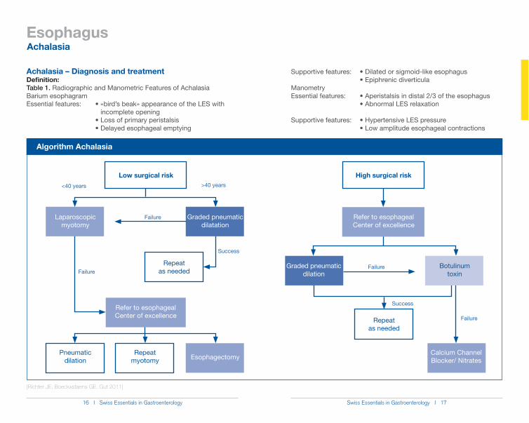
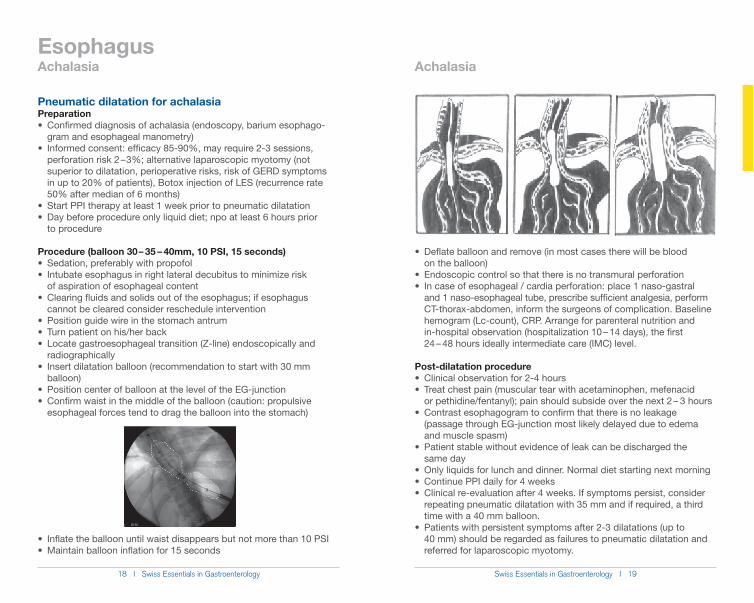
Achalasia – Diagnosis and treatmentDefinition:Table 1. Radiographic and Manometric Features of AchalasiaBarium esophagram
incomplete opening
Algorithm Achalasia
Low surgical risk
Repeat as needed
Repeat myotomy Esophagectomy
Manometry
Pneumatic dilation
Refer to esophagealCenter of excellence
Graded pneumatic dilatation
Laparoscopic myotomy
<40 years >40 years
Failure
Success
Failure
High surgical risk
Repeat as needed
Calcium Channel Blocker/ Nitrates
Failure
Success
Failure
Botulinum toxin
Graded pneumatic dilation
[Richter JE, Boeckxstaens GE. Gut 2011]
Achalasia
Refer to esophagealCenter of excellence
18 I Swiss Essentials in Gastroenterology Swiss Essentials in Gastroenterology I 19
Esophagus
Pneumatic dilatation for achalasiaPreparation
gram and esophageal manometry)
perforation risk 2 – 3%; alternative laparoscopic myotomy (not superior to dilatation, perioperative risks, risk of GERD symptoms in up to 20% of patients), Botox injection of LES (recurrence rate 50% after median of 6 months)
to procedure
Procedure (balloon 30 – 35 – 40mm, 10 PSI, 15 seconds)
of aspiration of esophageal content
cannot be cleared consider reschedule intervention
radiographically
balloon)
esophageal forces tend to drag the balloon into the stomach)
on the balloon)
and 1 naso-esophageal tube, prescribe sufficient analgesia, perform CT-thorax-abdomen, inform the surgeons of complication. Baseline hemogram (Lc-count), CRP. Arrange for parenteral nutrition and in-hospital observation (hospitalization 10 – 14 days), the first 24 – 48 hours ideally intermediate care (IMC) level.
Post-dilatation procedure
or pethidine/fentanyl); pain should subside over the next 2 – 3 hours
(passage through EG-junction most likely delayed due to edema and muscle spasm)
same day
repeating pneumatic dilatation with 35 mm and if required, a third time with a 40 mm balloon.
40 mm) should be regarded as failures to pneumatic dilatation and referred for laparoscopic myotomy.
Achalasia Achalasia
20 I Swiss Essentials in Gastroenterology Swiss Essentials in Gastroenterology I 21
Esophagus
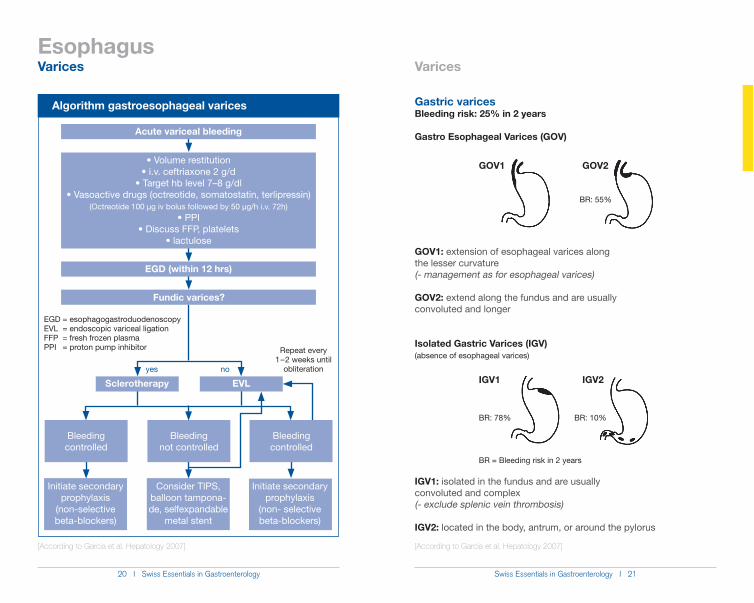
Algorithm gastroesophageal varices
Acute variceal bleeding
yes
Sclerotherapy
Bleeding controlled
Bleeding controlled
Bleeding not controlled
(Octreotide 100 µg iv bolus followed by 50 µg/h i.v. 72h)
EGD (within 12 hrs)
Fundic varices?
EVLno
Initiate secondary prophylaxis
(non-selective beta-blockers)
Initiate secondary prophylaxis
(non- selective beta-blockers)
Consider TIPS, balloon tampona-de, selfexpandable
metal stent
Repeat every1 – 2 weeks until
obliteration
EGD = esophagogastroduodenoscopyEVL = endoscopic variceal ligationFFP = fresh frozen plasmaPPI = proton pump inhibitor
[According to Garcia et al. Hepatology 2007]
Gastric varicesBleeding risk: 25% in 2 years
Gastro Esophageal Varices (GOV)
GOV1: extension of esophageal varices along the lesser curvature(- management as for esophageal varices)
GOV2: extend along the fundus and are usuallyconvoluted and longer
Isolated Gastric Varices (IGV)(absence of esophageal varices)
IGV1: isolated in the fundus and are usuallyconvoluted and complex (- exclude splenic vein thrombosis)
IGV2: located in the body, antrum, or around the pylorus
BR: 55%
BR: 10%BR: 78%
GOV1 GOV2
IGV1 IGV2
BR = Bleeding risk in 2 years
[According to Garcia et al. Hepatology 2007]
Varices Varices
22 I Swiss Essentials in Gastroenterology Swiss Essentials in Gastroenterology I 23
Esophagus
Esophageal varices – size classification and screeningSize classificationSmall varices < 5mmLarge varices > 5mm+ presence or absence of red signs (i.e. red wale marks, red spots)
_e «Red spots». f «Red wale marking».
Algorithm Varices
yes
Non-selective beta-blocker and/
or EVL (large varices or con-traindication for beta-blocker)
EGD yearly
Varices?
no
EGD = esophagogastroduodenoscopyEVL = endoscopic variceal ligation
Repeat EGD every 2 – 3 yrs
Episode of bleeding?
EVL
Cirrhosis
EGD Decompensation
[According to Garcia et al. Hepatology 2007]
Using a 100 mL syringe, slowly push air into the stomach balloon (300 mL) and seal the feeder with a clamp
noticeable (cardia approx. at 40 cm). Thereafter, slowly push 200 – 300 mL air into the stomach balloon again.
fix the tube under tension with a 500 g – 1 kg weight at the end of the bed.
with water in order to control the bleeding and aspirate the blood.
administered.
Varices Sengstaken – Tube
Esophagealballoon port
Gastricballoon port
Gastricaspiration port
Esophageal balloon (inflated)
Gastric balloon (inflated)
Gastric contentaspiration opening
Nasal cavity
Pharynx
Esophagus
Stomach
tension with 250 – 500 gweight
no yes
Linton – Tube
24 I Swiss Essentials in Gastroenterology Swiss Essentials in Gastroenterology I 25
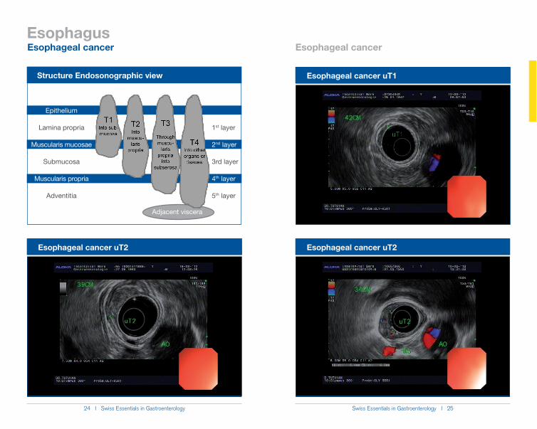
Structure Endosonographic view
Epithelium
Lamina propria
Muscularis mucosae
Submucosa
Muscularis propria
Adventitia
Adjacent viscera
1st layer
2nd layer
3rd layer
4th layer
5th layer
Esophageal cancer uT2
Esophageal cancer uT1
Esophagus
Esophageal cancer uT2
Esophageal cancer Esophageal cancer
26 I Swiss Essentials in Gastroenterology Swiss Essentials in Gastroenterology I 27
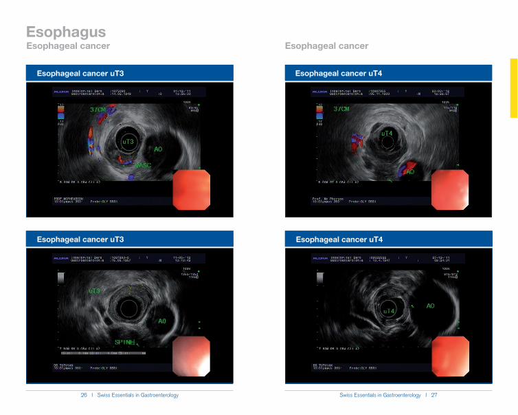
Esophageal cancer uT3
Esophageal cancer uT4Esophageal cancer uT3
Esophageal cancer uT4
EsophagusEsophageal cancer Esophageal cancer
28 I Swiss Essentials in Gastroenterology Swiss Essentials in Gastroenterology I 29
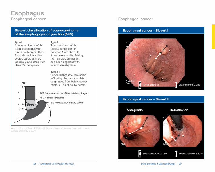
Type I:Adenocarcinoma of the distal esophagus with tumor center more than 1 cm above the endo-scopic cardia (Z-line). Generally originates from
[Adapted from HJ Stein, M Feith, JR Siewert. Cancer of the esophagogastric junction.Surgical Oncology 9 2000]
Siewert classification of adenocarcinoma of the esophagogastric junction (AEG)
Type II:True carcinoma of the cardia. Tumor center between 1 cm above to 2 cm below cardia. Arising from cardiac epithelium or a short segment with intestinal metaplasia.
Type III:Subcardial gastric carcinoma infiltrating the cardia ± distal esophagus from below (tumor center 2 – 5 cm below cardia)
{{{
5
1
0
2
5
cm
AEG I adenocarcinoma of the distal esophagus
AEG II cardia carcinoma
AEG III subcardiac gastric cancer
Esophageal cancer – Sievert I
Esophageal cancer – Sievert II
Tumor extension Distance from Z-Line
Extension above Z-Line Extension below Z-Line
Antegrade Retroflexion
EsophagusEsophageal cancer Esophageal cancer
30 I Swiss Essentials in Gastroenterology Swiss Essentials in Gastroenterology I 31
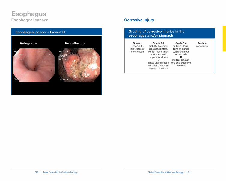
Esophageal cancer – Sievert III
Antegrade Retroflexion
Esophagus
Grading of corrosive injuries in the esophagus and/or stomach
Grade 1edema &
hyperemia of the mucosa
Grade 3 Amultiple ulcera-tions and small scattered areas
of necrosisB
multiple ulcerati-ons and extensive
necrosis
Grade 2 Afriability, bleeding, erosions, blisters,
whitish membranes, exudates, and
superficial ulcers B
grade 2a plus deep discrete or circum-ferential ulceration
Grade 4perforation
Esophageal cancer Corrosive injury
32 I Swiss Essentials in Gastroenterology Swiss Essentials in Gastroenterology I 33
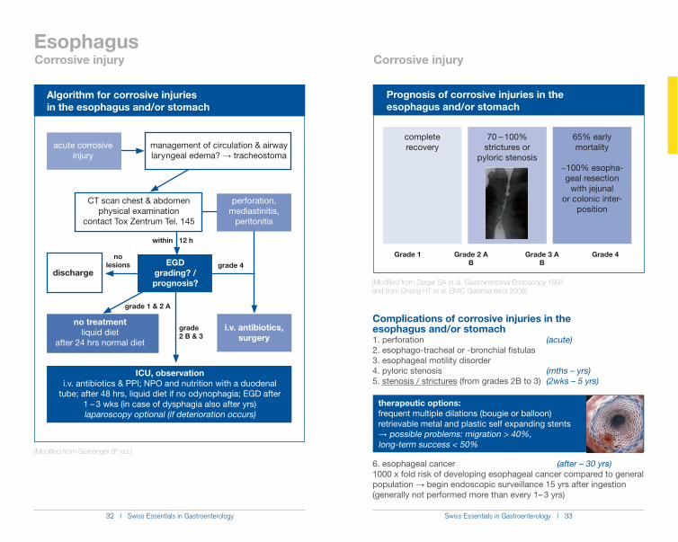
Complications of corrosive injuries in the esophagus and/or stomach1. perforation (acute)2. esophago-tracheal or -bronchial fistulas3. esophageal motility disorder4. pyloric stenosis (mths – yrs)5. stenosis / strictures (from grades 2B to 3) (2wks – 5 yrs)
6. esophageal cancer (after – 30 yrs)1000 x fold risk of developing esophageal cancer compared to general population ĺ begin endoscopic surveillance 15 yrs after ingestion (generally not performed more than every 1– 3 yrs)
Prognosis of corrosive injuries in the esophagus and/or stomach
Grade 1 Grade 3 AB
Grade 2 AB
Grade 4
complete recovery
70 – 100%strictures or
pyloric stenosis
65% early mortality
~100% esopha-geal resection
with jejunal or colonic inter-
position
therapeutic options:frequent multiple dilations (bougie or balloon)retrievable metal and plastic self expanding stentsĺ possible problems: migration > 40%, long-term success < 50%
[Modified from Zargar SA et al. Gastrointestinal Endoscopy 1991 and from Cheng HT et al. BMC Gastroenterol 2008]
Esophagus
Algorithm for corrosive injuries in the esophagus and/or stomach
acute corrosive injury
management of circulation & airway laryngeal edema? ĺ tracheostoma
perforation, mediastinitis,
peritonitis
dischargeEGD
grading? / prognosis?
no treatmentliquid diet
after 24 hrs normal diet
i.v. antibiotics,surgery
ICU, observationi.v. antibiotics & PPI; NPO and nutrition with a duodenal
tube; after 48 hrs, liquid diet if no odynophagia; EGD after 1 – 3 wks (in case of dysphagia also after yrs) laparoscopy optional (if deterioration occurs)
grade 4
grade 2 B & 3
grade 1 & 2 A
no lesions
[Modified from Sleisenger 8th ed.]
CT scan chest & abdomenphysical examination
contact Tox Zentrum Tel. 145
within 12 h
Corrosive injury Corrosive injury
34 I Swiss Essentials in Gastroenterology Swiss Essentials in Gastroenterology I 35
Esophagus
Chicago classification motility abnormalities
Individual swallowsIntegrity of contractionIntact contraction 20 mmHg isobaric contour without large or small breakWeak contraction a) Large break in the 20 mmHg isobaric contour (> 5cm in length) b) Small break in the 20 mmHg isobaric contour (2 – 5 cm in length)Failed peristalsis Minimal (< 3cm) integrity of the 20 mmHg isobaric contour distal to the proximal pressure through (P)Contraction pattern (for intact or weak peristalsis with small breaks)Premature contraction DL < 4.5 sHypercontractile DCI > 8000 mmHg-s-cmRapid contraction CFV > 9 cm s-1Normal contraction Not achieving any of the above diagnostic criteriaIntrabolus pressure pattern (30 mmHg isobaric contour)Panesophageal pressurization Uniform pressurization extending from the UES to the EGICompartmentalized eso- phageal pressurization Pressurization extending from the contractile front to a sphincterEGJ Pressurization Pressurization restricted to zone between the LES and CD in conjunction with hiatus herniaNormal pressurization No bolus pressurization > 30 mmHg
Interpretation of studiesDiagnosis Diagnostic CriteriaAchalasiaType I achalasia Classic achalasia: mean IRP > upper limit of normal, 100% failed peristalsisType II achalasia Achalasia with esophageal compression: mean IRP > upper limit of normal, no normal peristalsis, panesophageal pressurization with * 20% of swallows
Type III achalasia Mean IRP > upper limit of normal, no normal peristalsis, preserved fragments of distal peristalsis or premature (spastic) contrac- tions with * 20% of swallowsEGJ outflow obstruction Mean IRP > upper limit of normal, some instances of intact peristalsis or weak peristalsis with small breaks such that the criteria for achalasia are not metMotility Disorders (Patterns not observed in normal individuals) Distal esophageal spasm Normal mean IRP, * 20% premature contractionsHypercontractile esophagus At least one swallow DCI > 8000 mmHg- s-cm with single peaked or multipeaked (Jackhammer esophagus) contractionAbsent peristalsis Normal mean IRP, 100% of swallows with failed peristalsisPeristaltic abnormalities (Defined by exceeding statistical limits of normal)Weak peristalsis with large peristaltic Mean IRP < 15 mmHg and > 20% swallows with large breaks in the 20 mmHg defects isobaric contour (> 5cm in length)Weak peristalsis with small peristaltic Mean IRP < 15 mmHg and > 30% swallows with small breaks in the 20 mmHg defects isobaric contour (2-5cm in length)Frequent failed peristalsis > 30%, but < 100% of swallows with failed peristalsisRapid contractions with normal latency Rapid contraction with * 20% of swallows, DL > 4.5sHypertensive peristalsis Mean DCI > 5000 mmHg-s-cm, but not meeting criteria for hypercontractile (Nutcracker esophagus) esophagusNormal Not achieving any of the above diagnostic criteria[Bredenoord AJ, Fox M, Kahrilas PJ, Pandolfino JE, Schwizer W, Smout AJ; International High Resolution Manometry Working Group. Chicago classification criteria of esophageal motility disorders defined in high resolution esophageal pressure topography. Neurogastroenterol Motil. 2012]
Motility of the esophagus Motility of the esophagus
36 I Swiss Essentials in Gastroenterology Swiss Essentials in Gastroenterology I 37
Esophagus
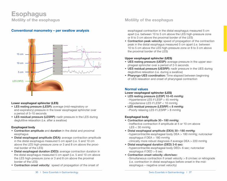
Conventional manometry – per swallow analysis
Lower esophageal sphincter (LES)LES resting pressure (LESP): average (mid-respiratory or
end-expiratory) pressure in the lower esophageal sphincter over a period of 5-10 seconds
LES residual pressure (LESRP): nadir pressure in the LES during deglutitive relaxation (i.e. after a swallow)
Esophageal bodyContraction amplitude and duration in the distal and proximal
esophagusDistal esophageal amplitude (DEA): average contraction amplitude
in the distal esophagus measured 5 cm apart (i.e. 5 and 10 cm above the LES high-pressure zone or 3 and 8 cm above the proxi- mal border of the LES)
Distal esophageal duration (DED): average contraction duration in the distal esophagus measured 5 cm apart (i.e. 5 and 10 cm above the LES high-pressure zone or 3 and 8 cm above the proximal border of the LES)
Contraction onset velocity: speed of propagation of the onset of
15 cm
10 cm
5 cm
LES (HPZ)
esophageal contraction in the distal esophagus measured 5 cm apart (i.e. between 10 to 5 cm above the LES high-pressure zone or 8 to 3 cm above the proximal border of the LES)
Contraction peak velocity: speed of propagation of the contraction peak in the distal esophagus measured 5 cm apart (i.e. between 10 to 5 cm above the LES high-pressure zone or 8 to 3 cm above the proximal border of the LES)
Upper esophageal sphincter (UES)UES resting pressure (UESP): average pressure in the upper eso-
phageal sphincter over a period of 2-5 secondsUES residual pressure (UESRP): nadir pressure in the UES during
deglutitive relaxation (i.e. during a swallow)Pharyngo-UES coordination: Time elapsed between beginning
of UES relaxation and onset of pharyngeal contraction
Normal valuesLower esophageal sphincter (LES)
LES resting pressure (LESP) 10-45 mmHg: - Hypertensive LES if LESP > 45 mmHg - Hypotensive LES if LESP < 10 mmHg
LES residual pressure (LESRP) < 8 mmHg: - Poorly relaxing LES if LESRP > 8 mmHg
Esophageal bodyContraction amplitude 30 – 180 mmHg:
- Ineffective contraction if amplitude at 5 or 10 cm above LES < 30 mmHg
Distal esophageal amplitude (DEA) 30 – 180 mmHg: - hypercontractile esophageal body DEA > 180 mmHg; nutcracker esophagus if DEA > 180 mmHg - clinically more robust diagnosis if average DEA > 220 mmHg
Distal esophageal duration (DED) 3-6 sec: - hypercontractile esophageal body DED> 6 sec; nutcracker esophagus if DED > 6 sec
Contraction onset velocity <8cm/sec: - Simultaneous contraction if onset velocity > 8 cm/sec or retrograde (i.e. contraction in distal esophagus before onset in the mid- esophagus – negative onset velocity)
Motility of the esophagus Motility of the esophagus
38 I Swiss Essentials in Gastroenterology Swiss Essentials in Gastroenterology I 39
Esophagus
Upper esophageal sphincter (UES)UES resting pressure (UESP) 30-180 mmHg:
- Hypertensive UES if UESP > 180 mmHg - Hypotensive UES if UESP < 30 mmHg
UES residual pressure (UESRP) < 8 mmHg: - Poorly relaxing UES if UESRP > 8 mmHg)
Pharyngo-UES coordination -300 to -500 msec: - Pharyngo-UES dyscoordination if delay shorter than -300 msec or onset of pharyngeal contraction before the begin of UES relaxation
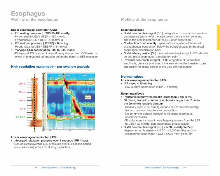
High resolution manometry – per swallow analysis
Lower esophageal sphincter (LES) Integrated relaxation pressure over 4 seconds (IRP 4-sec):
Sum of lowest average LES pressures over a 4-second period (not continuous) in the LES during deglutition
Esophageal body Distal contractile integral (DCI): Integration of contraction amplitu-
de, distance and time of the area below the transition zone and above the proximal border of the LES after deglutition
Contraction front velocity: speed of propagation of the onset of esophageal contraction below the transition zone to the distal esophageal deceleration point
Distal latency period (DL): time between beginning of UES relaxati- on and distal esophageal deceleration point
Proximal contractile integral (PCI): Integration of contraction amplitude, distance and time of the area above the transition zone and below the distal border of the UES after deglutition
Normal valuesLower esophageal sphincter (LES)
IRP 4-sec < 15 mmHg - EGJ outflow obstruction if IRP >15 mmHg
Esophageal bodyPeristaltic integrity: no breaks larger than 3 cm in the
30 mmHg isobaric contour or no breaks larger than 2 cm in the 20 mmHg isobaric contour - Breaks > 3 cm in 30 mmHg isobaric or > 2 cm in 20 mmHg isobaric contour: hypotensive contraction - No 30 mmHg isobaric contour in the distal esophagus: absent peristalsis - Simultaneous increase in esophageal pressure from the LES to UES > 30 mmHg: pan-esophageal pressurization
Distal contractile integral (DCI) < 5’000 mmHg*sec*cm - hypercontractile peristalsis if DCI > 5,000 mmHg*sec*cm - jackhammer esophagus if DCI > 8,000 mmHg*sec*cm
20 cm
15 cm
10 cm
5 cm
LES (HPZ)
Motility of the esophagus Motility of the esophagus
40 I Swiss Essentials in Gastroenterology Swiss Essentials in Gastroenterology I 41
Esophagus
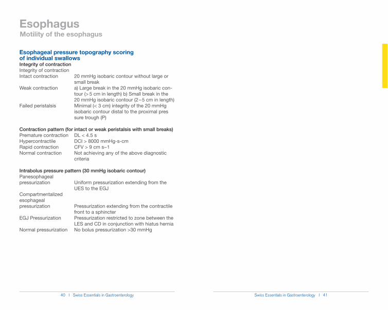
Esophageal pressure topography scoring of individual swallowsIntegrity of contractionIntegrity of contractionIntact contraction 20 mmHg isobaric contour without large or small breakWeak contraction a) Large break in the 20 mmHg isobaric con- tour (> 5 cm in length) b) Small break in the 20 mmHg isobaric contour (2 – 5 cm in length)Failed peristalsis Minimal (< 3 cm) integrity of the 20 mmHg isobaric contour distal to the proximal pres sure trough (P)
Contraction pattern (for intact or weak peristalsis with small breaks)Premature contraction DL < 4.5 sHypercontractile DCI > 8000 mmHg-s-cmRapid contraction CFV > 9 cm s!1Normal contraction Not achieving any of the above diagnostic criteria
Intrabolus pressure pattern (30 mmHg isobaric contour)Panesophageal pressurization Uniform pressurization extending from the UES to the EGJCompartmentalized esophageal pressurization Pressurization extending from the contractile front to a sphincterEGJ Pressurization Pressurization restricted to zone between the LES and CD in conjunction with hiatus herniaNormal pressurization No bolus pressurization >30 mmHg
Motility of the esophagus
42 I Swiss Essentials in Gastroenterology Swiss Essentials in Gastroenterology I 43
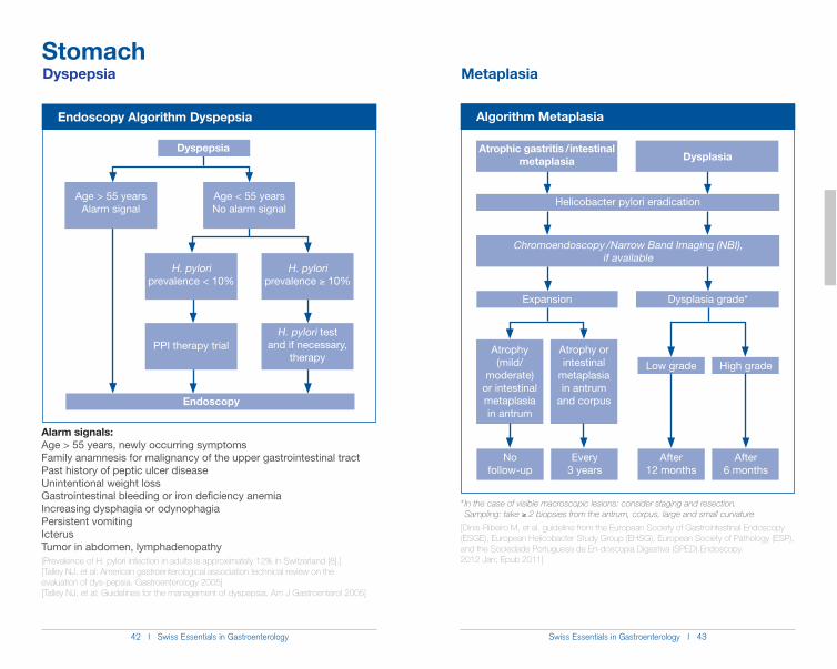
Endoscopy Algorithm Dyspepsia
Dyspepsia
Age < 55 yearsNo alarm signal
H. pylori prevalence < 10%
H. pylori prevalence * 10%
PPI therapy trialH. pylori test
and if necessary, therapy
Age > 55 yearsAlarm signal
Endoscopy
Stomach
Alarm signals:Age > 55 years, newly occurring symptomsFamily anamnesis for malignancy of the upper gastrointestinal tractPast history of peptic ulcer diseaseUnintentional weight lossGastrointestinal bleeding or iron deficiency anemiaIncreasing dysphagia or odynophagiaPersistent vomitingIcterusTumor in abdomen, lymphadenopathy[Prevalence of H. pylori infection in adults is approximately 12% in Switzerland [8].][Talley NJ, et al: American gastroenterological association technical review on the evaluation of dys-pepsia. Gastroenterology 2005][Talley NJ, et al: Guidelines for the management of dyspepsia. Am J Gastroenterol 2005]
Dyspepsia
Algorithm Metaplasia
Atrophy (mild/
moderate) or intestinal metaplasia in antrum
Helicobacter pylori eradication
Chromoendoscopy / Narrow Band Imaging (NBI), if available
Expansion Dysplasia grade*
Atrophy or intestinal
metaplasia in antrum
and corpus
Low grade High grade
After 12 months
After 6 months
No follow-up
Every 3 years
Metaplasia
Atrophic gastritis / intestinal metaplasia Dysplasia
* In the case of visible macroscopic lesions: consider staging and resection. Sampling: take * 2 biopsies from the antrum, corpus, large and small curvature
[Dinis-Ribeiro M, et al. guideline from the European Society of Gastrointestinal Endoscopy (ESGE), European Helicobacter Study Group (EHSG), European Society of Pathology (ESP), and the Sociedade Portuguesa de En-doscopia Digestiva (SPED).Endoscopy. 2012 Jan; Epub 2011]
44 I Swiss Essentials in Gastroenterology Swiss Essentials in Gastroenterology I 45
Stomach
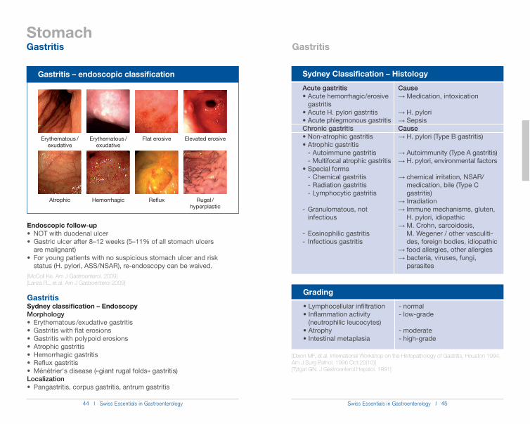
Gastritis – endoscopic classification
Erythematous /exudative
Erythematous /exudative
Flat erosive Elevated erosive
Atrophic Hemorrhagic Reflux Rugal / hyperplastic
Endoscopic follow-up
are malignant)
status (H. pylori, ASS/NSAR), re-endoscopy can be waived.[McColl Ke. Am J Gastroenterol. 2009][Lanza FL, et al. Am J Gastroenterol 2009]
GastritisSydney classification – EndoscopyMorphology
Localization
Gastritis
Sydney Classification – Histology
Grading
- normal- low-grade
- moderate- high-grade
(neutrophilic leucocytes)
Causeĺ Medication, intoxication
ĺ H. pyloriĺ SepsisCauseĺ H. pylori (Type B gastritis)
ĺ�Autoimmunity (Type A gastritis)ĺ H. pylori, environmental factors
ĺ chemical irritation, NSAR/ medication, bile (Type C gastritis)ĺ Irradiationĺ Immune mechanisms, gluten, H. pylori, idiopathicĺ M. Crohn, sarcoidosis, M. Wegener / other vasculiti- des, foreign bodies, idiopathicĺ food allergies, other allergiesĺ bacteria, viruses, fungi, parasites
Acute gastritis
gastritis
Chronic gastritis
- Autoimmune gastritis - Multifocal atrophic gastritis
- Chemical gastritis - Radiation gastritis - Lymphocytic gastritis
- Granulomatous, not infectious
- Eosinophilic gastritis- Infectious gastritis
[Dixon MF, et al. International Workshop on the Histopathology of Gastritis, Houston 1994. Am J Surg Pathol. 1996 Oct;20(10)][Tytgat GN. J Gastroenterol Hepatol. 1991]
Gastritis
46 I Swiss Essentials in Gastroenterology Swiss Essentials in Gastroenterology I 47
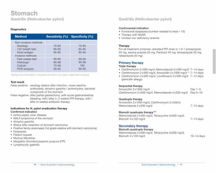
Stomach
Diagnostics
* Sydney recommendation: 6 biopsies (2x antrum, 2x corpus, large/small curvature)
Test resultFalse positive: serology (status after infection, cross-reactive antibodies), atrophic gastritis / achlorhydria, bacterial overgrowth of the stomachFalse negative: after partial gastrectomy, with acute gastrointestinal bleeding, with / after (< 2 weeks) PPI therapy, with / after (4 weeks) antibiotic therapy
Indications for H. pylori eradication therapyConfirmed indication
Method
Non-invasive methods Serology 13C breath test Stool antigenInvasive methods Fast urease test Histology* Culture PCR-analysis
Sensitivity (%)
70-9085-9585-95
90-9580-9870-9090-95
Specificity (%)
70-9085-9585-95
90-9590-98100
90-95
Gastritis (Helicobacter pylori)Gastritis (Helicobacter pylori)
Controversial indication
TherapyFor all treatment schemes: standard PPI dose is 1-0-1 (omeprazole 20 mg, esome-prazole 20 mg, Pantozol 40 mg, lansoprazole 30 mg, rabeprazole 20 mg)
Primary therapy Triple therapy
(penicillin allergy)
Sequential therapy Amoxicillin 2 x 1000 mg/d Day 1 – 5Clarithromycin 2 x 500 mg/d, Metronidazole 2 x 500 mg/d Day 6 – 10
Quadruple therapy Amoxicillin 2 x 1000 mg/d, Clarithromycin 2 x 500/d, Metronidazole 2 x 500 mg/d 7 – 14 days
Bismuth quadruple therapy*** Metronidazole 2 x 500 mg/d, Tetracycline 4x500 mg/d, Bismuth 4 x 120 mg/d 7 – 14 days
Secondary therapy Bismuth quadruple therapy Metronidazole 2 x 500 mg/d, Tetracycline 4x500 mg/d, Bismuth 4 x 120 mg/d 10 – 14 days
48 I Swiss Essentials in Gastroenterology Swiss Essentials in Gastroenterology I 49
Stomach
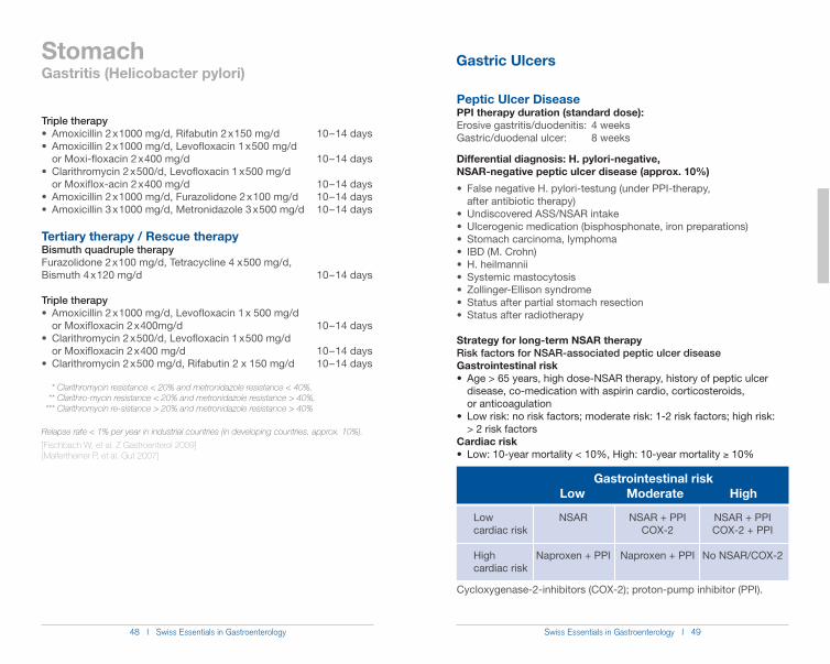
Triple therapy
or Moxi-floxacin 2 x 400 mg/d 10 – 14 days
or Moxiflox-acin 2 x 400 mg/d 10 – 14 days
Tertiary therapy / Rescue therapy Bismuth quadruple therapy Furazolidone 2 x 100 mg/d, Tetracycline 4 x 500 mg/d, Bismuth 4 x 120 mg/d 10 – 14 days
Triple therapy
or Moxifloxacin 2 x 400mg/d 10 – 14 days
or Moxifloxacin 2 x 400 mg/d 10 – 14 days
* Clarithromycin resistance < 20% and metronidazole resistance < 40%, ** Clarithro-mycin resistance < 20% and metronidazole resistance > 40%, *** Clarithromycin re-sistance > 20% and metronidazole resistance > 40%
Relapse rate < 1% per year in industrial countries (in developing countries, approx. 10%).
[Fischbach W, et al. Z Gastroenterol 2009][Malfertheiner P, et al. Gut 2007]
Gastritis (Helicobacter pylori)
Peptic Ulcer DiseasePPI therapy duration (standard dose):Erosive gastritis/duodenitis: 4 weeksGastric/duodenal ulcer: 8 weeks
Differential diagnosis: H. pylori-negative, NSAR-negative peptic ulcer disease (approx. 10%)
after antibiotic therapy)
Strategy for long-term NSAR therapyRisk factors for NSAR-associated peptic ulcer diseaseGastrointestinal risk
disease, co-medication with aspirin cardio, corticosteroids, or anticoagulation
> 2 risk factorsCardiac risk
* 10%
Cycloxygenase-2-inhibitors (COX-2); proton-pump inhibitor (PPI).
Gastrointestinal risk Low Moderate High
NSAR + PPICOX-2 + PPI
No NSAR/COX-2
Low cardiac risk
High cardiac risk
NSAR + PPICOX-2
Naproxen + PPI
NSAR
Naproxen + PPI
Gastric Ulcers
50 I Swiss Essentials in Gastroenterology Swiss Essentials in Gastroenterology I 51
Stomach
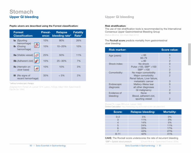
Peptic ulcers are described using the Forrest classification:
Forrest Preval- Relapse FatalityClassification: ence: bleeding rate* Rate*
* without endoscopic therapy
[Adapted from: Forrest JA: Lancet 1974; Laine L: N Engl J Med 1994; Katschinski B: Dig Dis Sci 1994]
Risk stratification: The use of risk stratification tools is recommended by the International Consensus Upper Gastrointestinal Bleeding Group [Barkun AN et al.: Ann Intern Med 2010]
The Rockall score predicts mortality from gastrointestinal ulcer bleeding:
Risk marker:
Age (years):
Shock index:
Comorbidity:
Endoscopic diagnosis:
Evidence of bleeding:
< 6060–79* 80
No shockPulse >100, SBP* >100
SBP* <100No major comorbidity
Major comorbidityRenal failure, Liver failure,
metastatic cancerMallory-Weiss tearall other diagnoses
GI malignancyNone
Blood, adherent clot, spurting vessel
Score value:
01201202
30120
2
26%
10%
11%
7%
3%
2%
Ia (Spurting hemorrhage)Ib (Oozing hemorrhage)
IIa (Visible vessel)
IIb (Adherent clot)
IIc (Hematin on ulcer base)
III (No signs of recent hemorrhage)
90%
10 – 20%
50%
25 – 30%
10%
< 5%
10%
10%
25%
10%
10%
35%
[Rockall TA, Logan RFA et al.: Lancet 1996]Online Calculator: http://www.bsg.org.uk/rockall-score-calculator.html
Score:
0–234567
8–11
Relapse bleeding:
5%11%14%24%33%44%42%
Mortality:
0%3%5%11%17%27%41%
CAVE: The Rockall score underscores the rate of recurrent bleeding*SBP = Systolic blood pressure [Church NI et al.: Gastrointest Endosc 2006]
Upper Gl bleeding Upper Gl bleeding
52 I Swiss Essentials in Gastroenterology Swiss Essentials in Gastroenterology I 53
Stomach
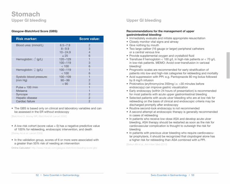
Glasgow-Blatchford Score (GBS):
The GBS is based only on clinical and laboratory variables and can be assessed in the ER without endoscopy [Blatchford O, Murray WR, Blatchford M: Lancet 2000]
A low-risk cohort (score value = 0) has a negative predictive value of 100% for rebleeding, endoscopic intervention, and death [Stanley AJ et al: Lancet 2009]
In the validation group, scores of 6 or more were associated with a greater than 50% risk of needing an interventionOnline Calculator: http://www.mdcalc.com/glasgow-blatchford-bleeding-score-gbs
Recommendations for the management of upper gastrointestinal bleeding:
or a central venous line
in low-risk patients. MEMO: Avoid over-transfusion in variceal
patients into low-and high-risk categories for rebleeding and mortality
by 8 mg/h infusion
endoscopy) can improve gastric visualization
for most patients with acute upper gastrointestinal bleeding
rebleeding on the basis of clinical and endoscopic criteria may be discharged promptly after endoscopy
in cases of rebleeding
bleeding, ASA therapy should be restarted as soon as the risk for cardiovascular complication is thought to outweigh the risk for bleeding
lar prophylaxis, it should be recognized that clopidogrel alone has a higher risk for rebleeding than ASA combined with a PPI.[Barkun AN et al.: Ann Intern Med 2010]
Risk marker:
Blood urea: (mmol/L)
Hemoglobin: ƃ (g/L)
Hemoglobin: Ƃ (g/L)
Systolic blood pressure:(mm Hg)
Pulse * 100 /min MelaenaSyncopeHepatic diseaseCardiac failure
6.5 – 7.98 – 9.9
10 – 24.9* 25
120 – 129100 – 119
< 100100 – 119
< 100100 – 10990 – 99< 90
Score value:
23461361612311222
Upper Gl bleeding Upper Gl bleeding
54 I Swiss Essentials in Gastroenterology Swiss Essentials in Gastroenterology I 55
Stomach
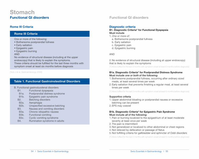
Rome III Criteria One or more of the following:
Bothersome postprandial fullness Early satiation Epigastric pain Epigastric burning
ANDNo evidence of structural disease (including at the upper endoscopy) that is likely to explain the symptoms.These criteria should be fulfilled for the last three months with symptom onset at least six months before diagnosis
Rome III Criteria
[Tack J et al. Gastroenterology 2006]
Diagnostic criteriaB1. Diagnostic Criteria* for Functional Dyspepsia Must include1. One or more of: a. Bothersome postprandial fullness b. Early satiation c. Epigastric pain d. Epigastric burning
AND
2. No evidence of structural disease (including at upper endoscopy) that is likely to explain the symptoms [*Criteria fulfilled for the last 3 months with symptom onset at least 6 months before diagnosis]
B1a. Diagnostic Criteria* for Postprandial Distress SyndromeMust include one or both of the following:1. Bothersome postprandial fullness, occurring after ordinary sized meals, at least several times per week2. Early satiation that prevents finishing a regular meal, at least several times per week[*Criteria fulfilled for the last 3 months with symptom onset at least 6 months before diagnosis]
Supportive criteria1. Upper abdominal bloating or postprandial nausea or excessive belching can be present2. EPS may coexist
B1b. Diagnostic Criteria* for Epigastric Pain SyndromeMust include all of the following:1. Pain or burning localized to the epigastrium of at least moderate severity at least once per week2. The pain is intermittent3. Not generalized or localized to other abdominal or chest regions4. Not relieved by defecation or passage of flatus5. Not fulfilling criteria for gallbladder and sphincter of Oddi disorders[*Criteria fulfilled for the last 3 months with symptom onset at least 6 months before diagnosis]
Functional GI disorders Functional GI disorders
Table 1. Functional Gastroinstestinal Disorders
B. Functional gastroduodenal disorders B1. Functional dyspepsia B1a. Postprandial distress syndrome B1b. Epigastric pain syndrome B2. Belching disorders B2a. Aerophagia B2b. Unspecified excessive belching B3. Nausea and vomiting disorders B3a. Chronic idiopathic nausea B3b. Functional vomiting B3c. Cyclic vomiting syndrome B4. Rumination syndrome in adults
56 I Swiss Essentials in Gastroenterology Swiss Essentials in Gastroenterology I 57
Stomach
Supportive criteria1. The pain may be of a burning quality but without a retrosternal competent2. The pain is commonly induced or relieved by ingestion of a meal but may occur while fasting3. Postprandial distress syndrome may coexist
B2a. Diagnostic Criteria* for AerophagiaMust include all of the following:1. Troublesome repetitive belching at least several times a week2. Air swallowing that is objectively observed or measured[*Criteria fulfilled for the last 3 months with symptom onset at least 6 months before diagnosis]
B2b. Diagnostic Criteria* for Unspecified Excessive BelchingMust include all of the following:1. Troublesome repetitive belching at least several times a week2. No evidence that excessive air swallowing underlies the symptom[*Criteria fulfilled for the last 3 months with symptom onset at least 6 months before diagnosis]
B4. Diagnostic Criteria* for Rumination SyndromeMust include both of the following:1. Persistent or recurrent regurgitation of recently ingested food into the mouth with subsequent spitting or remastication and swallowing2. Regurgitation is not preceded by retching[*Criteria fulfilled for the last 3 months with symptom onset at least 6 months before diagnosis]
Supportive criteria1. Regurgitation events are usually not preceded by nausea2. Cessation of the process when the regurgitated material becomes acidic3. Regurgitant contains recognizable food with a pleasant taste
B3a. Diagnostic Criteria* for Chronic Idiopathic NauseaMust include all of the following:1. Bothersome nausea, occurring at least several times per week2. Not usually associated with vomiting3. Absence of abnormalities at upper endoscopy or metabolic disease that explains the nausea[*Criteria fulfilled for the last 3 months with symptom onset at least 6 months before diagnosis]
B3b. Diagnostic Criteria* for Functional VomitingMust include all of the following:1. On average, 1 or more episodes of vomiting per week2. Absence of criteria for an eating disorder, rumination or major psychiatric disease according to DSM-IV3. Absence of self-induced vomiting and chronic cannabinoid use and absence of abnormalities in the central nervous system or metabolic diseases to explain the recurrent vomiting[*Criteria fulfilled for the last 3 months with symptom onset at least 6 months before diagnosis]
B3c. Diagnostic Criteria* for Cyclic Vomiting SyndromeMust include all of the following:1. Stereotypical episodes of vomiting regarding onset (acute) and duration (less than 1 week)2. Three or more discrete episodes in the prior year3. Absence of nausea and vomiting between episodes[*Criteria fulfilled for the last 3 months with symptom onset at least 6 months before diagnosis]
Supportive criterionHistory or family history of migraine headaches.
Functional GI disorders Functional GI disorders

58 I Swiss Essentials in Gastroenterology Swiss Essentials in Gastroenterology I 59
Biliopancreatic diversion with duodenal switch
Sleeve gastrectomy
Stomach
ResectedStomach
Stomach
Biliopancreatic limb
Alimentary limbCommon channel } 2.5 meter
Intestine
Roux-en-Y Gastric Bypass
Gastric pouch
Antecolic Rouxlimb (150cm)
Stomach
Biliopancreatic limb(50cm from Treitz)
Operation Techniques Operation Techniques
Gastric surgery 1
Billroth I Billroth II
60 I Swiss Essentials in Gastroenterology Swiss Essentials in Gastroenterology I 61
Gastric surgery 2
Roux-en-Ygastro-jejunostomy
Roux-en-Yesophago-jejunostomy
Jejuno-jejunostomyJejuno-jejunostomy
IntestineOperation Techniques
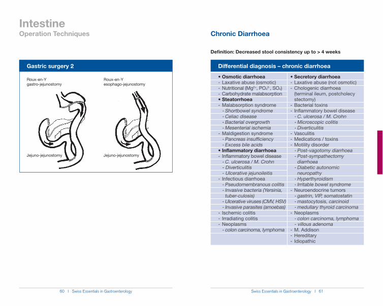
Definition: Decreased stool consistency up to > 4 weeks
Differential diagnosis – chronic diarrhoea
- Laxative abuse (osmotic)- Nutritional (Mg2+, PO4
3-, SO4)- Carbohydrate malabsorption
- Malabsorption syndrome - Shortbowel syndrome - Celiac disease - Bacterial overgrowth - Mesenterial ischemia- Maldigestion syndrome - Pancreas insufficiency - Excess bile acids
- Inflammatory bowel disease - C. ulcerosa / M. Crohn - Diverticulitis - Ulcerative jejunoileitis- Infectious diarrhoea - Pseudomembranous colitis - Invasive bacteria (Yersinia, tuber-culosis) - Ulcerative viruses (CMV, HSV) - Invasive parasites (amoebas) - Ischemic colitis- Irradiating colitis- Neoplasms - colon carcinoma, lymphoma
- Laxative abuse (not osmotic)- Chologenic diarrhoea (terminal ileum, postcholecy stectomy)- Bacterial toxins- Inflammatory bowel disease - C. ulcerosa / M. Crohn - Microscopic colitis - Diverticulitis- Vasculitis- Medications / toxins- Motility disorder - Post-vagotomy diarrhoea - Post-sympathectomy diarrhoea - Diabetic autonomic neuropathy - Hyperthyroidism - Irritable bowel syndrome- Neuroendocrine tumors - gastrin, VIP, somatostatin - mastocytosis, carcinoid - medullary thyroid carcinoma- Neoplasms - colon carcinoma, lymphoma - villous adenoma- M. Addison- Hereditary- Idiopathic
Chronic Diarrhoea
62 I Swiss Essentials in Gastroenterology Swiss Essentials in Gastroenterology I 63
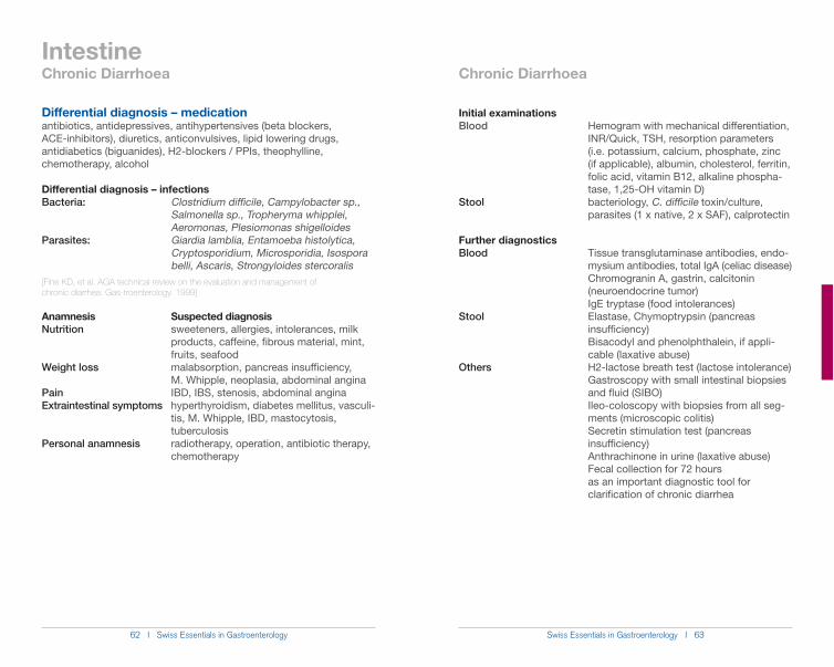
Differential diagnosis – medicationantibiotics, antidepressives, antihypertensives (beta blockers, ACE-inhibitors), diuretics, anticonvulsives, lipid lowering drugs, antidiabetics (biguanides), H2-blockers / PPIs, theophylline, chemotherapy, alcohol
Differential diagnosis – infectionsBacteria: Clostridium difficile, Campylobacter sp., Salmonella sp., Tropheryma whipplei, Aeromonas, Plesiomonas shigelloidesParasites: Giardia lamblia, Entamoeba histolytica, Cryptosporidium, Microsporidia, Isospora belli, Ascaris, Strongyloides stercoralis[Fine KD, et al. AGA technical review on the evaluation and management of chronic diarrhea. Gas-troenterology. 1999]
Anamnesis Suspected diagnosisNutrition sweeteners, allergies, intolerances, milk products, caffeine, fibrous material, mint, fruits, seafoodWeight loss malabsorption, pancreas insufficiency, M. Whipple, neoplasia, abdominal anginaPain IBD, IBS, stenosis, abdominal anginaExtraintestinal symptoms hyperthyroidism, diabetes mellitus, vasculi- tis, M. Whipple, IBD, mastocytosis, tuberculosisPersonal anamnesis radiotherapy, operation, antibiotic therapy, chemotherapy
IntestineChronic Diarrhoea
Initial examinationsBlood Hemogram with mechanical differentiation, INR/Quick, TSH, resorption parameters (i.e. potassium, calcium, phosphate, zinc (if applicable), albumin, cholesterol, ferritin, folic acid, vitamin B12, alkaline phospha- tase, 1,25-OH vitamin D)Stool bacteriology, C. difficile toxin/culture, parasites (1 x native, 2 x SAF), calprotectin
Further diagnosticsBlood Tissue transglutaminase antibodies, endo- mysium antibodies, total IgA (celiac disease) Chromogranin A, gastrin, calcitonin (neuroendocrine tumor) IgE tryptase (food intolerances)Stool Elastase, Chymoptrypsin (pancreas insufficiency) Bisacodyl and phenolphthalein, if appli- cable (laxative abuse)Others H2-lactose breath test (lactose intolerance) Gastroscopy with small intestinal biopsies and fluid (SIBO) Ileo-coloscopy with biopsies from all seg- ments (microscopic colitis) Secretin stimulation test (pancreas insufficiency) Anthrachinone in urine (laxative abuse) Fecal collection for 72 hours as an important diagnostic tool for clarification of chronic diarrhea
Chronic Diarrhoea
64 I Swiss Essentials in Gastroenterology Swiss Essentials in Gastroenterology I 65
Intestine
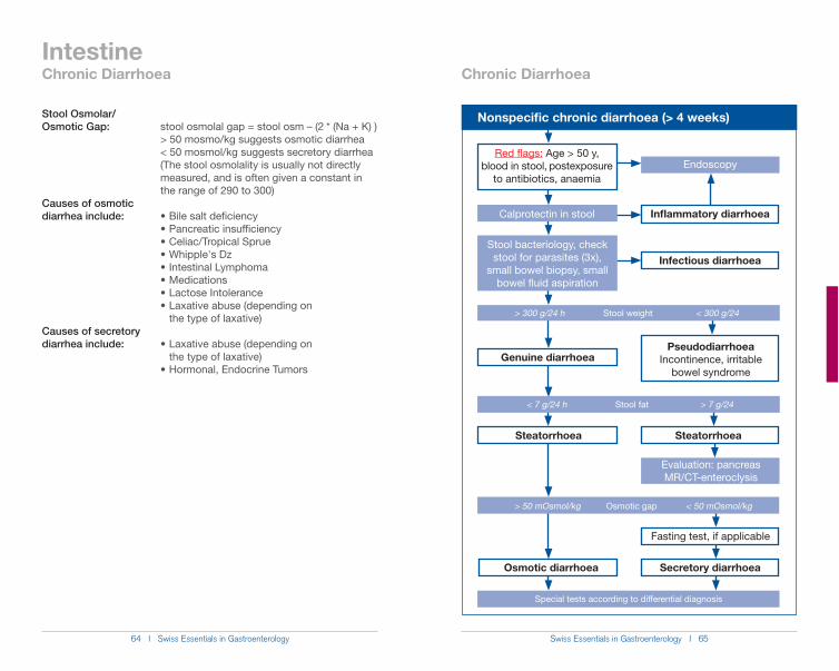
Stool Osmolar/Osmotic Gap: stool osmolal gap = stool osm – (2 * (Na + K) ) > 50 mosmo/kg suggests osmotic diarrhea < 50 mosmol/kg suggests secretory diarrhea (The stool osmolality is usually not directly measured, and is often given a constant in the range of 290 to 300)Causes of osmotic diarrhea include:
the type of laxative)Causes of secretory diarrhea include: the type of laxative)
Nonspecific chronic diarrhoea (> 4 weeks)
Red flags: Age > 50 y, blood in stool, postexposure
to antibiotics, anaemia
Endoscopy
Calprotectin in stool
Stool bacteriology, check stool for parasites (3x),
small bowel biopsy, small bowel fluid aspiration
Inflammatory diarrhoea
Infectious diarrhoea
> 300 g/24 h Stool weight < 300 g/24
< 7 g/24 h Stool fat > 7 g/24
> 50 mOsmol/kg Osmotic gap < 50 mOsmol/kg
Special tests according to differential diagnosis
Genuine diarrhoeaPseudodiarrhoea
Incontinence, irritable bowel syndrome
Steatorrhoea Steatorrhoea
Evaluation: pancreasMR/CT-enteroclysis
Osmotic diarrhoea Secretory diarrhoea
Fasting test, if applicable
Chronic Diarrhoea Chronic Diarrhoea
66 I Swiss Essentials in Gastroenterology Swiss Essentials in Gastroenterology I 67
Intestine
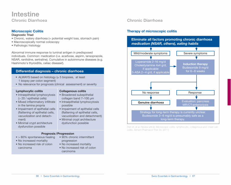
Lymphocytic colitis
(> 20 / epithelial cells)
in the lamina propria
(flattening of epithelial cells, vacuolization and detach- ment)
dysfunction possible
carcinoma
Collagenous colitis
collagen band 7–100 µm
possible
(flattening of epithelial cells, vacuolization and detachment)
dysfunction possible
progression
carcinoma
Differential diagnosis – chronic diarrhoea
Prognosis / Progression
Microscopic ColitisDiagnostic Triad
Abnormal immune response to luminal antigen in predisposed individuals. Common: medication (i.e. acarbose, aspirin, lansoprazole, NSAR, ranitidine, sertraline). Cumulative in autoimmune diseases (e.g.
* 5 biopsies, at least 1 biopsy per colon segment)
Eliminate all factors promoting chronic diarrhoeamedication (NSAR, others), eating habits
Mild/moderate symptoms
Loperamide 2-16 mg/dCholestyramine 4x4 g/d,
if applicable5-ASA 2 – 4 g/d, if applicable
Severe symptoms
No response
Genuine diarrhoea Evaluation: pancreasMR/CT-enteroclysis
Induction therapyBudesonide 9 mg/d
for 6 – 8 weeks
Response
Strategy for long-term therapy is currently unclear. Budesonide 3 – 6 mg/d is presumably safe as a
long-term therapy.
[Yen ef, et al. Review article: Microscopic colitis--lymphocytic, collagenous and «mast cell» colitis. Aliment Pharmacol Ther 34, 2011]
Chronic Diarrhoea Chronic Diarrhoea
Therapy of microscopic colitis
68 I Swiss Essentials in Gastroenterology Swiss Essentials in Gastroenterology I 69
Intestine
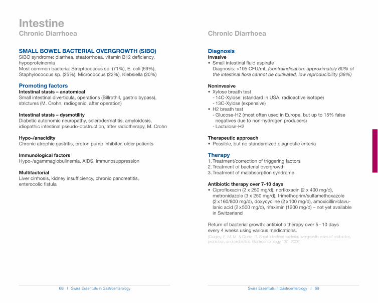
SMALL BOWEL BACTERIAL OVERGROWTH (SIBO)SIBO syndrome: diarrhea, steatorrhoea, vitamin B12 deficiency, hypoproteinemiaMost common bacteria: Streptococcus sp. (71%), E. coli (69%), Staphylococcus sp. (25%), Micrococcus (22%), Klebsiella (20%)
Promoting factorsIntestinal stasis – anatomicalSmall intestinal diverticula, operations (BillrothII, gastric bypass), strictures (M. Crohn, radiogenic, after operation)
Intestinal stasis – dysmotilityDiabetic autonomic neuropathy, sclerodermatitis, amyloidosis, idiopathic intestinal pseudo-obstruction, after radiotherapy, M. Crohn
Hypo-/anacidityChronic atrophic gastritis, proton pump inhibitor, older patients
Immunological factorsHypo-/agammaglobulinemia, AIDS, immunosuppression
MultifactorialLiver cirrhosis, kidney insufficiency, chronic pancreatitis, enterocolic fistula
Chronic Diarrhoea
DiagnosisInvasive
Diagnosis: >105 CFU/mL (contraindication: approximately 60% of the intestinal flora cannot be cultivated, low reproducibility (38%)
Noninvasive
- 14C-Xylose: (standard in USA, radioactive isotope) - 13C-Xylose (expensive)
- Glucose-H2 (most often used in Europe, but up to 15% false negatives due to non-hydrogen producers) - Lactulose-H2 Therapeutic approach
Therapy1. Treatment/correction of triggering factors2. Treatment of bacterial overgrowth3. Treatment of malabsorption syndrome
Antibiotic therapy over 7-10 days
metronidazole (3 x 250 mg/d), trimethoprim/sulfamethoxazole (2 x 160/800 mg/d), doxycycline (2 x 100 mg/d), amoxicillin/clavu- lanic acid (2 x 500 mg/d), rifaximin (1200 mg/d) – not yet available in Switzerland
Return of bacterial growth: antibiotic therapy over 5 – 10 days every 4 weeks using various medications.[Quigley, E. M. M. & Quera, R. Small intestinal bacterial overgrowth: roles of antibiotics, prebiotics, and probiotics. Gastroenterology 130, 2006]
Chronic Diarrhoea
70 I Swiss Essentials in Gastroenterology Swiss Essentials in Gastroenterology I 71
Intestine
Negative H2-Lactulose Breath Test
H2-
conc
entr
atio
n (p
pm)
H2-Content
120600 9030 150 180
time (min)
0
40
60
80
100
20
H2-Content
CH
4-co
ncen
trat
ion
(ppm
)
CH4-Content
120600 9030 150 180
time (min)
0
8
10
15
20
5
CH4-Content
Baseline 30 min 60 min 90 min 120 min 150 min 180 min0
0
0
0
0
0
0
0
0
0
0
0
0
0
0
0
0
0
0
0
0
0
0
0
0
0
0
0
0
0
0
0
0
0
0
NauseaflatulenceDiarrhea
BorgorytmusAbdominal pain
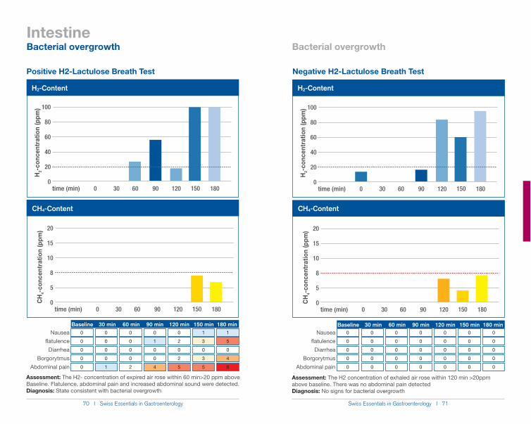
Assessment: The H2 concentration of exhaled air rose within 120 min >20ppm above baseline. There was no abdominal pain detectedDiagnosis: No signs for bacterial overgrowth
Assessment: The H2- concentration of expired air rose within 60 min>20 ppm above Baseline. Flatulence, abdominal pain and increased abdominal sound were detected. Diagnosis: State consistent with bacterial overgrowth
Positive H2-Lactulose Breath Test
H2-
conc
entr
atio
n (p
pm)
H2-Content
120600 9030 150 180
time (min)
0
40
60
80
100
20
H2-Content
CH
4-co
ncen
trat
ion
(ppm
)
CH4-Content
120600 9030 150 180
time (min)
0
8
10
15
20
5
CH4-Content
Baseline 30 min 60 min 90 min 120 min 150 min 180 min0
0
0
0
0
0
0
0
0
1
0
0
0
0
2
0
1
0
0
4
0
2
0
2
5
1
3
0
3
5
1
5
0
4
6
Nauseaflatulence
DiarrheaBorgorytmus
Abdominal pain
Bacterial overgrowth Bacterial overgrowth
72 I Swiss Essentials in Gastroenterology Swiss Essentials in Gastroenterology I 73
IntestineIBD Therapy
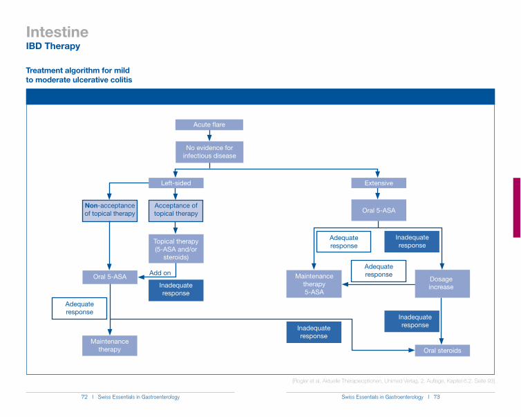
Treatment algorithm for mild to moderate ulcerative colitis
No evidence for infectious disease
Oral 5-ASAAcceptance of topical therapy
Acute flare
Extensive
Inadequate response
Inadequate response
Oral steroids
Adequate response
Maintenance therapy 5-ASA
Dosage increaseInadequate
response
Non-acceptance of topical therapy
Maintenance therapy
Topical therapy (5-ASA and/or
steroids)
Oral 5-ASA
Adequate response
Add on
Left-sided
Inadequate response
Adequate response
[Rogler et al, Aktuelle Therapieoptionen, Unimed Verlag, 2. Auflage, Kapitel 6.2. Seite 93]
74 I Swiss Essentials in Gastroenterology Swiss Essentials in Gastroenterology I 75
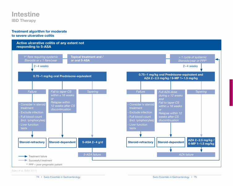
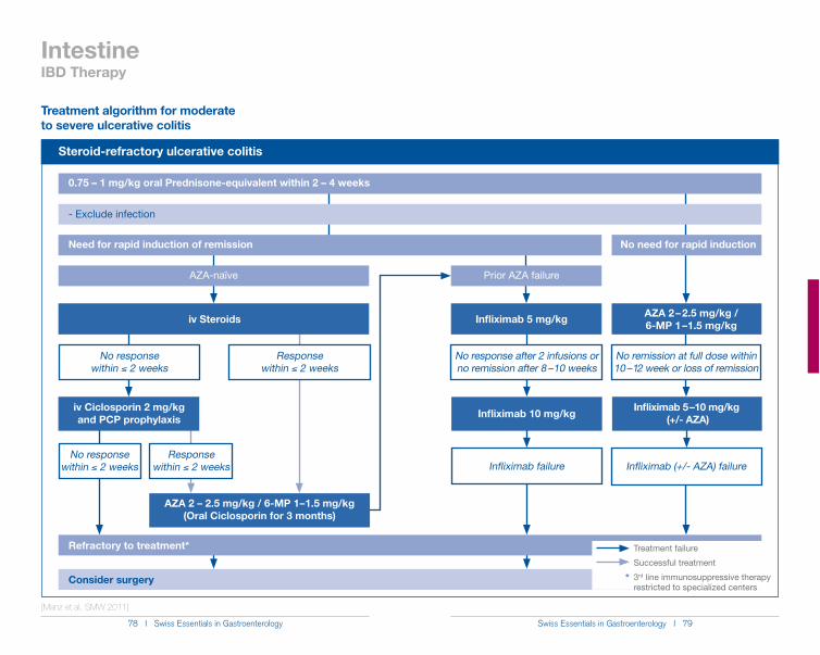
IntestineIBD Therapy
Active ulcerative colitis of any extent not responding to 5-ASA
2 – 4 weeks
Treatment algorithm for moderate to severe ulcerative colitis
Steroid-refractory Steroid-dependentSteroid-dependent
2 – 4 weeks
Steroid-refractory
Failure
- Consider iv steroid treatment- Exclude infection- Full blood count (incl. lymphocytes)- Liver function tests
Tapering
0.75 – 1 mg/kg oral Prednisone-equivalent
Fail to taper CS within ) 16 weeks or Relapse within 12 weeks after CS discontinuation
Topical treatment and / or oral 5-ASA
1st flare requiring systemic Steroids or ) 1 flare/year
> 1 cycle of systemic Steroids/year or PPP*
TaperingFailure
- Consider iv steroid treatment- Exclude infection- Full blood count (incl. lymphocytes)- Liver function tests
0.75 – 1 mg/kg oral Prednisone-equivalent and AZA 2 – 2.5 mg/kg / 6-MP 1 – 1.5 mg/kg
Full AZA dose during * 12 weeks and Fail to taper CS within ) 16 weeks or Relapse within 12 weeks after CS discontinuation
Treatment failure
Successful treatment
PPP = poor prognostic patient*
5-ASA failure
5-ASA 2 – 4 g/d
AZA failure
AZA 2 – 2.5 mg/kg / 6-MP 1 – 1.5 mg/kg
[Manz et al, SMW 2011]
76 I Swiss Essentials in Gastroenterology Swiss Essentials in Gastroenterology I 77
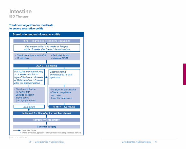
IntestineIBD Therapy
Steroid-dependent ulcerative colitis
0.75 – 1 mg/kg oral Prednisone-equivalent
Treatment algorithm for moderate to severe ulcerative colitis
Consider surgery
Fail to taper within ) 16 weeks or Relapse within 12 weeks after Steroid discontinuation
Full AZA/6-MP dose during * 12 weeks and Fail to taper CS within ) 16 weeks or Relapse within 12 weeks after CS discontinuation
AZA failure
- Check compliance to AZA/6-MP- Exclude infection- Blood count (incl. lymphocytes)
Gastrointestinal intolerance or flu-like syndrome
- No signs of pancreatitis- Check compliance and dose- Liver transaminases
AZA 2 – 2.5 mg/kg
- Check compliance to 5-ASA- Monitor blood
- Exclude infection- Measure TPMT
Infliximab 5 – 10 mg/kg (or oral Tacrolimus)
Refractory to treatment*
Treatment failure3rd line immunosuppressive therapy restricted to specialized centers*
6-MP 1 – 1.5 mg/kg
[Manz et al, SMW 2011]
78 I Swiss Essentials in Gastroenterology Swiss Essentials in Gastroenterology I 79
IntestineIBD Therapy
Steroid-refractory ulcerative colitis
Treatment algorithm for moderate to severe ulcerative colitis
AZA-naïve
Consider surgery
AZA 2 – 2.5 mg/kg / 6-MP 1 – 1.5 mg/kg
No remission at full dose within 10 – 12 week or loss of remission
No need for rapid induction
0.75 – 1 mg/kg oral Prednisone-equivalent within 2 – 4 weeks
- Exclude infection
Response within ) 2 weeks
Prior AZA failure
Infliximab 5 mg/kg
No response after 2 infusions or no remission after 8 – 10 weeks
Need for rapid induction of remission
No response within ) 2 weeks
iv Steroids
Response within ) 2 weeks
iv Ciclosporin 2 mg/kg and PCP prophylaxis
No response within ) 2 weeks
Infliximab 5 – 10 mg/kg (+/- AZA)Infliximab 10 mg/kg
Infliximab failure Infliximab (+/- AZA) failure
Refractory to treatment*
AZA 2 – 2.5 mg/kg / 6-MP 1 – 1.5 mg/kg (Oral Ciclosporin for 3 months)
Treatment failure
Successful treatment
3rd line immunosuppressive therapy restricted to specialized centers
*
[Manz et al, SMW 2011]
80 I Swiss Essentials in Gastroenterology Swiss Essentials in Gastroenterology I 81
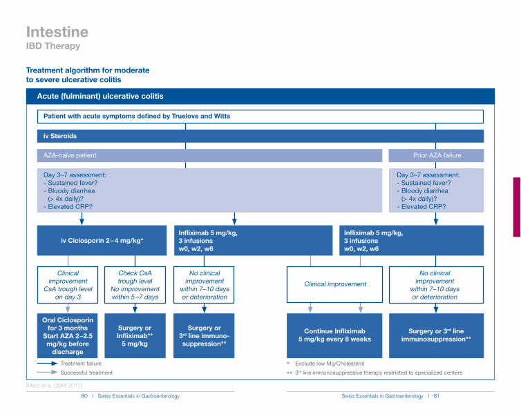
IntestineIBD Therapy
Acute (fulminant) ulcerative colitis
Treatment algorithm for moderate to severe ulcerative colitis
Prior AZA failure
iv Steroids
AZA-naïve patient
Treatment failure
Successful treatment***
Day 3–7 assessment:- Sustained fever?- Bloody diarrhea (> 4x daily)?- Elevated CRP?
Day 3–7 assessment:- Sustained fever?- Bloody diarrhea (> 4x daily)?- Elevated CRP?
Patient with acute symptoms defined by Truelove and Witts
No clinical improvement
within 7–10 days or deterioration
iv Ciclosporin 2 – 4 mg/kg*Infliximab 5 mg/kg, 3 infusions w0, w2, w6
Continue Infliximab 5 mg/kg every 8 weeks
Oral Ciclosporin for 3 months
Start AZA 2 – 2.5 mg/kg before
discharge
Surgery or 3rd line immuno-suppression**
Surgery or Infliximab**
5 mg/kg
Surgery or 3rd line immunosuppression**
Clinical improvement
CsA trough level on day 3
Check CsA trough level
No improvement within 5 –7 days
No clinical improvement
within 7–10 days or deterioration
Clinical improvement
Exclude low Mg/Cholesterol
3rd line immunosuppressive therapy restricted to specialized centers
Infliximab 5 mg/kg, 3 infusions w0, w2, w6
[Manz et al, SMW 2011]
82 I Swiss Essentials in Gastroenterology Swiss Essentials in Gastroenterology I 83
IntestineIBD Therapy
Treatment algorithm for maintenance of remission of ulcerative colitis
Chronic active cause(steroid-dependency)
Relapse
Relapse
Colectomy
In remission
5-ASA, topical or systemic or in combination;
1.5g or 3g
Azathioprin
Relapse
No Remission
Cylcosporin? Tacrolimus? Infliximab?
[Rogler et al, Aktuelle Therapieoptionen, Unimed Verlag, 2. Auflage, Kapitel 6.2. Seite 97]
84 I Swiss Essentials in Gastroenterology Swiss Essentials in Gastroenterology I 85
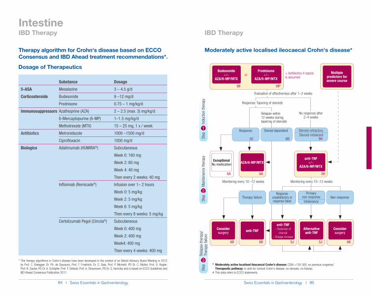
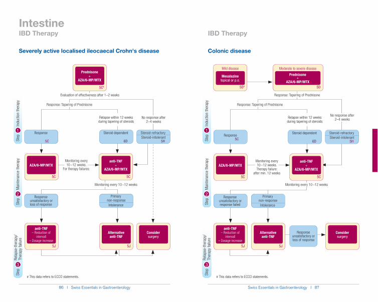
Intestine
Therapy algorithm for Crohn's disease based on ECCO Consensus and IBD Ahead treatment recommendations*.
Moderately active localised ileocaecal Crohn's disease*
IBD Therapy IBD Therapy
Step
Rel
apse
-ther
apy/
Ther
apy
failu
reSt
ep
M
aint
enan
ce th
erap
ySt
ep
I
nduc
tion
ther
apy
2
3
1
Dosage of Therapeutics
Substance Dosage
5-ASA Mesalazine 3 – 4.5 g/d
Corticosteroide Budesonide 9 –12 mg/d
Prednisone 0.75 – 1 mg/kg/d
Immunosuppressors Azathioprine (AZA) 2 – 2.5 (max. 3) mg/kg/d
6-Mercaptopurine (6-MP) 1–1.5 mg/kg/d
Methotrexate (MTX) 15 – 25 mg, 1 x / week
Antibiotics Metronidazole 1000 –1500 mg/d
Ciprofloxacin 1000 mg/d
Biologics Adalimumab (HUMIRA®) Subcutaneous
Week 0: 160 mg
Week 2: 80 mg
Week 4: 40 mg
Then every 2 weeks: 40 mg
Infliximab (Remicade®) Infusion over 1– 2 hours
Week 0: 5 mg/kg
Week 2: 5 mg/kg
Week 6: 5 mg/kg
Then every 8 weeks: 5 mg/kg
Certolizumab Pegol (Cimzia®) Subcutaneous
Week 0: 400 mg
Week 2: 400 mg
Week4: 400 mg
Then every 4 weeks: 400 mg
+ Antibiotics if sepsis is assumed
Response
5C
Steroid dependent
6D
6A
ExceptionalNo medication
6A
AZA/6-MP/MTX
5H
anti-TNF+/–
AZA/6-MP/MTX
6B
anti-TNF
5J
Alternativeanti-TNF
Response unsatisfactory or response failed
5J
anti-TNF– Reduction of
intervall– Dosage increase
5B 5B#
Budesonide+/–
AZA/6-MP/MTX
Prednisone+/–
AZA/6-MP/MTXor
6B
Consider surgery
Therapy failure
6B
Consider surgery
Non-response
Multiple predictors for severe course
Evaluation of effectiveness after 1–2 weeks
Relapse within 12 weeks during tapering of steroids
Monitoring every 10 –12 weeks
Primary non-response
Intolerance
Steroid-refractorySteroid-intolerant
5H
Monitoring every 10 –12 weeks
Response: Tapering of steroids
No response after 2–4 weeks
* Moderately active localised ileocaecal Crohn's disease: CDAI >150-300, no previous surgeries/ Therapeutic pathway: is valid for luminal Crohn's disease, no stenosis, no fistulas. # This data refers to ECCO statements.
* This therapy algorithms in Crohn‘s disease have been developed in the context of an Abbott Advisory Board Meeting in 2012 by Prof. C. Braegger, Dr. Ph. de Saussure, Prof. F. Froehlich, Dr. C. Gaia, Prof. P. Michetti, PD Dr. C. Mottet, Prof. G. Rogler, Prof. B. Sauter, PD Dr. A. Schöpfer, Prof. F. Seibold, Prof. A. Straumann, PD Dr. S. Vavricka and is based on ECCO Guidelines and IBD Ahead Consensus Publication 2011.
86 I Swiss Essentials in Gastroenterology Swiss Essentials in Gastroenterology I 87
Intestine
Severely active localised ileocaecal Crohn's disease Colonic disease
IBD Therapy IBD Therapy
Monitoring every 10 –12 weeks.
For therapy failures:
5C#
Prednisone+
AZA/6-MP/MTX
Response
5C
5C
anti-TNF+
AZA/6-MP/MTX
Response unsatisfactory or loss of response
5J
Alternativeanti-TNF
5J
anti-TNF– Reduction of
intervall– Dosage increase
Steroid dependent
6D
5C
AZA/6-MP/MTX
Consider surgery
Evaluation of effectiveness after 1–2 weeks
Monitoring every 10 –12 weeks
No response after 2–4 weeks
Response: Tapering of Prednisone
Relapse within 12 weeks during tapering of steroids
Steroid-refractorySteroid-intolerant
5H
Primary non-response Intolerance
Moderate to severe disease
5D
Prednisone+
AZA/6-MP/MTX
5J
Alternativeanti-TNF
5J
anti-TNF– Reduction of
intervall– Dosage increase
Mild disease
5D#
Mesalazinetopical or p.o.
Consider surgery
Response unsatisfactory or loss of response
Monitoring every 10 –12 weeks. Therapy failure:
after min. 12 weeks
Response5C
5C
anti-TNF+
AZA/6-MP/MTX
Response unsatisfactory or response failed
Steroid dependent
6D
5C
AZA/6-MP/MTX
Response: Tapering of Prednisone
Monitoring every 10 –12 weeks
No response after 2–4 weeks
Response: Tapering of Prednisone
Relapse within 12 weeks during tapering of steroids
Steroid-refractorySteroid-intolerant
5H
Primary non-responseIntolerance
# This data refers to ECCO statements. # This data refers to ECCO statements.
Step
Rel
apse
-ther
apy/
Ther
apy
failu
reSt
ep
M
aint
enan
ce th
erap
ySt
ep
I
nduc
tion
ther
apy
2
3
1
Step
Rel
apse
-ther
apy/
Ther
apy
failu
reSt
ep
M
aint
enan
ce th
erap
ySt
ep
I
nduc
tion
ther
apy
2
3
1
88 I Swiss Essentials in Gastroenterology Swiss Essentials in Gastroenterology I 89
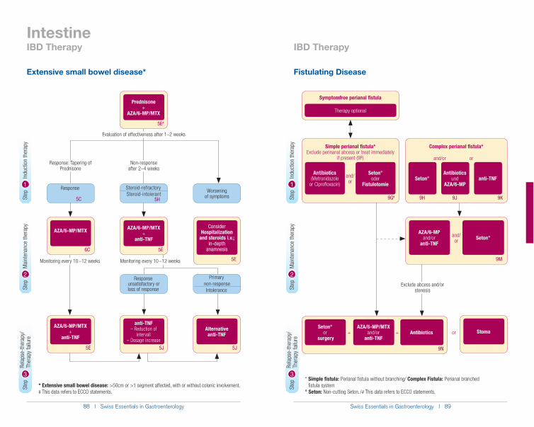
Intestine
Extensive small bowel disease* Fistulating Disease
IBD Therapy IBD Therapy
5J
anti-TNF– Reduction of
intervall– Dosage increase
5J
Alternativeanti-TNF
5E
AZA/6-MP/MTX +
anti-TNF
6C
AZA/6-MP/MTX
5E
AZA/6-MP/MTX +
anti-TNF
5E#
Prednisone+
AZA/6-MP/MTX
Response
5C
Response unsatisfactory or loss of response
Evaluation of effectiveness after 1–2 weeks
5E
Consider Hospitalization
and steroids i.v.; in-depth
anamnesis
Worsening of symptoms
* Extensive small bowel disease: >50cm or >1 segment affected, with or without colonic involvement.# This data refers to ECCO statements.
Non-response after 2–4 weeks
Steroid-refractorySteroid-intolerant
5H
Primary non-response Intolerance
Response: Tapering of Prednisone
Monitoring every 10 –12 weeks Monitoring every 10 –12 weeks
Simple perianal fistula*Exclude perinanal abcess or treat immediately
if present (9F)
9G#
Antibiotics (Metronidazole
or Ciprofloxacin)
Seton°oder
Fistulotomie
and/or
Complex perianal fistula*
9H 9J 9K
Seton°Antibiotics
undAZA/6-MP
anti-TNF
and/or or
Symptomfree perianal fistula
Therapy optional
9M
AZA/6-MPand/or
anti-TNFSeton°and/
or
9N
AZA/6-MP/MTXand/or
anti-TNFAntibiotics+
Seton°or
surgery+ or Stoma
* Simple fistula: Perianal fistula without branching/ Complex Fistula: Perianal branched fistula system
° Seton: Non-cutting Seton. / # This data refers to ECCO statements.
Exclude abcess and/or stenosis
Step
Rel
apse
-ther
apy/
Ther
apy
failu
reSt
ep
M
aint
enan
ce th
erap
ySt
ep
I
nduc
tion
ther
apy
2
3
1
Step
Rel
apse
-ther
apy/
Ther
apy
failu
reSt
ep
M
aint
enan
ce th
erap
ySt
ep
I
nduc
tion
ther
apy
2
3
1
90 I Swiss Essentials in Gastroenterology Swiss Essentials in Gastroenterology I 91
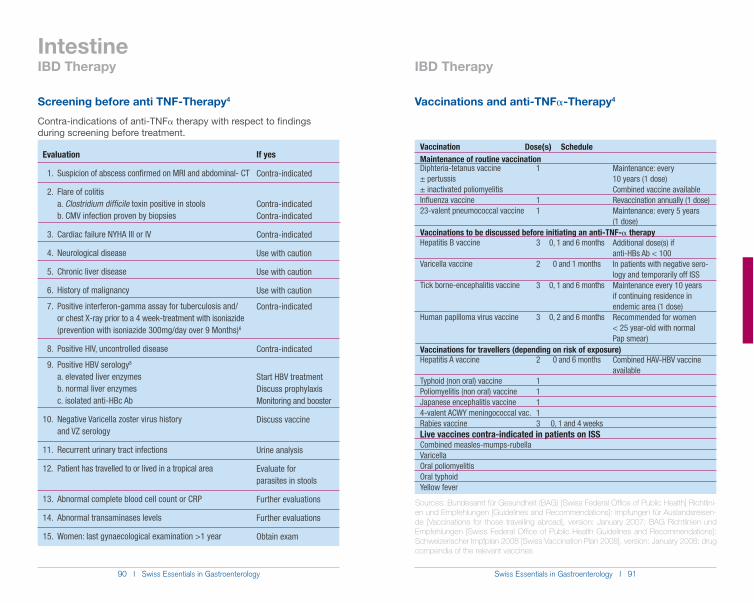
Intestine
Screening before anti TNF-Therapy4
Contra-indications of anti-TNF! therapy with respect to findings during screening before treatment.
Evaluation
1. Suspicion of abscess confirmed on MRI and abdominal- CT
2. Flare of colitis a. Clostridium difficile toxin positive in stools b. CMV infection proven by biopsies
3. Cardiac failure NYHA III or IV
4. Neurological disease
5. Chronic liver disease
6. History of malignancy
7. Positive interferon-gamma assay for tuberculosis and/ or chest X-ray prior to a 4 week-treatment with isoniazide (prevention with isoniazide 300mg/day over 9 Months)6
8. Positive HIV, uncontrolled disease
9. Positive HBV serology8
a. elevated liver enzymes b. normal liver enzymes c. isolated anti-HBc Ab
10. Negative Varicella zoster virus history and VZ serology
11. Recurrent urinary tract infections
12. Patient has travelled to or lived in a tropical area
13. Abnormal complete blood cell count or CRP
14. Abnormal transaminases levels
15. Women: last gynaecological examination >1 year
If yes
Contra-indicated
Contra-indicatedContra-indicated
Contra-indicated
Use with caution
Use with caution
Use with caution
Contra-indicated
Contra-indicated
Start HBV treatmentDiscuss prophylaxisMonitoring and booster
Discuss vaccine
Urine analysis
Evaluate for parasites in stools
Further evaluations
Further evaluations
Obtain exam
Vaccinations and anti-TNF!-Therapy4
Vaccination
Diphteria-tetanus vaccine± pertussis± inactivated poliomyelitisInfluenza vaccine23-valent pneumococcal vaccine
Hepatitis B vaccine
Varicella vaccine
Tick borne-encephalitis vaccine
Human papilloma virus vaccine
Hepatitis A vaccine
Typhoid (non oral) vaccinePoliomyelitis (non oral) vaccineJapanese encephalitis vaccine4-valent ACWY meningococcal vac.Rabies vaccine
Combined measles-mumps-rubellaVaricellaOral poliomyelitisOral typhoidYellow fever
Dose(s)
1
11
3
2
3
3
2
11113
Schedule
0, 1 and 6 months
0 and 1 months
0, 1 and 6 months
0, 2 and 6 months
0 and 6 months
0, 1 and 4 weeks
Maintenance of routine vaccinationMaintenance: every 10 years (1 dose)Combined vaccine availableRevaccination annually (1 dose)Maintenance: every 5 years (1 dose)
Additional dose(s) if anti-HBs Ab < 100In patients with negative sero- logy and temporarily off ISSMaintenance every 10 years if continuing residence in endemic area (1 dose)Recommended for women < 25 year-old with normal Pap smear)
Combined HAV-HBV vaccine available
Vaccinations to be discussed before initiating an anti-TNF-! therapy
Vaccinations for travellers (depending on risk of exposure)
Live vaccines contra-indicated in patients on ISS
Sources: Bundesamt für Gesundheit (BAG) [Swiss Federal Office of Public Health] Richtlini-en und Empfehlungen [Guidelines and Recommendations]: Impfungen für Auslandsreisen-de [Vaccinations for those travelling abroad], version: January 2007; BAG Richtlinien und Empfehlungen [Swiss Federal Office of Public Health Guidelines and Recommendations]: Schweizerischer Impfplan 2008 [Swiss Vaccination Plan 2008], version: January 2008; drug compendia of the relevant vaccines
IBD Therapy IBD Therapy
92 I Swiss Essentials in Gastroenterology Swiss Essentials in Gastroenterology I 93
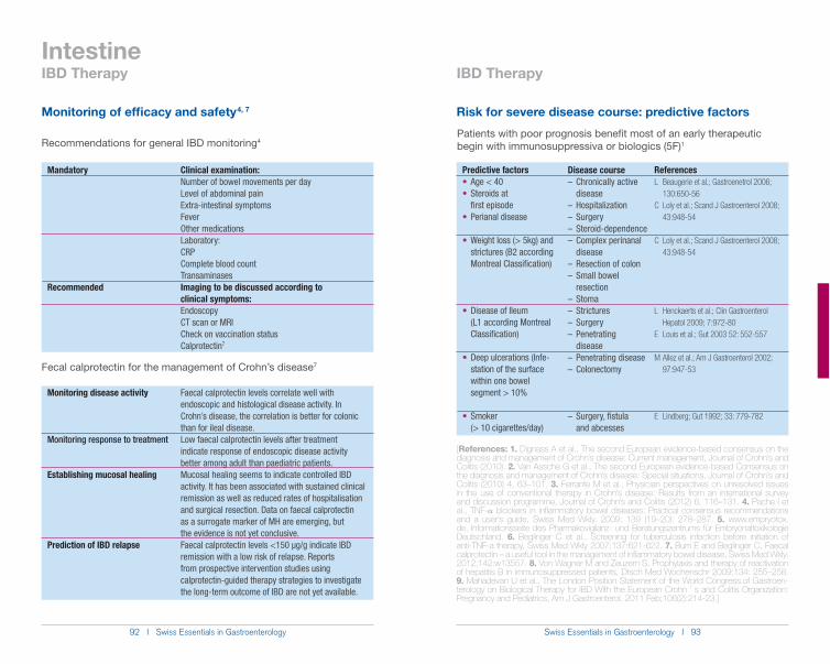
Intestine
Monitoring of efficacy and safety 4, 7
Recommendations for general IBD monitoring4
Mandatory Clinical examination: Number of bowel movements per day Level of abdominal pain Extra-intestinal symptoms Fever Other medications Laboratory: CRP Complete blood count TransaminasesRecommended Imaging to be discussed according to clinical symptoms: Endoscopy CT scan or MRI Check on vaccination status Calprotectin7
Monitoring disease activity Faecal calprotectin levels correlate well with endoscopic and histological disease activity. In Crohn’s disease, the correlation is better for colonic than for ileal disease.Monitoring response to treatment Low faecal calprotectin levels after treatment indicate response of endoscopic disease activity better among adult than paediatric patients.Establishing mucosal healing Mucosal healing seems to indicate controlled IBD activity. It has been associated with sustained clinical remission as well as reduced rates of hospitalisation and surgical resection. Data on faecal calprotectin as a surrogate marker of MH are emerging, but the evidence is not yet conclusive.Prediction of IBD relapse Faecal calprotectin levels <150 µg/g indicate IBD remission with a low risk of relapse. Reports from prospective intervention studies using calprotectin-guided therapy strategies to investigate the long-term outcome of IBD are not yet available.
7
Risk for severe disease course: predictive factors
Patients with poor prognosis benefit most of an early therapeutic begin with immunosuppressiva or biologics (5F)1
Predictive factors Age < 40 Steroids at
first episode Perianal disease
Weight loss (> 5kg) and strictures (B2 according Montreal Classification)
Disease of Ileum (L1 according Montreal Classification)
Deep ulcerations (Infe- station of the surface within one bowel segment > 10%
Smoker (> 10 cigarettes/day)
Disease course– Chronically active disease– Hospitalization– Surgery– Steroid-dependence– Complex perinanal disease– Resection of colon– Small bowel resection– Stoma– Strictures – Surgery– Penetrating disease– Penetrating disease– Colonectomy
– Surgery, fistula and abcesses
ReferencesL Beaugerie et al.; Gastroenetrol 2006; 130:650-56 C Loly et al.; Scand J Gastroenterol 2008; 43:948-54
C Loly et al.; Scand J Gastroenterol 2008; 43:948-54
L Henckaerts et al.; Clin Gastroenterol Hepatol 2009; 7:972-80E Louis et al.; Gut 2003 52: 552-557
M Allez et al.; Am J Gastroenterol 2002; 97:947-53
E Lindberg; Gut 1992; 33: 779-782
[References: 1. Dignass A et al., The second European evidence-based consensus on the diagnosis and management of Crohn’s disease: Current management, Journal of Crohn’s and Colitis (2010). 2. Van Assche G et al., The second European evidence-based Consensus on the diagnosis and management of Crohn’s disease: Special situations, Journal of Crohn’s and Colitis (2010) 4, 63–101. 3. Ferrante M et al., Physician perspectives on unresolved issues in the use of conventional therapy in Crohn’s disease: Results from an international survey and discussion programme, Journal of Crohn’s and Colitis (2012) 6, 116–131. 4. Pache I et al., TNF-! blockers in inflammatory bowel diseases: Practical consensus recommendations and a user’s guide, Swiss Med Wkly. 2009; 139 (19–20): 278–287. 5. www.empryotox.de, Informationsseite des Pharmakovigilanz- und Beratungszentrums für Embryonaltoxikologie Deutschland. 6. Beglinger C et al., Screening for tuberculosis infection before initiation of anti-TNF-a therapy, Swiss Med Wkly 2007;137:621-622. 7. Burri E and Beglinger C, Faecal calprotectin – a useful tool in the management of inflammatory bowel disease, Swiss Med Wkly. 2012;142:w13557. 8. Von Wagner M and Zeuzem S, Prophylaxis and therapy of reactivation of hepatitis B in immunosuppressed patients, Dtsch Med Wochenschr 2009;134: 255–258. 9. Mahadevan U et al., The London Position Statement of the World Congress.of Gastroen-terology on Biological Therapy for IBD With the European Crohn ’ s and Colitis Organization: Pregnancy and Pediatrics, Am J Gastroenterol. 2011 Feb;106(2):214-23.]
IBD Therapy IBD Therapy
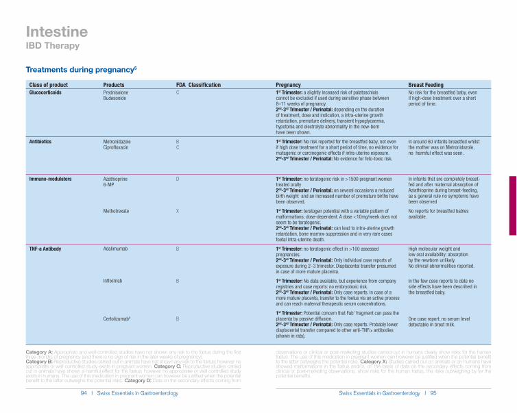
94 I Swiss Essentials in Gastroenterology Swiss Essentials in Gastroenterology I 95
Class of productGlucocorticoids
Antibiotics
Immuno-modulators
TNF-a Antibody
Products PrednisoloneBudesonide
MetronidazoleCiprofloxacin
Azathioprine6-MP
Methotrexate
Adalimumab
Infliximab
Certolizumab9
FDA ClassificationC
BC
D
X
B
B
B
Category A: Appropriate and well-controlled studies have not shown any risk to the fœtus during the first three months of pregnancy (and there is no sign of risk in the later weeks of pregnancy). Category B: Reproductive studies carried out in animals have not shown any risk to the fœtus; however no appropriate or well controlled study exists in pregnant women. Category C: Reproductive studies carried out in animals have shown a harmful effect for the fœtus; however no appropriate or well controlled study exists in humans. The use of this medication in pregnant women can however be justified when the potential benefit to the latter outweighs the potential risks. Category D: Data on the secondary effects coming from
IntestineIBD Therapy
Treatments during pregnancy5
Pregnancy 1st Trimester: a slightly inceased risk of palatoschisis cannot be excluded if used during sensitive phase between 8–11 weeks of pregnancy.2nd-3rd Trimester / Perinatal: depending on the duration of treatment, dose and indication, a intra-uterine growth retardation, premature delivery, transient hypoglycaemia, hypotonia and electrolyte abnormality in the new-born have been shown.
1st Trimester: No risk reported for the breastfed baby, not even if high dose treatment for a short period of time, no evidence for mutagenic or carcinogenic effects if intra-uterine exposure.2nd-3rd Trimester / Perinatal: No evidence for feto-toxic risk.
1st Trimester: no teratogenic risk in >1500 pregnant women treated orally 2nd-3rd Trimester / Perinatal: on several occasions a reduced birth weight and an increased number of premature births have been observed.
1st Trimester: teratogen potential with a variable pattern of malformations; dose-dependent. A dose <10mg/week does not seem to be teratogenic. 2nd-3rd Trimester / Perinatal: can lead to intra-uterine growth retardation, bone marrow suppression and in very rare cases foetal intra-uterine death.
1st Trimester: no teratogenic effect in >100 assessed pregnancies. 2nd-3rd Trimester / Perinatal: Only individual case reports of exposure during 2–3 trimester. Diaplacental transfer presumed in case of more mature placenta.
1st Trimester: No data available, but experience from company registries and case reports: no embryotoxic risk. 2nd-3rd Trimester / Perinatal: Only case reports. In case of amore mature placenta, transfer to the foetus via an active process and can reach maternal therapeutic serum concentrations.
1st Trimester: Potential concern that Fab' fragment can pass the placenta by passive diffusion. 2nd-3rd Trimester / Perinatal: Only case reports. Probably lower diaplacental transfer compared to other anti-TNF! antibodies (shown in rats).
Breast FeedingNo risk for the breastfed baby, even if high-dose treatment over a short period of time.
In around 60 infants breastfed whilst the mother was on Metronidazole, no harmful effect was seen.
In infants that are completely breast-fed and after maternal absorption of Aziathioprine during breast-feeding, as a general rule no symptoms have been observed
No reports for breastfed babies available.
High molecular weight and low oral availability: absorption by the newborn unlikely. No clinical abnormalities reported.
In the few case reports to date no side effects have been described in the breastfed baby.
One case report: no serum level detectable in brest milk.
observations or clinical or post-marketing studies carried out in humans clearly show risks for the human fœtus. The use of this medication in pregnant women can however be justified when the potential benefit to the latter outweighs the potential risks. Category X: Studies carried out on animals or on humans have showed malformations in the fœtus and/or, on the basis of data on the secondary effects coming from clinical or post-marketing observations, show risks for the human fœtus, the risks outweighing by far the potential benefits.
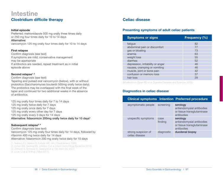
96 I Swiss Essentials in Gastroenterology Swiss Essentials in Gastroenterology I 97
Intestine
Initial episodePreferred: metronidazole 500 mg orally three times daily or 250 mg four times daily for 10 to 14 daysAlternative: vancomycin 125 mg orally four times daily for 10 to 14 days
First relapseConfirm diagnosis (see text)If symptoms are mild, conservative management may be appropriateIf antibiotics are needed, repeat treatment as in initial episode above
Second relapse1, 2
Confirm diagnosis (see text)Tapering and pulsed oral vancomycin (below), with or without probiotics (Saccharomyces boulardii 500mg orally twice daily). The probiotics may be overlapped with the final week of the taper and continued for two additional weeks in the absence of antibiotics.
125 mg orally four times daily for 7 to 14 days125 mg orally twice daily for 7 days125 mg orally once daily for 7 days125 mg orally every other day for 7 days125 mg orally every 3 days for 14 daysAlternative: fidaxomicin 200mg orally twice daily for 10 days4
Subsequent relapse3, 4
Confirm diagnosis (see text)Vancomycin 125 mg orally four times daily for 14 days, followed by rifaximin 400 mg twice daily for 14 daysAlternative: fidaxomicin 200 mg orally twice daily for 10 days
[1. Tedesco FJ, Gordon D, Fortson WC. Am J Gastroenterol 1985][2. Cohen SH, Gerding DN, Johnson S et al. Infect Control Hosp Epidemiol 2010] [3. Johnson S, Schriever C, Galang M et al. Clin Infect Dis 2007][4. Louie TJ et al: N Engl J Med 2011]
Presenting symptoms of adult celiac patients
Diagnostics in celiac disease
Symptoms or signs Frequency (%)
fatigueabdominal pain or discomfortgas or bloatinganemiaweight lossdiarrheadepression, irritability or angernausea, cramping or vomitingmuscle, joint or bone painconfusion or memory losshair loss
82777363555246464237 29
Clinical symptoms Intention Preferred procedure
asymptomatic people
unspecific symptoms
strong suspicion of celiac disease
screening
case finding
diagnostic
serologyantiendomysial antibodies or tissue transglutaminase antibodiesserologyantiendomysial antibodies or tissue transglutaminase antibodiesduodenal biopsy
[Modified from Zipser RD et al. Digestive Diseases and Sciences 2003]
Clostridium difficile therapy Celiac disease
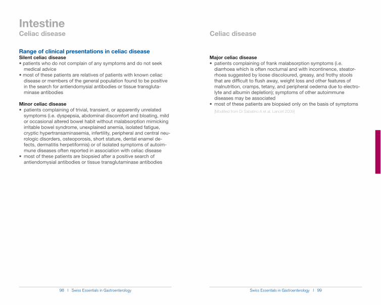
98 I Swiss Essentials in Gastroenterology Swiss Essentials in Gastroenterology I 99
Intestine
Range of clinical presentations in celiac diseaseSilent celiac disease
medical advice
disease or members of the general population found to be positive in the search for antiendomysial antibodies or tissue transgluta- minase antibodies
Minor celiac disease
symptoms (i.e. dyspepsia, abdominal discomfort and bloating, mild or occasional altered bowel habit without malabsorption mimicking irritable bowel syndrome, unexplained anemia, isolated fatigue, cryptic hypertransaminasemia, infertility, peripheral and central neu- rologic disorders, osteoporosis, short stature, dental enamel de- fects, dermatitis herpetiformis) or of isolated symptoms of autoim- mune diseases often reported in association with celiac disease
antiendomysial antibodies or tissue transglutaminase antibodies
Major celiac disease
diarrhoea which is often nocturnal and with incontinence, steator- rhoea suggested by loose discoloured, greasy, and frothy stools that are difficult to flush away, weight loss and other features of malnutrition, cramps, tetany, and peripheral oedema due to electro- lyte and albumin depletion); symptoms of other autoimmune diseases may be associated
[Modified from Di Sabatino A et al. Lancet 2009]
Celiac disease Celiac disease
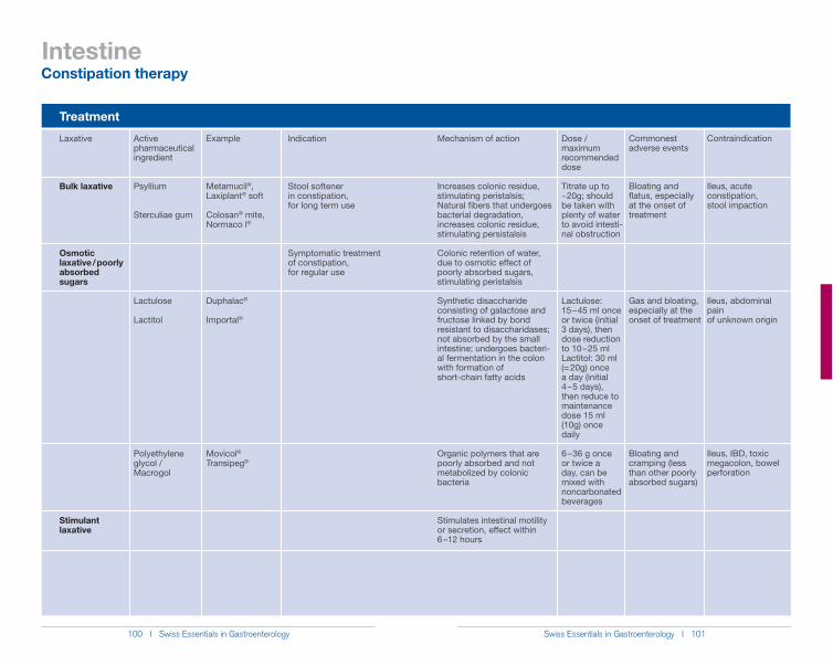
100 I Swiss Essentials in Gastroenterology Swiss Essentials in Gastroenterology I 101
Intestine
Treatment
Active pharmaceutical ingredient
Psyllium
Sterculiae gum
Lactulose
Lactitol
Polyethylene glycol / Macrogol
Laxative
Bulk laxative
Osmotic laxative / poorly absorbedsugars
Stimulant laxative
Mechanism of action
Increases colonic residue, stimulating peristalsis;Natural fibers that undergoes bacterial degradation, increases colonic residue, stimulating persistalsis
Colonic retention of water, due to osmotic effect of poorly absorbed sugars, stimulating peristalsis
Synthetic disaccharide consisting of galactose and fructose linked by bond resistant to disaccharidases; not absorbed by the small intestine; undergoes bacteri-al fermentation in the colon with formation of short-chain fatty acids
Organic polymers that are poorly absorbed and not metabolized by colonic bacteria
Stimulates intestinal motility or secretion, effect within 6 – 12 hours
Example
Metamucil®, Laxiplant® soft
Colosan® mite, Normaco l®
Duphalac®
Importal®
Movicol® Transipeg®
Indication
Stool softener in constipation, for long term use
Symptomatic treatment of constipation, for regular use
Dose / maximum recommended dose
Titrate up to ~20g; should be taken with plenty of water to avoid intesti-nal obstruction
Lactulose: 15 – 45 ml once or twice (initial 3 days), then dose reduction to 10 – 25 ml Lactitol: 30 ml (= 20g) once a day (initial 4 – 5 days), then reduce to maintenance dose 15 ml (10g) once daily
6 – 36 g once or twice a day, can be mixed with noncarbonated beverages
Commonest adverse events
Bloating and flatus, especially at the onset of treatment
Gas and bloating, especially at the onset of treatment
Bloating and cramping (less than other poorly absorbed sugars)
Contraindication
Ileus, acute constipation, stool impaction
Ileus, abdominal pain of unknown origin
Ileus, IBD, toxic megacolon, bowel perforation
Constipation therapy
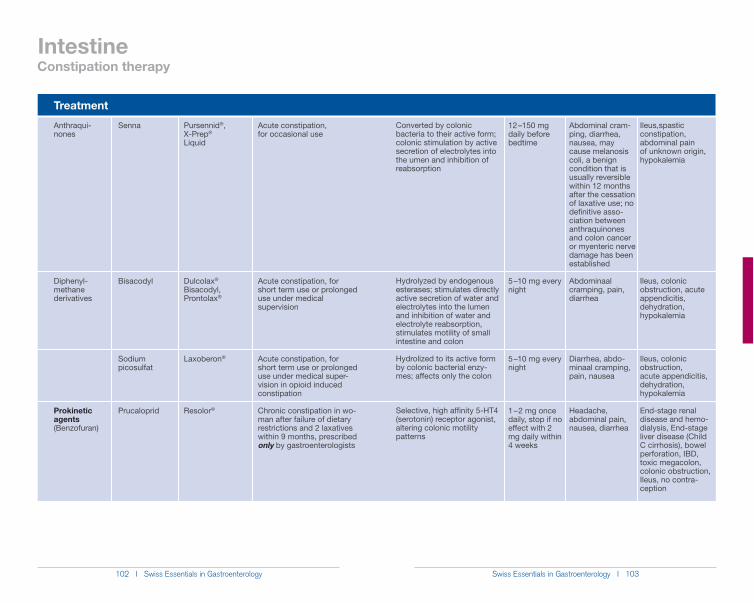
102 I Swiss Essentials in Gastroenterology Swiss Essentials in Gastroenterology I 103
Intestine
Treatment
Senna
Bisacodyl
Sodium picosulfat
Prucaloprid
Anthraqui-nones
Diphenyl-methane derivatives
Prokinetic agents (Benzofuran)
Pursennid®,X-Prep® Liquid
Dulcolax® Bisacodyl, Prontolax®
Laxoberon®
Resolor®
Acute constipation, for occasional use
Acute constipation, for short term use or prolonged use under medical supervision
Acute constipation, for short term use or prolonged use under medical super-vision in opioid induced constipation
Chronic constipation in wo-man after failure of dietary restrictions and 2 laxatives within 9 months, prescribed only by gastroenterologists
12 – 150 mg daily before bedtime
5 – 10 mg every night
5 – 10 mg every night
1 – 2 mg once daily, stop if no effect with 2 mg daily within 4 weeks
Abdominal cram-ping, diarrhea, nausea, may cause melanosis coli, a benign condition that is usually reversible within 12 months after the cessation of laxative use; no definitive asso-ciation between anthraquinones and colon cancer or myenteric nerve damage has been established
Abdominaal cramping, pain, diarrhea
Diarrhea, abdo-minaal cramping, pain, nausea
Headache, abdominal pain, nausea, diarrhea
Ileus,spastic constipation, abdominal pain of unknown origin, hypokalemia
Ileus, colonic obstruction, acute appendicitis, dehydration, hypokalemia
Ileus, colonic obstruction, acute appendicitis, dehydration, hypokalemia
End-stage renal disease and hemo-dialysis, End-stage liver disease (Child C cirrhosis), bowel perforation, IBD, toxic megacolon, colonic obstruction, Ileus, no contra-ception
Converted by colonic bacteria to their active form; colonic stimulation by active secretion of electrolytes into the umen and inhibition of reabsorption
Hydrolyzed by endogenous esterases; stimulates directly active secretion of water and electrolytes into the lumen and inhibition of water and electrolyte reabsorption, stimulates motility of small intestine and colon
Hydrolized to its active form by colonic bacterial enzy-mes; affects only the colon
Selective, high affinity 5-HT4 (serotonin) receptor agonist, altering colonic motility patterns
Constipation therapy
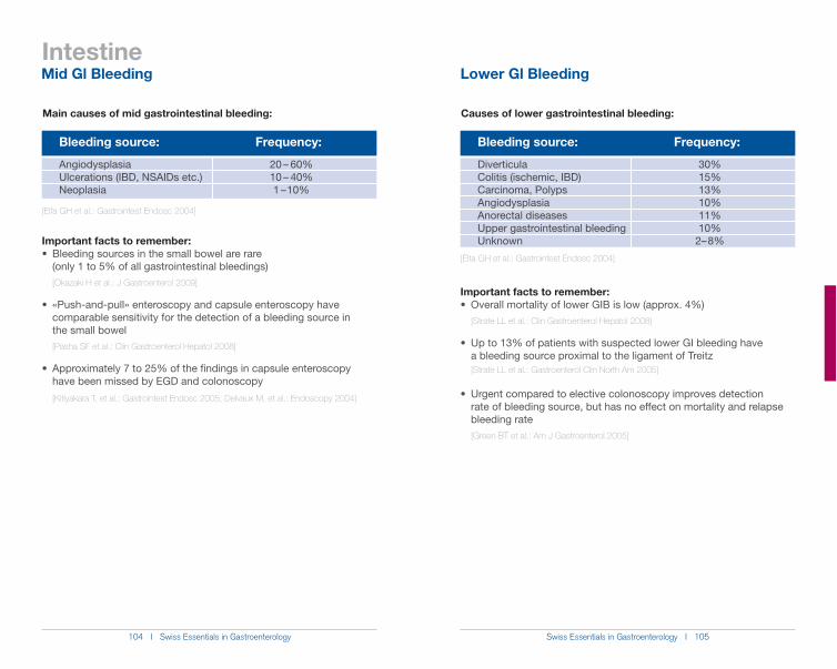
104 I Swiss Essentials in Gastroenterology Swiss Essentials in Gastroenterology I 105
Intestine
Causes of lower gastrointestinal bleeding:
Bleeding source: Frequency:
DiverticulaColitis (ischemic, IBD)Carcinoma, PolypsAngiodysplasiaAnorectal diseasesUpper gastrointestinal bleedingUnknown
30%15%13%10%11%10%
2– 8%
[Elta GH et al.: Gastrointest Endosc 2004]
Important facts to remember:
[Strate LL et al.: Clin Gastroenterol Hepatol 2008]
a bleeding source proximal to the ligament of Treitz [Strate LL et al.: Gastroenterol Clin North Am 2005]
rate of bleeding source, but has no effect on mortality and relapse bleeding rate [Green BT et al.: Am J Gastroenterol.2005]
Main causes of mid gastrointestinal bleeding:
Bleeding source: Frequency:
AngiodysplasiaUlcerations (IBD, NSAIDs etc.)Neoplasia
20 – 60%10 – 40%1 – 10%
[Elfa GH et al.: Gastrointest Endosc 2004]
Important facts to remember:
(only 1 to 5% of all gastrointestinal bleedings) [Okazaki H et al.: J Gastroenterol 2009]
comparable sensitivity for the detection of a bleeding source in the small bowel [Pasha SF et al.: Clin Gastroenterol Hepatol 2008]
have been missed by EGD and colonoscopy
[Kitiyakara T, et al.: Gastrointest Endosc 2005; Delvaux M, et al.: Endoscopy 2004]
Mid Gl Bleeding Lower Gl Bleeding
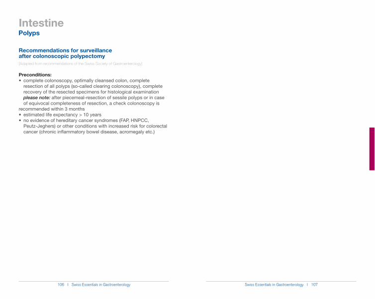
106 I Swiss Essentials in Gastroenterology Swiss Essentials in Gastroenterology I 107
Recommendations for surveillance after colonoscopic polypectomy[Adapted from recommendations of the Swiss Society of Gastroenterology]
Preconditions:complete colonoscopy, optimally cleansed colon, complete
resection of all polyps (so-called clearing colonoscopy), complete recovery of the resected specimens for histological examination please note: after piecemeal-resection of sessile polyps or in case of equivocal completeness of resection, a check colonoscopy is recommended within 3 months
estimated life expectancy > 10 yearsno evidence of hereditary cancer syndromes (FAP, HNPCC,
Peutz-Jeghers) or other conditions with increased risk for colorectal cancer (chronic inflammatory bowel disease, acromegaly etc.)
IntestinePolyps
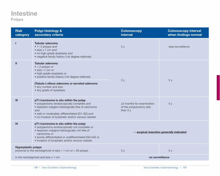
108 I Swiss Essentials in Gastroenterology Swiss Essentials in Gastroenterology I 109
Riskcategory
Tubular adenomaand
) 1 cm andand
Tubular adenomaoror
or
(Tubulo-) villous adenoma or serrated adenoma
ia
pT1 / carcinoma in situ within the polypand
andnd
pT1 / carcinoma in situ within the polypor
carcinoma oror
I
II
III
IV
5 y
3 y
)3 months for examination of the polypectomy site; then 3 y
3 y
stop surveillance
5 y
5 y
5 y
Polyp histology &secondary criteria
Colonoscopy interval
Colonoscopy interval when findings normal
Intestine
ĺ surgical resection generally indicated
no surveillance
Hyperplastic polypsproximal to the rectosigmoid or size > 1 cm or > 20 polyps
in the rectosigmoid and size ) 1 cm
Polyps
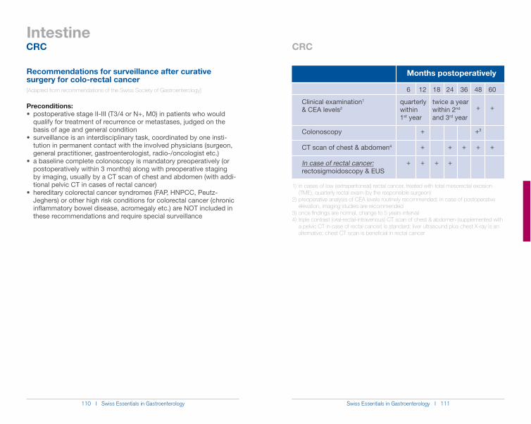
110 I Swiss Essentials in Gastroenterology Swiss Essentials in Gastroenterology I 111
Intestine
Recommendations for surveillance after curative surgery for colo-rectal cancer[Adapted from recommendations of the Swiss Society of Gastroenterology]
Preconditions:
qualify for treatment of recurrence or metastases, judged on the basis of age and general condition
tution in permanent contact with the involved physicians (surgeon, general practitioner, gastroenterologist, radio-/oncologist etc.)
postoperatively within 3 months) along with preoperative staging by imaging, usually by a CT scan of chest and abdomen (with addi- tional pelvic CT in cases of rectal cancer)
Jeghers) or other high risk conditions for colorectal cancer (chronic inflammatory bowel disease, acromegaly etc.) are NOT included in these recommendations and require special surveillance
Clinical examination1 & CEA levels2
Colonoscopy
CT scan of chest & abdomen4 In case of rectal cancer:rectosigmoidoscopy & EUS
12
+
+
+
18
+
24
+
+
36
+
48
+
+3
+
60
+
+
6
+
Months postoperatively
quarterly within 1st year
twice a year within 2nd
and 3rd year
1) in cases of low (extraperitoneal) rectal cancer, treated with total mesorectal excision (TME), quarterly rectal exam (by the responsible surgeon)2) preoperative analysis of CEA levels routinely recommended; in case of postoperative elevation, imaging studies are recommended3) once findings are normal, change to 5 years interval4) triple contrast (oral-rectal-intravenous) CT scan of chest & abdomen (supplemented with a pelvic CT in case of rectal cancer) is standard; liver ultrasound plus chest X-ray is an alternative; chest CT scan is beneficial in rectal cancer
CRC CRC
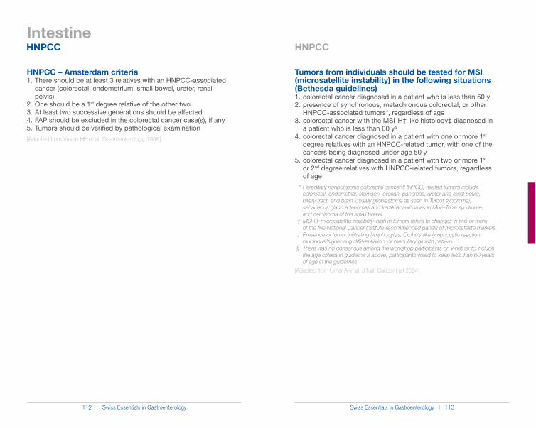
112 I Swiss Essentials in Gastroenterology Swiss Essentials in Gastroenterology I 113
Intestine
HNPCC – Amsterdam criteria1. There should be at least 3 relatives with an HNPCC-associated cancer (colorectal, endometrium, small bowel, ureter, renal pelvis)2. One should be a 1st degree relative of the other two3. At least two successive generations should be affected4. FAP should be excluded in the colorectal cancer case(s), if any5. Tumors should be verified by pathological examination
[Adapted from Vasen HF et al. Gastroenterology 1999]
Tumors from individuals should be tested for MSI (microsatellite instability) in the following situations (Bethesda guidelines)1. colorectal cancer diagnosed in a patient who is less than 50 y2. presence of synchronous, metachronous colorectal, or other HNPCC-associated tumors*, regardless of age3. colorectal cancer with the MSI-H† like histology‡ diagnosed in a patient who is less than 60 y§
4. colorectal cancer diagnosed in a patient with one or more 1st degree relatives with an HNPCC-related tumor, with one of the cancers being diagnosed under age 50 y5. colorectal cancer diagnosed in a patient with two or more 1st or 2nd degree relatives with HNPCC-related tumors, regardless of age * Hereditary nonpolyposis colorectal cancer (HNPCC)-related tumors include colorectal, endometrial, stomach, ovarian, pancreas, ureter and renal pelvis, biliary tract, and brain (usually glioblastoma as seen in Turcot syndrome), sebaceous gland adenomas and keratoacanthomas in Muir–Torre syndrome, and carcinoma of the small bowel. † MSI-H: microsatellite instability–high in tumors refers to changes in two or more of the five National Cancer Institute-recommended panels of microsatellite markers. ‡ Presence of tumor infiltrating lymphocytes, Crohn’s-like lymphocytic reaction, mucinous/signet-ring differentiation, or medullary growth pattern. § There was no consensus among the workshop participants on whether to include the age criteria in guideline 3 above; participants voted to keep less than 60 years of age in the guidelines.
[Adapted from Umar A et al. J Natl Cancer Inst 2004]
HNPCC HNPCC
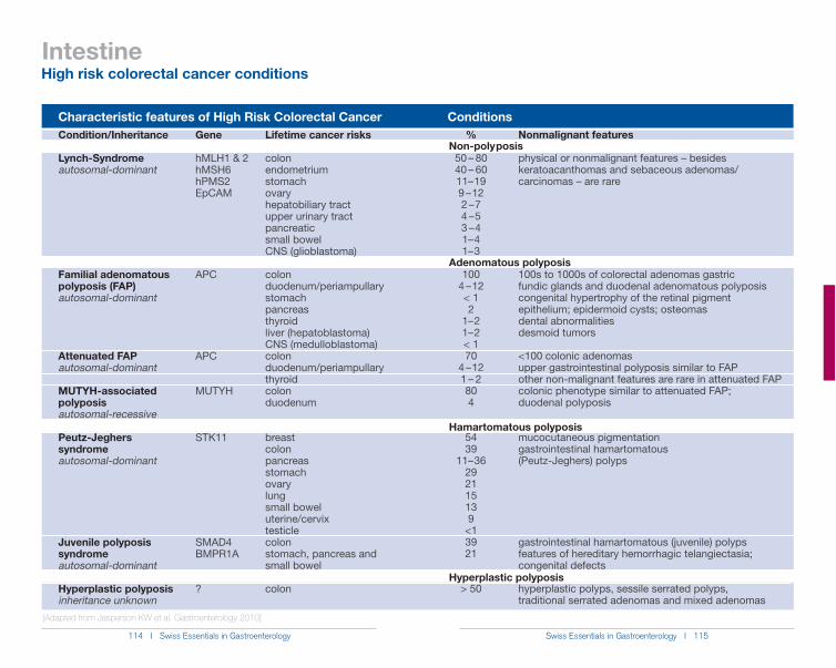
114 I Swiss Essentials in Gastroenterology Swiss Essentials in Gastroenterology I 115
Characteristic features of High Risk Colorectal Cancer Conditions
Intestine
Gene
hMLH1 & 2hMSH6hPMS2EpCAM
APC
APC
MUTYH
STK11
SMAD4BMPR1A
?
Condition/Inheritance
Lynch-Syndromeautosomal-dominant
Familial adenomatous polyposis (FAP)autosomal-dominant
Attenuated FAPautosomal-dominant
MUTYH-associated polyposisautosomal-recessive
Peutz-Jeghers syndromeautosomal-dominant
Juvenile polyposis syndromeautosomal-dominant
Hyperplastic polyposisinheritance unknown
Lifetime cancer risks
colonendometriumstomachovaryhepatobiliary tractupper urinary tractpancreaticsmall bowelCNS (glioblastoma) colonduodenum/periampullarystomachpancreasthyroidliver (hepatoblastoma)CNS (medulloblastoma)colonduodenum/periampullarythyroidcolonduodenum
breastcolonpancreasstomachovarylungsmall boweluterine/cervixtesticlecolonstomach, pancreas and small bowel
colon
Nonmalignant features
physical or nonmalignant features – besides keratoacanthomas and sebaceous adenomas/ carcinomas – are rare
100s to 1000s of colorectal adenomas gastric fundic glands and duodenal adenomatous polyposiscongenital hypertrophy of the retinal pigment epithelium; epidermoid cysts; osteomasdental abnormalitiesdesmoid tumors
<100 colonic adenomasupper gastrointestinal polyposis similar to FAPother non-malignant features are rare in attenuated FAPcolonic phenotype similar to attenuated FAP; duodenal polyposis
mucocutaneous pigmentationgastrointestinal hamartomatous (Peutz-Jeghers) polyps
gastrointestinal hamartomatous (juvenile) polypsfeatures of hereditary hemorrhagic telangiectasia; congenital defects
hyperplastic polyps, sessile serrated polyps, traditional serrated adenomas and mixed adenomas
%
50 – 8040 – 6011– 199 – 122 – 74 – 53 – 41– 41– 3
1004 – 12< 12
1– 21– 2< 170
4 – 121 – 2804
5439
11– 36292115139
<13921
> 50
[Adapted from Jasperson KW et al. Gastroenterology 2010]
Non-poly posis
Adenomatous polyposis
Hamartomatous polyposis
Hyperplastic polyposis
High risk colorectal cancer conditions
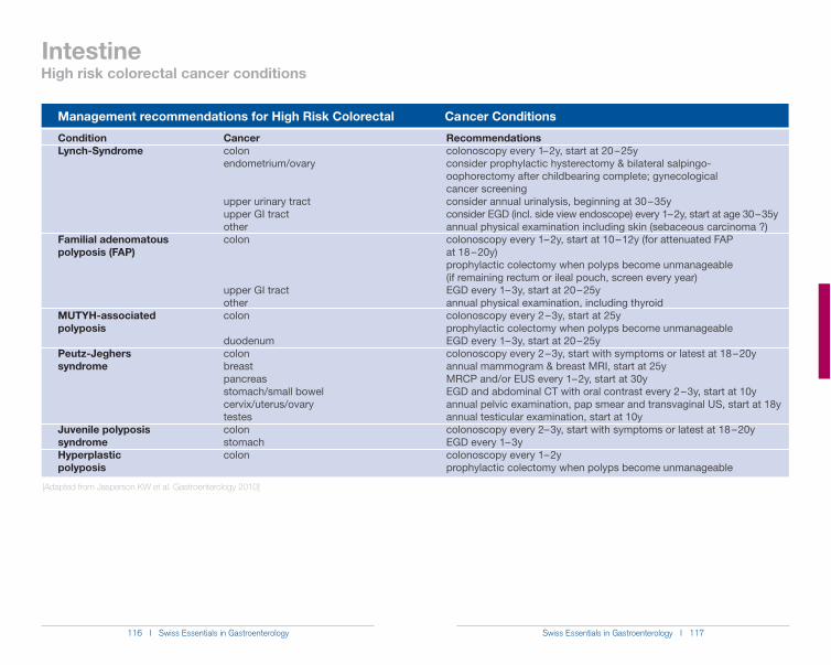
116 I Swiss Essentials in Gastroenterology Swiss Essentials in Gastroenterology I 117
Management recommendations for High Risk Colorectal Ca ncer Conditions
Intestine
[Adapted from Jasperson KW et al. Gastroenterology 2010]
ConditionLynch-Syndrome
Familial adenomatous polyposis (FAP)
MUTYH-associated polyposis
Peutz-Jeghers syndrome
Juvenile polyposis syndromeHyperplastic polyposis
Cancercolonendometrium/ovary
upper urinary tractupper GI tractothercolon
upper GI tractothercolon
duodenumcolonbreastpancreasstomach/small bowelcervix/uterus/ovarytestescolonstomachcolon
Recommendationscolonoscopy every 1– 2y, start at 20 – 25yconsider prophylactic hysterectomy & bilateral salpingo- oophorectomy after childbearing complete; gynecological cancer screeningconsider annual urinalysis, beginning at 30 – 35yconsider EGD (incl. side view endoscope) every 1– 2y, start at age 30 – 35yannual physical examination including skin (sebaceous carcinoma ?)colonoscopy every 1– 2y, start at 10 – 12y (for attenuated FAP at 18 – 20y) prophylactic colectomy when polyps become unmanageable (if remaining rectum or ileal pouch, screen every year)EGD every 1– 3y, start at 20 – 25yannual physical examination, including thyroidcolonoscopy every 2 – 3y, start at 25yprophylactic colectomy when polyps become unmanageableEGD every 1– 3y, start at 20 – 25ycolonoscopy every 2 – 3y, start with symptoms or latest at 18 – 20yannual mammogram & breast MRI, start at 25yMRCP and/or EUS every 1– 2y, start at 30yEGD and abdominal CT with oral contrast every 2 – 3y, start at 10yannual pelvic examination, pap smear and transvaginal US, start at 18y annual testicular examination, start at 10ycolonoscopy every 2– 3y, start with symptoms or latest at 18 – 20yEGD every 1– 3ycolonoscopy every 1– 2yprophylactic colectomy when polyps become unmanageable
High risk colorectal cancer conditions
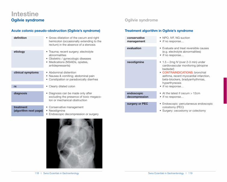
118 I Swiss Essentials in Gastroenterology Swiss Essentials in Gastroenterology I 119
Intestine
Acute colonic pseudo-obstruction (Ogilvie’s syndrome)
Gross dilatation of the cecum and right hemicolon (occasionally extending to the rectum) in the absence of a stenosis
Trauma; recent surgery; electrolyte abnormalities
Obstetric / gynecologic diseasesMedications (NSAIDs, opiates,
antidepressants)
Abdominal distentionNausea & vomiting; abdominal painConstipation or paradoxically diarrhea
Clearly dilated colon
Diagnosis can be made only after excluding the presence of toxic megaco- lon or mechanical obstruction
Conservative managementNeostigmineEndoscopic decompression or surgery
definition
etiology
clinical symptoms
rx
diagnosis
treatment(algorithm next page)
Treatment algorithm in Ogilvie’s syndrome
If no response…
(e.g. electrolyte abnormalities)
cardiovascular monitoring (atropine
CONTRAINDICATIONS: bronchial asthma, recent myocardial infarction, beta-blockers, bradyarrhythmias, hyperthyreosis
colostomy (PEC)
conservative management
evaluation
neostigmine
endoscopic decompression
surgery or PEC
Ogilvie syndrome Ogilvie syndrome
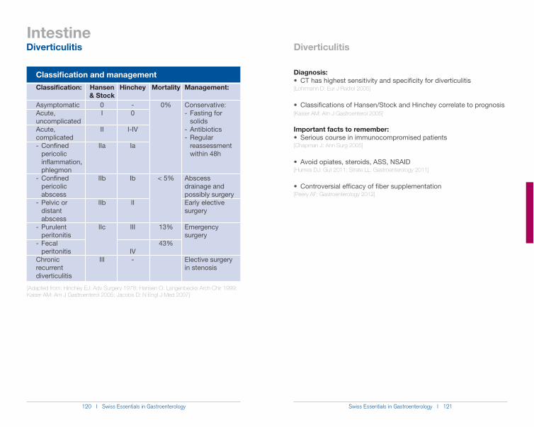
120 I Swiss Essentials in Gastroenterology Swiss Essentials in Gastroenterology I 121
Intestine
Diagnosis:
[Lohrmann D: Eur J Radiol 2005]
[Kaiser AM: Am J Gastroenterol 2005]
Important facts to remember:
[Chapman J: Ann Surg 2005]
[Humes DJ: Gut 2011; Strate LL: Gastroenterology 2011]
[Peery AF: Gastroenterology 2012]
Classification and managementClassification:
AsymptomaticAcute, uncomplicatedAcute, complicated- Confined pericolic inflammation, phlegmon- Confined pericolic abscess- Pelvic or distant abscess- Purulent peritonitis- Fecal peritonitisChronic recurrent diverticulitis
Management:
Conservative:- Fasting for solids- Antibiotics- Regular reassessment within 48h
Abscess drainage and possibly surgeryEarly elective surgery
Emergency surgery
Elective surgery in stenosis
Hansen & Stock
0I
II
IIa
IIb
IIb
IIc
III
Hinchey
-0
I-IV
Ia
Ib
II
III
IV-
Mortality
0%
< 5%
13%
43%
[Adapted from: Hinchey EJ: Adv Surgery 1978; Hansen O: Langenbecks Arch Chir 1999; Kaiser AM: Am J Gastroenterol 2005; Jacobs D: N Engl J Med 2007]
Diverticulitis Diverticulitis
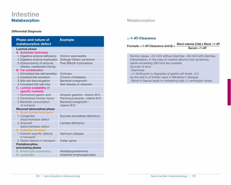
122 I Swiss Essentials in Gastroenterology Swiss Essentials in Gastroenterology I 123
Differential Diagnosis
Phase and nature of Example malabsorptive defect
Intestine
!-1-AT-Clearance
Stool volume (/3d) x Stool !1-ATFormula: !1-AT-Clearance (ml/d) = Serum !1-AT
Normal values. <24 ml/d without diarrhea; <56 ml/d with diarrhea. Interpretation. In the case of marked albumin loss syndrome, values exceeding 200 ml/d are possible. Sources of error - Diarrhoea - !1-Antitrypsin is degraded at gastric pH levels <3.5
- Blood in faeces leads to misleading high !1–antitrypsin levels.
Malabsorption Malabsorption
Luminal phaseA. Substrate hydrolysis 1. Digestive enzyme deficiency 2. Digestive enzyme inactivation 3. Dyssynchrony of enzyme release, inadequate mixingB. Fat solubilization 1. Diminished bile salt secretion 2. Impaired bile secretion 3. Bile salt deconjugation 4. Increased bile salt lossC. Luminal availability of specific nutrients 1. Diminished gastric acid 2. Diminished intrinsic factor 3. Bacterial consumption of nutrientsMucosal (absorptive) phaseA. Brush border hydrolysis* 1. Congenital disaccharidase defect 2. Acquired disaccharidase defectB. Epithelial transport 1. Nutrient-specific defects in transport 2. Global defects in transportPostabsorptive, processing phaseA. Enterocyte processingB. Lymphatic
Chronic pancreatitisZollinger-Ellison syndromePost Billroth II procedure
CirrhosisChronic cholestasisBacterial overgrowthIleal disease or resection
Atrophic gastritis – vitamin B12Pernicious anemia – vitamin B12Bacterial overgrowth – vitamin B12
Sucrase-isomaltase deficiency
Lactase deficiency
Celiac sprue
AbetalipoproteinemiaIntestinal lymphangiectasia
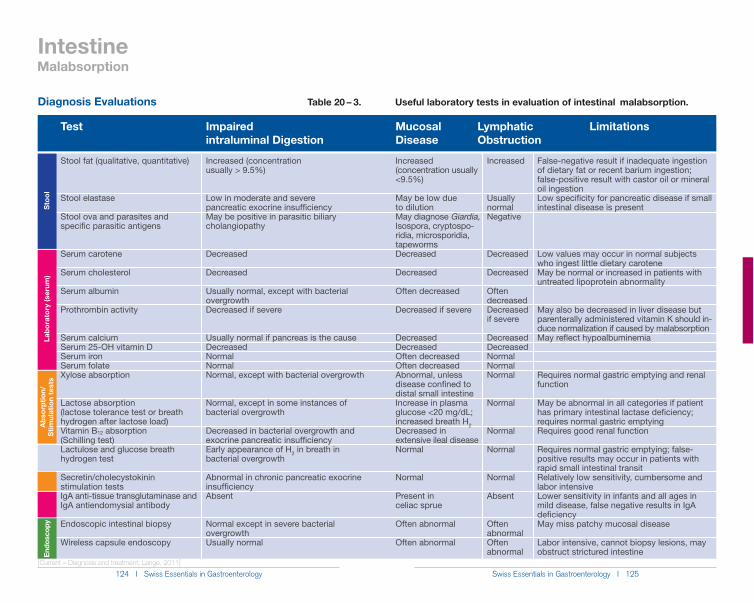
124 I Swiss Essentials in Gastroenterology Swiss Essentials in Gastroenterology I 125
Test Impaired intraluminal Digestion
Mucosal Disease
Lymphatic Obstruction
Intestine
Diagnosis Evaluations Table 20 – 3. Useful laboratory tests in evaluation of intestinal malabsorption.
Limitations
Increased (concentration usually > 9.5%)
Low in moderate and severe pancreatic exocrine insufficiencyMay be positive in parasitic biliary cholangiopathy
Decreased
Decreased
Usually normal, except with bacterial overgrowthDecreased if severe
Usually normal if pancreas is the causeDecreasedNormalNormalNormal, except with bacterial overgrowth
Normal, except in some instances of bacterial overgrowth
Decreased in bacterial overgrowth and exocrine pancreatic insufficiency Early appearance of H2 in breath in bacterial overgrowth
Abnormal in chronic pancreatic exocrine insufficiencyAbsent
Normal except in severe bacterial overgrowthUsually normal
Stool fat (qualitative, quantitative)
Stool elastase
Stool ova and parasites and specific parasitic antigens
Serum carotene
Serum cholesterol
Serum albumin
Prothrombin activity
Serum calciumSerum 25-OH vitamin DSerum ironSerum folateXylose absorption
Lactose absorption (lactose tolerance test or breath hydrogen after lactose load)Vitamin B12 absorption (Schilling test)Lactulose and glucose breath hydrogen test
Secretin/cholecystokinin stimulation testsIgA anti-tissue transglutaminase and IgA antiendomysial antibody
Endoscopic intestinal biopsy
Wireless capsule endoscopy
Increased (concentration usually <9.5%)
May be low due to dilutionMay diagnose Giardia, Isospora, cryptospo-ridia, microsporidia, tapewormsDecreased
Decreased
Often decreased
Decreased if severe
DecreasedDecreasedOften decreasedOften decreasedAbnormal, unless disease confined to distal small intestineIncrease in plasma glucose <20 mg/dL; increased breath H2Decreased in extensive ileal diseaseNormal
Normal
Present inceliac sprue
Often abnormal
Often abnormal
Increased
Usually normalNegative
Decreased
Decreased
Often decreasedDecreased if severe
DecreasedDecreasedNormalNormalNormal
Normal
Normal
Normal
Normal
Absent
Often abnormalOften abnormal
False-negative result if inadequate ingestion of dietary fat or recent barium ingestion; false-positive result with castor oil or mineral oil ingestionLow specificity for pancreatic disease if small intestinal disease is present
Low values may occur in normal subjects who ingest little dietary caroteneMay be normal or increased in patients with untreated lipoprotein abnormality
May also be decreased in liver disease but parenterally administered vitamin K should in-duce normalization if caused by malabsorptionMay reflect hypoalbuminemia
Requires normal gastric emptying and renal function
May be abnormal in all categories if patient has primary intestinal lactase deficiency; requires normal gastric emptyingRequires good renal function
Requires normal gastric emptying; false-positive results may occur in patients with rapid small intestinal transitRelatively low sensitivity, cumbersome and labor intensiveLower sensitivity in infants and all ages in mild disease, false negative results in IgA deficiencyMay miss patchy mucosal disease
Labor intensive, cannot biopsy lesions, may obstruct strictured intestine
Sto
olLa
bora
tory
(ser
um)
Abs
orpt
ion/
Stim
ulat
ion
test
sEn
dosc
opy
[Current – Diagnosis and treatment; Lange, 2011]
Malabsorption
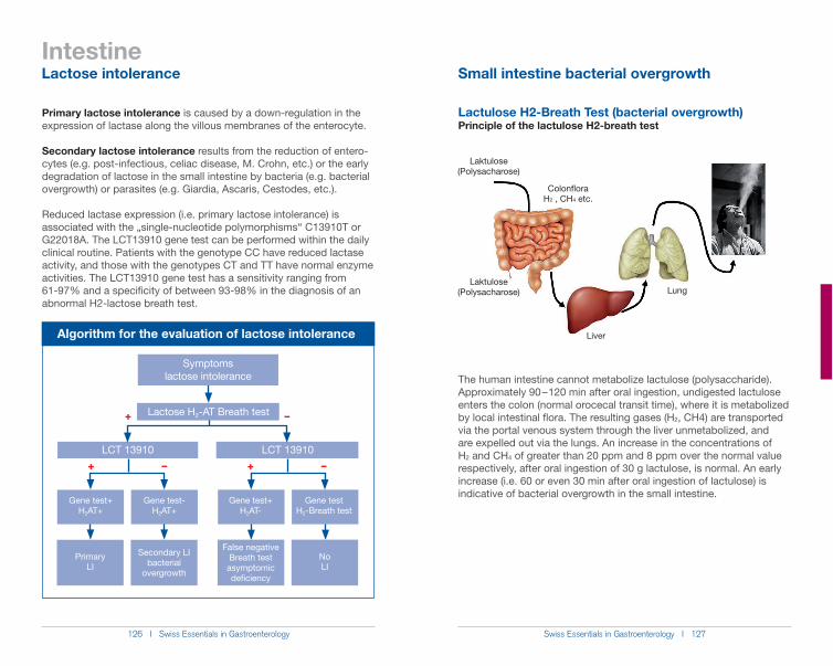
126 I Swiss Essentials in Gastroenterology Swiss Essentials in Gastroenterology I 127
Intestine
Lactulose H2-Breath Test (bacterial overgrowth)Principle of the lactulose H2-breath test
The human intestine cannot metabolize lactulose (polysaccharide). Approximately 90 – 120 min after oral ingestion, undigested lactulose enters the colon (normal orocecal transit time), where it is metabolized by local intestinal flora. The resulting gases (H2, CH4) are transported via the portal venous system through the liver unmetabolized, and are expelled out via the lungs. An increase in the concentrations of H2 and CH4 of greater than 20 ppm and 8 ppm over the normal value respectively, after oral ingestion of 30 g lactulose, is normal. An early increase (i.e. 60 or even 30 min after oral ingestion of lactulose) is indicative of bacterial overgrowth in the small intestine.
Laktulose(Polysacharose)
ColonfloraH2 , CH4 etc.
Laktulose(Polysacharose)
Liver
Lung
Primary lactose intolerance is caused by a down-regulation in the expression of lactase along the villous membranes of the enterocyte.
Secondary lactose intolerance results from the reduction of entero-cytes (e.g. post-infectious, celiac disease, M. Crohn, etc.) or the early degradation of lactose in the small intestine by bacteria (e.g. bacterial overgrowth) or parasites (e.g. Giardia, Ascaris, Cestodes, etc.).
Reduced lactase expression (i.e. primary lactose intolerance) is associated with the „single-nucleotide polymorphisms“ C13910T or G22018A. The LCT13910 gene test can be performed within the daily clinical routine. Patients with the genotype CC have reduced lactase activity, and those with the genotypes CT and TT have normal enzyme activities. The LCT13910 gene test has a sensitivity ranging from 61-97% and a specificity of between 93-98% in the diagnosis of an abnormal H2-lactose breath test.
Algorithm for the evaluation of lactose intolerance
Symptomslactose intolerance
LCT 13910 LCT 13910
Gene test+H2AT-
Gene testH2-Breath test
Gene test+H2AT+
Gene test-H2AT+
+ –
+ – + –
Lactose H2-AT Breath test
False negativeBreath testasymptomicdeficiency
NoLI
Primary LI
Secondary LIbacterial
overgrowth
Lactose intolerance Small intestine bacterial overgrowth
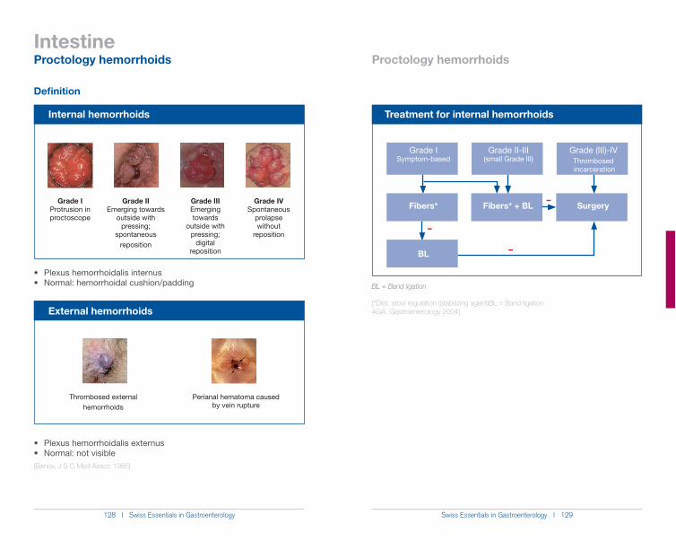
128 I Swiss Essentials in Gastroenterology Swiss Essentials in Gastroenterology I 129
Intestine
Definition
[Banov, J S C Med Assoc 1985]
Internal hemorrhoids
Grade IProtrusion in proctoscope
Grade IIIEmerging towards
outside withpressing;
digital reposition
Grade IIEmerging towards
outside with pressing;
spontaneous reposition
Grade IVSpontaneous
prolapsewithout
reposition
External hemorrhoids
Perianal hematoma caused by vein rupture
Thrombosed externalhemorrhoids
BL = Band ligation
[*Diet, stool regulation (stabilizing agent)BL = Band ligationAGA. Gastroenterology 2004]
Treatment for internal hemorrhoids
Grade I Symptom-based
–
–
–Fibers* Fibers* + BL Surgery
BL
Proctology hemorrhoids Proctology hemorrhoids
Grade II-III(small Grade III)
Grade (III)-IVThrombosed incarceration
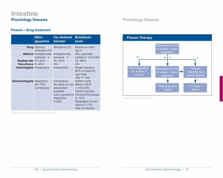
130 I Swiss Essentials in Gastroenterology Swiss Essentials in Gastroenterology I 131
Intestine
Fissure – Drug treatment
Nitro-glycerine
Glyceryl trinitrate 0.2%Intrasphincter pressure �27 – 85%0 – 43%Inexpensive
Headache 20 – 70%Compliance
Nifedipine 2%
Intrasphincter pressure �45 – 95%4%Inexpensive
ComplianceNo ready-to-use preparation availableLess experienceHeadache 0-20%
Botulinum toxin Typ ARev. sphincter paresis 2 – 3 months25 – 96%4%Single injection80% of patients pain-free after 1st dayPatient costs (Botox 100 E = 416 CHF)Painful injectionPerianal thrombosis 5 – 10%Reversible inconti-nence 3 – 12%Risk of infection
Ca-channel blocker
Botulinum toxin
Drug
Method
Healing rateRecurrence
Advantage(s)
Disadvantage(s)
[AGA technical review. Gastroenterology 2003]
Fissure Therapy
–
Nifedipine 2% for 6 weeks + stool
regulation
V-Y flaps + (Botox
–+
Follow-up at 6 weeks
Stool regulationfor further 3
months
Nifedipine 2% for 6 weeks + stool
regulation
Fissure debridement
(and/or Botox)
–
+
[Hetzer FA et al.: Praxis 2000]
Proctology fissures Proctology fissures
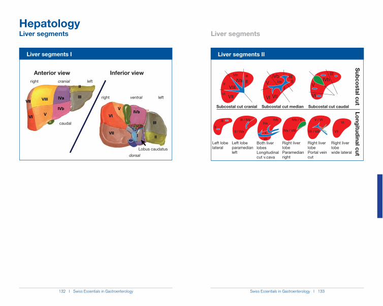
132 I Swiss Essentials in Gastroenterology Swiss Essentials in Gastroenterology I 133
Hepatology
Liver segments II
Subcostal cut cranial Subcostal cut median Subcostal cut caudal
Subcostal cut Longitudinal cut
Left lobe lateral
Left lobe paramedian left
Both liver lobesLongitudinal cut v.cava
Right liver lobeParamedian right
Right liver lobePortal vein cut
Right liver lobewide lateral
VI
V
IVb III
IIIVaVIII
VII VI VII
VIVb III
III
IVaV
VI
IVbIII
II
III III / IVb
II / IVa
IVbIVa
I
VII
IVa / VIII
IVb / V V / VI
VII / VIII VII
VI
Liver segments I
Anterior view Inferior viewright cranial left
right ventral left
caudal
Lobus caudatus
dorsal
Liver segments Liver segments
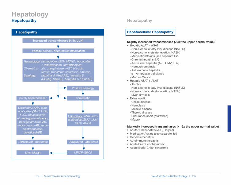
134 I Swiss Essentials in Gastroenterology Swiss Essentials in Gastroenterology I 135
Hepatology
Hepatopathy
Increased transaminases (< 5x ULN)
obesity, alcohol, hepatotoxic medication
Hematology: hemoglobin, MCV, MCHC, leucocytes + differentiation, thrombocytesChemistry: alk. phosphatase, y-GT, bilirubin, ferritin, transferrin saturation, albumin,Serology: hepatitis A (HAV-AB), hepatitis B (HBsAg, HBcAB), hepatitis C (HCV-AB)
purely hepatocellular
Laboratory: ANA, auto-antibodies (SMC, LKM, SLC), ceruloplasmin,
Į1-antitrypsin deficiency, transglutaminase-AB,
endomysium-AB, serum electrophoresis, genetics (HFE)
Ultrasound – abdomen
Liver biopsy
Positive serology
cholestatic
Laboratory: ANA, auto-antibodies (SMC, LKM,
SLC), ANCA
Ultrasound – abdomen
MRCP / ERCP
Hepatopathy Hepatopathy
Hepatocellular Hepatopathy
Slightly increased transaminases (< 5x the upper normal value)
- Non-alcoholic fatty liver disease (NAFLD) - Non-alcoholic steatohepatitis (NASH) - Medication/toxins (see separate list) - Chronic hepatitis B/C - Acute viral hepatitis (A-E, CMV, EBV) - Hemochromatosis - Autoimmune hepatitis -�Į1-Antitrypsin deficiency - Morbus Wilson
- Alcohol - Non-alcoholic fatty liver disease (NAFLD) - Non-alcoholic steatohepatitis (NASH) - Liver cirrhosis
- Celiac disease - Hemolysis - Muscle disease - Thyroid disease - Endurance sport (Marathon) - Macro
Markedly increased transaminases (> 15x the upper normal value)
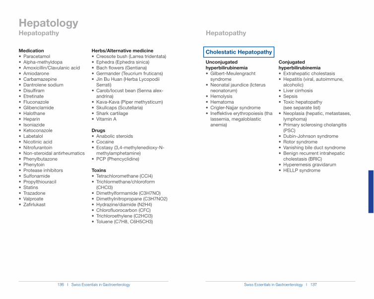
136 I Swiss Essentials in Gastroenterology Swiss Essentials in Gastroenterology I 137
Hepatology
Medication Herbs/Alternative medicine
Serrati)
andrina)
Drugs
methylamphetamine)
Toxins
(CHCl3)
Hepatopathy Hepatopathy
Cholestatic Hepatopathy
Unconjugated hyperbilirubinemia
syndrome
neonatorum)
lassemia, megaloblastic anemia)
Conjugated hyperbilirubinemia
alcoholic)
(see separate list)
lymphoma)
(PSC)
cholestasis (BRIC)
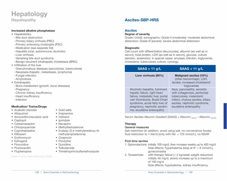
138 I Swiss Essentials in Gastroenterology Swiss Essentials in Gastroenterology I 139
Hepatology
Medication/ Toxins/Drugs
methylamphetamine)
Increased alkaline phosphatase
- Bile duct obstruction - Primary biliary cirrhosis (PBC) - Primary sclerosing cholangitis (PSC) - Medication (see separate list) - Hepatitis (viral, autoimmune, alcoholic) - Liver cirrhosis - Vanishing bile duct syndrome - Benign recurrent intrahepatic cholestasis (BRIC)
- Granulomatous diseases (sarcoidosis, tuberculosis) - Neoplasia (hepatic, metastases, lymphoma) - Fungal infection - Amyloidosis
- Bone metabolism (growth, bone diseases) - Pregnancy - Chronic kidney insufficiency - Heart insufficiency - Infection
[Green R, et al. Gastroenterology (2002)]
AscitesDegree of severityGrade I (mild): sonographic; Grade II (moderate): moderate abdominal distension; Grade III (severe): severe abdominal distension
DiagnosticCell count with differentiation (leucocytes), albumin (as well as in serum), total protein, LDH (as well as in serum), glucose, culture (aerobic, anaerobic). In special cases: amylase, bilirubin, triglyceride, cholesterol, tuberculosis culture, cytology.
Serum Ascites Albumin Gradient (SAAG) = Albumin Serum – Albumin Ascites
TherapyGeneral measuresSalt restriction (in addition, avoid using salt, no convenience foods), fluid restriction to 1 liter/d (only with Na+ < 125 mmol/L), no NSAR
First-time ascites1. Spironolactone: initially 100 mg/d, then increase weekly up to 400 mg/d Side effects: hyperkalemia (stop at K+ > 6 mmol/L), gynecomastia2. Torasemide: with therapy failure (< 2 kg/week weight reduction) initially 40 mg/d, slowly increase up to a maximum of 160 mg/d Side effects: hypokalemia, kidney insufficiency
SAAG > 11 g/L SAAG < 11 g/L
Liver cirrhosis (80%)
Alcoholic hepatitis, fulminant hepatic failure, right heart failure, metastatic liver, portal vein thrombosis, Budd-Chiari syndrome, acute fatty liver of pregnancy, nephrotic syndro-me, exudative enteropathy
Malignant ascites (10%)(often hemorrhagic; LDH,
lactate, increased cholesterol/triglyceride)
Ileus, pancreatitis, serositis with collagenosis, peritoneal tuberculosis, mesenteric infarct, chylous ascites, biliary ascites, nephrotic syndrome, exudative enteropathy
Ascites-SBP-HRS Hepatopathy
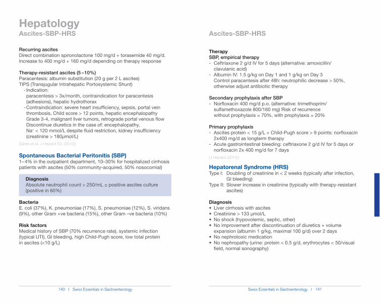
140 I Swiss Essentials in Gastroenterology Swiss Essentials in Gastroenterology I 141
Hepatology
TherapySBP, empirical therapy- Ceftriaxone 2 g/d IV for 5 days (alternative: amoxicillin/ clavulanic acid)- Albumin IV: 1.5 g/kg on Day 1 and 1 g/kg on Day 3 Control paracentesis after 48h: neutrophilic decrease > 50%, otherwise adjust antibiotic therapy
Secondary prophylaxis after SBP- Norfloxacin 400 mg/d p.o. (alternative: trimethoprim/ sulfamethoxazole 800/160 mg) Risk of recurrence without prophylaxis = 70%, with prophylaxis = 20%
Primary prophylaxis - Ascites protein < 15 g/L + Child-Pugh score > 9 points: norfloxacin 2x400 mg/d as longterm therapy- Acute gastrointestinal bleeding: ceftriaxone 2 g/d IV for 5 days or norfloxacin 2x 400 mg/d for 7 days[J Hepatol (2010)]
Hepatorenal Syndrome (HRS)Type I: Doubling of creatinine in < 2 weeks (typically after infection, GI bleeding) Type II: Slower increase in creatinine (typically with therapy-resistant ascites)
Diagnosis
expansion (albumin 1 g/kg, maximal 100 g/d) over 2 days
field, normal sonography)
Recurring ascites Direct combination spironolactone 100 mg/d + torasemide 40 mg/d. Increase to 400 mg/d + 160 mg/d depending on therapy response
Therapy-resistant ascites (5 – 10%)Paracentesis: albumin substitution (20 g per 2 L ascites)TIPS (Transjugular Intrahepatic Portosystemic Shunt) - Indication: paracentesis > 3x/month, contraindication for paracentesis (adhesions), hepatic hydrothorax - Contraindication: severe heart insufficiency, sepsis, portal vein thrombosis, Child score > 12 points, hepatic encephalopathy Grade 3-4, malignant liver tumors, retrograde portal venous flow Discontinue diuretics in the case of: encephalopathy, Na+ < 120 mmol/L despite fluid restriction, kidney insufficiency (creatinine > 180µmol/L)[Ginès et al. J Hepatol 53, (2010)]
Spontaneous Bacterial Peritonitis (SBP)1– 4% in the outpatient department, 10–30% for hospitalized cirrhosis patients with ascites (50% community-acquired, 50% nosocomial)
Diagnosis Absolute neutrophil count > 250/mL ± positive ascites culture (positive in 60%)
BacteriaE. coli (37%), K. pneumoniae (17%), S. pneumoniae (12%), S. viridans (9%), other Gram +ve bacteria (15%), other Gram –ve bacteria (10%)
Risk factorsMedical history of SBP (70% recurrence rate), systemic infection (typical UTI), GI bleeding, high Child-Pugh score, low total protein in ascites (<10 g/L)
Ascites-SBP-HRS Ascites-SBP-HRS
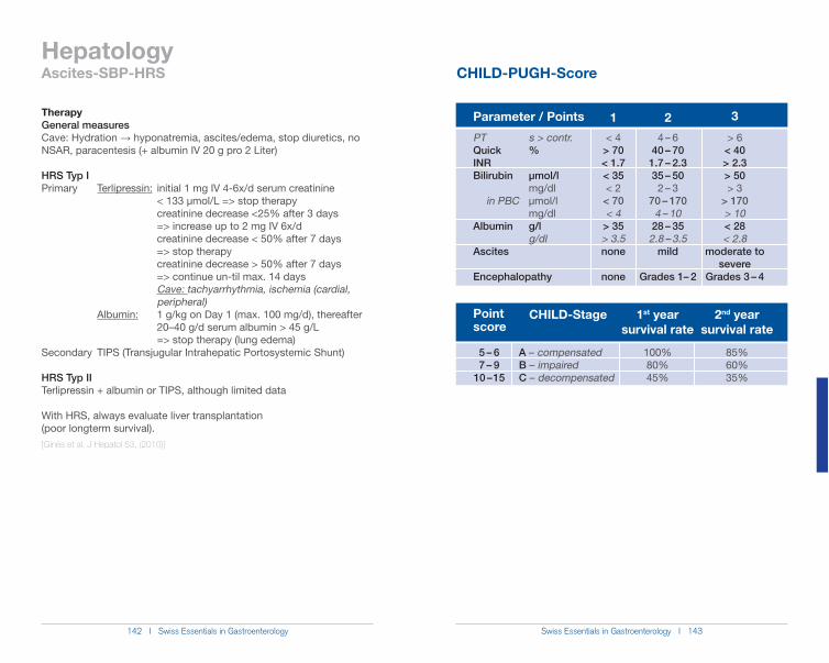
142 I Swiss Essentials in Gastroenterology Swiss Essentials in Gastroenterology I 143
Hepatology
TherapyGeneral measures Cave: Hydration ĺ hyponatremia, ascites/edema, stop diuretics, no NSAR, paracentesis (+ albumin IV 20 g pro 2 Liter)
HRS Typ IPrimary Terlipressin: initial 1 mg IV 4-6x/d serum creatinine < 133 µmol/L => stop therapy creatinine decrease <25% after 3 days => increase up to 2 mg IV 6x/d creatinine decrease < 50% after 7 days => stop therapy creatinine decrease > 50% after 7 days => continue un-til max. 14 days Cave: tachyarrhythmia, ischemia (cardial, peripheral) Albumin: 1 g/kg on Day 1 (max. 100 mg/d), thereafter 20–40 g/d serum albumin > 45 g/L => stop therapy (lung edema)Secondary TIPS (Transjugular Intrahepatic Portosystemic Shunt)
HRS Typ IITerlipressin + albumin or TIPS, although limited data
With HRS, always evaluate liver transplantation (poor longterm survival).[Ginès et al. J Hepatol 53, (2010)]
Parameter / Points
Point score
CHILD-Stage
A – compensatedB – impairedC – decompensated
1st year survival rate
100%80%45%
2nd year survival rate
85%60%35%
5 – 67 – 9
10 – 15
Ascites-SBP-HRS CHILD-PUGH-Score
1< 4
> 70< 1.7< 35< 2
< 70< 4
> 35> 3.5none
none
24 – 6
40 – 701.7 – 2.335 – 50
2 – 370 – 170
4 – 1028 – 35
2.8 – 3.5mild
Grades 1– 2
3
> 6< 40> 2.3> 50> 3
> 170> 10< 28< 2.8
moderate to severe
Grades 3 – 4
PT s > contr.Quick %INRBilirubin µmol/l mg/dl in PBC µmol/l mg/dlAlbumin g/l g/dlAscites
Encephalopathy
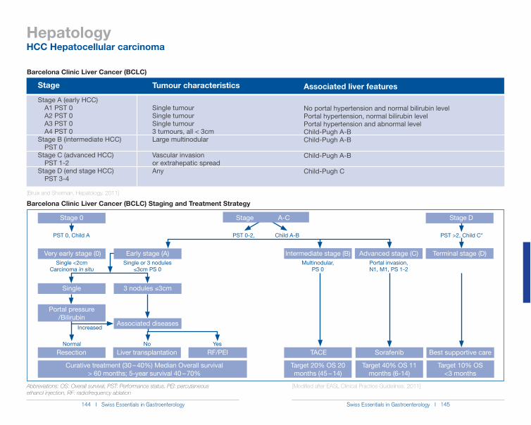
144 I Swiss Essentials in Gastroenterology Swiss Essentials in Gastroenterology I 145
Hepatology
Stage Tumour characteristics Associated liver features
Barcelona Clinic Liver Cancer (BCLC)
Stage A (early HCC) A1 PST 0 A2 PST 0 A3 PST 0 A4 PST 0Stage B (intermediate HCC) PST 0Stage C (advanced HCC) PST 1-2Stage D (end stage HCC) PST 3-4
Single tumourSingle tumourSingle tumour3 tumours, all < 3cmLarge multinodular
Vascular invasion or extrahepatic spreadAny
No portal hypertension and normal bilirubin levelPortal hypertension, normal bilirubin levelPortal hypertension and abnormal levelChild-Pugh A-BChild-Pugh A-B
Child-Pugh A-B
Child-Pugh C
[Bruix and Sherman, Hepatology, 2011]
Barcelona Clinic Liver Cancer (BCLC) Staging and Treatment Strategy
Stage 0 Stage D
PST 0, Child A PST 0-2, Child A-B PST >2, Child C*
Very early stage (0) Intermediate stage (B) Terminal stage (D)Advanced stage (C)Single <2cm
Carcinoma in situSingle or 3 nodules
)3cm PS 0Multinodular,
PS 0Portal invasion, N1, M1, PS 1-2
Single 3 nodules )3cm
Normal No Yes
Increased
Portal pressure/Bilirubin
Early stage (A)
Associated diseases
TACE Best supportive careSorafenibResection Liver transplantation RF/PEI
Curative treatment (30 – 40%) Median Overall survival > 60 months; 5-year survival 40 – 70%
Target 20% OS 20 months (45 – 14)
Target 40% OS 11 months (6-14)
Target 10% OS <3 months
Abbreviations: OS: Overall survival, PST: Performance status, PEI: percutaneous ethanol injection, RF: radiofrequency ablation
HCC Hepatocellular carcinoma
[Modified after EASL Clinical Practice Guidelines, 2011]
Stage A-C
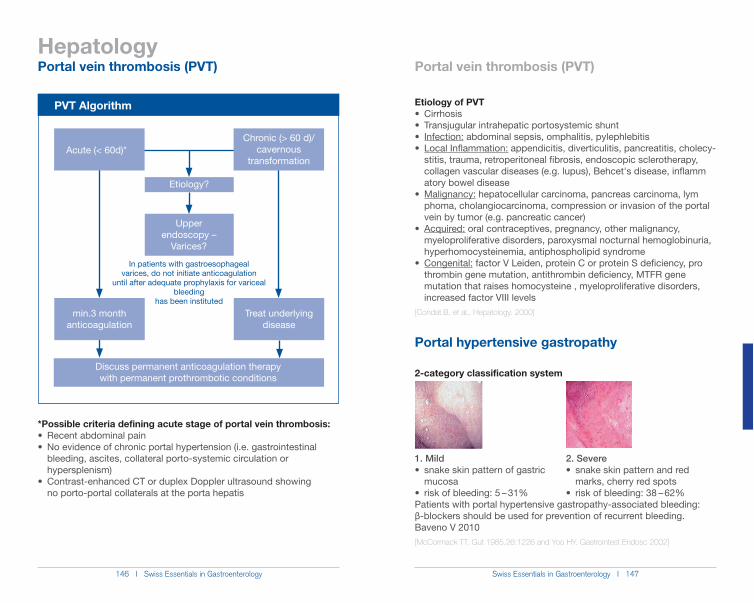
146 I Swiss Essentials in Gastroenterology Swiss Essentials in Gastroenterology I 147
Hepatology
In patients with gastroesophagealvarices, do not initiate anticoagulation
until after adequate prophylaxis for variceal bleeding
has been instituted
Treat underlying disease
min.3 month anticoagulation
Etiology?
Upper endoscopy –
Varices?
Discuss permanent anticoagulation therapywith permanent prothrombotic conditions
Chronic (> 60 d)/cavernous
transformationAcute (< 60d)*
*Possible criteria defining acute stage of portal vein thrombosis:Recent abdominal pain
bleeding, ascites, collateral porto-systemic circulation or hypersplenism)
no porto-portal collaterals at the porta hepatis
Etiology of PVT
Infection: abdominal sepsis, omphalitis, pylephlebitisLocal Inflammation: appendicitis, diverticulitis, pancreatitis, cholecy-
stitis, trauma, retroperitoneal fibrosis, endoscopic sclerotherapy,
atory bowel diseaseMalignancy: hepatocellular carcinoma, pancreas carcinoma, lym
phoma, cholangiocarcinoma, compression or invasion of the portal vein by tumor (e.g. pancreatic cancer)
Acquired: oral contraceptives, pregnancy, other malignancy, myeloproliferative disorders, paroxysmal nocturnal hemoglobinuria, hyperhomocysteinemia, antiphospholipid syndrome
Congenital: factor V Leiden, protein C or protein S deficiency, pro thrombin gene mutation, antithrombin deficiency, MTFR gene mutation that raises homocysteine , myeloproliferative disorders, increased factor VIII levels [Condat B, et al., Hepatology. 2000]
Portal hypertensive gastropathy
2-category classification system
Patients with portal hypertensive gastropathy-associated bleeding: !-blockers should be used for prevention of recurrent bleeding. Baveno V 2010[McCormack TT. Gut 1985.26:1226 and Yoo HY. Gastrointest Endosc 2002]
1. Mildsnake skin pattern of gastric
mucosarisk of bleeding: 5 – 31%
2. Severe
marks, cherry red spots – 62%
Portal vein thrombosis (PVT) Portal vein thrombosis (PVT)
PVT Algorithm
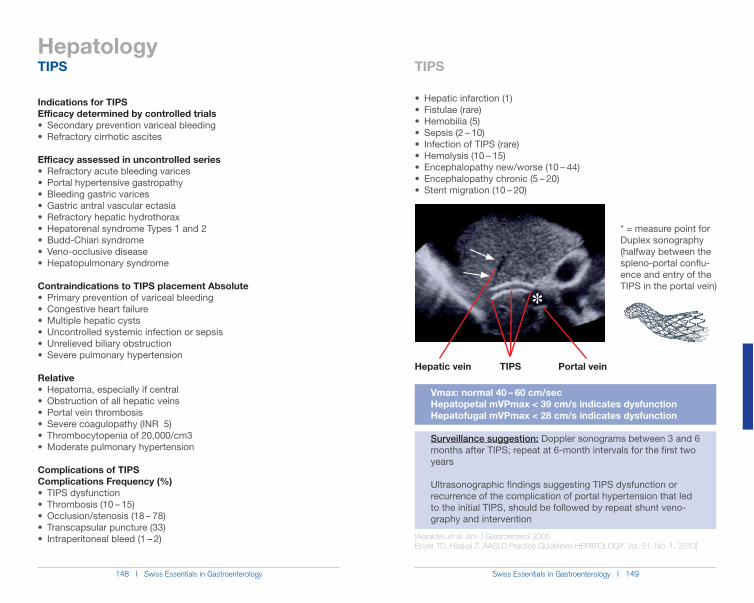
148 I Swiss Essentials in Gastroenterology Swiss Essentials in Gastroenterology I 149
Hepatology
Indications for TIPSEfficacy determined by controlled trials
Secondary prevention variceal bleedingRefractory cirrhotic ascites
Efficacy assessed in uncontrolled seriesRefractory acute bleeding varicesPortal hypertensive gastropathyBleeding gastric varicesGastric antral vascular ectasiaRefractory hepatic hydrothoraxHepatorenal syndrome Types 1 and 2Budd-Chiari syndromeVeno-occlusive diseaseHepatopulmonary syndrome
Contraindications to TIPS placement Absolute
Relative
Complications of TIPSComplications Frequency (%)
Hepatic vein TIPS Portal vein
Vmax: normal 40 – 60 cm/sec Hepatopetal mVPmax < 39 cm/s indicates dysfunction Hepatofugal mVPmax < 28 cm/s indicates dysfunction
Surveillance suggestion: Doppler sonograms between 3 and 6 months after TIPS; repeat at 6-month intervals for the first two years
Ultrasonographic findings suggesting TIPS dysfunction or recurrence of the complication of portal hypertension that led to the initial TIPS, should be followed by repeat shunt veno- graphy and intervention
[Abraldes et al. Am J Gastroenterol 2005Boyer TD, Haskal Z. AASLD Practice Guidelines HEPATOLOGY, Vol. 51, No. 1, 2010]
@
* = measure point for Duplex sonography (halfway between the spleno-portal conflu-ence and entry of the TIPS in the portal vein)
TIPS TIPS
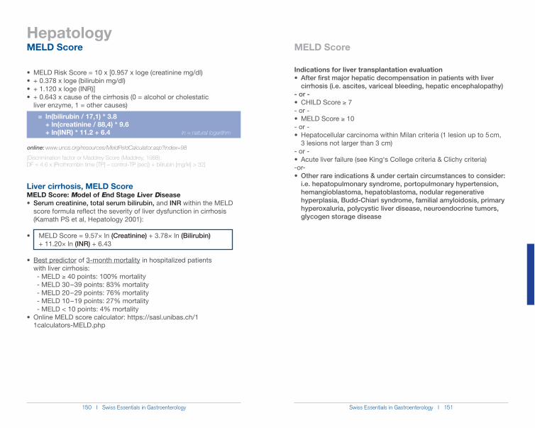
150 I Swiss Essentials in Gastroenterology Swiss Essentials in Gastroenterology I 151
Hepatology
MELD Risk Score = 10 x [0.957 x loge (creatinine mg/dl)+ 0.378 x loge (bilirubin mg/dl)+ 1.120 x loge (INR)]+ 0.643 x cause of the cirrhosis (0 = alcohol or cholestatic
liver enzyme, 1 = other causes)
= ln(bilirubin / 17,1) * 3.8 + ln(creatinine / 88,4) * 9.6 + ln(INR) * 11.2 + 6.4 ln = natural logarithm
online: www.unos.org/resources/MeldPeldCalculator.asp?index=98
[Discrimination factor or Maddrey Score (Maddrey, 1988):DF = 4.6 x (Prothrombin time [TP] – control-TP [sec]) + bilirubin [mg/kl] > 32]
MELD Score MELD Score
Liver cirrhosis, MELD ScoreMELD Score: Model of End Stage Liver Disease
Serum creatinine, total serum bilirubin, and INR within the MELD score formula reflect the severity of liver dysfunction in cirrhosis (Kamath PS et al, Hepatology 2001):
(Creatinine) (Bilirubin) (INR) + 6.43
Best predictor of 3-month mortality in hospitalized patients with liver cirrhosis: - MELD * 40 points: 100% mortality - MELD 30 – 39 points: 83% mortality - MELD 20 – 29 points: 76% mortality - MELD 10 – 19 points: 27% mortality - MELD < 10 points: 4% mortality
Online MELD score calculator: https://sasl.unibas.ch/1 1calculators-MELD.php
Indications for liver transplantation evaluation After first major hepatic decompensation in patients with liver
cirrhosis (i.e. ascites, variceal bleeding, hepatic encephalopathy) - or -
* 7- or -
* 10 - or -
3 lesions not larger than 3 cm) - or -
-or-Other rare indications & under certain circumstances to consider:
i.e. hepatopulmonary syndrome, portopulmonary hypertension, hemangioblastoma, hepatoblastoma, nodular regenerative hyperplasia, Budd-Chiari syndrome, familial amyloidosis, primary hyperoxaluria, polycystic liver disease, neuroendocrine tumors, glycogen storage disease
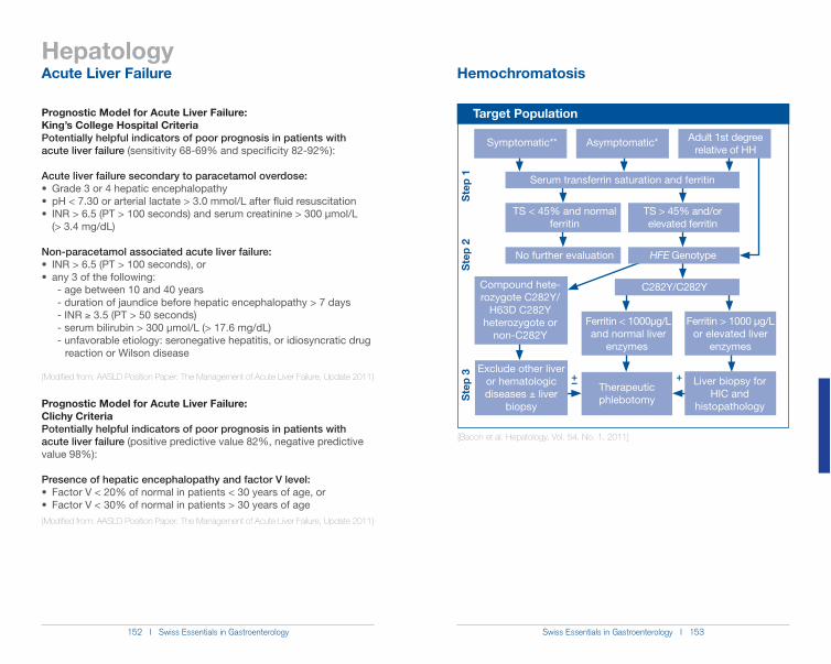
152 I Swiss Essentials in Gastroenterology Swiss Essentials in Gastroenterology I 153
Hepatology
Prognostic Model for Acute Liver Failure: King’s College Hospital CriteriaPotentially helpful indicators of poor prognosis in patients with acute liver failure (sensitivity 68-69% and specificity 82-92%):
Acute liver failure secondary to paracetamol overdose:Grade 3 or 4 hepatic encephalopathypH < 7.30 or arterial lactate > 3.0 mmol/L after fluid resuscitationINR > 6.5 (PT > 100 seconds) and serum creatinine > 300 µmol/L
(> 3.4 mg/dL)
Non-paracetamol associated acute liver failure:INR > 6.5 (PT > 100 seconds), orany 3 of the following:
- age between 10 and 40 years - duration of jaundice before hepatic encephalopathy > 7 days - INR * 3.5 (PT > 50 seconds) - serum bilirubin > 300 µmol/L (> 17.6 mg/dL) - unfavorable etiology: seronegative hepatitis, or idiosyncratic drug reaction or Wilson disease
[Modified from: AASLD Position Paper: The Management of Acute Liver Failure, Update 2011]
Prognostic Model for Acute Liver Failure: Clichy CriteriaPotentially helpful indicators of poor prognosis in patients with acute liver failure (positive predictive value 82%, negative predictive value 98%):
Presence of hepatic encephalopathy and factor V level:Factor V < 20% of normal in patients < 30 years of age, orFactor V < 30% of normal in patients > 30 years of age
[Modified from: AASLD Position Paper: The Management of Acute Liver Failure, Update 2011]
Acute Liver Failure Hemochromatosis
Target Population
+–
Asymptomatic*Symptomatic**
No further evaluation
Adult 1st degree relative of HH
TS < 45% and normal ferritin
TS > 45% and/or elevated ferritin
Therapeutic phlebotomy
Liver biopsy for HIC and
histopathology
Exclude other liver or hematologic diseases ± liver
biopsy
Serum transferrin saturation and ferritin
HFE Genotype
C282Y/C282Y
Ferritin < 1000µg/L and normal liver
enzymes
Ferritin > 1000 µg/L or elevated liver
enzymes
+
Ste
p 1
Ste
p 2
Ste
p 3
[Bacon et al. Hepatology, Vol. 54, No. 1, 2011]
Compound hete-rozygote C282Y/
H63D C282Y heterozygote or
non-C282Y
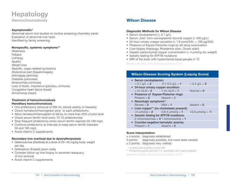
154 I Swiss Essentials in Gastroenterology Swiss Essentials in Gastroenterology I 155
Hepatology
Diagnostic Methods for Wilson DiseaseSerum ceruloplasmin (< 0.1 g/L)Serum „free“ (non-ceruloplasmin bound) copper (> 200 µg/L)24-hour urinary copper excretion (> 1.6 µmol/24h, > 100 µg/24h)Presence of Kayser-Fleischer rings by slit lamp examinationLiver biopsy (histology, Rhodanine stain, Orcein stain)Hepatic parenchymal copper concentration (> 4 µmol/g dry weight)Genetic testing for ATP7B mutationsMRI of the brain with hyperintense basal ganglia in T2
[Modified after EASL Clinical Practice Guidelines, 2012] Wilson Disease Scoring System (Leipzig Score)
< 0.1 g/L = 2 0.1-0.2 g/L = 1 > 0.2 g/L = 0
> 2x ULN = 2 1-2x ULN = 1 Normal = 0
Present = 2 Absent = 0
Severe = 2 Mild = 1 Absent = 0Liver copper** (no cholestasis present):
>4 µmol/g = 2 0.8-4 µmol/g = 1 <0.8 µmol/g = -1
2 chromosomes = 4 1 chromosome = 1
Present = 1 Absent = 0
Score interpretation: * 4 points: diagnosis established 3 points: diagnosis possible, but more tests needed) 2 points: diagnosis very unlikely * or typical abnormalities at brain MRI** Rhodanine-positive granules if no quantitative liver copper available
[Modified after EASL Clinical Practice Guidelines, 2012]
Hemochromatosis Wilson Disease
Asymptomatic*Abnormal serum iron studies on routine screening chemistry panelEvaluation of abnormal liver testsIdentified by family screening
Nonspecific, systemic symptoms**WeaknessFatigueLethargyApathyWeight lossSpecific, organ-related symptomsAbdominal pain (hepatomegaly)Arthralgias (arthritis)Diabetes (pancreas)Amenorrhea (cirrhosis)Loss of libido, impotence (pituitary, cirrhosis)Congestive heart failure (heart)Arrhythmias (heart)
Treatment of hemochromatosisHereditary hemochromatosis
50 and 100 mg/L
Secondary iron overload due to dyserythropoiesis
per day
of iron removal
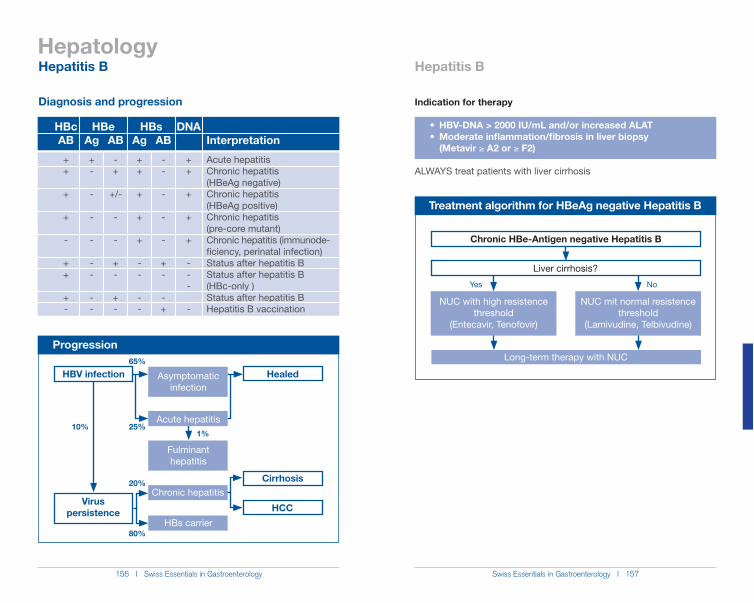
156 I Swiss Essentials in Gastroenterology Swiss Essentials in Gastroenterology I 157
Hepatology
Progression
Healed
Fulminant hepatitis
Chronic hepatitisCirrhosis
HBs carrier
HCCVirus
persistence
HBV infection Asymptomaticinfection
65%
25%
20%
80%
10%1%
Acute hepatitis
HBcAB
++
+
+
-
++
+-
Ag
+-
-
-
-
--
--
AB
-+
+/-
-
-
+-
+-
Ag
++
+
+
+
--
--
AB
--
-
-
-
+-
-+
DNA
++
+
+
+
---
-
Interpretation
Acute hepatitisChronic hepatitis (HBeAg negative)Chronic hepatitis (HBeAg positive)Chronic hepatitis (pre-core mutant)Chronic hepatitis (immunode-ficiency, perinatal infection)Status after hepatitis BStatus after hepatitis B (HBc-only )Status after hepatitis BHepatitis B vaccination
HBe HBs
Treatment algorithm for HBeAg negative Hepatitis B
Long-term therapy with NUC
Chronic HBe-Antigen negative Hepatitis B
Liver cirrhosis?
NUC with high resistence threshold
(Entecavir, Tenofovir)
NUC mit normal resistence threshold
(Lamivudine, Telbivudine)
Indication for therapy
(Metavir * A2 or * F2)
ALWAYS treat patients with liver cirrhosis
No
Hepatitis B Hepatitis B
Diagnosis and progression
Yes
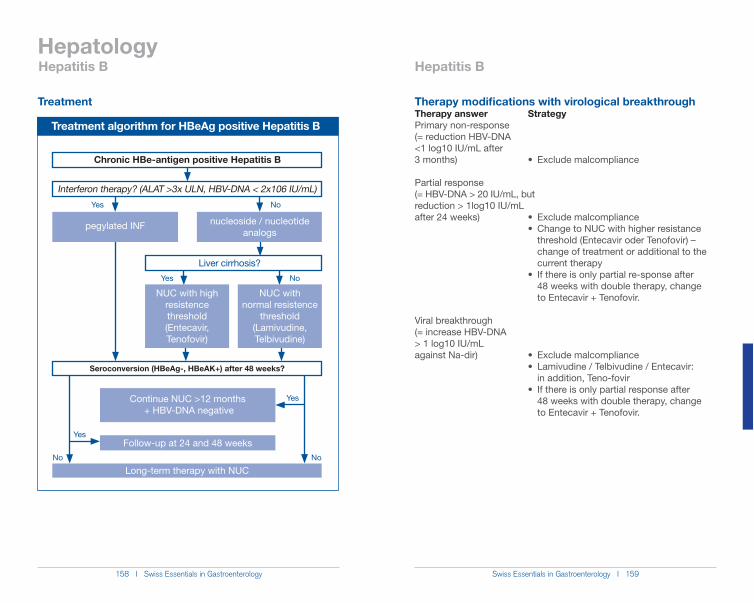
158 I Swiss Essentials in Gastroenterology Swiss Essentials in Gastroenterology I 159
Hepatology
Treatment algorithm for HBeAg positive Hepatitis B
Chronic HBe-antigen positive Hepatitis B
Interferon therapy? (ALAT >3x ULN, HBV-DNA < 2x10⁶ IU/mL)
pegylated INF nucleoside / nucleotide analogs
Liver cirrhosis?
NUC with high resistence threshold(Entecavir, Tenofovir)
NUC with normal resistence
threshold(Lamivudine, Telbivudine)
Continue NUC >12 months + HBV-DNA negative
Follow-up at 24 and 48 weeks
Long-term therapy with NUC
Seroconversion (HBeAg-, HBeAK+) after 48 weeks?
No
Yes
No
Yes
Yes No
NoYes
Therapy modifications with virological breakthroughTherapy answer StrategyPrimary non-response (= reduction HBV-DNA <1 log10 IU/mL after
Partial response (= HBV-DNA > 20 IU/mL, butreduction > 1log10 IU/mL
threshold (Entecavir oder Tenofovir) – change of treatment or additional to the current therapy
48 weeks with double therapy, change to Entecavir + Tenofovir.
Viral breakthrough (= increase HBV-DNA > 1 log10 IU/mL
in addition, Teno-fovir
48 weeks with double therapy, change to Entecavir + Tenofovir.
Hepatitis B Hepatitis B
Treatment
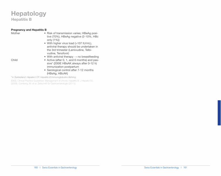
160 I Swiss Essentials in Gastroenterology Swiss Essentials in Gastroenterology I 161
Hepatology
Pregnancy and Hepatitis B
tive (70%), HBeAg negative (2-10%, HBc only (1%))
antiviral therapy should be undertaken in the 3rd trimester (Lamivudine, Telbi- vudine, Tenofovir)
ĺ no breastfeeding
sive* (200IE HBsAK always after 0-12 h) immunization postpartum
(HBsAg, HBcAK)* in Switzerland: Hepatect CP, Hepatitis B Immunoglobulins Behring
[EASL Clinical Practice Guidelines: Management of chronic hepatitis B. J Hepatol 50, (2009). Cornberg, M. et al. Zeitschrift für Gastroenterologie (2011)]
Hepatitis B
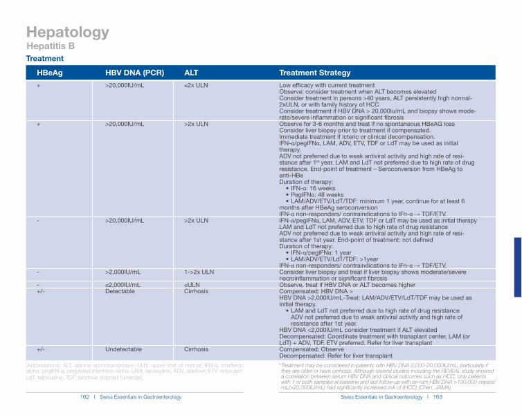
Swiss Essentials in Gastroenterology I 163 162 I Swiss Essentials in Gastroenterology
Hepatology
HBeAg HBV DNA (PCR) ALT Treatment Strategy
[Abbreviations: ALT, alanine aminotransferase; ULN, upper limit of normal; IFN-!, interferon alpha; pegIFN-!, pegylated interferon alpha; LAM, lamivudine, ADV, adefovir; ETV, entecavir; LdT, telbivudine; TDF, tenofovir disproxil fumarate]
* Treatment may be considered in patients with HBV DNA 2,000-20,000IU/mL, particularly if they are older or have cirrhosis. Although several studies including the REVEAL study showed a correlation between serum HBV DNA and clinical outcomes such as HCC, only patients with 1 or both samples at baseline and last follow-up with se-rum HBV DNA >100,000 copies/ mL(>20,000IU/mL) had significantly increased risk of (HCC) (Chen, JAMA)
+
+
-
-
-+/-
+/-
>20,000IU/mL
>20,000IU/mL
>20,000IU/mL
>2,000IU/mL
)2,000IU/mLDetectable
Undetectable
)2x ULN
>2x ULN
>2x ULN
1->2x ULN
)ULN Cirrhosis
Cirrhosis
Low efficacy with current treatmentObserve: consider treatment when ALT becomes elevatedConsider treatment in persons >40 years, ALT persistently high normal-2xULN, or with family history of HCCConsider treatment if HBV DNA > 20,000Iu/mL and biopsy shows mode-rate/severe inflammation or significant fibrosisObserve for 3-6 months and treat if no spontaneous HBeAG lossConsider liver biopsy prior to treatment if compensated.Immediate treatment if Icteric or clinical decompensation.IFN-"/pegIFN", LAM, ADV, ETV, TDF or LdT may be used as initial therapy.ADV not preferred due to weak antiviral activity and high rate of resi-stance after 1st year. LAM and LdT not preferred due to high rate of drug resistance. End-point of treatment – Seroconversion from HBeAg to anti-HBeDuration of therapy:
": 16 weeks": 48 weeks
months after HBeAg seroconversionIFN-" non-responders/ contraindications to IFn-" ĺ�TDF/ETV.IFN-"/pegIFN", LAM, ADV, ETV, TDF or LdT may be used as initial therapyLAM and LdT not preferred due to high rate of drug resistanceADV not preferred due to weak antiviral activity and high rate of resi-stance after 1st year. End-point of treatment: not definedDuration of therapy:
"/pegIFN": 1 year
IFN-" non-responders/ contraindications to IFn-" ĺ TDF/ETV.Consider liver biopsy and treat if liver biopsy shows moderate/severe necroinflammation or significant fibrosisObserve, treat if HBV DNA or ALT becomes higherCompensated: HBV DNA >HBV DNA >2,000IU/mL-Treat: LAM/ADV/ETV/LdT/TDF may be used as initial therapy.
ADV not preferred due to weak antiviral activity and high rate of resistance after 1st year.HBV DNA <2,000IU/mL consider treatment if ALT elevatedDecompensated: Coordinate treatment with transplant center, LAM (or LdT) + ADV, TDF, ETV preferred. Refer for liver transplantCompensated: ObserveDecompensated: Refer for liver transplant
Hepatitis BTreatment
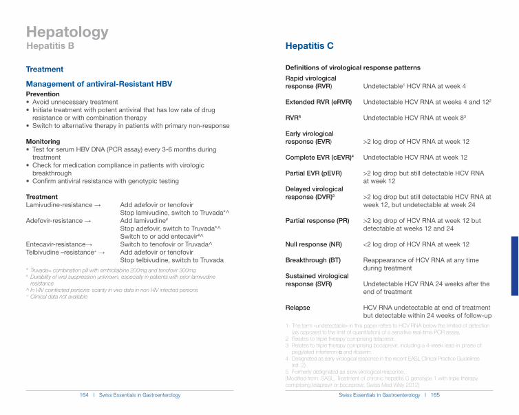
164 I Swiss Essentials in Gastroenterology Swiss Essentials in Gastroenterology I 165
Hepatology
Management of antiviral-Resistant HBVPrevention
resistance or with combination therapy
Monitoring
treatment
breakthrough
TreatmentLamivudine-resistance ĺ� Add adefovir or tenofovir Stop lamivudine, switch to Truvada*^Adefovir-resistance ĺ� Add lamivudine#
Stop adefovir, switch to Truvada*^ Switch to or add entecavir#^Entecavir-resistanceĺ� Switch to tenofovir or Truvada^Telbivudine –resistance+ ĺ Add adefovir or tenofovir Stop telbivudine, switch to Truvada* Truvada= combination pill with emtricitabine 200mg and tenofovir 300mg # Durability of viral suppression unknown, especially in patients with prior lamivudine resistance^ In HIV coinfected persons: scanty in vivo data in non HIV infected persons+ Clinical data not available
Hepatitis B Hepatitis C
Treatment Definitions of virological response patterns
Rapid virological response (RVR) Undetectable1 HCV RNA at week 4
Extended RVR (eRVR) Undetectable HCV RNA at weeks 4 and 122
RVR8 Undetectable HCV RNA at week 83
Early virological response (EVR) >2 log drop of HCV RNA at week 12
Complete EVR (cEVR)4 Undetectable HCV RNA at week 12
Partial EVR (pEVR) >2 log drop but still detectable HCV RNA at week 12Delayed virological response (DVR)5 >2 log drop but still detectable HCV RNA at week 12, but undetectable at week 24
Partial response (PR) >2 log drop of HCV RNA at week 12 but detectable at weeks 12 and 24
Null response (NR) <2 log drop of HCV RNA at week 12
Breakthrough (BT) Reappearance of HCV RNA at any time during treatmentSustained virological response (SVR) Undetectable HCV RNA 24 weeks after the end of treatment
Relapse HCV RNA undetectable at end of treatment but detectable within 24 weeks of follow-up1 The term «undetectable» in this paper refers to HCV RNA below the limited of detection (as opposed to the limit of quantitation) of a sensitive real-time PCR assay.2 Relates to triple therapy comprising telaprevir.3 Relates to triple therapy comprising boceprevir, including a 4-week lead-in phase of pegylated interferon-Į and ribavirin.4 Designated as early virological response in the recent EASL Clinical Practice Guidelines (ref. 2).5 Formerly designated as slow virological response.[Modified from: SASL, Treatment of chronic hepatitis C genotype 1 with triple therapy comprising telaprevir or boceprevir, Swiss Med Wkly 2012]
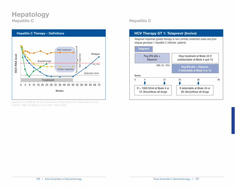
166 I Swiss Essentials in Gastroenterology Swiss Essentials in Gastroenterology I 167
Hepatology
HCV Therapy GT 1: Telaprevir (Incivo)
Telaprevir response-guided therapy in non-cirrhotic treatment-naïve and prior relapse genotype 1 hepatitis C-infected patients
Telaprevir
Peg-IFN alfa + Ribavirin
Peg IFN alfa + Ribavirin if detectable at Week 4 or 12
Stop treatment at Week 24 if undetectable at Week 4 and 12
SVR: 72 – 75%
0 4 12 24 36 48
Weeks
If > 1000 IU/ml at Week 4 or 12: discontinue all drugs
If detectable at Week 24 or 36: discontinue all drugs
Hepatitis C Hepatitis CH
CV
RN
A le
vel
64560 6028 68 7284 12 16 32 4436 40 48 5220 24
Weeks
Treatment
Null response
Partial response
Breakthrough
Relapse
Detection limit
2 log10 drop
Non
resp
onse
Hepatitis C Therapy – Definitions
[Adapted from Shiffman M. Curr Gastroenterol Rep 2006;8:46–52Neumann A, et al. Science 1998; De Bruijne J, et al. Neth J Med 2008]
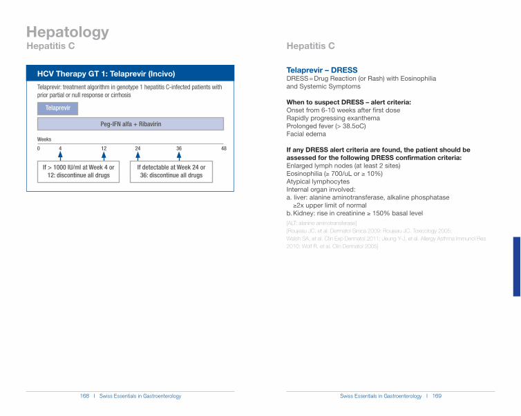
168 I Swiss Essentials in Gastroenterology Swiss Essentials in Gastroenterology I 169
Hepatology
Telaprevir – DRESSDRESS = Drug Reaction (or Rash) with Eosinophilia and Systemic Symptoms
When to suspect DRESS – alert criteria:Onset from 6-10 weeks after first doseRapidly progressing exanthemaProlonged fever (> 38.5oC)Facial edema
If any DRESS alert criteria are found, the patient should be assessed for the following DRESS confirmation criteria:Enlarged lymph nodes (at least 2 sites)Eosinophilia (* 700/uL or * 10%)Atypical lymphocytesInternal organ involved:a. liver: alanine aminotransferase, alkaline phosphatase *2x upper limit of normalb. Kidney: rise in creatinine * 150% basal level[ALT: alanine aminotransferase][Roujeau JC, et al. Dermatol Sinica 2009; Roujeau JC. Toxicology 2005;Walsh SA, et al. Clin Exp Dermatol 2011; Jeung Y-J, et al. Allergy Asthma Immunol Res 2010; Wolf R, et al. Clin Dermatol 2005]
Hepatitis C Hepatitis C
HCV Therapy GT 1: Telaprevir (Incivo)
Telaprevir: treatment algorithm in genotype 1 hepatitis C-infected patients with prior partial or null response or cirrhosis
Telaprevir
Peg-IFN alfa + Ribavirin
0 4 12 24 36 48
Weeks
If > 1000 IU/ml at Week 4 or 12: discontinue all drugs
If detectable at Week 24 or 36: discontinue all drugs
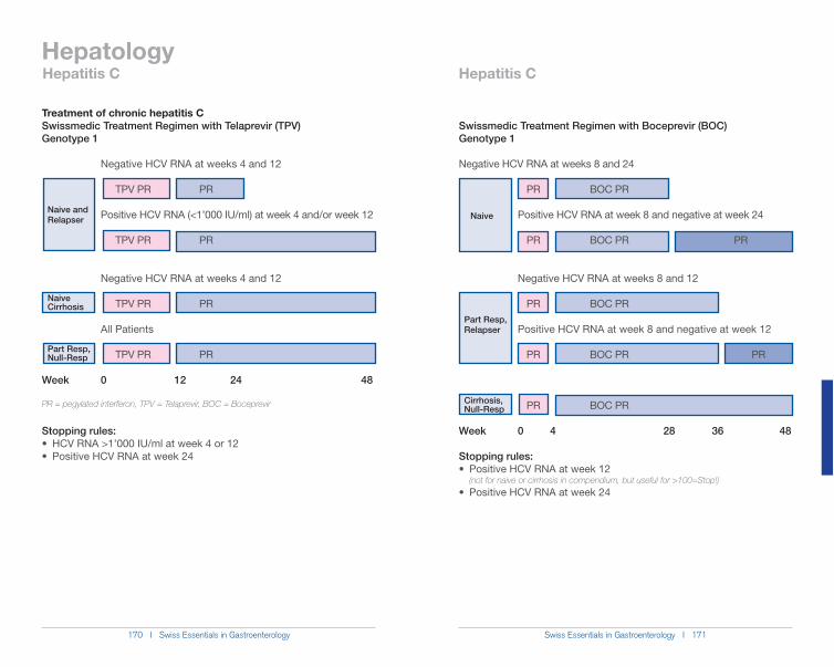
170 I Swiss Essentials in Gastroenterology Swiss Essentials in Gastroenterology I 171
Hepatology
Naive and Relapser
Naive Cirrhosis
Part Resp, Null-Resp
Part Resp, Relapser
Naive
Cirrhosis, Null-Resp
Hepatitis C Hepatitis C
Treatment of chronic hepatitis CSwissmedic Treatment Regimen with Telaprevir (TPV) Genotype 1
Negative HCV RNA at weeks 4 and 12
TPV PR PR
TPV PR PR
Negative HCV RNA at weeks 4 and 12
TPV PR PR
All Patients
TPV PR PR
Week 0 12 24 48
PR = pegylated interferon, TPV = Telaprevir, BOC = Boceprevir
Stopping rules:
Swissmedic Treatment Regimen with Boceprevir (BOC) Genotype 1
Negative HCV RNA at weeks 8 and 24
PR BOC PR
Positive HCV RNA at week 8 and negative at week 24
PR BOC PR PR
Negative HCV RNA at weeks 8 and 12
PR BOC PR
Positive HCV RNA at week 8 and negative at week 12
PR BOC PR PR
PR BOC PR
Week 0 4 28 36 48
Stopping rules:
(not for naive or cirrhosis in compendium, but useful for >100=Stop!)
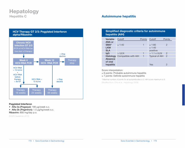
172 I Swiss Essentials in Gastroenterology Swiss Essentials in Gastroenterology I 173
Hepatology
HCV Therapy GT 2/3: Pegylated Interferon alpha/Ribavirin
Chronic HCV Infection GT 2/3
(PCR of HCV-RNA be-fore start of therapy)
Therapy 24 weeks
Therapy 48 weeks
Therapy 16 weeks
Week 4HCV-RNA PCR
HCV RNA < 15 IU/ml
+HCV RNA
before therapy < 8x10⁵
HCV RNA < 15 IU/ml
Therapy stop
< 2log decline
> 2log decline
Week 12HCV- RNA PCR
Pegylated Interferon 180 µg/week s.c.
1.5 µg/kg/week s.c. Ribavirin: 800 mg/day p.o.[DGVS Guidelines 2010]
Simplified diagnostic criteria for autoimmune hepatitis (AIH)
Variable Cutoff Points Cutoff Points ANA or SMA* * 1:40 1 * 1:80 2
* 1:40 SLA positive IgG > ULN 1 > 1.1 x ULN 2 Histology Compatible with AIH 1 Typical of AIH 2 Absence of viral hepatitis Yes 2
Score interpretation: * 6 points: Probable autoimmune hepatitis* 7 points: Definite autoimmune hepatitis* Maximal number of points for all autoantibodies is 2; AIH score maximum is 8.
[Modified from: Choi et al., Hepatology 2008]
Hepatitis C Autoimmune hepatitis
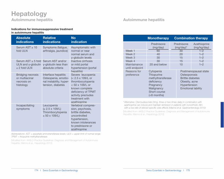
174 I Swiss Essentials in Gastroenterology Swiss Essentials in Gastroenterology I 175
Hepatology
* Alternative: Oral budesonide (3mg, three or two times daily) in combination with azathioprine can induce and maintain remission in patients with noncirrhotic AIH, with a low rate of steroid-specific side effects (Manns et al. Gastroenterology 2010)
[Modified from: AASLD Practice Guidelines: Diagnosis and Management of Autoimmune Hepatitis, Manns et al., Hepatology 2010]
Monotherapy Combination therapy
Week 1Week 2Week 3Week 4Maintainance until endpointReasons for preference
Prednisone (mg/day)
60403030
20 and below
Azathioprine (mg/kg/day)
1–21–21–21–21–2
Prednisone (mg/day)*
3020151510
Cytopenia Postmenopausal stateThiopurine Osteoporosismethyltransferase Brittle diabetesdeficiency Obesity, acnePregnancy HypertensionMalignancy Emotional labilityShort course (<6 months)
Absolute indications
Serum AST * 10 fold ULN
Serum AST * 5 fold ULN and Ȗ-globulin * 2 fold ULN
Bridging necrosis or multiacinar necrosis on histology
Incapacitating symptoms
Symptoms (fatigue, arthralgia, jaundice)
Serum AST and/or Ȗ-globulin less than absolute criteria
Interface hepatitisOsteopenia, emotio-nal instability, hyper-tension, diabetes
Leucopenia () 2.5 x 10⁹/L)Thrombocytopenia ) 50 x 109/L)
Asymptomatic with normal or near normal serum and Ȗ-globulin levelsInactive cirrhosis or mild portal hypertension (portal hepatitis)Severe leucopenia (< 2.5 x 10⁹/L or thrombocytopenia < 50 x 109/L or known complete deficiency of TPMT activity precludes treatment with azathioprineVertebral compres-sion, psychosis, brittle diabetes, uncontrolled hypertension, known intolerances to prednisone or azathioprine
Relative indications
No indication
Indications for immunosuppressive treatment in autoimmune hepatitis
Abbreviations: AST = aspartate aminotransferase levels; ULN = upper limit of normal range; TPMT = thiopurine methyltransferase.
[Modified from: AASLD Practice Guidelines: Diagnosis and Management of Autoimmune Hepatitis, Manns et al., Hepatology 2010]
Autoimmune hepatitis Autoimmune hepatitis
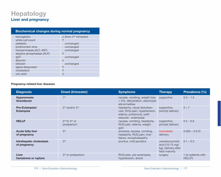
176 I Swiss Essentials in Gastroenterology Swiss Essentials in Gastroenterology I 177
Hepatology
Diagnosis
HyperemesisGravidarum
Pre-Eclampsia/ Eclampsia
HELLP
Acute fatty liverof pregnancy
Intrahepatic cholestasisof pregnancy
Liverhematoma or rupture
nausea, vomiting, weight loss > 5%, dehydration, electrolyte abnormalitiesheadache, visual disturban-cies, RUQ pain, hypertension, edema, proteinuria, (with seizures : eclampsia)nausea, vomiting, headache, RUQ pain, edema, weight gainanorexia, nausea, vomiting, headache, RUQ pain, liver failure, encephalopathypruritus, mild jaundice
RUQ pain, pre-eclampsia, hypotension, shock
1st
2nd and/or 3rd
2nd to 3rd or postpartum
3rd
3rd
3rd or postpartum
0.3 – 1.0
5 – 7
0.2 – 0.6
0.005 – 0.010
0.1 – 0.3
1 (in patients with HELLP)
supportive
supportive,prompt delivery
supportive,prompt delivery
immediate delivery
ursodeoxycholic acid (10-15 mg/kg), delivery after fetal maturitysurgery
Onset (trimester) Symptoms Therapy Prevalence (%)
Pregnancy-related liver diseases
Biochemical changes during normal pregnancy
hemoglobin ��(from 2nd trimester)white cell count ��platelets unchangedprothrombin time unchangedtransaminases (ALT, AST) unchangedalkaline phosphatase (ALP) �gGT unchangedalbumin �bilirubin unchangedalpha-fetoprotein �cholesterol �uric acid �
Liver and pregnancy
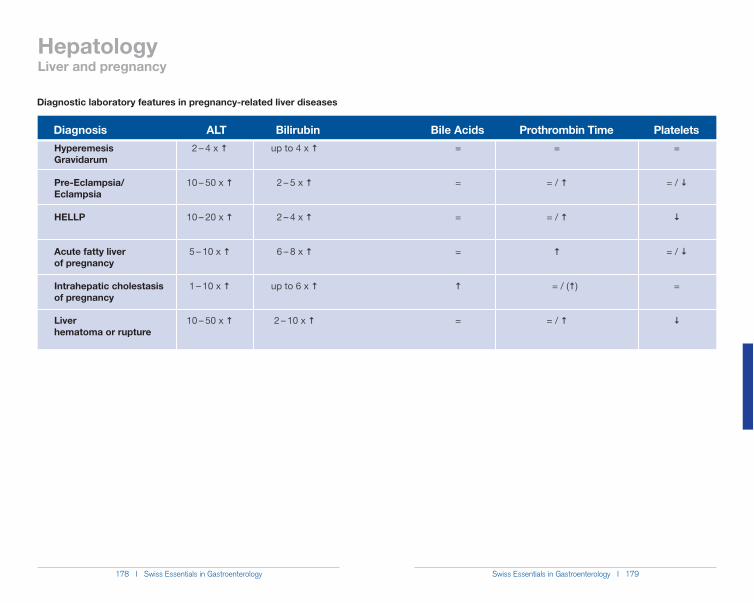
178 I Swiss Essentials in Gastroenterology Swiss Essentials in Gastroenterology I 179
Hepatology
DiagnosisHyperemesisGravidarum
Pre-Eclampsia/ Eclampsia
HELLP
Acute fatty liverof pregnancy
Intrahepatic cholestasisof pregnancy
Liverhematoma or rupture
up to 4 x �
2 – 5 x �
2 – 4 x �
6 – 8 x �
up to 6 x �
2 – 10 x �
2 – 4 x �
10 – 50 x �
10 – 20 x �
5 – 10 x �
1 – 10 x �
10 – 50 x �
=
= / �
�
= / �
=
�
=
=
=
=
�
=
ALT Bilirubin Bile Acids Platelets
Diagnostic laboratory features in pregnancy-related liver diseases
=
= / �
= / �
�
= / (�)
= / �
Prothrombin Time
Liver and pregnancy
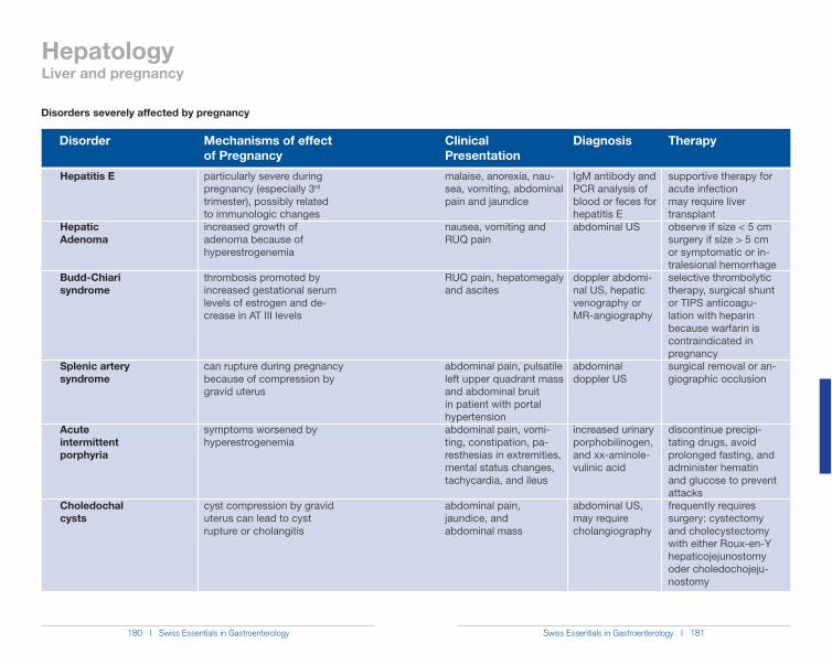
180 I Swiss Essentials in Gastroenterology Swiss Essentials in Gastroenterology I 181
Disorder Mechanisms of effectof Pregnancy
Clinical Presentation
Diagnosis Therapy
Disorders severely affected by pregnancy
Hepatitis E
HepaticAdenoma
Budd-Chiarisyndrome
Splenic artery syndrome
Acuteintermittentporphyria
Choledochalcysts
malaise, anorexia, nau-sea, vomiting, abdominal pain and jaundice
nausea, vomiting and RUQ pain
RUQ pain, hepatomegaly and ascites
abdominal pain, pulsatile left upper quadrant mass and abdominal bruit in patient with portal hypertensionabdominal pain, vomi-ting, constipation, pa-resthesias in extremities, mental status changes, tachycardia, and ileus
abdominal pain, jaundice, and abdominal mass
particularly severe during pregnancy (especially 3rd trimester), possibly related to immunologic changesincreased growth of adenoma because of hyperestrogenemia
thrombosis promoted by increased gestational serum levels of estrogen and de-crease in AT III levels
can rupture during pregnancy because of compression by gravid uterus
symptoms worsened by hyperestrogenemia
cyst compression by gravid uterus can lead to cyst rupture or cholangitis
supportive therapy for acute infectionmay require liver transplantobserve if size < 5 cmsurgery if size > 5 cm or symptomatic or in-tralesional hemorrhageselective thrombolytic therapy, surgical shunt or TIPS anticoagu-lation with heparin because warfarin is contraindicated in pregnancysurgical removal or an-giographic occlusion
discontinue precipi-tating drugs, avoid prolonged fasting, and administer hematin and glucose to prevent attacksfrequently requires surgery: cystectomy and cholecystectomy with either Roux-en-Y hepaticojejunostomy oder choledochojeju-nostomy
IgM antibody and PCR analysis of blood or feces for hepatitis Eabdominal US
doppler abdomi-nal US, hepatic venography orMR-angiography
abdominal doppler US
increased urinary porphobilinogen, and xx-aminole-vulinic acid
abdominal US, may require cholangiography
HepatologyLiver and pregnancy
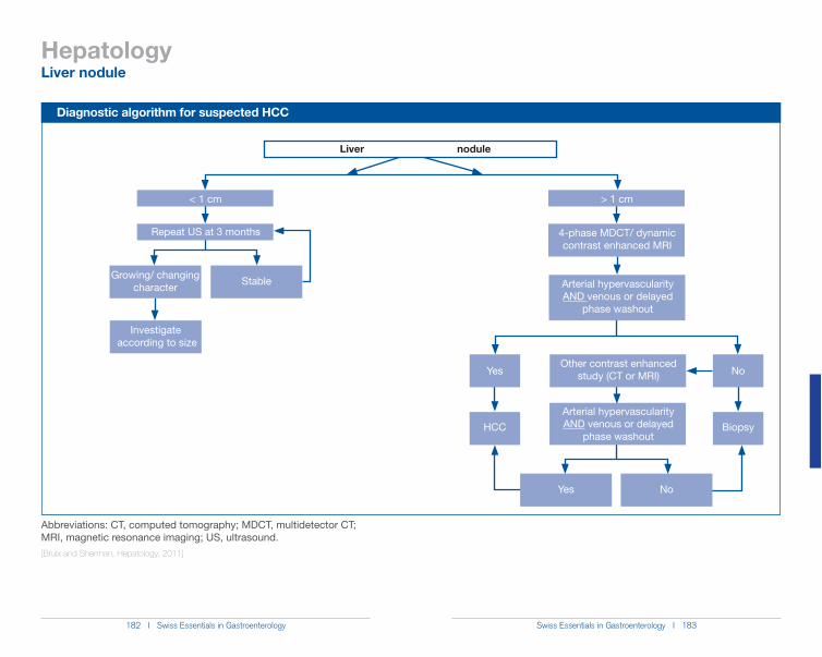
182 I Swiss Essentials in Gastroenterology Swiss Essentials in Gastroenterology I 183
Hepatology
Diagnostic algorithm for suspected HCC
Growing/ changing character
< 1 cm
Repeat US at 3 months
Investigate according to size
Stable
> 1 cm
4-phase MDCT/ dynamic contrast enhanced MRI
Arterial hypervascularity AND venous or delayed
phase washoutHCC Biopsy
Arterial hypervascularity AND venous or delayed
phase washout
Yes
Yes No
Abbreviations: CT, computed tomography; MDCT, multidetector CT; MRI, magnetic resonance imaging; US, ultrasound.[Bruix and Sherman, Hepatology, 2011]
Liver nodule
Liver nodule
NoOther contrast enhanced
study (CT or MRI)
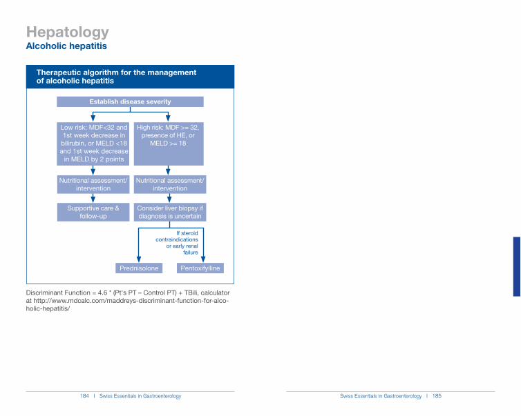
184 I Swiss Essentials in Gastroenterology Swiss Essentials in Gastroenterology I 185
Hepatology
Therapeutic algorithm for the management of alcoholic hepatitis
Low risk: MDF<32 and 1st week decrease in
bilirubin, or MELD <18 and 1st week decrease
in MELD by 2 points
High risk: MDF >= 32, presence of HE, or
MELD >= 18
Establish disease severity
Supportive care & follow-up
Nutritional assessment/ intervention
Nutritional assessment/ intervention
Consider liver biopsy if diagnosis is uncertain
Prednisolone Pentoxifylline
If steroid contraindications
or early renal failure
Alcoholic hepatitis
at http://www.mdcalc.com/maddreys-discriminant-function-for-alco-holic-hepatitis/
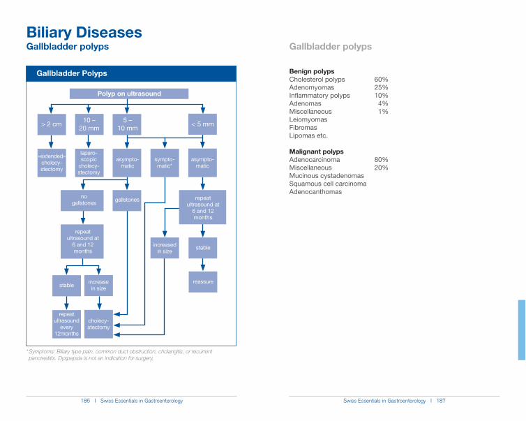
186 I Swiss Essentials in Gastroenterology Swiss Essentials in Gastroenterology I 187
Biliary Diseases
Gallbladder Polyps
«extended» cholecy-stectomy
laparo-scopic
cholecy-stectomy
asympto-matic
> 2 cm 10 –20 mm
5 –10 mm < 5 mm
asympto-matic
reassure
stable
repeat ultrasound at
6 and 12 months
stable increase in size
repeat ultrasound
every 12months
cholecy-stectomy
increased in size
Polyp on ultrasound
repeat ultrasound at
6 and 12 months
gallstonesno gallstones
sympto-matic*
* Symptoms: Biliary type pain, common duct obstruction, cholangitis, or recurrent pancreatitis. Dyspepsia is not an indication for surgery.
Benign polypsCholesterol polyps 60%Adenomyomas 25%Inflammatory polyps 10%Adenomas 4%Miscellaneous 1%LeiomyomasFibromasLipomas etc.
Malignant polypsAdenocarcinoma 80%Miscellaneous 20%Mucinous cystadenomasSquamous cell carcinomaAdenocanthomas
Gallbladder polyps Gallbladder polyps
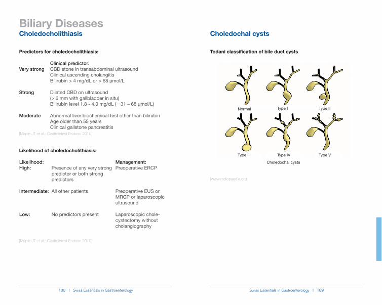
188 I Swiss Essentials in Gastroenterology Swiss Essentials in Gastroenterology I 189
Biliary Diseases
Predictors for choledocholithiasis:
Clinical predictor:Very strong CBD stone in transabdominal ultrasound Clinical ascending cholangitis Bilirubin > 4 mg/dL or > 68 µmol/L
Strong Dilated CBD on ultrasound (> 6 mm with gallbladder in situ) Bilirubin level 1.8 - 4.0 mg/dL (= 31 – 68 µmol/L) Moderate Abnormal liver biochemical test other than bilirubin Age older than 55 years Clinical gallstone pancreatitis[Maple JT et al.: Gastrointest Endosc 2010]
Likelihood of choledocholithiasis:
[Maple JT et al.: Gastrointest Endosc 2010]
Likelihood:High:
Intermediate:
Low:
Presence of any very strong predictor or both strong predictors
All other patients
No predictors present
Management: Preoperative ERCP
Preoperative EUS or MRCP or laparoscopic ultrasound
Laparoscopic chole-cystectomy without cholangiography
Todani classification of bile duct cysts
[www.radiopaedia.org]
Normal Type I Type II
Type III Type IV Type V
Choledochal cysts
Choledocholithiasis Choledochal cysts
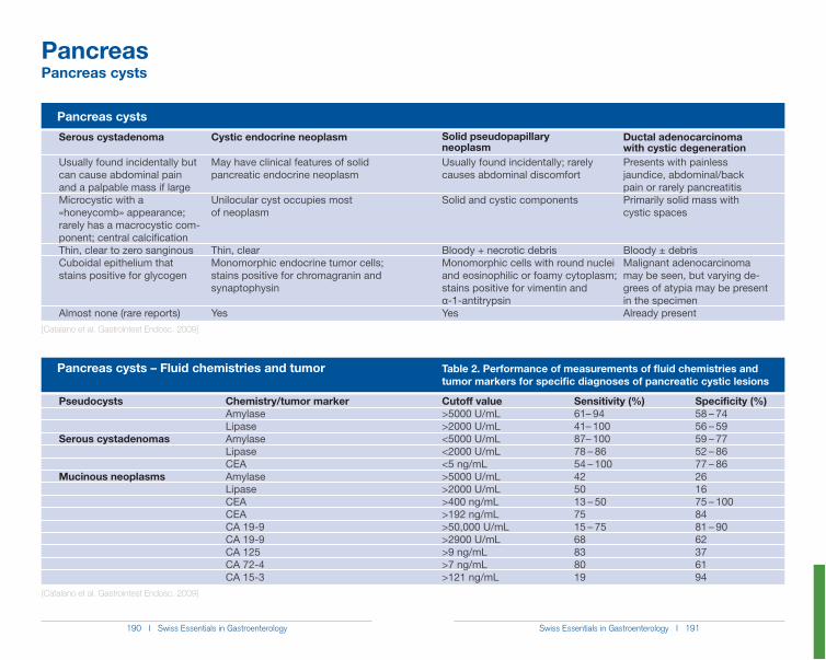
190 I Swiss Essentials in Gastroenterology Swiss Essentials in Gastroenterology I 191
Pancreas
Pancreas cysts
Pancreas cysts – Fluid chemistries and tumor
[Catalano et al. Gastrointest Endosc. 2009]
Table 2. Performance of measurements of fluid chemistries and tumor markers for specific diagnoses of pancreatic cystic lesions
[Catalano et al. Gastrointest Endosc. 2009]
Sensitivity (%)61– 9441– 10087– 10078 – 8654 – 100425013 – 507515 – 7568838019
Cutoff value>5000 U/mL>2000 U/mL<5000 U/mL<2000 U/mL<5 ng/mL>5000 U/mL>2000 U/mL>400 ng/mL>192 ng/mL>50,000 U/mL>2900 U/mL>9 ng/mL>7 ng/mL>121 ng/mL
Pseudocysts
Serous cystadenomas
Mucinous neoplasms
Chemistry/tumor markerAmylaseLipaseAmylaseLipaseCEAAmylaseLipaseCEACEACA 19-9CA 19-9CA 125CA 72-4CA 15-3
Specificity (%)58 – 7456 – 5959 – 7752 – 8677 – 86261675 – 1008481 – 9062376194
Pancreas cysts
Ductal adenocarcinoma with cystic degenerationPresents with painless jaundice, abdominal/back pain or rarely pancreatitisPrimarily solid mass with cystic spaces
Bloody ± debrisMalignant adenocarcinoma may be seen, but varying de-grees of atypia may be present in the specimenAlready present
Solid pseudopapillary neoplasmUsually found incidentally; rarely causes abdominal discomfort
Solid and cystic components
Bloody + necrotic debrisMonomorphic cells with round nuclei and eosinophilic or foamy cytoplasm; stains positive for vimentin and Į-1-antitrypsinYes
Serous cystadenoma
Usually found incidentally but can cause abdominal pain and a palpable mass if largeMicrocystic with a «honeycomb» appearance; rarely has a macrocystic com-ponent; central calcificationThin, clear to zero sanginousCuboidal epithelium that stains positive for glycogen
Almost none (rare reports)
Cystic endocrine neoplasm
May have clinical features of solid pancreatic endocrine neoplasm
Unilocular cyst occupies most of neoplasm
Thin, clearMonomorphic endocrine tumor cells; stains positive for chromagranin and synaptophysin
Yes
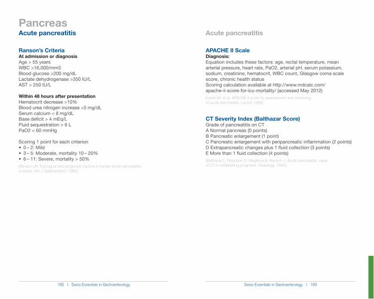
192 I Swiss Essentials in Gastroenterology Swiss Essentials in Gastroenterology I 193
Pancreas
Ranson’s CriteriaAt admission or diagnosisAge > 55 yearsWBC >16,000/mm3Blood glucose >200 mg/dLLactate dehydrogenase >350 IU/LAST > 250 IU/L
Within 48 hours after presentationHematocrit decrease >10%Blood urea nitrogen increase >5 mg/dLSerum calcium < 8 mg/dLBase deficit > 4 mEq/LFluid sequestration > 6 LPaO2 < 60 mmHg
Scoring 1 point for each criterion
[Ranson JH. Etiological and prognostic factors in human acute pancreatitis: a review. Am J Gastroenterol. 1982]
APACHE II ScaleDiagnosis:Equation includes these factors: age, rectal temperature, mean arterial pressure, heart rate, PaO2, arterial pH, serum potassium, sodium, creatinine, hematocrit, WBC count, Glasgow coma scale score, chronic health status Scoring calculation available at http://www.mdcalc.com/ apache-ii-score-for-icu-mortality/ (accessed May 2012)[Larvin M. et al. APACHE-II score for assessment and monitoring of acute pancreatitis. Lancet 1989]
CT Severity Index (Balthazar Score)Grade of pancreatitis on CTA Normal pancreas (0 points)B Pancreatic enlargement (1 point)C Pancreatic enlargement with peripancreatic inflammation (2 points)D Extrapancreatic changes plus 1 fluid collection (3 points)E More than 1 fluid collection (4 points)[Balthazar E, Robinson D, Megibow A, Ranson J. Acute pancreatitis: valueof CT in establishing prognosis. Radiology. 1990]
Acute pancreatitis Acute pancreatitis
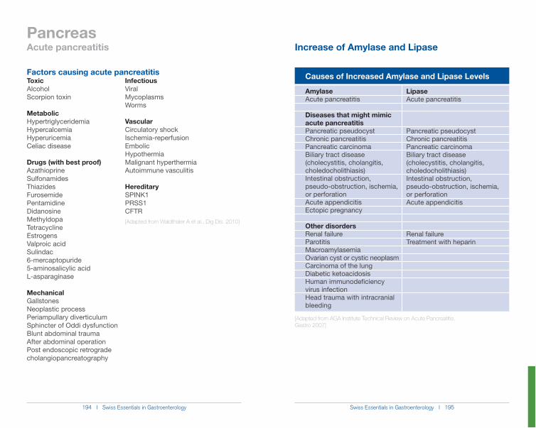
194 I Swiss Essentials in Gastroenterology Swiss Essentials in Gastroenterology I 195
[Adapted from AGA Institute Technical Review on Acute Pancreatitis. Gastro 2007]
Pancreas
Causes of Increased Amylase and Lipase Levels
AmylaseAcute pancreatitis
Diseases that might mimic acute pancreatitisPancreatic pseudocyst Chronic pancreatitis Pancreatic carcinoma Biliary tract disease (cholecystitis, cholangitis, choledocholithiasis)Intestinal obstruction, pseudo-obstruction, ischemia, or perforationAcute appendicitis Ectopic pregnancy
Other disordersRenal failure ParotitisMacroamylasemiaOvarian cyst or cystic neoplasmCarcinoma of the lungDiabetic ketoacidosisHuman immunodeficiency virus infectionHead trauma with intracranial bleeding
LipaseAcute pancreatitis
Pancreatic pseudocystChronic pancreatitisPancreatic carcinomaBiliary tract disease (cholecystitis, cholangitis, choledocholithiasis)Intestinal obstruction, pseudo-obstruction, ischemia, or perforationAcute appendicitis
Renal failureTreatment with heparin
Increase of Amylase and LipaseAcute pancreatitis
Factors causing acute pancreatitisToxicAlcoholScorpion toxin
MetabolicHypertriglyceridemiaHypercalcemiaHyperuricemiaCeliac disease
Drugs (with best proof)AzathioprineSulfonamidesThiazidesFurosemidePentamidineDidanosineMethyldopaTetracyclineEstrogensValproic acidSulindac6-mercaptopuride5-aminosalicylic acidL-asparaginase
MechanicalGallstonesNeoplastic processPeriampullary diverticulumSphincter of Oddi dysfunctionBlunt abdominal traumaAfter abdominal operationPost endoscopic retrograde cholangiopancreatography
InfectiousViralMycoplasmsWorms
VascularCirculatory shockIschemia-reperfusionEmbolicHypothermiaMalignant hyperthermiaAutoimmune vasculitis
HereditarySPINK1PRSS1CFTR[Adapted from Waldthaler A et al., Dig Dis. 2010]