Embed Size (px)
Citation preview
"Production and characterization of the recombinant wheat chitinase Wch1 and generation of chitin-specific antibodies"
Von der Fakultät für Mathematik, Informatik und Naturwissenschaften der Rheinisch-
Westfälischen Technischen Hochschule Aachen zur Erlangung des akademischen Grades einer Doktorin der Naturwissenschaften genehmigte Dissertation
vorgelegt von
Master of Science
Siham Agdour
Aus Algier, Algerien
Berichter: Universitätsprofessor Dr.rer.nat. R. Fischer
Universitätsprofessor Dr.rer.nat. F. Kreuzaler
Tag der mündlichen Prüfung: 31.08.2007
Diese Dissertation ist auf den Internetseiten der Hochschulbibliothek online verfügbar
Content
I
I Introduction ..............................................................................................................................1
I.1 Chitinases, chitin and chitosan..............................................................................................1
I.1.1 Chitinases ......................................................................................................................1
I.1.1.1 Classification of chitinases...................................................................................1
I.1.1.2 Structure, mechanism and substrate binding of chitinases...................................3
I.1.1.2.1 Structure and mechanism .................................................................................3
I.1.1.2.2 Substrate binding cleft of chitinases.................................................................5
I.1.1.3 The roles of chitinases..........................................................................................6
I.1.1.4 Applications of chitinases ....................................................................................8
I.1.2 Chitin and chitosan:.......................................................................................................9
I.1.2.1 Extraction and crystallographic structure of chitin ..............................................9
I.1.2.2 Biotechnological applications of chitin and chitosan.........................................11
I.2 Antibody engineering..........................................................................................................12
I.2.1 Antibody structure and function..................................................................................12
I.2.2 Generation of carbohydrate-specific antibodies..........................................................13
I.3 Aim of the thesis: ................................................................................................................15
II Materials and Methods........................................................................................................20
II.1 Materials...........................................................................................................................20
II.1.1 Chemicals and consumables........................................................................................20
II.1.2 Enzymes and reaction kits...........................................................................................20
II.1.3 Antibodies and substrates ............................................................................................20
II.1.4 Bacterial strains ...........................................................................................................21
II.1.5 Plants and animals .......................................................................................................22
II.1.6 Phage Libraries............................................................................................................22
II.1.7 DNA-Vectors...............................................................................................................22
II.1.8 Oligonucleotides..........................................................................................................23
II.1.9 Buffers, media and solutions .......................................................................................24
Content
II
II.1.10 Matrices and membranes.............................................................................................24
II.1.11 Equipment ...................................................................................................................24
II.2 Methods............................................................................................................................26
II.2.1 Recombinant DNA technologies.................................................................................26
II.2.1.1 Preparation of electrocompetent E. coli cells.....................................................26
II.2.1.2 Transformation of E. coli by electroporation .....................................................26
II.2.1.3 Preparation of electrocompetent A. tumefaciens cells........................................26
II.2.1.4 Transformation of A. tumefaciens by electroporation........................................27
II.2.1.5 Growth of recombinant A. tumefaciens and preparation of glycerol stocks ......27
II.2.1.6 Isolation of plasmid DNA from E. coli ..............................................................28
II.2.1.7 Agarose gel electrophoresis of DNA .................................................................28
II.2.1.8 Preparative agarose gel electrophoresis .............................................................28
II.2.1.9 PCR amplification ..............................................................................................28
II.2.1.10 DNA sequencing ................................................................................................29
II.2.2 Transient and stable transformation of tobacco plants ................................................30
II.2.2.1 Transient assay in tobacco leaves by vacuum infiltration..................................30
II.2.2.1.1 Preparation of recombinant agrobacteria .......................................................30
II.2.2.1.2 Vacuum infiltration of intact tobacco leaves..................................................30
II.2.2.2 Recombinant Agrobacterium-mediated stable transformation of tobacco plants 31
II.2.2.3 Growth of N. tabacum cv. Petite Havana SR1...................................................32
II.2.3 Expression and purification of recombinant proteins and monoclonal antibodies .....32
II.2.3.1 Optimization of expression conditions of recombinant Wch1 in bacteria .........32
II.2.3.2 Bacterial expression and purification of Wch1-MBP ........................................33
II.2.3.3 Bacterial expression and purification of his6-tagged Wch1 chitinase ...............34
II.2.3.4 Bacterial expression and purification of Strep-tagged Wch1.............................35
II.2.3.5 Bacterial expression and purification of the chitin-binding domain ..................35
II.2.3.6 Expression and purification of Wch1 chitinase from plants ..............................36
Content
III
II.2.3.7 Purification of monoclonal antibodies through Protein G matrix ......................37
II.2.4 Protein analysis............................................................................................................38
II.2.4.1 Extraction of total soluble proteins from plant leaves........................................38
II.2.4.2 Quantification of proteins...................................................................................38
II.2.4.3 SDS-PAA gel electrophoresis and Coomassie brilliant blue staining................38
II.2.4.4 Immunoblot analysis ..........................................................................................39
II.2.5 Characterization of chitinase activity ..........................................................................40
II.2.5.1 Colorimetric activity assay.................................................................................40
II.2.5.2 Optimum pH and temperature............................................................................40
II.2.5.3 Degradation of glycol-chitin in an overlay gel...................................................41
II.2.5.3.1 Preparation of glycol-chitin substrate ............................................................41
II.2.5.3.2 Isoelectric focusing (IEF) gel and overlayer substrate gel .............................41
II.2.5.4 Degradation of colloidal chitin...........................................................................42
II.2.5.5 Thin layer chromatography (TLC).....................................................................42
II.2.5.6 Extended depolymerization of chitosan .............................................................43
II.2.5.7 HPLC analysis....................................................................................................43
II.2.5.8 1H-NMR spectroscopy analysis .........................................................................43
II.2.5.9 Hydrolysis of (GlcNAc)6 oligosaccharide by Wch1 chitinase...........................44
II.2.5.10 Determination of the anomeric form of the hydrolytic products .......................44
II.2.5.11 Investigation of the anti-fungal activity of Wch1 ..............................................45
II.2.5.11.1 Isolation of fungal spores ...............................................................................45
II.2.5.11.2 In vitro anti-fungal assay................................................................................45
II.2.6 Classification of Wch1 ................................................................................................45
II.2.7 Immunization of mice .................................................................................................46
II.2.7.1 Chitosan-ovalbumin coupling ............................................................................46
II.2.7.2 Determination of antibody-binding titres by ELISA..........................................47
II.2.7.3 Competition ELISA using free peptides ............................................................48
Content
IV
II.2.8 Selection of phage displayed chitin-mimicking peptides............................................48
II.2.8.1 Phage displayed chitin-mimicking peptide selection .........................................48
II.2.9 Characterization of phage displayed chitin-mimicking peptides ................................50
II.2.9.1 Titration of amplified phages .............................................................................50
II.2.9.2 Phage ELISA......................................................................................................51
II.2.9.3 Inhibition ELISA of selected phage-displayed peptides ....................................52
II.2.9.4 Isolation of single stranded DNA from selected phages ....................................52
II.2.10 Generation of chitin/chitosan-specific monoclonal antibodies by hybridoma technology ................................................................................................................................53
II.2.10.1 Animal cell culture .............................................................................................53
II.2.10.2 Cryo-preservation of animal cells ......................................................................54
II.2.10.3 Isolation of mouse spleen cells...........................................................................54
II.2.10.4 Establishment of stable hybridoma cells ............................................................54
II.2.10.5 Limiting dilutions for isolation of monoclonal antibodies.................................55
II.2.10.6 Isotype determination of generated chitin-specific monoclonal antibodies.......55
II.2.11 Microscopic analysis ...................................................................................................56
II.2.11.1 Preparation of cover slides .................................................................................56
II.2.11.2 Preparation of fungal mycelia ............................................................................56
II.2.11.3 Coating of cover slides with fungal mycelia ......................................................56
II.2.11.4 Fluorescence microscopy ...................................................................................57
III Results ................................................................................................................................58
III.1.1 Generation of chitosan-specific antibodies .................................................................58
III.1.1.1 Coupling of chitosan to ovalbumin ....................................................................58
III.1.1.2 Immunization of mice with ova-coupled chitosan .............................................59
III.1.2 Generation of chitin-specific antibodies......................................................................60
III.1.2.1 Purification of chitin-binding domain ................................................................60
III.1.2.2 Selection of chitin mimicking peptides ..............................................................61
III.1.2.3 Sequence analysis of selected chitin-mimicking clones ....................................64
Content
V
III.1.2.4 Immunization of mice with ovalbumin coupled chitin-mimicking peptides .....64
III.1.2.5 Generation of chitin-specific monoclonal antibodies.........................................66
III.1.2.6 Determination of the isotype of generated monoclonal antibodies....................68
III.1.2.7 Immunofluorescent labelling of F. graminearum ..............................................68
III.1.2.8 Purification of selected monoclonal antibodies through protein G matrix ........69
III.1.2.9 Reactivity of non-purified and purified monoclonal antibodies to WGA7 G2 peptide and chitosan...............................................................................................................70
III.2 Production and characterization of recombinant Wch1 chitinase....................................73
III.2.1 Production of recombinant Wch1 chitinase in E. coli.................................................73
III.2.1.1 Cloning of Wch1 cDNA into bacterial expression vectors ................................73
III.2.1.2 Optimization of expression conditions in E. coli ...............................................73
III.2.1.3 Purification and quantification of bacterial produced chitinase.........................75
III.2.2 Production of recombinant Wch1 chitinase in tobacco plants ....................................76
III.2.2.1 Cloning of Wch1 cDNA into plant expression vectors ......................................76
III.2.2.2 Transient expression of Wch1 chitinase.............................................................77
III.2.2.3 Purification and quantification of plant produced chitinase...............................78
III.3 Generation and characterization of stable transformed tobacco plants producing Wch1 chitinase.......................................................................................................................................79
III.3.1 Generation and screening of stable transformed tobacco plants .................................79
III.3.2 Distribution of recombinant Wch1 chitinase in transgenic tobacco plants .................81
III.4 Characterization of Wch1 chitinase activity ....................................................................82
III.4.1 Glycol chitin enzymatic degradation after isoelectric focusing ..................................82
III.4.2 Colorimetric assay for quantification of chitinase activity .........................................83
III.4.3 Colloidal chitin Chitinase assay ..................................................................................84
III.5 Comparison of the yields and activity for the bacterial and plant produced Wch1 chitinases .....................................................................................................................................85
III.6 Optimum pH and temperature for chitinase activity........................................................86
III.7 In vitro antifungal activity................................................................................................88
Content
VI
III.8 Characterization of oligomers produced from enzymatic degradation of chitosan .........89
III.8.1 Thin layer chromatography (TLC) analysis ................................................................89
III.8.2 Time-course gel filtration of (GlcNac)6 oligosaccharide digestion by Wch1-apo......91
III.8.3 Size distribution of oliogmers after degradation of chitosan with Wch1-apo.............91
III.8.4 1HNMR spectroscopy analysis of selected oligomers.................................................93
III.8.5 Classification of Wch1 wheat chitinase ......................................................................95
III.8.6 Mechanism of action of Wch1 chitinase .....................................................................96
IV Discussion and future prospects .........................................................................................98
IV.1 Generation of chitosan- and chitin-specific monoclonal antibodies ................................98
IV.1.1 Generation of chitosan-specific antibodies .................................................................98
IV.1.2 Generation of chitin-specific monoclonal antibodies..................................................99
IV.2 Production and characterization of recombinant Wch1 chitinase..................................102
IV.2.1 Accumulation and purification of recombinant Wch1 from bacteria and tobacco ...102
IV.2.1.1 Accumulation and purification from bacteria ..................................................102
IV.2.1.2 Accumulation and purification from plant cells...............................................103
IV.2.2 Evaluation of the optimal system for production of Wch1 .......................................104
IV.2.3 Characterization of enzymatic Wch1-apo activity ....................................................106
IV.2.3.1 Optimum temperature and pH..........................................................................106
IV.2.3.2 Antifungal activity............................................................................................106
IV.2.3.3 Physico-chemical characterization of substrate degradation ...........................109
IV.2.3.3.1 Subsite cleavage of Wch1-apo chitinase......................................................109
IV.2.3.3.2 Anomeric configuration of degradation products and substrate preference 111
IV.2.3.3.3 Action pattern of Wch1-apo chitinase on chitosan ......................................113
V Summary.............................................................................................................................115
VI Bibliographic references ...................................................................................................117
VII Appendix..........................................................................................................................134
VII.1 Abbreviations ..............................................................................................................134

Content
VII
VII.2 Tables and figures .......................................................................................................137
VII.3 Schematic representation of vector maps....................................................................140
Introduction
1
I Introduction
I.1 Chitinases, chitin and chitosan
I.1.1 Chitinases
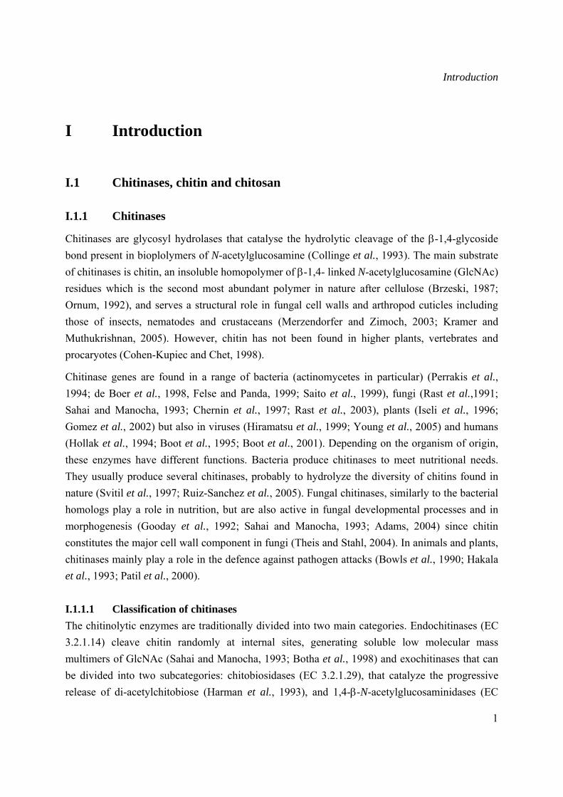
Chitinases are glycosyl hydrolases that catalyse the hydrolytic cleavage of the β-1,4-glycoside bond present in bioplolymers of N-acetylglucosamine (Collinge et al., 1993). The main substrate of chitinases is chitin, an insoluble homopolymer of β-1,4- linked N-acetylglucosamine (GlcNAc) residues which is the second most abundant polymer in nature after cellulose (Brzeski, 1987; Ornum, 1992), and serves a structural role in fungal cell walls and arthropod cuticles including those of insects, nematodes and crustaceans (Merzendorfer and Zimoch, 2003; Kramer and Muthukrishnan, 2005). However, chitin has not been found in higher plants, vertebrates and procaryotes (Cohen-Kupiec and Chet, 1998).
Chitinase genes are found in a range of bacteria (actinomycetes in particular) (Perrakis et al., 1994; de Boer et al., 1998, Felse and Panda, 1999; Saito et al., 1999), fungi (Rast et al.,1991; Sahai and Manocha, 1993; Chernin et al., 1997; Rast et al., 2003), plants (Iseli et al., 1996; Gomez et al., 2002) but also in viruses (Hiramatsu et al., 1999; Young et al., 2005) and humans (Hollak et al., 1994; Boot et al., 1995; Boot et al., 2001). Depending on the organism of origin, these enzymes have different functions. Bacteria produce chitinases to meet nutritional needs. They usually produce several chitinases, probably to hydrolyze the diversity of chitins found in nature (Svitil et al., 1997; Ruiz-Sanchez et al., 2005). Fungal chitinases, similarly to the bacterial homologs play a role in nutrition, but are also active in fungal developmental processes and in morphogenesis (Gooday et al., 1992; Sahai and Manocha, 1993; Adams, 2004) since chitin constitutes the major cell wall component in fungi (Theis and Stahl, 2004). In animals and plants, chitinases mainly play a role in the defence against pathogen attacks (Bowls et al., 1990; Hakala et al., 1993; Patil et al., 2000).
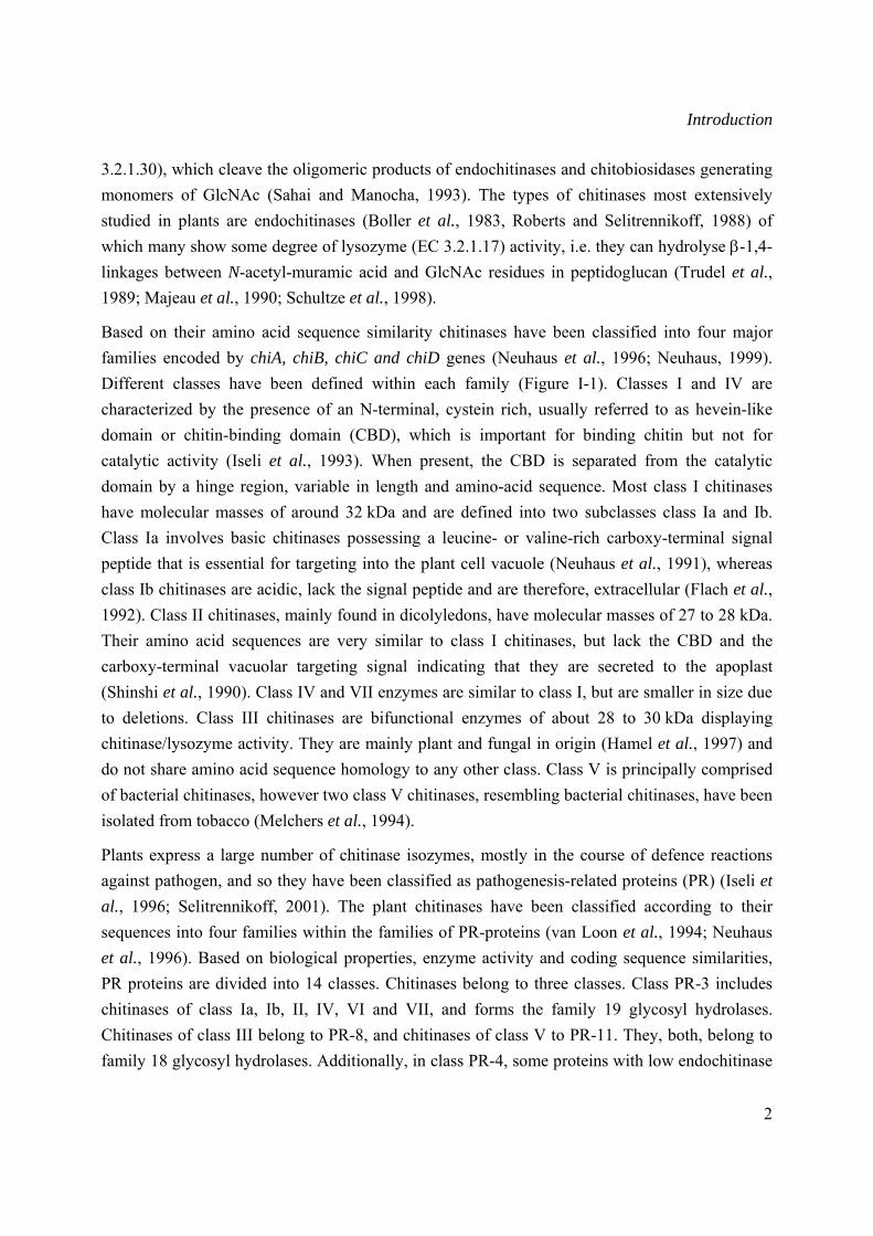
I.1.1.1 Classification of chitinases The chitinolytic enzymes are traditionally divided into two main categories. Endochitinases (EC 3.2.1.14) cleave chitin randomly at internal sites, generating soluble low molecular mass multimers of GlcNAc (Sahai and Manocha, 1993; Botha et al., 1998) and exochitinases that can be divided into two subcategories: chitobiosidases (EC 3.2.1.29), that catalyze the progressive release of di-acetylchitobiose (Harman et al., 1993), and 1,4-β-N-acetylglucosaminidases (EC
Introduction
2
3.2.1.30), which cleave the oligomeric products of endochitinases and chitobiosidases generating monomers of GlcNAc (Sahai and Manocha, 1993). The types of chitinases most extensively studied in plants are endochitinases (Boller et al., 1983, Roberts and Selitrennikoff, 1988) of which many show some degree of lysozyme (EC 3.2.1.17) activity, i.e. they can hydrolyse β-1,4-linkages between N-acetyl-muramic acid and GlcNAc residues in peptidoglucan (Trudel et al., 1989; Majeau et al., 1990; Schultze et al., 1998).
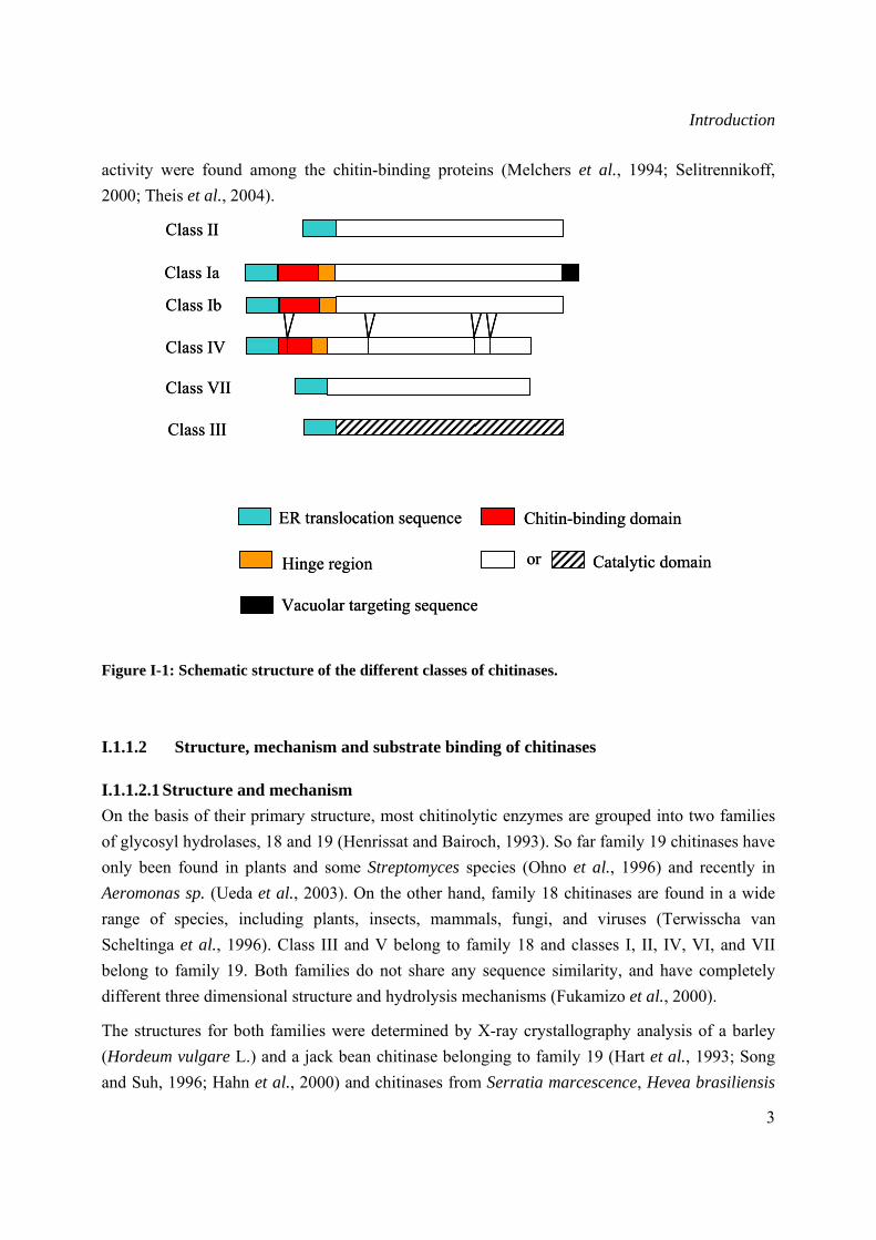
Based on their amino acid sequence similarity chitinases have been classified into four major families encoded by chiA, chiB, chiC and chiD genes (Neuhaus et al., 1996; Neuhaus, 1999). Different classes have been defined within each family (Figure I-1). Classes I and IV are characterized by the presence of an N-terminal, cystein rich, usually referred to as hevein-like domain or chitin-binding domain (CBD), which is important for binding chitin but not for catalytic activity (Iseli et al., 1993). When present, the CBD is separated from the catalytic domain by a hinge region, variable in length and amino-acid sequence. Most class I chitinases have molecular masses of around 32 kDa and are defined into two subclasses class Ia and Ib. Class Ia involves basic chitinases possessing a leucine- or valine-rich carboxy-terminal signal peptide that is essential for targeting into the plant cell vacuole (Neuhaus et al., 1991), whereas class Ib chitinases are acidic, lack the signal peptide and are therefore, extracellular (Flach et al., 1992). Class II chitinases, mainly found in dicolyledons, have molecular masses of 27 to 28 kDa. Their amino acid sequences are very similar to class I chitinases, but lack the CBD and the carboxy-terminal vacuolar targeting signal indicating that they are secreted to the apoplast (Shinshi et al., 1990). Class IV and VII enzymes are similar to class I, but are smaller in size due to deletions. Class III chitinases are bifunctional enzymes of about 28 to 30 kDa displaying chitinase/lysozyme activity. They are mainly plant and fungal in origin (Hamel et al., 1997) and do not share amino acid sequence homology to any other class. Class V is principally comprised of bacterial chitinases, however two class V chitinases, resembling bacterial chitinases, have been isolated from tobacco (Melchers et al., 1994).
Plants express a large number of chitinase isozymes, mostly in the course of defence reactions against pathogen, and so they have been classified as pathogenesis-related proteins (PR) (Iseli et al., 1996; Selitrennikoff, 2001). The plant chitinases have been classified according to their sequences into four families within the families of PR-proteins (van Loon et al., 1994; Neuhaus et al., 1996). Based on biological properties, enzyme activity and coding sequence similarities, PR proteins are divided into 14 classes. Chitinases belong to three classes. Class PR-3 includes chitinases of class Ia, Ib, II, IV, VI and VII, and forms the family 19 glycosyl hydrolases. Chitinases of class III belong to PR-8, and chitinases of class V to PR-11. They, both, belong to family 18 glycosyl hydrolases. Additionally, in class PR-4, some proteins with low endochitinase
Introduction
3
activity were found among the chitin-binding proteins (Melchers et al., 1994; Selitrennikoff, 2000; Theis et al., 2004).
Figure I-1: Schematic structure of the different classes of chitinases.
I.1.1.2 Structure, mechanism and substrate binding of chitinases
I.1.1.2.1 Structure and mechanism On the basis of their primary structure, most chitinolytic enzymes are grouped into two families of glycosyl hydrolases, 18 and 19 (Henrissat and Bairoch, 1993). So far family 19 chitinases have only been found in plants and some Streptomyces species (Ohno et al., 1996) and recently in Aeromonas sp. (Ueda et al., 2003). On the other hand, family 18 chitinases are found in a wide range of species, including plants, insects, mammals, fungi, and viruses (Terwisscha van Scheltinga et al., 1996). Class III and V belong to family 18 and classes I, II, IV, VI, and VII belong to family 19. Both families do not share any sequence similarity, and have completely different three dimensional structure and hydrolysis mechanisms (Fukamizo et al., 2000).
The structures for both families were determined by X-ray crystallography analysis of a barley (Hordeum vulgare L.) and a jack bean chitinase belonging to family 19 (Hart et al., 1993; Song and Suh, 1996; Hahn et al., 2000) and chitinases from Serratia marcescence, Hevea brasiliensis
Class II
Class Ib
Class IV
Class VII
Class III
or
ER translocation sequence
Hinge region
Chitin-binding domain
Catalytic domain
Class Ia
Vacuolar targeting sequence
Class II
Class Ib
Class IV
Class VII
Class III
or
ER translocation sequence
Hinge region
Chitin-binding domain
Catalytic domain
Class Ia
Vacuolar targeting sequence
Introduction
4
(rubber tree) and the pathogenic fungus Coccidioides immitis which are family 18 members (Perrakis et al., 1994; Terwisscha van Scheltinga et al., 1994; Hollis et al., 2000; Papanikolau et al., 2001). The overall fold of family 19 chitinases corresponds to a compact α-helical domain with three conserved disulfide bridges. The hypothetical binding cleft is composed of two α-helices and three-stranded β-sheet (Hart et al., 1995). The folding as well as substrate specificities of family 19 were reported to be very similar to those of lysozyme (Holm and Sander, 1994) leading to the proposal that both families have related catalytic mechanisms. On the other hand, family 18 chitinases have a typical (α/β)8 barrel structure composed of eight α-helices and an eight stranded β-sheet, with an additional N-terminal β-strand-rich domain and a small (α+β) domain (Terwisscha van Scheltinga and Dijikstra, 1996).
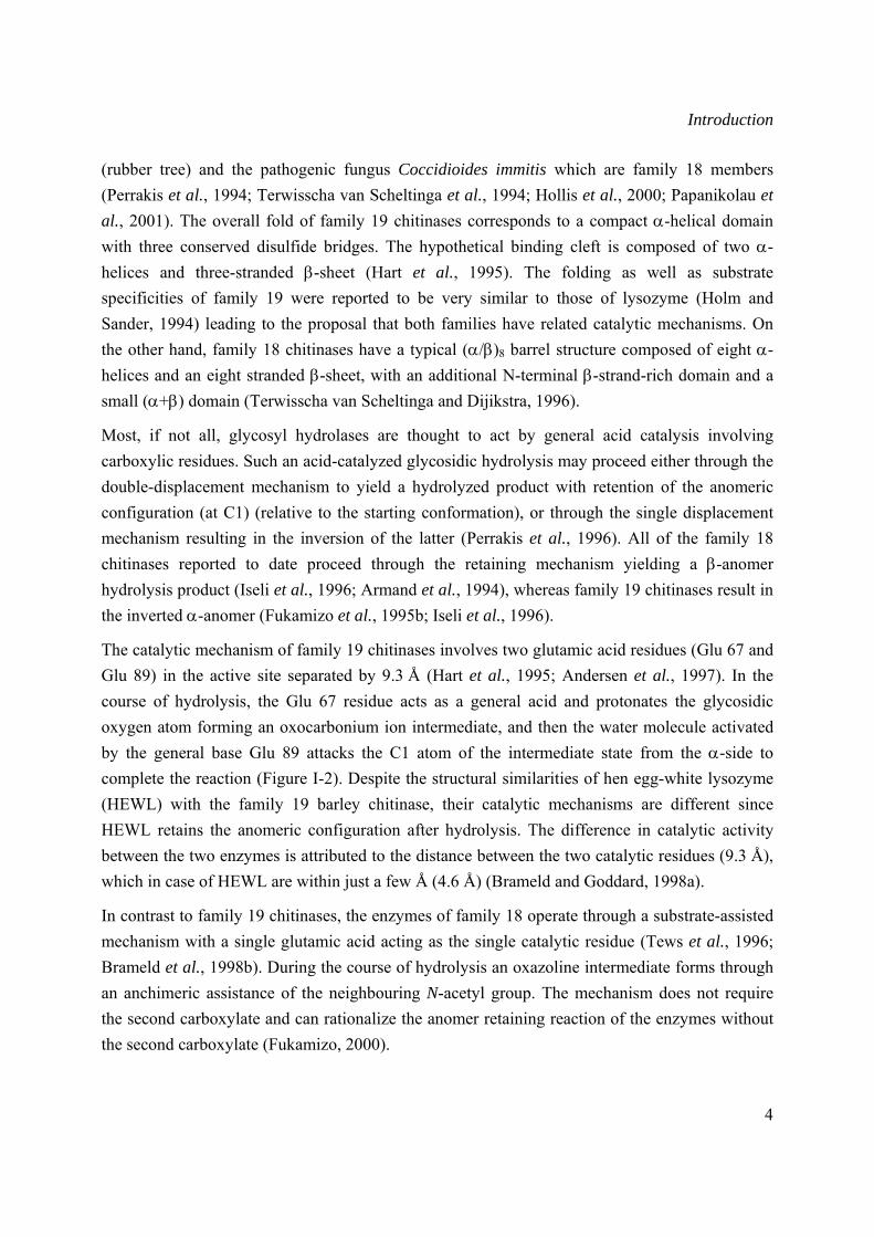
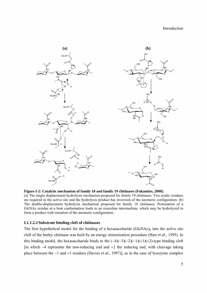
Most, if not all, glycosyl hydrolases are thought to act by general acid catalysis involving carboxylic residues. Such an acid-catalyzed glycosidic hydrolysis may proceed either through the double-displacement mechanism to yield a hydrolyzed product with retention of the anomeric configuration (at C1) (relative to the starting conformation), or through the single displacement mechanism resulting in the inversion of the latter (Perrakis et al., 1996). All of the family 18 chitinases reported to date proceed through the retaining mechanism yielding a β-anomer hydrolysis product (Iseli et al., 1996; Armand et al., 1994), whereas family 19 chitinases result in the inverted α-anomer (Fukamizo et al., 1995b; Iseli et al., 1996).
The catalytic mechanism of family 19 chitinases involves two glutamic acid residues (Glu 67 and Glu 89) in the active site separated by 9.3 Å (Hart et al., 1995; Andersen et al., 1997). In the course of hydrolysis, the Glu 67 residue acts as a general acid and protonates the glycosidic oxygen atom forming an oxocarbonium ion intermediate, and then the water molecule activated by the general base Glu 89 attacks the C1 atom of the intermediate state from the α-side to complete the reaction (Figure I-2). Despite the structural similarities of hen egg-white lysozyme (HEWL) with the family 19 barley chitinase, their catalytic mechanisms are different since HEWL retains the anomeric configuration after hydrolysis. The difference in catalytic activity between the two enzymes is attributed to the distance between the two catalytic residues (9.3 Å), which in case of HEWL are within just a few Å (4.6 Å) (Brameld and Goddard, 1998a).
In contrast to family 19 chitinases, the enzymes of family 18 operate through a substrate-assisted mechanism with a single glutamic acid acting as the single catalytic residue (Tews et al., 1996; Brameld et al., 1998b). During the course of hydrolysis an oxazoline intermediate forms through an anchimeric assistance of the neighbouring N-acetyl group. The mechanism does not require the second carboxylate and can rationalize the anomer retaining reaction of the enzymes without the second carboxylate (Fukamizo, 2000).
Introduction
5
(a) (b)
Figure I-2: Catalytic mechanism of family 18 and family 19 chitinases (Fukamizo, 2000). (a) The single displacement hydrolysis mechanism proposed for family 19 chitinases. Two acidic residues are required in the active site and the hydrolysis product has inversion of the anomeric configuration. (b) The double-displacement hydrolysis mechanism proposed for family 18 chitinases. Protonation of a GlcNAc residue in a boat conformation leads to an oxazoline intermediate, which may be hydrolyzed to form a product with retention of the anomeric configuration.
I.1.1.2.2 Substrate binding cleft of chitinases The first hypothetical model for the binding of a hexasaccharide (GlcNAc)6 into the active site cleft of the barley chitinase was built by an energy minimization procedure (Hart et al., 1995). In this binding model, the hexasaccharide binds to the (−4)(−3)(−2)(−1)(+1)(+2)-type binding cleft [in which −4 represents the non-reducing end and +2 the reducing end, with cleavage taking place between the −1 and +1 residues (Davies et al., 1997)], as in the case of lysozyme complex
Introduction
6
(Imoto et al., 1972). However the experimental time-course analysis of the (GlcNAc)6 hydrolysis reported by Honda and Fukamizo (1998), revealed a (−3)(−2)(−1)(+1)(+2)(+3) subsite instead. This subsite structure was proposed for all plant family 19 chitinases and was later confirmed by the results obtained with chitinases from rice (Oryza sativa L.) (Sasaki et al., 2002; Truong et al., 2003). For family 18, the entire substrate binding cleft was first revealed by superposition of the structure of H. brasiliensis chitinase complexed with (GlcNAc)4 and that of S. marcescens (Tews et al., 1997) and molecular dynamics simulations of (GlcNAc)6 binding to S. marcescens chitinase (Brameld and Goddard, 1998a) indicating binding cleft represented by (−4)(−3)(−2)(−1)(+1)(+2). Nevertheless, more recent experimental data analysis of enzymatic hydrolysis of (GlcNAc)6 by family 18 Coccidoides immitis, Bacillus circulans and Oryza sativa chitinases led to consider a different binding cleft represented by (−2)(−1)(+1)(+2)(+3)(+4) for microbial chitinases and (−4)(−3)(−2)(−1)(+1)(+2) for family 18 plant chitinases (Fukamizo, 2000, Sasaki et al., 2002, Aronson et al., 2003).
I.1.1.3 The roles of chitinases Chitinases and related enzymes have many roles in different biological processes, such as cuticle destabilization in crustaceans, protection from bacterial diseases, and other processes including chitinous polysaccharide degradation. Different biochemical studies established the aggressive and defensive roles of chitinases in different organisms. Both pathogens and predators of chitinous organisms as well as hosts of chitinous pathogens produce a wide range of chitinases (Gooday, 1996). Enzymes produced by bacterial and fungal pathogens of chitinous invertebrates have the dual role of aiding the penetration of the host exoskeleton, and provide nutrients both directly in the form of amino sugars, and indirectly by exposing other host materials. Fungal examples include the Oomycete Aphanomyces astaci, a pathogen of crayfish (Soderhall and Unestam, 1975), Paecilomyces lilacius, a pathogen of nematode eggs (Dackman et al., 1989), and entomopathogenic fungi, Metarhizium anisipliae and Nomurarea rileyi (El-Sayed et al., 1989; St Leger et al., 1991). Additionally, chitinases plays an important role in yeast and insect morphogenesis. They help in cell separation during cell growth in S. cerevisiae (Kuranda and Rubbins, 1991) and promote digestion of the structural polysaccharide of the insect exoskeletons and gut linings during the molting process (Kramer and Koga, 1986, Fukamizo et al., 2000).
Plants do not contain chitin in their cell walls, whereas major agricultural pests such as most fungi (ascomycetes, basidiomycetes, and deuteromycetes) and insects do (Collinge et al., 1993), leading to the assumption that plant chitinases are involved in defence mechanism against pathogens either directly through their antifungal properties or indirectly through the release of chitin oligomers capable of eliciting plant defensive responses (Collinge et al., 1993; Suarez et al., 2001; Gomez et al., 2002). Evidence has been reported that chitinases can degrade fungal cell
Introduction
7
walls and inhibit fungal growth particularly in combination with class I 1,3-β-glucanases (Schlumbaum et al., 1986; Mauch and Staehelin, 1989; Arlorio et al., 1992). The expression of a number of chitinase genes appeared to be induced upon fungal infection and they were shown to accumulate around hyphal walls of infection sites in planta (Wubben et al., 1992). Moreover, plants overexpressing chitinases showed decreased susceptibility to infection by some fungi that have chitin-containing cell walls (Broglie et al., 1991; Zhu et al., 1994; Jongedijk et al., 1995; Jach et al., 1995).
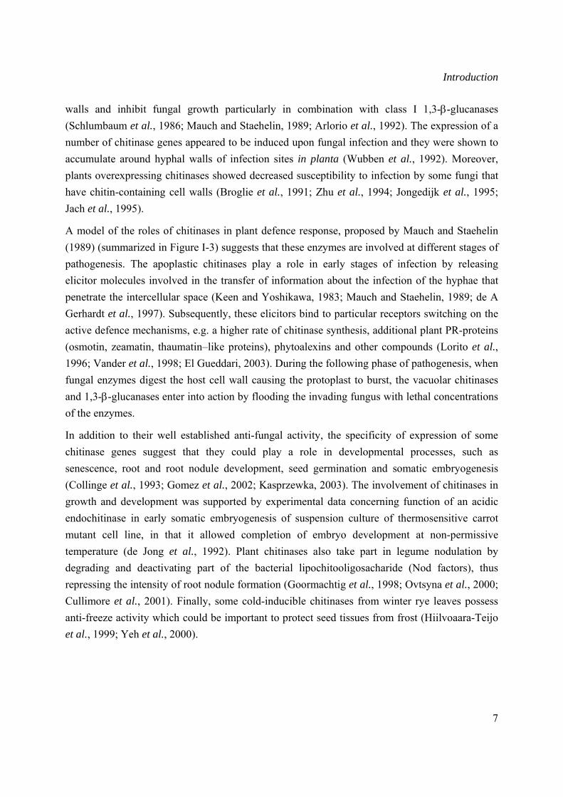
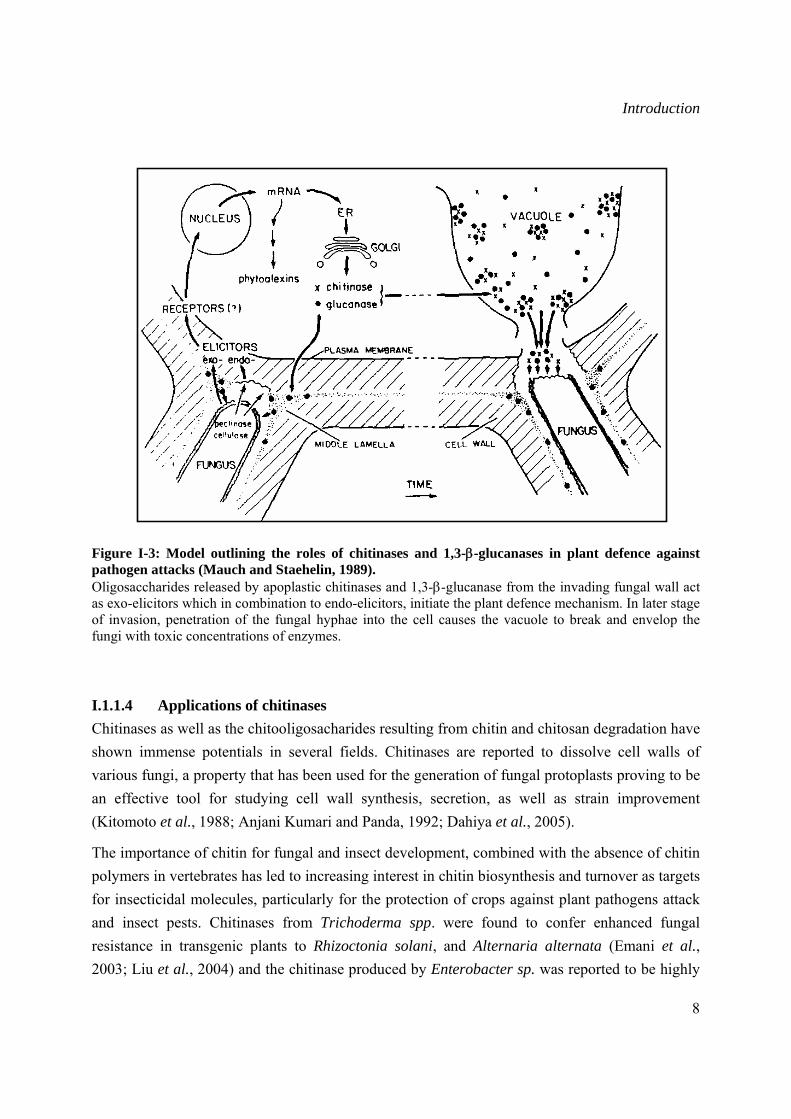
A model of the roles of chitinases in plant defence response, proposed by Mauch and Staehelin (1989) (summarized in Figure I-3) suggests that these enzymes are involved at different stages of pathogenesis. The apoplastic chitinases play a role in early stages of infection by releasing elicitor molecules involved in the transfer of information about the infection of the hyphae that penetrate the intercellular space (Keen and Yoshikawa, 1983; Mauch and Staehelin, 1989; de A Gerhardt et al., 1997). Subsequently, these elicitors bind to particular receptors switching on the active defence mechanisms, e.g. a higher rate of chitinase synthesis, additional plant PR-proteins (osmotin, zeamatin, thaumatin–like proteins), phytoalexins and other compounds (Lorito et al., 1996; Vander et al., 1998; El Gueddari, 2003). During the following phase of pathogenesis, when fungal enzymes digest the host cell wall causing the protoplast to burst, the vacuolar chitinases and 1,3-β-glucanases enter into action by flooding the invading fungus with lethal concentrations of the enzymes.
In addition to their well established anti-fungal activity, the specificity of expression of some chitinase genes suggest that they could play a role in developmental processes, such as senescence, root and root nodule development, seed germination and somatic embryogenesis (Collinge et al., 1993; Gomez et al., 2002; Kasprzewka, 2003). The involvement of chitinases in growth and development was supported by experimental data concerning function of an acidic endochitinase in early somatic embryogenesis of suspension culture of thermosensitive carrot mutant cell line, in that it allowed completion of embryo development at non-permissive temperature (de Jong et al., 1992). Plant chitinases also take part in legume nodulation by degrading and deactivating part of the bacterial lipochitooligosacharide (Nod factors), thus repressing the intensity of root nodule formation (Goormachtig et al., 1998; Ovtsyna et al., 2000; Cullimore et al., 2001). Finally, some cold-inducible chitinases from winter rye leaves possess anti-freeze activity which could be important to protect seed tissues from frost (Hiilvoaara-Teijo et al., 1999; Yeh et al., 2000).
Introduction
8
Figure I-3: Model outlining the roles of chitinases and 1,3-β-glucanases in plant defence against pathogen attacks (Mauch and Staehelin, 1989). Oligosaccharides released by apoplastic chitinases and 1,3-β-glucanase from the invading fungal wall act as exo-elicitors which in combination to endo-elicitors, initiate the plant defence mechanism. In later stage of invasion, penetration of the fungal hyphae into the cell causes the vacuole to break and envelop the fungi with toxic concentrations of enzymes.
I.1.1.4 Applications of chitinases Chitinases as well as the chitooligosacharides resulting from chitin and chitosan degradation have shown immense potentials in several fields. Chitinases are reported to dissolve cell walls of various fungi, a property that has been used for the generation of fungal protoplasts proving to be an effective tool for studying cell wall synthesis, secretion, as well as strain improvement (Kitomoto et al., 1988; Anjani Kumari and Panda, 1992; Dahiya et al., 2005).
The importance of chitin for fungal and insect development, combined with the absence of chitin polymers in vertebrates has led to increasing interest in chitin biosynthesis and turnover as targets for insecticidal molecules, particularly for the protection of crops against plant pathogens attack and insect pests. Chitinases from Trichoderma spp. were found to confer enhanced fungal resistance in transgenic plants to Rhizoctonia solani, and Alternaria alternata (Emani et al., 2003; Liu et al., 2004) and the chitinase produced by Enterobacter sp. was reported to be highly
Introduction
9
active toward Fusarium moniliforme, Aspergillus niger, Mucor rouxii and Rhizopus nigricans (Dahiya et al., 2005). Similarly, larval chitinases from tomato moth (Lacanobia oleracea), and tobacco hornworm (Manduca sexta) were shown to have insecticidal activity in vitro and in transgenic plants respectively (Fitches et al., 2004; Wang et al., 2005). Equally, a plant chitinase from yam (Dioscorea opposita) sprayed onto powdery mildew infected strawberry leaves and berries effectively degraded the pathogen (Karasuda et al., 2003). All these results demonstrate that chitinases might be a safe biodegradable biocontrol agent for use instead of conventional fungicides and pesticides.
In addition to their numerous applications as enzymes, the products resulting from the degradation of their major substrates, chitin and chitosan, have as well very important properties. Chitosan oligomers, β-1,4-N-glucosamine, and β-1,4-N-acetyl-glucosamine monomers have an immense potential in the pharmaceutical and plant protection fields.
I.1.2 Chitin and chitosan
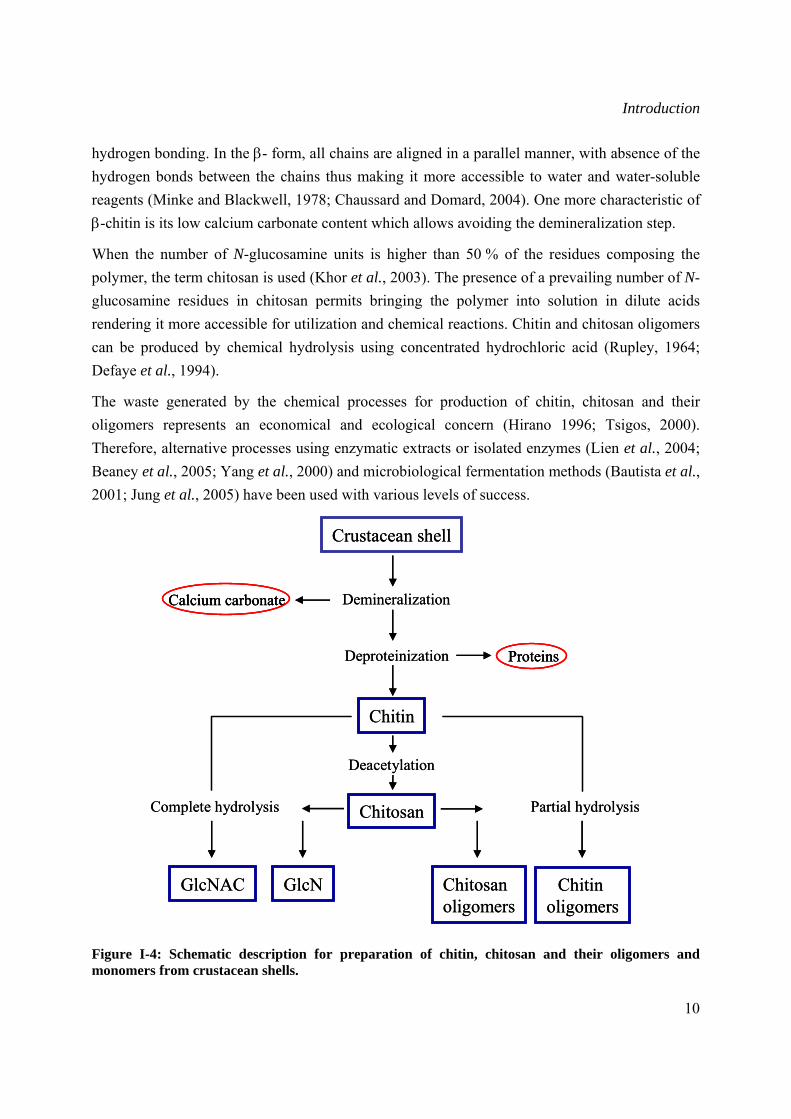
I.1.2.1 Extraction and crystallographic structure of chitin Chitin is the most abundant material after cellulose and it is estimated that at least 10 Gtons (1013 kg) of chitin are constantly present in the biosphere (Jeuniaux and Voss-Foucart, 1991). In the areas of fisheries, textile food and ecology, research was prompted to upgrade chitin in order to exploit a renewable resource and alleviate waste problems (Kumar et al., 2004). The shells of crabs, shrimps, prawns and lobsters coming from the peeling machines in canning factories are used for the industrial preparation of chitin (Skaugrud et al., 1991). Some insects such as the true fly and sulphur butterfly, or fungi like Aspergillus niger and Mucor rouxii represent an alternative source due to their high chitin content (Felt et al., 1998). The basic process for isolation of chitin is performed in a two-step process involving demineralization and deproteinization. First, calcium carbonate is removed by dilute hydrochloric acid, and then deproteinization is accomplished by extraction with a dilute sodium hydroxide solution at elevated temperature (No et al., 1997). Isolated chitin is a highly ordered copolymer of β-1,4-N-acetyl-glucosamine and β-1,4-N-glucosamine residues. Before the treatment, the shells are ground to make them more accessible, and after the completion of the manufacturing procedure chitin is dried so that it can be stored as a stable intermediate for chitosan production at a later stage (Singla and Chawla, 2001). Chitosan is produced by deacetylation of chitin with hot concentrated sodium hydroxide (No et al., 1997). A flow diagram depicting the manufacture of chitin, chitosan and their oligomers is given in Figure I-4.
The polymorphic forms of chitin differ in their crystallographic forms named α and β. The α-form chitin is characterized by cross-linked chains linked together by a dense network of
Introduction
10
hydrogen bonding. In the β- form, all chains are aligned in a parallel manner, with absence of the hydrogen bonds between the chains thus making it more accessible to water and water-soluble reagents (Minke and Blackwell, 1978; Chaussard and Domard, 2004). One more characteristic of β-chitin is its low calcium carbonate content which allows avoiding the demineralization step.
When the number of N-glucosamine units is higher than 50 % of the residues composing the polymer, the term chitosan is used (Khor et al., 2003). The presence of a prevailing number of N-glucosamine residues in chitosan permits bringing the polymer into solution in dilute acids rendering it more accessible for utilization and chemical reactions. Chitin and chitosan oligomers can be produced by chemical hydrolysis using concentrated hydrochloric acid (Rupley, 1964; Defaye et al., 1994).
The waste generated by the chemical processes for production of chitin, chitosan and their oligomers represents an economical and ecological concern (Hirano 1996; Tsigos, 2000). Therefore, alternative processes using enzymatic extracts or isolated enzymes (Lien et al., 2004; Beaney et al., 2005; Yang et al., 2000) and microbiological fermentation methods (Bautista et al., 2001; Jung et al., 2005) have been used with various levels of success.
Figure I-4: Schematic description for preparation of chitin, chitosan and their oligomers and monomers from crustacean shells.
Crustacean shell
Demineralization
Deproteinization
Chitin
Calcium carbonate
Proteins
Deacetylation
ChitosanComplete hydrolysis Partial hydrolysis
GlcNAC GlcN Chitosanoligomers
Chitin oligomers
Crustacean shell
Demineralization
Deproteinization
Chitin
Calcium carbonateCalcium carbonate
ProteinsProteins
Deacetylation
ChitosanComplete hydrolysis Partial hydrolysis
GlcNAC GlcN Chitosanoligomers
Chitin oligomers
Introduction
11
I.1.2.2 Biotechnological applications of chitin and chitosan Chitin and chitosan are inexpensive biopolymers that have been shown to be useful in many different areas, as anti-microbial compounds in agriculture and food industry, as flocculating agents in wastewater treatment, as hydrating agents in cosmetics, and more recently as a pharmaceutical agent in biomedicine (Sefarian and Martinez, 2001; Khor et al., 2003; Strand et al., 2003).
Chitin and chitosan have been tested to have both material and biological properties that might be beneficial to enhance wound healing (Muzzarelli et al., 1989a; Lahiji et al., 2000; Howling et al., 2001; Paul and Sharma, 2004). Several studies demonstrated that chitosan in form of cotton or bandages were able to accelerate wound healing and stop bleeding when used on injuries (Ueno et al., 1999; Mizuno et al., 2003; Shahidi and Abuzaytoun, 2005). The ability of wound healing is believed to be due to the tendency to form polyelectrolyte complexes with polyanion heparin, which posses anticoagulant and angiogenic properties. This complex extends the half-life of growth factors, thus supporting tissue growth and wound healing (Shahidi and Abuzaitoun, 2005). Another active area for chitosan application is in drug delivery. Recently the use of chitosan in formulation development has increased many folds. Chitosan microspheres and nanospheres have been formulated for site-specific drug delivery to the colon due to their biocompatibility and to their susceptibility to hydrolytic bacterial enzymes (Kumar et al., 2004). They shield the drug from the environment of the stomach and small intestine to deliver it to the colon where they are degraded by the microflora, thus releasing the drug (Sinha et al., 2001; Kumar et al., 2004).
The antimicrobial and antifungal properties of chitin and chitosan against a variety of microorganisms have attracted increasing interest. Chitosan and its derivative sulfobenzoyl chitosan were able to retard the growth of Coliforms, Pseudomonas, Aeromonas and Vibrio species on oysters (Singla and Chawla, 2000) as well as E. coli (Tsai and Su, 1999; Kumar et al., 2005). The bacterial action was proposed to be due to cross-linking between polycations of chitosan and the anion on bacterial surface, which altered the membrane permeability, thereby resulting in the leakage of glucose and lactate dehydrogenase from E. coli (Singla and Chawla, 2001; Kumar et al., 2005).
Chitosan has been utilized in soil amendment, in seed and foliar treatment to control different pathogens. It has a dual effect by inhibiting pathogen growth (Benhamou et al., 1994; Laflamme et al., 1999) as well as inducing various plant defence responses such as phytoalexins, lignification, phenylalanine-ammonia lyase (PAL), and peroxidase (POD) (Moerschbacher et al., 1986; El Gueddari et al., 2003; Kim et al., 2005). The antifungal properties combined with its safe oral administration make chitosan a very attractive material for food industry, were it is
Introduction
12
applied as antifungal coating material for post-harvest produce (El Ghaout et al., 1992; el Ghaout et al., 2000), as an additive to retard growth of spoilage microorganisms (Fang et al., 1994; Roller et al., 1999; Oh et al., 2001), and food wrapping materials (Rabea et al., 2003; Möller et al., 2004). Another promising industrial application of chitin and chitosan is the elimination of heavy metals such as cadmium, lead (Muzzarelli et al., 1989b, Lin and Lin, 2005), and copper (Deans and Dixon, 1992) from industrial waste water and contaminated soils, thus presenting an ecologically safe alternative to the traditional methods that may be inefficient and costly, particularly when metals are present at low concentrations (Volesky, 1987; Dean and Dixon, 1992).
I.2 Antibody engineering
I.2.1 Antibody structure and function
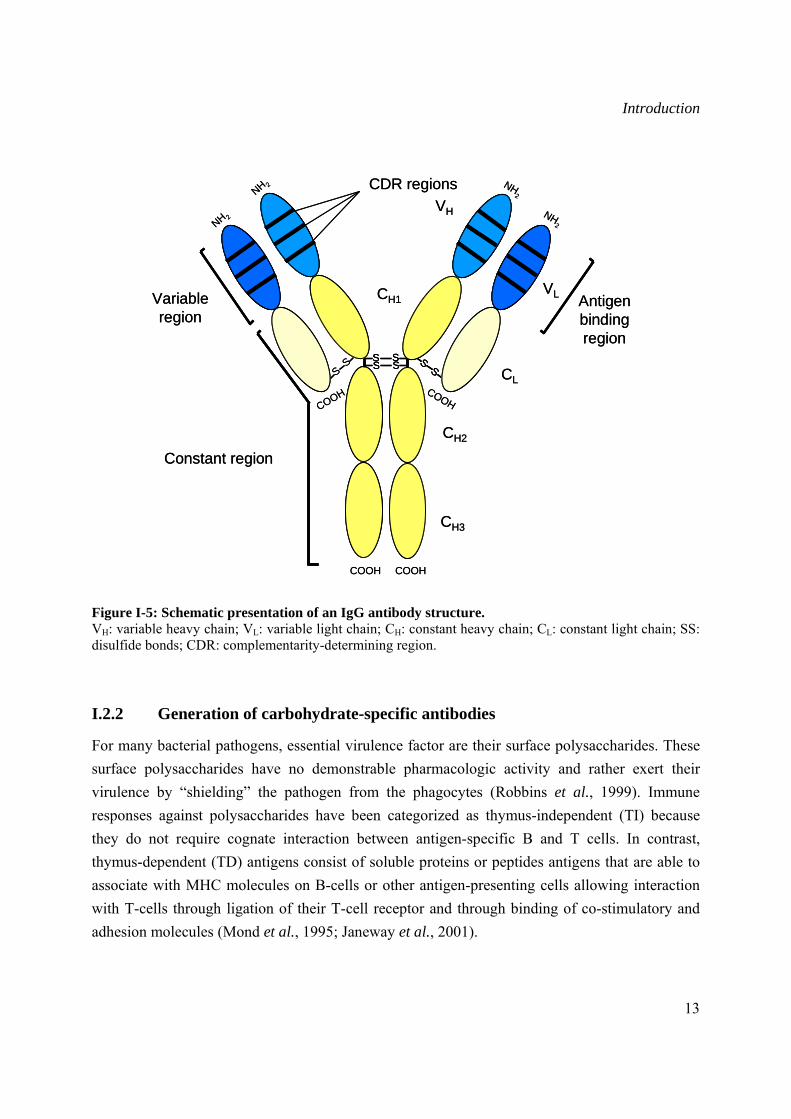
Antibody molecules, also referred to as immunoglobulin (Ig), are glycoproteins secreted by specialized B lymphocytes known as plasma cells. The basic structural molecule of an antibody consists of a “Y”-shaped structure composed of two identical heavy and light chains. Each of these chains contains multiple constant (C) and one variable (V) regions linked by disulfide bonds (Janeway et al., 2001) (Figure I-5). Depending on the Ig class, up to five structural molecules maybe combined. In mammals, there are five classes of Ig (IgG, IgM, IgA, IgD, and IgE), and in avian there are three classes (IgY, IgM, and IgA). In mammals, IgG and IgA are further subdivided into subclasses referred to as isotypes, due to polymorphism in the Fc region within the C domain. The Ig class determines both the type and the temporal nature of the immune response such as the half life in the serum, complement activation and ability to interact with the Fc receptor.
The essential functions of specific antibodies include direct activities such as toxin or viruse neutralization, and indirect activities that require other immune system components such as opsonization and complement activation (Lipman et al., 2005). The antigen binding activity of the antibody molecule is determined by the conformation of its amino acids in the complementarity determining regions (CDR) that are present in the variable regions of both the light and heavy chains of the antibody (Smith et al., 2005). The ability of antibodies to selectively bind a specific epitope present on a chemical, carbohydrate, protein or nucleic acid has been thoroughly exploited as a therapeutic tool to treat diseases, as well as to modulate physiological responses (Leenars and Hendriksen, 2005).
Introduction
13
Figure I-5: Schematic presentation of an IgG antibody structure. VH: variable heavy chain; VL: variable light chain; CH: constant heavy chain; CL: constant light chain; SS: disulfide bonds; CDR: complementarity-determining region.
I.2.2 Generation of carbohydrate-specific antibodies
For many bacterial pathogens, essential virulence factor are their surface polysaccharides. These surface polysaccharides have no demonstrable pharmacologic activity and rather exert their virulence by “shielding” the pathogen from the phagocytes (Robbins et al., 1999). Immune responses against polysaccharides have been categorized as thymus-independent (TI) because they do not require cognate interaction between antigen-specific B and T cells. In contrast, thymus-dependent (TD) antigens consist of soluble proteins or peptides antigens that are able to associate with MHC molecules on B-cells or other antigen-presenting cells allowing interaction with T-cells through ligation of their T-cell receptor and through binding of co-stimulatory and adhesion molecules (Mond et al., 1995; Janeway et al., 2001).
SS
SS
SS S
S
COOH COOH
Constant region
NH 2
NH 2 NH2
NH2
COOHCOOH
VL
CL
CH3
CH2
CH1
VH
Antigen bindingregion
CDR regions
Variable region
SS
SS
SS
SS
SS S
SS
S
COOH COOH
Constant region
NH 2
NH 2 NH2
NH2
COOHCOOH
VL
CL
CH3
CH2
CH1
VH
Antigen bindingregion
CDR regions
Variable region
Introduction
14
Efforts to overcome the low immunogenicity associated with polysaccharide antigens have focused on converting the TI antigens to TD antigens. The major strategy used to achieve a TD immune response to polysaccharides is through conjugation to a protein carrier (Lesinski and Westerink, 2001). The proposed mechanism of action involves the recognition of polysaccharide component through specific interactions with Ig surface receptors and internalization of the conjugate. Intercellularly, the protein is processed, peptide fragments are presented by MHC class II molecules and activation of peptide-specific T-cells results (Rijkers et al., 1998). Polysaccharide-protein conjugate vaccines have been remarkably effective in reducing the burden of disease caused by Haemophilus influenza and Streptococcus pneumoniae in children (Casadevall and Pirofski 2006). However, the general use of conjugation strategies is limited, in that eliciting carrier-specific T- and B-cells does not uniformly enhance polysaccharide immunogenicity (McCool et al., 1999). Variations in the immunogenicity of conjugate vaccines, even when the carrier remains the same, accounts for differences in the quality of the antibody elicited by different polysaccharide-conjugate vaccines. The use of carbohydrate-based vaccines might be further limited because oligosaccharides that are known to induce protective antibodies cannot, as yet, be totally designed (Benaissa-Trouw et al., 2001).
To facilitate vaccine design applications, there is an increased interest in molecular mimicry (Meloen et al., 2000; Deroo and Muller, 2001). Peptides in particular have raised considerable expectations as low-molecular weight substitutes for polysaccharide antigens. The major approaches used in identifying polyssacharide-mimicking peptides have been through screening of peptide libraries as well as the use of anti-idiotype antibodies (Apostolopoulos et al., 1999). The screening of combinatorial peptide libraries is the most successful method of isolating polysaccharide-mimicking peptides of importance in both tumour and microbial immunity. An early demonstration of peptide mimicry of polysaccharides was the isolation of concanavalin A binding peptide, acting as mimics of mannose-containing oligosaccharides (Oldenburg et al., 1992; Scott et al., 1992). Peptide libraries have also been used to isolate a peptide mimic of the Lewis Y (LeY) tumour antigen, with potential importance in the development of cancer vaccines (Hoess et al., 1993). More recent studies have demonstrated the ability of mimetic peptides to induce immune response against surface polysaccharides of N. meningitidis A (Grothaus et al., 2000), N. gonorrheae (Gulati et al., 2001), Streptococcus pneumoniae 4 (Lesinski et al., 2001), group B Streptococcus (Pincus et al., 1998), Cryptococcus neoformans (Fleuridor et al., 2001), and Candida albicans (Jouault et al., 2001). The second approach for augmentation of carbohydrate immunity is the potential use of anti-idiotypic antibodies. This approach requires that a polypeptide immunoglobulin variable region can mimic a polysaccharide determinant, providing a surrogate immunogen (Diakun and Matta, 1989; Sugiyama et al., 1991). Immunization with an antibody (Ab1) directed against a certain antigen (for example a
Introduction
15
polysaccharide) induces another antibody in response (Ab2). Ab2 is thought to be structurally similar to the original antigen, or in other words, to bear its ‘internal image’. This implies that Ab2 possesses similar shape to the original antigen and contacts the same residues in Ab1 binding site. Similarly, a peptide might be thought to be structurally similar to a polysaccharide, if it is able to bind to antibodies directed against the latter. Alternatively, the peptide might be structurally dissimilar, and interact with a different part of the antibody binding site. In this case, there is no structural mimicry, but functional mimicry, or specific cross-reactivity (Gulati et al., 2001; Johnson and Pinto, 2002). Immunization with anti-idiotope whose image mirrors a determinant of the pathogen being immunized against, has been used to raise protective antibodies in animals against polysaccharide capsules of E. coli K13 (Stein and Sonderstom, 1984), N. meningitidis group C (Westerink et al., 1988; Westerink et al., 1995), and S. pneumoniae (McNamara et al., 1984; McNamara-ward et al., 1987).
Not all peptides identified as carbohydrate mimics induce polysaccharide cross-reactive immune response, therefore, antigenic mimicry does not necessarily induce immunological mimicry (Cheng et al., 1988; Phalipon et al., 1997; Monzavi-Karbassi et al., 2002). Such failures suggest that commonly used parameters for selecting peptide mimetics, such as competition with native antigen for antibody binding, high-affinity binding to monoclonal antibody and/or induction of high titer of anti-peptide response, are insufficient and perhaps not predictive of whether a peptide is a polysaccharide mimotope (Fleuridor et al., 2001; Monzavi-Karbassi et al., 2002). Thus, a better understanding of the structural basis for carbohydrate mimicry and defining the degree of redundancy of potential mimotope sequences with respect to functional outcomes will facilitate the design of peptide immunogens that will incorporate a number of desirable features compressed in a relatively short peptide.
I.3 Aim of the thesis
In many regions of the world, microbial pathogens and pests continue to cause huge crop losses. Controlling crop diseases is very difficult and requires intensive use of potentially unsafe and environmentally harmful chemical plant protectants. Concern has been raised about both the environmental and the potential risk related to the use of these compounds. Therefore, considerable efforts have been made towards the development of alternative crop protectants. The European Commission has been actively encouraging the development and commercial implementation of new compounds known as ‘green chemicals’.
Several studies have demonstrated that plants have endogenous defence mechanisms that can be induced as a response to attack by insects and pathogens (Barber et al., 1989; Nishizawa et al., 1999; Anguelova-Merhar et al., 2001). In this context, chitin and chitosan are raising interest as
Introduction
16
potential elicitor of plant defences and active inhibitor of fungal growth (Wojdyla, 2004; Hadwiger, 1999; Orlikowski, 2004). Following the same research line, a project named CARAPAX (Chitosan Activates Resistance Against Pathogen After eXposure) (http://carapax.uni-muenster.de), supported by the fifth framework program of the European Union was aimed at the identification and detailed characterization of those specific chitin and chitosan oligomers with biological activity able to protect plants from microbial diseases, and develop a combination of chemical and enzymatic approaches to reliably prepare these bio-active oligomers with defined chemical and physical properties.
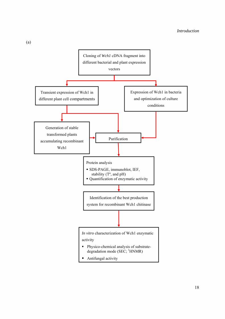
As part of the CARAPAX project, the aim of this study was the characterization of the enzymatic activity of the Wch1 wheat chitinase as well as the physico-chemical determination of substrate degradation products. Moreover, it included the generation of chitin/chitosan-specific monoclonal antibodies to serve as immunological tools to reliably identify the chitosans with different acetylation degrees, for quality control tests during chitin degradation. The objectives of the present study will be achieved by the following approaches:
• Production of the recombinant Wch1 chitinase and characterization of its enzymatic activity. The Wch1 chitinase cDNA gene will be cloned into various bacterial and plant expression vectors. The enzyme will be targeted to different cell compartments to verify expression rate and integrity of protein. Immunoblot analyses will be performed to investigate the accumulation levels and the stability of the heterologous enzyme. Depending on the accumulation levels in each compartment, stable transformed tobacco lines expressing apoplastic or ER-targeted enzyme will be generated. The enzymatic activity of the purified Wch1 chitinase will be qualitatively and quantitatively estimated by isoelectric focusing (IEF) and a colorimetric assay using glycol-chitin, colloidal chitin, and CM-chitin-RBV as substrates. The optimal temperature and pH conditions will be determined to ensure optimal enzymatic activity. The enzymatic mode of action as well as the anomeric configuration of the substrate after depolymerization will be established by size-exclusion chromatography and 1HNMR spectroscopy methods. The defensive role of Wch1 chitinase during fungal attack will be evaluated in vitro against Fusarium graminearum.
• In this study chitosan- and chitin-specific murine monoclonal antibodies will be generated following two strategies. Chitosan, being soluble, will be coupled to ovalbumin and used for immunization of mice. The insolubility of chitin makes the immunization with chitin-mimicking peptides a more suitable option. The chitin-mimicking peptides will be selected from linear and constrained commercially available phage peptide libraries (Ph.D.-12™, Ph.D.-7™ and Ph.D.-7C™). The libraries will be panned against the chitin-
Introduction
17
binding domain (CBD) protein and wheat germ agglutinin (WGA) as antigens. The ability of the peptides to mimic chitin will be assessed by competition-ELISA. Splenocytes from mice immunized with either coupled chitosan or candidate peptide (s) will be used for the development of hybridoma cell lines secreting chitin- and chitosan-specific mobs. These antibodies are to be purified by affinity chromatography and used for in vitro characterization using basic protein analysis methods (SDS, immunoblot, ELISA).
Introduction
18
(a)
Transient expression of Wch1 in different plant cell compartments
Identification of the best production
system for recombinant Wch1 chitinase
Generation of stable
transformed plants
accumulating recombinant Wch1
Expression of Wch1 in bacteria
and optimization of culture
conditions
In vitro characterization of Wch1 enzymatic
activity
Physico-chemical analysis of substrate-degradation mode (SEC; 1HNMR)
Antifungal activity
Purification
Protein analysis
SDS-PAGE, immunoblot, IEF, stability (Tº, and pH)
Quantification of enzymatic activity
Cloning of Wch1 cDNA fragment into different bacterial and plant expression
vectors
Introduction
19
(b)
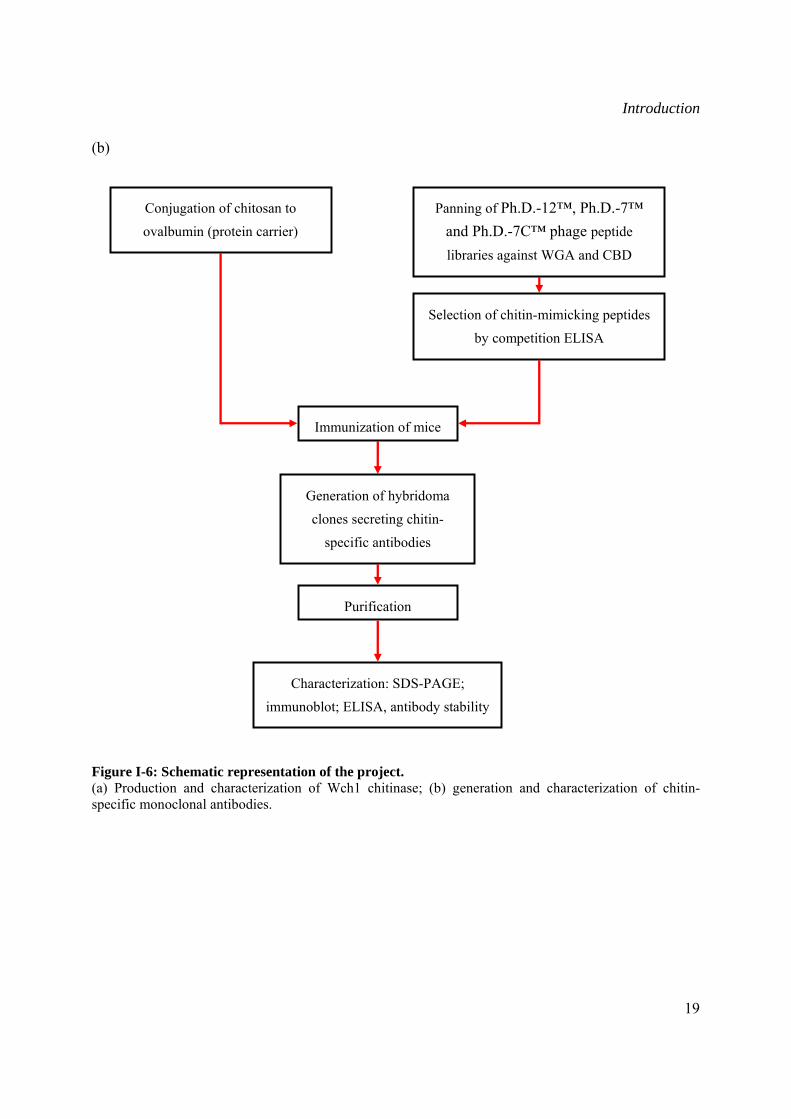
Figure I-6: Schematic representation of the project. (a) Production and characterization of Wch1 chitinase; (b) generation and characterization of chitin-specific monoclonal antibodies.
Generation of hybridoma
clones secreting chitin-
specific antibodies
Conjugation of chitosan to
ovalbumin (protein carrier)
Immunization of mice
Characterization: SDS-PAGE;
immunoblot; ELISA, antibody stability
Panning of Ph.D.-12™, Ph.D.-7™ and Ph.D.-7C™ phage peptide
libraries against WGA and CBD
Selection of chitin-mimicking peptides
by competition ELISA
Purification
Materials and Methods
20
II Materials and Methods
II.1 Materials
II.1.1 Chemicals and consumables
The chemicals used throughout the work were purchased from the following companies: Amersham Pharmacia Biotech (Freeburg, D), BD Biosciences (Heidelberg, D), Biotech (Freeburg, D), Boehringer Mannheim (Mannheim, D), Difco (Detroit, USA), Douche (Harlem, NL), Fluke (Neu-Ulm, D), IBA GmbH (Göttingen, D), Jerini GmbH (Berlin, D), LOEWE Biochemica GmbH (Sauerlach, D), Merck (Berlin, D), MWG (Ebersberg, D), New England Biolabs (Frankfurt, D), Pierce (Rockeford, IL, USA), Roche (Mannheim, D), Roth (Karlsruhe, D), Serva (Heidelberg, D), Sigma-Aldrich (Taufkirchen, D).
The consumables were obtained from:
Amicon (Witten, D), Bio-Rad Laboratories GmbH (München, D), Biozym Diagnostik GmbH (Hessisch Oldendorf, D), Eppendorf (Hamburg, D), Greiner (Solingen, D), Kodak (Stuttgart, D), Millipore (Schwalbach, D), Qiagen (Hilden, D), Schott Glaswerke (Mainz, D), Serva (Heidelberg, D), USB/Amersham (Braunschweig, D), Whatman (Maidstone, UK).
II.1.2 Enzymes and reaction kits
Restriction enzymes used for DNA digestion were purchased from New England Biolabs. Taq polymerase (Gibco BRL, Eggenstein, D) was used for PCR amplification of Wch1 chitinase gene and in control PCRs (II.2.1.9).
The kits used were as follows:
QIAprep® Spin Mini/Midiprep kit Qiagen
QIAquick® gel extraction kit Qiagen
QIAquick® PCR purification kit Qiagen
Bradford protein assay Roti®-Quant kit Roth
Mouse immunoglobulin Isotyping ELISA Kit BD Biosciences
II.1.3 Antibodies and substrates
Mouse anti-MBP monoclonal antibody (New England Biolabs) was used for detection of MBP-
Materials and Methods
21
Wch1 fusion protein by immunoblot analysis (II.2.4.4).
Mouse anti-his6 (Qiagen) was used for detection of His-tagged Wch1 expressed in plant and bacteria by immunoblot analysis (II.2.4.4).
Alkaline phosphatase (AP) or horseradish peroxidase (HRP)-conjugated to goat anti-mouse IgG (H+L, Fc) (Dianova, Hamburg, D) were used as secondary antibodies in immunoblot analysis (II.2.4.4) analysis and ELISA (II.2.7.2). Nitro-blue tetrazolium chloride/5-bromo-4-chloro-3’-indollyphosphate p-toluidine salt (NBT/BCIP) (Sigma) was used as alkaline phosphatase substrate for detection of immobilised proteins in immunoblot analysis. ABTS substrate tablets and buffer (Boehringer Mannheim) were used as chromophore for HRP-labelled antibodies, while p-Nitrophenyl phosphate disodium substrate tablets (Sigma) were used for AP-labelled antibodies.
Horseradish peroxidase (HRP) conjugated IgG raised against M13 bacteriophage (Dianova) was used for the detection of phages displaying peptides in ELISA (II.2.7.2).
Goat anti-rabbit Alexa Fluor® 594 (Molecular Probes) was used as secondary antibody in fluorescence microscopy (II.2.11).
II.1.4 Bacterial strains
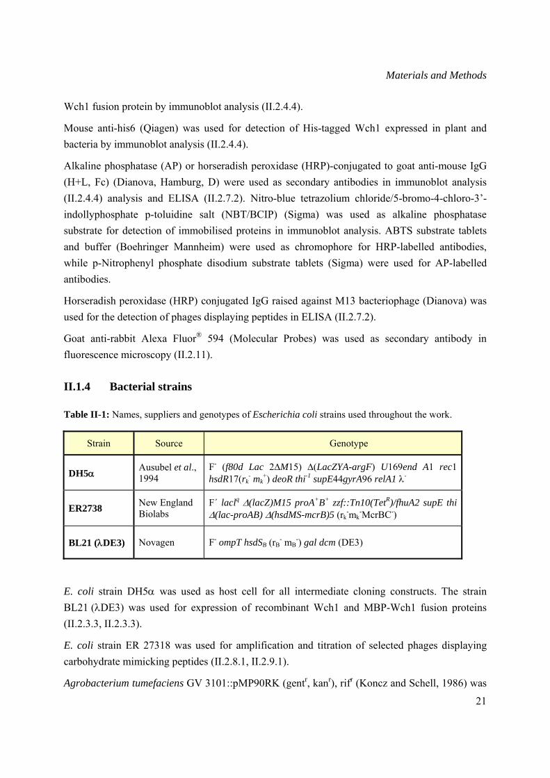
Table II-1: Names, suppliers and genotypes of Escherichia coli strains used throughout the work.
Strain Source Genotype
DH5α Ausubel et al., 1994
F- (f80d Lac 2∆M15) ∆(LacZYA-argF) U169end A1 rec1 hsdR17(rk
- mk+) deoR thi-1 supE44gyrA96 relA1 λ-
ER2738 New England Biolabs
F´ laclq Δ(lacZ)M15 proA+B+ zzf::Tn10(TetR)/fhuA2 supE thi Δ(lac-proAB) Δ(hsdMS-mcrB)5 (rk
-mk-McrBC-)
BL21 (λDE3) Novagen F- ompT hsdSB (rB- mB
-) gal dcm (DE3)
E. coli strain DH5α was used as host cell for all intermediate cloning constructs. The strain BL21 (λDE3) was used for expression of recombinant Wch1 and MBP-Wch1 fusion proteins (II.2.3.3, II.2.3.3).
E. coli strain ER 27318 was used for amplification and titration of selected phages displaying carbohydrate mimicking peptides (II.2.8.1, II.2.9.1).
Agrobacterium tumefaciens GV 3101::pMP90RK (gentr, kanr), rifr (Koncz and Schell, 1986) was
Materials and Methods
22
used for Agrobacterium-mediated gene transfer of tobacco plants (II.2.2).
II.1.5 Plants and animals
Nicotiana tabacum L. cv. Petite Havana SR1 was used for transient protein expression by agrobacterial vacuum infiltration (II.2.2.1.2) and generation of stable transformed plants (II.2.2.2).
Six to eight weeks old female Balb/c mice (Mus musculus) were used for immunization with 50-100 μg antigen mixed with adjuvant (GERBU Bichemicals GmbH, Gaiberg, D) (II.2.7).
II.1.6 Phage Libraries
The Ph.D.-12™, Ph.D.-7™ and Ph.D.-C7C™ peptide display phage libraries were purchased from New England Biolabs. These are combinatorial libraries of random dodeca- or heptapeptides fused N-terminal to the minor coat protein (pIII) of M13 phage. In contrast to the linear peptides displayed by the Ph.D.-12™ and Ph.D.-7™ libraries, the peptide sequences displayed by the Ph.D.-C7C™ library are flanqued by a pair of cystein residues that form, under non-reducing conditions, a disulfide cross-link resulting in phage display of cyclized peptides
II.1.7 DNA-Vectors
Schematic drawing of the vector maps are presented in the Appendix (VII.3)
pTRAkc-dhfr-AH and pTRAkc-dhfr-ERH, kindly provided by Dr. T. Rademacher (Institut für Biologie VII, RWTH Aachen, Germany), are optimized plant expression vectors containing the Cauliflower Mosaic Virus (CaMV) 35S promoter with duplicated enhancer region and the untranslated region from the CaMV 35S gene. These vectors were used for the accumulation of recombinant Wch1 chitinase in tobacco plants, secreted to the apoplast or retained in the ER (II.2.3.6).
pASKIB4 from IBA GmbH containing tetracycline promoter/operator, the ompA leader peptide sequence for periplasmic targeting and an N-terminal affinity tag named Strep-tag II, was used for bacterial expression of Wch1 recombinant chitinase (II.2.3.4).
pMYB5 vector (New England Biolabs) carries the E. coli malE gene encoding the maltose binding protein (MBP) fused in-frame to the region encoding intein and chitin binding domain (CBD) genes. Protein expression is under control of a T7 promoter directly followed by a lacI gene sequence encoding the lac repressor thus providing stringent control of the fusion gene expression. This plasmid allowed production of CBD protein (II.2.3.5) used for selection of chitin-mimicking peptides (II.2.8.1).
Materials and Methods
23
pET22b(+) vector (Novagen) containing the T7 promoter, the pelB coding sequence for periplasmic targeting and a his6 tag downstream of the multiple cloning site was used for bacterial expression of Wch1 recombinant chitinase (II.2.3.3).
pMAL-c2X vector from New England Biolabs containing the Ptac promoter was used for cloning the Wch1 chitinase cDNA downstream of the E. coli malE gene encoding for the maltose binding protein (MBP) gene and subsequent expression of Wch1-MBP fusion protein in the cytoplasm (II.2.3.2).
II.1.8 Oligonucleotides
Oligonucleotides used for sequence analysis and amplification of DNA are listed below. All oligonucleotides were synthesized by MWG (Ebersberg, D).
Primers used for sequencing of Wch1 cDNA cloned into pHENHi phagemid vector (kindly provided by Dr. D. Peschen, Institut für Biologie IV, RWTH Aachen, Germany).
pHEN-5’ 5′-GGA GACAGT CAT AAT GAA ATA CC-3′
pHEN-3’ 5′-GAC GTT AGT AAA TGA ATT TTC-3′ Primers used for control-PCRs of recombinant A. tumefaciens (II.2.1.5).
pSS-3’ 5’-AGA GAG AGA TAG ATT TGT AGA GA-3’
pSS-5’ 5’-ATC CTT CGC AAG ACC CTT CCT CT-3’
T7 universal primers were used for sequencing of the Wch1 cDNA cloned into the pET22b (+) vector (II.2.1.10).
Universe 5’-GTT GTA AAA CGA CGG CCA GT-3’
Reverse 5’-ACA CAG GAA ACA GCT ATG AC-3’
pMAL primers were designed to introduce EcoRI/BamHI and XbaI/SalI restriction sites for cloning the Wch1 cDNA into the pMAL-c2X vector (II.2.1.9).
pMAL-forw 5’-CGG AAT TCG GAT CCT CCA TGG AGC AGT GC-3’
(BamHI restriction site underlined)
pMAL-backw 5’-CGA CGT CGA CTC TAG ATT AGT CGA CGG CGA ACG G-3’
(XbaI restriction site underlined)
Reverse primer for sequencing of single stranded phage DNA (II.2.1.10).
96gIII 5’-CCC TCA TAG TTA GCG TAA CG-3’
Materials and Methods
24
II.1.9 Buffers, media and solutions
All standard solutions, buffers, and media were prepared according to Sambrook et al. (1996), Ausubel et al. (1995) and Coligan et al. (1995). Other special media and solutions are listed at the end of the respective method description. Media for bacterial and plant tissue cultures were sterilized by autoclaving (20 min/121oC/2 bar). All other solutions were sterile-filtered (0.2 μm). Thermo-labile components such as antibiotics were filter-sterilized (0.2 μm filter) and added to the autoclaved media or buffer after they were allowed to cool down to 60-50°C.
II.1.10 Matrices and membranes
Amylose matrix from New England Biolabs was used for purification of bacterial produced MBP-Wch1 fusion protein (II.2.3.2).
Ni-NTA agarose matrix (QIAGEN) was used for purification of bacterial (II.2.3.3) and plant produced (II.2.3.6) his6-tagged Wch1 by immobilized metal ion affinity chromatography (IMAC).
Strep-Tactin matrix (IBA GmbH) was used for purification of bacterial produced Strep-tagged Wch1 (II.2.3.4).
Protein G matrix (Amersham) was used for purification of anti-chitin monoclonal antibodies (mAbs) generated by hybridoma cultures (II.2.3.7).
HybondTM-C nitrocellulose membrane (0.45μm) from Amersham and Whatman no.1 paper from Whatman were used in immunoblot analysis (II.2.4.4).
II.1.11 Equipment
Cameras: E.A.S.Y 429K camera (Herolab, Wiesloch, D).
Centrifuges: AvantiTM J-30 I and Microfuge® R (Beckman, CA, USA), Varifuge 3.OR (Heraeus, Hanau), Centrifuge 5415 D (Eppendorf). Rotors: JA 30.50, JLA 16.250, F 241.5 (Beckman), F 45.24.11 (Eppendorf).
DNA gel electrophoresis apparatus: Wide mini and mini cells for DNA agarose electrophoresis and power supplies from Bio-Rad Laboratories GmbH.
DNA sequencer: 3730 DNA Analyzer and BigDye™ cycle sequencing terminator chemistry apparatus (Applied Biosystems, CA, USA).
Electroporation apparatus: “Gene pulserTM”, “Pulse controller” unit, Extender unit and 0.2 cm cuvettes (BioRad Laboratories GmbH).
Materials and Methods
25
Fluorescence microscope: Olympus BX40 (Olympus Austria GmbH, Wien, A).
IEF apparatus: Electrophoresis constant power supply ECPS 3000/150, Flat Bed Apparatus FBE-3000 (Pharmacia Fine Chemicals, Uppsala, S).
Incubator: Innova 4430 Incubator shaker (New Brunswick Scientific, NJ, USA).
PCR Thermocyclers: Primus and Primus 96 plus (MWG-Biotech).
Photometers: Eppendorf Biophotometer (Eppendorf), and multi-channel spectrophotometer Spectromax 340 (Molecular Devices, Sunnyvale, CA, USA).
Probe sonicator: Bandelin Sonopuls (Bandelin electronics, Berlin, D).
Protein gel electrophoresis equipment: Mini PROTEAN IITM electrophoresis system and Power Pac 300 from Bio-Rad Laboratories GmbH.
Refractive index (RI) detector: Schimadzu RID 6 A (Shimadzu Schweiz GmbH, Reinach, Ch) and Waters 410 differential refractometer (Waters, Gyancourt, F)
UV-Transilluminators: Wavelength 302 nm and UVT-20M (Herolab), UV-chamber (Bio-Rad Laboratories GmbH).
Software: Windows NT 4.0 operating system (Microsoft), Microsoft Office 2000 (Microsoft), Adobe Photoshop 6.0 (Adobe), Chromas, Excel, GCG (Wisconsin Package TM of Genetic Computer Group).
Materials and Methods
26
II.2 Methods
All experiments related to the genetic engineering were performed according to the regulations of “S1-Richtlinien” and were officially approved by the “Regierungspräsidium des Landes NRW” (RP-Nr.: 23.203.2 AC 12, 21/95) and “BGA” [AZ 521-K-1-8/98:AI3-04/1/0866/88 (S1) and 55.8867/-4/93 (greenhouse)].
General recombinant DNA techniques, i.e. DNA precipitation, restriction enzyme digestion, DNA ligation, DNA agarose gel electrophoresis, were performed according to the standard protocols described in Sambrook et al. (1996) and Ausubel et al. (1995).
II.2.1 Recombinant DNA technologies
II.2.1.1 Preparation of electrocompetent E. coli cells Electrocompetent bacterial cells were prepared from the E. coli strains DH5α and BL21 (λDE3) as described by Dower et al. (1988). A single bacterial colony from an LB plate was inoculated in 5 ml LB-broth and cultured o/n at 37°C. Three millilitre of the o/n culture was transferred into 500 ml of LB broth. The cells were cultured for 3-4 hours at 37°C until the mid-log phase (OD600nm= 0.5-0.8) was reached. Then the cells were placed on ice for 15-20 min and harvested by centrifugation (3,000 g/4°C/10 min). Cells were washed three times with sterile water and resuspended in ice-cold 10 % (v/v) glycerol to a 300-fold concentration from the original culture volume (>1010 cells/ml). 40 μl aliquots were stored at -80°C.
II.2.1.2 Transformation of E. coli by electroporation Electrocompetent cells (II.2.1.1) were thawed on ice and mixed with 1 pg to 300 ng of circular DNA in sterile dH2O. The cell/DNA mixture was transferred into a pre-chilled electroporation cuvette (0.2 cm) and assembled into a safety chamber. After application of the pulse (25 μF, 2.5 kV, 200 Ω), the cells were diluted in 1 ml of SOC medium and incubated shaking for 1 h at 37°C. Finally, 50 μl of the cells were plated onto LB agar containing appropriate antibiotics and incubated o/n at 37°C.
II.2.1.3 Preparation of electrocompetent A. tumefaciens cells A single colony of A. tumefaciens strain GV3101 grown on YEB agar plate containing 100 μg/ml rifampicin (Rif) and 25 μg/ml kanamycin (Km) (YEB-Rif-Km) was inoculated in 5 ml of YEB-Rif-Km medium in a 100 ml Erlenmeyer flask and incubated for two days at 28°C with shaking (250 rpm). One ml of the culture was transferred into 100 ml of YEB-Rif-Km medium and cultivated for 15-20 h at 28°C with shaking (250 rpm) until the OD600nm reached 1-1.5. The cells
Materials and Methods
27
were chilled on ice for 15 min and spun down by centrifugation (4,000 g/4°C/5 min). The culture medium was decanted and the cells were washed three times with 10 ml of dH2O by centrifugation and resuspended in 500 μl of sterile 10 % (v/v) glycerol. 45 μl-aliquots of the suspension were dispensed into pre-chilled microcentrifugation tubes, frozen immediately in liquid nitrogen and stored at -80°C.
YEB-Rif-Km medium: Nutrient broth 0.5 % (w/v) Yeast extract 0.1 % (w/v) Peptone 0.5 % (w/v) Sucrose 0.5 % (w/v) 2 mM MgSO4, 100 μg/ml rifampicin and 25 μg/ml kanamycin were added after autoclaving and cooling.
II.2.1.4 Transformation of A. tumefaciens by electroporation 0.2-1.0 μg of plasmid DNA (II.2.1.6) in sterile dH2O was added to a thawed aliquot of electrocompetent Agrobacterium cells (II.2.1.3) and placed on ice for 3 min. The cell/DNA mixture was transferred into a pre-chilled electroporation cuvette (0.2 cm) and assembled into a safety chamber. After application of the pulse (25 μF, 2.5 kV, 200 Ω), the cells were diluted in 1 ml of SOC medium in a 4-ml tube and incubated shaking (250 rpm) for 1 h at 28°C. Finally, 1-10 μl of the cells were plated on YEB agar containing 100 μg/ml rifampicin (Rif), 25 μg/ml kanamycin (Km) and 100 μg/ml carbenicillin (Carb) (YEB-Rif-Km-Carb) and incubated for 2-3 days at 28°C. As a control transformation of agrobacteria cells with dH2O was performed.
II.2.1.5 Growth of recombinant A. tumefaciens and preparation of glycerol stocks Single colonies of A. tumefaciens were examined for the presence of plasmids by control PCR (II.2.1.9). Positive colonies were inoculated in 10 ml of YEB-Rif-Km-Carb medium and cultivated for 2-3 days at 28°C with vigorous shaking at 250 rpm. The culture was transferred to 50 ml-falcon tubes and agrobacteria cells were pelleted by centrifugation at 4,000 g for 10 min at 15°C. The cells were resuspended in a 1:1 volume of YEB Rif-Km-Carb medium and glycerol stock media (GSM). The suspension was aliquoted (100 μl) and stored at –80°C for further experiments.
Glycerol stock media (GSM): Tris-HCl, pH 7.4 25 mM Glycerol l5 % (v/v) MgSO4 100 mM

Materials and Methods
28
II.2.1.6 Isolation of plasmid DNA from E. coli Recombinant plasmid DNA was purified with the Qiagen plasmid DNA Mini- and Midiprep kits (II.1.2) according to the manufacturer’s instructions based on the alkaline lysis method (Sambrook et al., 1996). Quality and yield of plasmid DNA was examined by reading the absorbance at 260 nm and 280 nm in a spectrophotometer according to Müller et al. (1993) and Sambrook et al. (1996). The integrity of DNA was verified by a control restriction digest followed by agarose gel electrophoresis (II.2.1.7). Isolated plasmid DNA was stored at -20°C.
II.2.1.7 Agarose gel electrophoresis of DNA Plasmid DNA (II.2.1.6) and PCR fragments (II.2.1.9) were separated in 1.2 % (w/v) agarose gels. Preparation of agarose gels and electrophoresis of the samples were carried out as described by Sambrook et al. (1996). Ethidium bromide was added to the gel solution and TBE electrophoresis running buffer prior to the experiment. Known amounts of DNA molecular markers such as 1 kb ladder (Gibco BRL) were used for evaluation and determination of DNA concentration and size. The DNA bands were visualised directly upon illumination with an UV transilluminator at 302 nm. Documentation of the DNA gels was performed by using a black and white E.A.S.Y 429K camera (Herolab).
II.2.1.8 Preparative agarose gel electrophoresis Preparative gel electrophoresis was used for large scale purification of a particular DNA fragment from a mixture of DNA fragments after restriction enzyme digestion. The agarose containing the DNA fragment of interest was excised from the gel on an UV transilluminator with a sterile scalpel. The DNA extraction was performed with QIAquick Gel extraction kit (II.1.2) according to the manufacturer’s guidelines. The concentration of recovered DNA was measured by a spectrophotometer or determined by agarose gel electrophoresis (II.2.1.8) and the DNA was used in further experiments.
II.2.1.9 PCR amplification Polymerase chain reaction (PCR) was used for introduction of EcoRI/BamHI and XbaI/SalI restriction sites in the Wch1 cDNA sequence using designed primers (II.1.8) and subsequent cloning into the pMAL-c2X vector. cDNA was amplified based on the protocol of Sambrook et al. (1996) with high fidelity DNA-polymerase and DNA polymerase buffer (Roche). The reactions were performed in 0.2 ml PCR reaction tubes (Biozym Diagnostik GmbH), using a DNA thermal Cycler (MWG). The cycler contained a heated lid to avoid the use of mineral oil.
For rapid identification of recombinant agrobacteria (II.2.1.4) or E. coli (II.2.1.2) control-PCR was carried out to detect cDNA insert as described by Jesnowski et al. (1995) using Taq
Materials and Methods
29
polymerase from Gibco BRL.
The optimal annealing temperature (Tp) of the primers was experimentally optimised or calculated based on the empiric formula (Wu et al., 1991):
Tp = {22 + 1,46 [2* (G + C) + (A + T)]}
PCR reactions were carried out in a total volume of 50 μl as described below:
Components Volume Final concentration
10x PCR buffer 5.0 μl 1x 50 mM MgCl2 1.5 μl 1.5 mM 10 mM dNTPs 1.0 μl 0.2 mM each 10 pmol forward primer 1.0 μl 10 pmol 10 pmol backward primer 1.0 μl 10 pmol Template DNA 0.5-5 μl 10-100 ng Taq DNA polymerase (5U/ml) 0.25 μl 1.25 units ddH2O to 50 μl
Amplification was carried out under the following conditions:
Initial denaturation 5 min 95°C Denaturation 1 min 95°C Primer annealing 1 min 55°C x 20-35 Primer extension 1 min 72°C Final extension 5 min 72°C
The performance of each PCR reaction was monitored by running 5 μl of each reaction on agarose gels using an appropriate DNA marker (II.2.1.7).
II.2.1.10 DNA sequencing About 200-400 ng of purified plasmid DNA (II.2.1.6) was sequenced using the Applied Biosystems 3730 DNA Analyzer and BigDyeTM cycle sequencing terminator chemistry using 20 pmol of the corresponding forward and backward primers (II.1.8).
Sequence analysis of selected phages displaying chitin-mimicking peptides (II.2.9.2) was achieved from isolated single stranded DNA (II.2.9.4) using commercial primer from New England Biolabs (II.1.8).
Chromas software package was used for displaying the chromatogram files. The sequences were edited and exported for further analysis with the Wisconsin PackageTM of the Genetic Computer Group (GCG).
Materials and Methods
30
II.2.2 Transient and stable transformation of tobacco plants
II.2.2.1 Transient transformation of tobacco leaves by vacuum infiltration Growth of recombinant A. tumefaciens (II.2.1.5) and vacuum infiltration of tobacco leaves was performed as described by Kapila et al. (1996) and Vaquero et al. (1999).
II.2.2.1.1 Preparation of recombinant agrobacteria One hundred ml of YEB-Km-Rif-Carb medium was inoculated with 100 μl of a glycerol stock from the selected recombinant agrobacteria carrying a plant expression vector (II.2.1.5). The culture was grown o/n at 28°C with shaking at 250 rpm. Next day, the cells were pelleted by centrifugation at 5,000 g for 10 min at 15°C and transferred into 250 ml of induction medium and cultivated at 28°C o/n with shaking at 250 rpm. Agrobacteria cells were centrifuged (4,000 g/15-25°C/15 min), resuspended in 50 ml of MMA solution and kept for 2 h at RT. A diluted aliquot (1:10) was measured at OD600nm and the cell suspension was adjusted to an OD600nm of 1. One hundred ml of the diluted cell suspension was used for vacuum infiltration of plant leaves (II.2.2.1.2).
Induction medium: YEB medium, pH 5.6 MES 10 mM
2 mM MgSO4, 25 μg/ml kanamycin, 100 μg/ml rifampicin, 100 μg/ml carbenicillin and 20 μM acetosyringone were added after autoclaving and cooling.
MMA solution: MS-salts (Murashige & Skoog, basic salt mixture) 0.43 % (w/v) MES, pH 5.6 10 mM Sucrose 2 % (w/v) Acetosyringone was added directly before use 200 μM
II.2.2.1.2 Vacuum infiltration of intact tobacco leaves Young N. tabacum cv. Petite Havana SR1 leaves (four leaves for each construct) were placed in 100 ml of agrobacteria suspension (II.2.2.1.1) in a “Weck” glass and a continuous vacuum (60-80 mbar) was applied for 20-30 min. The applied vacuum was released slowly and the leaves were briefly rinsed in distilled water and kept on wet Whatman paper no. 1 with the adaxial side upwards. The plastic tray was sealed with saran wrap and placed for 60 h at 22°C with a 16 h photoperiod. Leaves were weighed, frozen in liquid nitrogen and stored at -80°C until analysis. As control, leaves were infiltrated with agrobacteria suspension, which did not contain a pTRA expression plasmid.
Materials and Methods
31
II.2.2.2 Recombinant Agrobacterium-mediated stable transformation of tobacco plants Stable transformation of N. tabacum was performed with the help of Dr. Flora Schuster (RWTH Aachen, Institute für Biologie VII). Transgenic N. tabacum cv. Petite Havana SR1 were generated by leaf disc transformation using recombinant agrobacteria (II.2.1.5) transformed with a pTRA plasmid carrying the Wch1 cDNA sequence for apoplastic targeting (II.1.7). Transgenic T0 plants were regenerated from transformed callus (Fraley et al., 1983; Horsch et al., 1985). Briefly, wild type plants were grown on MS medium in “Weck” glasses and the youngest leaves (length up to 4 cm) were used for transformation. The agrobacteria suspension was prepared as described above (II.2.2.1.1) and the OD600nm was adjusted to at least 1.0 after dilution in MMA solution. The leaves were cut into 8-10 pieces and transferred into “Weck” glasses containing 50-100 ml of agrobacteria suspension and incubated for 30 min at RT. The leaf pieces were then transferred onto sterile pre-wetted Whatman filters in petri dishes closed with saran wrap and incubated for two days at 26-28°C in the dark. Following washing with distilled water containing 100 μg/ml kanamycin, 200 μg/ml claforan and 200 μg/ml betabactyl (Ticarcillin/Clavulanic acid, 25:1), leaf pieces were transferred onto MS II-plates and incubated for one week at 25°C in the dark and then for 2-3 weeks with a 16 h photoperiod. After shooting, the shoots were removed and transferred onto MS-III-plates and incubated for 10-14 days at 25°C with a 16 h photoperiod until roots developed. The small plants were transferred into “Weck’’ glasses containing MS-III medium and incubated for 2 weeks at 25°C in 16 h light rhythm until transferred into soil. The young leaves from regenerated transgenic plants were analysed by immunoblot analysis (II.2.4.4) for accumulation of recombinant proteins.
MS medium: MS-salts 0.43 % (w/v) Myo-Inosite 0.1 % (w/v) Sucrose 2 % (w/v) Thiamin-HCl 0.4 mg/l ddH2O add to 1000 ml
The pH was adjusted to 5.8 with 1 N NaOH (for preparation of solid medium, 0.8 % (w/v) agar was added). The solution was autoclaved and 500 μl of vitamin solution were added upon cooling to 55°C.
Vitamin solution: Glycin 2 mg/l Myo-Inisitol 100 mg/l Nicotinic acid 0.5 mg/l Pyridoxin 0.5 mg/l Thiamine-HCl 0.1 mg/l Filter sterilized and stored at 4°C.
Materials and Methods
32
MS-II medium: MS medium supplemented with: 6-Benzylaminopurin (BAP) (Sigma) 1 mg/l α-Naphtalenacetic acid (NAA) (Sigma) 0.1 mg/l Solution was autoclaved, then Kanamycin (100 mg/l) and Claforan (200 mg/l) were added.
MS-III medium: Autoclaved MS medium supplemented with: Kanamycin 100 mg/l Claforan 200-250 mg/l
II.2.2.3 Growth of N. tabacum cv. Petite Havana SR1 plants Tobacco plants were grown in ED73 standard soil (Patzer, Sinntal-Jossa, D) with 0-30 % (v/v) sand under the following conditions: 16 h artificial light, 25°C (or higher depending on the outside temperature), 10,000 Lux (plus the sun light) and 70-90 % humidity. To prevent pollination from other plants, flowers were covered with plastic bags with micro pores. Mature, dried seeds were stored in paper bags at RT.
II.2.3 Expression and purification of recombinant proteins and monoclonal antibodies
II.2.3.1 Optimization of expression conditions of recombinant Wch1 in bacteria Two conditions for expression of the different bacterial constructs (II.2.3.2, II.2.3.3, and II.2.3.4) were assayed in the attempt to set up the optimum conditions for increasing the recombinant protein yield. The analysed parameters were the concentration of the inducer and the incubation time after induction. The standard protocol was as follows:
Fifty ml LB medium containing 100 μg/ml ampicillin (LBA) and 1.5 % glucose (w/v) were inoculated with one freshly transformed E. coli BL21 (λDE3) colony (II.2.1.2) containing the plasmid of choice and incubated o/n at 37ºC. The next day, the cells were centrifuged and resuspended in 150 ml media containing the antibiotic and 0.1 % (w/v) glucose. The culture was allowed to grow until OD600nm of 2 was reached to increase the yield of produced recombinant protein as described by Galloway et al. (2003). The culture was split into 5 subcultures of 20 ml each.
Protein expression was induced with different concentrations of inducer for each subculture (2 mM, 1 mM, 0.5 mM, 0.3 mM or 0.1 mM for IPTG induced constructs) and (400 μg/L, 300 μg/L, 200 μg/L and 100 μg/L for the anhydrotetracycline induced construct). A non induced subculture was included as negative control. The optimum incubation time was determined by
Materials and Methods
33
collecting 1 ml aliquots after defined time points (3 h, 6 h and 16 h). Each aliquot was centrifuged (13,000 g/5 min/4˚C), the pellet was weighed and then mixed with Y-PER lysis buffer (Pierce, Bonn, D). The mix was incubated for 5 min before centrifugation (15,000 g/5 min/4˚C), the supernatant was carefully transferred to a fresh tube and the pellet was resuspended in the same volume of Y-PER lysis buffer. One μl from supernatants and resuspended pellets were analyzed by immunoblot analysis (II.2.4.4).
The following standard procedures (II.2.3.2, II.2.3.3, and II.2.3.4) were performed according to the optimized culture conditions.
II.2.3.2 Bacterial expression and purification of Wch1-maltose binding protein (Wch1-MBP)
The Wch1-MBP fused protein was expressed based on the manufacturer’s recommendations (New England Biolabs).
A freshly transformed single colony of E. coli strain BL21 (λDE3) (II.2.1.2) harbouring recombinant plasmid DNA was inoculated in 50 ml of fresh LBA medium containing 1.5 % (w/v) glucose and cultivated o/n at 37°C with vigorous shaking. The following day, the cells were pelleted (5,000 g/20 min/4°C), resuspended in rich broth medium containing ampicillin (100 μg/ml) and used for inoculation of 500 ml rich broth medium. The culture was grown to an OD600nm of 2. Expression of recombinant protein was then induced by addition of IPTG to a final concentration of 0.3 mM and incubated for 3 additional hours at 37°C. The cells were harvested by centrifugation (20 min/5,000 g/4°C). The supernatant was discarded and the pellet was frozen o/n at -20°C. The next day, cells were thawed on ice, resuspended in ice-cold column buffer (5 ml) and sonicated on ice 3 times for 2 min (cycle 9, power 70 % and 1 min intervals). Cell debris and insoluble components were removed by centrifugation (30,000 g/20 min/4°C) and the supernatant subjected to amylose affinity chromatography.
The crude extract was applied to a pre-equilibrated amylose matrix (New England Biolabs). Unbound protein was washed with 20-30 matrix volumes of column buffer. The MBP-Wch1 fusion protein was eluted in four fractions of 1 matrix volume with elution buffer. Ten ml from each step was tested by SDS-PAGE analysis (II.2.4.3) and immunoblot (II.2.4.4). Protein concentration was determined by the Bradford assay (II.2.4.1).
Rich broth medium: Tryptone 1 % (w/v) Yeast extract 0.5 % (w/v) NaCl 0.5% (w/v) Glucose 0,2 % (w/v)
Materials and Methods
34
Column buffer: Tris-HCl, pH 7.4 20 mM NaCl 200 mM EDTA 1 mM DTT 1 mM
Elution buffer: Tris-HCl, pH 7.4 20 mM NaCl 200 mM EDTA 1 mM DTT 1 mM Maltose 10 mM
II.2.3.3 Bacterial expression and purification of his6-tagged Wch1 chitinase The pET22b bacterial expression vector carrying the Wch1 cDNA was used to transform BL21 (λDE3) E. coli electrocompetent cells (II.2.1.2). One colony was grown in 50 ml of LBA medium containing 1.5 % (w/v) glucose o/n at 26°C. The following day, the o/n grown culture was centrifuged (5,000 g/20 min/4°C) and diluted with 500 ml fresh LBA medium containing 0.1 % (w/v) glucose. The culture was grown at 26°C with vigorous shaking until OD600nm of 2 was reached. IPTG at a final concentration of 2 mM was added and the culture was further grown for 3 hours at 26°C. The cells were harvested by centrifugation (5,000 g/20 min/4°C), resuspended in lysis buffer and then sonicated (2 x 2 min at 70 % power, cycle 9, 1 min pause). The crude extract was centrifuged and the supernatant was dialysed o/n against 1x PBS. Sodium chloride and imidazol were added to the dialysed extract to a final concentration of 0.5 M and 10 mM, respectively.
Five hundred μl Ni-NTA agarose matrix (Qiagen) was added in a disposable and equilibrated with 10-20 matrix volumes of washing buffer. The sample was passed twice through the pre-equilibrated Ni-NTA matrix. The column was washed with 100 matrix volumes of washing buffer and the proteins were eluted in three fractions with 1 matrix volume of elution buffer. Collected fractions were immediately dialysed against 1x PBS (pH 7.4) to remove imidazol and salts. To determine the yield and purity of the dialyzed Wch1 protein 10 μl from each step were run in a SDS-PAGE (II.2.4.3) and analyzed by immunoblot analysis (II.2.4.4).
Lysis buffer: Tris HCl, pH 8 75 mM NaCl 300 mM DTT 5 mM EDTA 10 mM Glycerol 10 % (v/v)
Materials and Methods
35
Washing buffer: 1x PBS pH 8 Imidazol 10 mM NaCl 500 mM
Elution buffer: 1x PBS, pH 4.5 Imidazol 250 mM
II.2.3.4 Bacterial expression and purification of Strep-tagged Wch1 E. coli BL21 (λDE3) cells transformed with pASK-IBA4 vector carrying the Wch1 cDNA (II.2.1.2) (kindly provided by Dr. A. Schiermeyer, Fraunhofer IME) were grown in 50 ml of LB medium as described above (II.2.3.3). After the culture reached an OD600nm of 2 the protein expression was induced with 200 μg/L anhydrotetracycline. The induced culture was incubated for 3 additional hours before harvesting the cells by centrifugation (5,000 g/20 min/4˚C). The pellet was resuspended in column buffer and sonicated (2x 2 min at 70 % power, cycle 9, with 1 min pause). The sonicate was centrifuged, the supernatant was applied to 1 ml pre-equilibrated Strep-Tactin column. The matrix was washed with 10 ml column buffer and Wch1 protein was eluted in four fractions with 500 μl elution buffer (column buffer containing 2.5 mM desbiotin). Ten μl were analysed in a SDS-PAGE (II.2.4.3) and immunoblot analysis (II.2.4.4). Protein concentration was determined using the Bradford assay (II.2.4.1).
Column buffer: Tris-HCl, pH 8 100 mM NaCl 150 mM EDTA 1 mM
Elution buffer: Tris-HCl, pH 8 100 mM NaCl 150 mM EDTA 1 mM Desbiotin 2.5 mM
II.2.3.5 Bacterial expression and purification of the chitin-binding domain Chitin-binding domain (CBD) was needed as antigen for selection of chitin-mimicking peptides (II.2.8.1). The expression and purification steps were performed according to the manufacturer’s recommendations (New England Biolabs).
E. coli BL21 (λDE3) competent cells were transformed with pMYB5 plasmid the CBD-MBP fusion (II.2.1.2), plated on LBA agar plates and incubated o/n at 37˚C. Ten ml LBA medium containing 1 % (w/v) glucose were inoculated with one colony and grown o/n at 37˚C. The next
Materials and Methods
36
day, 5 ml from the overnight grown culture were used to inoculate 500 ml LBA medium. The culture was incubated at 37˚C with shaking and the optical density (OD600nm) was monitored. When the culture reached log-phase (OD600nm 0.5-0.7), protein expression was induced with 0.3 mM IPTG. The temperature was reduced to 30˚C and the expression was carried out for a period of 3 h. The culture was centrifuged at 5,000 g for 20 min at 4˚C.
The pellet was resuspended in 10 ml lysis buffer and sonicated (2x 2 min at 70 % power, cycle 9, and 1 min pause). The sonicate was centrifuged (20,000 g/20 min/4˚C) and the supernatant was applied to 0.5 ml amylose matrix pre-equilibrated with 20 ml column buffer. Unbound protein was washed away with 40 ml column buffer. On-column cleavage of CBD protein from the MBP partner was achieved by passing 3 ml of column buffer supplemented with 50 mM DTT (freshly prepared). The cleaved CBD protein was eluted in five fractions with 500 μl column buffer each. To monitor the purification procedure, 10 μl from each step were run in a SDS-PAGE (II.2.4.2).
Lysis buffer: Tris-HCl, pH 7.5 20 mM NaCl 500 mM EDTA 1 mM Tween-20 0.1 % (v/v) Triton-X100 0.1 % (v/v)
Column buffer: Tris-HCl, pH 7.5 20 mM NaCl 500 mM EDTA 1 mM
II.2.3.6 Expression and purification of plant produced Wch1 chitinase For expression and purification of recombinant Wch1 chitinase, 100-1000 g of tobacco leaves were transiently transformed by vacuum infiltration (II.2.2.1) with agrobacteria containing the pTRA vector targeting the recombinant Wch1 to the apoplast (Wch1-apo) (II.2.1.5). Infiltrated leaves were mixed 1:1 (w/v) with total soluble protein extraction buffer and processed in an electric blender (Waring commercial, Torrington, Connecticut, USA). When the recombinant Wch1 chitinase (Wch1-ER) was extracted under denaturing conditions, urea was added to the buffer to a concentration of 8 M.
The plant extract was passed through a miracloth membrane and then centrifuged (10,800 g/40 min/4˚C) to remove the debris. The supernatant was transferred to a fresh container and 500 mM NaCl and 5 mM imidazol were added before adjusting the pH to 8. The mix was stored for 1 h at 4˚C to precipitate plant proteins and to obtain a cleaner extract for purification. The precipitate was removed by centrifugation (10,800 g/45 min/4˚C) and the pH was readjusted
Materials and Methods
37
to 8.
The Ni-NTA matrix was prepared as described above (II.2.3.3) and the crude extract was applied under pressure using a peristaltic pump at a flow of 1 ml/min. The matrix was washed with 100 ml washing buffer, and then the target protein was eluted in four fractions with 1 matrix volume of elution buffer. The pH of collected fractions was immediately adjusted to 7 with 1 M Tris solution (of unadjusted pH), then dialysed against 1x PBS (pH 7.4) to remove imidazol and salts. To control the yield and purity of the protein, 10 μl from each purification step were analysed in a SDS-PAGE (II.2.4.3) and immunoblot analysis (II.2.4.4). Protein concentration was determined using the Bradford assay (II.2.4.1).
Total soluble protein extraction buffer: 1x PBS, pH 6 Ascorbic acid 20 mM PVPP 1 % (w/v) β-mercaptoethanol 5 mM
Washing buffer: 1x PBS, pH 8 Imidazol 5 mM NaCl 500 mM
Elution buffer: 1x PBS, pH 4.5 Imidazol 250 mM
II.2.3.7 Purification of monoclonal antibodies through Protein G matrix Purification of anti-chitin monoclonal antibodies (mAbs) produced by hybridoma culture (II.2.10) was performed through protein G matrix following the manufacturer’s recommendations (Amersham biosciences).
Hybridoma supernatant was dialysed against 1x PBS (pH 7) and then filtered through a filter with a pore size of 0.45 μm to remove all cell debris. Five hundred μl protein G matrix was equilibrated with 1x PBS (pH 7). The filtered supernatant was applied using a peristaltic pump at a flow of 1 ml/min. The column was washed extensively with 40-50 ml 1x PBS (pH 7). Bound antibodies were eluted with four fractions of 300 μl elution buffer each. The pH was immediately adjusted to 7.5 with 1 M Tris buffer (of unadjusted pH). Ten μl from each purification step was analyzed by SDS-PAGE (II.2.4.3) and immunoblot analysis (II.2.4.4). The fractions containing the antibodies were joined, dialyzed against 1x PBS and stored at -20ºC until needed. The functionality of purified antibodies was tested by ELISA (II.2.7.2).
Materials and Methods
38
Elution buffer:
Glycin, pH 2.7 100 mM
II.2.4 Protein analysis
II.2.4.1 Extraction of total soluble proteins from plant leaves For the extraction of recombinant Wch1 from vacuum infiltrated tobacco leaves (II.2.2.1) or from stable transformed tobacco plants (II.2.2.2), frozen leaves were ground in liquid nitrogen to a fine powder with a mortar and pestle. Total soluble proteins were extracted using 1 ml of extraction buffer per gram leaf material. Cell debris were removed by two rounds of centrifugation (16,000 g/30 min/4°C) and the supernatant was used for expression analyses by immunoblot analysis (II.2.4.4).
Extraction buffer: Tris-HCl, pH 7.5 200 mM EDTA 5 mM DMSO 2 % (v/v) β-mercaptoethanol 10 mM
II.2.4.2 Quantification of proteins The concentration of purified proteins was determined by Bradford assay (Bradford, 1976). Briefly, the protein solution of interest was serially diluted in dilution buffer. Bovine serum albumin (BSA) was also serially diluted in the same buffer and used as standard. Ten μl of each dilution was transferred into the well of a low binding microtiter plate (Greiner). Ten μl of the buffer was used as a blank. Two hundred μl of Bradford reagent (Roth) were added to each well, mixed with the proteins and incubated at RT for 10 min followed by the measurement of OD595nm. For each dilution, measurements were performed in triplicate and the average was taken for the calculation of the protein concentration.
Dilution buffer: Tris-HCl, pH 7.4 20 mM NaCl 100 mM CaCl2 2.5 mM
II.2.4.3 SDS-PAA gel electrophoresis and Coomassie brilliant blue staining Protein samples were separated by sodium dodecylsulphate (SDS) polyacrylamide gel electrophoresis (stacking gel: T = 4 %, C = 2.6 %, pH 6.8; separating gel: T = 12 %, C = 2.6 %, pH 8.8) (Ausubel et al., 1995) in SDS-PAGE running buffer. Before loading onto the gel, protein samples were mixed with protein loading dye containing SDS and β-mercaptoethanol and were
Materials and Methods
39
denatured by boiling for 5-10 min. When the samples were to be analysed with immunoblot analysis a pre-stained broad range protein marker (P7708S, New England Biolabs.) boiled for 10 min was also loaded on the gel. The proteins were separated electrophoretically with 20 V/cm. Protein bands were revealed by staining with Coomassie brilliant blue or transfer to nitrocellulose membrane for immunoblot analysis (II.2.4.4). Proteins were detected after incubating the gel for 30 min in Coomassie staining solution at RT under constant rocking. Coomassie staining was removed by distaining solution until the protein bands were clearly visible.
SDS-PAGE running buffer: Tris-HCl, pH 8.3 125 mM Glycin 960 mM SDS 0.5 % (w/v)
Protein loading buffer: Tris-HCl, pH 6.8 200 mM Glycerol 40 % (v/v) Bromophenol blue 0.05 % (w/v) SDS 8 % (w/v) β-mercaptoethanol 5 % (v/v)
Coomassie staining solution: Coomassie brilliant blue G-250 0.25 % (w/v) Methanol 50 % (v/v) Glacial acetic acid 9 % (v/v)
Coomassie distaining solution: Methanol 10 % (v/v) Glacial acetic acid 10 % (v/v)
II.2.4.4 Immunoblot analysis Separated proteins were transferred from an SDS-PAA gel (II.2.4.2) to HybondTM-C nitrocellulose membrane (0.45 μm) in transfer buffer. After blotting the membrane was blocked with 1x PBS buffer containing 3 % (w/v) BSA. As primary antibody either anti-his6 or anti-MBP were used in a dilution of 1:5,000 in 1x PBST (1x PBS + 0.05 % Tween-20). Binding of the primary antibody was detected by addition of goat anti-mouse secondary polyclonal antibody coupled to alkaline phosphatase (AP). The target protein was finally revealed by addition of the substrate BCIP/NBT dissolved in AP buffer.
1x PBS buffer (pH 7.3): NaCl 137 mM KCl 2.7 mM Na2HPO4x2H2O 8.1 mM
Materials and Methods
40
KH2PO4 1.5 mM
Transfer buffer: Tris-HCl, pH 8.3 25 mM Glycin 92 mM Methanol 20 % (v/v)
AP buffer: Tris-HCl, pH 9.6 100 mM NaCl 100 mM MgCl2 5 mM
II.2.5 Characterization of chitinase activity
II.2.5.1 Colorimetric activity assay The activity of Wch1 chitinase, purified from plant and bacterial crude extracts (II.2.3.2, II.2.3.3, II.2.3.4, II.2.3.5, and II.2.3.6), was determined using carboxymethyl-chitin-Remazol Brilliant Violet (CM-chitin-RBV) as a substrate (Spindler, 1997). One hundred μl substrate (200 μg) were added to 1.5 ml-microcentrifuge tubes containing 300 μl of 100 mM sodium acetate buffer (pH 5.2). Fifty μl of recombinant chitinase (2-50 μg) was added and the mixtures were incubated at 37˚C for 1 h, respectively. However, determination of optimal temperature for Wch1-apo enzymatic activity, was performed by incubation of the samples at different temperatures (10, 20, 30, 40, 50, 60, and 70˚C) and monitoring the substrate hydrolysis every 10 minutes for 1 h. All assays were performed in triplicates. The reactions were stopped by addition of 100 μl 1 M HCl. After 10 min chilling on ice the mixture was centrifuged (15,000 g/10 min/4˚C) to precipitate the non-digested substrate and the absorbance at 585 nm of the supernatant, containing hydrolyzed and thus solubilized CM-chitin-RBV oligomers, was measured. Reaction mixtures containing heat-inactivated enzyme were set as controls for comparison. One enzyme unit was defined as a change in optical density at 585 nm (ΔOD585nm) of 1 in 1 h.
II.2.5.2 Optimum pH and temperature Determination of the Wch1 pH and temperature optima was performed with the colorimetric assay described in II.2.5.1.
The influence of the pH on the enzymatic activity was assayed in 100 mM glycin-HCl buffer (pH 2-3), 100 mM sodium acetate (pH 4-5), 100 mM phosphate buffer (pH 6-7) and 100 mM Tris-HCl buffer (pH 8-10).
To identify the optimum temperature, the enzyme activity was performed at various temperatures
Materials and Methods
41
from 10˚C to 70˚C in 100 mM acetate buffer (pH 5.2).
II.2.5.3 Degradation of glycol-chitin in an overlay gel
II.2.5.3.1 Preparation of glycol-chitin substrate Glycol-chitin substrate was prepared according to Trudel and Asselin (1989) and used as substrate in chitinase activity experiments (II.2.5.3.2). One gram glycol-chitosan (Sigma) was dissolved in 20 ml 10 % (v/v) acetic acid solution and stirred o/n at RT. The next day, 90 ml methanol was slowly added and the resulting suspension was filtered under vacuum. Chitosan was reacetylated by incubation of the filtrate with 1.5 ml anhydride acetic acid for 30 min at RT. The formed gel was homogenised with methanol in a blender and centrifuged (27,000 g/15 min/4˚C). The pellet was resuspended in methanol and centrifuged a second time. The pellet was resuspended in 100 ml ddH2O containing 0.02 % (w/v) NaN3 to avoid bacterial growth.
II.2.5.3.2 Isoelectric focusing (IEF) gel and overlayer substrate gel For determination of chitinase activity in an overlay gel, purified chitinase samples (II.2.3) and crude extract from transiently transformed tobacco leaves accumulating the enzyme in the plant cell apoplast or ER (II.2.2.4) were first separated according to their pI in an isoelectric focusing gel. Crude extract from wild type N. tabacum cv. Petite Havana SR1 tobacco leaves and IMAC-purified metal-binding proteins from wild type E. coli cells were used as negative controls.
The IEF gel was prepared according to Westermeier et al. (2001). A 4.2 % (w/v) polyacrylamide gel was prepared, washed with ddH2O for elimination of any non-polymerised products and allowed to dry o/n at RT. The next day, the gel was rehydrated in a solution containing ampholine with a pH gradient from 3-10 (Serva). Proteins were separated for 2 h at 2000 V, 7 mA and 7 W on a horizontal ceramic cooling plate (Serva). After electrophoresis, the gel was washed with 20 mM sodium acetate buffer (pH 5.2) to reactivate the enzymatic activity.
A 10 % (w/v) polyacrylamide overlay gel containing glycol-chitin as substrate (II.2.5.3.1) was applied on top of the IEF gel and followed by incubation o/n at 37˚C. The substrate gel was stained with fluorescent brightener 28 (Sigma) and extensively washed in distilled water. Chitinase activity was detected as dark spots under UV light.
IEF gel: 0.25 M Tris-HCl, pH 8.4 0.5 ml Acrylamide/bisacrylamide 2.7 ml Glycerin 87 % (w/v) 1 ml Temed 10 μl
Materials and Methods
42
APS 40 % (w/v) 20 μl
Rehydratation solution: Sorbitol 10 % (w/v) Ampholine 1.3 ml ddH2O till 20 ml
Glycol-chitin overlay substrate gel: 0.1 M sodium acetate, pH 5 10 ml Acrylamide/bisacrylamide 5 ml Glycol-chitin 1 % (w/v) 600 μl Temed 150 μl APS 40 % (w/v) 150 μl ddH2O to 15 ml
II.2.5.4 Degradation of colloidal chitin Colloidal shrimp shell chitin was kindly provided by Dr. N. El Gueddari (Institute for Biochemistry and Biotechnology of Plants, Westfaelische Wilhelms University of Münster, Germany). Hydrolysis of colloidal chitin was determined in microtiter plates (Greiner) by measuring the decrease in OD620nm.
125 μl of 100 mM sodium acetate buffer (pH 5.2) was dispensed into each microwell and 50 μl of a colloidal chitin suspension (40 mg/ml in 100 mM sodium acetate buffer (pH 5.2)) was added and preincubated for 15 min at 37ºC. The reaction was started by addition of 25 μl (120 μg) of purified recombinant Wch1 chitinase (II.2.3.6). The OD620nm was measured every 5 min for 1 h. Prior to each measurement the plate was shaken to ensure the colloidal chitin was well dispersed.
II.2.5.5 Thin layer chromatography (TLC) Thin layer chromatography (TLC) was used to analyse the oligomers resulting from the enzymatic hydrolysis of chitin oligosaccharides (GlcNac)2-6 substrates (Dextra laboratories, Reading, UK).
The reaction mixtures (50 μl) containing 700 μg substrate in 100 mM sodium acetate buffer (pH 5.2) were incubated with 1 μg purified recombinant chitinase (II.2.3.6) for 120 min at 37˚C. The enzyme was inactivated by incubating the reaction tubes for 5 min at 100˚C. Five μl of the reaction mixture were applied to silica Gel-60 TLC plates (20x20 cm, Merk) and chromatographed in n-butanol : formic acid : water (3:2:1) (v/v/v). The TLC plates were developed by spraying the plates with 10 % (v/v) sulphuric acid in water followed by heating for 10-20 min at 120°C to detect dark spots indicating chitin oligomers.
Materials and Methods
43
II.2.5.6 Extended depolymerization of chitosan Two hundred milligram highly acetylated (FA 0.65), high molecular weight chitosan (kindly provided by Prof. K. M. Vårum, NOBIPOL, Department of Biotechnology, NTNU, Trondheim, Norway) were dissolved in 40 ml 40 mM sodium acetate (pH 5.5) containing 100 mM NaCl and 0.1 mg/ml BSA. The sample was immersed in a shaking water bath for one week at 37˚C. The depolymerization reaction was started by addition of 150 μg purified recombinant Wch1 chitinase (II.2.3.6). The reaction was stopped after one week by immersion in boiling water for 2 min. The products of this reaction were separated by HPLC (II.2.5.7).
II.2.5.7 Size exclusion chromatography analysis The reaction mix from extended depolymerazion of chitosan (II.2.5.6) and enzymaticaly degraded (GlcNAc)6 (II.2.5.9) were filtered through a 0.20 μm filter to eliminate aggregates. The filtrates were diluted in 0.15 M ammonium acetate buffer (pH 4.5) and then injected to three pre-equilibrated columns connected in series, packed with Superdex™ 30 (Amersham Pharmacia Biotech, Uppsala, S), with an overall dimension of 2.60 x 180 cm. The oligomers were eluted with degassed 0.15 M ammonium acetate buffer (pH 4.5) at a flow rate of 0.8 ml/min. The effluent was monitored with an online refractive index (RI) detector (Shimadzu Schweiz GmbH) to determine the peaks corresponding to each produced oligomer.
Coupling of ovalbumin to chitosan (II.2.7.1) was verified by injection of 100-200 μg ova-chitosan as well as uncoupled chitosan (used as reference for comparison). Samples were injected in 0.15 M acetic acid/ammonium acetate buffer (pH 4.5) into a TSK GW 6000 (Tosohaas, Stuttgat, D) connected to a Protein-Pack 200SW (Waters, Gyroncourt, F) at a flow rate of 0.5 ml/min. For determination of coupling efficiency, molecular weights (MW) of coupled and uncoupled chitosan were compared to each other.
II.2.5.8 1H-NMR spectroscopy analysis Analysis of the gel filtrated oligomers (II.2.5.7) was performed at 90˚C after the samples were freeze-dried and resuspended in 1 ml deuterated water (D2O).
For the time-course enzymatic degradation of (GlcNAc)6 (II..2.5.10), purified Wch1-apo chitinase (II.2.3.6), (GlcNAc)6 oligomer as well as the buffer (10 mM sodium acetate) were freeze–dried and resuspended in deuterated water (D2O). The pD (the logarithmic measure of deuterium concentration) was adjusted to 4 with DCl or NaOD and 1H-NMR measurements were performed at 47˚C almost immediately after the enzyme and the substrate were mixed. 1H-NMR measurements were performed at 400.13 MHz on a Bruker Avance PDX spectrometer. Chemical shifts, which represent the difference between the resonance frequency of a nucleus
Materials and Methods
44
and a molecular standard, were determined relative to internal sodium-3-(trimethylsilyl) propionate-d4 (0.00 p.p.m.).
The average degree of polymerization (DPn) of enzyme-degraded chitosan was determined from the anomer (H-1) as follows:
DPn = [area of all H-1 resonances (internal and reducing end)]/(area of reducing end resonances).
II.2.5.9 Hydrolysis of (GlcNAc)6 oligosaccharide by Wch1 chitinase Degradation of fully acetylated hexamer (GlcNAc)6 with purified Wch1-apo (II.2.3.6) was monitored by gel filtration (II.2.5.7) to determine the subsite structure of the enzyme.
Four milligram (GlcNAc)6 substrate (Seikagaku corporation, Tokyo, JP) were dissolved in 2 ml H2O and added to 2 ml 0.08 M sodium acetate buffer (pH 5.5) containing 0.2 M NaCl and 0.4 mg BSA. The reaction mix was divided into two aliquots of 2 ml each. 1.4 μg purified recombinant Wch1-apo chitinase was added to each aliquot. The two reaction mixes were immersed in a shaking water bath at 37˚C. After 30 min, the first aliquot was retrieved and the reaction was terminated by immersion in boiling water for 2 min. The second aliquot was incubated for additional 30 min before the enzyme was inactivated. The hydrolysis products were analysed by HPLC (II.2.5.7).
II.2.5.10 Determination of the anomeric form of the hydrolytic products Classification of Wch1 chitinase, assigned from sequence analysis of Wch1 cDNA (II.2.5.12), was further confirmed by studying the hydrolysis mechanism which is a crucial factor differentiating the two main plant chitinase families 18 and 19. Determination of hydrolysis mechanism is achieved through detection of anomeric conformation (α− or β−) of the reducing end of the newly formed oligomer resulting from enzymatic degradation of (GlcNAc)6. The assay consisted in degrading fully acetylated hexamers with Wch1-apo followed by NMR-spectrometry as described by Fukamizo et al. (1995a).
Four mg (GlcNAc)6 substrate (Seikagaku Corporation), as well as the recombinant Wch1 chitinase (50 μg) were lyophilized twice from deuterated water (D2O) and resuspended in 700 μl and 10 μl deuterated 10 mM sodium acetate buffer (pD 4), respectively. The substrate solution was placed in a 5 mm NMR tube, and the enzyme was added. The NMR tube was immediately set into the NMR probe, which was thermostatically controlled at 47˚C. After 2 min incubation time 1H-NMR spectra measurements were started (II.2.5.8).
Materials and Methods
45
II.2.5.11 Investigation of the anti-fungal activity of Wch1
II.2.5.11.1 Isolation of fungal spores Two small Fusarium graminearum infected potato dextrose agar (PDA) pieces were transferred with an inoculation loop into 300 ml CM-sporulation media (Duvick et al., 1992) and shaken for 7 days at 28˚C and 175 rpm. The culture was regularly monitored under microscope for presence of spores. The culture was filtrated through two sterile layers of miracloth to remove the mycelia. The filtrate was centrifuged in sterile GS3-bottles (4,000 g/20 min/ 4˚C) and the spores were washed twice with 100 ml sterile 1x PBS. Washed spores were resuspended in 2 ml sterile 0.1 M sodium acetate buffer (pH 5.2) containing 25 % (v/v) glycerol and stored at –20˚C. The number of spores per millilitre was measured using a Thoma chamber.
CM-sporulation media: Carboxymethylcellulose 0.75 % (w/v) Yeast extract 0.05 % (w/v) MgSO4 7 H2O 0.025 % (w/v) NH4NO3 0.05 % (w/v) KH2PO4 0.05 % (w/v)
II.2.5.11.2 In vitro anti-fungal assay One hundred μl aliquots of potato dextrose broth (PDB) (Difco) containing 2.5x10⁴ of F. graminearum conidiospores (II.2.5.10.1) were germinated in the wells of a sterile microtiter plate (Greiner) for 16 h at 28˚C. Serial dilutions of plant produced and purified Wch1-apo chitinase (II.2.3.6) were prepared in PDB and added to the wells to a final concentration of 160 μg/ml, 80 μg/ml, 40 μg/ml and 20 μg/ml, respectively. Heat-inactivated Wch1-apo chitinase at a concentration of 160 μg/ml was used as a negative control. The plate was incubated for one week in the dark at 28˚C and the anti-fungal effect on mycelium growth was evaluated by measuring the optical density at 600nm.
II.2.6 Classification of Wch1
For classification of Wch1 within either family 18 or 19 of glycosyl hydrolases, its amino acid sequence was searched for the presence of highly conserved regions constituting the signature pattern of each family. The sequence of Wch1 was aligned with chitinase amino acid sequences that were retrieved from the ExPASy (Expert Protein Analysis System) proteomics server of the Swiss Institute of Bioinformatics database (www.expasy.org/enzyme/3.2.1.14).
Materials and Methods
46
The consensus sequences for family 18 and 19 chitinases are as follows:
Family 19 consensus pattern:
Pattern 1:
C - x(4.5) - F - Y - [ST] - x(3) - [FY] - [LIVMF] - x - A - x(3) - [YF] - x(2) - F - [GSA]
Pattern 2:
[LIVM] - [GSA] - F - x - [STAG](2) - [LIVMFY] - W - [FY] - W - [LIVM]
Family 18 consensus pattern:
[LIVMFY] - [DN] - G - [LIVMF] - [DN] - [LIVMF] - [DN] - x – E
The constant amino acid residues are indicated in green, the brackets designate possible residues found at a given position within the sequence, and the numbers specify the numbers of residues.
II.2.7 Immunization of mice
The treatment and maintenance of laboratory animals was approved by the ‘Regierungspräsidium des Landes NRW’ (RP-Nr.: 23.203.2 AC 12, 21/95) and supervised by Dr. Hirsch as responsible biosafety officer at the Institut für Biologie I, RWTH Aachen.
Female mice (Balb/c) were immunized with 100 μg ovalbumin-coupled chitin-mimicking peptides (WGA7 B5, G2, G3, F4 and F5) (III.1.2.3), or with ovalbumin-coupled chitosan (ova-chitosan) (II.2.7.1) mixed with 40 µl GERBU’s adjuvant. Two further injections into the tail vein were given at biweekly intervals with the same amount of antigen and 20 µl GERBU’s adjuvant per mouse. The final boost was performed four days prior to sacrifice. After the last boost, blood was taken from the tail vein using a 26-gauge needle (1-2 μl) and a capillary (Roth) for picking up blood. The blood was diluted 500-fold with 1x PBS for estimation of antibody titres (II.2.7.2, II.2.7.3).
II.2.7.1 Chitosan-ovalbumin coupling To enhance the immune response in mice, chitosan was coupled to a carrier protein such as ovalbumin. The linkage was performed using Bis(sulfosuccinimidyl) suberate (BS3), a homobifunctional cross-linker (Figure II-1).
BS3 coupling agent induces the formation of amid linkages involving the free amine groups present along the polymer chains of chitosan and the amino acids lysine and methionine of the protein carrier. Fifty milligrams chitosan with a degree of acetylation of 10 % (DA 10 %) (kindly
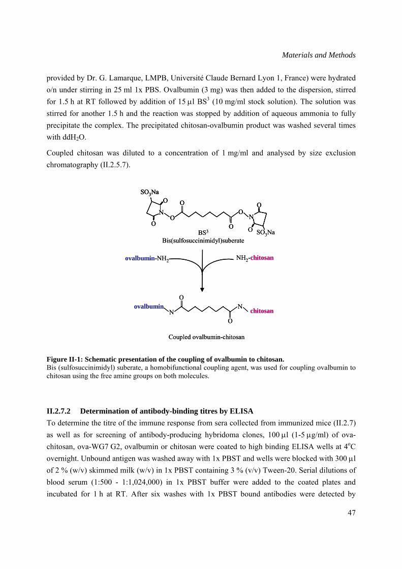
Materials and Methods
47
provided by Dr. G. Lamarque, LMPB, Université Claude Bernard Lyon 1, France) were hydrated o/n under stirring in 25 ml 1x PBS. Ovalbumin (3 mg) was then added to the dispersion, stirred for 1.5 h at RT followed by addition of 15 μl BS3 (10 mg/ml stock solution). The solution was stirred for another 1.5 h and the reaction was stopped by addition of aqueous ammonia to fully precipitate the complex. The precipitated chitosan-ovalbumin product was washed several times with ddH2O.
Coupled chitosan was diluted to a concentration of 1 mg/ml and analysed by size exclusion chromatography (II.2.5.7).
Figure II-1: Schematic presentation of the coupling of ovalbumin to chitosan. Bis (sulfosuccinimidyl) suberate, a homobifunctional coupling agent, was used for coupling ovalbumin to chitosan using the free amine groups on both molecules.
II.2.7.2 Determination of antibody-binding titres by ELISA To determine the titre of the immune response from sera collected from immunized mice (II.2.7) as well as for screening of antibody-producing hybridoma clones, 100 μl (1-5 μg/ml) of ova-chitosan, ova-WG7 G2, ovalbumin or chitosan were coated to high binding ELISA wells at 4oC overnight. Unbound antigen was washed away with 1x PBST and wells were blocked with 300 μl of 2 % (w/v) skimmed milk (w/v) in 1x PBST containing 3 % (v/v) Tween-20. Serial dilutions of blood serum (1:500 - 1:1,024,000) in 1x PBST buffer were added to the coated plates and incubated for 1 h at RT. After six washes with 1x PBST bound antibodies were detected by
BS3
Bis(sulfosuccinimidyl)suberate
O SO3NaO
OO
SO3Na
OO
OO
NN
ovalbumin-NH2 NH2-chitosan
ovalbuminchitosan
O
ON
N
Coupled ovalbumin-chitosan
BS3
Bis(sulfosuccinimidyl)suberate
O SO3NaO
OO
SO3Na
OO
OO
NN
O
OO
SO3Na
OO
OO
NN
ovalbumin-NH2 NH2-chitosan
ovalbuminchitosan
O
ON
N
Coupled ovalbumin-chitosan
Materials and Methods
48
addition of 1:5,000 diluted AP-labelled goat anti-mouse (GAM) polyclonal antibodies in 1x PBST buffer (1 h at RT) and p-nitrophenyl phosphate (pNPP) as substrate (Sigma). ELISA plates were incubated for 30 min h at 37°C followed by measurement of the OD405nm.
Analysis of the binding specificity of selected hybridoma clones (II.2.10) to the ovalbumin-coupled WGA7 G2 peptide was performed by capture ELISA. The set up of the experiment consisted in coating 100 μl (5 μg/ml) anti-ovalbumin to high binding ELISA wells overnight at 4oC. After washing the unbound antigen and blocking the remaining free binding sites on the wells as described above, 120 μl of ova-WGA7 G2 solution (5 μg/ml) were applied and incubated 1 h at RT. Unbound protein was washed with 1x PBS and 100 ml hybridoma supernatant were applied and incubated for 1 h at RT. The wells were once more washed with 1x PBS and the bound antibodies were detected with AP-labelled GAM antibodies and pNPP substrate, as described above.
II.2.7.3 Competition ELISA using free peptides To confirm the specificity of the immune response, serum from immunized mice showing high reactivity against chitin-mimicking peptides (WGA7 G2, G3, F4, F5 and B5) (III.1.2.3) were tested by competition ELISA. The experiment consisted in competing a constant concentration of blood serum with equal volumes of increasing concentrations of the corresponding free chitin-mimicking peptides.
For each peptide to be tested 100 ml of 1:1,000 diluted blood serum in 1x PBST was incubated with different concentrations of free peptides (12.5, 25, 50, 100 or 200 μg/ml) for 1 h at RT. Non competed 1:1,000 diluted serum was used as control and reference. One hundred μl from each dilution were applied to high binding microtiter wells coated with free or peptide-coupled ovalbumin (5 μg/ml) and incubated for 1 h at RT. Detection of bound antibodies was performed as described in II.2.7.2.
II.2.8 Selection of phage displayed chitin-mimicking peptides
II.2.8.1 Phage displayed chitin-mimicking peptide selection For isolation and enrichment of phages displaying chitin mimicking peptides, solid phase panning was performed as described in Figure II-2. High binding microtiter wells were coated o/n with 10 µg/ml antigen, wheat germ agglutinin (WGA) (Biomeda, CA, USA) or chitin-binding domain (CBD) (II.2.3.5) in coating buffer at 4°C. The wells were blocked for two hours with 1x TBST containing 2 % (w/v) skimmed milk. 4 x 10¹⁰ phages from each library (II.1.6) were mixed with 1x TBST to a volume of 100 μl and incubated o/n at 4°C with the coated antigens (WGA or CBD). For the first three rounds of selection unbound phages were washed away by rinsing
Materials and Methods
49
10 times with 1x TBST and 10 times with 1x TBS. In the fourth and fifth rounds of panning the stringency was increased and plates were washed 10 times with boric buffer (pH 9) and 10 times with acetate buffer (pH 4) with an incubation time of 1 min between each wash. Bound phages were eluted with 100 μl elution buffer followed by immediate neutralisation with 1 M Tris-HCl (pH 9.1).
The eluted phages were amplified by infection of 20 ml log phase E. coli ER2738 cells. The mixture was incubated for 4.5 h at 37˚C with shaking. The infected cells were pelleted by centrifugation at 5,000 g for 30 min at 4°C. The supernatant was transferred to a fresh tube containing 1/6 volume PEG/NaCl solution and incubated o/n at 4°C without shaking. The phages were centrifuged (15,000 g/15 min/4ºC), resuspended in 200 μl 1x TBS buffer and stored at 4°C. The selected phages were tittered (II.2.9.1) and further characterized by ELISA (II.2.9.2, II.2.9.3).
Acetate buffer: Sodium acetate, pH 4 100 mM NaCl 1 M Tween-20 0.1 % (v/v)
Boric buffer: Boric acid, pH 9 100 mM NaCl 1 M Tween-20 0.1 % (v/v)
Coating buffer:
NaHCO3, pH 8.6 0.1 M
Elution buffer: Glycin/HCl, pH 2.2 0.2 M BSA 1 mg/ml
PEG/NaCl solution: Polyethylene glycol-8000 20 % (w/v) NaCl 2.5 M Autoclaved and stored at RT
TBS buffer: Tris/HCl, pH 7.5 50 mM NaCl 150 mM Autoclaved and stored at RT
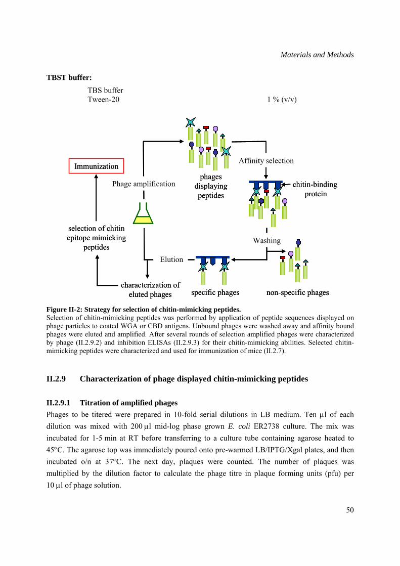
Materials and Methods
50
TBST buffer: TBS buffer Tween-20 1 % (v/v)
Figure II-2: Strategy for selection of chitin-mimicking peptides. Selection of chitin-mimicking peptides was performed by application of peptide sequences displayed on phage particles to coated WGA or CBD antigens. Unbound phages were washed away and affinity bound phages were eluted and amplified. After several rounds of selection amplified phages were characterized by phage (II.2.9.2) and inhibition ELISAs (II.2.9.3) for their chitin-mimicking abilities. Selected chitin-mimicking peptides were characterized and used for immunization of mice (II.2.7).
II.2.9 Characterization of phage displayed chitin-mimicking peptides
II.2.9.1 Titration of amplified phages Phages to be titered were prepared in 10-fold serial dilutions in LB medium. Ten μl of each dilution was mixed with 200 μl mid-log phase grown E. coli ER2738 culture. The mix was incubated for 1-5 min at RT before transferring to a culture tube containing agarose heated to 45°C. The agarose top was immediately poured onto pre-warmed LB/IPTG/Xgal plates, and then incubated o/n at 37°C. The next day, plaques were counted. The number of plaques was multiplied by the dilution factor to calculate the phage titre in plaque forming units (pfu) per 10 μl of phage solution.
Phage amplificationphages
displayingpeptides
Affinity selection
chitin-bindingprotein
characterization of eluted phages
selection of chitin epitope mimicking
peptides
Immunization
Washing
non-specific phagesspecific phages
Elution
Phage amplificationphages
displayingpeptides
Affinity selection
chitin-bindingprotein
characterization of eluted phages
selection of chitin epitope mimicking
peptides
Immunization
Washing
non-specific phagesspecific phages
Elution
Materials and Methods
51
LB/IPTG/Xgal agar plates: Bacto-Tryptone 1.0 % (w/v) Yeast extract 0.5 % (w/v) NaCl 0.5 % (w/v) Agar 1.5 % (w/v) After autoclaving, the medium was allowed to cool then 210 mM IPTG and 1 ml Xgal (40 mg/ml stock solution) were added. The plates were stored at 4°C in the dark.
Agarose top : Agarose 0.7 % (w/v) Bacto-Tryptone 1.0 % (w/v) Yeast extract 0.5 % (w/v) NaCl 0.5 % (w/v) MgCl2 6H2O 0.7 % (w/v) Autoclaved and stored at RT
II.2.9.2 Phage ELISA After several panning rounds eluted phages were tested for their binding to the antigen by phage ELISA. Log-phase E. coli ER2738 cells were infected with eluted phages and serial dilutions were plated on IPTG/Xgal agar plates as described in II.2.9.1. High binding microtier plates were coated with 100 μl (5 μg/ml) of the antigen of interest (WGA or CBD) and incubated o/n at 4˚C. The next day, ER2738 E. coli cells were cultivated o/n in 10 ml LB medium. The o/n grown culture was diluted 1/50 in LB medium and disposed in sterile microtiter wells (200 μl/well). For each panned peptide library thirty wells were infected with randomly selected phage plaques (1 plaque/well) and the plate was incubated 4.5 h at 37˚C. The o/n coated high binding plates were washed with 1x TBS and free binding sites were blocked for two hours with 300 ml 1x TBST containing 2 % (w/v) skimmed milk. After washing with 1x TBS each well was filled with 50 μl 1x TBST.
The microtiter plates containing the phage-producing clones were centrifuged (2700 rpm/20min/RT) and 50 μl supernatant were added to the wells coated with the corresponding antigen. The plates were incubated 1 h at RT then washed 6 times with 1x TBST. Hundred μl of 1:5,000 diluted anti-M13 HRP-labelled antibodies were used as primary antibody and incubated for 1 h at RT. The wells were washed and incubated with 100 μl of ABTS substrate for 20 min in the dark. The colour development was measured in an ELISA reader at OD405. Positive clones were selected and further characterized by inhibition ELISA (II.2.9.3). The peptide sequences were identified by sequencing (II.2.1.10) using single stranded DNA (II.2.9.4).
Materials and Methods
52
II.2.9.3 Inhibition ELISA of selected phage-displayed peptides Inhibition ELISA was performed to confirm the specificity of selected peptides (II.2.8.1) to the GlcNAc-epitope present on WGA or Datura stramonium lectins (Vector Laboratories, Ltd, Peterborough, UK). The experimental set up included incubation of constant amounts of phages displaying peptide with equal volumes of increasing concentration of chitin hydrolysate (inhibitor) (Vector Laboratories, Ltd).
High binding microtiter plates were coated o/n at 4˚C with 5 μg/ml WGA or D. stramonium lectins. Unbound antigen was washed with 1x PBS, then the wells were blocked with 300 ml 1x PBS containing 3 % (v/v) Tween-20 for 1 h at RT. Serial dilutions from 1:20 to 1:160 of chitin hydrolysate were prepared in 1x PBST.
Ten μl of the phage solution (II.2.8.1) were applied to each well and 90 μl from each chitin hydrolysatedilution was added. The plate was incubated 1 h at RT and unbound phages were washed away with 1x PBST. One hundred μl of HRP-labelled anti-M13 antibody (1:5,000 diluted in 1x PBST) were applied to each well and incubated for 1 h at RT. Unbound antibodies were removed by washing with 1x PBST. Bound phages were detected with 100 μl ABTS substrate. After 20 min incubation at RT in the dark, the developed colour was measured at OD405nm. The levels of inhibition were compared to the absorbance of the control wells which did not contain the inhibitor.
II.2.9.4 Isolation of single stranded DNA from selected phages Amino acid sequences of selected phage-displayed chitin–mimicking peptides (II.2.8.1) were identified by extraction and sequencing of single stranded DNA.
Phage clones to be sequenced were amplified as described above (II.2.8.1). Four ml of an o/n incubated phage-containing supernatant-PEG/NaCl mix were transferred to a fresh tube and centrifuged (15,000 g/15 min/4°C). The phage pellet was resuspended into 100 μl iodide buffer and 250 μl 100 % ethanol. The mixture was incubated for 10 min at RT before spinning for 20 min. The pellet was washed in 70 % ethanol and briefly dried under vacuum. The single stranded DNA was resuspended in 20 μl ddH2O and stored at -20°C.
Iodide buffer: Tris-HCl, pH 8 10 mM EDTA 1 mM NaI 4 M Stored at RT in the dark.
Materials and Methods
53
II.2.10 Generation of chitin/chitosan-specific monoclonal antibodies by hybridoma technology
Generation of chitin-specific mAbs was achieved through fusion of myeloma cells with spleen cells from mice immunized with ova-WGA7 G2 peptide (II.2.7).
II.2.10.1 Animal cell culture The described animal cell culture work was performed at 22°C under sterile conditions in a laboratory with safety class S1.
The growth of myeloma cells and generated hybridoma cells (II.2.10.4) was performed in a CO2 incubator at 37°C, 5 % CO2 and 100 % relative humidity to have ideal growth conditions and a correct buffering of the medium. The cells were cultivated in 96-and 24-well-tissue culture plates, petri dishes or tissue culture flasks (Greiner). As basis medium RPMI-1640 (Cytogen, Sinn-Fleisbach, D) was used, to which diverse substances were added to make a full medium. RPMI-1640 was also used as wash medium for the preparation of spleen and myeloma cells. The selection of the HGPRT(−) myeloma cells was performed in full medium containing 130 µM 8-Azaguanin in order to avoid HGPRT(+) revertant. The selection of hybridoma cells was done for at least 14 days after the fusion in HAT-medium (Littlefield, 1964). The first limiting dilution (LD) was performed in HT-Medium (II.2.10.5) containing 80 U/ml interleukin-6 (IL-6; Boehringer Mannheim GmbH, Mannheim, D) as a growth stimulator (Bazin and Lemieux, 1989; Rieckmann et al., 1997). Depending on the growth of the hybridoma cells the concentration of IL-6 was slowly decreased and then it was completely suppressed. Cryo-preservation of animal cells (II.2.10.2) was performed in cryo-medium in liquid nitrogen. All media were made with sterile, tissue culture-approved solutions, supplements and buffer-components. Media were kept at 4°C in the dark and were equilibrated to 37°C for three hours in the CO2 incubator before use.
R10 (complete medium): RPMI-1640 (Cytogen) 1xRPMI-1640 Fetal calf serum (FCS) (Cytogen) 10 % (v/v) L-glutamin (Gibco BRL) 2 mM β-mercaptoethanol 50 μM Penicillin/streptomycin 100 U/100 μg/ml Before use, each FCS-charge was tested for its usibility for cultivation of hybridoma cells.
HAT-medium/HT-medium: Complete medium Hypoxanthin (Sigma) 100 μM Aminopterin (Sigma) 10 μM Thymidin (Sigma) 16 μM
Materials and Methods
54
Cryo-medium: RPMI-1640 FCS 20 % (v/v) Dimethylsulfoxide 10 % (v/v)
II.2.10.2 Cryo-preservation of animal cells Freezing and thawing of myeloma cells (II.2.10.4) and antibody-secreting hybridoma cells was performed as described (Batt, 1997) to create a cell bank stored in liquid nitrogen ensuring constant supply of mAbs-producing cells
II.2.10.3 Isolation of mouse spleen cells All steps for isolation of murine spleen cells were performed under sterile conditions. Spleen cells of immunised mice showing an adequate titer (II.2.7.2) were isolated for fusion (II.2.10.4) with myeloma cells one day after the last injection (II.2.7). The mouse was sacrificed with Isofuran, surface sterilised with 70 % (v/v) ethanol and fixed onto a preparation board. Skin and abdominal wall were opened and the spleen was removed under sterile conditions. To obtain single cells, the spleen was passed with the help of a plunger of a sterile 5 ml syringe (Becton-Dickinson) through a stainless steel sieve (mesh size: 200 µm) into a petri dish containing 30 ml equilibrated RPMI-1640 medium. The cell suspension was carefully pipetted up and down a few times and then incubated for 1 min in the CO2 incubator to sediment larger particles. The supernatant was pipetted into a new tube and centrifuged (200 g/5 min/RT). The cell sediment was prepared for fusion by washing once in 40 ml pre-equilibrated RPMI-1640 medium (3 h incubation at 37ºC in CO2 incubator). The cell sediment was resuspended in 10 ml pre-equilibrated RPMI-1640 medium and the cell number was determined under the microscope using a Thoma chamber. The ready-to use cell suspension was used for fusion to murine myeloma cells for generation of hybridoma clones (II.2.10.4).
II.2.10.4 Establishment of stable hybridoma cells For establishment of stable antibody-producing hybridoma clones, isolated spleen cells (II.2.10.3) were fused with exponentially growing SP2/0-Ag14 myeloma cells (Schulman et al., 1978).
Before the fusion procedure, isolated spleen cells as well as myeloma cells were washed in RPMI-1640 medium and then resuspended in 30 ml RPMI-1640 medium. Both cell samples were mixed in a ratio of 4:1 (1x10⁸ spleen cells and 3x10⁷ myeloma cells). The mix was centrifuged (300 g/5min/RT) and 1 ml pre-warmed PEG-1500 (Roche) was carefully added (drop by drop) over 1 min time while rocking. The cells were incubated for 90 seconds in a 37°C water-bath while rocking, then 3 ml RPMI-1640 medium were gradually added. Additional 10 ml RPMI-1640 medium were carefully supplied and incubated for 5 minutes in the CO2 incubator before
Materials and Methods
55
centrifugation (300 g/5 min/RT). Fused cells were resuspended in 60 ml HAT medium and 120 μl per well were disposed into a 96-well tissue culture plate.
The cultivation and the selection of hybridoma cells (Littlefield, 1964) was performed by incubation of the plates for a period of seven days without opening the incubators. At day eight, cells were fed with fresh HAT medium (200 μl/well) and allowed to grow for four additional days before the hybridoma supernatant could be tested by ELISA (II.2.7.2) for the presence of specific antibodies.
II.2.10.5 Limiting dilutions for isolation of monoclonal antibodies Cloning by limiting dilution is a procedure for separating cells based on the assumption that if a suspension of cells is diluted with enough culture medium, a concentration of cells will be produced such that an accurately measured volume of the diluted suspension will contain one cell.
One hundred μl HT-medium per well were prepared in a 96-well tissue culture plate. Fifty μl of positive hybridoma clones were applied to the first well of the first column in the plate. Fifty μl from that well were mixed with fresh HT-medium in the next well in the same column (dilution 1:2). One hundred ml fresh HT-medium were added to the wells of the first column then 100 ml were transferred to the wells of the next column with a multi-channel pipette. This operation was repeated for all columns. The plate was incubated at 37ºC in 5 % CO2 for 4-7 days without any change of the medium. The supernatant from each well was screened by ELISA (II.2.7.2) for determination of wells producing desired antibodies. Positive clones were transferred into larger volumes of medium (24-well plates).
II.2.10.6 Isotype determination of generated chitin-specific monoclonal antibodies The isotype determination of selected mAbs (II.2.10.5) was performed by ELISA (II.2.7.2) using a commercially available kit following the manufacturer’s protocol (BD Biosciences).
Fifty μl of each purified isotype-specific rat-anti-mouse monoclonal antibody, diluted 1:50 in coating buffer (1x PBS), were added to the wells of a high binding microtiter plate. The plate was covered and incubated for 1 h at 37˚C. The wells were carefully washed with 1x PBST and remaining free binding sites were blocked with 200 μl 1 % (w/v) BSA for 1 h at RT. One hundred μl of each hybridoma cell culture supernatant were pipetted to the wells of the prepared microtiter plate and samples were incubated for 1 h at RT. A positive reference antigen mixture provided in the kit was used as control. Wells in which hybridoma cell culture supernatant was replaced by 1x PBS were included as negative control.
Materials and Methods
56
The plate was washed thoroughly 6 times with 1x PBST, soaking the wells each time for 1 min. HRP-labelled rat anti-mouse Ig mAb solution (100 μl) diluted 1:100 in 1 % (w/v) BSA were added and incubated for 1 h at RT. One hundred μl substrate solution, prepared by mixing equal volumes of reagent A and reagent B (both provided in the kit), were added to each well and incubated for 3-10 min at RT. When reactions developed a greenish-blue colour, the reaction was stopped with 50 μl stop solution provided in the kit. The plates were immediately measured in an ELISA reader at OD405nm.
II.2.11 Microscopic analysis
Fluorescence microscopy was carried out to determine the binding of selected mAbs (II.2.11.1) to native chitin and chitosan present in the fungal cell wall.
II.2.11.1 Preparation of cover slides Cover slides were cleaned with 70 % (v/v) ethanol, then immediately dropped in poly-L-lysine solution (Sigma) for 5 min. The slides were washed with ddH2O before using them for antigen coating (II.2.11.3).
II.2.11.2 Preparation of fungal mycelia Spores from F. graminearum (105/ml) (II.2.5.10.1) were germinated in 10 ml PDB (Difco) for 3 days at 28˚C shaking at 200 rpm. The produced mycelia were washed 3 times with 10 ml sterile water (5,000 g/20 min/4˚C) to remove the media and secreted proteins.
II.2.11.3 Coating of cover slides with fungal mycelia The poly-L-lysine-coated cover slides (II.2.11.1) were deposited with tweezers into pre-blocked 12-well tissue culture plate. One ml of the prepared mycelia solution (II.2.11.2) was pipetted onto the cover slides. The plate was centrifuged in an ELISA centrifuge (Rotina, Hettich) (2,000 g/15 min/RT) to ensure a tight coating. The supernatant was removed with a pipette and the slides were carefully washed 3 times with 1x PBST to remove unbound mycelia. 250 μl of hybridoma supernatant containing monoclonal antibodies was added onto the cover slides and incubated for 1 h at RT. The cover slides were washed again 3 times with 1x PBST, then incubated with 250 μl of 1:100 diluted Alexa Fluor® 594 labelled goat anti-rabbit (Molecular Probes) for 1 h at RT. Unspecific antibodies were removed by washing with 1x PBST. A negative control included mycelia incubated only with labelled antibody.
Materials and Methods
57
II.2.11.4 Fluorescence microscopy The prepared cover slides (II.2.11.3) were transferred up side down onto a glass slide and fixed around with clear nail polish to inhibit the drying out of the probes. The samples were analysed with an Olympus BX40 fluorescence microscope (Olympus Austria GmbH) for analysis of immunofluorescence.
Results
58
III Results
This doctoral thesis was part of an interdisciplinary research project supported by the European Union with a primary objective to identify biologically active chitooligomers that induce plant defense mechanisms to develop non-toxic, biodegradable pesticides. These oligomers with a defined range of degree of polymerization and acetylation are then to be produced by enzymatic hydrolysis of chitin with chitinase in an upscaled process. Additionally, this project aimed at the establishement of an antibody-based monitoring of the enzymatiquely produced oligomers.
The objective of this work was the generation and characterization of chitin/chitosan-specific monoclonal antibodies as well as the production and characterization of Wch1 wheat chitinase.
Two different strategies were followed for the generation of chitin-specific antibodies in order to circumvent the limitations caused by the insolubility and low immunogenicity of chitin. The first approach consisted in coupling a soluble variant of chitin, chitosan, to the protein carrier ovalbumin and subsequent immunization of Balb/c mice (III.1.1). In the second strategy, chitin-mimicking peptides were selected by panning peptide libraries against chitin-binding protein or wheat germ agglutinin (WGA) (III.1.2). Selected peptides were coupled to ovalbumin and then used for immunization. The resulting monoclonal antibodies were characterized by ELISA and immunoblot analysis.
The Wch1 cDNA gene was cloned into bacterial and plant expression vectors fused at the C-terminal end of Strep-tag as well as the N-terminal end of maltose binding protein (MBP) and his6 tag. The accumulation levels and enzymatic activity of Wch1 produced in E. coli and tobacco leaves were quantifed to select the best production system (III.2, III.4, III.5). Moreover, the anti-fungal property of Wch1 was assessed (III.7) and the physico-chemical characterization of substrate degradation was performed (III.8).
III.1 Generation of chitin- and chitosan-specific antibodies
III.1.1 Generation of chitosan-specific antibodies
III.1.1.1 Coupling of chitosan to ovalbumin Due to its poor ability in triggering the immune system, chitosan (DA 10 %) was coupled to the protein carrier ovalbumin using the homo-bifunctional Bis(sulfosuccinimidyl) suberate (BS3) cross-linker (II.2.7.1) for improvement of its immunogenicity. Carrier-hapten (ova-chitosan)
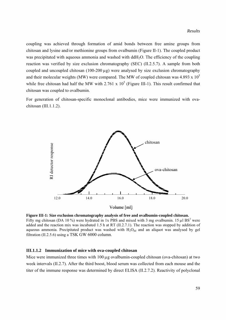
Results
59
coupling was achieved through formation of amid bonds between free amine groups from chitosan and lysine and/or methionine groups from ovalbumin (Figure II-1). The coupled product was precipitated with aqueous ammonia and washed with ddH2O. The efficiency of the coupling reaction was verified by size exclusion chromatography (SEC) (II.2.5.7). A sample from both coupled and uncoupled chitosan (100-200 μg) were analysed by size exclusion chromatography and their molecular weights (MW) were compared. The MW of coupled chitosan was 4.893 x 10⁵ while free chitosan had half the MW with 2.761 x 10⁵ (Figure III-1). This result confirmed that chitosan was coupled to ovalbumin.
For generation of chitosan-specific monoclonal antibodies, mice were immunized with ova-chitosan (III.1.1.2).
Figure III-1: Size exclusion chromatography analysis of free and ovalbumin-coupled chitosan. Fifty mg chitosan (DA 10 %) were hydrated in 1x PBS and mixed with 3 mg ovalbumin. 15 μl BS3 were added and the reaction mix was incubated 1.5 h at RT (II.2.7.1). The reaction was stopped by addition of aqueous ammonia. Precipitated product was washed with H2Odd and an aliquot was analysed by gel filtration (II.2.5.6) using a TSK GW 6000 column.
III.1.1.2 Immunization of mice with ova-coupled chitosan Mice were immunized three times with 100 μg ovalbumin-coupled chitosan (ova-chitosan) at two week intervals (II.2.7). After the third boost, blood serum was collected from each mouse and the titer of the immune response was determined by direct ELISA (II.2.7.2). Reactivity of polyclonal
chitosan
ova-chitosan
12.0 14.0 16.0 18.0 20.0
Volume [ml]
RI d
etec
torr
espo
nse chitosan
ova-chitosan
chitosan
ova-chitosan
12.0 14.0 16.0 18.0 20.0
Volume [ml]
RI d
etec
torr
espo
nse
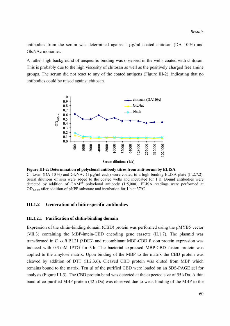
Results
60
antibodies from the serum was determined against 1 μg/ml coated chitosan (DA 10 %) and GlcNAc monomer.
A rather high background of unspecific binding was observed in the wells coated with chitosan. This is probably due to the high viscosity of chitosan as well as the positively charged free amine groups. The serum did not react to any of the coated antigens (Figure III-2), indicating that no antibodies could be raised against chitosan.
Figure III-2: Determination of polyclonal antibody titres from anti-serum by ELISA. Chitosan (DA 10 %) and GlcNAc (1 μg/ml each) were coated to a high binding ELISA plate (II.2.7.2). Serial dilutions of sera were added to the coated wells and incubated for 1 h. Bound antibodies were detected by addition of GAMAP polyclonal antibody (1:5,000). ELISA readings were performed at OD405nm after addition of pNPP substrate and incubation for 1 h at 37ºC.
III.1.2 Generation of chitin-specific antibodies
III.1.2.1 Purification of chitin-binding domain
Expression of the chitin-binding domain (CBD) protein was performed using the pMYB5 vector (VII.3) containing the MBP-intein-CBD encoding gene cassette (II.1.7). The plasmid was transformed in E. coli BL21 (λDE3) and recombinant MBP-CBD fusion protein expression was induced with 0.3 mM IPTG for 3 h. The bacterial expressed MBP-CBD fusion protein was applied to the amylose matrix. Upon binding of the MBP to the matrix the CBD protein was cleaved by addition of DTT (II.2.3.6). Cleaved CBD protein was eluted from MBP which remains bound to the matrix. Ten µl of the purified CBD were loaded on an SDS-PAGE gel for analysis (Figure III-3). The CBD protein band was detected at the expected size of 55 kDa. A thin band of co-purified MBP protein (42 kDa) was observed due to weak binding of the MBP to the
0.00.10.20.30.40.50.60.70.80.91.0
500
1000
2000
4000
8000
1600
0
3200
0
6400
0
1280
00
2560
00
5120
00
1024
000
Serum dilutions (1/x)
chitosan (DA10%)GlcNacblank
OD
405n
m
0.00.10.20.30.40.50.60.70.80.91.0
500
1000
2000
4000
8000
1600
0
3200
0
6400
0
1280
00
2560
00
5120
00
1024
000
Serum dilutions (1/x)
chitosan (DA10%)GlcNacblank
chitosan (DA10%)GlcNacblank
OD
405n
m
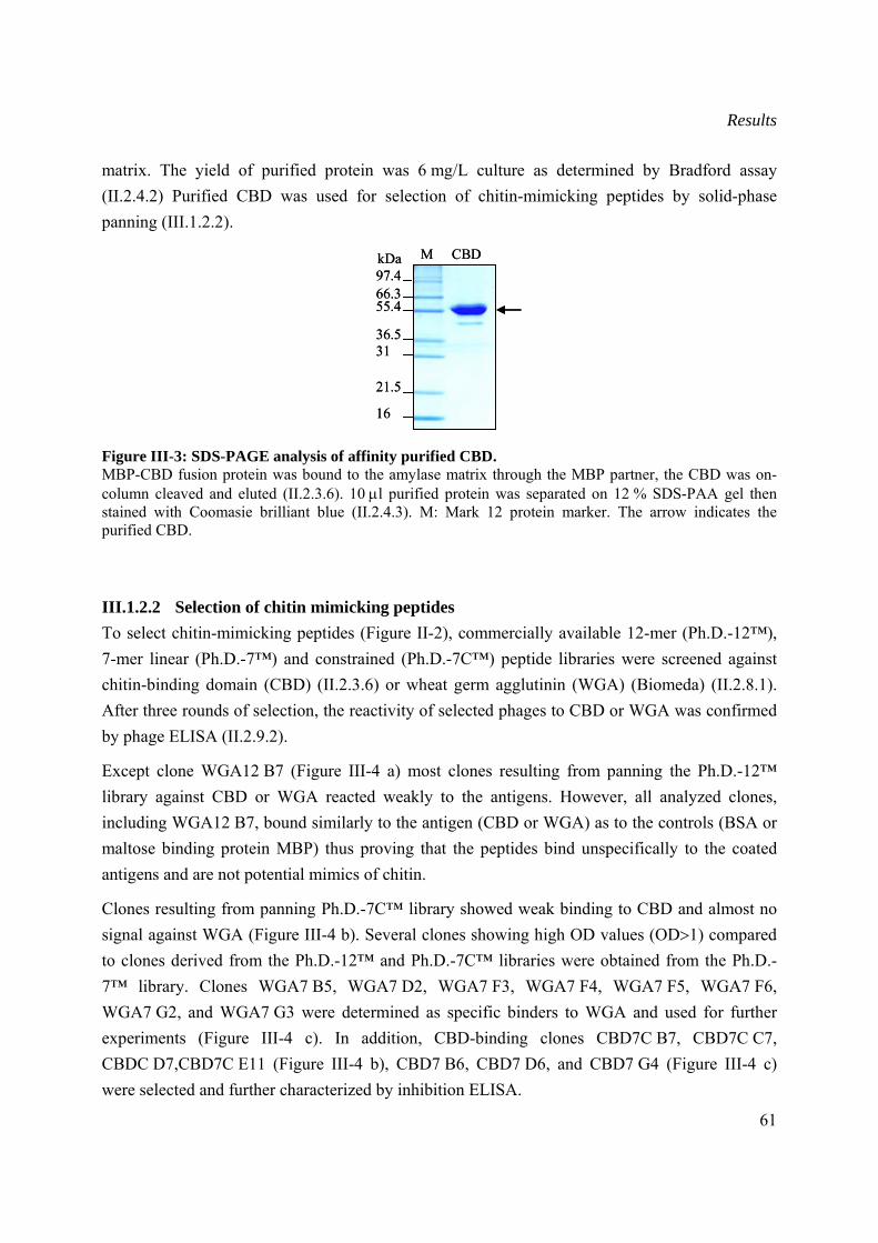
Results
61
matrix. The yield of purified protein was 6 mg/L culture as determined by Bradford assay (II.2.4.2) Purified CBD was used for selection of chitin-mimicking peptides by solid-phase panning (III.1.2.2).
Figure III-3: SDS-PAGE analysis of affinity purified CBD. MBP-CBD fusion protein was bound to the amylase matrix through the MBP partner, the CBD was on-column cleaved and eluted (II.2.3.6). 10 μl purified protein was separated on 12 % SDS-PAA gel then stained with Coomasie brilliant blue (II.2.4.3). M: Mark 12 protein marker. The arrow indicates the purified CBD.
III.1.2.2 Selection of chitin mimicking peptides To select chitin-mimicking peptides (Figure II-2), commercially available 12-mer (Ph.D.-12™), 7-mer linear (Ph.D.-7™) and constrained (Ph.D.-7C™) peptide libraries were screened against chitin-binding domain (CBD) (II.2.3.6) or wheat germ agglutinin (WGA) (Biomeda) (II.2.8.1). After three rounds of selection, the reactivity of selected phages to CBD or WGA was confirmed by phage ELISA (II.2.9.2).
Except clone WGA12 B7 (Figure III-4 a) most clones resulting from panning the Ph.D.-12™ library against CBD or WGA reacted weakly to the antigens. However, all analyzed clones, including WGA12 B7, bound similarly to the antigen (CBD or WGA) as to the controls (BSA or maltose binding protein MBP) thus proving that the peptides bind unspecifically to the coated antigens and are not potential mimics of chitin.
Clones resulting from panning Ph.D.-7C™ library showed weak binding to CBD and almost no signal against WGA (Figure III-4 b). Several clones showing high OD values (OD>1) compared to clones derived from the Ph.D.-12™ and Ph.D.-7C™ libraries were obtained from the Ph.D.-7™ library. Clones WGA7 B5, WGA7 D2, WGA7 F3, WGA7 F4, WGA7 F5, WGA7 F6, WGA7 G2, and WGA7 G3 were determined as specific binders to WGA and used for further experiments (Figure III-4 c). In addition, CBD-binding clones CBD7C B7, CBD7C C7, CBDC D7,CBD7C E11 (Figure III-4 b), CBD7 B6, CBD7 D6, and CBD7 G4 (Figure III-4 c) were selected and further characterized by inhibition ELISA.
97.466.355.4
36.531
21.5
16
kDa M CBD97.466.355.4
36.531
21.5
16
kDa M CBD
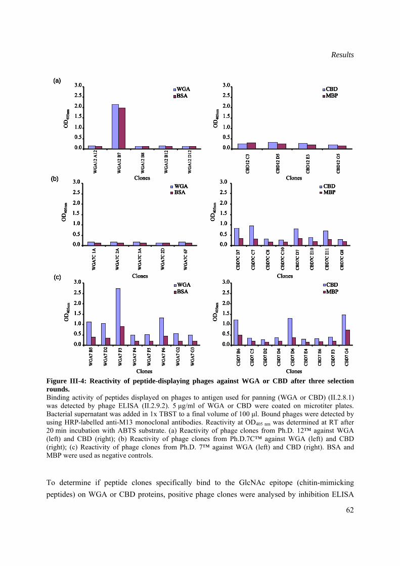
Results
62
Figure III-4: Reactivity of peptide-displaying phages against WGA or CBD after three selection rounds. Binding activity of peptides displayed on phages to antigen used for panning (WGA or CBD) (II.2.8.1) was detected by phage ELISA (II.2.9.2). 5 μg/ml of WGA or CBD were coated on microtiter plates. Bacterial supernatant was added in 1x TBST to a final volume of 100 μl. Bound phages were detected by using HRP-labelled anti-M13 monoclonal antibodies. Reactivity at OD405 nm was determined at RT after 20 min incubation with ABTS substrate. (a) Reactivity of phage clones from Ph.D. 12™ against WGA (left) and CBD (right); (b) Reactivity of phage clones from Ph.D.7C™ against WGA (left) and CBD (right); (c) Reactivity of phage clones from Ph.D. 7™ against WGA (left) and CBD (right). BSA and MBP were used as negative controls.
To determine if peptide clones specifically bind to the GlcNAc epitope (chitin-mimicking peptides) on WGA or CBD proteins, positive phage clones were analysed by inhibition ELISA
Clones
OD
405n
m
0.0
0.5
1.0
1.5
2.0
2.5
3.0
WG
A7C
1A
WG
A7C
2A
WG
A7C
3A
WG
A7C
2D
WG
A7C
6F
WGABSA
Clones
OD
405n
m
0.0
0.5
1.0
1.5
2.0
2.5
3.0
CBD
7C B
7
CBD
7C C
7
CBD
7C C
8
CBD
7C C
10
CBD
7C D
7
CBD
7C E
10
CBD
7C E
11
CBD
7C G
8
CBDMBP
(b)
Clones
OD
405n
m
0.0
0.5
1.0
1.5
2.0
2.5
3.0
CBD
7 B6
CBD
7 C
5
CBD
7 D
2
CBD
7 D
4
CBD
7 D
6
CBD
7 E4
CBD
7 E6
CBD
7 F3
CBD
7 G
4
CBDMBP
Clones
OD
405n
m
0.0
0.5
1.0
1.5
2.0
2.5
3.0
WG
A7
B5
WG
A7
D2
WG
A7
F3
WG
A7
F4
WG
A7
F5
WG
A7
F6
WG
A7
G2
WG
A7
G3
WGABSA
(c)
WG
A12
A12
WG
A12
B7
WG
A12
B8
WG
A12
B12
WG
A12
D12
Clones
OD
405n
m
3.0
0.0
0.5
1.0
1.5
2.0
2.5WGABSA
Clones
OD
405n
m
0.0
0.5
1.0
1.5
2.0
2.5
3.0
CBD
12 C
3
CBD
12 D
5
CBD
12 E
3
CBD
12 G
5
CBDMBP
(a)
Clones
OD
405n
m
0.0
0.5
1.0
1.5
2.0
2.5
3.0
WG
A7C
1A
WG
A7C
2A
WG
A7C
3A
WG
A7C
2D
WG
A7C
6F
WGABSA
Clones
OD
405n
m
0.0
0.5
1.0
1.5
2.0
2.5
3.0
CBD
7C B
7
CBD
7C C
7
CBD
7C C
8
CBD
7C C
10
CBD
7C D
7
CBD
7C E
10
CBD
7C E
11
CBD
7C G
8
CBDMBP
(b)
Clones
OD
405n
m
0.0
0.5
1.0
1.5
2.0
2.5
3.0
WG
A7C
1A
WG
A7C
2A
WG
A7C
3A
WG
A7C
2D
WG
A7C
6F
WGABSA
Clones
OD
405n
m
0.0
0.5
1.0
1.5
2.0
2.5
3.0
WG
A7C
1A
WG
A7C
2A
WG
A7C
3A
WG
A7C
2D
WG
A7C
6F
WGABSA
0.0
0.5
1.0
1.5
2.0
2.5
3.0
WG
A7C
1A
WG
A7C
2A
WG
A7C
3A
WG
A7C
2D
WG
A7C
6F
WGABSAWGABSAWGABSA
Clones
OD
405n
m
0.0
0.5
1.0
1.5
2.0
2.5
3.0
CBD
7C B
7
CBD
7C C
7
CBD
7C C
8
CBD
7C C
10
CBD
7C D
7
CBD
7C E
10
CBD
7C E
11
CBD
7C G
8
CBDMBP
Clones
OD
405n
m
0.0
0.5
1.0
1.5
2.0
2.5
3.0
CBD
7C B
7
CBD
7C C
7
CBD
7C C
8
CBD
7C C
10
CBD
7C D
7
CBD
7C E
10
CBD
7C E
11
CBD
7C G
8
CBDMBP
0.0
0.5
1.0
1.5
2.0
2.5
3.0
CBD
7C B
7
CBD
7C C
7
CBD
7C C
8
CBD
7C C
10
CBD
7C D
7
CBD
7C E
10
CBD
7C E
11
CBD
7C G
8
CBDMBPCBDMBPCBDMBP
(b)
Clones
OD
405n
m
0.0
0.5
1.0
1.5
2.0
2.5
3.0
CBD
7 B6
CBD
7 C
5
CBD
7 D
2
CBD
7 D
4
CBD
7 D
6
CBD
7 E4
CBD
7 E6
CBD
7 F3
CBD
7 G
4
CBDMBP
Clones
OD
405n
m
0.0
0.5
1.0
1.5
2.0
2.5
3.0
WG
A7
B5
WG
A7
D2
WG
A7
F3
WG
A7
F4
WG
A7
F5
WG
A7
F6
WG
A7
G2
WG
A7
G3
WGABSA
(c)
Clones
OD
405n
m
0.0
0.5
1.0
1.5
2.0
2.5
3.0
CBD
7 B6
CBD
7 C
5
CBD
7 D
2
CBD
7 D
4
CBD
7 D
6
CBD
7 E4
CBD
7 E6
CBD
7 F3
CBD
7 G
4
CBDMBP
Clones
OD
405n
m
0.0
0.5
1.0
1.5
2.0
2.5
3.0
CBD
7 B6
CBD
7 C
5
CBD
7 D
2
CBD
7 D
4
CBD
7 D
6
CBD
7 E4
CBD
7 E6
CBD
7 F3
CBD
7 G
4
CBDMBP
0.0
0.5
1.0
1.5
2.0
2.5
3.0
CBD
7 B6
CBD
7 C
5
CBD
7 D
2
CBD
7 D
4
CBD
7 D
6
CBD
7 E4
CBD
7 E6
CBD
7 F3
CBD
7 G
4
CBDMBPCBDMBPCBDMBP
Clones
OD
405n
m
0.0
0.5
1.0
1.5
2.0
2.5
3.0
WG
A7
B5
WG
A7
D2
WG
A7
F3
WG
A7
F4
WG
A7
F5
WG
A7
F6
WG
A7
G2
WG
A7
G3
WGABSA
Clones
OD
405n
m
0.0
0.5
1.0
1.5
2.0
2.5
3.0
WG
A7
B5
WG
A7
D2
WG
A7
F3
WG
A7
F4
WG
A7
F5
WG
A7
F6
WG
A7
G2
WG
A7
G3
WGABSA
0.0
0.5
1.0
1.5
2.0
2.5
3.0
WG
A7
B5
WG
A7
D2
WG
A7
F3
WG
A7
F4
WG
A7
F5
WG
A7
F6
WG
A7
G2
WG
A7
G3
WGABSAWGABSAWGABSA
(c)
WG
A12
A12
WG
A12
B7
WG
A12
B8
WG
A12
B12
WG
A12
D12
Clones
OD
405n
m
3.0
0.0
0.5
1.0
1.5
2.0
2.5WGABSA
Clones
OD
405n
m
0.0
0.5
1.0
1.5
2.0
2.5
3.0
CBD
12 C
3
CBD
12 D
5
CBD
12 E
3
CBD
12 G
5
CBDMBP
(a)
WG
A12
A12
WG
A12
B7
WG
A12
B8
WG
A12
B12
WG
A12
D12
Clones
OD
405n
m
3.0
0.0
0.5
1.0
1.5
2.0
2.5WGABSA
WG
A12
A12
WG
A12
B7
WG
A12
B8
WG
A12
B12
WG
A12
D12
Clones
OD
405n
m
3.0
0.0
0.5
1.0
1.5
2.0
2.5WGABSA
3.0
0.0
0.5
1.0
1.5
2.0
2.5WGABSAWGABSAWGABSA
Clones
OD
405n
m
0.0
0.5
1.0
1.5
2.0
2.5
3.0
CBD
12 C
3
CBD
12 D
5
CBD
12 E
3
CBD
12 G
5
CBDMBP
Clones
OD
405n
m
0.0
0.5
1.0
1.5
2.0
2.5
3.0
CBD
12 C
3
CBD
12 D
5
CBD
12 E
3
CBD
12 G
5
CBDMBP
0.0
0.5
1.0
1.5
2.0
2.5
3.0
CBD
12 C
3
CBD
12 D
5
CBD
12 E
3
CBD
12 G
5
CBDMBPCBDMBPCBDMBP
(a)
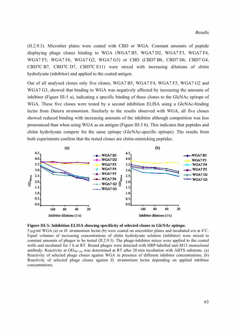
Results
63
(II.2.9.3). Microtiter plates were coated with CBD or WGA. Constant amounts of peptide displaying phage clones binding to WGA (WGA7 B5, WGA7 D2, WGA7 F3, WGA7 F4, WGA7 F5, WGA7 F6, WGA7 G2, WGA7 G3) or CBD (CBD7 B6, CBD7 D6, CBD7 G4, CBD7C B7, CBD7C D7, CBD7C E11) were mixed with increasing dilutions of chitin hydrolysate (inhibitor) and applied to the coated antigen.
Out of all analysed clones only five clones, WGA7 B5, WGA7 F4, WGA7 F5, WGA7 G2 and WGA7 G3, showed that binding to WGA was negatively affected by increasing the amounts of inhibitor (Figure III-5 a), indicating a specific binding of these clones to the GlcNAc epitope of WGA. These five clones were tested by a second inhibition ELISA using a GlcNAc-binding lectin from Datura stramonium. Similarly to the results observed with WGA, all five clones showed reduced binding with increasing amounts of the inhibitor although competition was less pronounced than when using WGA as an antigen (Figure III-5 b). This indicates that peptides and chitin hydrolysate compete for the same epitope (GlcNAc-specific epitope). The results from both experiments confirm that the tested clones are chitin-mimicking peptides.
Figure III-5: Inhibition ELISA showing specificity of selected clones to GlcNAc epitope. 5 μg/ml WGA (a) or D. stramonium lectin (b) were coated on microtitter plates and incubated o/n at 4˚C. Equal volumes of increasing concentrations of chitin hydrolysate solution (inhibitor) were mixed to constant amounts of phages to be tested (II.2.9.3). The phage-inhibitor mixes were applied to the coated wells and incubated for 1 h at RT. Bound phages were detected with HRP-labelled anti-M13 monoclonal antibody. Reactivity at OD405 nm was determined at RT after 20 min incubation with ABTS substrate. (a) Reactivity of selected phage clones against WGA in presence of different inhibitor concentrations; (b) Reactivity of selected phage clones against D. stramonium lectin depending on applied inhibitor concentrations.
WGA7 B5WGA7 D2WGA7 F3WGA7 F4WGA7 F5WGA7 F6WGA7 G2WGA7 G3
0.0
0.51.0
1.52.0
2.5
3.0
3.54.04.5
160 80 40 20
Inhibitor dilutions (1/x)
OD
405n
m
(a)
WGA7 B5WGA7 F3WGA7 F4WGA7 F5WGA7 G2WGA7 G3
0.00.5
1.0
1.52.0
2.53.0
3.5
4.04.5
160 80 40 20
Inhibitor dilutions (1/x)
OD
405n
m
(b)
WGA7 B5WGA7 D2WGA7 F3WGA7 F4WGA7 F5WGA7 F6WGA7 G2WGA7 G3
0.0
0.51.0
1.52.0
2.5
3.0
3.54.04.5
160 80 40 20
Inhibitor dilutions (1/x)
OD
405n
m
(a)
WGA7 B5WGA7 D2WGA7 F3WGA7 F4WGA7 F5WGA7 F6WGA7 G2WGA7 G3
WGA7 B5WGA7 B5WGA7 D2WGA7 D2WGA7 F3WGA7 F3WGA7 F4WGA7 F4WGA7 F5WGA7 F5WGA7 F6WGA7 F6WGA7 G2WGA7 G2WGA7 G3WGA7 G3
0.0
0.51.0
1.52.0
2.5
3.0
3.54.04.5
160 80 40 20
Inhibitor dilutions (1/x)
OD
405n
m
(a)
WGA7 B5WGA7 F3WGA7 F4WGA7 F5WGA7 G2WGA7 G3
0.00.5
1.0
1.52.0
2.53.0
3.5
4.04.5
160 80 40 20
Inhibitor dilutions (1/x)
OD
405n
m
(b)
WGA7 B5WGA7 F3WGA7 F4WGA7 F5WGA7 G2WGA7 G3
WGA7 B5WGA7 B5WGA7 F3WGA7 F3WGA7 F4WGA7 F4WGA7 F5WGA7 F5WGA7 G2WGA7 G2WGA7 G3WGA7 G3
0.00.5
1.0
1.52.0
2.53.0
3.5
4.04.5
160 80 40 20
Inhibitor dilutions (1/x)
OD
405n
m
(b)
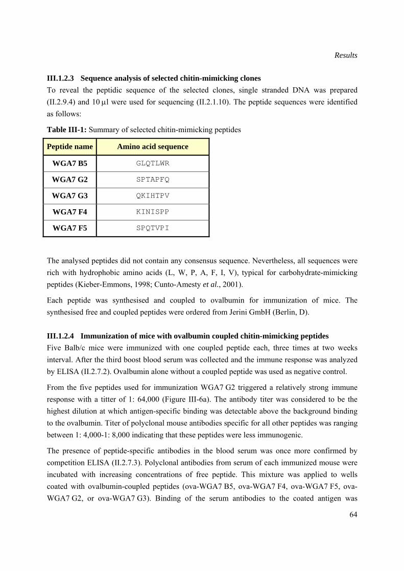
Results
64
III.1.2.3 Sequence analysis of selected chitin-mimicking clones To reveal the peptidic sequence of the selected clones, single stranded DNA was prepared (II.2.9.4) and 10 μl were used for sequencing (II.2.1.10). The peptide sequences were identified as follows:
Table III-1: Summary of selected chitin-mimicking peptides
Peptide name Amino acid sequence
WGA7 B5 GLQTLWR
WGA7 G2 SPTAPFQ
WGA7 G3 QKIHTPV
WGA7 F4 KINISPP
WGA7 F5 SPQTVPI
The analysed peptides did not contain any consensus sequence. Nevertheless, all sequences were rich with hydrophobic amino acids (L, W, P, A, F, I, V), typical for carbohydrate-mimicking peptides (Kieber-Emmons, 1998; Cunto-Amesty et al., 2001).
Each peptide was synthesised and coupled to ovalbumin for immunization of mice. The synthesised free and coupled peptides were ordered from Jerini GmbH (Berlin, D).
III.1.2.4 Immunization of mice with ovalbumin coupled chitin-mimicking peptides Five Balb/c mice were immunized with one coupled peptide each, three times at two weeks interval. After the third boost blood serum was collected and the immune response was analyzed by ELISA (II.2.7.2). Ovalbumin alone without a coupled peptide was used as negative control.
From the five peptides used for immunization WGA7 G2 triggered a relatively strong immune response with a titter of 1: 64,000 (Figure III-6a). The antibody titer was considered to be the highest dilution at which antigen-specific binding was detectable above the background binding to the ovalbumin. Titer of polyclonal mouse antibodies specific for all other peptides was ranging between 1: 4,000-1: 8,000 indicating that these peptides were less immunogenic.
The presence of peptide-specific antibodies in the blood serum was once more confirmed by competition ELISA (II.2.7.3). Polyclonal antibodies from serum of each immunized mouse were incubated with increasing concentrations of free peptide. This mixture was applied to wells coated with ovalbumin-coupled peptides (ova-WGA7 B5, ova-WGA7 F4, ova-WGA7 F5, ova-WGA7 G2, or ova-WGA7 G3). Binding of the serum antibodies to the coated antigen was
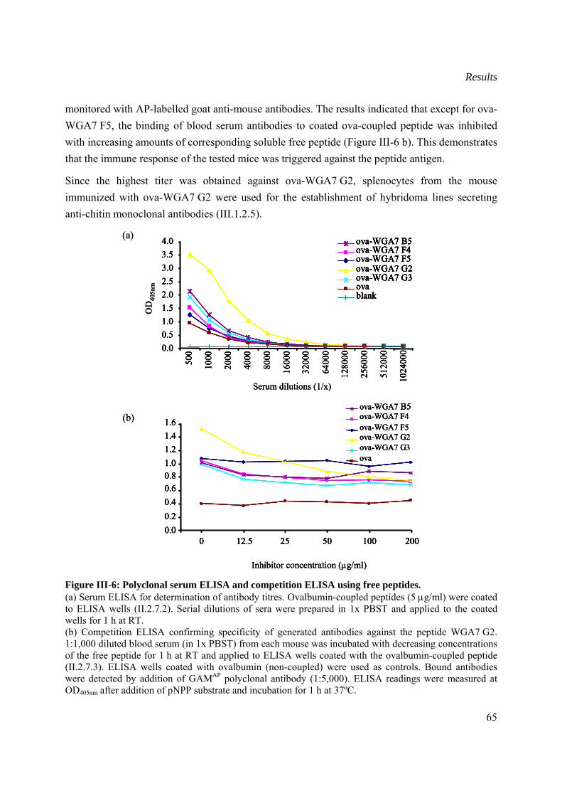
Results
65
monitored with AP-labelled goat anti-mouse antibodies. The results indicated that except for ova-WGA7 F5, the binding of blood serum antibodies to coated ova-coupled peptide was inhibited with increasing amounts of corresponding soluble free peptide (Figure III-6 b). This demonstrates that the immune response of the tested mice was triggered against the peptide antigen.
Since the highest titer was obtained against ova-WGA7 G2, splenocytes from the mouse immunized with ova-WGA7 G2 were used for the establishment of hybridoma lines secreting anti-chitin monoclonal antibodies (III.1.2.5).
Figure III-6: Polyclonal serum ELISA and competition ELISA using free peptides. (a) Serum ELISA for determination of antibody titres. Ovalbumin-coupled peptides (5 μg/ml) were coated to ELISA wells (II.2.7.2). Serial dilutions of sera were prepared in 1x PBST and applied to the coated wells for 1 h at RT. (b) Competition ELISA confirming specificity of generated antibodies against the peptide WGA7 G2. 1:1,000 diluted blood serum (in 1x PBST) from each mouse was incubated with decreasing concentrations of the free peptide for 1 h at RT and applied to ELISA wells coated with the ovalbumin-coupled peptide (II.2.7.3). ELISA wells coated with ovalbumin (non-coupled) were used as controls. Bound antibodies were detected by addition of GAMAP polyclonal antibody (1:5,000). ELISA readings were measured at OD405nm after addition of pNPP substrate and incubation for 1 h at 37ºC.
0.00.20.40.60.81.01.21.41.6
0 12.5 25 50 100 200
Inhibitor concentration (μg/ml)
ova-WGA7 F5ova-WGA7 F4
ova-WGA7 G2ova-WGA7 G3
ova-WGA7 B5
ova
(b)
0.00.20.40.60.81.01.21.41.6
0 12.5 25 50 100 200
Inhibitor concentration (μg/ml)
ova-WGA7 F5ova-WGA7 F4
ova-WGA7 G2ova-WGA7 G3
ova-WGA7 B5
ova
0.00.20.40.60.81.01.21.41.6
0 12.5 25 50 100 2000.00.20.40.60.81.01.21.41.6
0 12.5 25 50 100 200
Inhibitor concentration (μg/ml)
ova-WGA7 F5ova-WGA7 F4
ova-WGA7 G2ova-WGA7 G3
ova-WGA7 B5
ova
ova-WGA7 F5ova-WGA7 F4
ova-WGA7 G2ova-WGA7 G3
ova-WGA7 B5
ova
(b)
Serum dilutions (1/x)
OD
405n
m
0.00.51.01.52.02.53.03.54.0
500
1000
2000
4000
8000
1600
0
3200
0
6400
0
1280
00
2560
00
5120
00
1024
000
ova-WGA7 F5ova-WGA7 F4
ova-WGA7 G2ova-WGA7 G3
ova-WGA7 B5
ovablank
(a)
Serum dilutions (1/x)
OD
405n
m
0.00.51.01.52.02.53.03.54.0
500
1000
2000
4000
8000
1600
0
3200
0
6400
0
1280
00
2560
00
5120
00
1024
000
ova-WGA7 F5ova-WGA7 F4
ova-WGA7 G2ova-WGA7 G3
ova-WGA7 B5
ovablank
Serum dilutions (1/x)
OD
405n
m
0.00.51.01.52.02.53.03.54.0
500
1000
2000
4000
8000
1600
0
3200
0
6400
0
1280
00
2560
00
5120
00
1024
000
ova-WGA7 F5ova-WGA7 F4
ova-WGA7 G2ova-WGA7 G3
ova-WGA7 B5
ovablank
0.00.51.01.52.02.53.03.54.0
500
1000
2000
4000
8000
1600
0
3200
0
6400
0
1280
00
2560
00
5120
00
1024
000
ova-WGA7 F5ova-WGA7 F4
ova-WGA7 G2ova-WGA7 G3
ova-WGA7 B5
ovablank
ova-WGA7 F5ova-WGA7 F5ova-WGA7 F4ova-WGA7 F4
ova-WGA7 G2ova-WGA7 G2ova-WGA7 G3ova-WGA7 G3
ova-WGA7 B5ova-WGA7 B5
ovaovablankblank
(a)
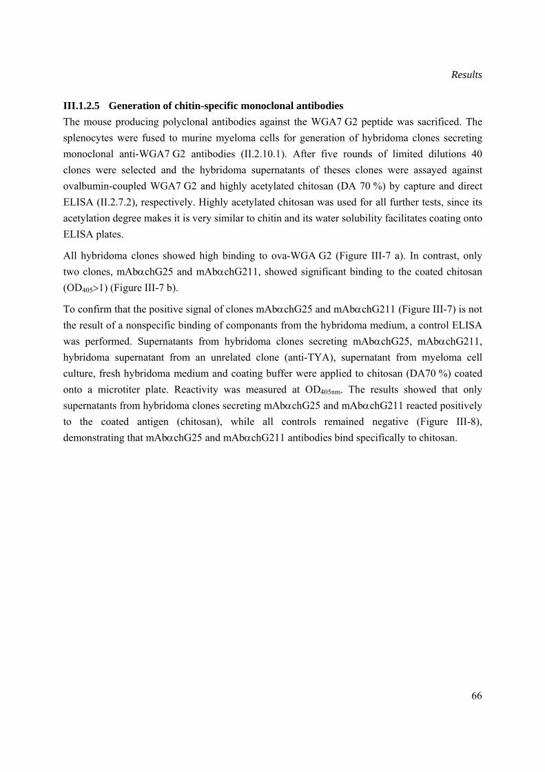
Results
66
III.1.2.5 Generation of chitin-specific monoclonal antibodies The mouse producing polyclonal antibodies against the WGA7 G2 peptide was sacrificed. The splenocytes were fused to murine myeloma cells for generation of hybridoma clones secreting monoclonal anti-WGA7 G2 antibodies (II.2.10.1). After five rounds of limited dilutions 40 clones were selected and the hybridoma supernatants of theses clones were assayed against ovalbumin-coupled WGA7 G2 and highly acetylated chitosan (DA 70 %) by capture and direct ELISA (II.2.7.2), respectively. Highly acetylated chitosan was used for all further tests, since its acetylation degree makes it is very similar to chitin and its water solubility facilitates coating onto ELISA plates.
All hybridoma clones showed high binding to ova-WGA G2 (Figure III-7 a). In contrast, only two clones, mAbαchG25 and mAbαchG211, showed significant binding to the coated chitosan (OD405>1) (Figure III-7 b).
To confirm that the positive signal of clones mAbαchG25 and mAbαchG211 (Figure III-7) is not the result of a nonspecific binding of componants from the hybridoma medium, a control ELISA was performed. Supernatants from hybridoma clones secreting mAbαchG25, mAbαchG211, hybridoma supernatant from an unrelated clone (anti-TYA), supernatant from myeloma cell culture, fresh hybridoma medium and coating buffer were applied to chitosan (DA70 %) coated onto a microtiter plate. Reactivity was measured at OD405nm. The results showed that only supernatants from hybridoma clones secreting mAbαchG25 and mAbαchG211 reacted positively to the coated antigen (chitosan), while all controls remained negative (Figure III-8), demonstrating that mAbαchG25 and mAbαchG211 antibodies bind specifically to chitosan.
Results
67
Figure III-7: Reactivity of generated monoclonal antibodies against ova-WG7 G2 (a) and highly acetylated chitosan (b). Highly acetylated chitosan (DA 70 %) (1 μg/ml) were coated on ELISA plate o/n at 4ºC. Hundred μl hybridoma culture supernatant were applied to coated wells and incubated 1 h at RT (II.2.7.2). Bound antibodies were detected by addition of HRP-labelled GAM polyclonal antibody (1:5,000). ELISA readings were performed at OD405nm after addition of ABTS substrate and incubation for 20 min at RT in the dark. A blank microtiter well coated with 1x PBS was used as negative control.
clones
OD
504n
m
0.0
0.5
1.0
1.5
2.0
2.5
3.0
3.5 ova-WGA7 G2ovalbumin
mA
bαch
G21
mA
bαch
G22
mA
bαch
G23
mA
bαch
G24
mA
bαch
G25
mA
bαch
G26
mA
bαch
G27
mA
bαch
G28
mA
bαch
G29
mA
bαch
G21
0m
Abα
chG
211
mA
bαch
G21
2m
Abα
chG
213
mA
bαch
G21
4m
Abα
chG
215
mA
bαch
G21
6m
Abα
chG
217
mA
bαch
G21
8m
Abα
chG
219
mA
bαch
G22
0m
Abα
chG
221
mA
bαch
G22
2m
Abα
chG
223
mA
bαch
G22
4m
Abα
chG
225
mA
bαch
G22
6m
Abα
chG
227
mA
bαch
G22
8m
Abα
chG
229
mA
bαch
G23
0m
Abα
chG
231
mA
bαch
G23
2m
Abα
chG
233
mA
bαch
G23
4m
Abα
chG
235
mA
bαch
G23
6m
Abα
chG
237
mA
bαch
G23
8m
Abα
chG
239
mA
bαch
G24
0
(a)
clones
OD
504n
m
0.0
0.5
1.0
1.5
2.0
2.5
3.0
3.5 ova-WGA7 G2ovalbumin
mA
bαch
G21
mA
bαch
G22
mA
bαch
G23
mA
bαch
G24
mA
bαch
G25
mA
bαch
G26
mA
bαch
G27
mA
bαch
G28
mA
bαch
G29
mA
bαch
G21
0m
Abα
chG
211
mA
bαch
G21
2m
Abα
chG
213
mA
bαch
G21
4m
Abα
chG
215
mA
bαch
G21
6m
Abα
chG
217
mA
bαch
G21
8m
Abα
chG
219
mA
bαch
G22
0m
Abα
chG
221
mA
bαch
G22
2m
Abα
chG
223
mA
bαch
G22
4m
Abα
chG
225
mA
bαch
G22
6m
Abα
chG
227
mA
bαch
G22
8m
Abα
chG
229
mA
bαch
G23
0m
Abα
chG
231
mA
bαch
G23
2m
Abα
chG
233
mA
bαch
G23
4m
Abα
chG
235
mA
bαch
G23
6m
Abα
chG
237
mA
bαch
G23
8m
Abα
chG
239
mA
bαch
G24
0
clones
OD
504n
m
0.0
0.5
1.0
1.5
2.0
2.5
3.0
3.5 ova-WGA7 G2ovalbumin
mA
bαch
G21
mA
bαch
G22
mA
bαch
G23
mA
bαch
G24
mA
bαch
G25
mA
bαch
G26
mA
bαch
G27
mA
bαch
G28
mA
bαch
G29
mA
bαch
G21
0m
Abα
chG
211
mA
bαch
G21
2m
Abα
chG
213
mA
bαch
G21
4m
Abα
chG
215
mA
bαch
G21
6m
Abα
chG
217
mA
bαch
G21
8m
Abα
chG
219
mA
bαch
G22
0m
Abα
chG
221
mA
bαch
G22
2m
Abα
chG
223
mA
bαch
G22
4m
Abα
chG
225
mA
bαch
G22
6m
Abα
chG
227
mA
bαch
G22
8m
Abα
chG
229
mA
bαch
G23
0m
Abα
chG
231
mA
bαch
G23
2m
Abα
chG
233
mA
bαch
G23
4m
Abα
chG
235
mA
bαch
G23
6m
Abα
chG
237
mA
bαch
G23
8m
Abα
chG
239
mA
bαch
G24
00.0
0.5
1.0
1.5
2.0
2.5
3.0
3.5 ova-WGA7 G2ovalbuminova-WGA7 G2ovalbuminova-WGA7 G2ovalbumin
mA
bαch
G21
mA
bαch
G22
mA
bαch
G23
mA
bαch
G24
mA
bαch
G25
mA
bαch
G26
mA
bαch
G27
mA
bαch
G28
mA
bαch
G29
mA
bαch
G21
0m
Abα
chG
211
mA
bαch
G21
2m
Abα
chG
213
mA
bαch
G21
4m
Abα
chG
215
mA
bαch
G21
6m
Abα
chG
217
mA
bαch
G21
8m
Abα
chG
219
mA
bαch
G22
0m
Abα
chG
221
mA
bαch
G22
2m
Abα
chG
223
mA
bαch
G22
4m
Abα
chG
225
mA
bαch
G22
6m
Abα
chG
227
mA
bαch
G22
8m
Abα
chG
229
mA
bαch
G23
0m
Abα
chG
231
mA
bαch
G23
2m
Abα
chG
233
mA
bαch
G23
4m
Abα
chG
235
mA
bαch
G23
6m
Abα
chG
237
mA
bαch
G23
8m
Abα
chG
239
mA
bαch
G24
0
(a)
clones
OD
405n
m
0.0
0.2
0.4
0.6
0.8
1.0
1.2
1.4
mA
bαch
G21
mA
bαch
G22
mA
bαch
G23
mA
bαch
G24
mA
bαch
G25
mA
bαch
G26
mA
bαch
G27
mA
bαch
G28
mA
bαch
G29
mA
bαch
G21
0m
Abα
chG
211
mA
bαch
G21
2m
Abα
chG
213
mA
bαch
G21
4m
Abα
chG
215
mA
bαch
G21
6m
Abα
chG
217
mA
bαch
G21
8m
Abα
chG
219
mA
bαch
G22
0m
Abα
chG
221
mA
bαch
G22
2m
Abα
chG
223
mA
bαch
G22
4m
Abα
chG
225
mA
bαch
G22
6m
Abα
chG
227
mA
bαch
G22
8m
Abα
chG
229
mA
bαch
G23
0m
Abα
chG
231
mA
bαch
G23
2m
Abα
chG
233
mA
bαch
G23
4m
Abα
chG
235
mA
bαch
G23
6m
Abα
chG
237
mA
bαch
G23
8m
Abα
chG
239
mA
bαch
G24
0bl
ank
(b)
clones
OD
405n
m
0.0
0.2
0.4
0.6
0.8
1.0
1.2
1.4
mA
bαch
G21
mA
bαch
G22
mA
bαch
G23
mA
bαch
G24
mA
bαch
G25
mA
bαch
G26
mA
bαch
G27
mA
bαch
G28
mA
bαch
G29
mA
bαch
G21
0m
Abα
chG
211
mA
bαch
G21
2m
Abα
chG
213
mA
bαch
G21
4m
Abα
chG
215
mA
bαch
G21
6m
Abα
chG
217
mA
bαch
G21
8m
Abα
chG
219
mA
bαch
G22
0m
Abα
chG
221
mA
bαch
G22
2m
Abα
chG
223
mA
bαch
G22
4m
Abα
chG
225
mA
bαch
G22
6m
Abα
chG
227
mA
bαch
G22
8m
Abα
chG
229
mA
bαch
G23
0m
Abα
chG
231
mA
bαch
G23
2m
Abα
chG
233
mA
bαch
G23
4m
Abα
chG
235
mA
bαch
G23
6m
Abα
chG
237
mA
bαch
G23
8m
Abα
chG
239
mA
bαch
G24
0bl
ank
(b)
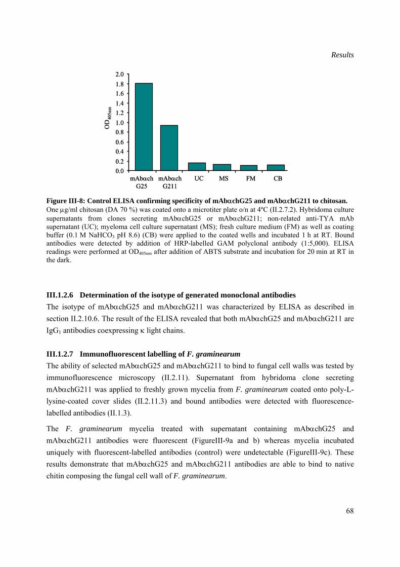
Results
68
Figure III-8: Control ELISA confirming specificity of mAbαchG25 and mAbαchG211 to chitosan. One μg/ml chitosan (DA 70 %) was coated onto a microtiter plate o/n at 4ºC (II.2.7.2). Hybridoma culture supernatants from clones secreting mAbαchG25 or mAbαchG211; non-related anti-TYA mAb supernatant (UC); myeloma cell culture supernatant (MS); fresh culture medium (FM) as well as coating buffer (0.1 M NaHCO3 pH 8.6) (CB) were applied to the coated wells and incubated 1 h at RT. Bound antibodies were detected by addition of HRP-labelled GAM polyclonal antibody (1:5,000). ELISA readings were performed at OD405nm after addition of ABTS substrate and incubation for 20 min at RT in the dark.
III.1.2.6 Determination of the isotype of generated monoclonal antibodies The isotype of mAbαchG25 and mAbαchG211 was characterized by ELISA as described in section II.2.10.6. The result of the ELISA revealed that both mAbαchG25 and mAbαchG211 are IgG1 antibodies coexpressing κ light chains.
III.1.2.7 Immunofluorescent labelling of F. graminearum The ability of selected mAbαchG25 and mAbαchG211 to bind to fungal cell walls was tested by immunofluorescence microscopy (II.2.11). Supernatant from hybridoma clone secreting mAbαchG211 was applied to freshly grown mycelia from F. graminearum coated onto poly-L-lysine-coated cover slides (II.2.11.3) and bound antibodies were detected with fluorescence-labelled antibodies (II.1.3).
The F. graminearum mycelia treated with supernatant containing mAbαchG25 and mAbαchG211 antibodies were fluorescent (FigureIII-9a and b) whereas mycelia incubated uniquely with fluorescent-labelled antibodies (control) were undetectable (FigureIII-9c). These results demonstrate that mAbαchG25 and mAbαchG211 antibodies are able to bind to native chitin composing the fungal cell wall of F. graminearum.
0.00.20.40.60.81.01.21.41.61.82.0
OD
405n
m
mAbαchG25
mAbαchG211
UC MS FM CB0.00.20.40.60.81.01.21.41.61.82.0
OD
405n
m
mAbαchG25
mAbαchG211
UC MS FM CB
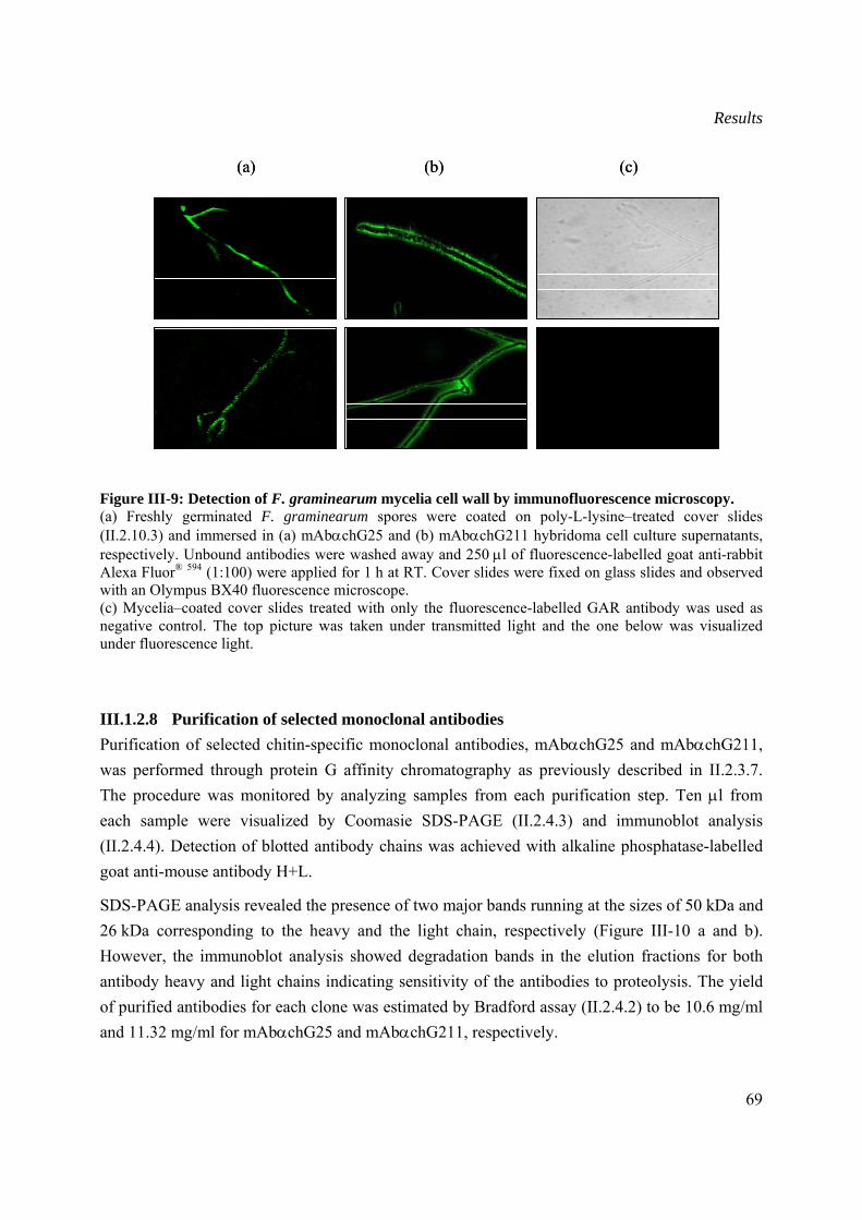
Results
69
Figure III-9: Detection of F. graminearum mycelia cell wall by immunofluorescence microscopy. (a) Freshly germinated F. graminearum spores were coated on poly-L-lysine–treated cover slides (II.2.10.3) and immersed in (a) mAbαchG25 and (b) mAbαchG211 hybridoma cell culture supernatants, respectively. Unbound antibodies were washed away and 250 μl of fluorescence-labelled goat anti-rabbit Alexa Fluor® 594 (1:100) were applied for 1 h at RT. Cover slides were fixed on glass slides and observed with an Olympus BX40 fluorescence microscope. (c) Mycelia–coated cover slides treated with only the fluorescence-labelled GAR antibody was used as negative control. The top picture was taken under transmitted light and the one below was visualized under fluorescence light.
III.1.2.8 Purification of selected monoclonal antibodies Purification of selected chitin-specific monoclonal antibodies, mAbαchG25 and mAbαchG211, was performed through protein G affinity chromatography as previously described in II.2.3.7. The procedure was monitored by analyzing samples from each purification step. Ten μl from each sample were visualized by Coomasie SDS-PAGE (II.2.4.3) and immunoblot analysis (II.2.4.4). Detection of blotted antibody chains was achieved with alkaline phosphatase-labelled goat anti-mouse antibody H+L.
SDS-PAGE analysis revealed the presence of two major bands running at the sizes of 50 kDa and 26 kDa corresponding to the heavy and the light chain, respectively (Figure III-10 a and b). However, the immunoblot analysis showed degradation bands in the elution fractions for both antibody heavy and light chains indicating sensitivity of the antibodies to proteolysis. The yield of purified antibodies for each clone was estimated by Bradford assay (II.2.4.2) to be 10.6 mg/ml and 11.32 mg/ml for mAbαchG25 and mAbαchG211, respectively.
(a) (b) (c)(a) (b) (c)
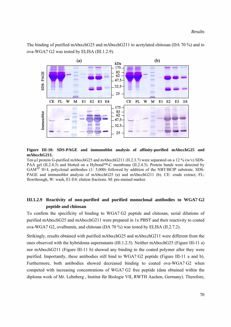
Results
70
The binding of purified mAbαchG25 and mAbαchG211 to acetylated chitosan (DA 70 %) and to ova-WGA7 G2 was tested by ELISA (III.1.2.9).
Figure III-10: SDS-PAGE and immunoblot analysis of affinity-purified mAbαchG25 and mAbαchG211. Ten μl protein G-purified mAbαchG25 and mAbαchG211 (II.2.3.7) were separated on a 12 % (w/v) SDS-PAA gel (II.2.4.3) and blotted on a Hybond™-C membrane (II.2.4.3). Protein bands were detected by GAMAP H+L polyclonal antibodies (1: 5,000) followed by addition of the NBT/BCIP substrate. SDS-PAGE and immunoblot analysis of mAbαchG25 (a) and mAbαchG211 (b). CE: crude extract, FL: flowthrough, W: wash, E1-E4: elution fractions. M: pre-stained marker.
III.1.2.9 Reactivity of non-purified and purified monoclonal antibodies to WGA7 G2 peptide and chitosan
To confirm the specificity of binding to WGA7 G2 peptide and chitosan, serial dilutions of purified mAbαchG25 and mAbαchG211 were prepared in 1x PBST and their reactivity to coated ova-WGA7 G2, ovalbumin, and chitosan (DA 70 %) was tested by ELISA (II.2.7.2).
Strikingly, results obtained with purified mAbαchG25 and mAbαchG211 were different from the ones observed with the hybridoma supernatants (III.1.2.5). Neither mAbαchG25 (Figure III-11 a) nor mAbαchG211 (Figure III-11 b) showed any binding to the coated polymer after they were purified. Importantly, these antibodies still bind to WGA7 G2 peptide (Figure III-11 a and b). Furthermore, both antibodies showed decreased binding to coated ova-WGA7 G2 when competed with increasing concentrations of WGA7 G2 free peptide (data obtained within the diploma work of Mr. Lehnberg , Institut für Biologie VII, RWTH Aachen, Germany). Therefore,
8362
47.532.5
25
175
kDa
8362
47.5
32.5
25
175
CE FL W E1 E2 E3 E4 CE FL W E1 E2 E3 E4
Imm
unob
lot
SDS-
PAG
E
M M
(a) (b)
8362
47.532.5
25
175
kDa
8362
47.5
32.5
25
175
CE FL W E1 E2 E3 E4 CE FL W E1 E2 E3 E4
Imm
unob
lot
SDS-
PAG
E
M M
(a) (b)
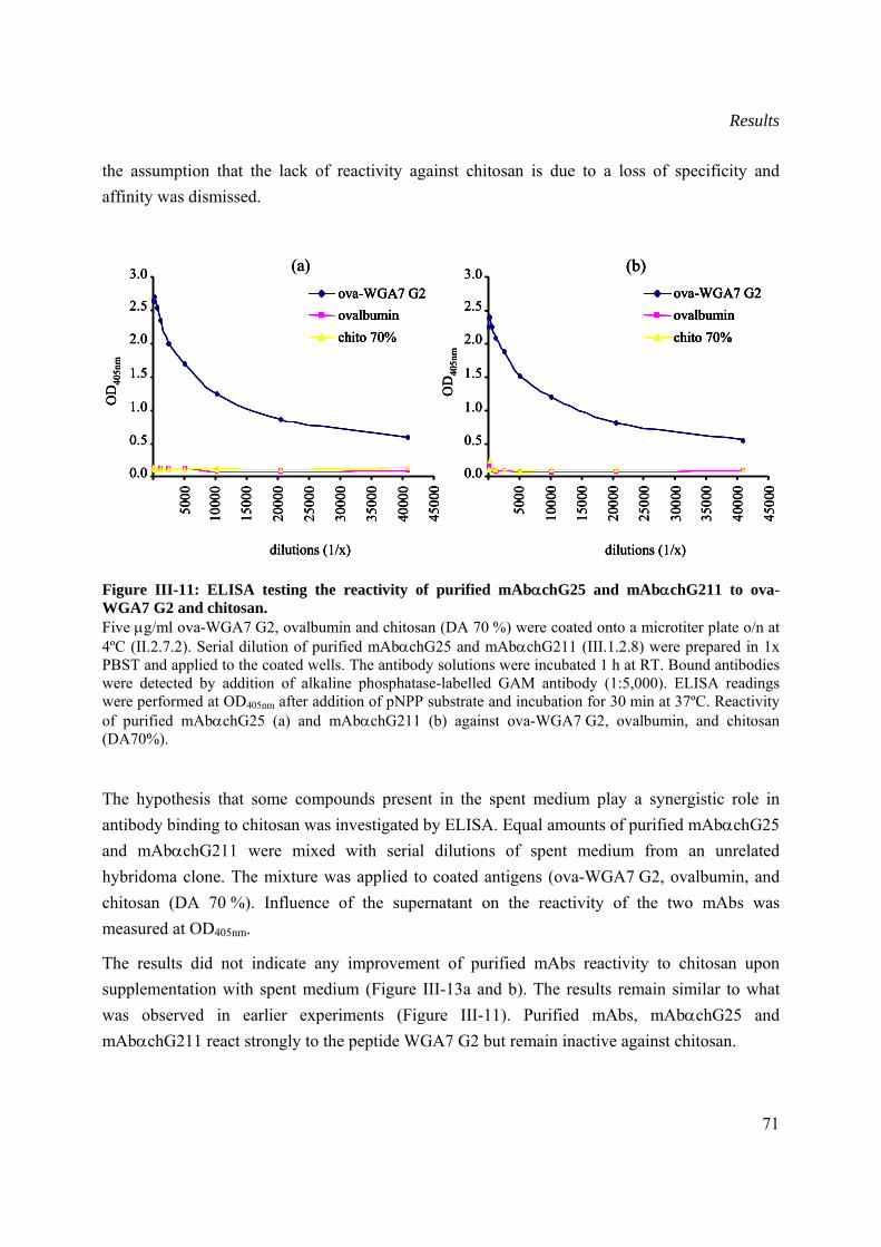
Results
71
the assumption that the lack of reactivity against chitosan is due to a loss of specificity and affinity was dismissed.
Figure III-11: ELISA testing the reactivity of purified mAbαchG25 and mAbαchG211 to ova-WGA7 G2 and chitosan. Five μg/ml ova-WGA7 G2, ovalbumin and chitosan (DA 70 %) were coated onto a microtiter plate o/n at 4ºC (II.2.7.2). Serial dilution of purified mAbαchG25 and mAbαchG211 (III.1.2.8) were prepared in 1x PBST and applied to the coated wells. The antibody solutions were incubated 1 h at RT. Bound antibodies were detected by addition of alkaline phosphatase-labelled GAM antibody (1:5,000). ELISA readings were performed at OD405nm after addition of pNPP substrate and incubation for 30 min at 37ºC. Reactivity of purified mAbαchG25 (a) and mAbαchG211 (b) against ova-WGA7 G2, ovalbumin, and chitosan (DA70%).
The hypothesis that some compounds present in the spent medium play a synergistic role in antibody binding to chitosan was investigated by ELISA. Equal amounts of purified mAbαchG25 and mAbαchG211 were mixed with serial dilutions of spent medium from an unrelated hybridoma clone. The mixture was applied to coated antigens (ova-WGA7 G2, ovalbumin, and chitosan (DA 70 %). Influence of the supernatant on the reactivity of the two mAbs was measured at OD405nm.
The results did not indicate any improvement of purified mAbs reactivity to chitosan upon supplementation with spent medium (Figure III-13a and b). The results remain similar to what was observed in earlier experiments (Figure III-11). Purified mAbs, mAbαchG25 and mAbαchG211 react strongly to the peptide WGA7 G2 but remain inactive against chitosan.
0.0
0.5
1.0
1.5
2.0
2.5
3.0
5000
1000
0
1500
0
2000
0
2500
0
3000
0
3500
0
4000
0
4500
0
dilutions (1/x)
OD
405n
m
ova-WGA7 G2ovalbuminchito 70%
(a)
0.0
0.5
1.0
1.5
2.0
2.5
3.0
5000
1000
0
1500
0
2000
0
2500
0
3000
0
3500
0
4000
0
4500
0
dilutions (1/x)
OD
405n
m
ova-WGA7 G2ovalbuminchito 70%
(b)
0.0
0.5
1.0
1.5
2.0
2.5
3.0
5000
1000
0
1500
0
2000
0
2500
0
3000
0
3500
0
4000
0
4500
0
dilutions (1/x)
OD
405n
m
ova-WGA7 G2ovalbuminchito 70%
(a)
0.0
0.5
1.0
1.5
2.0
2.5
3.0
5000
1000
0
1500
0
2000
0
2500
0
3000
0
3500
0
4000
0
4500
0
dilutions (1/x)
OD
405n
m
ova-WGA7 G2ovalbuminchito 70%
ova-WGA7 G2ovalbuminchito 70%
(a)
0.0
0.5
1.0
1.5
2.0
2.5
3.0
5000
1000
0
1500
0
2000
0
2500
0
3000
0
3500
0
4000
0
4500
0
dilutions (1/x)
OD
405n
m
ova-WGA7 G2ovalbuminchito 70%
(b)
0.0
0.5
1.0
1.5
2.0
2.5
3.0
5000
1000
0
1500
0
2000
0
2500
0
3000
0
3500
0
4000
0
4500
0
dilutions (1/x)
OD
405n
m
ova-WGA7 G2ovalbuminchito 70%
ova-WGA7 G2ovalbuminchito 70%
(b)
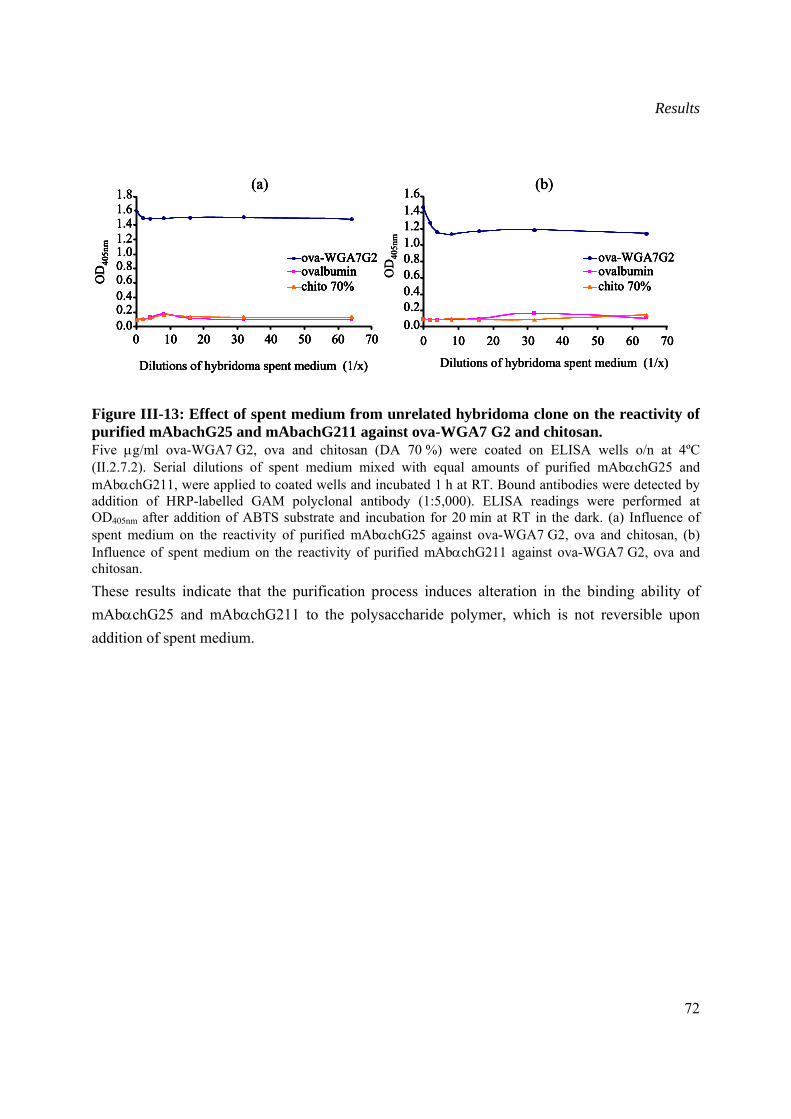
Results
72
Figure III-13: Effect of spent medium from unrelated hybridoma clone on the reactivity of purified mAbachG25 and mAbachG211 against ova-WGA7 G2 and chitosan. Five μg/ml ova-WGA7 G2, ova and chitosan (DA 70 %) were coated on ELISA wells o/n at 4ºC (II.2.7.2). Serial dilutions of spent medium mixed with equal amounts of purified mAbαchG25 and mAbαchG211, were applied to coated wells and incubated 1 h at RT. Bound antibodies were detected by addition of HRP-labelled GAM polyclonal antibody (1:5,000). ELISA readings were performed at OD405nm after addition of ABTS substrate and incubation for 20 min at RT in the dark. (a) Influence of spent medium on the reactivity of purified mAbαchG25 against ova-WGA7 G2, ova and chitosan, (b) Influence of spent medium on the reactivity of purified mAbαchG211 against ova-WGA7 G2, ova and chitosan. These results indicate that the purification process induces alteration in the binding ability of mAbαchG25 and mAbαchG211 to the polysaccharide polymer, which is not reversible upon addition of spent medium.
0.00.20.40.60.81.01.21.41.61.8
0 10 20 30 40 50 60 70
Dilutions of hybridoma spent medium (1/x)
OD
405n
m
ova-WGA7G2ovalbuminchito 70%
(a)
OD
405n
m
0.00.20.40.60.81.01.21.41.6
0 10 20 30 40 50 60 70
ova-WGA7G2ovalbuminchito 70%
(b)
Dilutions of hybridoma spent medium (1/x)
0.00.20.40.60.81.01.21.41.61.8
0 10 20 30 40 50 60 70
Dilutions of hybridoma spent medium (1/x)
OD
405n
m
ova-WGA7G2ovalbuminchito 70%
(a)
0.00.20.40.60.81.01.21.41.61.8
0 10 20 30 40 50 60 70
Dilutions of hybridoma spent medium (1/x)
OD
405n
m
ova-WGA7G2ovalbuminchito 70%
0.00.20.40.60.81.01.21.41.61.8
0 10 20 30 40 50 60 70
Dilutions of hybridoma spent medium (1/x)
OD
405n
m
ova-WGA7G2ovalbuminchito 70%
ova-WGA7G2ovalbuminchito 70%
(a)
OD
405n
m
0.00.20.40.60.81.01.21.41.6
0 10 20 30 40 50 60 70
ova-WGA7G2ovalbuminchito 70%
(b)
Dilutions of hybridoma spent medium (1/x)
OD
405n
m
0.00.20.40.60.81.01.21.41.6
0 10 20 30 40 50 60 70
ova-WGA7G2ovalbuminchito 70%
0.00.20.40.60.81.01.21.41.6
0 10 20 30 40 50 60 70
ova-WGA7G2ovalbuminchito 70%
(b)
Dilutions of hybridoma spent medium (1/x)
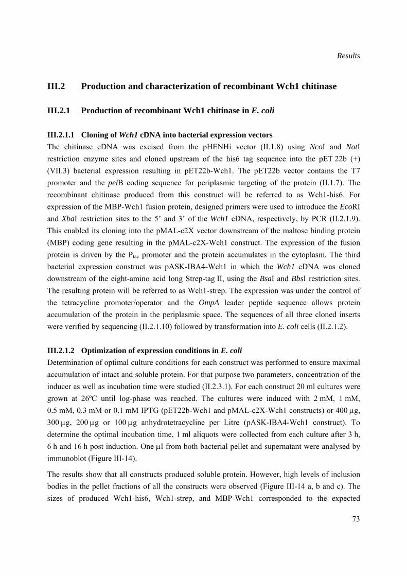
Results
73
III.2 Production and characterization of recombinant Wch1 chitinase
III.2.1 Production of recombinant Wch1 chitinase in E. coli
III.2.1.1 Cloning of Wch1 cDNA into bacterial expression vectors The chitinase cDNA was excised from the pHENHi vector (II.1.8) using NcoI and NotI restriction enzyme sites and cloned upstream of the his6 tag sequence into the pET 22b (+) (VII.3) bacterial expression resulting in pET22b-Wch1. The pET22b vector contains the T7 promoter and the pelB coding sequence for periplasmic targeting of the protein (II.1.7). The recombinant chitinase produced from this construct will be referred to as Wch1-his6. For expression of the MBP-Wch1 fusion protein, designed primers were used to introduce the EcoRI and XbaI restriction sites to the 5’ and 3’ of the Wch1 cDNA, respectively, by PCR (II.2.1.9). This enabled its cloning into the pMAL-c2X vector downstream of the maltose binding protein (MBP) coding gene resulting in the pMAL-c2X-Wch1 construct. The expression of the fusion protein is driven by the Ptac promoter and the protein accumulates in the cytoplasm. The third bacterial expression construct was pASK-IBA4-Wch1 in which the Wch1 cDNA was cloned downstream of the eight-amino acid long Strep-tag II, using the BsaI and BbsI restriction sites. The resulting protein will be referred to as Wch1-strep. The expression was under the control of the tetracycline promoter/operator and the OmpA leader peptide sequence allows protein accumulation of the protein in the periplasmic space. The sequences of all three cloned inserts were verified by sequencing (II.2.1.10) followed by transformation into E. coli cells (II.2.1.2).
III.2.1.2 Optimization of expression conditions in E. coli Determination of optimal culture conditions for each construct was performed to ensure maximal accumulation of intact and soluble protein. For that purpose two parameters, concentration of the inducer as well as incubation time were studied (II.2.3.1). For each construct 20 ml cultures were grown at 26ºC until log-phase was reached. The cultures were induced with 2 mM, 1 mM, 0.5 mM, 0.3 mM or 0.1 mM IPTG (pET22b-Wch1 and pMAL-c2X-Wch1 constructs) or 400 μg, 300 μg, 200 μg or 100 μg anhydrotetracycline per Litre (pASK-IBA4-Wch1 construct). To determine the optimal incubation time, 1 ml aliquots were collected from each culture after 3 h, 6 h and 16 h post induction. One μl from both bacterial pellet and supernatant were analysed by immunoblot (Figure III-14).
The results show that all constructs produced soluble protein. However, high levels of inclusion bodies in the pellet fractions of all the constructs were observed (Figure III-14 a, b and c). The sizes of produced Wch1-his6, Wch1-strep, and MBP-Wch1 corresponded to the expected
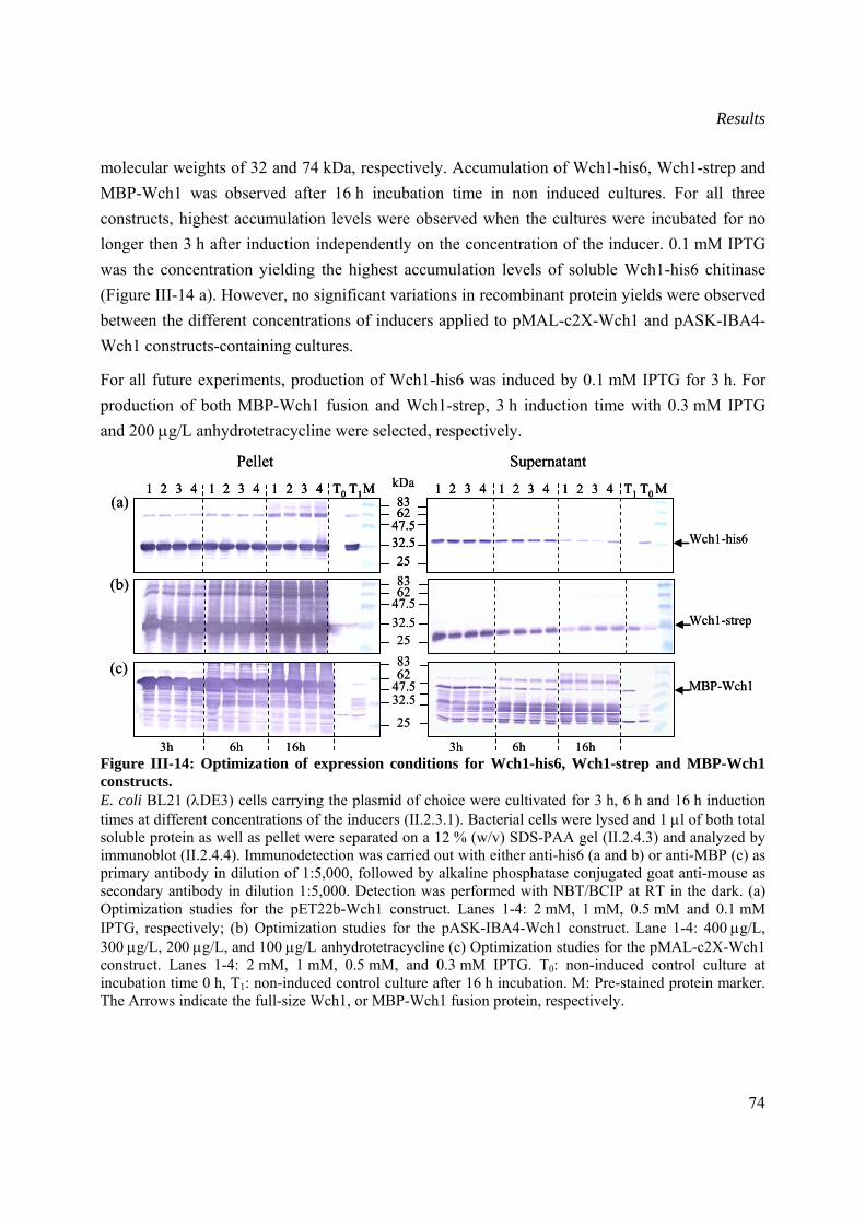
Results
74
molecular weights of 32 and 74 kDa, respectively. Accumulation of Wch1-his6, Wch1-strep and MBP-Wch1 was observed after 16 h incubation time in non induced cultures. For all three constructs, highest accumulation levels were observed when the cultures were incubated for no longer then 3 h after induction independently on the concentration of the inducer. 0.1 mM IPTG was the concentration yielding the highest accumulation levels of soluble Wch1-his6 chitinase (Figure III-14 a). However, no significant variations in recombinant protein yields were observed between the different concentrations of inducers applied to pMAL-c2X-Wch1 and pASK-IBA4-Wch1 constructs-containing cultures.
For all future experiments, production of Wch1-his6 was induced by 0.1 mM IPTG for 3 h. For production of both MBP-Wch1 fusion and Wch1-strep, 3 h induction time with 0.3 mM IPTG and 200 μg/L anhydrotetracycline were selected, respectively.
Figure III-14: Optimization of expression conditions for Wch1-his6, Wch1-strep and MBP-Wch1 constructs. E. coli BL21 (λDE3) cells carrying the plasmid of choice were cultivated for 3 h, 6 h and 16 h induction times at different concentrations of the inducers (II.2.3.1). Bacterial cells were lysed and 1 μl of both total soluble protein as well as pellet were separated on a 12 % (w/v) SDS-PAA gel (II.2.4.3) and analyzed by immunoblot (II.2.4.4). Immunodetection was carried out with either anti-his6 (a and b) or anti-MBP (c) as primary antibody in dilution of 1:5,000, followed by alkaline phosphatase conjugated goat anti-mouse as secondary antibody in dilution 1:5,000. Detection was performed with NBT/BCIP at RT in the dark. (a) Optimization studies for the pET22b-Wch1 construct. Lanes 1-4: 2 mM, 1 mM, 0.5 mM and 0.1 mM IPTG, respectively; (b) Optimization studies for the pASK-IBA4-Wch1 construct. Lane 1-4: 400 μg/L, 300 μg/L, 200 μg/L, and 100 μg/L anhydrotetracycline (c) Optimization studies for the pMAL-c2X-Wch1 construct. Lanes 1-4: 2 mM, 1 mM, 0.5 mM, and 0.3 mM IPTG. T0: non-induced control culture at incubation time 0 h, T1: non-induced control culture after 16 h incubation. M: Pre-stained protein marker. The Arrows indicate the full-size Wch1, or MBP-Wch1 fusion protein, respectively.
Pellet Supernatant
8362
47.532.525
kDa8362
47.532.525
T0 T1 T0T1 MM1 2 3 4 1 2 3 4 1 2 3 4 1 2 3 4 1 2 3 4 1 2 3 4(a)
(b)
Wch1-his6
Wch1-strep
3h 6h 16h
8362
47.532.5
25
3h 6h 16h
(c)MBP-Wch1
Pellet Supernatant
8362
47.532.525
kDa8362
47.532.525
T0 T1 T0T1 MM1 2 3 4 1 2 3 4 1 2 3 4 1 2 3 4 1 2 3 4 1 2 3 48362
47.532.525
T0 T1 T0T1 MM1 2 3 4 1 2 3 4 1 2 3 4 1 2 3 4 1 2 3 4 1 2 3 4(a)
(b)
Wch1-his6
Wch1-strep
3h 6h 16h
8362
47.532.5
25
3h 6h 16h
(c)MBP-Wch1
Results
75
III.2.1.3 Purification and quantification of bacterial produced chitinase Wch1 cDNA cloned into the three bacterial chitinase constructs pMAL-c2X-Wch1, pET22b-Wch1 and pASK-IBA4-Wch1 was expressed applying the optimum conditions determined in III.1.1 and subsequently purified through amylose (II.2.3.2), Ni-NTA (II.2.3.3) and Strep-Tactin matrices (II.2.3.4), respectively (II.2.3). The integrity as well as the purity of each protein was examined by both SDS-PAGE (II.2.4.3) and immunoblot analysis (II.2.4.4). The purification was repeated three times and the average yields of purified protein were determined by Bradford assay (II.2.4.1).
The size of the bacterial produced MBP-Wch1 fusion protein corresponded to the expected molecular weight of 74 kDa (Figure III-15 a). SDS-PAGE analysis revealed presence of several bands of smaller size then the full-size fusion protein. These bands were proven, by immunoblot, to be degradation products. The average yield of the MBP-Wch1 protein in the crude extract was estimated to be 30 mg per litre culture medium and 4 mg per litre after purification.
The affinity-purified Wch1-strep protein band was detected at the expected size of 32 kDa by immunoblot analysis (Figure III-15 b). However, SDS-PAGE analysis revealed a band of slightly higher size. The Wch1-strep chitinase was the highest in purity and stability as neither contamination nor degradation bands were detected by SDS-PAGE. This result was confirmed by immunoblot analysis. The main drawback of the protein produced from the pASK-IBA-Wch1 construct was the low yield estimated to be 1.5 mg and 0.7 mg per litre culture medium before and after purification, respectively.
Despite extensive washing steps during purification of his6-tagged chitinase (Wch1-his6), several bands of high molecular weight were observed in the elution fractions analysed by SDS-PAGE (Figure III-15 c). These bands were bigger than the expected size of 32 kDa for the Wch1-his6 and they were not present on the immunoblot indicating that they probably represent E. coli metallo-binding proteins which unspecifically bind to the Ni-NTA matrix. The estimation of purified Wch1-his6 protein levels using the Bradford assay was not possible due to the contaminating E. coli proteins that would lead to biased results. The lack of purity of the chitinase obtained with this construct constituted a drawback for all future characterization studies. Therefore, no further experiments were performed with Wch1-his6 chitinase.
Purified Wch1-strep and MBP-Wch1 proteins were separated in an IEF gel and their enzymatic activity was tested on an overly gel containing glycol chitin as substrate (III.4.1). Chitinase activity of both proteins was quantified by colorimetric assay (III.4.2).
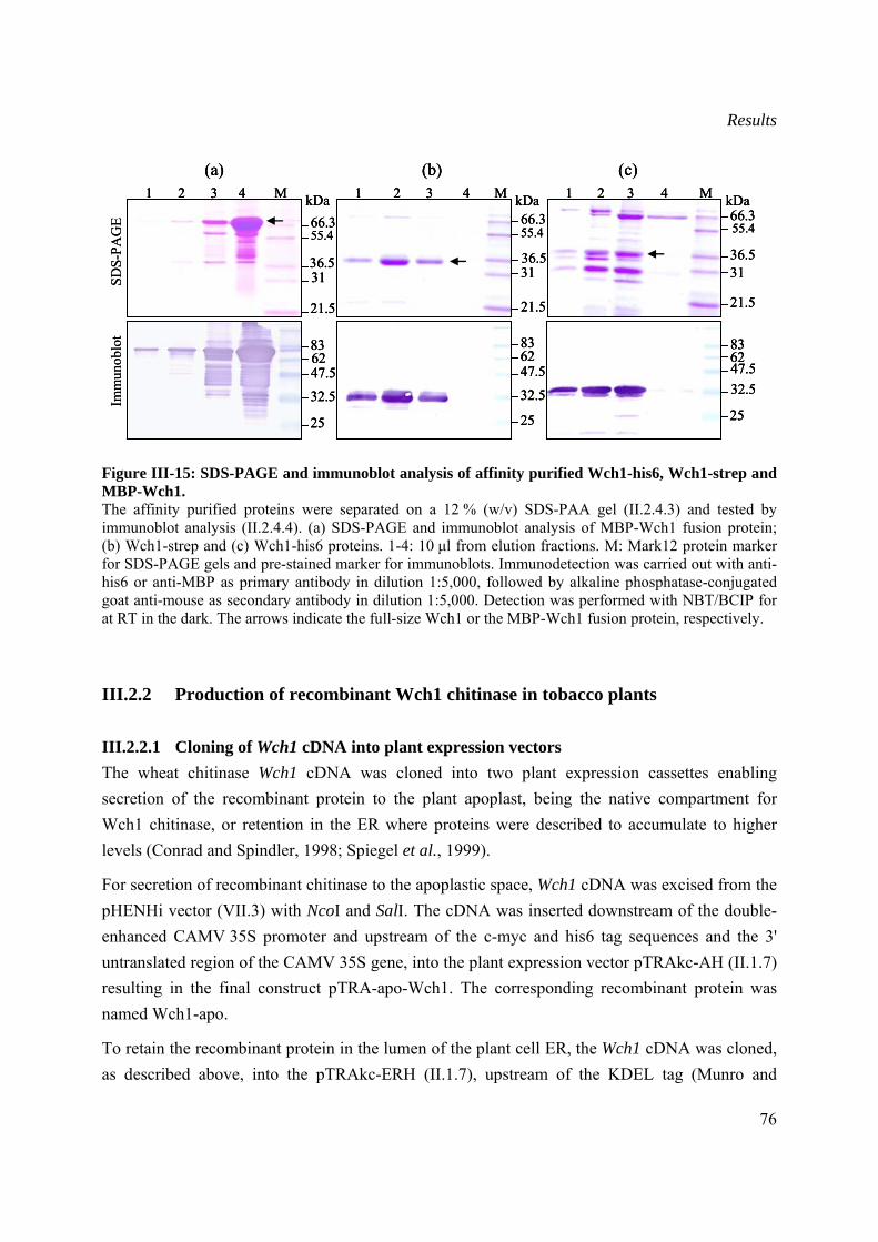
Results
76
Figure III-15: SDS-PAGE and immunoblot analysis of affinity purified Wch1-his6, Wch1-strep and MBP-Wch1. The affinity purified proteins were separated on a 12 % (w/v) SDS-PAA gel (II.2.4.3) and tested by immunoblot analysis (II.2.4.4). (a) SDS-PAGE and immunoblot analysis of MBP-Wch1 fusion protein; (b) Wch1-strep and (c) Wch1-his6 proteins. 1-4: 10 μl from elution fractions. M: Mark12 protein marker for SDS-PAGE gels and pre-stained marker for immunoblots. Immunodetection was carried out with anti-his6 or anti-MBP as primary antibody in dilution 1:5,000, followed by alkaline phosphatase-conjugated goat anti-mouse as secondary antibody in dilution 1:5,000. Detection was performed with NBT/BCIP for at RT in the dark. The arrows indicate the full-size Wch1 or the MBP-Wch1 fusion protein, respectively.
III.2.2 Production of recombinant Wch1 chitinase in tobacco plants
III.2.2.1 Cloning of Wch1 cDNA into plant expression vectors The wheat chitinase Wch1 cDNA was cloned into two plant expression cassettes enabling secretion of the recombinant protein to the plant apoplast, being the native compartment for Wch1 chitinase, or retention in the ER where proteins were described to accumulate to higher levels (Conrad and Spindler, 1998; Spiegel et al., 1999).
For secretion of recombinant chitinase to the apoplastic space, Wch1 cDNA was excised from the pHENHi vector (VII.3) with NcoI and SalI. The cDNA was inserted downstream of the double-enhanced CAMV 35S promoter and upstream of the c-myc and his6 tag sequences and the 3' untranslated region of the CAMV 35S gene, into the plant expression vector pTRAkc-AH (II.1.7) resulting in the final construct pTRA-apo-Wch1. The corresponding recombinant protein was named Wch1-apo.
To retain the recombinant protein in the lumen of the plant cell ER, the Wch1 cDNA was cloned, as described above, into the pTRAkc-ERH (II.1.7), upstream of the KDEL tag (Munro and
SDS-
PAG
EIm
mun
oblo
t
47.5
55.4
32.5
36.5
66.3kDa
21.5
8362
25
31
M1 2 3 4(c)
6247.532.5
25
83
3136.5
55.466.3
(a)
kDaM1 2 3 4
21.5
47.5
36.5
(b)
62
32.5
25
83
M1 2 3 4
31
55.466.3
kDa
21.5
SDS-
PAG
EIm
mun
oblo
t
47.5
55.4
32.5
36.5
66.3kDa
21.5
8362
25
31
M1 2 3 4(c)
47.5
55.4
32.5
36.5
66.3kDa
21.5
8362
25
31
M1 2 3 4(c)
6247.532.5
25
83
3136.5
55.466.3
(a)
kDaM1 2 3 4
21.5
6247.532.5
25
83
3136.5
55.466.3
(a)
kDaM1 2 3 4
21.5
47.5
36.5
(b)
62
32.5
25
83
M1 2 3 4
31
55.466.3
kDa
21.5
47.5
36.5
(b)
62
32.5
25
83
M1 2 3 4
31
55.466.3
kDa
21.5
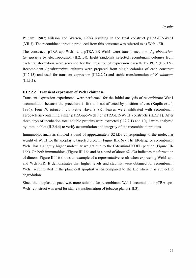
Results
77
Pelham, 1987; Nilsson and Warren, 1994) resulting in the final construct pTRA-ER-Wch1 (VII.3). The recombinant protein produced from this construct was referred to as Wch1-ER.
The constructs pTRA-apo-Wch1 and pTRA-ER-Wch1 were transformed into Agrobacterium tumefaciens by electroporation (II.2.1.4). Eight randomly selected recombinant colonies from each transformation were screened for the presence of expression cassette by PCR (II.2.1.9). Recombinant Agrobacterium cultures were prepared from single colonies of each construct (II.2.15) and used for transient expression (III.2.2.2) and stable transformation of N. tabacum (III.3.1).
III.2.2.2 Transient expression of Wch1 chitinase Transient expression experiments were performed for the initial analysis of recombinant Wch1 accumulation because the procedure is fast and not affected by position effects (Kapila et al., 1996). Four N. tabacum cv. Petite Havana SR1 leaves were infiltrated with recombinant agrobacteria containing either pTRA-apo-Wch1 or pTRA-ER-Wch1 constructs (II.2.2.1). After three days of incubation total soluble proteins were extracted (II.2.2.1) and 10 μl were analyzed by immunoblot (II.2.4.4) to verify accumulation and integrity of the recombinant proteins.
Immunoblot analysis showed a band of approximately 32 kDa corresponding to the molecular weight of Wch1 for the apoplastic targeted protein (Figure III-16a). The ER-targeted recombinant Wch1 has a slightly higher molecular weight due to the C-terminal KDEL peptide (Figure III-16b). On both immunoblots (Figure III-16a and b) a band of about 62 kDa indicates the formation of dimers. Figure III-16 shows an example of a representative result when expressing Wch1-apo and Wch1-ER. It demonstrates that higher levels and stability were obtained for recombinant Wch1 accumulated in the plant cell apoplast when compared to the ER where it is subject to degradation.
Since the apoplastic space was more suitable for recombinant Wch1 accumulation, pTRA-apo-Wch1 construct was used for stable transformation of tobacco plants (III.3).
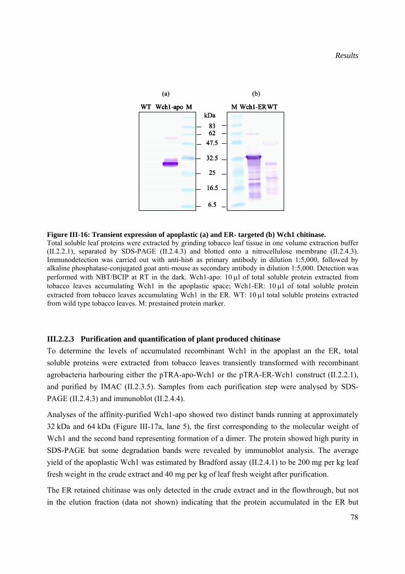
Results
78
Figure III-16: Transient expression of apoplastic (a) and ER- targeted (b) Wch1 chitinase. Total soluble leaf proteins were extracted by grinding tobacco leaf tissue in one volume extraction buffer (II.2.2.1), separated by SDS-PAGE (II.2.4.3) and blotted onto a nitrocellulose membrane (II.2.4.3). Immunodetection was carried out with anti-his6 as primary antibody in dilution 1:5,000, followed by alkaline phosphatase-conjugated goat anti-mouse as secondary antibody in dilution 1:5,000. Detection was performed with NBT/BCIP at RT in the dark. Wch1-apo: 10 μl of total soluble protein extracted from tobacco leaves accumulating Wch1 in the apoplastic space; Wch1-ER: 10 μl of total soluble protein extracted from tobacco leaves accumulating Wch1 in the ER. WT: 10 μl total soluble proteins extracted from wild type tobacco leaves. M: prestained protein marker.
III.2.2.3 Purification and quantification of plant produced chitinase To determine the levels of accumulated recombinant Wch1 in the apoplast an the ER, total soluble proteins were extracted from tobacco leaves transiently transformed with recombinant agrobacteria harbouring either the pTRA-apo-Wch1 or the pTRA-ER-Wch1 construct (II.2.2.1), and purified by IMAC (II.2.3.5). Samples from each purification step were analysed by SDS-PAGE (II.2.4.3) and immunoblot (II.2.4.4).
Analyses of the affinity-purified Wch1-apo showed two distinct bands running at approximately 32 kDa and 64 kDa (Figure III-17a, lane 5), the first corresponding to the molecular weight of Wch1 and the second band representing formation of a dimer. The protein showed high purity in SDS-PAGE but some degradation bands were revealed by immunoblot analysis. The average yield of the apoplastic Wch1 was estimated by Bradford assay (II.2.4.1) to be 200 mg per kg leaf fresh weight in the crude extract and 40 mg per kg of leaf fresh weight after purification.
The ER retained chitinase was only detected in the crude extract and in the flowthrough, but not in the elution fraction (data not shown) indicating that the protein accumulated in the ER but
8362
47.5
32.5
25
kDaMWT Wch1-apo M WTWch1-ER
16.5
6.5
(a) (b)
8362
47.5
32.5
25
kDaMWT Wch1-apo M WTWch1-ER
16.5
6.5
8362
47.5
32.5
25
kDaMWT Wch1-apo MWT Wch1-apo M WTWch1-ERM WTWch1-ER
16.5
6.5
(a) (b)
Results
79
could not bind to the Ni-NTA matrix, probably due to the inaccessibility of the his6 tag to the matrix. However, the protein was successfully purified (Figure III-17 b) after denaturation with 8 M urea (II.2.3.5). SDS-PAGE showed contamination with a protein, corresponding to the size (56 kDa) of the plant derived ribulose-1,5-bisphosphate carboxylase/oxygenase (rubisco).
Due to the impossibility to purify Wch1-ER chitinase in its native state no further experiments were carried out with the Wch1-ER protein.
The Wch1-apo chitinase was tested on glycol chitin gel after IEF (III.4.1) and the enzymatic activity was quantified by colorimetric assay (III.4.2).
Figure III-17: SDS-PAGE and immunoblot analysis of affinity purified Wch1-apo and Wch1-ER via IMAC. Ten μl IMAC-purified apoplastic and ER-targeted Wch1 chitinase (II.2.3.5) were separated on a 12 % (w/v) SDS-PAA gel (II.2.4.3) and analysed by immunoblot (II.2.4.4). (a) SDS-PAGE and immunoblot analysis of Wch1-apo. 1: crude extract, 2: flowthrough, 3: wash, 4-8: elution fractions; (b) SDS-PAGE and immunoblot analysis of urea-denatured Wch1-ER. 1: crude extract, 2: flowthrough, 3-4: wash fractions, 5-13: elution fractions. M: Mark12 protein marker for SDS-PAGE gels and pre-stained marker for immunoblots. Immunodetection was carried out with anti-his6 monoclonal antibody as primary antibody and goat anti-mouse polyclonal antibodies conjugated to alkaline phosphatase as secondary antibody (both diluted to 1:5,000) followed by addition of the NBT/BCIP substrate.
III.3 Generation and characterization of stable transformed tobacco plants producing Wch1 chitinase
III.3.1 Generation and screening of stable transformed tobacco plants
Wch1-apo construct was stably transformed into tobacco plants by Agrobacterium-mediated transformation (II.2.2.2) to ensure constant supply of recombinant Wch1 from plants. Twenty-
SDS-
PAG
EIm
mun
oblo
t
1 2 3 M
3136.5
55.466.3
kDa
(b)
21.5
6247.5
32.52516.5
4 5 6 7 8 9 101112 131 2 3 4 5 6 7M
3136.5
55.466.3
kDa
(a)
21.5
6247.532.5
25
16.5
SDS-
PAG
EIm
mun
oblo
t
1 2 3 M
3136.5
55.466.3
kDa
(b)
21.5
6247.5
32.52516.5
4 5 6 7 8 9 101112 131 2 3 M
3136.5
55.466.3
kDa
(b)
21.5
6247.5
32.52516.5
4 5 6 7 8 9 101112 131 2 3 4 5 6 7M
3136.5
55.466.3
kDa
(a)
21.5
6247.532.5
25
16.5
1 2 3 4 5 6 7M
3136.5
55.466.3
kDa
(a)
21.5
6247.532.5
25
16.5
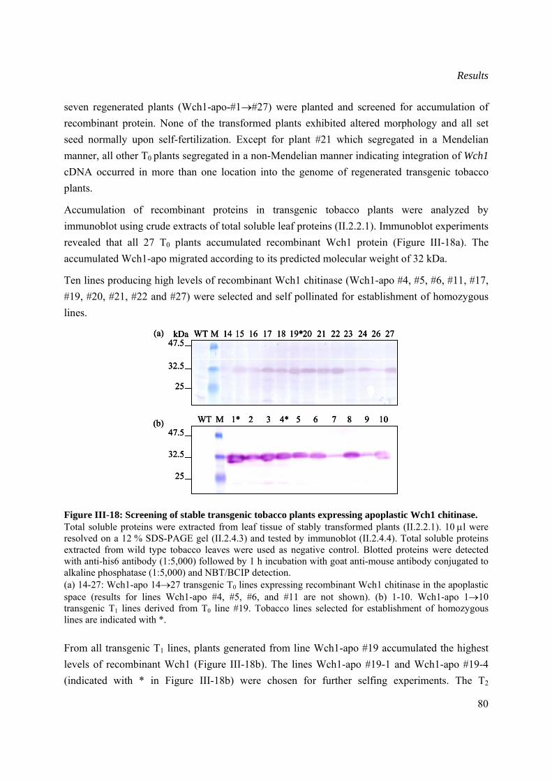
Results
80
seven regenerated plants (Wch1-apo-#1→#27) were planted and screened for accumulation of recombinant protein. None of the transformed plants exhibited altered morphology and all set seed normally upon self-fertilization. Except for plant #21 which segregated in a Mendelian manner, all other T0 plants segregated in a non-Mendelian manner indicating integration of Wch1 cDNA occurred in more than one location into the genome of regenerated transgenic tobacco plants.
Accumulation of recombinant proteins in transgenic tobacco plants were analyzed by immunoblot using crude extracts of total soluble leaf proteins (II.2.2.1). Immunoblot experiments revealed that all 27 T0 plants accumulated recombinant Wch1 protein (Figure III-18a). The accumulated Wch1-apo migrated according to its predicted molecular weight of 32 kDa.
Ten lines producing high levels of recombinant Wch1 chitinase (Wch1-apo #4, #5, #6, #11, #17, #19, #20, #21, #22 and #27) were selected and self pollinated for establishment of homozygous lines.
Figure III-18: Screening of stable transgenic tobacco plants expressing apoplastic Wch1 chitinase. Total soluble proteins were extracted from leaf tissue of stably transformed plants (II.2.2.1). 10 μl were resolved on a 12 % SDS-PAGE gel (II.2.4.3) and tested by immunoblot (II.2.4.4). Total soluble proteins extracted from wild type tobacco leaves were used as negative control. Blotted proteins were detected with anti-his6 antibody (1:5,000) followed by 1 h incubation with goat anti-mouse antibody conjugated to alkaline phosphatase (1:5,000) and NBT/BCIP detection. (a) 14-27: Wch1-apo 14→27 transgenic T0 lines expressing recombinant Wch1 chitinase in the apoplastic space (results for lines Wch1-apo #4, #5, #6, and #11 are not shown). (b) 1-10. Wch1-apo 1→10 transgenic T1 lines derived from T0 line #19. Tobacco lines selected for establishment of homozygous lines are indicated with *. From all transgenic T1 lines, plants generated from line Wch1-apo #19 accumulated the highest levels of recombinant Wch1 (Figure III-18b). The lines Wch1-apo #19-1 and Wch1-apo #19-4 (indicated with * in Figure III-18b) were chosen for further selfing experiments. The T2
32.5
25
47.5kDa WT 14 15 16 17 18 19*20 21 22 23 24 26 27M(a)
32.5
25
47.5WT 1* 2 3 4* 5 6 7 8 9 10M(b)
32.5
25
47.5kDa WT 14 15 16 17 18 19*20 21 22 23 24 26 27M(a)
32.5
25
47.5kDa WT 14 15 16 17 18 19*20 21 22 23 24 26 27M(a)
32.5
25
47.5WT 1* 2 3 4* 5 6 7 8 9 10M(b)
32.5
25
47.5WT 1* 2 3 4* 5 6 7 8 9 10M(b)
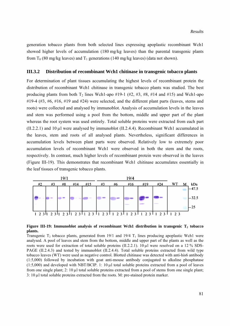
Results
81
generation tobacco plants from both selected lines expressing apoplastic recombinant Wch1 showed higher levels of accumulation (180 mg/kg leaves) than the parental transgenic plants from T0 (80 mg/kg leaves) and T1 generations (140 mg/kg leaves) (data not shown).
III.3.2 Distribution of recombinant Wch1 chitinase in transgenic tobacco plants
For determination of plant tissues accumulating the highest levels of recombinant protein the distribution of recombinant Wch1 chitinase in transgenic tobacco plants was studied. The best producing plants from both T2 lines Wch1-apo #19-1 (#2, #3, #8, #14 and #15) and Wch1-apo #19-4 (#3, #6, #16, #19 and #24) were selected, and the different plant parts (leaves, stems and roots) were collected and analysed by immunoblot. Analysis of accumulation levels in the leaves and stem was performed using a pool from the bottom, middle and upper part of the plant whereas the root system was used entirely. Total soluble proteins were extracted from each part (II.2.2.1) and 10 μl were analysed by immunoblot (II.2.4.4). Recombinant Wch1 accumulated in the leaves, stem and roots of all analysed plants. Nevertheless, significant differences in accumulation levels between plant parts were observed. Relatively low to extremely poor accumulation levels of recombinant Wch1 were observed in both the stem and the roots, respectively. In contrast, much higher levels of recombinant protein were observed in the leaves (Figure III-19). This demonstrates that recombinant Wch1 chitinase accumulates essentially in the leaf tissues of transgenic tobacco plants.
Figure III-19: Immunoblot analysis of recombinant Wch1 distribution in transgenic T2 tobacco plants. Transgenic T2 tobacco plants, generated from 19/1 and 19/4 T1 lines producing apoplastic Wch1 were analysed. A pool of leaves and stem from the bottom, middle and upper part of the plants as well as the roots were used for extraction of total soluble proteins (II.2.2.1). 10 μl were resolved on a 12 % SDS-PAGE (II.2.4.3) and tested by immunoblot (II.2.4.4). Total soluble proteins extracted from wild type tobacco leaves (WT) were used as negative control. Blotted chitinase was detected with anti-his6 antibody (1:5,000) followed by incubation with goat anti-mouse antibody conjugated to alkaline phosphatase (1:5,000) and developed with NBT/BCIP. 1: 10 μl total soluble proteins extracted from a pool of leaves from one single plant; 2: 10 μl total soluble proteins extracted from a pool of stems from one single plant; 3: 10 μl total soluble proteins extracted from the roots. M: pre-stained protein marker.
32.5
47.5
25
kDa#2 #3 #8 #14 #15 #3 #6 #16 #19 #24 WT M19/1 19/4
1 2 3 1 2 3 1 2 3 1 2 3 1 2 3 1 2 3 1 2 3 1 2 3 1 2 3 1 2 3 1 2 3
32.5
47.5
25
kDa#2 #3 #8 #14 #15 #3 #6 #16 #19 #24 WT M19/1 19/4
1 2 3 1 2 3 1 2 3 1 2 3 1 2 3 1 2 3 1 2 3 1 2 3 1 2 3 1 2 3 1 2 3
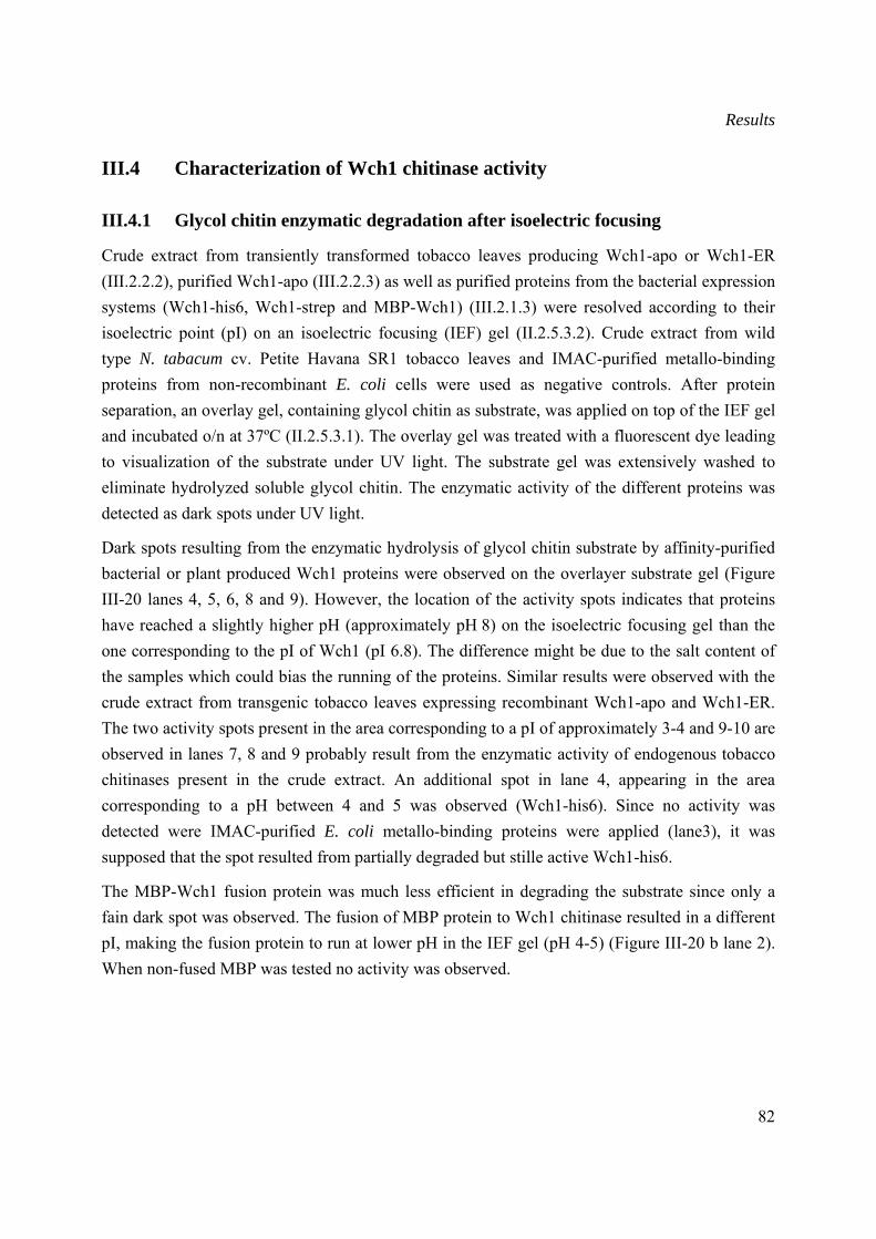
Results
82
III.4 Characterization of Wch1 chitinase activity
III.4.1 Glycol chitin enzymatic degradation after isoelectric focusing
Crude extract from transiently transformed tobacco leaves producing Wch1-apo or Wch1-ER (III.2.2.2), purified Wch1-apo (III.2.2.3) as well as purified proteins from the bacterial expression systems (Wch1-his6, Wch1-strep and MBP-Wch1) (III.2.1.3) were resolved according to their isoelectric point (pI) on an isoelectric focusing (IEF) gel (II.2.5.3.2). Crude extract from wild type N. tabacum cv. Petite Havana SR1 tobacco leaves and IMAC-purified metallo-binding proteins from non-recombinant E. coli cells were used as negative controls. After protein separation, an overlay gel, containing glycol chitin as substrate, was applied on top of the IEF gel and incubated o/n at 37ºC (II.2.5.3.1). The overlay gel was treated with a fluorescent dye leading to visualization of the substrate under UV light. The substrate gel was extensively washed to eliminate hydrolyzed soluble glycol chitin. The enzymatic activity of the different proteins was detected as dark spots under UV light.
Dark spots resulting from the enzymatic hydrolysis of glycol chitin substrate by affinity-purified bacterial or plant produced Wch1 proteins were observed on the overlayer substrate gel (Figure III-20 lanes 4, 5, 6, 8 and 9). However, the location of the activity spots indicates that proteins have reached a slightly higher pH (approximately pH 8) on the isoelectric focusing gel than the one corresponding to the pI of Wch1 (pI 6.8). The difference might be due to the salt content of the samples which could bias the running of the proteins. Similar results were observed with the crude extract from transgenic tobacco leaves expressing recombinant Wch1-apo and Wch1-ER. The two activity spots present in the area corresponding to a pI of approximately 3-4 and 9-10 are observed in lanes 7, 8 and 9 probably result from the enzymatic activity of endogenous tobacco chitinases present in the crude extract. An additional spot in lane 4, appearing in the area corresponding to a pH between 4 and 5 was observed (Wch1-his6). Since no activity was detected were IMAC-purified E. coli metallo-binding proteins were applied (lane3), it was supposed that the spot resulted from partially degraded but stille active Wch1-his6.
The MBP-Wch1 fusion protein was much less efficient in degrading the substrate since only a fain dark spot was observed. The fusion of MBP protein to Wch1 chitinase resulted in a different pI, making the fusion protein to run at lower pH in the IEF gel (pH 4-5) (Figure III-20 b lane 2). When non-fused MBP was tested no activity was observed.
Results
83
Figure III-20: Overlayer substrate gel performed after isoelectric focusing of Wch-apo or Wch1-ER. Crude extract from tobacco leaves producing Wch1-apo or Wch1-ER (II.2.2.4) as well as 5 μg affinity purified Wch1-his6, Wch1-strep, MBP-Wch1 fusion protein (II.2.3), MBP and E. coli metallo-binding proteins were separated according to their pI on an IEF gel and their enzymatic activity was tested on an overlayer gel containing glycol chitin substrate (II.2.5.3.2). Crude extract from wild type tobacco leaves, IMAC-purified metallo-proteins from non-recombinant E. coli cells and MBP were used as negative controls. 1: MBP; 2: MBP-Wch1; 3: IMAC purified E. coli metallo-binding proteins; 4: Wch1-his6; 5: Wch1-strep; 6: Wch1-apo; 7: total soluble extract from wild type tobacco leaves; 8: total soluble extract from tobacco leaves producing Wch1-apo; 9: total soluble extract from tobacco leaves producing Wch1-ER. The arrows indicate the dark spots resulting from enzymatic degradation of the glycol chitin substrate.
III.4.2 Colorimetric assay for quantification of chitinase activity
Quantitative evaluation of the enzymatic activity of the purified chitinase from bacteria (Wch1-strep and MBP-Wch1) and plants (Wch1-apo) was achieved with a colorimetric assay using CM-chitin-RBV, a water soluble chitin substrate linked to the dye ramazol brilliant violet, as substrate (II.2.5.1). Two hundred μg of the substrate were subjected to enzymatic hydrolysis by incubation with 2 μg purified Wch1-strep, Wch1-apo or 50 μg purified MBP-Wch1, respectively. At different time points (5, 10, 20, 30, 40 and 60 min) samples were deducted and the enzymatic reactions were stopped. Undigested substrate was precipitated by treatment with 1 M HCl. The supernatant containing soluble CM-chitin-RBV was collected and measured at OD585nm. One unit active chitinase was assigned as the change in OD585 reading of 1 per hour.
The Wch1-strep showed the highest activity with 1.25 units per μg protein followed by the plant purified Wch1-apo producing 0.75 units per μg protein. The fusion of Wch1 chitinase to MBP proved to be detrimental to the enzymatic activity of Wch1 chitinase as the activity decreased to 0.01 units per μg protein.
1 2 3 4 5 6 7 8 9 pH 10
pH 3
1 2 3 4 5 6 7 8 9 pH 10
pH 3
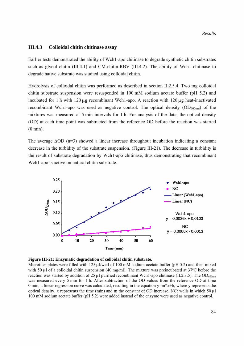
Results
84
III.4.3 Colloidal chitin chitinase assay
Earlier tests demonstrated the ability of Wch1-apo chitinase to degrade synthetic chitin substrates such as glycol chitin (III.4.1) and CM-chitin-RBV (III.4.2). The ability of Wch1 chitinase to degrade native substrate was studied using colloidal chitin.
Hydrolysis of colloidal chitin was performed as described in section II.2.5.4. Two mg colloidal chitin substrate suspension were resuspended in 100 mM sodium acetate buffer (pH 5.2) and incubated for 1 h with 120 μg recombinant Wch1-apo. A reaction with 120 μg heat-inactivated recombinant Wch1-apo was used as negative control. The optical density (OD600nm) of the mixtures was measured at 5 min intervals for 1 h. For analysis of the data, the optical density (OD) at each time point was subtracted from the reference OD before the reaction was started (0 min).
The average ΔOD (n=3) showed a linear increase throughout incubation indicating a constant decrease in the turbidity of the substrate suspension. (Figure III-21). The decrease in turbidity is the result of substrate degradation by Wch1-apo chitinase, thus demonstrating that recombinant Wch1-apo is active on natural chitin substrate.
Figure III-21: Enzymatic degradation of colloidal chitin substrate. Microtiter plates were filled with 125 μl/well of 100 mM sodium acetate buffer (pH 5.2) and then mixed with 50 μl of a colloidal chitin suspension (40 mg/ml). The mixture was preincubated at 37ºC before the reaction was started by addition of 25 μl purified recombinant Wch1-apo chitinase (II.2.3.5). The OD620nm was measured every 5 min for 1 h. After subtraction of the OD values from the reference OD at time 0 min, a linear regression curve was calculated, resulting in the equation y=m*x+b, where y represents the optical density, x represents the time (min) and m the constant of OD increase. NC: wells in which 50 μl 100 mM sodium acetate buffer (pH 5.2) were added instead of the enzyme were used as negative control.
Wch1-apoy = 0,0036x + 0,0103
NCy = 0,0006x - 0,0013
ΔO
D62
0nm
0.00
0.05
0.10
0.15
0.20
0.25
0 10 20 30 40 50 60
Time (min)
Wch1-apoNCLinear (Wch1-apo)Linear (NC)
Wch1-apoy = 0,0036x + 0,0103
NCy = 0,0006x - 0,0013
ΔO
D62
0nm
0.00
0.05
0.10
0.15
0.20
0.25
0 10 20 30 40 50 60
Time (min)
ΔO
D62
0nm
0.00
0.05
0.10
0.15
0.20
0.25
0 10 20 30 40 50 600.00
0.05
0.10
0.15
0.20
0.25
0 10 20 30 40 50 60
Time (min)
Wch1-apoNCLinear (Wch1-apo)Linear (NC)
Wch1-apoNCLinear (Wch1-apo)Linear (NC)
Results
85
III.5 Comparison of the yields and activity for the bacterial and plant produced Wch1 chitinases
To determine the most appropriate production system, purified recombinant Wch1 from bacteria (III.2.1.3) and tobacco plant (III.2.2.3) were compared in terms of protein yield, and enzymatic activity (Table III-1).
Comparison of the protein yields produced in plants and bacteria demonstrated that tobacco plants were the best production system with an average of 200 mg and 40 mg of Wch1-apo per kg fresh weight leaves before and after purification, respectively (III.2.2.3). In bacterial cultures, MBP-Wch1 was as well produced to high levels reaching an average of 30 mg and 4 mg protein per litre culture before and after purification, respectively (III.2.1.3). In contrast to the MBP-Wch1 fusion protein, Wch1-strep accumulated in relatively low amounts of protein ranging to 1.5 mg per litre culture prior purification and 0.7 mg per litre culture purified protein (III.2.1.3). Differences in purification efficiency were as well observed between the tag systems. Purification through the Strep-Tactin matrix was the most efficient as 47 % of Wch1-strep was recovered from the crude extract. The efficiency of the IMAC purification system using his6 tag was twice as less efficient as the Strep-Tactin counterpart as only 20 % of overall produced Wch1-apo could be purified. Yet, the lowest efficiency among all was observed when purifying the MBP-Wch1 through the amylose matrix with 13 % of the fusion protein was recovered.
Comparison of the enzymatic activity (III.4.2) revealed that the most active chitinase was Wch1-strep showing 1.25 U per μg purified protein. Plant produced Wch1-apo enzyme was less active with 0.75 U per μg purified protein. Fusion of chitinase to MBP had a negative effect on the enzymatic activity leading to the loss of most of the enzymatic activity (0.01 U per μg).
Although the bacterial-produced Wch1-strep was the most active of all three produced proteins, estimation of the total enzymatic units produced from each system showed that tobacco plants are the best production system with an average of 30,000 U produced per kg fresh weight leaves compared to 1,875 U per litre bacterial culture produced from purified Wch1-strep (Table III-2).
Following these results all further characterization studies such as pH and temperature optimum (III.6), in-vitro antifungal activity (III.7) as well as analysis of oligomers resulting from Wch1 enzymatic hydrolysis of different substrates (III.8) were performed with purified plant produced Wch1-apo.
Results
86
Table III-2: Summary of protein yields and enzymatic activities of bacterial and plant produced Wch1.
Protein Tag Protein in the crude extract
(mg/L or Kg)
Purified protein
(mg/L or
Kg)
Protein recovery
(%)
Activity of purified
protein
(U/μg)
Overall activity (U/L
or Kg leaves)
Wch1-strep Strep-tag II 1.5 0.7 47 1.25 1,875
MBP-Wch1 MBP 30 4.0 13 0.01 40
Wch1-apo His6 200 40 20 0.75 30,000
III.6 Optimum pH and temperature for chitinase activity
The optimal pH and temperature of plant produced and purified Wch1-apo chitinase activity were determined by a colorimetric assay using the CM-chitin-RBV substrate (II.2.5.1). Enzymatic activity of Wch1-apo chitinase was tested under different pH conditions ranging from pH 2 to pH 10 (II.2.5.2) and after 1 h incubation at 37ºC the non hydrolyzed substrate was precipitated and the solubilised fraction was measured at OD585nm. For each tested pH the ΔOD was calculated (ΔOD = OD from enzymatic reaction containing active Wch1-apo − OD from the reaction containing heat-inactivated Wch1-apo) and plotted against the pH values.
The pH of the reaction mix had a clear influence on the enzymatic activity of Wch1-apo. Within the pH range of 2 to 5, enzymatic activity increased until reaching its optimum activity at pH 5 (Figure III-22). Any further increase in pH resulted in decreased activity. At pH 2 and pH 10 the Wch1-apo chitinase was inactive. The optimum pH 5 was used to identify the optimal temperature for Wch1 enzymatic activity.
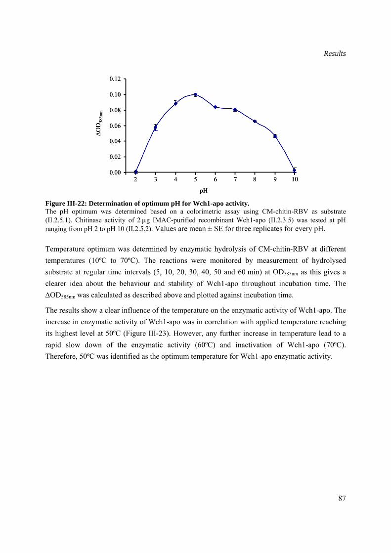
Results
87
Figure III-22: Determination of optimum pH for Wch1-apo activity. The pH optimum was determined based on a colorimetric assay using CM-chitin-RBV as substrate (II.2.5.1). Chitinase activity of 2 μg IMAC-purified recombinant Wch1-apo (II.2.3.5) was tested at pH ranging from pH 2 to pH 10 (II.2.5.2). Values are mean ± SE for three replicates for every pH. Temperature optimum was determined by enzymatic hydrolysis of CM-chitin-RBV at different temperatures (10ºC to 70ºC). The reactions were monitored by measurement of hydrolysed substrate at regular time intervals (5, 10, 20, 30, 40, 50 and 60 min) at OD585nm as this gives a clearer idea about the behaviour and stability of Wch1-apo throughout incubation time. The ΔOD585nm was calculated as described above and plotted against incubation time.
The results show a clear influence of the temperature on the enzymatic activity of Wch1-apo. The increase in enzymatic activity of Wch1-apo was in correlation with applied temperature reaching its highest level at 50ºC (Figure III-23). However, any further increase in temperature lead to a rapid slow down of the enzymatic activity (60ºC) and inactivation of Wch1-apo (70ºC). Therefore, 50ºC was identified as the optimum temperature for Wch1-apo enzymatic activity.
0.00
0.02
0.04
0.06
0.08
0.10
0.12
2 3 4 5 6 7 8 9 10
pH
ΔO
D58
5nm
0.00
0.02
0.04
0.06
0.08
0.10
0.12
2 3 4 5 6 7 8 9 10
pH
ΔO
D58
5nm
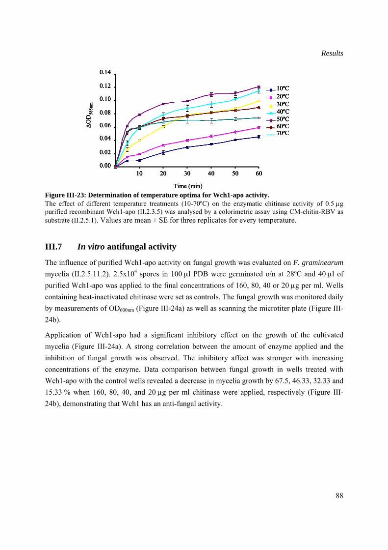
Results
88
Figure III-23: Determination of temperature optima for Wch1-apo activity. The effect of different temperature treatments (10-70ºC) on the enzymatic chitinase activity of 0.5 μg purified recombinant Wch1-apo (II.2.3.5) was analysed by a colorimetric assay using CM-chitin-RBV as substrate (II.2.5.1). Values are mean ± SE for three replicates for every temperature.
III.7 In vitro antifungal activity
The influence of purified Wch1-apo activity on fungal growth was evaluated on F. graminearum mycelia (II.2.5.11.2). 2.5x10⁴ spores in 100 μl PDB were germinated o/n at 28ºC and 40 μl of purified Wch1-apo was applied to the final concentrations of 160, 80, 40 or 20 μg per ml. Wells containing heat-inactivated chitinase were set as controls. The fungal growth was monitored daily by measurements of OD600nm (Figure III-24a) as well as scanning the microtiter plate (Figure III-24b).
Application of Wch1-apo had a significant inhibitory effect on the growth of the cultivated mycelia (Figure III-24a). A strong correlation between the amount of enzyme applied and the inhibition of fungal growth was observed. The inhibitory affect was stronger with increasing concentrations of the enzyme. Data comparison between fungal growth in wells treated with Wch1-apo with the control wells revealed a decrease in mycelia growth by 67.5, 46.33, 32.33 and 15.33 % when 160, 80, 40, and 20 μg per ml chitinase were applied, respectively (Figure III-24b), demonstrating that Wch1 has an anti-fungal activity.
0.00
0.02
0.04
0.06
0.08
0.10
0.12
0.14
10 20 30 40 50 60
Time (min)
ΔO
D58
5nm
10ºC20ºC30ºC40ºC50ºC60ºC70ºC
0.00
0.02
0.04
0.06
0.08
0.10
0.12
0.14
10 20 30 40 50 60
Time (min)
ΔO
D58
5nm
0.00
0.02
0.04
0.06
0.08
0.10
0.12
0.14
10 20 30 40 50 600.00
0.02
0.04
0.06
0.08
0.10
0.12
0.14
10 20 30 40 50 60
Time (min)
ΔO
D58
5nm
10ºC20ºC30ºC40ºC50ºC60ºC70ºC
10ºC10ºC20ºC20ºC30ºC30ºC40ºC40ºC50ºC50ºC60ºC60ºC70ºC70ºC
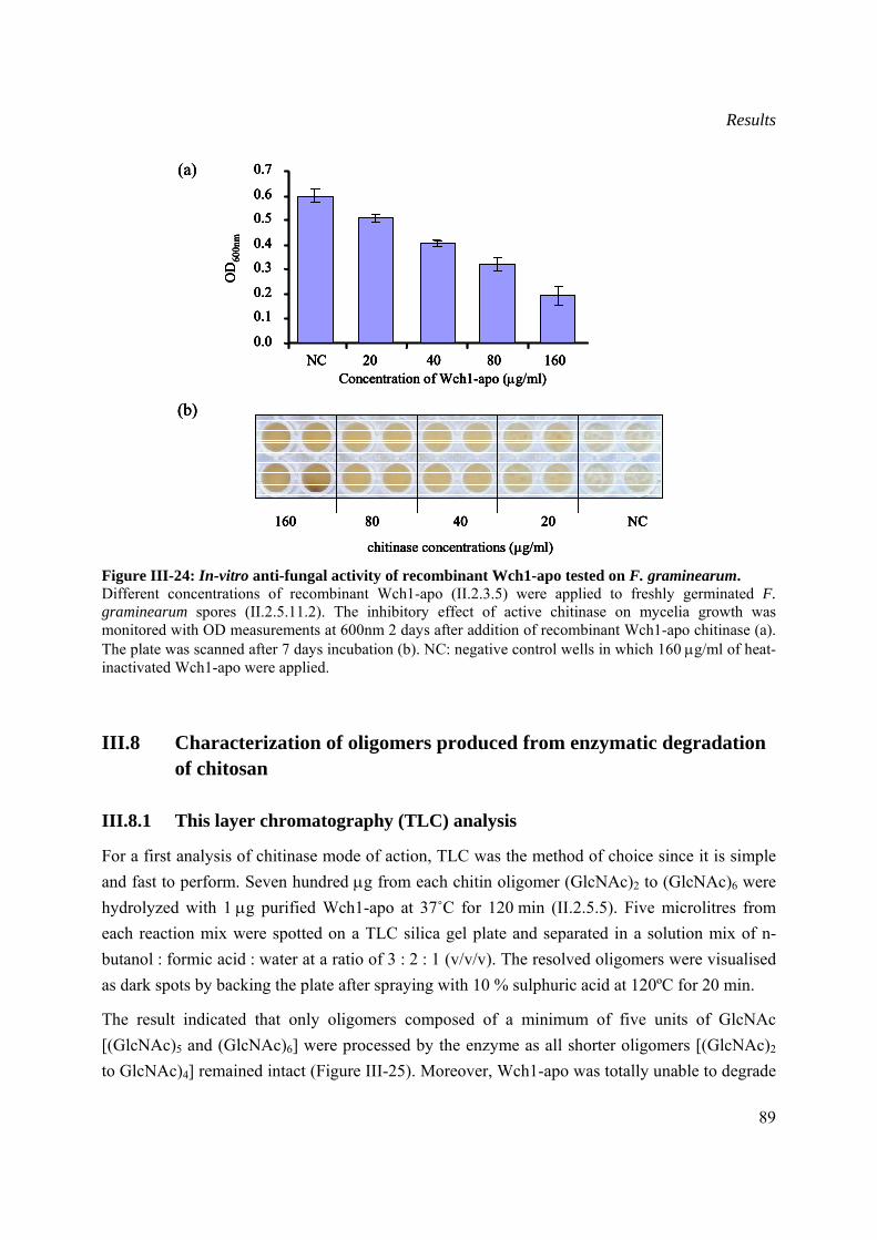
Results
89
Figure III-24: In-vitro anti-fungal activity of recombinant Wch1-apo tested on F. graminearum. Different concentrations of recombinant Wch1-apo (II.2.3.5) were applied to freshly germinated F. graminearum spores (II.2.5.11.2). The inhibitory effect of active chitinase on mycelia growth was monitored with OD measurements at 600nm 2 days after addition of recombinant Wch1-apo chitinase (a). The plate was scanned after 7 days incubation (b). NC: negative control wells in which 160 μg/ml of heat-inactivated Wch1-apo were applied.
III.8 Characterization of oligomers produced from enzymatic degradation of chitosan
III.8.1 This layer chromatography (TLC) analysis
For a first analysis of chitinase mode of action, TLC was the method of choice since it is simple and fast to perform. Seven hundred μg from each chitin oligomer (GlcNAc)2 to (GlcNAc)6 were hydrolyzed with 1 μg purified Wch1-apo at 37˚C for 120 min (II.2.5.5). Five microlitres from each reaction mix were spotted on a TLC silica gel plate and separated in a solution mix of n-butanol : formic acid : water at a ratio of 3 : 2 : 1 (v/v/v). The resolved oligomers were visualised as dark spots by backing the plate after spraying with 10 % sulphuric acid at 120ºC for 20 min.
The result indicated that only oligomers composed of a minimum of five units of GlcNAc [(GlcNAc)5 and (GlcNAc)6] were processed by the enzyme as all shorter oligomers [(GlcNAc)2 to GlcNAc)4] remained intact (Figure III-25). Moreover, Wch1-apo was totally unable to degrade
0.0
0.1
0.2
0.3
0.4
0.5
0.6
0.7
NC 20 40 80 160Concentration of Wch1-apo (μg/ml)
OD
600n
m
(a)
(b)
160 80 40 20 NC
chitinase concentrations (µg/ml)
0.0
0.1
0.2
0.3
0.4
0.5
0.6
0.7
NC 20 40 80 160Concentration of Wch1-apo (μg/ml)
OD
600n
m
(a)
0.0
0.1
0.2
0.3
0.4
0.5
0.6
0.7
NC 20 40 80 160Concentration of Wch1-apo (μg/ml)
OD
600n
m
0.0
0.1
0.2
0.3
0.4
0.5
0.6
0.7
NC 20 40 80 1600.0
0.1
0.2
0.3
0.4
0.5
0.6
0.7
NC 20 40 80 160Concentration of Wch1-apo (μg/ml)
OD
600n
m
(a)
(b)
160 80 40 20 NC
chitinase concentrations (µg/ml)
(b)
160 80 40 20 NC
chitinase concentrations (µg/ml)
160 80 40 20 NC160 80 40 20 NC
chitinase concentrations (µg/ml)
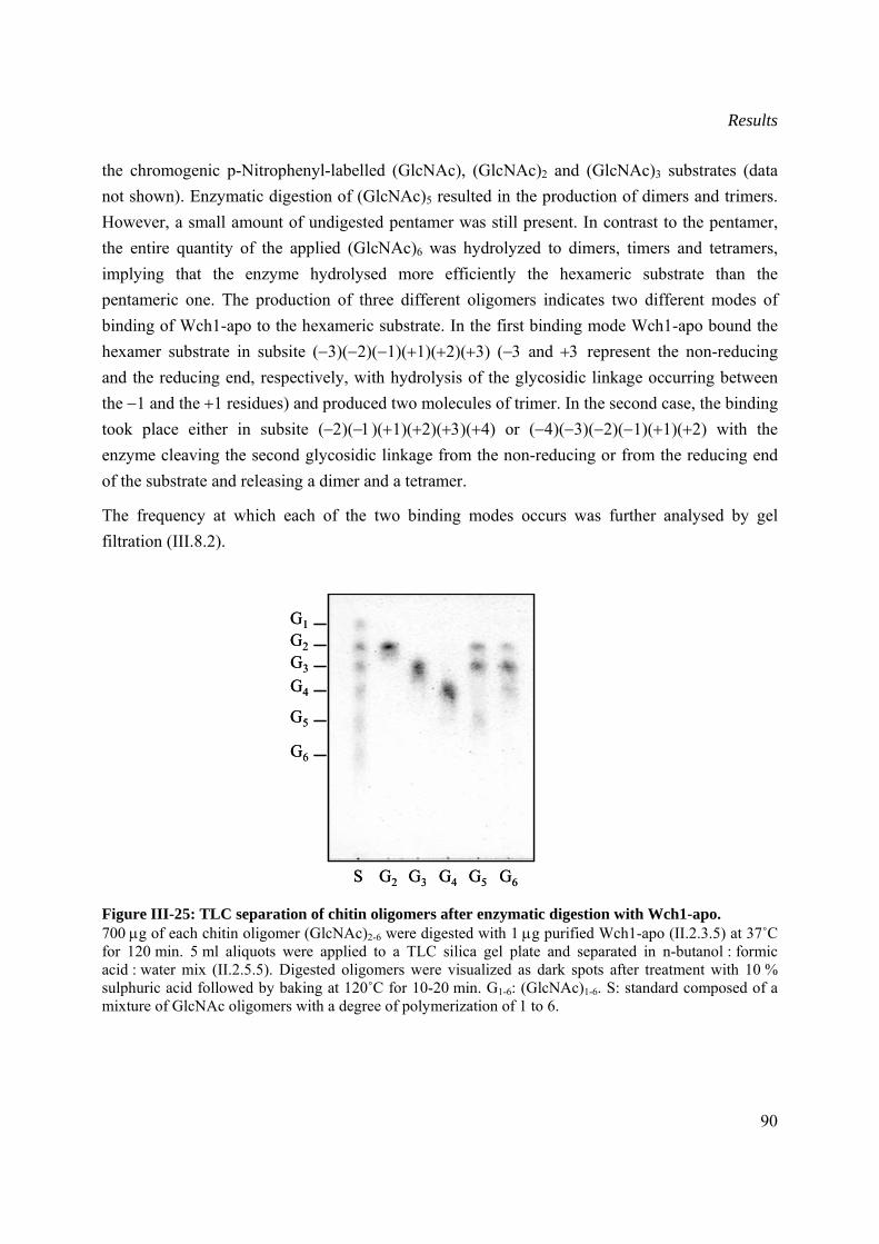
Results
90
the chromogenic p-Nitrophenyl-labelled (GlcNAc), (GlcNAc)2 and (GlcNAc)3 substrates (data not shown). Enzymatic digestion of (GlcNAc)5 resulted in the production of dimers and trimers. However, a small amount of undigested pentamer was still present. In contrast to the pentamer, the entire quantity of the applied (GlcNAc)6 was hydrolyzed to dimers, timers and tetramers, implying that the enzyme hydrolysed more efficiently the hexameric substrate than the pentameric one. The production of three different oligomers indicates two different modes of binding of Wch1-apo to the hexameric substrate. In the first binding mode Wch1-apo bound the hexamer substrate in subsite (−3)(−2)(−1)(+1)(+2)(+3) (−3 and +3 represent the non-reducing and the reducing end, respectively, with hydrolysis of the glycosidic linkage occurring between the −1 and the +1 residues) and produced two molecules of trimer. In the second case, the binding took place either in subsite (−2)(−1)(+1)(+2)(+3)(+4) or (−4)(−3)(−2)(−1)(+1)(+2) with the enzyme cleaving the second glycosidic linkage from the non-reducing or from the reducing end of the substrate and releasing a dimer and a tetramer.
The frequency at which each of the two binding modes occurs was further analysed by gel filtration (III.8.2).
Figure III-25: TLC separation of chitin oligomers after enzymatic digestion with Wch1-apo. 700 μg of each chitin oligomer (GlcNAc)2-6 were digested with 1 μg purified Wch1-apo (II.2.3.5) at 37˚C for 120 min. 5 ml aliquots were applied to a TLC silica gel plate and separated in n-butanol : formic acid : water mix (II.2.5.5). Digested oligomers were visualized as dark spots after treatment with 10 % sulphuric acid followed by baking at 120˚C for 10-20 min. G1-6: (GlcNAc)1-6. S: standard composed of a mixture of GlcNAc oligomers with a degree of polymerization of 1 to 6.
G1G2G3
G4
G5
G6
S G2 G3 G4 G5 G6
G1G2G3
G4
G5
G6
S G2 G3 G4 G5 G6
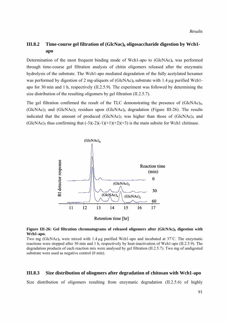
Results
91
III.8.2 Time-course gel filtration of (GlcNac)6 oligosaccharide digestion by Wch1-apo
Determination of the most frequent binding mode of Wch1-apo to (GlcNAc)6 was performed through time-course gel filtration analysis of chitin oligomers released after the enzymatic hydrolysis of the substrate. The Wch1-apo mediated degradation of the fully acetylated hexamer was performed by digestion of 2 mg-aliquots of (GlcNAc)6 substrate with 1.4 μg purified Wch1-apo for 30 min and 1 h, respectively (II.2.5.9). The experiment was followed by determining the size distribution of the resulting oligomers by gel filtration (II.2.5.7).
The gel filtration confirmed the result of the TLC demonstrating the presence of (GlcNAc)4, (GlcNAc)3 and (GlcNAc)2 residues upon (GlcNAc)6 degradation (Figure III-26). The results indicated that the amount of produced (GlcNAc)3 was higher than those of (GlcNAc)4 and (GlcNAc)2 thus confirming that (-3)(-2)(-1)(+1)(+2)(+3) is the main subsite for Wch1 chitinase.
Figure III-26: Gel filtration chromatograms of released oligomers after (GlcNAc)6 digestion with Wch1-apo. Two mg (GlcNAc)6 were mixed with 1.4 μg purified Wch1-apo and incubated at 37˚C. The enzymatic reactions were stopped after 30 min and 1 h, respectively by heat-inactivation of Wch1-apo (II.2.5.9). The degradation products of each reaction mix were analysed by gel filtration (II.2.5.7). Two mg of undigested substrate were used as negative control (0 min).
III.8.3 Size distribution of oliogmers after degradation of chitosan with Wch1-apo
Size distribution of oligomers resulting from enzymatic degradation (II.2.5.6) of highly
Retention time [hr]
RI d
etec
tor r
espo
nse
Reaction time (min)
(GlcNAc)6
11 12 13 14 15 16 17
0
30
60(GlcNAc)4
(GlcNAc)3
(GlcNAc)2
Retention time [hr]
RI d
etec
tor r
espo
nse
Reaction time (min)
(GlcNAc)6
11 12 13 14 15 16 17
0
30
60(GlcNAc)4
(GlcNAc)3
(GlcNAc)2
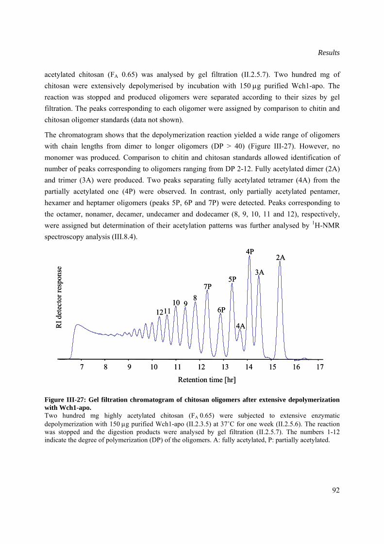
Results
92
acetylated chitosan (FA 0.65) was analysed by gel filtration (II.2.5.7). Two hundred mg of chitosan were extensively depolymerised by incubation with 150 μg purified Wch1-apo. The reaction was stopped and produced oligomers were separated according to their sizes by gel filtration. The peaks corresponding to each oligomer were assigned by comparison to chitin and chitosan oligomer standards (data not shown).
The chromatogram shows that the depolymerization reaction yielded a wide range of oligomers with chain lengths from dimer to longer oligomers (DP > 40) (Figure III-27). However, no monomer was produced. Comparison to chitin and chitosan standards allowed identification of number of peaks corresponding to oligomers ranging from DP 2-12. Fully acetylated dimer (2A) and trimer (3A) were produced. Two peaks separating fully acetylated tetramer (4A) from the partially acetylated one (4P) were observed. In contrast, only partially acetylated pentamer, hexamer and heptamer oligomers (peaks 5P, 6P and 7P) were detected. Peaks corresponding to the octamer, nonamer, decamer, undecamer and dodecamer (8, 9, 10, 11 and 12), respectively, were assigned but determination of their acetylation patterns was further analysed by 1H-NMR spectroscopy analysis (III.8.4).
Figure III-27: Gel filtration chromatogram of chitosan oligomers after extensive depolymerization with Wch1-apo. Two hundred mg highly acetylated chitosan (FA 0.65) were subjected to extensive enzymatic depolymerization with 150 μg purified Wch1-apo (II.2.3.5) at 37˚C for one week (II.2.5.6). The reaction was stopped and the digestion products were analysed by gel filtration (II.2.5.7). The numbers 1-12 indicate the degree of polymerization (DP) of the oligomers. A: fully acetylated, P: partially acetylated.
Retention time [hr]
RI d
etec
torr
espo
nse
2A
3A
4P
4A
5P
6P
7P8
9101112
7 8 9 10 11 12 13 14 15 16 17
Retention time [hr]
RI d
etec
torr
espo
nse
2A
3A
4P
4A
5P
6P
7P8
9101112
7 8 9 10 11 12 13 14 15 16 17
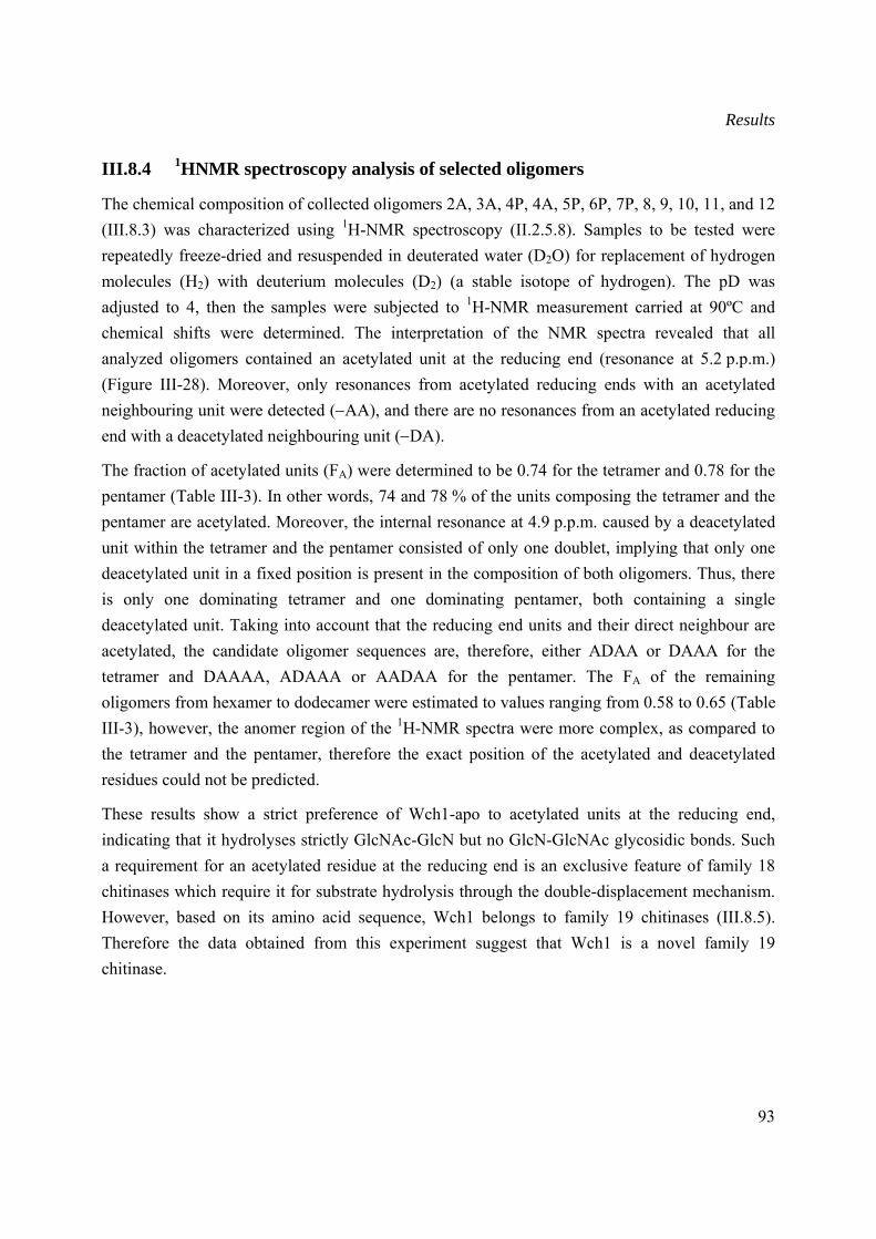
Results
93
III.8.4 1HNMR spectroscopy analysis of selected oligomers
The chemical composition of collected oligomers 2A, 3A, 4P, 4A, 5P, 6P, 7P, 8, 9, 10, 11, and 12 (III.8.3) was characterized using 1H-NMR spectroscopy (II.2.5.8). Samples to be tested were repeatedly freeze-dried and resuspended in deuterated water (D2O) for replacement of hydrogen molecules (H2) with deuterium molecules (D2) (a stable isotope of hydrogen). The pD was adjusted to 4, then the samples were subjected to 1H-NMR measurement carried at 90ºC and chemical shifts were determined. The interpretation of the NMR spectra revealed that all analyzed oligomers contained an acetylated unit at the reducing end (resonance at 5.2 p.p.m.) (Figure III-28). Moreover, only resonances from acetylated reducing ends with an acetylated neighbouring unit were detected (−AA), and there are no resonances from an acetylated reducing end with a deacetylated neighbouring unit (−DA).
The fraction of acetylated units (FA) were determined to be 0.74 for the tetramer and 0.78 for the pentamer (Table III-3). In other words, 74 and 78 % of the units composing the tetramer and the pentamer are acetylated. Moreover, the internal resonance at 4.9 p.p.m. caused by a deacetylated unit within the tetramer and the pentamer consisted of only one doublet, implying that only one deacetylated unit in a fixed position is present in the composition of both oligomers. Thus, there is only one dominating tetramer and one dominating pentamer, both containing a single deacetylated unit. Taking into account that the reducing end units and their direct neighbour are acetylated, the candidate oligomer sequences are, therefore, either ADAA or DAAA for the tetramer and DAAAA, ADAAA or AADAA for the pentamer. The FA of the remaining oligomers from hexamer to dodecamer were estimated to values ranging from 0.58 to 0.65 (Table III-3), however, the anomer region of the 1H-NMR spectra were more complex, as compared to the tetramer and the pentamer, therefore the exact position of the acetylated and deacetylated residues could not be predicted.
These results show a strict preference of Wch1-apo to acetylated units at the reducing end, indicating that it hydrolyses strictly GlcNAc-GlcN but no GlcN-GlcNAc glycosidic bonds. Such a requirement for an acetylated residue at the reducing end is an exclusive feature of family 18 chitinases which require it for substrate hydrolysis through the double-displacement mechanism. However, based on its amino acid sequence, Wch1 belongs to family 19 chitinases (III.8.5). Therefore the data obtained from this experiment suggest that Wch1 is a novel family 19 chitinase.
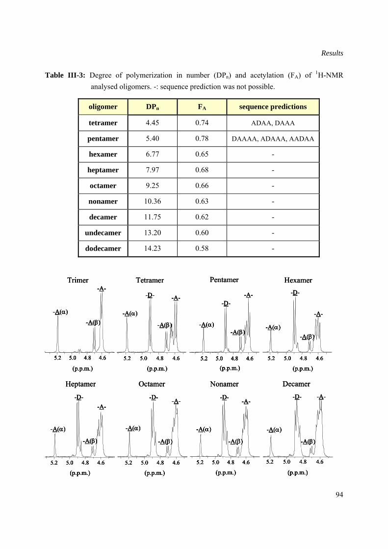
Results
94
Table III-3: Degree of polymerization in number (DPn) and acetylation (FA) of 1H-NMR analysed oligomers. -: sequence prediction was not possible.
oligomer DPn FA sequence predictions
tetramer 4.45 0.74 ADAA, DAAA
pentamer 5.40 0.78 DAAAA, ADAAA, AADAA
hexamer 6.77 0.65 -
heptamer 7.97 0.68 -
octamer 9.25 0.66 -
nonamer 10.36 0.63 -
decamer 11.75 0.62 -
undecamer 13.20 0.60 -
dodecamer 14.23 0.58 -
Heptamer
(p.p.m.)4.64.85.05.2
-A(α)
-A(β)
-A--D-
Octamer
(p.p.m.)4.64.85.05.2
-D-
-A(α)
-A(β)
-A-
Nonamer
(p.p.m.)4.64.85.05.2
-D-
-A(α)
-A(β)
-A-
Decamer
(p.p.m.)
4.64.85.05.2
-D-
-A(α)
-A(β)
-A-
Heptamer
(p.p.m.)4.64.85.05.2
-A(α)
-A(β)
-A--D-
Heptamer
(p.p.m.)4.64.85.05.2
-A(α)
-A(β)
-A--D-
(p.p.m.)4.64.85.05.2
-A(α)
-A(β)
-A--D-
Octamer
(p.p.m.)4.64.85.05.2
-D-
-A(α)
-A(β)
-A-
Octamer
(p.p.m.)4.64.85.05.2
-D-
-A(α)
-A(β)
-A-
(p.p.m.)4.64.85.05.2
-D-
-A(α)
-A(β)
-A-
Nonamer
(p.p.m.)4.64.85.05.2
-D-
-A(α)
-A(β)
-A-
Nonamer
(p.p.m.)4.64.85.05.2
-D-
-A(α)
-A(β)
-A-
(p.p.m.)4.64.85.05.2
-D-
-A(α)
-A(β)
-A-
Decamer
(p.p.m.)
4.64.85.05.2
-D-
-A(α)
-A(β)
-A-
Decamer
(p.p.m.)
4.64.85.05.2
-D-
-A(α)
-A(β)
-A-
(p.p.m.)
4.64.85.05.2
-D-
-A(α)
-A(β)
-A-
Trimer
(p.p.m.)
4.64.85.05.2
-A(α)
-A(β)
-A-Tetramer
(p.p.m.)4.64.85.05.2
-A(α)
-D- -A-
-A(β)
Hexamer
(p.p.m.)4.64.85.05.2
-A(α)
-D-
-A(β)
-A-
Pentamer
(p.p.m.)4.64.85.05.2
-A(α)
-D-
-A(β)
-A-
Trimer
(p.p.m.)
4.64.85.05.2
-A(α)
-A(β)
-A-Trimer
(p.p.m.)
4.64.85.05.2
-A(α)
-A(β)
-A-
(p.p.m.)
4.64.85.05.2
-A(α)
-A(β)
-A-Tetramer
(p.p.m.)4.64.85.05.2
-A(α)
-D- -A-
-A(β)
Tetramer
(p.p.m.)4.64.85.05.2
-A(α)
-D- -A-
-A(β)
(p.p.m.)4.64.85.05.2
-A(α)
-D- -A-
-A(β)
Hexamer
(p.p.m.)4.64.85.05.2
-A(α)
-D-
-A(β)
-A-
Hexamer
(p.p.m.)4.64.85.05.2
-A(α)
-D-
-A(β)
-A-
(p.p.m.)4.64.85.05.2
-A(α)
-D-
-A(β)
-A-
Pentamer
(p.p.m.)4.64.85.05.2
-A(α)
-D-
-A(β)
-A-
Pentamer
(p.p.m.)4.64.85.05.2
-A(α)
-D-
-A(β)
-A-
(p.p.m.)4.64.85.05.2
-A(α)
-D-
-A(β)
-A-
Results
95
Figure III-28: 1H-NMR spectra of gel filtration-resolved oligomers resulting from depolymerization of chitosan with Wch1-apo. Oligomers resulting from extensive depolymerisation of highly acetylated chitosan (DP 3 to 12) (II.2.5.6) were freeze-dried and resuspended in deuterated water. The samples were transferred to appropriate tubes and analysed by 1H-NMR (II.2.5.8). –A(α)/A(β): α/β acetylated unit on reducing end; –D–: deacetylated internal unit; A– acetylated non-reducing end. p.p.m.: Chemical shift of 1H nuclear resonance determined relative to internal sodium-3-(trimethylsilyl) propionate-d4 and expressed in parts per million.
III.8.5 Classification of Wch1 wheat chitinase
The classification of Wch1 within the nomenclature of glycosyl hydrolases was achieved based on its amino acid sequence homology to highly conserved regions representing the signature patterns of family 18 and 19 chitinases (http://www.expasy.org/enzyme/3.2.1.14) (II.2.6).
After amino acid sequence alignment of conserved regions from chitinases belonging to either family 18 or 19 with Wch1, a very high homology of Wch1 sequence to the two signature patterns described for family 19 was observed (Figure III-29). Furthermore, no similarity to the consensus sequence of family 18 could be identified. Therefore, it was concluded that Wch1 belongs to the family 19 chitinases.
Undecamer
(p.p.m.)
4.64.85.05.2
-A(α)
-D-
-A(β)
-A-
Dodecamer
(p.p.m.)
4.64.85.05.2
-A(α)
-A(β)
-D--A-
Undecamer
(p.p.m.)
4.64.85.05.2
-A(α)
-D-
-A(β)
-A-
Undecamer
(p.p.m.)
4.64.85.05.2
-A(α)
-D-
-A(β)
-A-
(p.p.m.)
4.64.85.05.2
-A(α)
-D-
-A(β)
-A-
Dodecamer
(p.p.m.)
4.64.85.05.2
-A(α)
-A(β)
-D--A-
Dodecamer
(p.p.m.)
4.64.85.05.2
-A(α)
-A(β)
-D--A-
(p.p.m.)
4.64.85.05.2
-A(α)
-A(β)
-D--A-
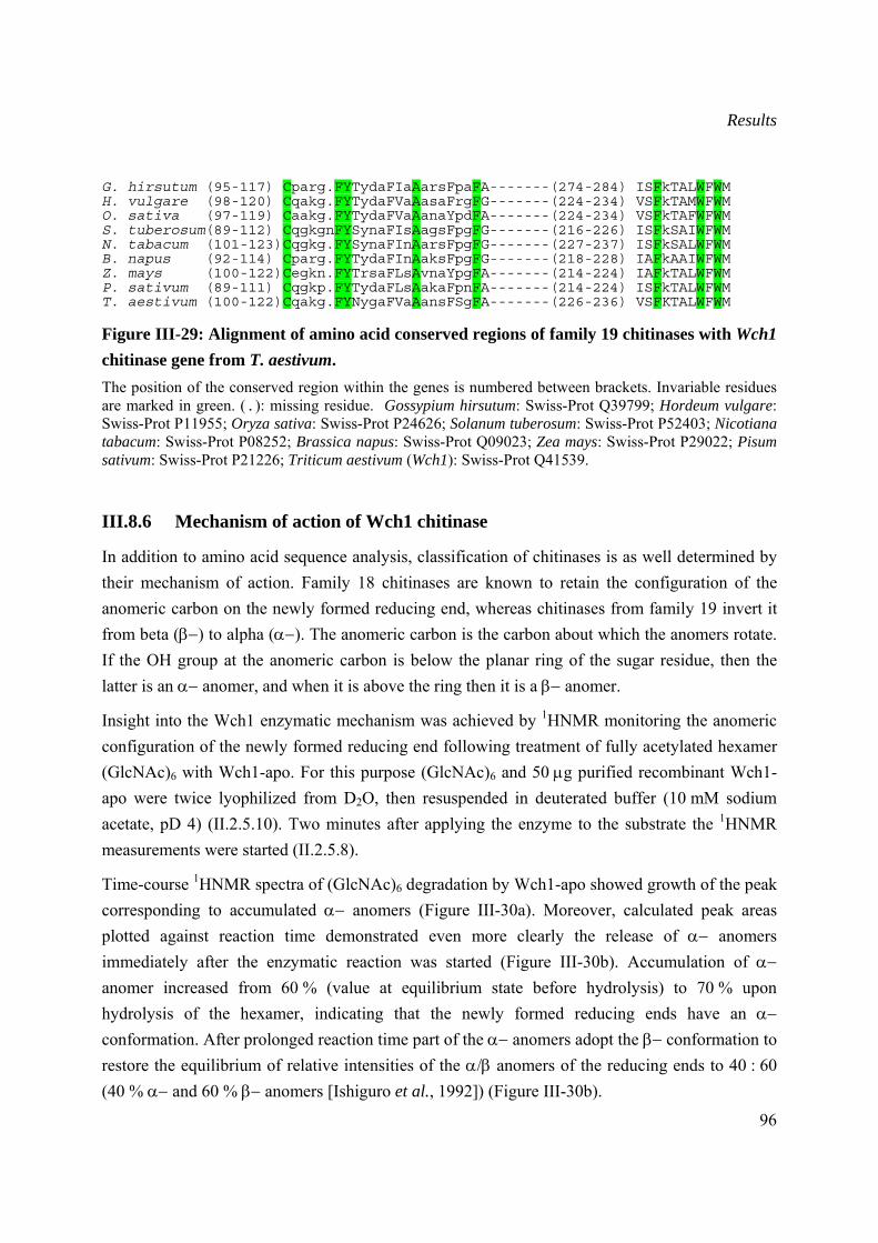
Results
96
G. hirsutum (95-117) Cparg.FYTydaFIaAarsFpaFA-------(274-284) ISFkTALWFWM H. vulgare (98-120) Cqakg.FYTydaFVaAasaFrgFG-------(224-234) VSFkTAMWFWM O. sativa (97-119) Caakg.FYTydaFVaAanaYpdFA-------(224-234) VSFkTAFWFWM S. tuberosum(89-112) CqgkgnFYSynaFIsAagsFpgFG-------(216-226) ISFkSAIWFWM N. tabacum (101-123)Cqgkg.FYSynaFInAarsFpgFG-------(227-237) ISFkSALWFWM B. napus (92-114) Cparg.FYTydaFInAaksFpgFG-------(218-228) IAFkAAIWFWM Z. mays (100-122)Cegkn.FYTrsaFLsAvnaYpgFA-------(214-224) IAFkTALWFWM P. sativum (89-111) Cqgkp.FYTydaFLsAakaFpnFA-------(214-224) ISFkTALWFWM T. aestivum (100-122)Cqakg.FYNygaFVaAansFSgFA-------(226-236) VSFKTALWFWM
Figure III-29: Alignment of amino acid conserved regions of family 19 chitinases with Wch1 chitinase gene from T. aestivum. The position of the conserved region within the genes is numbered between brackets. Invariable residues are marked in green. (.): missing residue. Gossypium hirsutum: Swiss-Prot Q39799; Hordeum vulgare: Swiss-Prot P11955; Oryza sativa: Swiss-Prot P24626; Solanum tuberosum: Swiss-Prot P52403; Nicotiana tabacum: Swiss-Prot P08252; Brassica napus: Swiss-Prot Q09023; Zea mays: Swiss-Prot P29022; Pisum sativum: Swiss-Prot P21226; Triticum aestivum (Wch1): Swiss-Prot Q41539.
III.8.6 Mechanism of action of Wch1 chitinase
In addition to amino acid sequence analysis, classification of chitinases is as well determined by their mechanism of action. Family 18 chitinases are known to retain the configuration of the anomeric carbon on the newly formed reducing end, whereas chitinases from family 19 invert it from beta (β−) to alpha (α−). The anomeric carbon is the carbon about which the anomers rotate. If the OH group at the anomeric carbon is below the planar ring of the sugar residue, then the latter is an α− anomer, and when it is above the ring then it is a β− anomer.
Insight into the Wch1 enzymatic mechanism was achieved by 1HNMR monitoring the anomeric configuration of the newly formed reducing end following treatment of fully acetylated hexamer (GlcNAc)6 with Wch1-apo. For this purpose (GlcNAc)6 and 50 μg purified recombinant Wch1-apo were twice lyophilized from D2O, then resuspended in deuterated buffer (10 mM sodium acetate, pD 4) (II.2.5.10). Two minutes after applying the enzyme to the substrate the 1HNMR measurements were started (II.2.5.8).
Time-course 1HNMR spectra of (GlcNAc)6 degradation by Wch1-apo showed growth of the peak corresponding to accumulated α− anomers (Figure III-30a). Moreover, calculated peak areas plotted against reaction time demonstrated even more clearly the release of α− anomers immediately after the enzymatic reaction was started (Figure III-30b). Accumulation of α− anomer increased from 60 % (value at equilibrium state before hydrolysis) to 70 % upon hydrolysis of the hexamer, indicating that the newly formed reducing ends have an α− conformation. After prolonged reaction time part of the α− anomers adopt the β− conformation to restore the equilibrium of relative intensities of the α/β anomers of the reducing ends to 40 : 60 (40 % α− and 60 % β− anomers [Ishiguro et al., 1992]) (Figure III-30b).
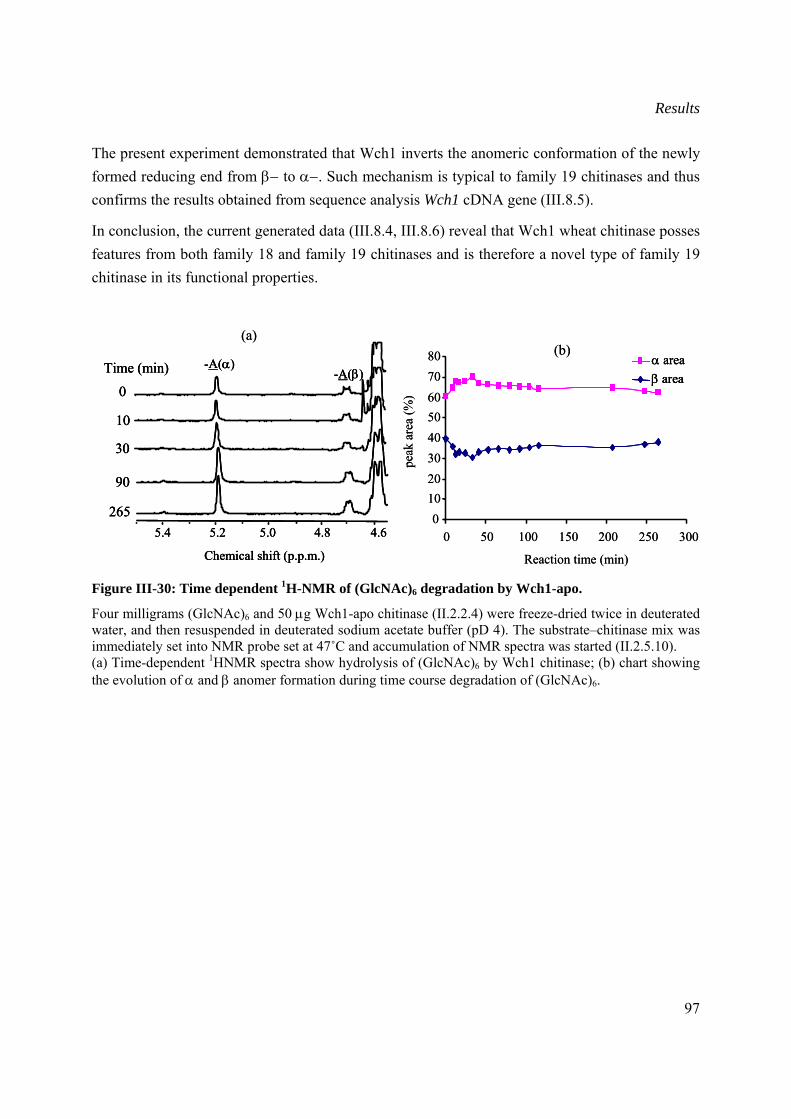
Results
97
The present experiment demonstrated that Wch1 inverts the anomeric conformation of the newly formed reducing end from β− to α−. Such mechanism is typical to family 19 chitinases and thus confirms the results obtained from sequence analysis Wch1 cDNA gene (III.8.5).
In conclusion, the current generated data (III.8.4, III.8.6) reveal that Wch1 wheat chitinase posses features from both family 18 and family 19 chitinases and is therefore a novel type of family 19 chitinase in its functional properties.
Figure III-30: Time dependent 1H-NMR of (GlcNAc)6 degradation by Wch1-apo.
Four milligrams (GlcNAc)6 and 50 μg Wch1-apo chitinase (II.2.2.4) were freeze-dried twice in deuterated water, and then resuspended in deuterated sodium acetate buffer (pD 4). The substrate–chitinase mix was immediately set into NMR probe set at 47˚C and accumulation of NMR spectra was started (II.2.5.10). (a) Time-dependent 1HNMR spectra show hydrolysis of (GlcNAc)6 by Wch1 chitinase; (b) chart showing the evolution of α and β anomer formation during time course degradation of (GlcNAc)6.
0
10
30
90
Chemical shift (p.p.m.)
4.64.85.05.25.4265
-A(α)-A(β)Time (min)
(a)
0
10
30
90
Chemical shift (p.p.m.)
4.64.85.05.25.4265
-A(α)-A(β)Time (min)
0
10
30
90
Chemical shift (p.p.m.)
4.64.85.05.25.4265
-A(α)-A(β)Time (min)
(a)
Reaction time (min)
01020304050607080
0 50 100 150 200 250 300
peak
area
(%)
β areaα area
(b)
Reaction time (min)
01020304050607080
0 50 100 150 200 250 300
peak
area
(%)
β areaα areaβ areaα area
(b)
Discussion and future prospects
98
IV Discussion and future prospects
IV.1 Generation of chitosan- and chitin-specific monoclonal antibodies
One of the main purposes of this thesis was to provide antibodies specifically binding to glucosamine and N-acetyl-glucosamine residues for monitoring the quality of produced oligomers during chitin degradation. In addition, cell wall polysaccharide structures (chitin, chitosan and glucans) of phytopathogenic fungi play an important role in the initiation of the infection process and subsequent induction of defence mechanism in the invaded plant (Boller, 1995). They constitute the initial targets of antimicrobial hydrolases (chitinases and glucanases). Rational design of antibodies targeted to essential components of fungal cell wall was shown to complement the palette of antifungal resistance strategies (Peschen et al., 2005; Torosantucci et al., 2005). Moreover, such antibodies proved valuable in identification of structural evolution of infection structures during penetration and growth in vitro (El Gueddari et al., 2002). One major drawback in antibody-targeting of polysaccharide components is their poor immunogenicity, and their inability to stimulate MHC class II-dependent T cell help, resulting in generation of only short-lived IgM antibodies (Mond et al., 1995). Immunization with protein conjugates (Ada et al., 2003; Casadevall and Pirofski, 2006) or with polysaccharide mimotopes proved to be efficient approaches that elicit T cell-dependent immune response (Cunto-Amesty et al., 2001; Pirofsky, 2001). For generation of chitosan and chitin specific antibodies, both approaches were investigated.
IV.1.1 Generation of chitosan-specific antibodies
To generate chitosan-specific antibodies, water soluble polymeric chitosan (DA 20 %) was conjugated to ovalbumin, and subcutaneously injected into Balb/c mice. The ELISA screening of blood serum against chitosan indicated that the antigen failed to trigger any IgG maturation from the immune system. Different reasons such as the choice of the animal or chitosan used for the immunizations might be responsible for the absence of the immune response. To our knowledge, no chitosan-specific monoclonal antibodies are available; however, few authors reported successful generation of polyclonal IgG antibodies using coupled and uncoupled chitosans, although the titers were low (Walker et al., 1990; Kim et al., 2000; Sorlier et al., 2003). The choice of animal species to be used for the experiment is of importance since all chitosan-specific polyclonal antibodies reported to date were obtained from immunized rabbit (New Zealand or Chincilla-bastard) suggesting that mice are not the animal of choice when immunizing with
Discussion and future prospects
99
carbohydrate antigens. In fact several studies showed that the success in triggering an immune response to carbohydrate antigens depends both on the strain of mice as well as on the antigen used (composition, protein carrier, etc). The work of Barbor et al. (2003) showed that C57BL/6 mice were more reactive to glyco-epitopes of horseradish peroxidase than Balb/c mice. A recent study, however, reported efficient elicitation of immune response to glucans of fungal cell wall in Balb/c mice with laminarin (a polymer of α-1,3-, and α-1,6- linked glucans from algae) conjugated to diphtheria toxoid (Torosantucci et al., 2005). All these results underline the importance of certain crucial aspects like the competence of the animals used for immunization as well as the choice of an efficient conjugate partner for triggering an immunological response to polysaccharides.
Following the work of Torosantucci et al. (2005), it would be interesting to use diphteria or cholera toxoid-conjugated chitosan as these protein carriers seem to be more potent inducers of the immune response to carbohydrates than bovine serum albumin (BSA) or ovalbumin, particularly when using mice for immunization.
IV.1.2 Generation of chitin-specific monoclonal antibodies
In contrast to chitosan, chitin is insoluble and cannot be coupled to a protein carrier. Thus, it was necessary to use an alternative strategy for generation of polysaccharide-specific antibodies. One of the most used strategies involves the screening of combinatorial peptide libraries for selection of carbohydrate mimics. An early demonstration of peptide mimicry of carbohydrates was achieved by screening a large and diverse peptide library against concanavalin (ConA), and selection of a 12-mer peptide that was shown to bind the lectin with an affinity comparable to that of the methyl α-D-mannopyranoside (Oldenburg et al., 1992; Scott et al., 1992). Also, polyclonal antibodies raised against the dodecapeptide bound the carbohydrate residue. This demonstrated the validity of peptide mimicry as an efficient alternative to protein carrier coupling for generation of carbohydrate-specific antibodies.
In this thesis the same approach was followed to select peptides that mirror the GlcNAc-binding receptor on wheat germ agglutinin (WGA) lectin and thus serve as a mimotope for the generation of chitin-specific antibodies. A combinatorial 7-mer linear peptide library expressed on the surface of filamentous phages was screened against WGA, and GlcNAc-mimicking peptides were identified by competition ELISA (III.1.2.2). Five peptides inhibiting the binding of the carbohydrate in a concentration-dependent manner were sequenced and their peptide composition was identified. The peptides were very different in sequence suggesting that they bind the WGA in different manners. Contrarily to peptides mimicking carbohydrate epitopes such as mannose (YPY), Glucose (WRY), Lewis Y (WLY) and group C polysaccharide (YRY), that have shown
Discussion and future prospects
100
carbohydrate-mimicking motifs composed typically of phenylalanine (F), tyrosine (Y) and tryptophan (W) aromatic amino acid residues (Kieber-Emmons, 1998; Gulati et al., 2001). The selected GlcNAc mimicking peptides did not display any of such carbohydrate-mimetic motifs. However, except for the selected peptide WGA7 G3, all sequences contained aromatic residues such as phenylalanine (F) (WGA7 G2), tryptophan (W) (WGA7 B5), and serine (S) (WGA7 G2, WGA7 F4, and WGA7 F5) that were described to occupy the volume of the GlcNAc moiety (Cunto-Amesty et al., 2001).
Analysis of serum from immunized mice indicated that the peptides had different levels of antigenicity (the magnitude of antibody response), with WGA7 G2 being the most antigenic peptide mimetic (III.1.2.4). Hybridoma cell lines derived from splenocytes of the mouse immunized with WGA7 G2 were analysed. Among the 30 cell lines generated, two clones (mAbαchG25 and mAbαchG211) cross-reacted with water soluble, highly acetylated chitosan (FA 0.65). Moreover, unpurified mAbαchG25 and mAbαchG211 were able to recognize native chitin and/or chitosan-containing F. graminearum cell walls in an immunofluorescence microscopy assay, thus providing a valuable tool for exploration of their distribution in fungal cell wall. Important insights were obtained on the composition of fungal cell wall using lectins (O’Connell, 1991) and chitinases (Toki et al., 2002), but polyclonal anti-chitosan antibodies used by El Gueddari et al. (2002) provided more detailed information on structural changes undergone during growth and infection process. The preliminary results demonstrated the potential application mAbαchG25 and mAbαchG211 antibodies for fungal cell wall analysis, therefore, more detailed studies determining their substrate specificity in terms of degree of acetylation and polymerization should be performed.
The reactivity and specificity of mAbαchG25 and mAbαchG211 to highly acetylated chitosan were demonstrated by ELISA and by microscopy using hybridoma cell culture supernatant. Surprisingly, ELISA analysis of purified aliquots showed complete inactivation toward the polymer while they remained highly active against the peptide (III.1.2.9). Such results raised many questions about the molecular basis of antibody-carbohydrate recognition, and on the effectiveness of WGA7 G2 as chitin-mimotope. It is possible that the peptide WGA7 G2 used for immunization acted more as a functional mimetic of the chitin/chitosan rather than on the basis of structural similarity. Structural mimicry implies that both peptide and carbohydrate bind the active site on the lectin through interaction with the same residues, while functional mimicry engage interaction of both ligands with different sets of residues (Johnson and Pinto, 2002). Additionally, sequence differences of selected GlcNAc–mimetics suppose that each of them may interact with the GlcNAc-binding site of WGA through different residues and probably in different conformations. It is likely that the carbohydrate and the peptide interaction with the
Discussion and future prospects
101
GlcNAc-active site uses different sets of amino acid residues on the lectin, which may have important implications on peptide selection that is done on the basis of lectin-peptide interacting residues that may differ from those involved in lectin-carbohydrate binding. Immunization with such a peptide would result in elicitation of antibodies with high affinity to their peptide antigen but with compromised reactivity to the carbohydrate as observed by Beehouwer et al. (2002). The author reported that immunization of mice with a functional mimotope of glucuronoxylomannan (GXM) polysaccharide capsule of Cryptococcus neoformans, induced very high anti-peptide, but low GXM titers. More detailed crystallographic analysis revealed that the peptide mimotope did not correctly engage the carbohydrate-binding residues, resulting in lower affinity of the antibodies.
Considering the data, it is highly possible that the antibody-chitosan interaction involves very limited number of residues resulting in a weak binding (Vyas et al., 2003). It is supposed that the physico-chemical conditions of the hybridoma cell medium favours these interactions. After purification the antibodies are in different physico-chemical environment which does not sustain the binding. Nevertheless, further studies for identification of structural and functional basis of WGA7 G2 mimicry of GlcNAc, as well as antibody-peptide and antibody carbohydrate recognition have to be performed to verify this assumption.
Although the chitin and/or chitosan-specific antibodies showed weak binding to the carbohydrate, it is the first report of monoclonal antibodies directed against this polymer. Lectin can adopt different conformations and binding mechanisms to carbohydrates (Jain et al., 2000a, Jain et al., 2000b) thus biasing the selection of effective mimotopes. Therefore, alternative strategies for the selection of carbohydrate mimotopes could be assayed. One possible approach is through screening phage peptide libraries against the combinatorial site of IgM or IgG antibodies raised against polysaccharide antigens. This strategy has proved to be efficient for identification of mimotopes of cryptococcal capsular polysaccharide (Fleuridor et al., 2001) and cell-surface polysaccharide mimotopes of Mycobacterium tuberculosis (Gevorkian, et al., 2005). A second method would be the identification of anti-idiotypic mimics. This approach requires that the polysaccharide induces an immune response which leads to production of antibodies (Ab1) whose idiotopes are capable of acting as antigens and can in turn stimulate an anti-idiotope response (Ab2). Ideally, Ab2 bears an idiotope that is an internal image of Ab1 and would be a perfect mimic of the polysaccharide (Kieber-Emmons et al., 1986; Gulati et al., 2001). Although tedious and time-consuming, this method has been successfully used to raise antibodies against capsular polysaccharides of E. coli (Stein and Soderstrom, 1984), Streptococcus pneumoniae (McNamara-Ward et al., 1987), and N. meningitidis group C (Westerink et al., 1995).
Discussion and future prospects
102
IV.2 Production and characterization of recombinant Wch1 chitinase
Enzymatic depolymerization of chitin substrate from crab or shrimp shell with chitinases represents a very interesting, environment-friendly, alternative to the currently used aggressive chemical process, which generates polluting wastes with detrimental effect on the environment (Hirano, 1996; Bautista et al., 2005; Beaney et al., 2005). Additionally, chitinases hydrolyse their substrates through precise mechanisms thus ensuring production of high quality, well defined polymers, in contrast to the chemically processed, commercially available counterpart. To ensure adequate supply of chitinase for industrial scale application, optimization of accumulation levels, purification, enzymatic activity as well as careful characterization of catalytic mechanism must be assessed.
IV.2.1 Accumulation and purification of recombinant Wch1 from bacteria and tobacco
For optimization of accumulation levels, degree of purity and enzymatic activity, over-expression of Wch1 chitinase was tested in both, E. coli cells and tobacco plants.
IV.2.1.1 Accumulation and purification from bacteria For bacterial expression of recombinant Wch1 chitinase, the cDNA fragment was cloned into three bacterial expression vectors containing either a C-terminal his6 tag, an N-terminal Strep-tag II or an N-terminal MBP since the purity and yields of obtained proteins differ substantially from one tag systems to another (Lichty et al., 2005). Before proceeding to large scale expression, culture conditions for each construct were optimised. The optimal concentrations of the inducer and incubation time were determined to ensure maximum protein yield (III.2.1.2).
Levels of recombinant protein accumulation in the crude extract as well as after purification steps were analysed by immunoblot and SDS-PAGE, revealing differences in yield as well as purity between the different tags. In accordance with the results observed by Lichty et al. (2005), his6-tagged Wch1 from E. coli resulted in higher protein amounts when compared to the Strep-tagged version, but the degree of purity was rather poor. Improving the purity of his6-tagged proteins from E. coli crude extracts requires several chromatography steps following the affinity purification which would decrease the final protein yield. Even though slightly smaller quantities were obtained with the Strep-tagged Wch1 chitinase, its purity was very high. Alternative expression of the pASKIBA-Wch1 construct should be tested using a different E. coli strain for improved accumulation of soluble chitinase. Higher accumulation levels of a class I chitinase from Leucaena leucocephala were obtained by Kaomek et al. (2003) using Oregami (DE3), a
Discussion and future prospects
103
redox-deficient E. coli strain allowing correct disulfide bond formation in the bacterial cytoplasm.
Fusion of proteins to large affinity tags such as glutathione-S-transferase (GST), thioredoxine (TRX) or MBP are described as advantageous in terms of increased expression, enhanced solubility and protection from proteolysis (Chen and Gouaux, 1996; Jacquet et al., 1999, Smyth et al. 2003; and Li et al., 2004). The fusion of Wch1 to MBP partner dramatically increased the level of soluble protein accumulation. The yield in crude extract was 15 and 20 fold higher then those of Wch1-his6 and Wch1-strep, respectively. However, MBP did not seem to protect the protein from proteolysis as many degradation bands were observed.
IV.2.1.2 Accumulation and purification from plant cells Different studies were performed to investigate the effect of subcellular localization on the accumulation levels of recombinant proteins produced in plants. The main compartments usually investigated are the apoplast, vacuole, ER, cytosol and chloroplasts. The optimal compartment may change depending on the protein expressed. Ziegelhoffer et al. (2001) observed higher apoplastic accumulation of recombinant endo-1,4-β-glucanase and 1,4-β-cellobiosidase from Acidothermus cellulolyticus, compared to the chloroplast or cytosol. While the work of other group demonstrated higher accumulations of proteins in the ER (Schouten et al., 1996; Conrad and Fiedler, 1998; Ramirez et al., 2002). Moreover, the optimal compartment may differ depending on the plant organ targeted as described by Yang et al. (2005) who reported higher accumulation of a silk-like protein in the apoplast of leaf cells, while the protein was undetected in the seeds. The same protein was shown to accumulate at high levels in the ER when expressed in the seeds. Furthermore the protein expressed nicely in the vacuole of seed cells but could not accumulate in leaves. These results resume the importance of testing different compartments for each recombinant protein to achieve the highest accumulation possible.
Wch1 is a wheat chitinase belonging to class Ib chitinases. The enzymes of this family miss the C-terminal peptide extension responsible for vacuolar targeting of the protein, thus Wch1 is naturally secreted into the apoplast (Liao et al., 1994). To investigate accumulation of Wch1 chitinase in different compartments of tobacco cells, Wch1 cDNA was cloned into two different pTRA plant expression vectors for apoplastic or ER targeting. Preliminary evaluation of gene expression was achieved by transient expression in tobacco leaves. Although the transient state of transgene accumulation in vacuum-infiltrated leaves may not be fully comparable to results after stable integration into tobacco chromosomes and extended screening of different individual lines, it provides a rapid tool for verification of a gene product before moving into transgenic plants (Kapila et al., 1996; Fischer et al., 1999). In addition, transient expression is not influenced by
Discussion and future prospects
104
positional effects that potentially bias gene expression levels in stable transformed plants (Kapila et al., 1996; Scholthof et al., 1996).
Immunoblot analysis of the crude extracts showed higher accumulation of Wch1 chitinase in the apoplast then in the ER, which could easily be explained by the fact that the apoplastic space is the native compartment. Moreover, additional differences were observed after affinity purification. Wch1-apo was successfully purified while Wch1-ER was unable to bind the matrix and was entirely recovered in the flowthrough (III.2.2.3). Instead, Wch1-ER could be purified after urea-denaturation, thus indicating a probable modification in protein conformation enabling correct interaction of the his6-tag with the Ni-NTA resin. Similar results were reported by Fiore et al. (2000) who demonstrated that the accessibility of the C-terminus can be influenced by conformational changes of the protein carrier. Despite few degradation bands visualized on immunoblot, SDS-PAGE analysis showed that Wch1-apo was pure as no contaminating bands were observed (III.2.2.3). Compared to Wch1 expressed in bacteria, Wch1-apo showed the highest purity.
To ensure constant supply and to facilitate the upscaled production of Wch1 chitinase, transgenic tobacco plants were generated. Given the inaccessibility of the his6-tag on Wch1-ER and the subsequent problems of its purification under native conditions, only stable transgenic tobacco plants accumulating recombinant Wch1 in the apoplast were generated. Immunoblot analyses showed that all 27 generated transgenic lines produced the Wch1-apo, but only 10 lines accumulated the recombinant protein to relatively high levels (around 80 mg/kg fresh weight leaves). These lines were selected and self pollinated for establishment of homozygous lines. Further screening was carried out in the T1 and T2 generation to identify the lines with the highest accumulation levels (140 and 180 mg per kg fresh weight leaves, respectively).
The accumulation levels of the T1 and T2 best producing lines correlated with the accumulation levels obtained in the transient agroinfiltration thereby corroborating the suitability of stable transgenic plants for upscaled production of Wch1-apo chitinase.
IV.2.2 Evaluation of the optimal system for production of Wch1
The aim for Wch1 chitinase production is its application as technical enzyme in industrial process for production of chitin oligomers from shrimp, squad or crab shell. Thus it was necessary to determine the most appropriate production system providing large quantities of active enzyme.
The endo-enzymatic activities of Wch1-strep, MBP-Wch1, and Wch1-apo were quantified by a colorimetric assay using CM-chitin-RBV (III.4.2) and then compared to each other. Activity of purified bacterial Wch1-strep was 1.73 folds higher then for purified plant derived Wch1-apo. It is possible that the difference resulted from the purification procedure required for each system
Discussion and future prospects
105
rather than from the protein quality per se. IMAC purification of plant extract required roughly between 12 to 16 h (at a flow rate of 1 ml/min) and about just 1 h using bacterial crude extract due to larger volumes and presence of leaf debris that slow down the transfer of the crude extract through the resin, thus resulting in longer exposition of the enzyme to proteolytic activity as well as to oxidation.
While fusion proteins are usually described as helpful tools for enhancing accumulation of soluble active enzymes (Kaomek et al., 2003, Huang and Chen, 2005), fusion of Wch1 chitinase to a large protein partner like MBP lead to a drastic decrease in enzymatic activity that was 125 and 72 folds lower than that of purified Wch1-strep and Wch1-apo, respectively. The reduced enzymatic activity could be explained by conformational changes induced by the MBP, masking the active site on Wch1. Another possible reason could be the production of misfolded inactive fusion protein that is prevented from precipitation by the presence of the highly soluble fusion partner as reported by Nominé et al. (2001) and Martinez-Torrecuadrada et al. (2005). Periplasmic targeting of an MBP-fused chitinase was reported by Southworth et al. (1996) to be more favourable, as they speculated that the environment of this compartment resulted in improved folding of recombinant chitinase and recovery of enzymatic activity.
For identification of the best system for production of active recombinant Wch1 chitinase, the amounts of produced protein as well as enzymatic activity were taken into consideration. Given that the degree of bacterial Wch1-his6 purity was unsatisfactory and bacterial MBP-Wch1 possessed a very poor activity, both systems were dismissed. Bacterial produced Wch1-strep was more active than plant produced Wch1-apo with 1.25 and 0.75 U per μg protein, respectively. Nevertheless, comparing the total amount of purified active protein produced from one kilogram fresh weight leaves (40 mg) with the one produced from one litre E. coli culture (0.7 mg), it becomes clear that significantly more enzymatic units can be produced from plants (30,000 U), than from bacteria (1,875 U). Therefore, tobacco plants are the best production system for the wheat chitinase Wch1. This is very beneficial since tobacco plants present the advantage of simple upscaling of Wch1 production to industrial level. However, some limitation could arise from the consumable Ni-NTA resin cost. Two recently described systems, polyhydroxybuterate (PHB) and elastine-like polypeptide (ELP), were reported to be effective and cost-effective for large scale manufacture of recombinant proteins (Scheller et al., 2004; Banki and Wood, 2005). According to the estimations reported by the authors, PHB and ELP systems reduce the purification costs by 82 % and 90 %, respectively, compared to the IMAC procedure. Therefore, they represent major potential alternative candidates to the his6 tag system. However, a careful investigation on the impact of these two tags on accumulation levels and enzymatic activity will be necessary to assess their compatibility for Wch1 chitinase.
Discussion and future prospects
106
IV.2.3 Characterization of enzymatic Wch1-apo activity
IV.2.3.1 Optimum temperature and pH Similar to other pathogen-related (PR) proteins, chitinases have typical physiochemical properties enabling them to resist acidic pH and proteolytic cleavage, and thus survive in the harsh environments where they occur (vacuole or intercellular spaces) (Stintzi et al., 1993). Most chitinases display optimum activity at temperatures of 40ºC and 50ºC and pH between 4 and 6 (Yamaoka et al., 1999; Ano et al., 2003; Okazaki et al., 2004, Hoell et al., 2005). However, some chitinases from rice and pineapple displayed strong activity at higher temperatures (70ºC) and extreme pH (2 and 9) (Taira et al., 2005a; Park et al., 2001).
In this study the enzymatic activity of recombinant Wch1-apo was quantified in reaction buffers of pH ranging from 2 to 10. (III.6). The enzyme was active within a wide pH range. However, higher activities between pH 4 to 7 were observed, with a pH optimum of 5. The recombinant Wch1-apo chitinase retained 41 % and 47 % of its activity at pH 3 and 9, respectively, but was inactivated at pH 2 and 10.
The influence of the temperature on the enzymatic activity was assayed between 10ºC and 70ºC. The enzyme was active at temperatures ranging from 10ºC to 60 ºC, after which the enzyme was rapidly inactivated. The optimal conditions for Wch1 chitinase activity (50ºC) are in agreement with chitinases from Beauveria bassiana (Zhang et al., 2004), Phaseolus mungo (Wang et al., 2005), Bacillus chitinolyticus and Streptomyces griseus (Hoster et al., 2005), Serratia marcescens (Nima) (Ruiz-sanchez et al., 2005) and Pseudomonas aeruginosa (Folders et al., 2001), showing optimal activity at the same temperature. The activity of Wch1-apo chitinase with a wide temperature and pH range makes it very suitable for production of chitin oligomers in a fermentation process at industrial scale.
IV.2.3.2 Antifungal activity Plant chitinases are considered to play a major role in defence mechanisms by degrading the chitin of the cell wall of many pathogenic fungi (Bowls, 1990; Linthorst, 1991; Theis and Stahl, 2004). Apoplastic chitinases play a role in the early stages of pathogenesis, when the hyphae penetrate the intercellular space, by the release of elicitor molecules that informs the cell about the infection (Inui et al., 1996; de A Gerhardt et al., 1997). In contrast vacuolar chitinases, mainly class Ia chitinases, are involved later in the infection, when the hyphae penetrate and destroy the cell, causing the protoplast to burst and releasing the vacuolar content into the extracellular compartment (Collinge et al., 1993). Vacuolar chitinases are more active than apoplastic ones against crystalline chitin, whereas apoplastic forms better hydrolyse soluble chitin (Taira et al., 2002; Kasprzewska, 2003). The increased efficacy is believed to be conferred
Discussion and future prospects
107
by the chitin-binding domain (CBD) which can bind chitin in the fungal cell wall by hydrophobic interaction, thus assisting the antifungal action of the enzyme (Iseli et al., 1993; Truong et al., 2003; Taira et al., 2005 b).
The wheat chitinase Wch1 is an acidic class Ib chitinase lacking the C-terminal extension that is responsible for vacuolar targeting (Neuhaus et al., 1991, Melchers et al., 1993), thus it is secreted extracellularly. The activation of the Wch1 gene upon fungal infection with Puccinia graminis was demonstrated by Liao et al. (1994) which indicates its potential involvement in defence against pathogens. The ability of Wch1-apo to inhibit fungal growth of F. graminearum was investigated in vitro in a microtiter plate assay (III.7). Mycelia growth monitored by absorbance after 48 h incubation showed strong inhibitory effect of plant produced recombinant Wch1 chitinase as the growth was reduced by 67.5 % compared to the control culture, thus confirming the defensive role of Wch1 chitinase against fungal pathogens. Moreover, TLC analysis of hydrolytic activities of Wch1 chitinase toward N-acetylchitooligosacharides showed high activity toward (GlcNAc)5 and (GlcNAc)6 in opposition to its total inactivity toward shorter oligomer chains (III.8.1). These results are an additional indication to the role of Wch1 as a generator of chitin oligomers from the cell wall of infecting fungi, which in turn induce series of defence reactions such as phytoalexin production (Yamada et al., 1993), transient depolarization of membrane potential (Kikuyama et al., 1997), ion flux (Kuchitsu et al., 1997), transient generation of reactive oxygen species (Kuchitsu et al., 1995) and jasmonic acid (Nojiri et al., 1996), induction of chitinases (Roby et al., 1987; Inui et al., 1996; Nishizawa et al., 1999), ribosome inactivating proteins (RIP) and some membrane-affecting compounds (MAC) (osmotin, zeamatin and other thaumatin-like proteins) (Lorito et al., 1996).
The inhibitory effect of Wch1-apo chitinase on fungal growth was higher than the one reported for a class Ib chitinase from Castanea sativa (Allona et al., 1996). Nevertheless, when compared with vacuolar class I chitinases from rice and secale, higher concentrations of purified Wch1-apo enzyme were required to inhibit fungal growth (Taira et al., 2001; Truong et al., 2003). Yet, it should be taken into consideration that basic vacuolar class I chitinases are more potent than the apoplastic counterpart. Furthermore, the results can be influenced by the cell wall structure of the pathogen used. The activity of both rice and secale chitinases was assayed on Trichoderma species, whereas the present study was performed on Fusarium which are known to be refractory species to the action of cell wall-degrading enzymes, possibly because of their high protein content (Barbosa and Kemmelmeier, 1993; Dahlleen et al., 2001), and clustering of acetylated glucosamine residues which restricts chitinase-accessible regions to a small part of the chitinous component (Fukamizo et al., 1996).
Discussion and future prospects
108
The requirement for high amounts of active enzyme for fungal inhibition may as well be explained by the fact that in vitro assays do not reflect the in vivo situation which involves synergistic and complementary action of different compounds in the inhibitory process. Apoplastic fluid extract from Fusarium infected wheat plants contained pathogen-related proteins (PR) belonging to families of β-1,3-glucanase, chitinases and thaumatin-like protein (Anand et al., 2004). When this fluid was used in an in vitro test it inhibited growth of F. graminaerum and Gaeumannomyces graminis var. tritici. Moreover, Suo and Leung (2001) observed a rapid increase in activity of both chitinases and β-1,3-glucanase, in the intercellular fluid of rose leaves infected with Diplocarpon rosae. Several in vitro studies analyzing the inhibitory effect of chitinase and β-1,3-glucanase combinations highlighted their strong synergism and increased combinatory effect on fungal growth (Sela-Buurlage et al., 1993; Melchers et al., 1994). Thus, it is possible to enhance Wch1-apo inhibitory effect by combining it to a synergistic PR protein. Given the rapidity of in vitro tests, it would be of great interest to screen several proteins to identify the most potent combinations. The most successful combinations would then be co-expressed in transgenic plants and tested for their resistance to fungal pathogens.
Chitinases have been the focus of numerous studies aiming to develop genetically engineered fungal resistance in crops of economical importance. Transgenic plants expressing chitinases from plants (Broglie et al., 1991; Jach et al., 1995), bacteria (Itoh et al., 2003), yeast (Carsten et al., 2003) and mycoparasitic fungi (Bolar et al., 2001; Liu et al., 2004a) had improved resistance to fungal attacks. Transgenic tobacco plants overexpressing Wch1 were generated (II.3.1) but their resistance to fungal attack has still to be assessed. However, a first indication of the antifungal activity of Wch1 was recently reported by Peschen et al. (2004). In this study, the antifungal potency of Wch1 against Fusarium spp was enhanced both in vitro and in transgenic Arabidopsis thaliana by fusion of Wch1 to a Fusarium-specific antibody (CPW2). It is believed that the antibody would concentrate the chitinase activity at the fungal cell wall leading to a more efficient degradation of the chitin substrate. The work of Peschen et al. (2004) emphasizes the utility of combined action of different defence elements for improved efficacy against pathogenic attacks. Actually, several groups investigating the effect of simultaneous co-expression of chitinases with other PR proteins (mainly glucanases) reported enhanced protection levels in transgenic plants (Jach et al., 1995; Rodriguez Herrera et al., 1999).
It is suggested that co-expression of chitinases and glucanases can limit fungal growth by their combined lytic action on the fungal cell wall. The action of glucanases supports chitinase activity by weakening the mature cell wall, composed of cross-linked chitin-glucan fibers; thus facilitating access of the chitinous substrate (Anguelova-Merhar et al., 2001). Additionally, by releasing glucan oligosaccharides they activate phytoalexin production (Keen and Yoshikawa, 1983; Okinaka et al., 1995; Cosio et al., 1996) and induce the hypersensitive lignification
Discussion and future prospects
109
response in intact (Moerschbacher et al., 1986; Vander et al., 1998; Anguelova-Merhar et al., 2001) and wounded wheat leaves (Barber et al., 1989).
In the light of the results reported by Peschen et al. (2004), and the findings of other groups, engineering of transgenic plants overexpressing combinations of recombinant scFv CPW2-Wch1-apo fusion with various PR proteins would be of great interest towards enhanced phytopathogen resistance.
IV.2.3.3 Physico-chemical characterization of substrate degradation Biotechnological exploitation of chitinases requires detailed knowledge of the catalytic mechanism and the enzyme-substrate interactions. To achieve this goal, the hydrolytic activity of Wch1 chitinase was investigated on highly acetylated chitosan (FA 0.65) as well as on short N-acetylated chitooligomers (GlcNAc)2-6 and the resulting products were analyzed by different physico-chemical methods such as thin layer chromatography, HPLC and 1HNMR spectroscopy.
IV.2.3.3.1 Subsite cleavage of Wch1-apo chitinase To assess the activity of recombinant Wch1-apo on native substrate, colloidal chitin was incubated with the enzyme in a microtiter plate and the reaction was monitored by spectrophotometry (III.4.3). The drop in optical density observed as factor of incubation time, indicated successful degradation of the substrate. This result demonstrates the ability of Wch1 to depolymerize native substrate, and thus, have significant implication on the potential use of Wch1-apo for depolymerization of chitinous substrate from natural sources in a fermentation process.
A preliminary examination of Wch1-apo activity on oligomeric substrate was performed by degradation of fully N-acetylated oligomers ranging from the dimer to the hexamer (GlcNAc)2-6 (III.8). The resulting products were analyzed by thin layer chromatography. Wch1-apo exhibited an endo-type hydrolytic activity most common to plant chitinases (Kasprzewska, 2003). It required a minimum of four glycosidic bonds for hydrolysis and had a greater affinity towards larger oligomers as it degraded (GlcNAc)6 more efficiently than (GlcNAc)5 but showed no activity towards oligomers shorter then a pentamer.
More detailed information about the cleavage mode was obtained by size exclusion chromatography (III.8.2). The degradation reaction of (GlcNAc)6 as a function of incubation period was monitored, and each resulting oligomer was quantified by calculating the corresponding peak area. From the results of this experiment it was clear that the cleavage of (GlcNAc)6 into two (GlcNAc)3 molecules was more predominant than (GlcNAc)4 + (GlcNAc)2 indicating that the enzyme has the binding cleft represented by (−3)(−2)(−1)(+1)(+2)(+3) with +3
Discussion and future prospects
110
representing the reducing end and −3 the non-reducing end of (GlcNAc)6. This result is in line with family 19 chitinases from rice (Sasaki et al., 2003; Truong et al., 2003) and from barley (Honda and Fukamizo, 1998) which show the same cleavage pattern.
Subsite structures may differ within the same family 18 or 19 depending on whether the chitinase is from plant or microbial origin. As reported by Sasaki et al. (2002), the subsite structure of microbial family 18 chitinases is (−2)(−1)(+1)(+2)(+3)(+4), while that of plant family 18 chitinases is (−4)(−3)(−2)(−1)(+1)(+2). Similar situation occurs between plant and microbial family 19 chitinases. Plant family 19 chitinases are characterized with (−3)(−2)(−1)(+1)(+2)(+3) motif, whereas microbial chitinases have a binding cleft represented by (-2)(-1)(+1)(+2)(+3)(+4) (Ueda et al., 2003). More detailed molecular dynamic simulations and kinetic studies on family 19 plant chitinases from a basic class I rice and class II barley (Sasaki et al., 2003; Honda and Fukamizo, 1998) suggested that the substrate binding mode involves high affinity interaction of substrate-enzyme at (−2) and (+2) sites with a weaker binding at subsites (−3) and (+3) and cleavage taking place at (−1) and (+1) sites. Considering the high amino acid sequence homology between Wch1 chitinase, the barley (80 %) and rice (73 %) chitinases (Figure IV-1), it is highly possible that interactions of Wch1 with the chitin hexamer occur in a similar manner. However, crystallographic analysis of Wch1-apo-(GlcNAc)6 complex will be necessary to confirm this assumption.
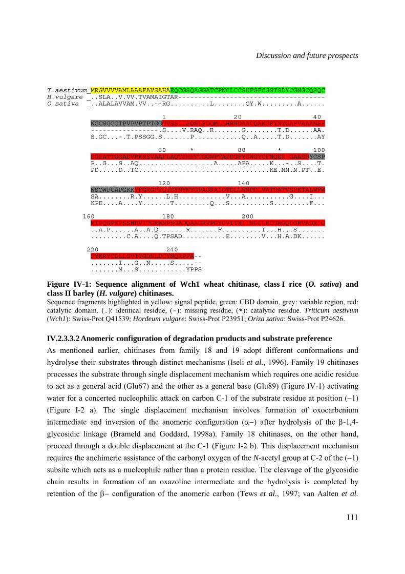
Discussion and future prospects
111
T.aestivum_MRGVVVVAMLAAAFAVSAHAEQCGSQAGGATCPNCLCCSKFGFCGSTSDYCGNGCQSQC H.vulgare _..SLA..V.VV.TVAMAIGTAR------------------------------------- O.sativa _..ALALAVVAM.VV..--RG..........L........QY.W.........A...... 1 20 40 NGCSGGGTPVPVPTPTGGGVSSIISQSLFDQMLLHRNDAACQAKGFYNYGAFVAAANSF -----------------.S....V.RAQ..R.......G........T.D......AA. S.GC...-.T.PSSGG.S.......P............Q..A.....T.D.......AY 60 * 80 * 100 SGFATTGGADVRKREVAAFLAQTSHETTGGWPTAPDGPYSWGYCFNQER-GAASDYCSP P..G...S..AQ...................A.....AFA.....K...-..S....T. PD.....D..TC.................................KE.NN.N.PT..E. 120 140 NSQWPCAPGKKYFGRGPIQISYNYNYGPAGRAIGTDLLNNPDLVATDATVSFKTALWFW SA........R.Y......L.H............V...A...........G....I... KPE....A....Y.......T.........Q...S..........S.........F... 160 180 200 MTPQSPKPSSHDVITGRWSPSGADQAAGRVPGYGVITNIINGGLECGRGQDGRVADRIG ..A.P......A..A.Q.......R.......F..........I...H...S....... .........C.A....Q.TPSAD...........E........V...H.A.DK...... 220 240 FYKRYCDLLGVTYGDNLDCYNQRPFA-- .......I...G..N.....S.....-- .......M...S............YPPS Figure IV-1: Sequence alignment of Wch1 wheat chitinase, class I rice (O. sativa) and class II barley (H. vulgare) chitinases. Sequence fragments highlighted in yellow: signal peptide, green: CBD domain, grey: variable region, red: catalytic domain. (.): identical residue, (-): missing residue, (*): catalytic residue. Triticum aestivum (Wch1): Swiss-Prot Q41539; Hordeum vulgare: Swiss-Prot P23951; Oriza sativa: Swiss-Prot P24626.
IV.2.3.3.2 Anomeric configuration of degradation products and substrate preference As mentioned earlier, chitinases from family 18 and 19 adopt different conformations and hydrolyse their substrates through distinct mechanisms (Iseli et al., 1996). Family 19 chitinases processes the substrate through single displacement mechanism which requires one acidic residue to act as a general acid (Glu67) and the other as a general base (Glu89) (Figure IV-1) activating water for a concerted nucleophilic attack on carbon C-1 of the substrate residue at position (−1) (Figure I-2 a). The single displacement mechanism involves formation of oxocarbenium intermediate and inversion of the anomeric configuration (α−) after hydrolysis of the β-1,4-glycosidic linkage (Brameld and Goddard, 1998a). Family 18 chitinases, on the other hand, proceed through a double displacement at the C-1 (Figure I-2 b). This displacement mechanism requires the anchimeric assistance of the carbonyl oxygen of the N-acetyl group at C-2 of the (−1) subsite which acts as a nucleophile rather than a protein residue. The cleavage of the glycosidic chain results in formation of an oxazoline intermediate and the hydrolysis is completed by retention of the β− configuration of the anomeric carbon (Tews et al., 1997; van Aalten et al.
Discussion and future prospects
112
2001; Aronson et al., 2003). As a consequence to their distinct mechanisms, chitinases from both families differ in their preference for acetylated (A−) and deacetylated (D−) units on each side of the scissile glycosidic bond, and consequently differ with respect to their activities towards chitosan.
The anomeric form of enzymatic products was investigated in a time course 1HNMR monitoring hydrolysis of chitin hexasaccharide (Fukamizo et al., 1995a). Wch1-apo exhibited an anomer inversion as it produced oligomers with α− anomers at the reducing ends, which is in agreement with previously reported inverting mechanism of family 19 chitinases form rice (Sasaki et al., 2003), papaya (Subroto et al., 1999), Streptomyces griseus (Ohno et al., 1996) and bean (Iseli et al., 1996). However, 1HNMR analysis of hetero-chitooligomers (trimer to dodecamer) resulting from extensive depolymerization of highly acetylated chitosan, revealed an absolute preference of Wch1-apo for −AA residues at the reducing end (−1 and −2 subsites). Furthermore carbon NMR spectroscopy analysis demonstrated that all produced chitooligomers contained an acetylated (–A) residue at the non-reducing end (Prof. Vårum, NOBIPOL, Department of Biotechnology, NTNU, Trondheim, Norway, personal communication). Since the non-reducing end residue of an oligomer was bound in a (+1) subsite before cleavage, the occurrence of acetylated residues at both (−2)(−1) and (+1) subsites show that there is clear selectivity toward sequences of acetylated units, on both sides of the glycosidic bond, for cleavage. These findings were surprising because absolute requirement for –A residues at the (−1) subsite is normally attributed to family 18 chitinases, as it is a prerequisite for the substrate-assisted catalytic hydrolysis (Honda et al., 2004; Synstad et al., 2004). As a result of this mechanism, family 18 chitinases process only GlcNAc-GlcNAc and GlcNAc-GlcN linkages (Mitsutomi et al., 1995, Tanabe et al., 2000; Sørbotten et al., 2005), whereas family 19 chitinases cleave both GlcNAc-GlcNAc and GlcN-GlcNAc sequences (Ohno et al., 1996). Moreover, substrate preference of Wch1-apo chitinase for acetylated units at subsites (−2)(−1) and (+1) was similar to that of hen egg white lysozyme (HEWL) (CL, EL and EL), which, contrarily to Wch1 chitinase, acts through the double displacement mechanism (Vårum et al., 1996).
Structural studies on model family 19 chitinase from barley (Hart et al., 1995; Brameld and Goddard, 1998a) identified Lys165, Asn124, Asn199, Glu67 and Glu89 amino acid residues (Figure IV-1) as critical for enzyme-substrate interaction and catalysis. Mutations of Glu67 and Glu89 to Gln resulted in complete loss of activity and mutation of Asn 124 to Ala reduced enzyme activity to 0.82 % of the wild type (Andersen et al., 1997). These residues are found to be strongly conserved among family 19 plant chitinases and are all present in Wch1 sequence (Figure IV-1). Considering the high homology of Wch1-apo and the chitinase from barley seed in respect to their amino acid coding sequences as well as inverting mechanism, we would assume that their protein structures would be similar. Structural resemblance between animal lysozyme,
Discussion and future prospects
113
phage lysozyme and barley chitinase has been previously reported (Holm and Sander, 1994, Monzingo et al., 1996). Superposition of the three-dimensional structures from barley and HEWL revealed coincidence of their catalytic residues, Glu67 and Glu35, respectively. However, contrarily to the barley chitinase, HEWL proceeds through double displacement mechanism as a result of the proximity of its second acidic residue (Asp52) to the oxacarbenium intermediate thus forcing retention of the anomeric configuration (Brameld and Goddard, 1998a; Dao-Pin et al., 1989;). Wch1-apo chitinase exhibits characteristics of family 19 chitinases in respect to sequence homology and catalytic mechanism, but has substrate preferences similar to HEWL and family 18 chitinases. Thus, the wheat Wch1-apo enzyme is a novel type of family 19 chitinases in respect to its functional properties. These results suggest differences in the structure of catalytic domain of Wch1-apo compared to other family 19 chitinases. More detailed crystallographic and mutagenesis studies should be carried out to resolve the structural basis of Wch1-apo–substrate interaction and define the residues engaged in the hydrolysis process.
IV.2.3.3.3 Action pattern of Wch1-apo chitinase on chitosan Polymeric chitin and chitosan have many industrial, agricultural, and pharmaceutical applications (Khor and Lim, 2003; Shahidi and Abuzaytoun, 2005). Similarly, chitin and chitosan oligomers possess a number of potential applications such as elicitation of disease resistance in plants (Vander et al., 1998; El Gueddari and Moerschbacher, 2003), anti-tumor activity (Tsukada et al., 1990; Suzuki et al., 1996), immuno-enhancing effects (Suzuki et al., 1992; Pae et al., 2001), anti-fungal activity (Cheah and Page, 1997; Kendra et al., 1989; Muzzarelli et al., 2001), and antimicrobial activity (Rhoades and Roller, 2000; Tsai et al., 2000; Tsai et al., 2002; Gil et al., 2004). Furthermore, oligomers may be more advantageous than chitin and chitosan in physiological functional foods because chitin and chitosan are not degraded in the human intestine due to the absence of enzymes such as chitinase and chitosanase (Jeon et al., 2000).
In this study, the possibility of producing chito-oligomer mixtures was explored by analysis of enzymatic degradation of highly acetylated chitosan with Wch1-apo chitinase, which resulted in production of a wide range of chitooligomers (DP 2 to DP>40) along with low molecular weight chitosan (LMWC). This was in accordance with the finding that enzymatic hydrolysis yields chitin and chitosan oligomers of high degree of polymerization (Uchida et al., 1989). Depending on their DA, DP and sequential distribution of the co-units, the properties and applications of the different oligomers and LMWC may differ (Kasaai and Malaekeh, 2004). Oligomeric and polymeric chitosans resulting from the enzymatic depolymerization with Wch1-apo may have various potential applications in the medical and agricultural fields. Chitin oligomers from (GlcNAc)4 to (GlcNAc)7 were found to have strong attracting responses to peritoneal exudate cells in BALB/c mice (Suzuki et al., 1985). Moreover, chitin and chitosan oligomers had a
Discussion and future prospects
114
positive effect on wound healing by increasing type I collagenase (MMP-1) release from fibroblasts (Okamura et al., 2005). However, for antimicrobial applications, highly deacetylated chitosan/chitooligomers have shown better efficiency, essentially due to free –NH3
+ groups which are responsible for binding of negatively charged microbial cell membranes, leading to formation of pores and thus permeabilization of cytoplasmic content, resulting in antibacterial activity (Chen et al., 1998; Vishu Kumar et al., 2005). Production of such highly deacetylated chitosan oligomers with Wch1-apo should be possible by degradation of polymeric chitosans which has lower degree of acetylation (DA).
The disease resistance eliciting effect of different chitin and chitosan oligomers was investigated by El Gueddari and Moerschbacher (2003). The authors identified series of polymeric and oligomeric chitosans for their ability to enhance activity of phenylalanine ammonia-lyase (PAL) and peroxidase (POD), two key enzymes in lignin biosynthesis, as well as triggering of the oxidative burst in wheat. PAL activity was elicited by polymeric chitosans (DP>70) and most strongly at intermediate DAs between 15 and 50 %, whereas POD activities were enhanced by both polymeric (DAs 60 to 70 %) and GlcNAc oligomers (DP>6). GlcNAc oligomers with DP>4, and polymeric chitosans (DP 100-300) with DAs ranging from 35 to 50 % elicited most strongly the oxidative burst. These findings suggest that polymeric and oligomeric chitosans resulting from enzymatic degradation of high molecular weight chitosan with Wch1 could be possibly act as inducers of plant defence system against fungal attacks and thus serve as a non-toxic, biodegradable plant protectant pesticides.
Summary
115
V Summary
The purpose of this study was the production and characterization of the wheat chitinase Wch1 with respect to its enzymatic activity as well as the physico-chemical characteristics of generated chitooligomers. The second focus of this work concerned the generation of chitin/chitosan-specific monoclonal antibodies to establish an ELISA assay allowing simple and rapid detection of chitooligosaccharides released from the degradation of chitin substrate in a fermenter. This assay would constitute a valuable tool for monitoring as well as quality assessment of the oligomers throughout the of the production process.
Two different strategies were investigated for the generation of chitin- and chitosan-specific monoclonal antibodies. First of all, water soluble chitosan was coupled to ovalbumin and used for immunization of Balb/c mice. However, the coupled polymer was unable to trigger any immunogenic response. The second strategy consisted in using peptide mimetics as substitutes of the insoluble and non-immunogenic chitin polymer. For this purpose, three peptide libraries(7-mer linear, 7-mer constrained, and 12-mer linear) were panned against wheat germ agglutinin (WGA) or chitin-binding domain (CBD). Five heptameric linear peptides, namely WGA7 B5, WGA7 F4, WGA7 F5, WGA7 G2 and WGA7 G3 were selected after showing specificity to the GlcNAc binding site on both WGA and D. stramonium lectin in an inhibition ELISA. All peptides were coupled to ovalbumin as the protein carrier and injected into mice. High antibody titers showed successful immunization with WGA7 G2 but low responses for the rest of the peptides. Consequently, splenocytes from WGA7 G2-immunized mouse were used for generation of hybridoma clones secreting WGA7 G2-specific monoclonal antibodies. Forty clones specifically binding to WGA7 G2 were selected after several screening rounds by direct and capture ELISA. Two clones, mAbαchG25 and mAbαchG211 showed binding to highly acetylated chitosan polymer. Moreover, the two monoclonal antibodies were able to bind to the hyphal cell walls of F. graminearum as demonstrated by immunofluorescence microscopy assay. Their isotype was determined to be IgG1 harbouring κ light chains, and were purified by protein G affinity chromatography. Surprisingly, both antibodies displayed a complete loss of binding capacity towards the polymer but not toward the peptide after affinity purification. This result suggested WGA7 G2 mimicry was based on functional rather than on a structural resemblance of chitin binding to WGA, thus making the possibility of generating strong chitin-specific binders very difficult. Although further structural studies are required to understand the molecular basis of mAbαchG25 and mAbαchG211 interactions with WGA and chitin, this study is the first report of hybridoma-generated monoclonal antibodies specifically binding to chitin.
Summary
116
The cDNA fragment of Wch1 was cloned into three different bacterial expression vectors fused at the N-terminal end of both a his6 tag and a Strep-tag II and as a C-terminal fusion with the maltose binding protein (MBP). Furthermore, the cDNA gene was cloned into plant expression vectors, enabling accumulation of the recombinant protein in the plant cell apoplast (Wch1-apo) and endoplasmatic reticulum (Wch1-ER). Quantification of protein yields as well as enzymatic units produced by each construct lead to the conclusion that the apoplastic space of tobacco leaves was the most appropriate compartment for production of recombinant Wch1. Fusion of Wch1 to MBP resulted in a drastic loss in enzymatic activity whereas targeting to the plant ER lead to conformational modifications and inaccessibility of the his6 tag to the Ni-NTA matrix, making protein purification under native conditions impossible. Stable transformed tobacco plants overexpressing heterologous Wch1 in the apoplast were generated to ensure a constant source for Wch1 production in the future.
The pH and temperature optima for plant produced and purified Wch1-apo enzymatic activity, as estimated with a colorimetric assay using CM-chitin-RBV as substrate were found to be pH 5 and 50ºC, respectively. In addition to CM-chitin-RBV, Wch1-apo was active on different substrates such as glycol chitin, highly acetylated chitosan, and colloidal chitin. The enzyme produced oligomers with degrees of polymerization ranging from DP2 to DP>40 but was unable to hydrolyze chitin oligomers that were shorter than the pentamer, thus, indicating an endo-chitinase activity. Moreover, inhibition of mycelia growth of F. graminearum suggests a role of Wch1 in antifungal resistance both by degrading fungal cell wall as well by releasing chitooligosaccharide elicitors of plant defense mechanisms.
From the deduced amino acid sequence, Wch1 enzyme was found to belong to family 19 chitinases. The enzymatic mode of action resolved by TLC, HPLC and 1HNMR analysis of released oligomers following hydrolysis of (GlcNAc)6 that Wch1 inverts the anomeric conformation of newly formed reducing ends and possessed the subsite structure (−3)(−2)(−1)(+1)(+2)(+3). Theses finding were in accordance with those of family 19 chitinases. However, unexpected data from the physico-chemical analysis of oligomers resulting from the extended depolymerization of highly acetylated chitosan revealed an exclusive preference of Wch1 to acetylated units at the (−1) residues. This substrate preference is a feature exclusively attributed to family 18 chitinases and differed strongly with the mechanism described for family 19 chitinases. The data presented in this study clearly demonstrate that Wch1 is a novel family 19 chitinase in its hydrolytic mechanism. Its stability in a wide range of temperatures and pH as well as its high specificity for GlcNAc residues makes it a promising candidate for upscaled enzymatic depolymerization of chitin and chitosan to privide mixtures of oligosaccharides with predicted chemical composition having defined biological functions.
Bibliographic references
117
VI Bibliographic references
Ada G. and Isaacs D. (2003) Carbohydrate-protein conjugate vaccines. Clinl microbiol. Infect., 9: 79-85.
Adams D.J. (2004) Fungal cell wall chitinases and glucanases. Microbiology, 150: 2029-2035.
Allona I., Collada C., Casado R., Paz-Ares J., and Aragoncillo C. (1996) Bacterial expression of an active class Ib chitinase from Castanea sativa cotyledons. Plant Mol. Biol., 32: 1171-1176.
Anand A., Lei Z., Summer L.W., Mysore K.S., Arakane Y., Bockus W.W., and Muthukrishnan S. (2004). Apoplastic extracts from a transgenic wheat line exhibiting lesion-mimic phenotype have multiple pathogenesis-related proteins that are antifungal. Mol. Plant Microbe Interact., 17: 1306-1317.
Andersen M., Jensen A., Robertus J.D., Leah R., and Skriver K. (1997) Heterologous expression and characterization of wild-type and mutant forms of a 26 kDa endochitinase from barley (Hordeum vulgare L.). Biochem. J., 322: 815-822.
Anguelova-Merhar V.S., van der Westhuizen A.J., and Pretorius Z.A. (2001) β-1,3-glucanase and chitinase activities and the resistance response of wheat to leaf rust. J. Phytopathol., 149: 381-384.
Anjani Kumari J., and Panda T. (1992) Studies on critical analysis of factors influencing improved production of protoplast from Trichoderma reesei mycelium. Enzyme Microb. Technol., 14: 241-248
Ano A., Takayanagi T., Uchibori T., Okuda T., and Yokotsuka K. (2003) Characterization of a class III chitinase from Vitis vinifera cv. Koshu. J. Biosci. Bioeng., 95: 645-647.
Apostolopoulos V., Sandrin M.S., and McKenzie I.F.C. (1999) Carbohydrate/peptide mimics: effect on MUC1 cancer immunotherapy. J. Mol. Med., 77: 427-436.
Arlorio M., Ludwig A. Boller T., and Bonafonte P. (1992) Inhibition of fungal growth by plant chitinases and β-1,3-glucanases: a morphological study. Protoplasma, 171: 34-43.
Armand S., Tomita H., Heynaud A, Grey C., Watanabe T., and Henrissat B. (1994) Stereochemical course of the hydrolysis reaction catalyzed by chitinase A1 and D from Bacillus circulans WL-12. FEBS Lett., 343: 177-180.
Aronson N.N. Jr, Halloran B.A., Alexyev M.F., Amable L., Madura J.D., Pasupulati L., Worth C., and Van Roey P. (2003) Family 18 chitinase-oligosaccharide substrate interaction: subsite preference and anomer selectivity of Serratia marcescens chitinase A. Biochem. J., 376: 87-95.
Ausubel F.M., Brent R., Kingston R.F., Moore D.D., Seidman J.G., Smith J.A., and Struhl K. (1995) Current protocols in molecular biology. John Wiley & Sons, Inc., New York.
Barber M.S., Bertram R.E., and Ride J.P. (1989) Chitin oligosaccharides elicit lignification in wounded wheat leaves. Physiol. Mol. Plant Pathol., 34: 3-12.
Barbor M., Faveeuw C., Fitchette A.C, Gilbert D., Galas L., Trottein F., and Faye L., and Lerouge P. (2003) Immunoreactivity in mammals of two typical plant glyco-epitopes, core α(1,3)-fucose and core xylose. Glycobiology, 13: 427-434.
Barbosa I.P., and Kemmelmeier C. (1993) Chemical composition of the hyphal wall from Fusarium graminearum. Exp. Mycol., 17: 274-283.
Bautista J., Jover M., Gutierrez J.F., Corpas R., Cremades O., Fontiveros E., Iglesias F., and Vega J. (2001) Preparation of crayfish chitin by in situ lactic acid production. Process Biochem., 37: 229-234.

Bibliographic references
118
Beaney P., Lizardi-Mendoza J., and Healey M. (2005) Comparison of chitins produced by chemical and bioprocessing methods. J. Chem. Technol. Biotechnol., 80: 145-150.
Beenhouwer D.O., May R.J., Valadon P., and Scharff M.D. (2002) High affinity mimotope of the polysaccharide capsule of Cryptococcus neoformans identified from an evolutionary phage peptide library. J. Immunol., 169: 6992-6999.
Benhamou N., Lafontaine P.J., and Nicole M. (1994) Induction of systemic resistance of Fusarium crown and root rot in tomato plants by seed treatment with chitosan. Phytopathology, 84: 1432-1444.
Benaissa-Trouw B., Lefeber D.J., Kamerling J.P., Vliegenthart J.F., Kreeijeveld K., and Snippe H. (2001) Synthetic polysaccharide type 3-related di-, tri-, and tetrasaccharide-CRM (197) conjugates induce protection against Streptococcus pneumoniae type 3 in mice. Infect. Immun., 69: 4698-4701.
Bolar J.P., Norelli J.L., Harman G.E., Brown S.K., and Aldwinckle H.S. (2001) Synergistic activity of endochitinase and exochitinase from Trichoderma atroviride (T. harzanum) against the pathogenic fungus (Venturia inaequalis) in transgenic apple plants. Transgenic Res., 10: 533-543.
Boller T. (1995) Chemoreception of microbial signals in plant cells. Ann. Rev. Plant Physiol. Plant Mol. Biol., 46: 189-214.
Boller T., Gehri A., Mauch F., and Vögeli U. (1983) Chitinase in bean leaves: induction by ethylene, purification, properties, and possible function. Planta, 157: 22-31.
Boot R.G., Renkema G.H., Strijland A., van Zonneeld A.J., and Aerts J.M. (1995) Cloning of a cDNA encoding chitotriosidase, a human chitinase produced by macrophages., J. Biol. Chem., 270: 26252-26256.
Boot R:G., Blommaart E.F.C:, Swart E., Ghauharali-van der Vlugt K., Bijl N., Moe C., Place A., and Aerts J.M.F.G. (2001) Identification of a novel acidic mammalian chitinase distinct from chitobiosidase. J. Biol. Chem., 276: 6770-6778.
Botha A-M., Nagel M.A.C., Van der Westhuizen, and Botha F.C. (1998) Chitinase isoenzymes in near-isogenic wheat lines challenged with Russian wheat aphid, exogenous ethylene, and mechanical wounding. Bot. Bull. Acad. Sin., 39: 99-106.
Bowls D. (1990) Defense-related proteins in higher plants. Ann. Rev. Biochem., 59: 873-907.
Brameld K.A., and Goddard III W.A. (1998a) The role of enzyme distortion in the single displacement mechanism of family 19 chitinases. Proc. Natl. Acad. Sci. USA, 95: 4276-4281.
Brameld A.K., Shrader W.D., Imperilali B., and Goddard III W.A. (1998b) Substrate assistance in the mechanism of family 18 chitinases: theoretical studies of potential intermediates and inhibitors. J. Mol. Biol., 280: 913-923.
Broglie K., Chet I., Holliday M., Cressman R., Biddle P., Knowlton S., Mauvais C.J., and Broglie R. (1991) Transgenic plants with enhanced resistance to the fungal pathogen Rhizoctonia solani. Science, 254: 1194-1197.
Brzeski, M.M. (1987) Chitin and chitosan-putting waste to good use. Infofish Int., 5: 31-33.
Carstens M., Vivier M.A., and Pretorius I.S. (2003) The Saccharomyces cerevisiae chitinase, encoded by the CTS1-2 gene, confers antifungal activity against Botritis cinerea to transgenic tobacco. Transgenic Res., 12: 497-508.
Casadevall A., and Pirofski L.A. (2006) Polysaccharide-containing conjugate vaccines for fungal diseases. Trends Mol. Med., 12: 6-9.
Chaussard G., and Domard A. (2004) New aspects of the extraction of chitin from squid pens. Biomacromolecules, 5: 559-564.
Bibliographic references
119
Cheah L.H., and Page B.B.C. (1997) Chitosan coating for inhibition of Sclerotinia rot of carrots. New Zealand J. Crop Hortic. Sci., 25: 89-92.
Chen C., Liau W., and Tsai G. (1998) Antibacterial effects of N-sulfonated and N-sulfobenzoyl chitosan and application to oyster preservation. J. Food Protect., 61: 1124-1128.
Chen G.Q. and Gouaux, J.E. (1996) Overexpression of bacterio-opsin in Escherichia coli as a water-soluble fusion to binding protein: Efficient regeneration of the fusion protein and the selective cleavage with trypsin. Protein Sci., 5:456-467.
Cheng H.L., Sood A.K., Ward R.E., Kieber-Emmons T., and Kohler H. (1990) Structural basis of stimulatory anti-idiotypic antibodies. Mol. Immunol., 25: 33-40.
Chernin L., Gafni A., Mozes-Koch R., Gerson U., and Sztejnberg A. (1997) Chitinolytic activity of the acaropathogenic fungi Hirsutella thompsonii and Hisuthella necatrix. Can. J. Microbiol., 43: 440-446.
Cohen-Kupiec R., and Chet I. (1998) The molecular biology of chitin digestion. Curr. Opin. Biotechnol., 9: 270-277.
Collinge D.B., Kragh K.M., Mikkelsen J.D., Nielsen K.K., Rasmussen U., and Vad K. (1993) Plant chitinases. The Plant Journal, 3: 31-40.
Conrad U., and Fiedler U. (1998) Compartment-specific accumulation of recombinant immunoglobulins in plant cells: an essential tool for antibody production and immunomodulation of physiological functions and pathogen activity. Plant Mol. Biol., 38: 101-109.
Cosio E.G., Feger M.G., Miller C.J., Antelo L., and Ebel J. (1996) High-affinity binding of fungal β-glucan elicitors to cell membranes of species of the plant family Fabaceae. Planta, 200: 92-99.
Cullimore J.V., Ranjeva R., and Bono J.J. (2001) Perception of lipochitooligosaccharidic Nod factors in legumes. Trends Plant Sci., 6: 24-30.
Cunto-amesty G., Luo P., Monzavi-Karbassi B., and Kieber-Emmons T. (2001) Exploiting molecular mimicry: Defining rules of the game. Intern. Rev. Immunol., 20: 157-180.
Dackman C., Chet I., and Nordbring-Hertz B. (1989) Fungal parasitism of the cyst nematode Heterodera schachtii: infection and enzymatic activity. FEMS Microbiol. Ecol., 62: 201-208.
Dahiya N., Tewari R., Tiwari R.P., and Hoondal G.S. (2005) Production of an antifungal chitinase from Enterobacter sp. NRG4 and its application in protoplast production. World J. Microbiol. Biotechnol., 21: 1611-1616
Dahleen L.S., Okubara P.A., and Blechl A.E. (2001) Transgenic approaches to combat Fusarium head blight in wheat and barley. Crop Sci., 41: 628-637.
Dao-Pin s., Liao D.I., and Remington S. (1989) Electrostatic fields in the active sites of lysozymes. Proc. Natl. Acad. Sci. USA, 86:5361-5365.
Davies G.J., and Henrissat B. (1995) Structures and mechanisms of glycosyl hydrolases. Structures, 3: 853-859.
Davies G.J., Wilson K.S., and Henrissat B. (1997) Nomenclature for sugar-binding subsites in glycosyl hydrolases. Biochem. J., 321: 557-559.
de A. Gerhardt L.B., Sachetto–Martins G., Contarini M.G., Sandorini M., de P. Ferreira R., de Lima V.M., Cordeiro M.C., de Oliveira D.E., and Margis-Pinheiro M. (1997) Arabidopsis thaliana class IV chitinase is early induced during the interaction with Xanthomonas campestris. FEBS Lett., 419: 69-75.
Deans J.R., and Dixon B.G. (1992) Bioabsorbents for waste-water treatment. In “Advances in Chitin and Chitosan”, Brine C.J., Sandford P.A., and Zikakis J.P., eds, pp. 648-656. Elsevier Applied Science, Oxford, UK.
Bibliographic references
120
de Boer W., Gunnewiek PJAK, Lafeber P., Janse J.D., Spit B.E., and Woldendorp F.W. (1998) Anti-fungal properties of chitinolytic dune soil bacteria. Soil Biol. Biochem., 30: 193-203.
Defaye J., Gadelle A., and Pedersen C. (1994) A convenient acess to beta-(1 4)-linked 2-amino-2deoxy-D-glucopyranosyl fluoride oligosaccharides and beta-(1 4)-linked 2-amino-2-deoxy-D-glucopyranosyl oligosaccharides by fluorolysis and fluorohydrolysis of chitosan. Carbohydr. Res., 261: 267-277.
Deroo S., and Muller C.P. (2001) Antigenic and immunogenic phage displayed mimotopes as substitute antigens: applications and limitations. Comb. Chem. High Throughput Screen., 4: 75-110.
de Jong A.J., Cordewener J., Lo Shiavo F., Terezi M., Vandekerckhove J., van Kammen A., and de Vries S.C. (1992) A carrot somatic embryo mutant is rescued by chitinase. Plant Cell, 4: 425-433.
Diakun K.R., and Matta K.L. (1989) Synthetic antigen as immunogens: Part III. Specificity analysis of an anti-anti-idiotypic antibody to a carbohydrate tumor-associated antigen. J. Immunol., 142: 2037-2040.
El Ghaout A., Arul J., Asselin, and Benhamou N. (1992) Antifungal activity of chitosan on two post-harvest pathogens: Induction of morphological and cytological alterations on Rhizopus stolonifer. Mycol. Res., 96: 769-779.
El Ghaout A., Smilanick J.L., Brown G.E., Ippolito A., Wisniewski M., and Wilson C.L. (2000) Application of Candida saitoana and glycolchitosan for the control of postharvest diseases of apple and citrus fruit under semi-commercial conditions. Plant Dis., 84:243-248.
El Gueddari N.E., Moerschbacher B.M. (2003) A bioactivity matrix for chitosans as elicitors of disease resistance reactions in wheat. In “Advances in chitin science” (I. Boucher, K. Jamieson, A. Retnakaran, eds), Proceedings of the 9th ICCC, Vol.VII, pp. 56-57.
El Gueddari N.E., Rauchhaus U., Moerschbacher B.M., and Deising H.B. (2002) Developmentally regulated conversion of surface-exposed chitin to chitosan in cell walls of plant pathogenic fungi. New physiologist, 156: 103-112.
El-Sayed G.N., Coudron T.A., Ignoffo C.M., and Riba G. (1989) Chitinolytic activity and virulence associated with native and mutant isolates of an entomopathogenic fungus Nomuraea rileyi. J. Invert. Path., 54: 394-403.
Emani C., Garcia J.M., Lopata-Finch, Pozo M.J., Uribe P., Kim D.J., Sunilkumar G., Cook D.R., Kenerley C.M., and Rathore K. (2003) Enhanced fungal resistance in transgenic cotton expressing an endochitinase gene from Trichoderma virens. Plant Biotechnol. J., 1: 321-336.
Fang S.W., Li C.F., and Shihi D.Y.C. (1994) Antifungal activity of chitosan and its derivative effect on low-sugar candies kumquat. J. Food Protect., 56: 136-140.
Felse P.A., and Panda T. (1999) Regulation and cloning of microbial chitinase genes. Appl. Microbiol. Biotechnol., 51: 141-151.
Fiore C., Trezeguet V., Roux P., Le Saux A., Noel F., Schwimmwr C., Arlot D., Dianoux A.C., Lauquin G.J., and Brandolin G. (2000) Purification of histidine-tagged mitochondrial ADP/ATP carrier: influence of the conformational states of the C-terminal region. Protein Exp. Purif., 19: 57-65.
Fischer R., Liao, Y.C., Hoffmann K., Schillberg S., and Emans N. (1999) Molecular farming of recombinant antibodies in plants. Biol. Chem., 380: 825-839.
Fitches E., Wilkinson H., Bell H., Bown D.P., Gatehouse J.A., and Edwards J.P. (2004) Cloning, expression and functional characterization of chitinase from larvae of tomato moth (Lacanobia oleracea): a demonstration of insecticidal activity of insect chitinase. Insect Biochem. Mol. Biol., 34: 1037-1050.
Flach J., Pilet P.E., and Jollés P. (1992) What’s new in chitinase research?. Experientia, 48:701-716.
Fleuridor R., Lees A., and Pirofski L. (2001) A cryptococcal capsular polysaccharide mimotope prolongs the survival of mice with Cryptoccocus neoformans infection. J. Immunol., 166:1087-1096.
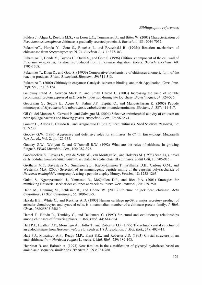
Bibliographic references
121
Folders J., Algra J., Roelofs M.S., van Loon L.C., Tommassen J., and Bitter W. (2001) Characterization of Pseudomonas aeroginosa chitinase, a gradually secreted protein. J. Bacteriol., 183: 7044-7052.
FukamizoT., Honda Y., Goto S., Boucher I., and Brzezinski R. (1995a) Reaction mechanism of chitosanase from Streptomyces sp. N174. Biochem J., 311: 377-383.
Fukamizo T., Honda Y., Toyoda H., Ouchi S., and Goto S. (1996) Chitinous component of the cell wall of Fusarium oxysporum, its structure deduced from chitosanase digestion. Biosci. Biotech. Biochem., 60: 1705-1708.
Fukamizo T., Koga D., and Goto S. (1995b) Comparative biochemistry of chitinases-anomeric form of the reaction products. Biosci. Biotechnol. Biochem., 59: 311-313.
Fukamizo T. (2000) Chitinolytic enzymes: Catalysis, substrate binding, and their Application. Curr. Prot. Pept. Sci., 1: 105-124.
Galloway Chad A., Sowden Mark P., and Smith Harold C. (2003) Increasing the yield of soluble recombinant protein expressed in E. coli by induction during late log phase. Biotechniques, 34: 524-526.
Gevorkian G., Segura E., Acero G., Palma J.P., Espitia C., and Manoutcharian K. (2005) Peptide mimotopes of Mycobacterium tuberculosis carbohydrate imunodeterminants. Biochem. J., 387: 411-417.
Gil G., del Monaco S., Cerrutti P., and Galvagno M. (2004) Selective antimicrobial activity of chitosan on beer spoilage bacteria and brewing yeasts. Biotechnol. Lett., 26: 569-574.
Gomez L., Allona I., Casado R., and Aragoncillo C. (2002) Seed chitinases. Seed Sciences Research, 12: 217-230.
Gooday G.W. (1996) Aggressive and defensive roles for chitinases. In Chitin Enzymology, Muzzarelli R.A.A., ed., Vol. 2, pp. 125-135.
Gooday G.W., Wei-yun Z, and O’Donnell R.W. (1992) What are the roles of chitinase in growing fungus?. FEMS Microbiol. Lett., 100: 387-392.
Goormachtig S., Lievens S., van de Velde W., van Montagu M., and Holsters M. (1998) Srchi13, a novel early nodulin from Sesbania rostrata, is related to acidic class III chitinases. Plant Cell, 10: 905-915.
Grothaus M.C. Srivastava N., Smithson S.L., Kieber-Emmon T., Williams D.B., Carlone G.M., and Westerink M.A. (2000) Selection of an immunogenic peptide mimic of the capsular polysaccharide of Neisseria meningitidis serogroup A using a peptide display library. Vaccine, 18: 1253-1263.
Gulati S., Ngampasutadol J., Yamasaki R., McQuillen D.P., and Rice P.A. (2001) Strategies for mimicking Neisserial saccharides epitopes as vaccines. Intern. Rev. Immunol., 20: 229-250.
Hahn M., Henning M., Schlesier B., and Höhne W. (2000) Structure of jack bean chitinase. Acta Crystallogr. D Biol. Crystallogr., 56: 1096-1099.
Hakala B.E., White C., and Recklies A.D. (1993) Human cartilage gp-39, a major secretory product of articular chondrocytes and synovial cells, is a mammalian member of a chitinase protein family. J. Biol. Chem., 268:25803-25810.
Hamel F., Boivin R., Temblay C., and Bellemare G. (1997) Structural and evolutionary relationships among chitinases of flowering plants. J. Mol. Evol., 44: 614-624.
Hart P.J., Heather D.P., Monzingo A., Hollis T., and Robertus J.D. (1995) The refined crystal structure of an endochitinase from Hordeum vulgare L. seeds at 1.8 Å resolution. J. Mol. Biol., 248: 402-413.
Hart P.J., Monzingo A.F., Ready M.P., Ernst S.R., and Robertus J.D. (1993) Crystal structure of an endochitinase from Hordeum vulgare L. seeds. J. Mol. Biol., 229: 189-193.
Henrissat B. and Bairoch A. (1993) New families in the classification of glycosyl hydrolases based on amino acid sequence similarities. Biochem J., 293: 781-788.
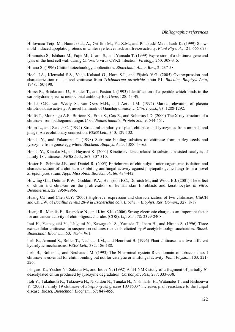
Bibliographic references
122
Hiilovaara-Teijo M., Hannukkala A., Griffith M., Yu X.M., and Pihakaski-Maunsbach K. (1999) Snow-mold-induced apoplatic proteins in winter rye leaves lack antifreeze activity. Plant Physiol., 121: 665-673.
Hiramatsu S., Ishihara M., Fujie M., Usami S., and Yamada T. (1999) Expression of a chitinase gene and lysis of the host cell wall during Chlorella virus CVK2 infection. Virology, 260: 308-315.
Hirano S. (1996) Chitin biotechnology applications. Biotechnol. Annu. Rev., 2: 237-58.
Hoell I.A., Klemsdal S.S., Vaaje-Kolstad G., Horn S.J., and Eijsink V.G. (2005) Overexpression and characterization of a novel chitinase from Trichoderma atroviride strain P1. Biochim. Biophys. Acta, 1748: 180-190.
Hoess R., Brinkmann U., Handel T., and Pastan I. (1993) Identification of a peptide which binds to the carbohydrate-specific monoclonal antibody B3. Gene, 128: 43-49.
Hollak C.E., van Weely S., van Oers M.H., and Aerts J.M. (1994) Marked elevation of plasma chitotriosidase activity. A novel hallmark of Gaucher disease. J. Clin. Invest., 93, 1288-1292.
Hollis T., Monzingo A.F., Bortone K., Ernst S., Cox R., and Robertus J.D. (2000) The X-ray structure of a chitinase from pathogenic fungus Coccidioides immitis. Protein Sci., 9: 544-551.
Holm L., and Sander C. (1994) Structural similarity of plant chitinase and lysozymes from animals and phage: An evolutionary connection. FEBS Lett., 340: 129-132.
Honda Y., and Fukamizo T. (1998) Substrate binding subsites of chitinase from barley seeds and lysozyme from goose egg white. Biochem. Biophys. Acta, 1388: 53-65.
Honda Y., Kitaoka M., and Hayashi K. (2004) Kinetic evidence related to substrate-assisted catalysis of family 18 chitinases. FEBS Lett., 567: 307-310.
Hoster F., Schmitz J.E., and Daniel R. (2005) Enrichment of chitinolytic microorganisms: isolation and characterization of a chitinase exhibiting antifungal activity against phytopathogenic fungi from a novel Streptomyces strain. Appl. Microbiol. Biotechnol., 66: 434-442.
Howling G.I., Dettmar P.W:, Goddard P.A:, Hampson F.C., Dornish M., and Wood E.J. (2001) The effect of chitin and chitosan on the proliferation of human skin fibroblasts and keratinocytes in vitro. Biomaterials, 22: 2959-2966.
Huang C.J, and Chen C.Y. (2005) High-level expression and characterization of two chitinases, ChiCH and ChiCW, of Bacillus cereus 28-9 in Escherichia coli. Biochem. Biophys. Res. Comun., 327: 8-17.
Huang R., Mendis E., Rajapakse N., and Kim S.K. (2006) Strong electronic charge as an important factor for anticancer activity of chitooligosaccharides (COS). Life Sci., 78: 2399-2408.
Inui H., Yamaguchi Y., Ishigami Y., Kawaguchi S., Yamada T., Ihara H., and Hirano S. (1996) Three extracellular chitinases in suspension-cultures rice cells elicited by N-acetylchitooligosaccharides. Biosci. Biotechnol. Biochem., 60: 1956-1961.
Iseli B., Armand S., Boller T., Neuhaus J.M., and Henrissat B. (1996) Plant chitinases use two different hydrolytic mechanisms. FEBS Lett., 382: 186-188.
Iseli B., Boller T., and Neuhaus J.M. (1993) The N-terminal cystein-Rich domain of tobacco class I chitinase is essential for chitin binding but not for catalytic or antifungal activity. Plant Physiol., 103: 221-226.
Ishiguro K., Yoshie N., Sakurai M., and Inoue Y. (1992) A 1H NMR study of a fragment of partially N-deacetylated chitin produced by lysozyme degradation. Carbohydr. Res., 237: 333-338.
Itoh Y., Takahashi K., Takizawa H., Nikaidou N., Tanaka H., Nishihashi H., Watanabe T., and Nishizawa Y. (2003) Family 19 chitinase of Streptomyces griseus HUT6037 increases plant resistance to the fungal disease. Biosci. Biotechnol. Biochem., 67: 847-855.

Bibliographic references
123
Jach G., Görnhart B., Mundy J., Logemann J., Leah R., Schell J., and Maas C. (1995) Enhanced quantitative resistance against fungal disease by combinatorial expression of different barley antifungal proteins in transgenic tobacco. Plant J., 8: 97-109.
Jain D., Kaur K., Goel M., and Salunk D.M. (2000b) Structural basis of functional mimicry between carbohydrate and peptide ligands of ConA. Biochem. Biophys. Res. Commun, 272: 843-849.
Jain D., Kaur K., Sundaravadivel B., and Salunk D.M. (2000a) Structural and functional consequences of peptide-carbohydrate mimicry. J. Biol. Chem., 275: 16098-16102.
Jacquet A., Daminet V., Haumont M., Garcia L., Chaudoir S., Bollen A., and Biemans R. (1999) Expression of a recombinant Toxoplasma gondii ROP2 fragment as a fusion proteolytic degradation. Protein Expr. Purif., 17: 392-400.
Jeon Y.J., Shahidi F., and Kim S.K. (2000) Preparation of chitin and chitosan oligomers and their applications in physiological functional foods. Food Rev. Int., 16: 159-176.
Janeway C.A., Travers P., Walport M., and Shlomchik M. (2001) Immuno biology 5: The immune system in health and disease. 5th edition, Garland Publishing.
Jeuniaux C., and Voss-Foucart M.F. (1991) Chitin biomass and production in the marine environment. Biochem. Syst. Ecol., 19: 347-356.
Johnson M.A., and Pinto M. (2002) Molecular mimicry of carbohydrates by peptides. Aust. J. chem., 55: 13-25.
Jongedijk E., Tigelaar H., van Roekel J.S.C., Bres-Vloemans S.A., Dekker I., van den Elzen P.J.M., Cornelissen B.J.C., and Melchers L.S. (1995) Synergistic activity of chitinases and β-1,3-glucanases enhances fungal resistance in transgenic tomato plants. Euphytica, 85: 173-177.
Jouault T., Fradin C., Dzierszinski F., Borg-Von-Zepelin M., Tomavo S., Corman R., Trinel P.A., Kerckaert J.P, and Poulain D. (2001) Peptides that mimic Candida albicans-derived β-1,2-linked mannosides. Glycobiology, 11: 693-701.
Jung W.J., Jo G.H., Kuk J.H., Kim K.Y., Park R.D. (2005) Extraction of chitin from red crabe shell waste by cofermentation with Lactobacillus paracasei subsp. Tolerans KCTC-3074 and Serratia marcesecns FS-3. Appl. Microbiol. Biotechnol., Online First issue.
Kaomek M., Mizuno K., Fujimura T., Sriyotha P., and Ketudat C.J. (2003) Cloning, Expression, and characterization of an antifungal chitinase from Leucaena leucocephala de Wit. Biosci. Biotechnol. Biochem., 67:667-676.
Kapila, J., De Rycke, R., van Montagu, and M., and Agenon, G. (1996) An Agrobacterium mediated transient gene expression system for intact leaves. Plant Sci., 122: 101-108.
Karasuda S., Tanaka S., Kajihara H., Yamamoto Y., and Koga D. (2003) Plant chitinase as possible biocontrol agent for use instead of chemical fungicides. Biosci. Biotechnol. Biochem., 67: 221-224.
Kasaai M.R., Malaekeh M. (2003) The effect of degree of acetylation of the chemical structure and the properties of chitin and chitosan: a short review. In “Advances in chitin science” I. Boucher, K. Jamieson, A. Retnakaran, eds., Proceedings of the 9th ICCC, Vol.VII, pp. 178-179.
Kasprzewska A. (2003) Plant chitinases-Regulation and function. Cell. Mol. Biol. Lett., 8: 809-824.
Keen N.T., and Yoshikawa M. (1989) β-1,3-endoglucanase from soybean releases elicitor-active carbohydrates from fungus cell wall. Plant Physiol., 71: 460-465.
Kendra D.F., Christian D., and Hadwiger L.A. (1989) Chitosan oligomers from Fusarium solani/pea interactions, chitinase/β-glucanase digestion of sporelings from fungal wall chitin actively inhibit fungal growth and enhance disease resistance. Physiol. Mol. Plant Path., 35: 215-230.
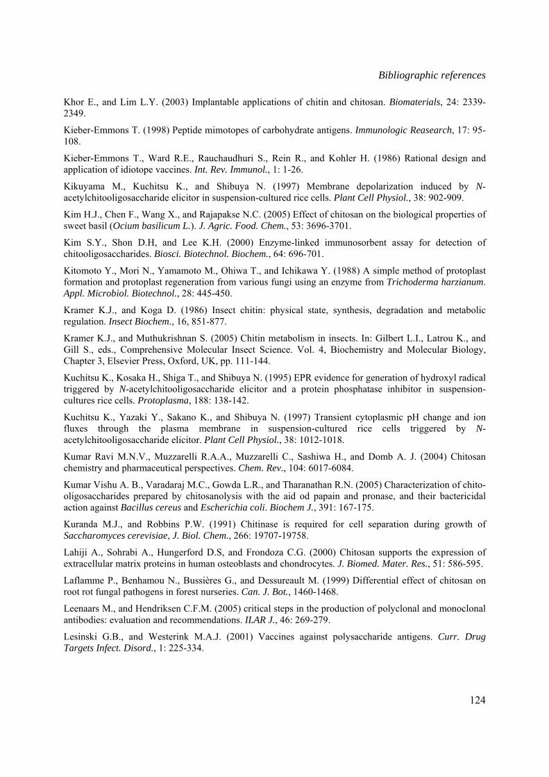
Bibliographic references
124
Khor E., and Lim L.Y. (2003) Implantable applications of chitin and chitosan. Biomaterials, 24: 2339-2349.
Kieber-Emmons T. (1998) Peptide mimotopes of carbohydrate antigens. Immunologic Reasearch, 17: 95-108.
Kieber-Emmons T., Ward R.E., Rauchaudhuri S., Rein R., and Kohler H. (1986) Rational design and application of idiotope vaccines. Int. Rev. Immunol., 1: 1-26.
Kikuyama M., Kuchitsu K., and Shibuya N. (1997) Membrane depolarization induced by N-acetylchitooligosaccharide elicitor in suspension-cultured rice cells. Plant Cell Physiol., 38: 902-909.
Kim H.J., Chen F., Wang X., and Rajapakse N.C. (2005) Effect of chitosan on the biological properties of sweet basil (Ocium basilicum L.). J. Agric. Food. Chem., 53: 3696-3701.
Kim S.Y., Shon D.H, and Lee K.H. (2000) Enzyme-linked immunosorbent assay for detection of chitooligosaccharides. Biosci. Biotechnol. Biochem., 64: 696-701.
Kitomoto Y., Mori N., Yamamoto M., Ohiwa T., and Ichikawa Y. (1988) A simple method of protoplast formation and protoplast regeneration from various fungi using an enzyme from Trichoderma harzianum. Appl. Microbiol. Biotechnol., 28: 445-450.
Kramer K.J., and Koga D. (1986) Insect chitin: physical state, synthesis, degradation and metabolic regulation. Insect Biochem., 16, 851-877.
Kramer K.J., and Muthukrishnan S. (2005) Chitin metabolism in insects. In: Gilbert L.I., Latrou K., and Gill S., eds., Comprehensive Molecular Insect Science. Vol. 4, Biochemistry and Molecular Biology, Chapter 3, Elsevier Press, Oxford, UK, pp. 111-144.
Kuchitsu K., Kosaka H., Shiga T., and Shibuya N. (1995) EPR evidence for generation of hydroxyl radical triggered by N-acetylchitooligosaccharide elicitor and a protein phosphatase inhibitor in suspension-cultures rice cells. Protoplasma, 188: 138-142.
Kuchitsu K., Yazaki Y., Sakano K., and Shibuya N. (1997) Transient cytoplasmic pH change and ion fluxes through the plasma membrane in suspension-cultured rice cells triggered by N-acetylchitooligosaccharide elicitor. Plant Cell Physiol., 38: 1012-1018.
Kumar Ravi M.N.V., Muzzarelli R.A.A., Muzzarelli C., Sashiwa H., and Domb A. J. (2004) Chitosan chemistry and pharmaceutical perspectives. Chem. Rev., 104: 6017-6084.
Kumar Vishu A. B., Varadaraj M.C., Gowda L.R., and Tharanathan R.N. (2005) Characterization of chito-oligosaccharides prepared by chitosanolysis with the aid od papain and pronase, and their bactericidal action against Bacillus cereus and Escherichia coli. Biochem J., 391: 167-175.
Kuranda M.J., and Robbins P.W. (1991) Chitinase is required for cell separation during growth of Saccharomyces cerevisiae, J. Biol. Chem., 266: 19707-19758.
Lahiji A., Sohrabi A., Hungerford D.S, and Frondoza C.G. (2000) Chitosan supports the expression of extracellular matrix proteins in human osteoblasts and chondrocytes. J. Biomed. Mater. Res., 51: 586-595.
Laflamme P., Benhamou N., Bussières G., and Dessureault M. (1999) Differential effect of chitosan on root rot fungal pathogens in forest nurseries. Can. J. Bot., 1460-1468.
Leenaars M., and Hendriksen C.F.M. (2005) critical steps in the production of polyclonal and monoclonal antibodies: evaluation and recommendations. ILAR J., 46: 269-279.
Lesinski G.B., and Westerink M.A.J. (2001) Vaccines against polysaccharide antigens. Curr. Drug Targets Infect. Disord., 1: 225-334.
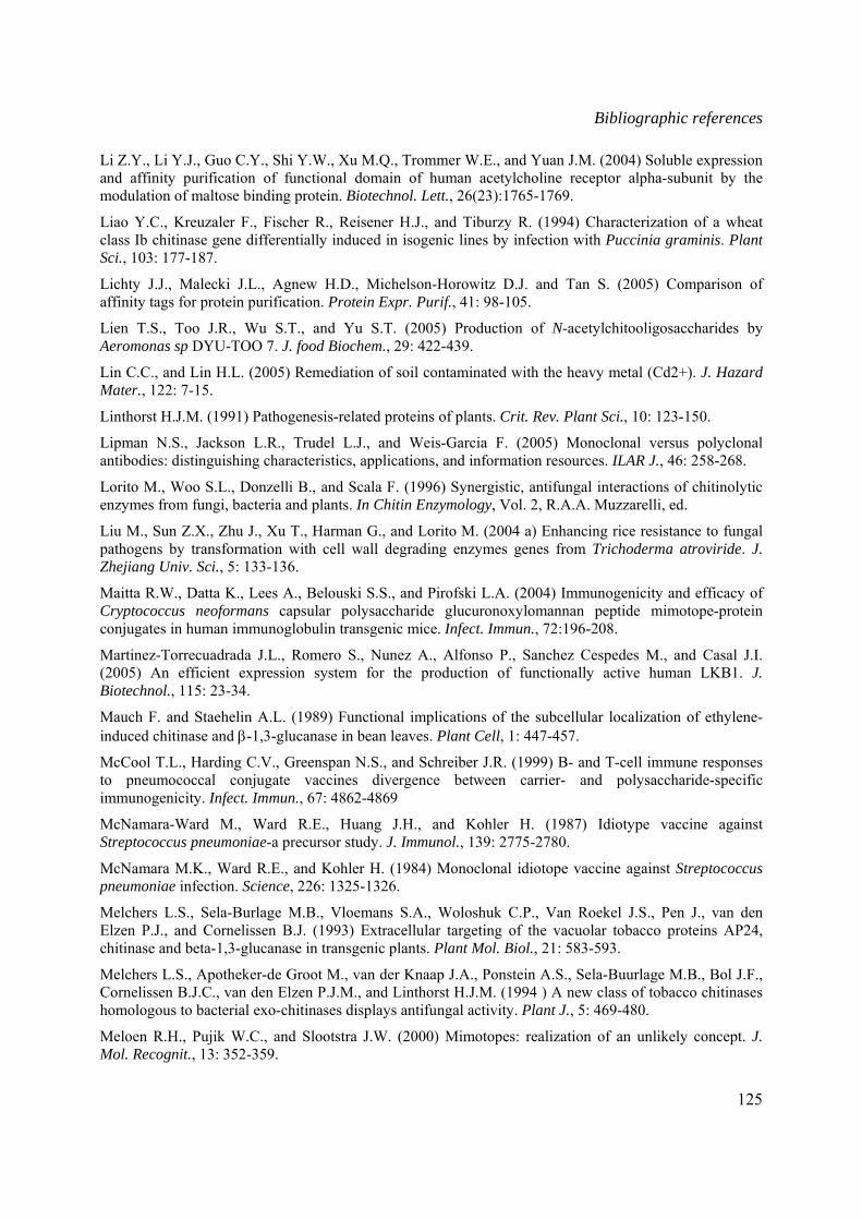
Bibliographic references
125
Li Z.Y., Li Y.J., Guo C.Y., Shi Y.W., Xu M.Q., Trommer W.E., and Yuan J.M. (2004) Soluble expression and affinity purification of functional domain of human acetylcholine receptor alpha-subunit by the modulation of maltose binding protein. Biotechnol. Lett., 26(23):1765-1769.
Liao Y.C., Kreuzaler F., Fischer R., Reisener H.J., and Tiburzy R. (1994) Characterization of a wheat class Ib chitinase gene differentially induced in isogenic lines by infection with Puccinia graminis. Plant Sci., 103: 177-187.
Lichty J.J., Malecki J.L., Agnew H.D., Michelson-Horowitz D.J. and Tan S. (2005) Comparison of affinity tags for protein purification. Protein Expr. Purif., 41: 98-105.
Lien T.S., Too J.R., Wu S.T., and Yu S.T. (2005) Production of N-acetylchitooligosaccharides by Aeromonas sp DYU-TOO 7. J. food Biochem., 29: 422-439.
Lin C.C., and Lin H.L. (2005) Remediation of soil contaminated with the heavy metal (Cd2+). J. Hazard Mater., 122: 7-15.
Linthorst H.J.M. (1991) Pathogenesis-related proteins of plants. Crit. Rev. Plant Sci., 10: 123-150.
Lipman N.S., Jackson L.R., Trudel L.J., and Weis-Garcia F. (2005) Monoclonal versus polyclonal antibodies: distinguishing characteristics, applications, and information resources. ILAR J., 46: 258-268.
Lorito M., Woo S.L., Donzelli B., and Scala F. (1996) Synergistic, antifungal interactions of chitinolytic enzymes from fungi, bacteria and plants. In Chitin Enzymology, Vol. 2, R.A.A. Muzzarelli, ed.
Liu M., Sun Z.X., Zhu J., Xu T., Harman G., and Lorito M. (2004 a) Enhancing rice resistance to fungal pathogens by transformation with cell wall degrading enzymes genes from Trichoderma atroviride. J. Zhejiang Univ. Sci., 5: 133-136.
Maitta R.W., Datta K., Lees A., Belouski S.S., and Pirofski L.A. (2004) Immunogenicity and efficacy of Cryptococcus neoformans capsular polysaccharide glucuronoxylomannan peptide mimotope-protein conjugates in human immunoglobulin transgenic mice. Infect. Immun., 72:196-208.
Martinez-Torrecuadrada J.L., Romero S., Nunez A., Alfonso P., Sanchez Cespedes M., and Casal J.I. (2005) An efficient expression system for the production of functionally active human LKB1. J. Biotechnol., 115: 23-34.
Mauch F. and Staehelin A.L. (1989) Functional implications of the subcellular localization of ethylene-induced chitinase and β-1,3-glucanase in bean leaves. Plant Cell, 1: 447-457.
McCool T.L., Harding C.V., Greenspan N.S., and Schreiber J.R. (1999) B- and T-cell immune responses to pneumococcal conjugate vaccines divergence between carrier- and polysaccharide-specific immunogenicity. Infect. Immun., 67: 4862-4869
McNamara-Ward M., Ward R.E., Huang J.H., and Kohler H. (1987) Idiotype vaccine against Streptococcus pneumoniae-a precursor study. J. Immunol., 139: 2775-2780.
McNamara M.K., Ward R.E., and Kohler H. (1984) Monoclonal idiotope vaccine against Streptococcus pneumoniae infection. Science, 226: 1325-1326.
Melchers L.S., Sela-Burlage M.B., Vloemans S.A., Woloshuk C.P., Van Roekel J.S., Pen J., van den Elzen P.J., and Cornelissen B.J. (1993) Extracellular targeting of the vacuolar tobacco proteins AP24, chitinase and beta-1,3-glucanase in transgenic plants. Plant Mol. Biol., 21: 583-593.
Melchers L.S., Apotheker-de Groot M., van der Knaap J.A., Ponstein A.S., Sela-Buurlage M.B., Bol J.F., Cornelissen B.J.C., van den Elzen P.J.M., and Linthorst H.J.M. (1994 ) A new class of tobacco chitinases homologous to bacterial exo-chitinases displays antifungal activity. Plant J., 5: 469-480.
Meloen R.H., Pujik W.C., and Slootstra J.W. (2000) Mimotopes: realization of an unlikely concept. J. Mol. Recognit., 13: 352-359.
Bibliographic references
126
Merzendorfer H., and Zimoch L. (2003) Chitin metabolism in insects: structure, function and regulation of chitin synthases and chitinases. J. Exp. Biol., 206: 4393-4412.
Minke R., and Blackwell J. (1978) The structure of alpha-chitin. J. Mol. Biol., 120: 167-181.
Mizuno K., Yamamura K., Yano K., Osada T., Saeki S., Takimoto N., Sakura T., and Nimura Y. (2003) Effect of chitosan film containing basic fibroplast growth factor on wound healing in genetically diabetic mice. J. Biomed. Mater. Res., 64: 177-181.
Mitsutomi M., Kidoh H., Tomita H., and Watanabe T. (1995) The action of Bacillus circulans WL-12 on partially N-acetylated chitosan. Biosci. Biotechnol. Biochem., 59: 529-531.
Moerschbabacher B.M., Kogel K.H., Noll U., and Reisener H.-J. (1986) An elicitor of the hypersensitive lignification response in wheat leaves isolated from the rust fungus Puccinia graminis f.sp. tritici. I. Partial purification and characterization. Z. Naturforsch., 41c: 830-838.
Moerschbacher B.M., Flott B.E., Noll U., Reisener H.J. (1989) On the specificity of an elicitor preparation from stem rust which induces lignification in wheat leaves. Plant Physiol. Biochem., 27: 305-314.
Möller H., Grelier S., Pardon P., and Coma V. (2004) Antimicrobial and physicochemical properties of chitosan-HPMC-based films. J. Agric. Chem., 52: 6585-6591.
Mond J.J., Lees A., and Snapper C.M. (1995) T cell-independent antigens type 2. Annu. Rev. immunol., 13: 655-692.
Monzavi-Karbassi B., Cunto-Amesty G., Luo P., and Kieber-Emmons T. (2002) Peptide mimotopes as surrogate antigens of carbohydrates in vaccine discovery. Trends Biotechnol., 20: 207-214.
Monzingo A.F., Marcotte E.M., Hart P.J., and Robertus J.D. (1996) Chitinases, chitosanases, and lysozymes can be divided into procaryotic and eucaryotic families sharing a conserved core. Nat. Struct. Biol., 3: 133-140.
Muzzarelli R.A.A., Muzzarelli C., Tarsi R., Miliani M., Gabbanelli F., and Cartolari M. (2001) Fungistatic activity of modified chitosans against Saprolegnia parasitica. Biomacromolecules, 2: 165-169.
Muzzarelli R.A.A., Biagini G., Pugnaloni A., Filippini O., Baldassarre V., Castaldini C., and Rizzoli C. (1989a) Reconstruction of parodontal tissue with chitosan. Biomaterials, 10: 598-603.
Muzzarelli R.A.A., Weckx M., and Fillipini O. (1989b) Removal of trace metal ions from industrial waters, unclear effluents and drinking water, with the aid of cross-linked N-carboxymethyl chitosan. Carbohydr. Poly., 11: 293-296.
Neuhaus J.M., Sticher L., Meins F.Jr., and Boller T. (1991) A short C-terminal sequence is necessary and sufficient for the targeting of chitinases to the plant vacuole. Proc. Natl. Acad. Sci. U.S.A., 88: 10362-1066.
Neuhaus J.M., Fritig B., Linthorst H.J.M., Meins F., Mikkelsen J.D., and Ryals J. (1996) A revised nomenclature for chitinase genes. Plant Mol. Biol. Rep., 14: 102-104.
Neuhaus J.M. (1999) Plant chitinases (PR-3, PR-4, PR-8, PR-11). In: Datta S.K., Muthukrishnan S., eds., Pathogenesis-related proteins in plants. Boca Raton, FL: CRC Press, pp. 77-105.
Nishizawa Y., Kawakami A., Habi T., He D.Y., Shibuya N., and Minami E. (1999) Regulation of the chitinase gene expression in suspension-cultures rice cells by N-acetylchitooligosaccharides: differences in the signal transduction pathways leading to the activation of elicitor-responsive genes. Plant Mol. Biol., 39: 907-914.
No H.K., Mayers S.P. (1997) Preparation of chitin and chitosan. In: Chitin Handbook, Muzarelli R.A.A., Peter M.G., eds, European Chitin Society, pp 475-489.
Bibliographic references
127
Nojiri H., Sugimori M., Yamane H., Nishimura Y., Yamada A., and Shibuya N. (1996) Involvement of jasmonic acid in elicitor-induced phytoalexin production in suspension-cultured rice cells. Plant Physiol., 110: 387-392.
Nominé Y., Ristriani T., Laurent C., Lefevre J.F., Weiss E., and Trave G. (2001) Formation of soluble inclusion bodies by hpv e6 oncoprotein fused to maltose-binding protein. Protein Expr. Purif., 23: 22-32.
O’Conell R.J. (1991) Cytochemical analysis of infection structures of Colletotrichum lindemuthinum using fluorochrome-labelled lectins. Physiol. Mol. Plant Pathol., 39:189-200.
Oh H., Kim Y.J., Chang E.J., and Kim J.Y. (2001) Antimicrobial characteristics of chitosan against food spoilage microorganisms in liquid media and mayonnaise. Biosci. Biotachnol. Biochem., 65: 2378-2383.
Ohno T., Armand S., Hata T., Nikaidou N., Henrissat B., Mitsutomi M., and Watanabe T. (1996) A modular family 19 chitinase found in the procaryotic organism Streptomyces griseus HUT 6037. Journal of Bacteriology, 178: 5065-5070.
Oldenbourg K.R., Longanathan D., Goldstein I.J., Schultz P.G., and Galop M.A. (1992) Peptide ligands for a sugar-binding protein isolated from a random peptide library. Proc. Natl. Acad. Sci. USA, 89: 5393-5397.
Ovtsyna A.O., Schultze M., Tikhonovich I.A., Spaink H.P., Kondorosi E., Kondorosi A., and Staehelin C. (2000) Nod factors of Rhizobium leguminosarum cv. Viciae and their fucosylated derivatives stimulate a Nod factor cleaving activity in pea roots and are hydrolyzed in vitro by plant chitinases at different rates. Mol. Plant Microbe Interac., 13: 799-807.
Okazaki K., Yamashita Y., Noda M., Sueyoshi N., Kameshita I., and Hayakawa S. (2004) Molecular cloning and expression of the gene encoding family 19 chitinase from Streptomyces sp. J-13-3. Biosci. Biotechnol. Biochem., 68: 341-351.
Okinaka Y., Mimori K., Takeo K., Kitamura S., Takeuchi Y., Yamaoka N., Yoshikawa M. (1995) A structural model for the mechanisms of elicitor release from fungal cell walls by plant β-1,3-endoglucanase. Plant Physiol., 109: 119-123.
Okamura Y., Nomura A., Minami S., Okamoto Y. (2005) Effects of chitin/chitosan and their oligomers/monomers on release of type I collagenase from fibroblasts. Biomacromolecules, 6: 2382-2384.
Ornum J.V. (1992) Shrimp waste-must it be wasted. Infofish Int., 6: 48-52.
Pae H.O., Seo W.G., Kim N.Y., Oh G.S., Kim G.E., Kim Y.H., Kwak H.J., Yun Y.G., Jun C.D., and Chung H.T. (2001) Induction of granulocytic differentiation in acute promyelocytic leukemia cells (HL-60) by water-soluble chitosan oligomer. Leuk Res., 25: 339-346.
Papanikolau Y., Prag G., Tavlas G., Vorgias C.E., Oppenheim A.B., and Petratos K. (2001) High resolution structural analyses of mutant chitinase A complexes with substrates provide new insight into the mechanism of catalysis. Biochemistry, 40: 11338-11343.
Patil R.S., Ghormade V., and Deshpande M.V. (2000) Chitinolytic enzymes: An exploration. Enzyme Microb. Technol., 26: 473-483.
Park H.Y., Pan C.H., So M.Y., Ahn J.H., Jo D.H., and Kim S.I. (2001) Purification, characterization, and cDNA cloning of rice class III chitinase. Mol. Cell., 13: 69-76.
Park I, Choi I. H., Kim S. J., and Shin J. S. (2004) Peptide mimotopes of Neisseria meningitidis group B capsular polysaccharide. Yonsei Med. J., 45: 755-758.
Pashov A., Canziani G., Monzavi-Karbassi B., Kaveri S.V., MacLeod S., Saha R., Perry M., VanCott T.C., and Kieber-Emmons T. (2005) Antigenic properties of peptide mimotopes of HIV-1 associated carbohydrate antigens. J. Biol. Chem., 280: 28959-28965.
Bibliographic references
128
Paul W., and Sharma C.P. (2004) Chitosan and alginate wound dressing: A short review. Trends Biomater. Artif. Organs, 18: 18-23.
Perrakis A., Tews I., Dauter Z., Oppenheim A.B., Chet I., Wilson K.S., and Vogias E. (1994) Crystal structure of a bacterial chitinase at 2.3-Angstrom resolution. Structure, 2: 1169-1180.
Perrakis A., Tews I., Wilson K.S., and Vorgias C.E. (1996) Structural aspects on the catalytic mechanism of chitinases, hevamine, and chitobiases “Far away and yet so close?”. In Chitin Enzymology, Muzzarelli R.A.A., ed., Vol 2, pp. 109-122.
Peschen D., Li H.P., Fischer R., Kreuzaler F., and Liao Y.C. (2004) Fusion proteins comprising a Fusarium-specific antibody linked to antifungal peptides protect plants against a fungal pathogen. Nat. Biotechnol., 22:732-738.
Phalipon A., Folgori A., Arondel J., Sgaramella G., Fortugno P., Cortese R., Sansonetti P.J., and Felici F. (1997) Induction of anti-carbohydrate antibodies by phage library-selected peptide mimics. Eur. J. Immunol., 27: 2620-2625
Pincus S.H., Smith M.J., Jennings H.J., Burritt J.B., and Glee P.M. (1998) Peptides that mimic the group B streptococcal type II capsular polysaccharide antigen. J. Immunol., 160: 293-298.
Pirofski L.A. (2001) Polysaccharides, mimotopes and vaccines for fungal and encapsulated pathogens. Trends Microbiol., 9: 445-451.
Prins M., Lohuis D., Schots A., and Goldbach R. (2005) Phage display-selected single-chain antibodies confer high levels of resistance against tomato spotted wilt virus. J. Gen. Virol., 86: 2107-2113.
Rabea E.I., Badawy M.E.T., Stevens C.V., Smagghe G., and Steurbaut W. (2003) Chitosan as antimicrobial agent: Applications and mode of action. Biomacromolecules, 4: 1457-1465.
Ramirez N., Avala M., Lorenzo D., Palenzuela D., Herrera L., Doreste V., Perez M., Gavilond J.V., and Oramas P. (2002) Expression of a single–chain Fv antibody fragment specific for the hepatitis B surface antigen in transgenic tobacco plants. Transgenic Res., 11: 61-64.
Rast D.M., Baumgartner D., Mayer C., and Hollenstein G.O. (2003) Cell wall-associated enzymes in fungi. Phytochemistry, 64: 339-366.
Rast D.M., Horsh M., Furter R., and Gooday G.W. (1991) A complex chitinolytic system in exponentially growing mycellium of Mucor rouxii: properties and function. J. Gen. Microbiol., 137: 2797-2810.
Rhoades J., and Roller S. (2000) Antimicrobial actions of degraded and native chitosan against spoilage organisms in laboratory media and food. Appl. Environ. Microbiol., 66: 80-86.
Rijkers G.T., Sanders E.A., Breukels M.A., Zegers B.J. (1998) Infant B cell responses to polysaccharide determinants. Vaccine, 16: 1396-1400
Roberts W.K., and Selitrennikoff C.P. (1988) Plant and bacterial chitinases differ in antifungal activity. J. Gen. Microbiol., 134: 169-176.
Robbins J.B., Schneerson R., Szu S.C., and Pozsgay V. (1999) Bacterial polysaccharide-protein conjugate vaccines. Pure Appl. Chem., 71:745-754.
Roby D., Gadelle A., and Toppan A. (1987) Chitin oligosaccharides as elicitors of chitinase activity in melon plants. Biochem. Biophys. Res. Commun., 143: 885-892.
Rodriguez-Herrera R., Waniska R.D., and Rooney W.L. (1999) Antifungal protein and grain mold resistance in sorghum with nonpigmented testa. J. Agric. Food. Chem., 47: 4802-4806.
Roller S. (1999) Physiology of food spoilage organisms. Int. J. Food Microbiol., 50: 151-153.
Bibliographic references
129
Ruiz-Sanchez A., Cruz-Camarillo R., Salcedo-Hernandez R., Ibarra J.E., and Barboza-Corona J.E. (2005) Molecular cloning and purification of an endochitinase from Serratia marcescens (Nima). Mol. Biotechnol., 31: 103-111.
Rupley J.A. (1964) The hydrolysis of chitin by concentrated hydrochloric acid, and the preparation of low-molecular substrate for lysozym. Biochem. Biophys. Acta, 83: 245-255.
Sahai A.S., and Manocha M.S. (1993) Chitinases of fungi and plants: their involvement in morphogenesis and host-parasite interaction. FEMS Microbiol. Rev., 11:317-338.
Saito A., Fujii T., Yoneyama T., Redenbach M., Ohne T., Watanabe T., and Miyashita K. (1999) High-multiplicity of chitinase genes in Streptomyces coelicolor A3(2). Biosci. Biotechnol. Biochem., 63: 710-718.
Sambrook J., Fritsch E.F., and Maniatis T. (1996) Molecular cloning: A laboratory manual. Cold Spring Harbor Laboratory Press. Cold Spring Harbor, New York.
Sasaki C. Yokoyama A., Itoh Y., Hashimoto M., Watanabe T., and Fukamizo T. (2002) Comparative study of the reaction mechanism of family 18 chitinases from plants and microbes. J. Biochem., 131: 557-564.
Sasaki C., Itoh Y., Takehara H., Kuhara S., and Fukamizo T. (2003) Family 19 chitinase from rice (Oryza sativa L.): substrate-binding subsites demonstrated by kinetic and molecular modeling studies. Plant Mol. Biol., 52: 43-52.
Schahidi F., and Abuzaytoun R. (2005) Chitin, Chitosan, and co-products: chemistry, production, applications, and health effects. In “Advances in food and nutrition research” (S. Tylor, ed), Vol. 49, pp. 93-134. Elsevier.
Scheller J., Henggeler D., Viviani A., and Conrad U. (2004) Purification of spider silk-elastin from transgenic plants and application for human chondrocyte proliferation. Transgenic Res., 13: 51-57.
Schillberg S., Zimmermann S., Findlay K., and Fischer R. (2000) Plasma membrane display of anti-viral single chain Fv fragments confers resistance to tobacco mosaic virus. Mol. Breed, 6: 317-326.
Schlumbaum A., Mauch F., Vögeli U., and Boller T. (1986) Plant chitinases are potent inhibitors of fungal growth. Nature, 324: 365-367.
Scholthof, H.B., Scholthof, K.B., and Jackson, A.O. (1996) Plant virus gene vectors for transient expression of foreign proteins in plants. Annu. Rev. Phytopathol., 34: 299-323
Schouten A., Roosien J., Van Engelen F.A., de Jong G.A., Borst-Vrenssen A.W., Zilverentant J.F., Bosch D., Stiekema W.J., Gommers F.J., Schots A., and Bakker J. (1996) The C-terminal KDEL sequence increases the expression level of a single-chain antibody designed to be targeted to both the cytosol and the secretory pathway in transgenic tobacco. Plant Mol Biol., 30:781-793.
Schultze M., Staehelin C., Brunner F., Genetet I., Legrand M., Fritig B., Kondorosi E., and Kondorosi A. (1998) lant chitinase/lysozyme isoforms show distinct substrate specificity and cleavage site preference towards lipochitooligosaccharide Nod signals. Plant J., 16: 571-580.
Scott K.K., Loganathan D., Easley R.B., Gong X., and Goldstein I.J. (1992) A family of concanavalin A-binding peptides from a hexapeptide epitope library. Proc. Natl. Acad. Sci. USA, 89: 5398-5402.
Sefarian P.G. and Martinez M.L. (2001) Immune stimulating activity of two new chitosan containing adjuvant formulations. Vaccine, 19: 661-668.
Sela-Buurlage M., Ponstein A.S., Bres-Vloemans S.A, Melchers L.S., van den Elzen P.J.M, and Cornelissen B.J.C. (1993) Only specific tobacco (Nicotiana tabacum) chitinase and β-1,3-glucanases exhibit antifungal activity. Plant Physiol., 101: 857-863.
Bibliographic references
130
Shahidi F., and Abuzaytoun R. (2005) Chitin, chitosan, and co-products: chemistry, production, applications, and health effects. Advances in food and nutrition research, 49: 93-135.
Shinshi H., Neuhaus J.M., Ryals J., and Meins F. Jr. (1990) Structure of a tobacco chitinase gene: evidence that different chitinase genes can arise by transposition of sequences encoding a cysteine-rich domain. Plant Mol. Biol., 14: 357-368.
Singla A.K., and Chawla M. (2001) Chitosan: some pharmaceutical and biological aspects- an update. J. Pharm. Pharmacol., 53: 1047-1067.
Sinha V.R., and Kumria R. (2001) Polysaccharides in colon-specific drug delivery. Int. J. Pharm., 224: 19-38.
Skaugrud O. (1991) Chitosan – New biopolymer for cosmetics and drugs. Drug Cosmetic Ind., 148: 24-29.
Smith J., Kontermann R.E., Embleton J., and Kumar S. (2005) Antibody phage display technologies with special reference to angiogenesis. FASEB J., 19: 331-341.
Smyth D.R., Mrozkiewicz M.K., McGrath W.J., Listwan P. and Kobe B. (2003) Crystal structures of fusion proteins with large-affinity tags. Protein Sci., 12: 1313-1322.
Soderhall K. and Unestam T. (1975) Properties of extracellular enzymes from Aphanomyces astaci and their relevance in the penetration process of crayfish cuticle. Physiol. Plant., 35: 40-46.
Song H.K., and Suh S.W. (1996) Refined structure of the chitinase from barley seeds at 2.0 Å resolution. Acta Crystallogr. D Biol. Crystallogr., 52: 289-298.
Sørbotten A., Horn S.J., Eijsink V.G.H., and Vårum K.M. (2005) Degradation of chitosans with chitinase B from Serratia marscescens. Production of chito-oligosaccharides and insight into enzyme processivity. FEBS J., 272: 538-549.
Sorlier P., Hartmann D.J., Denuzière A., Viton C., and Domard A. (2003) Preparation and development of anti-chitosan antibodies. J. Biomed. Mater. Res. A, 67: 766-774.
Southworth M.V., Fuhrman J.A., Robbins P.W., Beauregard K., and Perler F.B. (1996) Gene cloning and production of active recombinant Brugia malayi microfilarial chitinase. Gene, 177: 55-58.
Spindler K.D. (1997) Chitinase and chitosanase assays. Chitin handbook, R.A.A Muzzarelli and M.G. Peters, eds. European Chitin Society, pp. 229-235.
Stein K.E., and Soderstrom T. (1984) Neonatal administration of idiotype or antiidiotype primes for protection against Escherichia coli infection in mice. J. Exp. Med., 160: 1001-1011.
Stinzi A., Heitz T., Prasad V., Wiedemann-Merdinoglu S., Kaufmann S., Geoffroy P., Legrand M., and Fritig B. (1993) Plant pathogenesis-related proteins and their role in defense against pathogens. Biochimie, 75: 687-706.
St.Leger R.J., Goettel M.S., Roberts D.W., and Staples R.C. (1991) Prepenetration events during infection of host cuticle by Metarhizium anisopliae. J. Invert. Pathol., 58: 168-179.
Strand S.P., Varum K.M., and Ostgaard K. (2003) Interactions between chitosan and bacterial suspensions: adsorption and flocculation. Colloids Surf. B Biointerfaces, 27: 71-81.
Suarez V., Staehelin C., Arango R., Holtorf H., Hofsteenge J., and Meins F. Jr. (2001) Substrate specificity and antifungal activity of recombinant tobacco class I chitinases. Plant Mol. Biol., 45: 609-618.
Subroto T., Sufiati S., and Beintema J.J. (1999) Papaya (Carica papaya) lysozyme is a member of the family 19 (basic, class II) chitinases. J. Mol. Evol., 49: 819-821.
Bibliographic references
131
Sugiyama T., Imai K., Ono A., Takayama Y., Tsujisaki M., Taki T., Hinoda Y., and Yachi A. (1991) Conformational structure of a monoclonal anti-idiotypic antibody to the monoclonal anti-adenocarcinoma-associated carbohydrate antibody YH206. J. Immunol., 146: 3097-3101.
Suo Y., and Leung D.W.M. (2001) Elevation of extracellular β-1,3-glucanase and chitinase activities in rose in response to treatment with acibenzolar-S-methyl and infection by D. rosae. J. Plant Physiol., 158: 971-976.
Suzuki S. (1996) Studies on biological effects of water soluble lower homologous oligosaccharides of chitin and chitosan. Fragrance J., 15: 61-68.
Suzuki K., Tokoro A., Okawa Y., Suzuki S., and Suzuki M. (1985) Enhancing effects of N-acetyl chitooligosaccharides on the active oxygen-generating and microbicidal activities of peritoneal exudates cells in mice. Chem. Pharm. Bull., 33: 886-888.
Suzuki S., Watanabe T., Mikami T., Matsumoto T., and Suzuki M. (1992) Immuno-enhancing effects of N-acetylchitohexaose. In “Advances in chitin and chitosan” (C.J. Brine, P.A. Sandford and J.P. Zikakis, eds), pp. 96-105, Elsevier Science. Publishers LTD.
Svitil A.L., Chadhain S.M.N., Moore J.A., and Kirchman D.L. (1997) Chitin degradation proteins produced by the marine bacterium Vibrio harveyi growing on different forms of chitin. Appl. Environ. Microbiol., 63: 408-413.
Synstad B., Gåseidnes S., van Aalten D.M.F., Vriend G., Nielsen J.E., and Eijsink G.H. (2004) Mutational and computational analysis of the role of conserved residues in the active site of a family 18 chitinase. Eur. J. Biochem., 271: 253-262.
Taira T., Ohnuma T., Yamagami T., Aso Y., Ishiguro M., and Ishihara M. (2002) Antifungal activity of rye (Secale cereal) seed chitinases: The different binding manners of class I and class II chitinases to the fungal cell wall. Biosci. Biotechnol. Biochem., 66: 970-977.
Taira T., Toma N., and Ishihara M. (2005a) Purification, Characterization, and Antifungal Activity of Chitinases from Pineapple (Ananas comosus) Leaf. Biosci. Biotechnol. Biochem., 69: 189-196.
Taira T., Ohdomari A., Nakama N., Shimoji M., and Ishihsra M. (2005b) Characterization and antifungal activity of Gazyumaru (Ficus microcarpa) latex chitinases: both the chitin-binding and the antifungal activities of class I chitinase are reinforced with increasing ionic strength. Biosci. Biotechnol. Biochem., 69: 811-818.
Tanabe T., Kawase T., Watanabe T., Uchida Y., and Mitsutomi M. (2000) Purification and characterization of a 49-kDa chitinase from Streptomyces griseus HUT 6037. J. Biosci. Bioeng., 89: 27-32.
Tang S.S., Tan W. S., Devi S., Wang L.F., Pang T., and Thong K.L. (2003) Mimotopes of the Vi antigen of Salmonella enterica serovar typhi identified from phage display peptide library. Clin. Diagn. Lab. Immunol., 10: 1078-1084.
Terwisscha van Scheltinga A.C., and Dijkstra B.W. (1996) Three-dimensional structure and mechanism of chitinase. In Chitin Enzymology, Vol. 2, Muzzarelli R.A.A., ed., pp. 143-146.
Terwisscha van Scheltinga A.C., Kalk K.H., Beintema J.J, and Dijkstra B.W. (1994) Crystal structures of hevamine, a plant defence protein with chitinase and lysozyme activity, and its complex with an inhibitor. Structure, 2: 1181-1189.
Tews I., Perrakis A., Oppenheim A., Dauter Z., Wilson K.S., and Vorgias C.E. (1996) Bacterial chitobiase structure provides insight into catalytic mechanism and the basis of Tay-Sachs disease. Nature Struct. Biol., 3: 638-648.
Tews I., van Scheltinga A.C.T., Perrakis A., Wilson K.S., and Dijkstra B.W. (1997) Substrate-assisted catalysis unifies two families of chitinolytic enzymes. J. Am. Chem. Soc., 119: 7954-7959.
Bibliographic references
132
Theis T., and Stahl U. (2004) Antifungal proteins: targets, mechanisms and prospective applications. Cell. Mol. Life Sci., 61: 437-455.
Tiburzy R., Noll U., and Reisener H.J. (1990) Resistance of wheat to Puccinia graminis f. sp. Tritici:histological investigation of resistance caused by the Sr5 gene. Physiol. Mol. Plant Pathol., 36: 95-108.
Torosantucci A., Bromuro C., Chiani P., De Bernardis F., Berti F., Galli C., Norelli F., Belluci C., Polonelli L., Costantino P., Rappuoli, and Cassone A. (2005) A novel glyco-conjugate vaccine against fungal pathogens. J. Exp. Med., 202: 597-606.
Trudel J. and Asselin A. (1989) Detection of chitinase activity after polyacrylamide gel electrophoresis. Anal. Biochem., 178: 362-366.
Truong N.H., Park S.M., Nishizawa Y., Watanabe T., Sasaki T., and Itohi Y. (2003) Structure, heterologous expression, and properties of rice (Oryza sativa L.) family 19 chitinases. Biosci. Biotechnol., Biochem., 67: 1063-1070.
Tsai G.J., Su W.H., Chen H.C., and Pan C.L. (2002) Antimicrobial activity of shrimp chitin and chitosan from different treatments and applications of fish preservation. Fisheries Sci., 68: 170-177.
Tsai G.J., Wu Z.Y., and Su W.H. (2000) Antibacterial activity of a chitooligosaccharide mixture prepared by cellulase digestion of shrimp chitosan and its application to milk preservation. J. Food Prot., 63: 747-752.
Tsigos I., Martinou A. Kafetzopoulos D., and Bouriotis V. (2000) Chitin deacetylases: New versatile tools in biotechnology. Trends Biotechnol., 18: 305-312.
Tsukada K., Matsumoto T., Aizawa K., Tokoro A., Naruse R., Suzuki S., and Suzuki M. (1990) Antimetastatic and growth-inhibitory effects of N-acetylchitohexaose in mice bearing Lewis lung carcinoma. Jpn. J. Cancer Res., 81: 259-265.
Ueno H., Yamada H., Tanaka I., Kaba N., Matsuura M., Okumura M., Kadosawa T., and Fujinaga T. (1999) Accelerating effects of chitosan for healing at early phase of experimental open wound in dogs. Biomaterials, 20: 1407-1414.
Uchida Y., Izume M., and Ohtakara A. (1989) Preparation of chitosan oligomers with purified chitosanase and its application. In “Chitin and Chitosan” (G. Skjak-braek, T. Anthonsen, and P. Sandford, eds), pp. 373-382. Elsevier, London, UK.
Ueda M., Kojima M., Yoshikawa T., Mitsuda N., Araki K., Kawaguchi T., Miyatake K., Arai M., and Fukamizo T. (2003) A novel type of family 19 chitinase from Aeromonas sp. No.10S-24. Cloning, sequence, expression, and the enzymatic properties. Eur. J. Biochem., 270: 2513-2520.
van Aalten D.M.F., Komander D., Synstad B., Gåseindnes S., Peter M.G., and Eijsink V.G.H. (2001) Structural insights into the catalytic mechanism of a family 18 exo-chitinase. Proc. Natl. Acad. Sci. U.S.A., 98: 8979-8984.
van Loon L.C., Pierpoint W.S., Boller T., and Conejero V. (1994) Recommendations for naming plant pathogenesis-related proteins. Plant Mol. Biol. Rep., 12: 245-264.
Vander P., Varum K.M., Domard A., Gueddari N.E.E., and Moerschbacher B.M. (1998) Comparison of the ability of partially N-acetylated chitosans and chitooligosaccharides to elicit resistance reactions in wheat leaves. Plant Physiol., 118: 1353-1359.
Vårum K.M, Holme H.K., Izume M., Stokke B.T., and Smidrød O. (1996) Determination of enzymatic hydrolysis specificity of partially N-acetylated chitosan. Biochim. Biophys. Acta, 1291: 5-15.
Bibliographic references
133
Vishu Kumar A.B., Varadaraj M.C., Gowda L.R., and Tharanathan R.N. (2005) Characterization of chito-oligosaccharides prepared by chitosanalysis with the aid of papain and pronase, and their bactericidal action against Bacillus cereus and Escherichia coli. Biochem. J., 391: 167-175.
Volesky B. (1987) Biosorbents for metal recovery. Trends Biotechnol., 5: 96-99.
Vyas N.K., Vyas M.N., Chevenak M.C., Bundle D.R., Pinto B.M., and Quiocho F.A. (2003) Structural basis of peptide-carbohydrate mimicry in an antibody-combining site. Proc. Natl. Acad. Sci. U.S.A., 100: 15023-15028.
Walker A.N., Garner R.E., and Horst M.N. (1990) Immunocytochemical detection of chitin in Pneumocystis carnii. Infect. Immun., 58: 412-415.
Wang S., Wu J., Rao P., Ng T.B., and Ye X. (2005) A chitinase with antifungal activity from the mung bean. Protein Expr. Purif., 40: 230-236.
Wang J., Chen Z., Du J., Sun Y., and Liang A. (2005) Novel insect resistance in Brassica napus developed by transformation of chitinase and scorpion toxin genes. Plant Cell Rep., 24: 549-555.
Westerink M.A.J., Campagnari A.A., Wirth M.A., and Apicella M.A. (1988) Development and characterization of an anti-idiotype antibody to the capsular polysaccharide of Neissiria meningitidis serogroup C. Infect. Immun., 56: 1120-1127.
Westerink M.A.J., Giardin P.C., Apicella M.A., and Kieber-Emmons T. (1995) Peptide mimicry of the meningococcal group C capsular polysaccharide. Proc. Natl. Acad. Sci. U.S.A., 92: 4021-4025.
Westermeier R. (2001) Electrophoresis in Practice, Third Edition.©WILEY-VCH.
Wubben J.P., Joosten M.H.A.J., van Kan J.A.L., and De Wit P.J.G.M. (1992) Subcellular localization of plant chitinases and 1,3-beta-glucanases in Cladosporium fulvum (syn. Fulvia fulva) infected tomato leaves. Phys. Mol. Path., 41: 23-32.
Yamaoka H., Hayashi H., Karita S., kimura T., Salkka K., and Ohmiya K. (1999) Purification and some properties of a chitinase from Xanthomonas sp. Strain AK. J. Biosci. Nioeng., 88: 328-330.
Yang J., Barr L.A., Fahnestock S.R., and Liu Z.B. (2005) High yield recombinant silk-like protein production in transgenic plants through protein targeting. Transgenic Res., 14: 313-324.
Yang J.K., Shih I.L., Tzeng Y.M., and Wang S.L. (2000) Production and purification of protease from Bacillus subtilis that can deproteinaze crustacean wastes. Enzyme Microb. Technol., 26: 406-413.
Yeh S., Moffat B.A., Griffith M., Xiong F., Yang D.S.C., Wiseman S.B., Sarhan F., Danyluk J., Xue Y.Q., Hew C.L., Deherty-Kirby A., and Lajoie G. (2000) Chitinase genes responsive to cold encode antifreeze proteins in winter cereals. Plant Physiol., 124: 1251-1263.
Young V.L., Simpson R.M., and Ward V.K. (2005) Characterization of an exochitinase from Epiphyas postvittana nucleopolyhedrovirus (family Baculoviridae). J. Gen. Virol., 86: 3253-3261.
Zhang J., Cai J., Wu K., Jin S., Pan R., and Fan M. (2004) Production and properties of chitinase from Beauveria bassiana Bb174 in solid state fermentation. Ying Yong Sheng Tai Xue Bao, 15: 863-866.
Zhu Q., Maher E.A., Masoud S., Dixon R.A., and Lamb C.J. (1994) Enhanced protection against attack by constitutive co-expression of chitinase and glucanase genes in transgenic tobacco. Biotechnology (N.Y.), 12: 807-812.
Ziegelhoffer T., Raasch J.A., and Austin-Phillips S. (2001) Dramatic effects of truncation and sub-cellular targeting on the accumulation of recombinant microbial cellulase in tobacco. Mol. Breed., 8: 147-158.
Appendix
134
VII Appendix
VII.1 Abbreviations
Å Angstrom
ABTS 2,2’-Azino-di-(3-ethylbenzthiazoline sulphonate)
AP Alkaline phosphatase
APS Ammoniumpersulfate
BCIP 5-bromo-4-chloro-3-indolyl phosphate
BSA Bovine serum albumin
ºC Degree celsius
CaCl2 Calcium chloride
CaMV Cauliflower mosaic virus
Carb Carbenicillin
CBD Chitin binding domain
cDNA Complementary DNA
cfu Colony-forming units
CHS Chalcone synthase
CI Chloroform/isoamyl alcohol (24:1)
cm centimeter
cv. Cultivar
DMSO Dimethyl sulfoxide
DNA Deoxyribonucleic acid
d Distilled
dd Double distilled
dNTP Deoxyribonucleoside triphosphate
DTT 1,4-dithiothreitol
E. coli Escherichia coli
EDTA Ethylenediamine tetraacetic acid
ELISA Enzyme-linked immunosorbent assay
ER Endoplasmatic reticulum
Fc Fragment crystalline
Appendix
135
g Relative centrifugal force (RCF)
gIII Gene III (encoding minor coat protein III)
GlcNAc N-acetyl-glucosamine
GST Glutathione S-transferase
GSM Agrobacteria glycerol stock media
h Hour(s)
HCl Hydrochloride
his Histidine
HRP Horse radish peroxidase
Ig Immunoglobulin
IPTG Isopropyl B-D-thiogalactopyranoside
IMAC Immobilized metal ion affinity cgromatography
kb Kilobase pair
KCl Potassium chloride
kDa Kilodalton
KDEL ER retention signal
KH2PO4 Potassium phosphate
Km Kanamycin
kV Kilovolt
l Litre
M Molarity
mAb monoclonal antibody
MBP Maltose binding protein
mg Milligramm
μg microgramm
min Minute(s)
ml Millilitre
μl Microliter
MgCl2 Magnesium chloride
MgSO4 Magnesium sulfate
mRNA Messenger RNA
N NormalSodium hydroxide
Appendix
136
NaCl Sodium chloride
NaHCO3 Sodium bicarbonate
Na2HPO4 di-Sodiumphosphate
NaN3 Sodium azide
NaOH Sodium hydroxide
nm nanometer
NMR Nuclear magnetic resonance
OD Optical density
Ω Ohm
o/n Overnight
PBS Phosphate buffered saline
PBST 0.05% (v/v) Tween-20 in PBS
PCR Polymerase chain reaction
pD A logarithmic measure of deuterium ion concentration
PEG Polyethylene glycol
Pfu plaque forming units
pH A logarithmic measure of hydrogen ion concentration
pIII Phage minor protein III
pNPP p-nitrophenyl phosphate
ppm part per million
PVPP Polyvinylpolypirrolidone
Rif Rifampicilin
rpm Rounds per minute
RT Reverse transcription
SDS-PAGE Sodium dodecyl sulphate-polyacrylamide gel electrophoresis Tº Temperature TBS Tris buffer saline
TBE Tri- buffered saline electrophoresis buffer
TEMED N,N,N′, N′-tetramethylene-ethylenediamine
UTR untranslated region
v/v Volume per volume
w/v Weight per volume
w/w Weight per weight
Appendix
137
VII.2 Tables and figures
Figure I-1: Schematic structure of the different classes of chitinases…………………………….3
Figure I-2: Catalytic mechanism of family 18 and family 19 chitinases (Fukamizo, 2000)……...5
Figure I-3: Model outlining the roles of chitinases and 1,3-β-glucanases in plant defence against pathogen attacks (Mauch and Staehelin, 1989)………………………….……………8
Figure I-4: Schematic description for preparation of chitin, chitosan and their oligomers and monomers from crustacean shells……..…………………………………………...10
Figure I-5: Schematic presentation of an IgG antibody structure……………………………….13
Figure I-6: Schematic representation of the project……………………………………………..18
Figure II-1: Schematic presentation of the coupling of ovalbumin to chitosan…………………46
Figure II-2: Strategy for selection of chitin-mimicking peptides………………………………..49
Figure III-1: Size exclusion chromatography analysis of free and ovalbumin-coupled chitosan.57
Figure III-2: Determination of polyclonal antibody titres from anti-serum by ELISA…………58
Figure III-3: SDS-PAGE analysis of affinity purified CBD…………………………………….59
Figure III-4: Reactivity of peptide-displaying phages against WGA or CBD after three selection rounds….………………………………………………………………………….60
Figure III-5: Inhibition ELISA showing specificity of selected clones to GlcNAc epitope…….61
Figure III-6: Polyclonal serum ELISA and competition ELISA using free peptides…………...63
Figure III-7: Reactivity of generated monoclonal antibodies against highly acetylated chitosan64
Figure III-8: Control ELISA confirming specificity of mAbαchG25 and mAbαchG211 to chitosan……………………………………………………………………….....65
Figure III-9: Detection of F. graminearum mycelia cell wall by immunofluorescence microscopy………………………………………………………………………66
Figure III-10: SDS-PAGE and immunoblot analysis of Affinity-purified mAbαchG25 and mAbαchG211…………………………………………………………………...67
Figure III-11: ELISA testing the reactivity of purified mAbαchG25 and mAbαchG211 to ova-WGA7 G2 and chitosan…………………………………………………………68
Figure III-13: Effect of spent medium from unrelated hybridoma clone on the reactivity of purified mAbachG25 and mAbachG211 against ova-WGA7 G2 and chitosan...69
Appendix
138
Figure III-14: Optimization of expression conditions for Wch1-his6, Wch1-strep and MBP-Wch1 constructs………………………………………………………………...71
Figure III-15: SDS-PAGE and immunoblot analysis of affinity purified Wch1-his6, Wch1-strep and MBP-Wch1…………………………………………………………………73
Figure III-16: Transient expression of apoplastic (a) and ER- targeted (b) Wch1 chitinase……75
Figure III-17: SDS-PAGE and immunoblot analysis of affinity purified Wch1-apo and Wch1-ER via IMAC………………………………………………………………….76
Figure III-18: Screening of stable transgenic tobacco plants expressing apoplastic Wch1 chitinase………………………………………………………………………..77
Figure III-19: Immunoblot analysis of recombinant Wch1 distribution in transgenic T2 tobacco plants…………………………………………………………………………….78
Figure III-20: Overlayer substrate gel performed after isoelectric focusing of Wch-apo or Wch1-ER……………………………………………………………………………….80
Figure III-21: Enzymatic degradation of colloidal chitin substrate……………………………..81
Figure III-22: Determination of optimum pH for Wch1-apo activity…………………………...84
Figure III-23: Determination of temperature optima for Wch1-apo activity……………………83
Figure III-24: In-vitro anti-fungal activity of recombinant Wch1-apo tested on F. graminearum……………………………………………………………….85
Figure III-25: TLC separation of chitin oligomers after enzymatic digestion with Wch1-apo…86
Figure III-26: Gel filtration chromatograms of released oligomers after (GlcNAc)6 digestion with Wch1-apo………………………………………………………………….87
Figure III-27: Gel filtration chromatogram of chitosan oligomers after extensive depolymerization with Wch1-apo……………………………………………88
Figure III-28: 1H-NMR spectra of gel filtration-resolved oligomers resulting from depolymerization of chitosan with Wch1-apo……………………………….90
Figure III-29: Alignment of amino acid conserved regions of family 19 chitinases with Wch1..91
Figure III-30: Time dependent 1H-NMR of (GlcNAc)6 degradation by Wch1-apo…………….92
Figure IV-1: Sequence alignment of Wch1 wheat chitinase, class I rice (O. sativa) and class II barley (H. vulgare) chitinases…………………………………………………...107
Figure VII-1: Vector map of pTRA-dhfr-AH (a) and pTRAdhfr-ERH (b)……………………145
Appendix
139
Figure VII-2: Vector map of pASK-Wch1…………………………………………………….145
Figure VII-3: Vector map of pMYB5………………………………………………………….145
Figure VII-4: Vector map of pET22b (+) (a) and its multiple cloning sites (b)……………….145
Figure VII-5: Vector map of pHENHi (a) and its multiple cloning sites (b)…………………..145
Figure VII-6: Vector map of pMALc2x (a) and its multiple cloning sites (b)..……………….145
Table II-1: Names, suppliers and genotypes of Escherichia coli strains used throughout the work………………………………………………………………………………20
Table III-1: Summary of selected chitin-mimicking peptides…………………………………..64
Table III-2: Summary of protein yields and enzymatic activities of plant and bacterial produced Wch1…………………………………………………………………………….....83
Table III-3: Degree of polymerization in number (DPn) and acetylation (FA) of 1H-NMR analysed oligomers……………………………………………………………….89
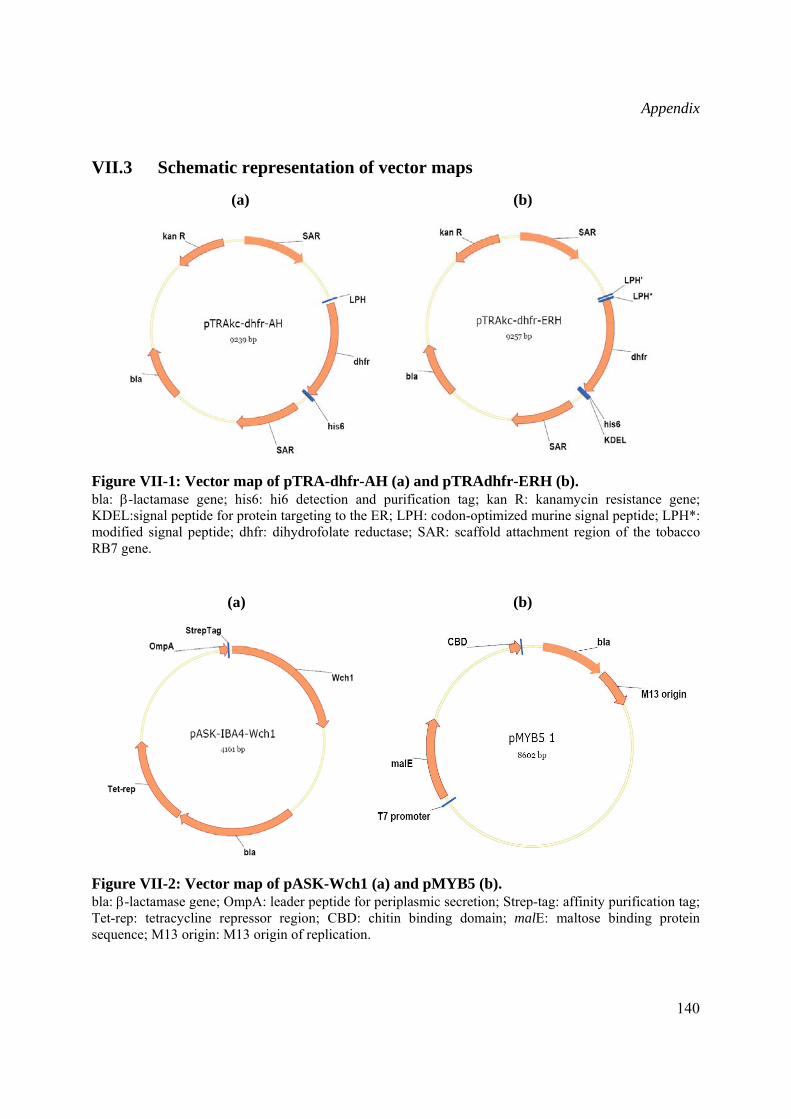
Appendix
140
VII.3 Schematic representation of vector maps
(a) (b)
Figure VII-1: Vector map of pTRA-dhfr-AH (a) and pTRAdhfr-ERH (b). bla: β-lactamase gene; his6: hi6 detection and purification tag; kan R: kanamycin resistance gene; KDEL:signal peptide for protein targeting to the ER; LPH: codon-optimized murine signal peptide; LPH*: modified signal peptide; dhfr: dihydrofolate reductase; SAR: scaffold attachment region of the tobacco RB7 gene.
(a) (b)
Figure VII-2: Vector map of pASK-Wch1 (a) and pMYB5 (b). bla: β-lactamase gene; OmpA: leader peptide for periplasmic secretion; Strep-tag: affinity purification tag; Tet-rep: tetracycline repressor region; CBD: chitin binding domain; malE: maltose binding protein sequence; M13 origin: M13 origin of replication.
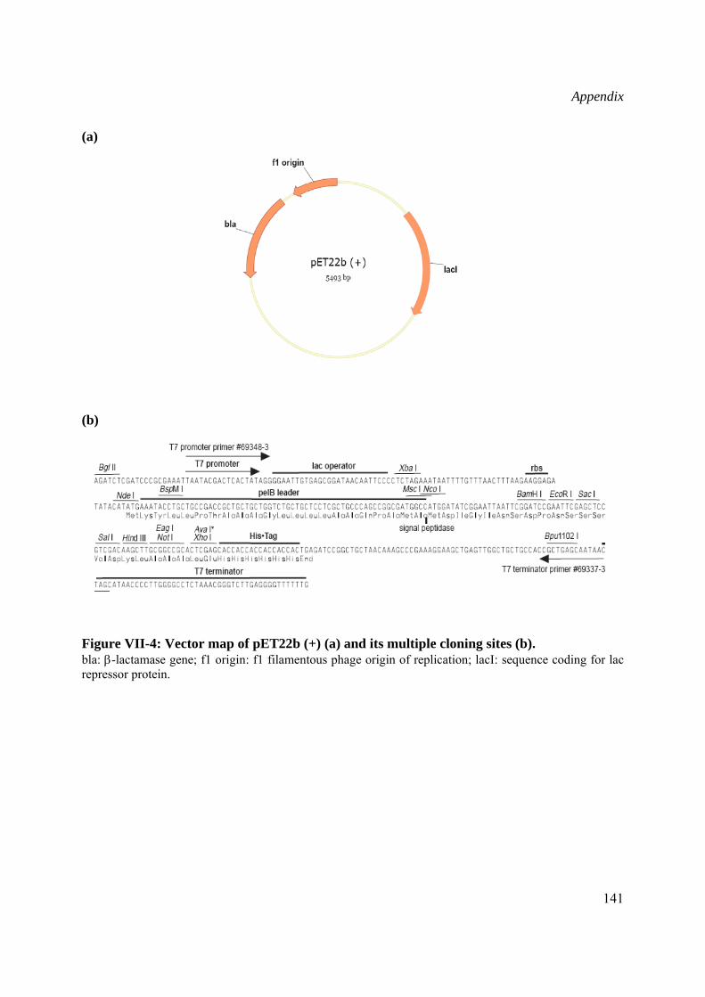
Appendix
141
(a)
(b)
Figure VII-4: Vector map of pET22b (+) (a) and its multiple cloning sites (b). bla: β-lactamase gene; f1 origin: f1 filamentous phage origin of replication; lacI: sequence coding for lac repressor protein.
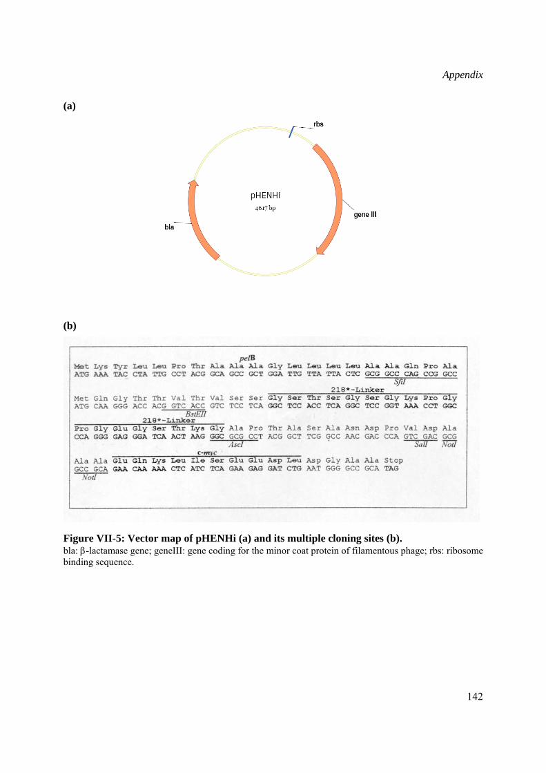
Appendix
142
(a)
(b)
Figure VII-5: Vector map of pHENHi (a) and its multiple cloning sites (b). bla: β-lactamase gene; geneIII: gene coding for the minor coat protein of filamentous phage; rbs: ribosome binding sequence.
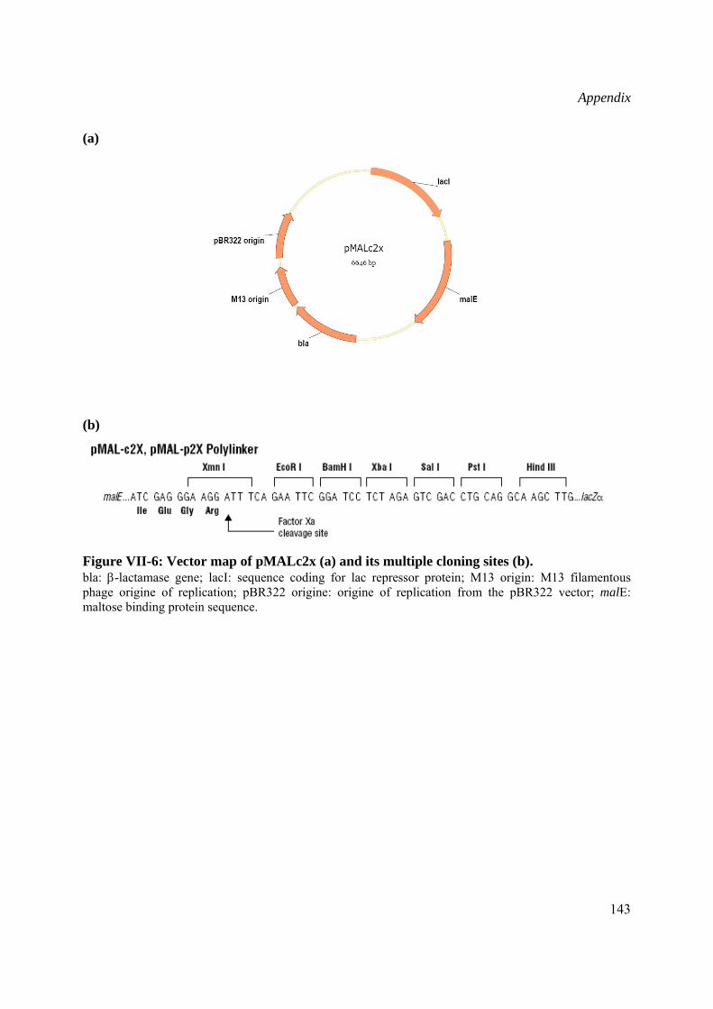
Appendix
143
(a)
(b)
Figure VII-6: Vector map of pMALc2x (a) and its multiple cloning sites (b). bla: β-lactamase gene; lacI: sequence coding for lac repressor protein; M13 origin: M13 filamentous phage origine of replication; pBR322 origine: origine of replication from the pBR322 vector; malE: maltose binding protein sequence.
Appendix
144
Lebenslauf
Persönliche Daten
Name Siham Agdour
Geburtsdatum 17. Oktober 1974
Staatangehörigkeit: Französisch
Ausbildung
2002-2007 Institut für Biologie VII RWTH Aachen Promotion unter Anleitung von Prof. Rainer Fischer. Titel: “Production and characterization of the recombinant wheat chitinase Wch1 and generation of chitin-specific antibodies”
2000 – 2001 Institut für Biologie VII RWTH Aachen Diplomarbeit unter Anleitung von Prof. Rainer Fischer Titel: „Engineering of scFv fragments with affinity to human spermine
synthase" 1999- 2000 Escuela Técnica Superior de Ingenieros Agrónomos y Montes,
Universidad de Córdoba (Spanien) Bachelor- Studium 1998 – 1999 Mediterranean Agronomic Institute of Chania, Kreta, Griechenland
Bachelor-Studium
1991 – 1997 Institut National Agronomique, Algers, Algerien Studium zum Agraringenieur Berufstätigkeit 01/2002-12/2003 Wissenschaftliche Hilfskraft am Institut für Biologie VII der RWTH
Aachen 01/2004-03/2006 Wissenschaftliche Angestellte am Institut für Biologie VII der RWTH
Aachen Seit 04/2006 Wissenschaftliche Angestellte am Fraunhofer Institut für
Molekularbiologie und Angewandte Ökologie, Aachen