Embed Size (px)
Citation preview
cells
Article
Rifampicin and Its Derivative Rifampicin QuinoneReduce Microglial Inflammatory Responses andNeurodegeneration Induced In Vitro by α-SynucleinFibrillary Aggregates
Leonardo Acuña 1,2, Sabah Hamadat 1 , Natalia S. Corbalán 3,4, Florencia González-Lizárraga 4,Mauricio dos-Santos-Pereira 1,5, Jérémy Rocca 1, Julia Sepúlveda Díaz 1, Elaine Del-Bel 5,Dulce Papy-García 3, Rosana N. Chehín 4, Patrick P. Michel 1 and Rita Raisman-Vozari 1,*
1 Institut du Cerveau et de la Moelle épinière (ICM), Inserm U 1127, CNRS UMR 7225, Sorbonne Université,F-75013 Paris, France
2 Instituto de Patología Experimental, CONICET/Universidad Nacional de Salta (UNSa),Salta A4408FVY, Argentina
3 Laboratoire Croissance, Régénération, Réparation et Régénération Tissulaires (CRRET)/ EAC CNRS 7149,Université Paris Est Créteil, Université Paris Est, 94010 Créteil, France
4 Instituto de Medicina Molecular y Celular Aplicada (IMMCA) CONICET/UNT and SIPROSA,Tucumán T4000ILI, Argentina
5 Faculdade de Medicina de Ribeirão Preto, Universidade de São Paulo, São Paulo 04023-062, Brazil* Correspondence: [email protected]; Tel.: +33(0)157274550 or +33(0)624543583
Received: 12 June 2019; Accepted: 18 July 2019; Published: 25 July 2019�����������������
Abstract: Aggregated forms of the synaptic protein α-synuclein (αS) have been proposed to operateas a molecular trigger for microglial inflammatory processes and neurodegeneration in Parkinson´sdisease. Here, we used brain microglial cell cultures activated by fibrillary forms of recombinanthuman αS to assess the anti-inflammatory and neuroprotective activities of the antibiotic rifampicin(Rif) and its autoxidation product rifampicin quinone (RifQ). Pretreatments with Rif and RifQ reducedthe secretion of prototypical inflammatory cytokines (TNF-α, IL-6) and the burst of oxidative stressin microglial cells activated with αS fibrillary aggregates. Note, however, that RifQ was constantlymore efficacious than its parent compound in reducing microglial activation. We also established thatthe suppressive effects of Rif and RifQ on cytokine release was probably due to inhibition of bothPI3K- and non-PI3K-dependent signaling events. The control of oxidative stress appeared, however,essentially dependent on PI3K inhibition. Of interest, we also showed that RifQ was more efficientthan Rif in protecting neuronal cells from toxic factors secreted by microglia activated by αS fibrils.Overall, data with RifQ are promising enough to justify further studies to confirm the potential ofthis compound as an anti-parkinsionian drug.
Keywords: aggregation; α-synuclein; microglia; neuroinflammation; Parkinson’s disease; cytokines;neuronal survival
1. Introduction
Parkinson’s disease (PD) is a neurodegenerative disease clinically characterized in part by motorsymptoms resulting from the loss of dopaminergic neurons in the substantia nigra pars compacta.In addition to neuronal demise, inflammatory processes and intraneuronal protein accumulationare characteristic histopathological hallmarks of PD [1]. Proteinaceous inclusions localized in theneuronal perikarya (Lewy bodies) and in neuronal processes (Lewy neurites) are primarily composed ofα-synuclein (αS) [2]. The aggregation ofαS in the central nervous system (CNS) is a pathological process
Cells 2019, 8, 776; doi:10.3390/cells8080776 www.mdpi.com/journal/cells
Cells 2019, 8, 776 2 of 17
of fundamental importance to the development and progression of PD [3] and other synucleinopathies.The αS aggregates are thought to favor neurodegeneration either by directly damaging neurons orby inducing the production of inflammatory mediators (e.g., TNF-α and reactive oxygen species(ROS)) that are neurotoxic [4,5]. These aggregates are generally surrounded by activated microglia andimmune cells in the CNS [6,7], which suggests that they may operate as a trigger for neuroinflammatoryprocesses and neurodegeneration to promote PD progression [8].
Besides their bactericidal activity, some antibiotic molecules also possess anti-inflammatory,anti-aggregant, or antioxidant properties, which may be of potential interest for PD treatment [9–13].Such is the case, for instance, of rifampicin (Rif), a macrocyclic bactericidal antibiotic that is widely usedin the treatment of infectious diseases, especially those caused by Mycobacterium, including tuberculosisand leprosy. Rif has been reported, for example, to attenuate cerebrospinal fluid (CSF) concentrationsof inflammation markers as well as neuronal damage in children with bacterial meningitis [14]. Rif wasalso the first antibiotic reported to have therapeutic effects in chronic neurodegenerative conditions.In particular, leprosy patients subjected to a chronic treatment with Rif displayed a significantlydecreased prevalence of dementia [15,16]. To date, a number of studies have reported anti-inflammatoryeffects of Rif in paradigms that are pertinent to neurological diseases [17–20]. Still, mechanisms throughwhich Rif could reduce neuroinflammation processes observed in neurodegenerative diseases remainto be clarified.
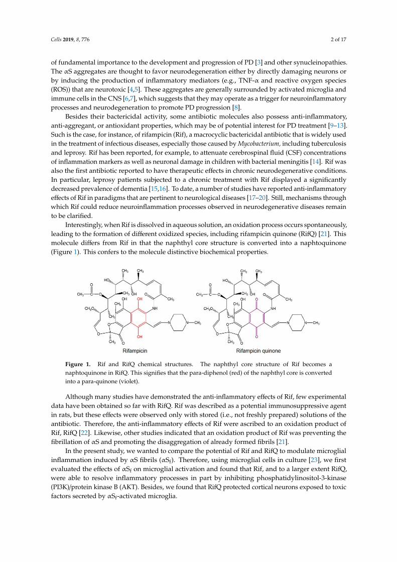
Interestingly, when Rif is dissolved in aqueous solution, an oxidation process occurs spontaneously,leading to the formation of different oxidized species, including rifampicin quinone (RifQ) [21]. Thismolecule differs from Rif in that the naphthyl core structure is converted into a naphtoquinone(Figure 1). This confers to the molecule distinctive biochemical properties.
Figure 1. Rif and RifQ chemical structures. The naphthyl core structure of Rif becomes anaphtoquinone in RifQ. This signifies that the para-diphenol (red) of the naphthyl core is convertedinto a para-quinone (violet).
Although many studies have demonstrated the anti-inflammatory effects of Rif, few experimentaldata have been obtained so far with RifQ. Rif was described as a potential immunosuppressive agentin rats, but these effects were observed only with stored (i.e., not freshly prepared) solutions of theantibiotic. Therefore, the anti-inflammatory effects of Rif were ascribed to an oxidation product ofRif, RifQ [22]. Likewise, other studies indicated that an oxidation product of Rif was preventing thefibrillation of αS and promoting the disaggregation of already formed fibrils [21].
In the present study, we wanted to compare the potential of Rif and RifQ to modulate microglialinflammation induced by αS fibrils (αSf). Therefore, using microglial cells in culture [23], we firstevaluated the effects of αSf on microglial activation and found that Rif, and to a larger extent RifQ,were able to resolve inflammatory processes in part by inhibiting phosphatidylinositol-3-kinase(PI3K)/protein kinase B (AKT). Besides, we found that RifQ protected cortical neurons exposed to toxicfactors secreted by αSf-activated microglia.
Cells 2019, 8, 776 3 of 17
2. Materials and Methods
Leibovitz’s L-15 medium, Dulbecco’s modified Eagle’s medium (DMEM), Trypsin-EDTA0.05%, and penicillin/streptomycin cocktail were all purchased from Invitrogen Life Technologies(Saint Aubin, France). Fetal calf serum (FCS) was obtained from Biowest LLC (Eurobio, Les Ulis,France). Poly(ethyleneimine) (PEI) (average molar mass 750,000, P3143), lipopolysaccharide(LPS; E. coli strain O26:B6; L8274), Rif R0700000, RifQ R0800000, LY-294,002 hydrochloride (LY),2′(3′)-O-(4-Benzoylbenzoyl)adenosine 5′-triphosphate triethylammonium salt (bz-ATP), Hoechst33342, the Cell Counting Kit-8, the rabbit Anti-Glyceraldehyde-3-phosphate dehydrogenase antibody(anti-GAPDH), and the Superoxide anion assay kit were all purchased from Sigma Aldrich (L’Isled’Abeau Chesnes, France). The interleukin (IL)-6 and tumor necrosis factor (TNF)-α enzyme-linkedimmunosorbent assay (ELISA) kits, the M-PER™Mammalian Protein Extraction Reagent, Pierce™,the Limulus Amebocyte Lysate (LAL) Chromogenic Endotoxin Quantitation Kit, the Pierce BCAProtein Assay Kit, and the Pre-stained Protein Ladder were obtained from ThermoFisher Scientific(Saint-Herblain, France). The rabbit anti-ionized calcium binding adaptor molecule-1 (IBA-1)antibody (#019-19741) was from Wako (Neuss, Germany). The rabbit phospho-AKT (Ser473)antibody (#9271) and rabbit AKT antibody (#9272) were purchased from Cell Signaling Technology(Saint-Quentin Yvelines, France). The monoclonal IgG isotype 2a anti-mTLR2-IgG antibody(α-TLR2) and the TLR2 agonist Pam3CSK4 were obtained from InvivoGen (Toulouse, France), and2-(Phenylthio)-N-[[tetrahydro-4-(4-phenyl-1-piperazinyl)-2H-pyran-4-yl]methyl-3-pyridinecarboxamideJNJ 47965567 (JNJ) was purchased from Tocris Biosciences (Bristol, UK).
2.1. Preparation of α-synuclein
Expression and purification of recombinant human αS was performed as previously described [24]and endotoxins potentially present were removed using a high capacity endotoxin removal resin(ThermoFisher) following the manufacturer´s instructions. Then, protein samples were filtered,centrifuged for 30 min at 12,000× g, and residual endotoxins were quantified using the LimulusAmebocyte Lysate assay. MonomericαS stock solutions containing less than 0.1 endotoxin unit (EU)/mgprotein were prepared in 20 mM 4-(2-Hydroxyethyl)piperazine-1-ethanesulfonic acid (HEPES), 150 mMNaCl, pH 7.4. The protein concentration was determined by measurement of absorbance at 280 nmusing an extinction coefficient ε275 of 5600 cm−1 M−1. Protein aggregation was performed using αSsolutions (1 mg/ml) diluted in 20 mM HEPES, 150 mM NaCl, pH 7.4. Samples were incubated in aThermomixer C (Eppendorf) at 37 ◦C under constant orbital agitation (600 revs/min) to obtain fibrillaryaggregates. The aggregates were then sonicated for 2 min in an ultrasonic bath and kept at −20 ◦Cuntil further use.
2.2. Transmission Electron Microscopy
Samples (50 µl) of a 1 mg/ml αS solution were adsorbed onto glow-discharged 200 meshformvar/carbon coated copper grids (Electron Microscopy Sciences, Hatfield, PA) and stained withuranyLess (Electron Microscopy Sciences). Excess liquid was removed and grids were allowed to airdry. Samples were viewed and imaged using a Hitachi 7700 transmission electron microscope (Hitachi,Tokyo, Japan).
2.3. Cell Culture Protocols
2.3.1. Microglial Cell Isolation
Animals were housed, handled, and cared for according to the recommendations of the EuropeanUnion Council Directives (2010/63/EU). The experimental procedures were authorized by the ethicalcommittee for animal experiments Charles Darwin n◦5.
Pure microglial cell cultures were obtained as previously described using a technique that relieson the preferential adhesion of microglia to the polycation polyethyleneimine (PEI) [23]. Briefly,
Cells 2019, 8, 776 4 of 17
the brains of postnatal day 1 C57BL/6J mouse pups (Janvier LABS, Le Genest St Isles, France) wereharvested, and the meninges stripped away, after which brain tissue was mechanically dissociated byrepeated pipetting. After two rounds of trituration, the supernatant containing the dissociated cellswas centrifuged at 1000 rpm for 5 min at 4 ◦C. The resulting pellet was triturated and resuspended inDMEM supplemented with 10% heat-inactivated FCS and 1% penicillin/streptomycin solution (definedas complete medium). Then, a cell suspension obtained by trituration of 2 mouse brains was plated ineach PEI-coated T-75 culture flask (Sigma-Aldrich) containing complete medium. The cultures werewashed once with complete medium after 2 days in vitro and microglial cells were then maintainedat 37 ◦C in a humidified atmosphere with 5% CO2 without any other culture medium change untilcompletion of isolation. The isolation was generally obtained 14–18 days after plating under theseconditions. The average yield was approximately 4–5.106 cells/T-75 culture flask with this protocol.
2.3.2. Microglial Cell Stimulation and Treatments
After isolation, microglial cells were harvested by trypsinization and seeded onto uncoated 48-wellplates (Nunc) at a density of 105 living cells per well, unless otherwise specified. The αS species(70 µg/ml), bz-ATP (500 µM), and Pam3CSK4 (1 µg/ml) were applied the day after plating for 24 h.Unless otherwise specified, Rif and RifQ were used at 100 µM, a concentration found to elicit optimalanti-inflammatory effects in preliminary experiments. The concentrations used for LY (2.5 µM), α-TLR2(2.5 µM), and JNJ (20 µM) were based on previous work [25]. Treatments with Rif, RifQ, LY, α-TLR2,and JNJ were initiated 3 h before applying the inflammogens to the cultures.
2.3.3. Primary Cortical Neuron Cultures
Primary cultures of cortical neurons from embryonic day 16 C57BL/6J mouse fetuses were preparedaccording to the protocol employed by Fifre et al. (2006) [26]. Dissociated cells were seeded at a densityof ~105 cells/cm2 onto 48-well plastic plates that were precoated with 1.5 g/ml of poly-DL-ornithine.Cortical cells were cultured without serum using a chemically defined DMEM-F12 medium (LifeTechnologies) that was supplemented with salts, hormones, and proteins, as described previously [26].Cultures were maintained at 37 ◦C in a humidified 5% CO2 atmosphere.
2.3.4. Neurotoxicity Assays
After 6 days in vitro, the seeding medium of cortical neurons was replaced by a culture mediumconditioned by microglial cultures that were exposed or not to treatments of interest. Then, after48 h of incubation with microglial conditioned medium (CM), cell viability was monitored with theCell Counting Kit-8 (CCK-8, Sigma-Aldrich) according to the manufacturer’s instructions. Briefly,CM was removed and the CCK-8 solution (15 µl of CCK-8 reagent in 150 µl culture medium) wasadded to each well for an incubation of 3 h at 37 ◦C in a humidified atmosphere with 5% CO2. Aftertermination of the incubation, the supernatant from each well was transferred into a 96-well microplate,and the absorbance measured at 450 nm using a Tecan’s Infinite®1000 spectrometer (Tecan Group,Männedorf, Switzerland).
2.3.5. Microglial-Conditioned Media Preparation
Primary microglial cells were plated and maintained with the same chemically-defined mediumas that used for cortical cultures. After 16 h, microglial cells were treated for 3 h with Rif or RifQfollowed by a stimulation with αSf for 24 h. After that, the supernatants were immediately transferredto cortical cultures for neurotoxicity assays. To verify the degree of activation of microglial cells inthis setting, we measured the TNF-α and IL-6 levels in the supernatants. Note that Rif and RifQ wereused at 10 µM in CM experiments. This is because we found that these concentrations are sufficient toinhibit microglial cell responses elicited by αSf in serum-free conditions.
Cells 2019, 8, 776 5 of 17
2.4. Protein Detection by Immunofluorescence
After the termination of treatment, the cultures were fixed with 4% formaldehyde (12 min, 4 ◦C),washed with PBS, and incubated overnight with antibodies against IBA-1 (1:500 in PBS with 0.02%Triton X-100) to detect microglial cells. As secondary antibody, we used a goat anti-rabbit AlexaFluor -555 (Invitrogen). When needed, nuclei of labelled cells were counterstained with Hoechst33342 (10 µM). Phase contrast and fluorescent images were acquired using a Nikon TE 2000 invertedmicroscope (Nikon, Tokyo, Japan) equipped with an ORCA-ER digital camera and HCImage imagingsoftware (Hamamatsu Corp., Bridgewater, NJ, USA).
2.5. Western Blot Analysis
The culture supernatants were removed, and microglial cells were washed with PBS and lysedusing the M-PER™ Mammalian Protein Extraction Reagent. Protein contents of the samples werequantified using a bicinchoninic acid (BCA) Protein Assay kit. Equal amounts of protein wereseparated by electrophoresis on a 10% sodium dodecyl sulfate polyacrylamide gel electrophoresis(SDS-PAGE), followed by transfer to a nitrocellulose membrane. The membranes first blocked inPBS containing 50% Odyssey blocking buffer (blocking solution) (LI-COR®Bioscience), were thenincubated overnight at 2–8 ◦C with primary antibodies (1:1000) against Glyceraldehyde-3-phosphatedehydrogenase, anti-ionized calcium binding adaptor molecule-1, phospho-AKT, or AKT dilutedin the blocking solution. After incubation, the membranes were washed with a Tris-buffered salineTween-20 solution and incubated with an adequate amount of Infrared Fluorescent (IR)Dye secondaryantibody (LI-COR®). We used an Odyssey CLx near-infrared fluorescence imaging system (LI-COR®)for Western blot imaging and quantification. Immunofluorescent signals were normalized accordingto protein levels in control conditions (untreated cells).
2.6. NADPH Oxidase Activity
Reduced nicotinamide adenine dinucleotide phosphate (NADPH) oxidase activity was determinedwith the superoxide anion assay kit (Sigma Aldrich) by measuring chemiluminescence products formedthrough the oxidation of luminol by superoxide anions. Briefly, cells were seeded (1.5.105 cells/well)in white opaque 96 wells microplates, pre-incubated 1 hour with Rif (100 µM), RifQ (100 µM), or LY(2.5 µM), and stimulated with αSf. Twenty-four hours later, the culture medium was removed and thecultures washed with a Hank’s balanced salt solution. Living cells were then incubated with 100 µl ofmedium assay provided by the assay kit. The enzymatic reaction was triggered by addition of luminoland the chemiluminescence signal was monitored at 37 ◦C using a microplate reader SpectraMax M4(Molecular Devices, Sunnyvale, CA, USA), with one acquisition every 2 s for 300 min.
2.7. Statistical Analysis
Data were analyzed by one-way ANOVA followed by the Tukey post-hoc test. All data arepresented as mean ± SEM of at least 3 independent experiments, except when noted. Statistical analysiswas performed with the Statistix 9.0 software.
3. Results
3.1. α-Synuclein Fibrils Induce Pro-Inflammatory Cytokine Release in Microglial Cell Cultures
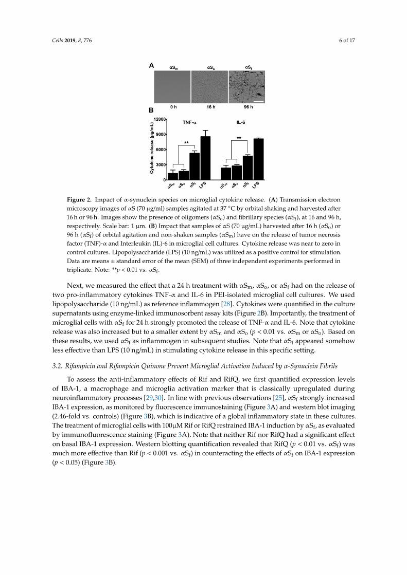
Recombinant monomeric αS (αSm) (70 µg/ml) was agitated by orbital shaking as previouslydescribed [10], and aggregation kinetics monitored by measurement of Thioflavin T (ThT)fluorescence [27] (not shown). The αS oligomers (αSo) and αSf were harvested after 16 and 96 hof incubation at 37 ◦C, respectively, and structural states were confirmed by transmission electronmicroscopy (TEM) (Figure 2A).
Cells 2019, 8, 776 6 of 17
Figure 2. Impact of α-synuclein species on microglial cytokine release. (A) Transmission electronmicroscopy images of αS (70 µg/ml) samples agitated at 37 ◦C by orbital shaking and harvested after16 h or 96 h. Images show the presence of oligomers (αSo) and fibrillary species (αSf), at 16 and 96 h,respectively. Scale bar: 1 µm. (B) Impact that samples of αS (70 µg/mL) harvested after 16 h (αSo) or96 h (αSf) of orbital agitation and non-shaken samples (αSm) have on the release of tumor necrosisfactor (TNF)-α and Interleukin (IL)-6 in microglial cell cultures. Cytokine release was near to zero incontrol cultures. Lipopolysaccharide (LPS) (10 ng/mL) was utilized as a positive control for stimulation.Data are means ± standard error of the mean (SEM) of three independent experiments performed intriplicate. Note: **p < 0.01 vs. αSf.
Next, we measured the effect that a 24 h treatment with αSm, αSo, or αSf had on the release oftwo pro-inflammatory cytokines TNF-α and IL-6 in PEI-isolated microglial cell cultures. We usedlipopolysaccharide (10 ng/mL) as reference inflammogen [28]. Cytokines were quantified in the culturesupernatants using enzyme-linked immunosorbent assay kits (Figure 2B). Importantly, the treatment ofmicroglial cells with αSf for 24 h strongly promoted the release of TNF-α and IL-6. Note that cytokinerelease was also increased but to a smaller extent by αSm and αSo (p < 0.01 vs. αSm or αSo). Based onthese results, we used αSf as inflammogen in subsequent studies. Note that αSf appeared somehowless effective than LPS (10 ng/mL) in stimulating cytokine release in this specific setting.
3.2. Rifampicin and Rifampicin Quinone Prevent Microglial Activation Induced by α-Synuclein Fibrils
To assess the anti-inflammatory effects of Rif and RifQ, we first quantified expression levelsof IBA-1, a macrophage and microglia activation marker that is classically upregulated duringneuroinflammatory processes [29,30]. In line with previous observations [25], αSf strongly increasedIBA-1 expression, as monitored by fluorescence immunostaining (Figure 3A) and western blot imaging(2.46-fold vs. controls) (Figure 3B), which is indicative of a global inflammatory state in these cultures.The treatment of microglial cells with 100µM Rif or RifQ restrained IBA-1 induction byαSf, as evaluatedby immunofluorescence staining (Figure 3A). Note that neither Rif nor RifQ had a significant effecton basal IBA-1 expression. Western blotting quantification revealed that RifQ (p < 0.01 vs. αSf) wasmuch more effective than Rif (p < 0.001 vs. αSf) in counteracting the effects of αSf on IBA-1 expression(p < 0.05) (Figure 3B).
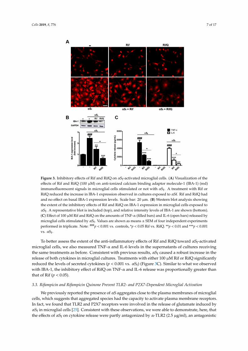
Cells 2019, 8, 776 7 of 17
Figure 3. Inhibitory effects of Rif and RifQ on αSf-activated microglial cells. (A) Visualization of theeffects of Rif and RifQ (100 µM) on anti-ionized calcium binding adaptor molecule-1 (IBA-1) (red)immunofluorescent signals in microglial cells stimulated or not with αSf. A treatment with Rif orRifQ reduced the increase in IBA-1 expression observed in cultures exposed to αSf. Rif and RifQ hadand no effect on basal IBA-1 expression levels. Scale bar: 20 µm. (B) Western blot analysis showingthe extent of the inhibitory effects of Rif and RifQ on IBA-1 expression in microglial cells exposed toαSf. A representative blot is included (top), and relative intensity levels of IBA-1 are shown (bottom).(C) Effect of 100 µM Rif and RifQ on the amounts of TNF-α (filled bars) and IL-6 (open bars) released bymicroglial cells stimulated by αSf. Values are shown as means ± SEM of four independent experimentsperformed in triplicate. Note: ###p < 0.001 vs. controls, *p < 0.05 Rif vs. RifQ. **p < 0.01 and ***p < 0.001vs. αSf.
To better assess the extent of the anti-inflammatory effects of Rif and RifQ toward αSf-activatedmicroglial cells, we also measured TNF-α and IL-6 levels in the supernatants of cultures receivingthe same treatments as before. Consistent with previous results, αSf caused a robust increase in therelease of both cytokines in microglial cultures. Treatments with either 100 µM Rif or RifQ significantlyreduced the levels of secreted cytokines (p < 0.001 vs. αSf) (Figure 3C). Similar to what we observedwith IBA-1, the inhibitory effect of RifQ on TNF-α and IL-6 release was proportionally greater thanthat of Rif (p < 0.05).
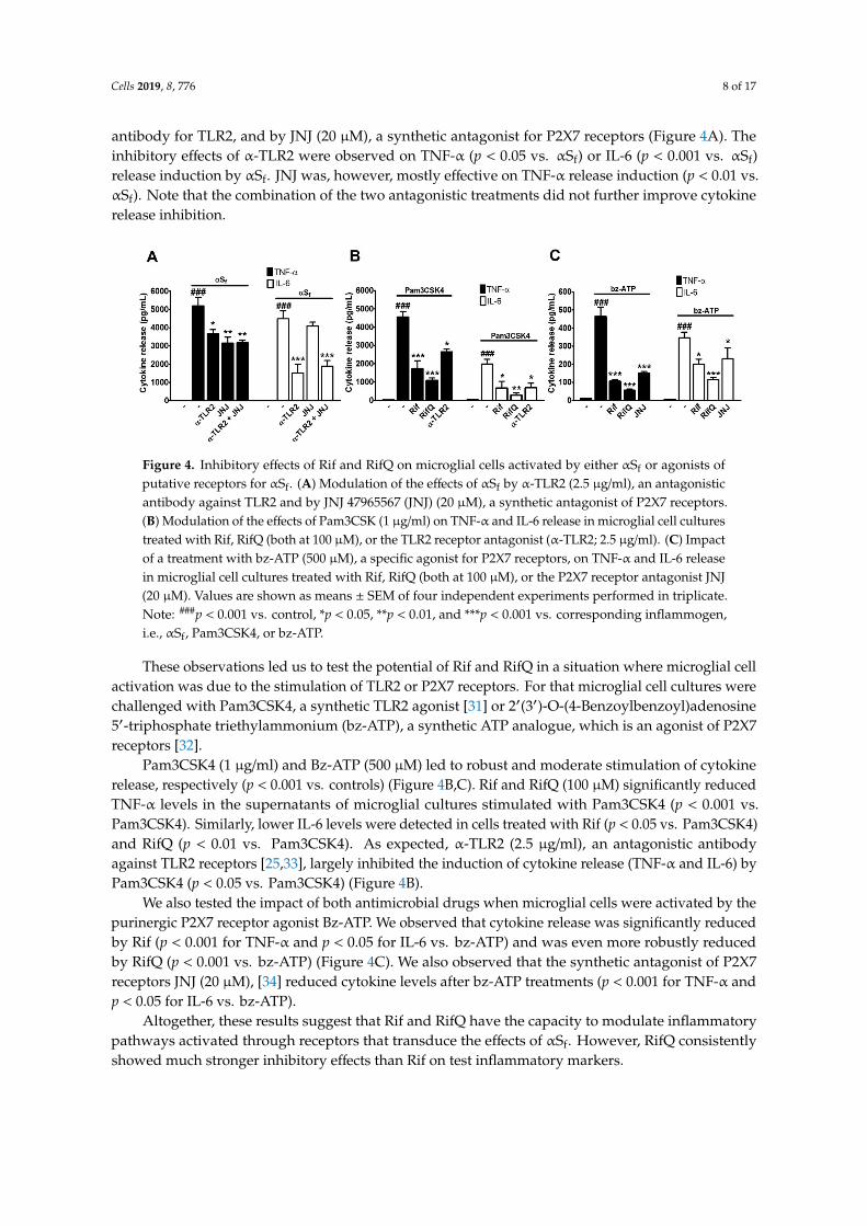
3.3. Rifampicin and Rifampicin Quinone Prevent TLR2- and P2X7-Dependent Microglial Activation
We previously reported the presence of αS aggregates close to the plasma membranes of microglialcells, which suggests that aggregated species had the capacity to activate plasma membrane receptors.In fact, we found that TLR2 and P2X7 receptors were involved in the release of glutamate induced byαSf in microglial cells [25]. Consistent with these observations, we were able to demonstrate, here, thatthe effects of αSf on cytokine release were partly antagonized by α-TLR2 (2.5 µg/ml), an antagonistic
Cells 2019, 8, 776 8 of 17
antibody for TLR2, and by JNJ (20 µM), a synthetic antagonist for P2X7 receptors (Figure 4A). Theinhibitory effects of α-TLR2 were observed on TNF-α (p < 0.05 vs. αSf) or IL-6 (p < 0.001 vs. αSf)release induction by αSf. JNJ was, however, mostly effective on TNF-α release induction (p < 0.01 vs.αSf). Note that the combination of the two antagonistic treatments did not further improve cytokinerelease inhibition.
Figure 4. Inhibitory effects of Rif and RifQ on microglial cells activated by either αSf or agonists ofputative receptors for αSf. (A) Modulation of the effects of αSf by α-TLR2 (2.5 µg/ml), an antagonisticantibody against TLR2 and by JNJ 47965567 (JNJ) (20 µM), a synthetic antagonist of P2X7 receptors.(B) Modulation of the effects of Pam3CSK (1 µg/ml) on TNF-α and IL-6 release in microglial cell culturestreated with Rif, RifQ (both at 100 µM), or the TLR2 receptor antagonist (α-TLR2; 2.5 µg/ml). (C) Impactof a treatment with bz-ATP (500 µM), a specific agonist for P2X7 receptors, on TNF-α and IL-6 releasein microglial cell cultures treated with Rif, RifQ (both at 100 µM), or the P2X7 receptor antagonist JNJ(20 µM). Values are shown as means ± SEM of four independent experiments performed in triplicate.Note: ###p < 0.001 vs. control, *p < 0.05, **p < 0.01, and ***p < 0.001 vs. corresponding inflammogen,i.e., αSf, Pam3CSK4, or bz-ATP.
These observations led us to test the potential of Rif and RifQ in a situation where microglial cellactivation was due to the stimulation of TLR2 or P2X7 receptors. For that microglial cell cultures werechallenged with Pam3CSK4, a synthetic TLR2 agonist [31] or 2′(3′)-O-(4-Benzoylbenzoyl)adenosine5′-triphosphate triethylammonium (bz-ATP), a synthetic ATP analogue, which is an agonist of P2X7receptors [32].
Pam3CSK4 (1 µg/ml) and Bz-ATP (500 µM) led to robust and moderate stimulation of cytokinerelease, respectively (p < 0.001 vs. controls) (Figure 4B,C). Rif and RifQ (100 µM) significantly reducedTNF-α levels in the supernatants of microglial cultures stimulated with Pam3CSK4 (p < 0.001 vs.Pam3CSK4). Similarly, lower IL-6 levels were detected in cells treated with Rif (p < 0.05 vs. Pam3CSK4)and RifQ (p < 0.01 vs. Pam3CSK4). As expected, α-TLR2 (2.5 µg/ml), an antagonistic antibodyagainst TLR2 receptors [25,33], largely inhibited the induction of cytokine release (TNF-α and IL-6) byPam3CSK4 (p < 0.05 vs. Pam3CSK4) (Figure 4B).
We also tested the impact of both antimicrobial drugs when microglial cells were activated by thepurinergic P2X7 receptor agonist Bz-ATP. We observed that cytokine release was significantly reducedby Rif (p < 0.001 for TNF-α and p < 0.05 for IL-6 vs. bz-ATP) and was even more robustly reducedby RifQ (p < 0.001 vs. bz-ATP) (Figure 4C). We also observed that the synthetic antagonist of P2X7receptors JNJ (20 µM), [34] reduced cytokine levels after bz-ATP treatments (p < 0.001 for TNF-α andp < 0.05 for IL-6 vs. bz-ATP).
Altogether, these results suggest that Rif and RifQ have the capacity to modulate inflammatorypathways activated through receptors that transduce the effects of αSf. However, RifQ consistentlyshowed much stronger inhibitory effects than Rif on test inflammatory markers.
Cells 2019, 8, 776 9 of 17
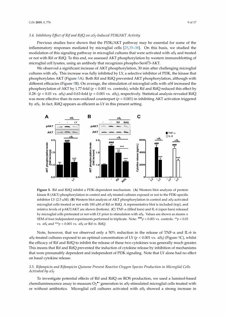
3.4. Inhibitory Effect of Rif and RifQ on αSf-Induced PI3K/AKT Activity
Previous studies have shown that the PI3K/AKT pathway may be essential for some of theinflammatory responses mediated by microglial cells [25,35–38]. On this basis, we studied themodulation of this signaling pathway in microglial cultures that were activated with αSf and treatedor not with Rif or RifQ. To this end, we assessed AKT phosphorylation by western immunoblotting ofmicroglial cell lysates, using an antibody that recognizes phospho-Ser473-AKT.
We observed a significant increase of AKT phosphorylation, 30 min after challenging microglialcultures with αSf. This increase was fully inhibited by LY, a selective inhibitor of PI3K, the kinase thatphosphorylates AKT (Figure 5A). Both Rif and RifQ prevented AKT phosphorylation, although withdifferent efficacies (Figure 5B). On average, the stimulation of microglial cells with αSf increased thephosphorylation of AKT by 1.77-fold (p < 0.001 vs. controls), while Rif and RifQ reduced this effect by0.28- (p < 0.01 vs. αSf) and 0.63-fold (p < 0.001 vs. αSf), respectively. Statistical analysis revealed RifQwas more effective than its non-oxidized counterpart (p < 0.001) in inhibiting AKT activation triggeredby αSf. In fact, RifQ appears as efficient as LY in this present setting.
Figure 5. Rif and RifQ inhibit a PI3K-dependent mechanism. (A) Western blot analysis of proteinkinase B (AKT) phosphorylation in control and αSf-treated cultures exposed or not to the PI3K-specificinhibitor LY (2.5 µM). (B) Western blot analysis of AKT phosphorylation in control and αSf-activatedmicroglial cells treated or not with 100 µM of Rif or RifQ. A representative blot is included (top), andrelative levels of pAKT/AKT are shown (bottom). (C) TNF-α (filled bars) and IL-6 (open bars) releasedby microglial cells pretreated or not with LY prior to stimulation with αSf. Values are shown as means ±SEM of four independent experiments performed in triplicate. Note: ###p < 0.001 vs. controls. **p < 0.01vs. αSf and ***p < 0.001 vs. αSf or Rif vs. RifQ.
Note, however, that we observed only a 50% reduction in the release of TNF-α and IL-6 inαSf-treated cultures exposed to an optimal concentration of LY (p < 0.001 vs. αSf) (Figure 5C), whilstthe efficacy of Rif and RifQ to inhibit the release of these two cytokines was generally much greater.This means that Rif and RifQ prevented the induction of cytokine release by inhibition of mechanismsthat were presumably dependent and independent of PI3K signaling. Note that LY alone had no effecton basal cytokine release.
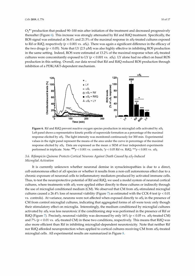
3.5. Rifampicin and Rifampicin Quinone Prevent Reactive Oxygen Species Production in Microglial CellsActivated by αSf
To investigate potential effects of Rif and RifQ on ROS production, we used a luminol-basedchemiluminescence assay to measure O2
•−generation in αSf-stimulated microglial cells treated withor without antibiotics. Microglial cell cultures activated with αSf showed a strong increase in
Cells 2019, 8, 776 10 of 17
O2•−production that peaked 90–100 min after initiation of the treatment and decreased progressively
thereafter (Figure 6). This increase was strongly attenuated by Rif and RifQ treatment. Specifically, theROS signal was estimated at 36.4% and 21.5% of the maximal response in αSf-treated cultures exposedto Rif or RifQ, respectively (p < 0.001 vs. αSf). There was again a significant difference in the efficacy ofthe two drugs (p < 0.05). Note that LY (2.5 µM) was also highly effective in inhibiting ROS productionin the same setting. Indeed, ROS were estimated at 13.2% of the maximal response when αSf-treatedcultures were concomitantly exposed to LY (p < 0.001 vs. αSf). LY alone had no effect on basal ROSproduction in this setting. Overall, our data reveal that Rif and RifQ reduced ROS production throughinhibition of a PI3K/AKT-dependent mechanism.
Figure 6. Rif and RifQ prevent reactive oxygen species production in microglial cells activated by αSf.
Left panel shows a representative kinetic profile of superoxide formation as a percentage of the maximalresponse elicited by αSf. The light intensity was monitored continuously for 300 min. Experimentalvalues in the right panel represent the means of the area under the curve in percentage of the maximalresponse elicited by αSf. Data are expressed as the mean ± SEM of four independent experimentsperformed in triplicate. Note: ###p < 0.001 vs. controls, *p < 0.05 Rif vs. RifQ, ***p < 0.001 vs. αSf.
3.6. Rifampicin Quinone Protects Cortical Neurons Against Death Caused by αSf-InducedMicroglial Activation
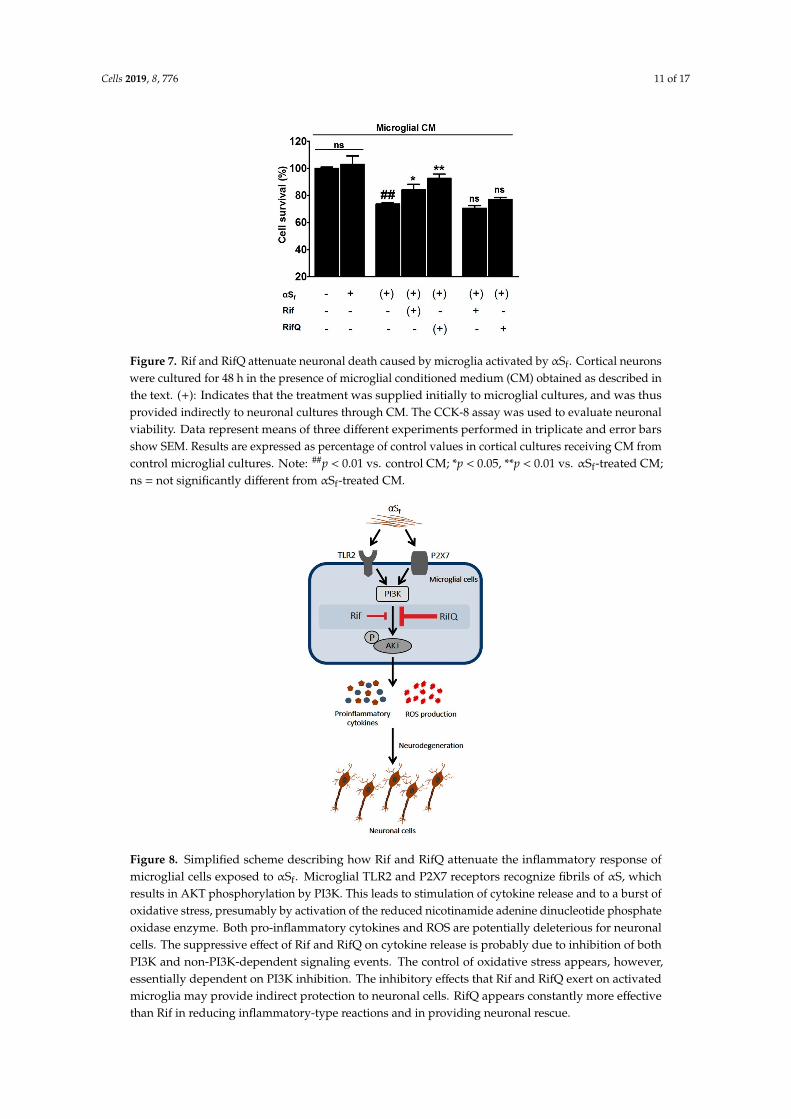
It is currently unknown whether neuronal demise in synucleinopathies is due to a direct,cell-autonomous effect of αS species or whether it results from a non-cell autonomous effect due to achronic exposure of neuronal cells to inflammatory mediators produced by activated immune cells.Thus, to test the neuroprotective potential of Rif and RifQ, we used a model system of neuronal corticalcultures, where treatments with αSf were applied either directly to these cultures or indirectly throughthe use of microglial conditioned medium (CM). We observed that CM from αSf-stimulated microglialcultures caused a 26.4% loss of neuronal viability (Figure 7) as estimated with the CCK-8 test (p < 0.01vs. controls). At variance, neurons were not affected when exposed directly to αSf in the presence ofCM from control microglial cultures, indicating that aggregated forms of αS were toxic only thoughtheir stimulatory effect on microglia. Interestingly, the medium conditioned by microglial culturesactivated by αSf was less neurotoxic if the conditioning step was performed in the presence of Rif orRifQ (Figure 7). Precisely, neuronal viability was decreased by only 16% (p < 0.05 vs. αSf-treated CM)and 7% (p < 0.01 vs. αSf-treated CM) in these two conditions, respectively. This means that RifQ wasalso more efficient than Rif in inhibiting microglial-dependent neurotoxicity. Note that neither Rifnor RifQ afforded neuroprotection when applied to cortical cultures receiving CM from αSf-treatedmicroglial cells. All experimental results are summarized in Figure 8.
Cells 2019, 8, 776 11 of 17
Figure 7. Rif and RifQ attenuate neuronal death caused by microglia activated by αSf. Cortical neuronswere cultured for 48 h in the presence of microglial conditioned medium (CM) obtained as described inthe text. (+): Indicates that the treatment was supplied initially to microglial cultures, and was thusprovided indirectly to neuronal cultures through CM. The CCK-8 assay was used to evaluate neuronalviability. Data represent means of three different experiments performed in triplicate and error barsshow SEM. Results are expressed as percentage of control values in cortical cultures receiving CM fromcontrol microglial cultures. Note: ##p < 0.01 vs. control CM; *p < 0.05, **p < 0.01 vs. αSf-treated CM;ns = not significantly different from αSf-treated CM.
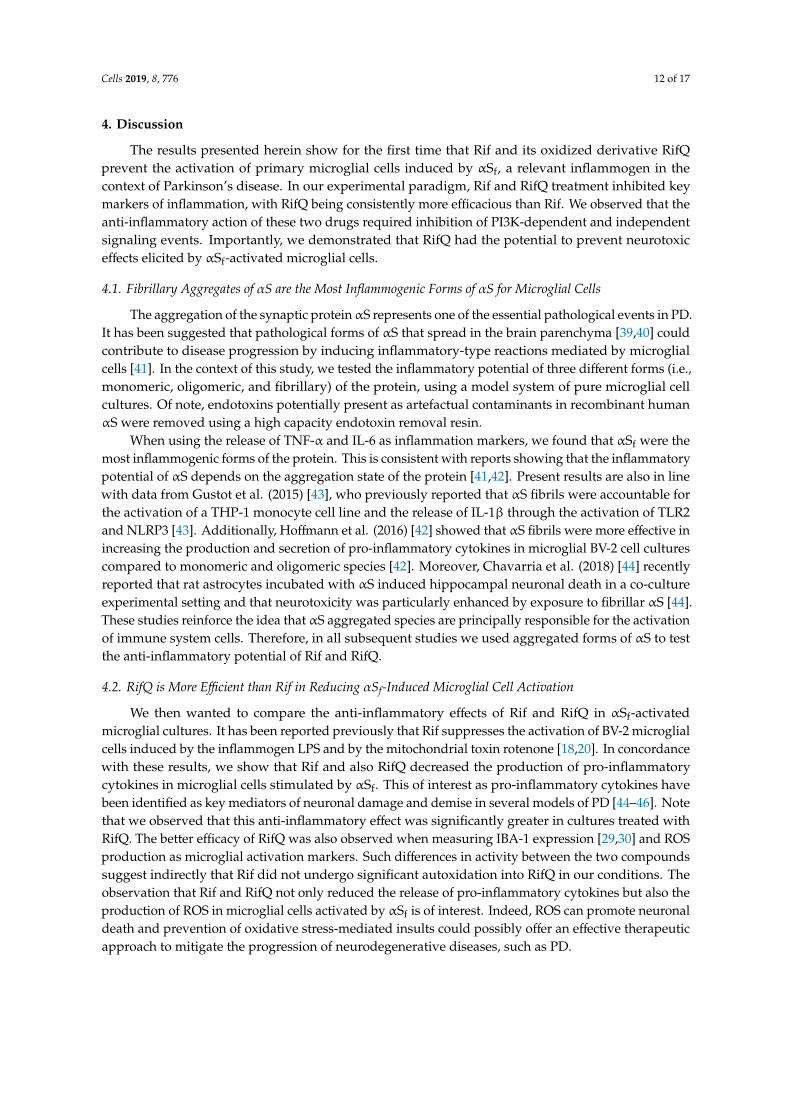
Figure 8. Simplified scheme describing how Rif and RifQ attenuate the inflammatory response ofmicroglial cells exposed to αSf. Microglial TLR2 and P2X7 receptors recognize fibrils of αS, whichresults in AKT phosphorylation by PI3K. This leads to stimulation of cytokine release and to a burst ofoxidative stress, presumably by activation of the reduced nicotinamide adenine dinucleotide phosphateoxidase enzyme. Both pro-inflammatory cytokines and ROS are potentially deleterious for neuronalcells. The suppressive effect of Rif and RifQ on cytokine release is probably due to inhibition of bothPI3K and non-PI3K-dependent signaling events. The control of oxidative stress appears, however,essentially dependent on PI3K inhibition. The inhibitory effects that Rif and RifQ exert on activatedmicroglia may provide indirect protection to neuronal cells. RifQ appears constantly more effectivethan Rif in reducing inflammatory-type reactions and in providing neuronal rescue.
Cells 2019, 8, 776 12 of 17
4. Discussion
The results presented herein show for the first time that Rif and its oxidized derivative RifQprevent the activation of primary microglial cells induced by αSf, a relevant inflammogen in thecontext of Parkinson’s disease. In our experimental paradigm, Rif and RifQ treatment inhibited keymarkers of inflammation, with RifQ being consistently more efficacious than Rif. We observed that theanti-inflammatory action of these two drugs required inhibition of PI3K-dependent and independentsignaling events. Importantly, we demonstrated that RifQ had the potential to prevent neurotoxiceffects elicited by αSf-activated microglial cells.
4.1. Fibrillary Aggregates of αS are the Most Inflammogenic Forms of αS for Microglial Cells
The aggregation of the synaptic proteinαS represents one of the essential pathological events in PD.It has been suggested that pathological forms of αS that spread in the brain parenchyma [39,40] couldcontribute to disease progression by inducing inflammatory-type reactions mediated by microglialcells [41]. In the context of this study, we tested the inflammatory potential of three different forms (i.e.,monomeric, oligomeric, and fibrillary) of the protein, using a model system of pure microglial cellcultures. Of note, endotoxins potentially present as artefactual contaminants in recombinant humanαS were removed using a high capacity endotoxin removal resin.
When using the release of TNF-α and IL-6 as inflammation markers, we found that αSf were themost inflammogenic forms of the protein. This is consistent with reports showing that the inflammatorypotential of αS depends on the aggregation state of the protein [41,42]. Present results are also in linewith data from Gustot et al. (2015) [43], who previously reported that αS fibrils were accountable forthe activation of a THP-1 monocyte cell line and the release of IL-1β through the activation of TLR2and NLRP3 [43]. Additionally, Hoffmann et al. (2016) [42] showed that αS fibrils were more effective inincreasing the production and secretion of pro-inflammatory cytokines in microglial BV-2 cell culturescompared to monomeric and oligomeric species [42]. Moreover, Chavarria et al. (2018) [44] recentlyreported that rat astrocytes incubated with αS induced hippocampal neuronal death in a co-cultureexperimental setting and that neurotoxicity was particularly enhanced by exposure to fibrillar αS [44].These studies reinforce the idea that αS aggregated species are principally responsible for the activationof immune system cells. Therefore, in all subsequent studies we used aggregated forms of αS to testthe anti-inflammatory potential of Rif and RifQ.
4.2. RifQ is More Efficient than Rif in Reducing αSf-Induced Microglial Cell Activation
We then wanted to compare the anti-inflammatory effects of Rif and RifQ in αSf-activatedmicroglial cultures. It has been reported previously that Rif suppresses the activation of BV-2 microglialcells induced by the inflammogen LPS and by the mitochondrial toxin rotenone [18,20]. In concordancewith these results, we show that Rif and also RifQ decreased the production of pro-inflammatorycytokines in microglial cells stimulated by αSf. This of interest as pro-inflammatory cytokines havebeen identified as key mediators of neuronal damage and demise in several models of PD [44–46]. Notethat we observed that this anti-inflammatory effect was significantly greater in cultures treated withRifQ. The better efficacy of RifQ was also observed when measuring IBA-1 expression [29,30] and ROSproduction as microglial activation markers. Such differences in activity between the two compoundssuggest indirectly that Rif did not undergo significant autoxidation into RifQ in our conditions. Theobservation that Rif and RifQ not only reduced the release of pro-inflammatory cytokines but also theproduction of ROS in microglial cells activated by αSf is of interest. Indeed, ROS can promote neuronaldeath and prevention of oxidative stress-mediated insults could possibly offer an effective therapeuticapproach to mitigate the progression of neurodegenerative diseases, such as PD.
Cells 2019, 8, 776 13 of 17
4.3. Rif and RifQ Inhibit an Activation Process Mediated by TLR2 and P2X7 Receptors
The ability of amyloid-like structures to bind cell membrane components probably explainswhy αSf have been found to interact with transmembrane signal transducing receptors [47]. Wepreviously found [25] that glutamate release induced by αSf was largely reduced in the presence ofan antagonistic antibody against TLR2, a TLR subtype that is probably involved in PD-associatedbrain inflammation [48–50]. Glutamate release induced by αSf was also curtailed by JNJ, a syntheticantagonist of the purinergic P2X7 receptor, reported by others as a putative receptor for αS [35]. Wealso found here that both receptors were involved in the proinflammatory effects of αSf. With thisin mind, we tested the efficacy of Rif and RifQ against specific agonists of TLR2 (Pam3CSK4) andP2X7 receptors (bz-ATP) and found that Rif and RifQ inhibited cytokine release induced throughactivation of each receptor. Note that in this context, RifQ was again more efficacious than Rif inreducing microglial inflammatory responses.
These data also suggest indirectly that Rif and RifQ controlled the effects of αSf by presumablyinterfering with the signaling pathways activated by TLR2 and P2X7 receptors rather than by reducingthe inflammogenic potential of fibrils by possibly forming complexes with them. However, thepossibility that Rif and RifQ could bind directly to TLR2 and P2X7 receptors, thereby preventing theirstimulation by αSf, is not totally excluded. We should finally mention that we did not test the effects ofRif and RifQ against other membrane receptors that might also participate in αSf-microglial activation.In that respect, β1-integrins and TLR4 have been reported to also convey some inflammatory effects ofαS species [51–53].
4.4. Rifampicin and Rifampicin Quinone act as Inhibitors of PI3K/AKT-Dependent Signaling
To further understand how Rif and RifQ antagonized the effects of αSf, we treated microglial cellswith αSf and estimated expression levels of phospho(activated)-AKT. We showed that αSf induced thephosphorylation of AKT, which is consistent with the implication of this protein kinase in signalingevents mediated by TLR2 and P2X7 receptors [35,54], i.e., putative receptors for αSf. Interestingly,treatments with Rif and RifQ were effective in reducing AKT activation, with the efficacy of RifQ beingequivalent to that of the PI3K inhibitor LY and more pronounced than that of Rif. This suggested thatthe inhibition of PI3K/AKT signaling accounted for the anti-inflammatory effects of Rif and RifQ. Itshould be noted, however, that an optimal concentration of LY was proportionally more efficacious inlowering oxidative stress than cytokine release, suggesting that non-PI3K-dependent signaling eventsalso intervened in the inhibitory effects that Rif and RifQ exert on cytokine release.
4.5. Rifampicin Quinone Provides Neuroprotection through its Anti-Inflammatory Activity
Despite the fact that midbrain dopamine neurons are considered to be the main target of thedisease process in PD [55], there are other brain neuronal populations also affected in this disorder, inparticular in the cerebral cortex [56,57]. In that regard, there is an increasing amount of evidence forcortical involvement in early and prodromal stages of PD [58], making primary cortical neurons inculture, a valuable tool for studying neuroprotection by Rif and RifQ [59].
Previous studies have shown that Rif was able to improve neuronal survival by inhibitionof inflammatory processes induced by LPS-activated microglial cells [17,18]. Here, we wanted todetermine whether inflammation products released by microglial cells exposed to αSf could induceneuronal death and whether RifQ and Rif could prevent these effects. We demonstrated that CMfrom αSf-activated microglia promoted neuronal cell death in cortical cultures, whereas CM fromnon-activated (control) microglia failed to do so. This is in agreement with other studies showingthat microglial cells activated with αSf or other αS species can induce neuronal damage in otherexperimental settings [35,60,61].
We found that the medium that was conditioned by microglial cells exposed to αSf in the presenceof RifQ caused less neuronal damage than the medium conditioned by microglial cells solely exposed
Cells 2019, 8, 776 14 of 17
to αSf. Rif was more modestly protective in the same paradigm. When RifQ and also Rif wereadded directly to cortical cultures receiving CM from αSf-treated microglia, no neuronal rescue wasobserved, suggesting that neuroprotection was indirect and resulted from an anti-inflammatory effecton microglia. The fact that RifQ had better efficacy than Rif in reducing inflammatory responses inαSf-treated microglial cell cultures explains most probably why neuroprotection was preponderantwith RifQ in the present setting.
Present results reinforce the idea that inflammatory microglial cells release molecules that arepotentially toxic to neurons and that compounds such as RifQ may prevent neuronal damage throughtheir suppressive effect on microglia. Note, however, that in a PC12 culture model, Rif was reportedto reduce the overexpression of αS provoked by exposure to the neurotoxicant MPP+ [62,63], whichsuggests that in a pathological context, Rif or RifQ may also protect neuronal cells by reducing theαS load.
Overall, present data demonstrate that RifQ exerts more potent anti-inflammatory effects than itsparent compound Rif, in a setting where microglial cells are activated by fibrillary aggregates of αS, apotential trigger for PD-induced neurodegeneration. The immunosuppressive effect of RifQ on αSf-activated microglial cells appeared to be sufficient for providing protection against neuronal cell death.Thus, data with RifQ appear promising enough to justify further studies to confirm the potential ofthis compound as an anti-parkinsionian drug. In particular, these studies should demonstrate whetherneuroprotection remains achievable after delayed intervention with RifQ in relevant PD models.
Author Contributions: Conceptualization, L.A., S.H., N.S.C., J.S.D., R.C., P.P.M., and R.R.V.; Data curation, L.A.,S.H., and J.R.; Formal analysis, L.A., P.P.M., and R.R.V.; funding acquisition, R.C., P.P.M., and R.R.V.; Investigation,L.A., S.H., N.S.C., J.R., J.S.D., P.P.M., and R.R.V.; Methodology, L.A., S.H., N.S.C., and M.S.P.; Resources, L.A., S.H.,N.S.C., F.G.L., M.S.P., J.R., J.S.D., D.P.G., E.D.B., R.C., P.P.M., and R.R.V.; Supervision, P.P.M. and R.R.V.; Validation,L.A.; writing—original draft, L.A.; Writing—review and editing, L.A., N.S.C., P.P.M., and R.R.V. All authors haveapproved the final manuscript.
Funding: This research was funded by program Investissements d’Avenir (Agence Nationale de laRecherche-10-IAIHU-06) and the Translational Research Infrastructure for Biotherapies in Neurosciences(ANR-11-INBS-0011-NeurATRIS). This work benefited from equipment and services from the CELIS and ICMQuantcore facilities, at Institut du Cerveau et de la Moelle épinière. L.A. and N.S.C. received a fellowship from theBernardo Houssay Program, MINCyT-CONICET-CAMPUS FRANCE. M.S.P. received a fellowship from Fundaçãode Amparo à Pesquisa do Estado de São Paulo.
Conflicts of Interest: The authors declare no conflict of interest.
References
1. Forno, L.S. Neuropathology of Parkinson’s disease. J. Neuropathol. Exp. Neurol. 1996, 55, 259–272. [CrossRef][PubMed]
2. Goedert, M.; Spillantini, M.G.; Del Tredici, K.; Braak, H. 100 years of Lewy pathology. Nat. Rev. Neurol. 2013,9, 13–24. [CrossRef] [PubMed]
3. Maries, E.; Dass, B.; Collier, T.J.; Kordower, J.H.; Steece-Collier, K. The role of alpha-synuclein in Parkinson’sdisease: Insights from animal models. Nat. Rev. Neurosci. 2003, 4, 727–738. [CrossRef] [PubMed]
4. Lee, H.-J.; Bae, E.-J.; Lee, S.-J. Extracellular α-Snuclein-a novel and crucial factor in Lewy body diseases. Nat.Rev. Neurol. 2014, 10, 92–98. [CrossRef] [PubMed]
5. Glass, C.K.; Saijo, K.; Winner, B.; Marchetto, M.C.; Gage, F.H. Mechanisms underlying inflammation inneurodegeneration. Cell 2010, 140, 918–934. [CrossRef] [PubMed]
6. McGeer, P.L.; Itagaki, S.; Akiyama, H.; McGeer, E.G. Rate of cell death in parkinsonism indicates activeneuropathological process. Ann. Neurol. 1988, 24, 574–576. [CrossRef] [PubMed]
7. Iseki, E.; Marui, W.; Akiyama, H.; Uéda, K.; Kosaka, K. Degeneration process of Lewy bodies in the brainsof patients with dementia with Lewy bodies using alpha-synuclein-immunohistochemistry. Neurosci. Lett.2000, 286, 69–73. [CrossRef]
8. Sanchez-Guajardo, V.; Tentillier, N.; Romero-Ramos, M. The relation between α-synuclein and microglia inParkinson’s disease: Recent developments. Neuroscience 2015, 302, 47–58. [CrossRef] [PubMed]
Cells 2019, 8, 776 15 of 17
9. Yulug, B.; Hanoglu, L.; Kilic, E.; Schabitz, W.R. RIFAMPICIN: An antibiotic with brain protective function.Brain Res. Bull. 2014, 107, 37–42. [CrossRef]
10. González-Lizárraga, F.; Socías, S.B.; Ávila, C.L.; Torres-Bugeau, C.M.; Barbosa, L.R.S.; Binolfi, A.;Sepúlveda-Díaz, J.E.; del-Bel, E.; Fernandez, C.O.; Papy-Garcia, D.; et al. Repurposing doxycycline forsynucleinopathies: Remodelling of α-synuclein oligomers towards non-toxic parallel beta-sheet structuredspecies. Sci. Rep. 2017, 7, 41755. [CrossRef]
11. Forloni, G.; Artuso, V.; Roiter, I.; Morbin, M.; Tagliavini, F. Therapy in prion diseases. Curr. Top. Med. Chem.2013, 13, 2465–2476. [CrossRef] [PubMed]
12. Stoilova, T.; Colombo, L.; Forloni, G.; Tagliavini, F.; Salmona, M. A new face for old antibiotics: Tetracyclinesin treatment of amyloidoses. J. Med. Chem. 2013, 56, 5987–6006. [CrossRef] [PubMed]
13. Santa-Cecília, F.V.; Socias, B.; Ouidja, M.O.; Sepulveda-Diaz, J.E.; Acuña, L.; Silva, R.L.; Michel, P.P.; Del-Bel, E.;Cunha, T.M.; Raisman-Vozari, R. Doxycycline Suppresses Microglial Activation by Inhibiting the p38 MAPKand NF-kB Signaling Pathways. Neurotox. Res. 2016, 29, 447–459. [CrossRef]
14. Uppal, L.; Singhi, S.; Singhi, P.; Aggarwal, R. Role of Rifampin in Reducing Inflammation and NeuronalDamage in Childhood Bacterial Meningitis: A Pilot Randomized Controlled Trial. Pediatr. Infect. Dis. J. 2017,36, 556–559. [CrossRef]
15. McGeer, P.L.; Harada, N.; Kimura, H.; McGeer, E.G.; Schulzer, M. Prevalence of Dementia amongst ElderlyJapanese with Leprosy: Apparent Effect of Chronic Drug Therapy. Dement. Geriatr. Cognit. Disord. 1992, 3,146–149. [CrossRef]
16. Chui, D.H.; Tabira, T.; Izumi, S.; Koya, G.; Ogata, J. Decreased beta-amyloid and increased abnormal Taudeposition in the brain of aged patients with leprosy. Am. J. Pathol. 1994, 145, 771–775. [PubMed]
17. Bi, W.; Zhu, L.; Jing, X.; Zeng, Z.; Liang, Y.; Xu, A.; Liu, J.; Xiao, S.; Yang, L.; Shi, Q.; et al. Rifampicin improvesneuronal apoptosis in LPS-stimulated co-cultured BV2 cells through inhibition of the TLR-4 pathway. Mol.Med. Rep. 2014, 10, 1793–1799. [CrossRef]
18. Bi, W.; Zhu, L.; Wang, C.; Liang, Y.; Liu, J.; Shi, Q.; Tao, E. Rifampicin inhibits microglial inflammation andimproves neuron survival against inflammation. Brain Res. 2011, 1395, 12–20. [CrossRef]
19. Kilic, U.; Kilic, E.; Lingor, P.; Yulug, B.; Bähr, M. Rifampicin inhibits neurodegeneration in the optic nervetransection model in vivo and after 1-methyl-4-phenylpyridinium intoxication in vitro. Acta Neuropathol.2004, 108, 65–68. [CrossRef]
20. Liang, Y.; Jing, X.; Zeng, Z.; Bi, W.; Chen, Y.; Wu, X.; Yang, L.; Liu, J.; Xiao, S.; Liu, S.; et al. Rifampicinattenuates rotenone-induced inflammation via suppressing NLRP3 inflammasome activation in microglia.Brain Res. 2015, 1622, 43–50. [CrossRef]
21. Li, J.; Zhu, M.; Rajamani, S.; Uversky, V.N.; Fink, A.L. Rifampicin inhibits alpha-synuclein fibrillation anddisaggregates fibrils. Chem. Biol. 2004, 11, 1513–1521. [CrossRef] [PubMed]
22. Konrad, P.; Stenberg, P. Rifampicin quinone is an immunosuppressant, but not rifampicin itself. Clin.Immunol. Immunopathol. 1988, 46, 162–166. [CrossRef]
23. Sepulveda-Diaz, J.E.; Ouidja, M.O.; Socias, S.B.; Hamadat, S.; Guerreiro, S.; Raisman-Vozari, R.; Michel, P.P.A simplified approach for efficient isolation of functional microglial cells: Application for modelingneuroinflammatory responses in vitro. Glia 2016, 64, 1912–1924. [CrossRef] [PubMed]
24. Hoyer, W.; Antony, T.; Cherny, D.; Heim, G.; Jovin, T.M.; Subramaniam, V. Dependence of alpha-synucleinaggregate morphology on solution conditions. J. Mol. Biol. 2002, 322, 383–393. [CrossRef]
25. Dos-Santos-Pereira, M.; Acuña, L.; Hamadat, S.; Rocca, J.; González-Lizárraga, F.; Chehín, R.;Sepulveda-Diaz, J.; Del-Bel, E.; Raisman-Vozari, R.; Michel, P.P. Microglial glutamate release evokedby α-synuclein aggregates is prevented by dopamine. Glia 2018, 66, 2353–2365. [CrossRef] [PubMed]
26. Fifre, A.; Sponne, I.; Koziel, V.; Kriem, B.; Potin, F.T.Y.; Bihain, B.E.; Olivier, J.-L.; Oster, T.; Pillot, T.Microtubule-associated Protein MAP1A, MAP1B, and MAP2 Proteolysis during Soluble Amyloidβ-Peptide-induced Neuronal Apoptosis Synergistic Involvement of Calpain and Caspase-3. J. Biol. Chem.2006, 281, 229–240. [CrossRef] [PubMed]
27. LeVine, H. Quantification of beta-sheet amyloid fibril structures with thioflavin T. Methods Enzymol. 1999,309, 274–284. [PubMed]
28. Lu, Y.-C.; Yeh, W.-C.; Ohashi, P.S. LPS/TLR4 signal transduction pathway. Cytokine 2008, 42, 145–151.[CrossRef]
Cells 2019, 8, 776 16 of 17
29. Ito, D.; Tanaka, K.; Suzuki, S.; Dembo, T.; Fukuuchi, Y. Enhanced expression of Iba1, ionized calcium-bindingadapter molecule 1, after transient focal cerebral ischemia in rat brain. Stroke 2001, 32, 1208–1215. [CrossRef]
30. Singh, V.; Mitra, S.; Sharma, A.K.; Gera, R.; Ghosh, D. Isolation and characterization of microglia from adultmouse brain: Selected applications for ex vivo evaluation of immunotoxicological alterations followingin vivo xenobiotic exposure. Chem. Res. Toxicol. 2014, 27, 895–903. [CrossRef]
31. St Paul, M.; Barjesteh, N.; Paolucci, S.; Pei, Y.; Sharif, S. Toll-like receptor ligands induce the expression ofinterferon-gamma and interleukin-17 in chicken CD4+ T cells. BMC Res. Notes 2012, 5, 616. [CrossRef][PubMed]
32. Young, M.T.; Pelegrin, P.; Surprenant, A. Amino acid residues in the P2X7 receptor that mediate differentialsensitivity to ATP and BzATP. Mol. Pharmacol. 2007, 71, 92–100. [CrossRef] [PubMed]
33. Meng, G.; Grabiec, A.; Rutz, M.; Metzger, J.; Luppa, P.B.; Wagner, H.; Bauer, S.; Kirschning, C.J. Murine TLR2expression analysis and systemic antagonism by usage of specific monoclonal antibodies. Immunol. Lett.2005, 98, 200–207. [CrossRef] [PubMed]
34. Letavic, M.A.; Lord, B.; Bischoff, F.; Hawryluk, N.A.; Pieters, S.; Rech, J.C.; Sales, Z.; Velter, A.I.; Ao, H.;Bonaventure, P.; et al. Synthesis and Pharmacological Characterization of Two Novel, Brain PenetratingP2X7 Antagonists. ACS Med. Chem. Lett. 2013, 4, 419–422. [CrossRef] [PubMed]
35. Jiang, T.; Hoekstra, J.; Heng, X.; Kang, W.; Ding, J.; Liu, J.; Chen, S.; Zhang, J. P2X7 receptor is criticalin α-synuclein—Mediated microglial NADPH oxidase activation. Neurobiol. Aging 2015, 36, 2304–2318.[CrossRef]
36. Lin, H.-Y.; Tang, C.-H.; Chen, Y.-H.; Wei, I.-H.; Chen, J.-H.; Lai, C.-H.; Lu, D.-Y. Peptidoglycan enhancesproinflammatory cytokine expression through the TLR2 receptor, MyD88, phosphatidylinositol 3-kinase/AKTand NF-kappaB pathways in BV-2 microglia. Int. Immunopharmacol. 2010, 10, 883–891. [CrossRef] [PubMed]
37. Saponaro, C.; Cianciulli, A.; Calvello, R.; Dragone, T.; Iacobazzi, F.; Panaro, M.A. The PI3K/Akt pathway isrequired for LPS activation of microglial cells. Immunopharmacol. Immunotoxicol. 2012, 34, 858–865. [CrossRef]
38. Vergara, D.; Nigro, A.; Romano, A.; de Domenico, S.; Damato, M.; Franck, J.; Coricciati, C.; Wistorski, M.;Cardon, T.; Fournier, I.; et al. Distinct Protein Expression Networks are Activated in Microglia Cells afterStimulation with IFN-γ and IL-4. Cells 2019, 8, 580. [CrossRef]
39. Bernis, M.E.; Babila, J.T.; Breid, S.; Wüsten, K.A.; Wüllner, U.; Tamgüney, G. Prion-like propagation ofhuman brain-derived alpha-synuclein in transgenic mice expressing human wild-type alpha-synuclein. ActaNeuropathol. Commun. 2015, 3, 75. [CrossRef]
40. Dijkstra, A.A.; Voorn, P.; Berendse, H.W.; Groenewegen, H.J.; Netherlands Brain Bank; Rozemuller, A.J.M.;van de Berg, W.D.J. Stage-dependent nigral neuronal loss in incidental Lewy body and Parkinson’s disease.Mov. Disord. Off. J. Mov. Disord. Soc. 2014, 29, 1244–1251. [CrossRef]
41. Couch, Y.; Alvarez-Erviti, L.; Sibson, N.R.; Wood, M.J.A.; Anthony, D.C. The acute inflammatory responseto intranigral α-synuclein differs significantly from intranigral lipopolysaccharide and is exacerbated byperipheral inflammation. J. Neuroinflamm. 2011, 8, 166. [CrossRef] [PubMed]
42. Hoffmann, A.; Ettle, B.; Bruno, A.; Kulinich, A.; Hoffmann, A.-C.; von Wittgenstein, J.; Winkler, J.; Xiang, W.;Schlachetzki, J.C.M. Alpha-synuclein activates BV2 microglia dependent on its aggregation state. Biochem.Biophys. Res. Commun. 2016, 479, 881–886. [CrossRef] [PubMed]
43. Gustot, A.; Gallea, J.I.; Sarroukh, R.; Celej, M.S.; Ruysschaert, J.-M.; Raussens, V. Amyloid fibrils are themolecular trigger of inflammation in Parkinson’s disease. Biochem. J. 2015, 471, 323–333. [CrossRef] [PubMed]
44. Chavarría, C.; Rodríguez-Bottero, S.; Quijano, C.; Cassina, P.; Souza, J.M. Impact of monomeric, oligomericand fibrillar alpha-synuclein on astrocyte reactivity and toxicity to neurons. Biochem. J. 2018, 475, 3153–3169.[CrossRef] [PubMed]
45. Bi, W.; Zhu, L.; Jing, X.; Liang, Y.; Tao, E. Rifampicin and Parkinson’s disease. Neurol. Sci. Off. J. Ital. Neurol.Soc. Ital. Soc. Clin. Neurophysiol. 2013, 34, 137–141. [CrossRef] [PubMed]
46. Hughes, C.D.; Choi, M.L.; Ryten, M.; Hopkins, L.; Drews, A.; Botía, J.A.; Iljina, M.; Rodrigues, M.;Gagliano, S.A.; Gandhi, S.; et al. Picomolar concentrations of oligomeric alpha-synuclein sensitizes TLR4 toplay an initiating role in Parkinson’s disease pathogenesis. Acta Neuropathol. 2019, 137, 103–120. [CrossRef][PubMed]
47. Shrivastava, A.N.; Aperia, A.; Melki, R.; Triller, A. Physico-Pathologic Mechanisms Involved inNeurodegeneration: Misfolded Protein-Plasma Membrane Interactions. Neuron 2017, 95, 33–50. [CrossRef]
Cells 2019, 8, 776 17 of 17
48. Watson, M.B.; Richter, F.; Lee, S.K.; Gabby, L.; Wu, J.; Masliah, E.; Effros, R.B.; Chesselet, M.-F.Regionally-specific microglial activation in young mice over-expressing human wildtype alpha-synuclein.Exp. Neurol. 2012, 237, 318–334. [CrossRef]
49. Drouin-Ouellet, J.; St-Amour, I.; Saint-Pierre, M.; Lamontagne-Proulx, J.; Kriz, J.; Barker, R.A.; Cicchetti, F.Toll-like receptor expression in the blood and brain of patients and a mouse model of Parkinson’s disease.Int. J. Neuropsychopharmacol. 2014, 18. [CrossRef]
50. Kim, C.; Lee, H.-J.; Masliah, E.; Lee, S.-J. Non-cell-autonomous Neurotoxicity of α-synuclein ThroughMicroglial Toll-like Receptor 2. Exp. Neurobiol. 2016, 25, 113–119. [CrossRef]
51. Fellner, L.; Irschick, R.; Schanda, K.; Reindl, M.; Klimaschewski, L.; Poewe, W.; Wenning, G.K.; Stefanova, N.Toll-like receptor 4 is required for α-synuclein dependent activation of microglia and astroglia. Glia 2013, 61,349–360. [CrossRef] [PubMed]
52. Kim, C.; Cho, E.-D.; Kim, H.-K.; You, S.; Lee, H.-J.; Hwang, D.; Lee, S.-J. β1-integrin-dependent migration ofmicroglia in response to neuron-released α-synuclein. Exp. Mol. Med. 2014, 46, e91. [CrossRef] [PubMed]
53. Shao, Q.-H.; Yan, W.-F.; Zhang, Z.; Ma, K.-L.; Peng, S.-Y.; Cao, Y.-L.; Yuan, Y.-H.; Chen, N.-H. Nurr1: A vitalparticipant in the TLR4-NF-κB signal pathway stimulated by α-synuclein in BV-2 cells. Neuropharmacology2019, 144, 388–399. [CrossRef] [PubMed]
54. Ifuku, M.; Buonfiglioli, A.; Jordan, P.; Lehnardt, S.; Kettenmann, H. TLR2 controls random motility, whileTLR7 regulates chemotaxis of microglial cells via distinct pathways. Brain. Behav. Immun. 2016, 58, 338–347.[CrossRef] [PubMed]
55. Michel, P.P.; Hirsch, E.C.; Hunot, S. Understanding Dopaminergic Cell Death Pathways in Parkinson Disease.Neuron 2016, 90, 675–691. [CrossRef] [PubMed]
56. Mendes, M.O.; Rosa, A.I.; Carvalho, A.N.; Nunes, M.J.; Dionísio, P.; Rodrigues, E.; Costa, D.; Duarte-Silva, S.;Maciel, P.; Rodrigues, C.M.P.; et al. Neurotoxic effects of MPTP on mouse cerebral cortex: Modulation ofneuroinflammation as a neuroprotective strategy. Mol. Cell. Neurosci. 2019, 96, 1–9. [CrossRef]
57. Garcia-Esparcia, P.; Koneti, A.; Rodríguez-Oroz, M.C.; Gago, B.; del Rio, J.A.; Ferrer, I. Mitochondrial activityin the frontal cortex area 8 and angular gyrus in Parkinson’s disease and Parkinson’s disease with dementia.Brain Pathol. 2018, 28, 43–57. [CrossRef]
58. Foffani, G.; Obeso, J.A. A Cortical Pathogenic Theory of Parkinson’s Disease. Neuron 2018, 99, 1116–1128.[CrossRef]
59. Falkenburger, B.H.; Saridaki, T.; Dinter, E. Cellular models for Parkinson’s disease. J. Neurochem. 2016, 139(Suppl. 1), 121–130. [CrossRef]
60. Bussi, C.; Ramos, J.M.P.; Arroyo, D.S.; Gaviglio, E.A.; Gallea, J.I.; Wang, J.M.; Celej, M.S.; Iribarren, P.Autophagy down regulates pro-inflammatory mediators in BV2 microglial cells and rescues both LPS andalpha-synuclein induced neuronal cell death. Sci. Rep. 2017, 7, 43153. [CrossRef]
61. Wang, S.; Chu, C.-H.; Guo, M.; Jiang, L.; Nie, H.; Zhang, W.; Wilson, B.; Yang, L.; Stewart, T.; Hong, J.-S.;et al. Identification of a specific α-synuclein peptide (α-Syn 29-40) capable of eliciting microglial superoxideproduction to damage dopaminergic neurons. J. Neuroinflamm. 2016, 13, 158. [CrossRef] [PubMed]
62. Xu, J.; Wei, C.; Xu, C.; Bennett, M.C.; Zhang, G.; Li, F.; Tao, E. Rifampicin protects PC12 cells againstMPP+-induced apoptosis and inhibits the expression of an alpha-Synuclein multimer. Brain Res. 2007, 1139,220–225. [CrossRef] [PubMed]
63. Kim, C.; Ho, D.-H.; Suk, J.-E.; You, S.; Michael, S.; Kang, J.; Joong Lee, S.; Masliah, E.; Hwang, D.; Lee, H.-J.;et al. Neuron-released oligomeric α-synuclein is an endogenous agonist of TLR2 for paracrine activation ofmicroglia. Nat. Commun. 2013, 4, 1562. [CrossRef] [PubMed]
© 2019 by the authors. Licensee MDPI, Basel, Switzerland. This article is an open accessarticle distributed under the terms and conditions of the Creative Commons Attribution(CC BY) license (http://creativecommons.org/licenses/by/4.0/).


![3 Simulink.ppt [Kompatibilitätsmodus]sobe/InfoMB_Jg14/Vo/3_Simulink.pdf · Derivative, Differenzierer, gibt 1. Ableitung des Eingangssignals aus Transport Delay, verzögert Signal](https://img.pdfslide.org/doc/110x75/605c2feab80d9a5db7638eb0/3-kompatibilittsmodus-sobeinfombjg14vo3simulinkpdf-derivative-differenzierer.jpg)