Embed Size (px)
Citation preview
Aus dem Institut für Molekularbiologie und Tumorforschung der Philipps-Universität Marburg
Geschäftsführender Direktor: Professor Dr. R. Müller
The Gene Encoding Human SCGB 2A1 is under Indirect Androgen Control Operating through an Sp
Family Binding Site in Prostate Cells
Inaugural Dissertation zur Erlangung des Doktorgrades der Humanbiologie
dem Fachbereich Medizin der Philipps-Universität Marburg
vorgelegt von
Fei Xiao
aus JiangSu, V.R.China
Marburg 2003

Angenommen vom Fachbereich Humanbiologie der Philipps-Universität Marburg am 01.10.2003 Gedruckt mit Genehmigung des Fachbereichs Dekan: Professor Dr. Bernhard Maisch Referent: Professor Dr. Miguel Beato Correferent: Professor Dr.Gerhard Aumüller
2
Contents
CONTENTS
CONTENTS ................................................................................................................... I
1. ABBREVIATIONS.................................................................................1
2. INTRODUCTION...................................................................................3
2.1 Transcriptional regulation in eukaryotic cells...........................................3
2.1.1 Transcription factors.................................................................................4
2.1.2 Chromatin structure and the regulation of transcription..........................7
2.2 Steroid hormones and their receptors .......................................................9
2.2.1 Structure and distribution of SHRs ...........................................................9
2.2.2 Interaction between SHRs and other factors ..........................................11
2.2.3 Androgens and androgen receptor..........................................................13
2.2.4 Non-genomic effects of androgens ..........................................................16
2.3 Secretoglobins.........................................................................................17
2.3.1 The secretoglobin family of proteins.......................................................17
2.3.2 Human members of the secretoglobin family..........................................20
2.4 Aim of the Project...................................................................................22
3. MATERIALS AND METHODS .........................................................23
3.1 Materials .................................................................................................23
3.1.1 Chemicals and equipment .......................................................................23
3.1.2 Cell lines .................................................................................................25
3.1.3 Buffers and solutions...............................................................................26
3.1.4 Enzymes and Antibodies .........................................................................27
3.1.5 Oligonucleotides .....................................................................................28
3.1.6 Plasmids..................................................................................................29
3.2 Methods...................................................................................................31
3.2.1 Cell culture and preparation of charcoal treated FCS ...........................31
3.2.2 Purification of nucleic acids ...................................................................31
3.2.2.1 Preparation of high molecular weight DNA from cultured cells ............31 3.2.2.2 Preparation of total RNA from cultured cells .........................................32
3.2.3 Gel electrophoresis .................................................................................32
3.2.3.1 DNA agarose gel electrophoresis............................................................32
I
Contents
3.2.3.2 SDS polyacrylamide gel electrophoresis and Coomassie Blue staining....................................................................................................32
3.2.4 Northern blotting analysis ......................................................................34
3.2.4.1 Electrophoresis of glyoxylated RNA through agarose gels.....................34 3.2.4.2 Transfer and fixation of denatured RNA to membranes..........................35 3.2.4.3 Northern hybridization............................................................................35
3.2.5 Southern blotting analysis.......................................................................36
3.2.5.1 Electrophoresis of DNA through agarose gels........................................36 3.2.5.2 Transfer and fixation of denatured DNA to membranes .........................36 3.2.5.3 Southern hybridization............................................................................37
3.2.6 Mapping of DNase I hypersensitive sites................................................37
3.2.7 Cloning and subcloning ..........................................................................38
3.2.8 PCR-mediated mutagenesis ....................................................................39
3.2.9 Preparation of competent E. coli and transformation ............................40
3.2.10 Transfections and reporter gene assays..................................................42
3.2.10.1 Transfection of LNCaP and HeLa cells with the calcium phosphate method.....................................................................................................42
3.2.10.2 Luciferase assay......................................................................................43 3.2.10.3 β-Galactosidase assay ............................................................................43
3.2.11 Preparation of nuclear extracts ..............................................................44
3.2.12 DNase I footprinting ...............................................................................45
3.2.12.1 End-labeling of the DNA probe...............................................................45 3.2.12.2 Maxam-Gilbert sequencing reactions.....................................................46 3.2.12.3 DNase I digestion....................................................................................46
3.2.13 Oligonucleotides and EMSA...................................................................47
3.2.14 Protein expression and purification........................................................48
3.2.14.1 Preparation of the expression construct .................................................48 3.2.14.2 Expression of SCGB 2A1 in E. coli.........................................................49 3.2.14.3 Purification of histidine-tagged SCGB 2A1 protein ...............................50
3.2.15 Antibody preparation ..............................................................................50
3.2.16 Western blotting ......................................................................................51
3.2.17 Immunostaining.......................................................................................51
4. RESULTS ..............................................................................................53
4.1 SCGB 2A1 is expressed in the prostate ..................................................53
4.1.1 SCGB 2A1 expression is localized to the glandular epithelium .............53
4.1.2 Expression of SCGB 2A1 in LNCaP cells is induced by androgen.........53
4.2 A hormone dependent chromatin alteration occurs in the SCGB 2A1 promoter ..........................................................................................56
II
Contents
4.3 The proximal promoter (-136/+50) of the SCGB 2A1 gene is sufficient for both constitutive and androgen-induced expression .........58
4.4 The dim-IR-GC box of the SCGB 2A1 promoter specifically responds to androgens but not to glucocorticoids...................................60
4.5 DNase I footprints with nuclear extracts from LNCaP and HeLa cells identify three functional elements ..................................................62
4.6 The ubiquitous proteins NF-Y, NF1 and Sp family factors bind to the SCGB 2A1 promoter in vitro ............................................................65
4.6.1 NF-Y binds to the CCAAT-box of the SCGB 2A1 promoter ....................65
4.6.2 NF1 recognizes a binding site with one substitution in each canonical half site ...................................................................................67
4.6.3 Sp family proteins recognize a dimeric inverted repeat type GC box.....69
4.7 NF-Y is functionally important for basal promoter activity whereas binding of NF1 participates in and binding of Sp factors mediates the androgen response.............................................................................73
4.8 Expression and purification of SCGB 2A1 and preparation of a rabbit antiserum ......................................................................................75
4.9 Localization of SCGB 2A1 in prostate tissue .........................................77
5. DISCUSSION ........................................................................................80
5.1 Expression of SCGB 2A1 in the prostate ...............................................80
5.2 SCGB 2A1 is a new target gene for androgens in the prostate with a different kinetics of mRNA induction than the gene encoding prostate specific antigen..........................................................................82
5.3 The kinetics of androgen induction of a DNase I hypersensitive site in the SCGB 2A1 promoter indicates an indirect non-genomic hormone response ...................................................................................84
5.4 Promoter deletion analyses identify a dimeric inverted repeat GC box as the “androgen responsive element” .............................................85
5.5 Unresponsiveness of the dim-IR-GC box towards glucocorticoids excludes the presence of a cryptic HRE .................................................88
5.6 NF-Y is functionally important for full promoter activity......................88
5.7 NF1 participates in the androgen response .............................................91
5.8 Sp family factors confer androgen regulation of the SCGB 2A1 gene .........................................................................................................95
III

Contents
5.9 How can Sp 1/3 indirectly mediate androgen induction of the SCGB 2A1 gene?....................................................................................99
6. Summary..............................................................................................104
7. References............................................................................................106
8. List of Academic Teachers .................................................................124
9. Acknowledgements .............................................................................125
10. Curriculum Vitae ................................................................................126
IV
Abbreviations
1. ABBREVIATIONS
All units of measurement are abbreviated according to the International System of units (SI). A Adenosine
AR Androgen receptor
ARE Androgen responsive element
ATP Adenosine triphosphate
bp Base pair
BSA Bovine serum albumin
C Cytosine
cDNA Complementary DNA
CIP Calf intestinal phosphatase
DAPI 4', 6'-diamidino-2-phenylindole, dihydrochloride
DBD DNA binding domain
DEX Dexamethasone
DHS DNase I hypersensitive site
DHT Dihydrotestosterone
dI-dC Poly(deoxyinosine-deoxycytidine)
DMEM Dulbecco’s Minimal Essential Medium
DNA Deoxyribonucleic acid
DNase Deoxyribonuclease
dNTPs 2'-deoxynucleoside-5'-triphosphates
DTT Dithiothreitol
et al. and others
EDTA Ethylene diaminetetraacetic acid
EMSA Electrophoretic mobility shift assay
EtBr Ethidium bromide
FCS Fetal calf serum
G Guanosine or gravity force
GR Glucocorticoid receptor
HEPES (2-Hydroxyethyl)-1-piperazineethanesulphonic acid
HRP Horse radish peroxidase
1
Abbreviations
kb Kilobase pair
kD Kilodalton
MMTV Mouse mammary tumor virus
NaCl Sodium chloride
ONPG ο-nitrophenyl-β-D-galactopyranoside
PAGE Polyacrylamide gel electrophoresis
PBS Phosphate buffered saline
PCR Polymerase chain reaction
PIPES 1,4-Piperazinediethanesulfonic acid
PMSF Phenylmethylsulfonyl fluoride
PSA Prostate specific antigen
RLU Relative luminescence unit
RSV Rous sarcoma virus
RNA Ribonucleic acid
RNase Ribonuclease
rpm Revolutions per minute
RT-PCR Reverse transcription PCR
SDS Sodium-dodecyl-sulphate
SSC Standard sodium citrate buffer
T Thymine
TAE Tris-acetate-EDTA buffer
TBE Tris-borate-EDTA buffer
TE Tris-EDTA
Tris Tris(hydroxymethyl)-amino-methane
U Unit
UV Ultraviolet
wt Wild type
2
Introduction
2. INTRODUCTION
2.1 Transcriptional regulation in eukaryotic cells
Transcription is defined as the process of RNA synthesis complementary to a DNA template. In eukaryotic cells transcription is performed by three different RNA polymerases. Ribosomal RNA is transcribed by RNA polymerase I, messenger RNA by RNA polymerase II, tRNAs and other small RNAs by RNA polymerase III. Because the investigated gene SCGB 2A1 is encoding an mRNA, transcriptional regulation operating through RNA polymerase II will be reviewed here.
Whereas bacterial RNA polymerase can bind to promoters and initiate transcription on its own and accessory factors are only needed for initiation, but are not required subsequently (McClure, 1985), eukaryotic RNA polymerase II requires the presence of additional transcription factors. These factors must bind to the promoter before the polymerase is incorporated into a preinitiation complex via protein-protein interactions. These transcription factors, rather than the polymerase itself, are thus responsible for the recognition of a eukaryotic promoter.
All eukaryotic RNA polymerases are multi-subunit complexes with a molecular weight of 500,000 Daltons or more. RNA polymerase II transcribes heterogeneous nuclear RNA (hnRNA), the precursor for mRNA (Young, 1991). In electron microscopical analyses, actively transcribing polymerase II complexes always appear as globular particles with a single RNA tail. But most transcription complexes appear as single globular units, without RNA-tail. This observation indicates that most genes are transcribed only infrequently, so that one polymerase finishes transcription before another round of transcription begins. In fact, only few genes are transcribed at high frequency.
In eukaryotes mature mRNA is produced in several steps. The precursors of mRNA, hnRNA, are freshly synthesized by RNA polymerase II. These precursors are defined as primary transcripts and covalently modified at both their 5' and 3' ends subsequently. Those modifications clearly distinguish them from the transcripts made by other RNA polymerases. The 5' end is capped immediately after synthesis beginning with the addition of a methylated G nucleotide, which is not only used as a signal in translation but also protects the growing RNA transcript from degradation. Modification of the 3' end begins with cleavage of the nascent RNA chain downstream of the polyadenylation signal AAUAAA. Subsequently a poly-A tail is added by poly (A) polymerase. Poly (A) tail formation is a significant component of
3
Introduction
3' processing, a link in the chain of events, including transcription, splicing, and cleavage/polyadenylation of hnRNA. Transcription, capping, splicing, polyadenylation, and transport take place as coupled processes that can regulate each other. Although the rate of production of hnRNA typically accounts for about half of a cell's RNA synthesis, the mRNA eventually produced represents only about 3% of the steady-state quantity of RNA in a cell (Lodish et al., 1995).
2.1.1 Transcription factors
Different cells in an individual contain the same genome, but show a different gene expression pattern, even if those cells are of the same cell type. Cells can quickly change their expression pattern in response to a plethora of changes in their environment, like in temperature, light conditions, redox potential, nutrient supply or in response to external signaling molecules like hormones, growth factors and cytokines. Although gene expression can in principle be regulated at all steps including opening of chromatin structure, initiation of transcription, translation of mRNA and post-translational modification of proteins, most genes appear to be regulated at the transcriptional level.
Because RNA polymerase II cannot initiate transcription itself, it is absolutely dependent on auxiliary transcription factors. There is a plethora of protein factors that can act in conjunction with RNA polymerase II. Those factors can be divided into three general groups. The first group contains the general transcription factors, which are required for the initiation of RNA synthesis on all promoters. They form a complex with RNA polymerase II surrounding the transcription start point, and determine the site of initiation. The second group is called the upstream factors. They are DNA-binding proteins that recognize specific short DNA elements located upstream of the transcription start point. They increase the efficiency of initiation, and are required for the full activity of many promoters. Those factors are not regulated and are present in all cells. The third group is the inducible factors. They function like the upstream factors, but are synthesized or activated at specific times or in specific tissues.
In eukaryotic cells, the basal transcription apparatus is needed for transcription from all promoters. It is assembled by the general transcription factors (TF) of RNA polymerase II (TFII) named as TFIIX, where ‘X’ is a letter that identifies the individual factor that originates from early chromatographic purification schemes. These factors assemble in a particular order as illustrated in Figure 2.1. Complex
4
Introduction
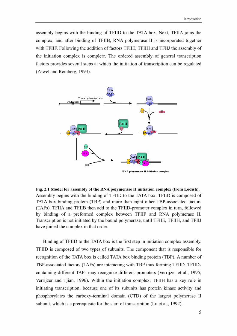
assembly begins with the binding of TFIID to the TATA box. Next, TFIIA joins the complex; and after binding of TFIIB, RNA polymerase II is incorporated together with TFIIF. Following the addition of factors TFIIE, TFIIH and TFIIJ the assembly of the initiation complex is complete. The ordered assembly of general transcription factors provides several steps at which the initiation of transcription can be regulated (Zawel and Reinberg, 1993).
Fig. 2.1 Model for assembly of the RNA polymerase II initiation complex (from Lodish). Assembly begins with the binding of TFIID to the TATA box. TFIID is composed of TATA box binding protein (TBP) and more than eight other TBP-associated factors (TAFs). TFIIA and TFIIB then add to the TFIID-promoter complex in turn, followed by binding of a preformed complex between TFIIF and RNA polymerase II. Transcription is not initiated by the bound polymerase, until TFIIE, TFIIH, and TFIIJ have joined the complex in that order.
Binding of TFIID to the TATA box is the first step in initiation complex assembly.
TFIID is composed of two types of subunits. The component that is responsible for recognition of the TATA box is called TATA box binding protein (TBP). A number of TBP-associated factors (TAFs) are interacting with TBP thus forming TFIID. TFIIDs containing different TAFs may recognize different promoters (Verrijzer et al., 1995; Verrijzer and Tjian, 1996). Within the initiation complex, TFIIH has a key role in initiating transcription, because one of its subunits has protein kinase activity and phosphorylates the carboxy-terminal domain (CTD) of the largest polymerase II subunit, which is a prerequisite for the start of transcription (Lu et al., 1992).
5
Introduction
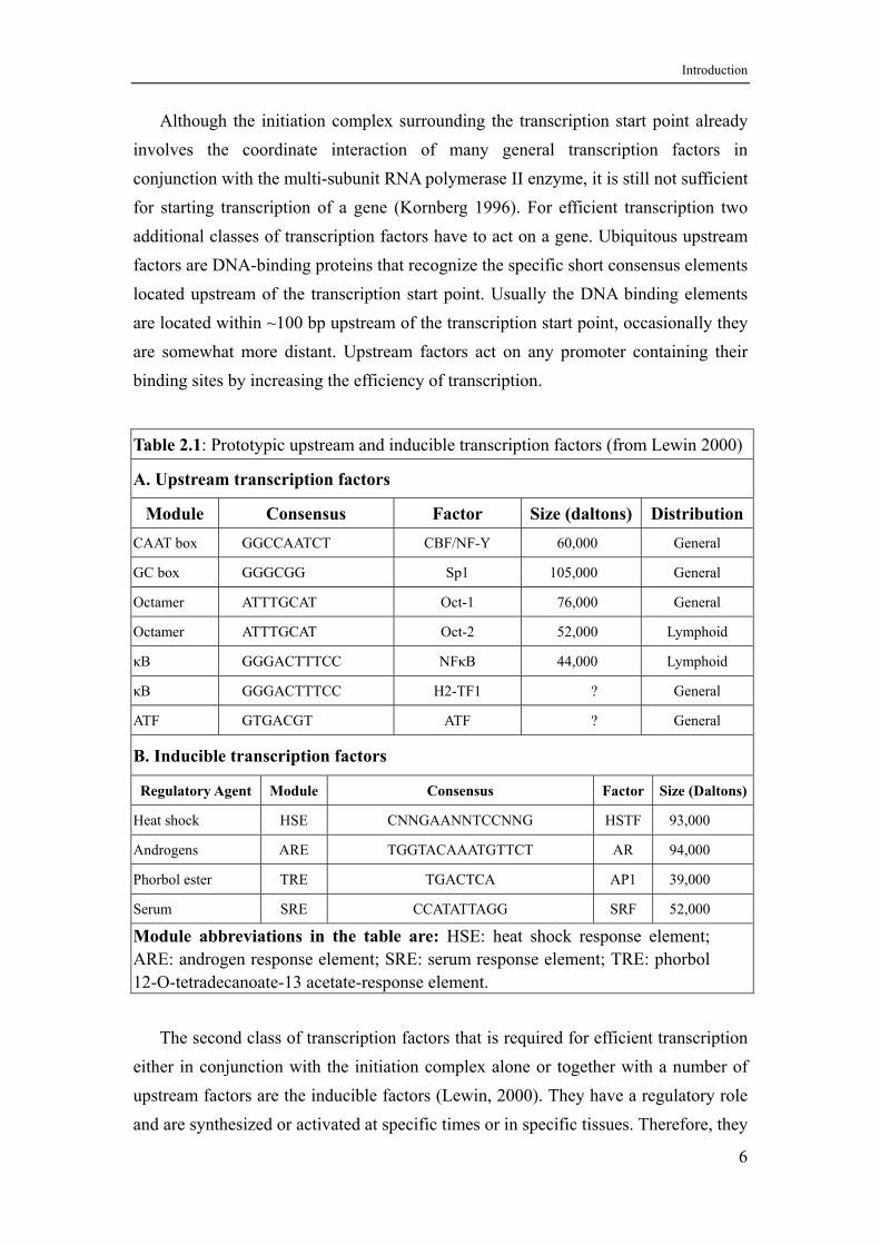
Although the initiation complex surrounding the transcription start point already involves the coordinate interaction of many general transcription factors in conjunction with the multi-subunit RNA polymerase II enzyme, it is still not sufficient for starting transcription of a gene (Kornberg 1996). For efficient transcription two additional classes of transcription factors have to act on a gene. Ubiquitous upstream factors are DNA-binding proteins that recognize the specific short consensus elements located upstream of the transcription start point. Usually the DNA binding elements are located within ~100 bp upstream of the transcription start point, occasionally they are somewhat more distant. Upstream factors act on any promoter containing their binding sites by increasing the efficiency of transcription.
Table 2.1: Prototypic upstream and inducible transcription factors (from Lewin 2000)
A. Upstream transcription factors
Module Consensus Factor Size (daltons) Distribution CAAT box GGCCAATCT CBF/NF-Y 60,000 General
GC box GGGCGG Sp1 105,000 General
Octamer ATTTGCAT Oct-1 76,000 General
Octamer ATTTGCAT Oct-2 52,000 Lymphoid
κB GGGACTTTCC NFκB 44,000 Lymphoid
κB GGGACTTTCC H2-TF1 ? General
ATF GTGACGT ATF ? General
B. Inducible transcription factors
Regulatory Agent Module Consensus Factor Size (Daltons)
Heat shock HSE CNNGAANNTCCNNG HSTF 93,000
Androgens ARE TGGTACAAATGTTCT AR 94,000
Phorbol ester TRE TGACTCA AP1 39,000
Serum SRE CCATATTAGG SRF 52,000
Module abbreviations in the table are: HSE: heat shock response element; ARE: androgen response element; SRE: serum response element; TRE: phorbol 12-O-tetradecanoate-13 acetate-response element.
The second class of transcription factors that is required for efficient transcription
either in conjunction with the initiation complex alone or together with a number of upstream factors are the inducible factors (Lewin, 2000). They have a regulatory role and are synthesized or activated at specific times or in specific tissues. Therefore, they
6
Introduction
are responsible for the control of transcription patterns in space and time. The DNA-binding elements of inducible factors that are regulated in response to certain stimuli are called response elements, such as the heat shock response element (HSE), the steroid hormone response element (SHR), or the serum response element (SRE). Binding of factors to their DNA elements is followed by protein-protein interactions with other components of the general transcription apparatus. Any one of several different elements can independently or synergistically activate gene transcription. In Table 2.1 a few prototypic upstream and inducible transcription factors are listed.
Some genes may be further stimulated by enhancers, which can act at a distance, and independent of orientation and position - upstream or downstream of the transcription start site. Enhancers consist of a number of DNA-binding elements for upstream and/or inducible factors that are arranged in a compact manner. Enhancers probably function by assembling a protein complex that interacts with promoter bound transcription factors, which requires that the intervening DNA is ‘looped out’.
2.1.2 Chromatin structure and the regulation of transcription
The human genome has to be compacted into a tiny nucleus using structures that can reversibly fold and unfold within a chromosome. Due to its staining properties this compacted DNA is called chromatin. The fundamental units of chromatin are the nucleosomes consisting of DNA and basic histone proteins. Using nucleases DNA in chromatin is degraded to a series of discrete fragments differing by multiples of 180-200 bp in size. DNA of this repeat unit length is wrapped around a histone octamer containing two copies each of the core histones H2A, H2B, H3, and H4. 146 bp of DNA are tightly wrapped around a histone octamer forming the nucleosome core particle. Some 40-50 bp of linker DNA are needed to connect one core particle to the next (Wolffe, 1999). A single 'linker'-histone H1 molecule is associated with each nucleosome core particle forming the chromatosome that contains some 160-170 bp of DNA and significantly stabilizes the DNA within the core particle. Histone H2A forms a heterodimer with H2B, and H3 forms a heterodimer with H4. Two H3/H4 heterodimers form the tetramer core of a nucleosome that is flanked by two H2A/H2B heterodimers on opposing sides. At last nucleosomes are packed into regular arrays forming a 30 nm fiber. Some chromosomal regions have properties distinct from the rest of the chromosome. This heterochromatin was found to be highly condensed and to replicate late in S-phase. The most common observed consequence of heterochromatin formation is the repression of transcription (Hennig, 1999).
7
Introduction
How do gene regulatory proteins and the general transcription factors gain access to DNA that is packaged into chromatin? Generally, nucleosomes do not present a serious barrier for either gene regulatory proteins or RNA polymerases. Enhancers can still function, positioned nucleosomes that block a promoter can be rearranged, and, once transcription has begun, polymerase II can transcribe through nucleosomes without dislodging them. The rearrangement could either be due to a separate activity of the gene activator proteins or could be an indirect consequence of the activator contacting the general transcription factors to facilitate their assembly on DNA (Croston and Kadonaga, 1993).
For gene expression, the chromatin in the vicinity of a gene must be decondensed before and during transcription. Sometimes a locus control region (LCR) that lies far upstream from a gene cluster is required for an extensive change in chromatin structure and for enabling expression of all genes in that domain. DNase I hypersensitive site experiments have confirmed the poor chromatin structure in such a region (Cockerill, 2000). Although several proteins that specifically bind to the LCR have been identified, the mechanism that alters the chromatin structure is still unknown. Some forms of higher-order DNA packaging render the DNA inaccessible both to gene regulatory proteins and to the general transcription factors. Higher-order DNA packaging thus plays a crucial part in the control of gene expression in eukaryotes, serving to silence large sections of the genome, in some cases reversibly, in other cases not. These inactive forms of chromatin, including the highly condensed heterochromatin (Croston and Kadonaga, 1993), are assumed to contain special proteins that make the DNA unusually inaccessible.
There is increasing evidence that modification of histones is associated with structural changes that occur in chromatin during replication and transcription. Acetylation and methylation occur on the free amino group of lysine, which removes its positive charge. Methylation occurs on arginine and histidine (Razin and Cedar, 1993). Phosphorylation occurs on the hydroxyl group of serine and also histidine, which introduces a negative charge. Acetylation is associated with changes in chromatin similar to those found during gene activation (Sternglanz, 1996) and which render the chromatin more sensitive to deoxyribonuclease. A cycle of phosphorylation occurs with H1, but its timing is different from the modification cycle of the other histones (Wolffe, 1999). All of these modifications affect internal residues and are transient. They change the charge of the protein molecule and have been viewed as potentially able to change the functional properties of the histones. Recent data
8
Introduction
indicate an indirect regulatory pathway in which ubiquitination of H2B (Lys 123) is a prerequisite for the methylation of H3 (Lys 4) and leads to transcriptional silencing (Sun and Allis, 2002).
Methylation of C residues in CpG dinucleotides also occurs in special regions called CpG islands, which show an accumulation of CpG doublets and often surround the promoters of constitutively expressed genes, although they are also found in the promoters of regulated genes (Antequera and Bird, 1993). An island included in a promoter must be unmethylated to be able to initiate transcription. A specific protein binds to the methylated CpG doublets and prevents initiation of transcription.
In conclusion, the genes are regulated in two steps. In the first step the chromatin of the target gene locus is decondensed, which is presumed to allow some of the gene regulatory proteins to access the DNA. In the second step the remaining gene regulatory proteins assemble on the DNA and direct the expression of individual genes.
2.2 Steroid hormones and their receptors
The gonads and adrenal gland produce five major groups of steroid hormones (SHs): estrogens, progestins, androgens, glucocorticoids, and mineralocorticoids. Steroid hormones can pass the cell membrane by simple diffusion due to their lipophilic nature. Within the target cells, SHs bind to high affinity steroid hormone receptors (SHRs). All unliganded SHRs are associated with a large multiprotein complex of chaperones, including heat shock protein 90 (Hsp90), which maintains the receptor in an inactive state but keeps it well prepared for hormone binding (Pratt and Toft, 1997). Most likely these chaperones play an active role in keeping the SHRs functional. Hormone binding activates the hormone receptor complex in order to induce expression of hormone responsive genes. Therefore, SHRs are ligand induced transcription factors.
2.2.1 Structure and distribution of SHRs
All steroid hormone receptors are modular proteins composed of distinct regions, which correspond to functional and structural units called domains. The most important domains are a central DNA-binding domain (DBD), which targets the receptor to the hormone response elements, and a ligand binding domain (LBD), required for switching the receptors’ function (Beato, 1989).
9
Introduction
Comparison of the amino acid sequences of various hormone receptors reveals a remarkable conservation. The DBD maps near the center of the primary sequence, and comprises ~80 amino acids containing the C2-C2- zinc-finger motif. Only very few amino acids within the first SHR zinc finger are responsible for specific recognition of the cognate HRE (Beato and Klug, 2000).
The ligand binding domain lies near the carboxy-terminal end of the receptor. The lipophilic steroid hormones and synthetic compounds with agonistic or antagonistic effects can bind to this domain and induce a transformation of the heterocomplexes of chaperones and SHR. Hormone induced transformatioin of the SHR heterocomplexes is associated with an increase in affinity for DNA, and a decrease in the size of the complex. The ligand binding domain is required for switching the receptors’ functions. When this domain is deleted in the glucocorticoid receptor (GR), the remaining DBD is in a constitutively active state, and no longer requires steroids for activity (Cadepond et al., 1991). Contrary to the GR, the ER is unable to activate transcription, when the LBD is deleted, although it continues to bind to an ERE (Gandini et al., 1997).
Ligand binding confers transcriptional competence onto SHRs that is exerted in most receptors by two independent transactivation functions, a constitutively active one located close to the DBD, referred to as activation function 1 (AF-1) and a ligand-inducible activation function in the LBD, called AF-2. The two AFs act synergistically and connect the receptor to the transcription apparatus through direct interactions with basal transcription factors, sequence-specific transcription factors and/or transcriptional co-activators (Beato and Klug, 2000).
Most members of the SHR family bind as homodimers to the palindromic hormone response elements (HREs) within the promoters of target genes, in contrast to other members of the nuclear receptor superfamily, which bind as heterodimers to their cognate recognition sequences.
The intracellular distribution of steroid receptors is the result of nuclear cytoplasmic export and ATP-dependent cytoplasmic-nuclear shuttling (Guiochon et al., 1991). At equilibrium the majority of ER, AR and PR is in the nucleus due to the presence of so-called nuclear localization signals (NLSs) that are believed to be required for nuclear pore recognition. The number and location of NLSs varies among SHRs.
10
Introduction
2.2.2 Interaction between SHRs and other factors
To regulate transcription, the activated SHRs must interact with other factors including general transcription factors (GTFs), co-activators, co-repressors, sequence specific factors or chromatin factors (see Figure 2.2).
The interaction between SHRs and GTFs can be achieved either by a direct contact (Beato and Sanchez-Pacheco, 1996) or by means of co-activators, also called transcription intermediary factors (TIFs), mediators or bridging factors. Co-activators are supposed to bridge between DNA-bound sequence-specific transcription factors and GTFs. Today, many of these co-activators have been characterized. One of them is the steroid receptor co-activator-1 (SRC-1) (Onate et al., 1995). SRC-1 interacts
with AF-2 of PR, GR and ERα in a ligand dependent fashion and enhances their hormone dependent transcriptional activities without altering the basal activity of a target promoter. Another important and general co-activator shown to interact with SHRs and SRC-1 is the CREB (for cAMP responsive element binding protein) binding protein (CBP), and the related protein p300 (Kamei et al., 1996). Some other co-activators have also been identified recently, which can interact with sequence-specific transcription factors or with general transcription factors and are necessary for the transactivation process.
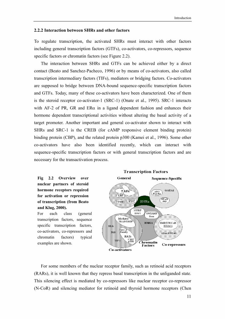
Fig 2.2 Overview over nuclear partners of steroid hormone receptors required for activation or repression of transcription (from Beato and Klug, 2000). For each class (general transcription factors, sequence specific transcription factors, co-activators, co-repressors and chromatin factors) typical examples are shown.
For some members of the nuclear receptor family, such as retinoid acid receptors
(RARs), it is well known that they repress basal transcription in the unliganded state. This silencing effect is mediated by co-repressors like nuclear receptor co-repressor (N-CoR) and silencing mediator for retinoid and thyroid hormone receptors (Chen
11
Introduction
and Evans, 1995; Hörlein et al., 1995). Transactivation by SHRs not only appears to be a simple net activation but the sum of relief from repression by co-repressors and activation by co-activators. The switch from the repressed to the activated state is promoted by the hormone ligand through an allosteric change in the SHR structure.
In addition to their HRE-mediated effects, SHRs control the activity of natural promoters also through positive and negative interactions with other sequence specific transcription factors (Beato et al., 1995). It is known that the interaction between GR and the heterodimeric transcription factor activator protein-1 (AP-1) can repress the activity of both partners. Similar repressive interactions have been described between
GR and the p65 subunit of the transcription factor NF-κB and the transcription factor GATA-1. Because most immunomodulatory genes and genes involved in
inflammation are positively regulated by the transcription factors AP-1 and NF-κB, it is well conceivable that the immunosuppressive and anti-inflammatory activities of
glucocorticoids are mediated through inhibition of AP-1 and NF-κB mediated transactivation by GR.
The interaction of SHRs with DNA and other factors takes place in the nucleus with its DNA compacted into chromatin. Genetic analyses have demonstrated a wide spread involvement of chromatin structure in gene regulation in general. Some nucleosome remodeling factors are ATP-dependent chromatin remodeling machines required for the transactivation of SHRs. In an in vitro system derived from Drosophila embryos, receptor binding to minichromosomes recruits ISWI and NURF 38 and triggers a chromatin remodeling event that facilitates access of NF1. NF1 plays only a structural role acting as a wedge to stabilize the open conformation of chromatin, thus facilitating full occupancy of the HREs and full transactivation (Di Croce et al., 1999; Eisfeld et al., 1997).
All observations lead to the current two step model for transcriptional activation by SHRs: (i) the hormone mediated recruitment of co-activators and other transcription factors with chromatin remodeling results in the local destabilization of repressive histone-DNA interactions, (ii) direct or most likely co-activator-mediated interactions with the basal transcription machinery initiate transcription (Beato and Klug, 2000).
12
Introduction
2.2.3 Androgens and androgen receptor
Androgens, most notably testosterone and dihydrotestosterone, have numerous clinically improtant actions in the developing embryo as well as in the pubertal and adult male. Androgens are important for the development and maintenance of characteristic male properties and specific reproductive organs and tissues. The action of androgens in target cells depends on the concentration in the serum and within the cells, the metabolic conversion of testosterone to dihydrotestosterone within the cells, the interactions with the receptor protein, and the action of the androgen receptor on the genomic level and on other signal transduction pathways.
In humans, the major androgen is testosterone, most of which is synthesized by the Leydig cells of the testis (Ewing and Zirkin, 1983). The adrenal cortex also contributes to the production of androgens. In brief, the steroidogenic cascade starts with the cleavage of the side chain of cholesterol. The biologically active androgens are generated by a stepwise degradation of the cleavage product pregnenolone. The final step in the biosynthetic pathway of testosterone is the reduction of the 17-keto group by 17β-hydroxysteroid dehydrogenase (17β-HSD) (Labrie et al., 1997). In target cells, the prohormone testosterone is converted to the active androgen
5α-dihydrotestosterone (DHT) by the enzyme 5α-reductase, which is localized at the surface of the nuclear membrane.
The major action of androgens is through direct activation of gene tanscription via high affinity interaction with the androgen receptor. The androgen receptor is a member of the steroid hormone receptor family. It is localized to the long arm of the X chromosome at Xq11-q12 (Brown et al., 1989). The AR gene is a single copy gene that spans 75-90 kilobases of genomic DNA and comprises 8 exons (Lubahn et al., 1989). In man, androgen receptor is detectable in testis, prostate, liver, cardiac muscle, sweat glands, hair follicles, pineal gland, vascular cells and various cell types in the temporal cortex. The prostate cancer cell line LNCaP and the breast cancer cell lines T47D and MCF-7 are also androgen receptor positive.
The predominant form of the AR is a 100-114 kDa protein of 918 amino acids. The full length AR is a single polypeptide comprised of discrete functional domains: an amino-terminal domain; a DNA-binding domain (DBD); a hinge region; and a ligand-binding domain (LBD). (i) The amino-terminal domain is the most variable in size and least homologous in sequence to other members of the steroid hormone receptor family. Within this domain resides a transcription activation region called
13
Introduction
activation function 1 (AF1). This region is important in transcriptional regulation via protein-protein interactions with other transcription factors (McEwan and Gustafsson, 1997). (ii) There are four cysteine residues invariably present in the DNA-binding domains of all steroid receptors, which bind a zinc ion with each of two loop structures known as zinc fingers. The DBD determines the specificity of receptor binding to DNA, with each of the zinc fingers serving distinct functions. The first zinc finger is responsible for recognition of the target DNA sequence, the second zinc finger stabilizes the DNA-receptor interaction by contacting the DNA phosphate backbone (Berg, 1989). (iii) The hinge region, located between DBD and LBD, is a region of low sequence homology. It provides an interface for interactions with other proteins such as c-Jun (Bubulya et al., 1996). In addition, the hinge contains one of the AR phosphorylation sites required for optimal transcriptional activity (Zhou et al., 1995). (iv) The ligand-binding domain encompasses approximately the carboxy-terminal one third of the protein. A principal function of the LBD is the specific, high-affinity binding of androgens. The ligand binding domain is folded into
a three-layered antiparallel α-helical sandwich that creates a wedge-shaped molecular scaffold forming the ligand binding cavity. This cavity is completely partitioned from the external environment and closed by helices 11 and 12 of the LBD, operating as a ‘lid’ after androgen has entered the binding pocket (Wurtz et al., 1996). It was suggested that ligand binding alters the structure of the LBD to a more compact one, less sensitive to the actions of proteases (Renaud et al., 1995).
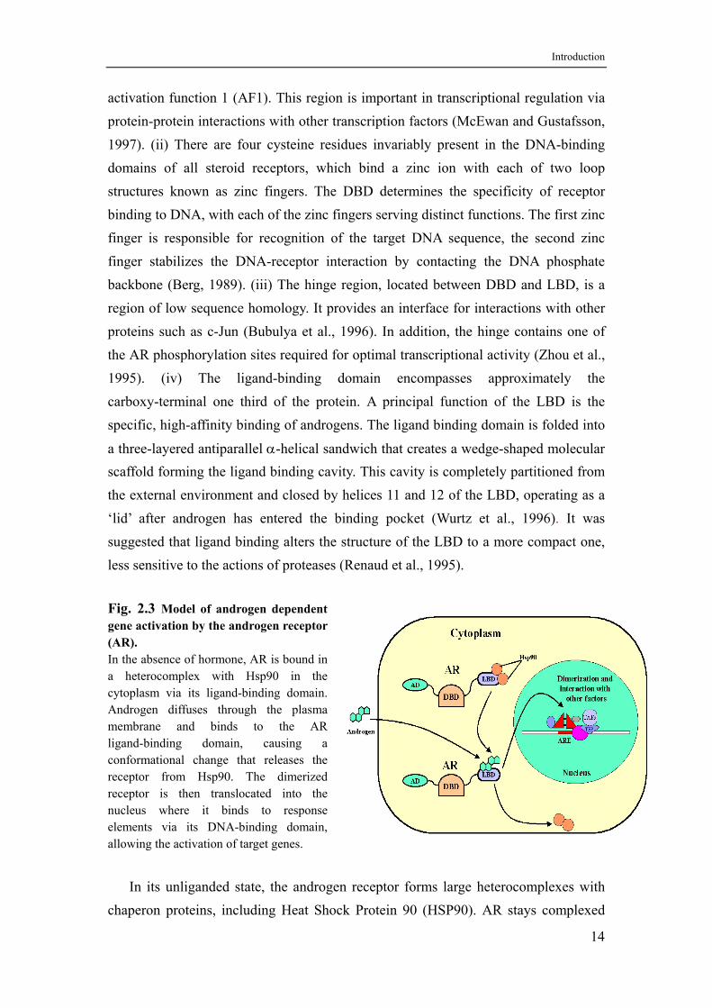
Fig. 2.3 Model of androgen dependent gene activation by the androgen receptor (AR). In the absence of hormone, AR is bound in a heterocomplex with Hsp90 in the cytoplasm via its ligand-binding domain. Androgen diffuses through the plasma membrane and binds to the AR ligand-binding domain, causing a conformational change that releases the receptor from Hsp90. The dimerized receptor is then translocated into the nucleus where it binds to response elements via its DNA-binding domain, allowing the activation of target genes.
In its unliganded state, the androgen receptor forms large heterocomplexes with
chaperon proteins, including Heat Shock Protein 90 (HSP90). AR stays complexed
14
Introduction
with HSP90 until androgen binds the ligand-binding pocket. The interaction with HSP90 retains the AR in an inactive state and is important for maintenance of a conformation optimal for high affinity ligand binding (Smith and Toft, 1993). It is known that androgen binding induces a conformational change, as a result of removal of proteins such as HSP90, unmasking certain functional domains. These changes facilitate receptor dimerization, nuclear transport, interaction with target DNA, and activation of target gene transcription (Figure 2.3). Whereas the LBD mediates activation in the presence of hormone, it seems to repress AR function in the absence of androgens. This is suggested by experiments using a mutant AR with its LBD deleted that is constitutively active in the absence of hormone (Jenster et al., 1991).
Phosphorylation by protein kinases A and C is a common post-translational modification in steroid hormone receptors. AR is phosphorylated at serine and threonine residues particularly in the amino-terminal domain and hinge region. AR phosphorylation is increased by androgen binding and appears to enhance the transcriptional activity of the receptor, perhaps by altering the interaction of ligand-activated AR with other proteins in the transcription complex (Ikonen et al., 1994).
AR is located in the cytoplasmic compartment prior to the binding of androgen. Nuclear uptake of the AR is an androgen dependent process mediated by a nuclear localization signal located in the C-terminal segment of the second zinc finger and the hinge region (Jenster et al., 1993). AR dimerization is also an androgen dependent process. The interaction between the amino terminus of one AR molecule and the LBD of another results in an anti-parallel homodimer (Langley, et al. 1995).
Like other steroid hormone receptors, AR specifically binds to DNA sequences called androgen responsive elements (AREs). AREs are also recognized by GR and PR. Therefore the induction of an ARE containing gene depends on the receptor status of the cell. In addition, some studies suggest that the AR may heterodimerize with GR so that they can influence each other’s transcriptional activity (Chen et al., 1997). AREs have been identified in the promoter regions of a number of androgen regulated genes, including prostate-specific antigen (PSA) and kallikrein-2 (KLK-2) (Riegman et al., 1991), sex-limited protein (Adler et al., 1991), and probasin (Rennie et al., 1993).
In order to regulate transcription, AR has to interact with many proteins as was similarly shown for other SHRs. AR directly interacts with the basal transcription factors TFIIF and TBP via AF1, and this interaction appears to stimulate recruitment
15
Introduction
of the transcriptional machinery to the promoter region of the target gene (McEwan and Gustafsson, 1997). The c-Jun component of AP-1 directly interacts with AR, probably through the leucine zipper region of c-Jun and the DBD of AR, thereby repressing AR transactivation (Sato et al., 1997). Similarly, interaction between the
amino terminal region of AR and RelA, a member of the NF-κB family of transcription factors, represses AR mediated transactivation (Palvimo et al., 1996).
Interestingly, there is evidence for ligand independent activation of AR transcriptional activity by peptide growth factors, such as insulin-like growth factor I (IGF-I) and epidermal growth factor (EGF) (Culig et al., 1995). In addition, the protein kinase A activator, forskolin, can induce AR in the absence of androgens, which likely involves receptor phosphorylation. This activation requires the DBD of the receptor and may be important in androgen-independent growth of prostatic tumors (Nazareth and Weigel, 1996).
2.2.4 Non-genomic effects of androgens
Until recently it was believed that transcription activation by AR is mainly mediated by genomic actions, i.e. by binding of AR to AREs in the promoter of target genes.
Meanwhile, a number of rapid non-genomic effects of steroids, often involving ion fluxes, have been reported, like for progesterone (Baldi et al., 1995), estrogens (Aronica et al., 1994), corticosterone (Ibarrola et al., 1991), and aldosterone (Wehling et al., 1994). Androgens have also been shown to induce rapid calcium fluxes in a variety of classical androgen-dependent cell types. In the human prostate cancer cell line LNCaP for example intracellular calcium concentrations are increased within two
minutes after addition of 5α-dihydrotestosterone or testosterone (Steinsapir et al., 1991). In most of these studies it could be shown that this change in intracellular calcium concentration is caused by a transmembrane influx of extracellular calcium through the plasma membrane, but the structure of the receptive unit and the biological significance of these membrane effects are not known yet.
Another example, which cannot be explained by the classical androgen response pathway, is that testosterone can modify the susceptibility of T-cells to infectious diseases. Recently, effects of testosterone and testosterone covalently coupled to albumin on the calcium flux through the plasma membrane of T-cells were reported (Benten et al., 1997). Since T-cells do not possess classical androgen receptors, this biological response indicates that plasma membrane receptors are involved in this non-genomic androgen effect.
16
Introduction
The fact that spermatogenesis depends on high levels of testosterone also can’t be easily explained on the basis of classical androgen receptor action, because the concentration of testosterone required for the maintenance of spermatogenesis is much higher than required for saturating the androgen receptor. Therefore an alternative sensing system, different from the androgen receptor, might be active in spermatogenesis. Some data support the view that alternative low-affinity interactions of steroids with yet undefined receptive structures in the plasma membrane may be essential for maintenance of spermatogenesis and the coordination of this process with steroidogenesis (Gorczynska and Handelsman, 1995).
2.3 Secretoglobins
The secretoglobins form a family of small secretory proteins with unclear physiological functions. After the first member of this family, uteroglobin, was identified from rabbit uterus secretions some 30 years ago (Beier, 1968; Krishnan and Daniel, 1967), more than 20 members were found in mammals within the last few years (Ni et al., 2000).
2.3.1 The secretoglobin family of proteins
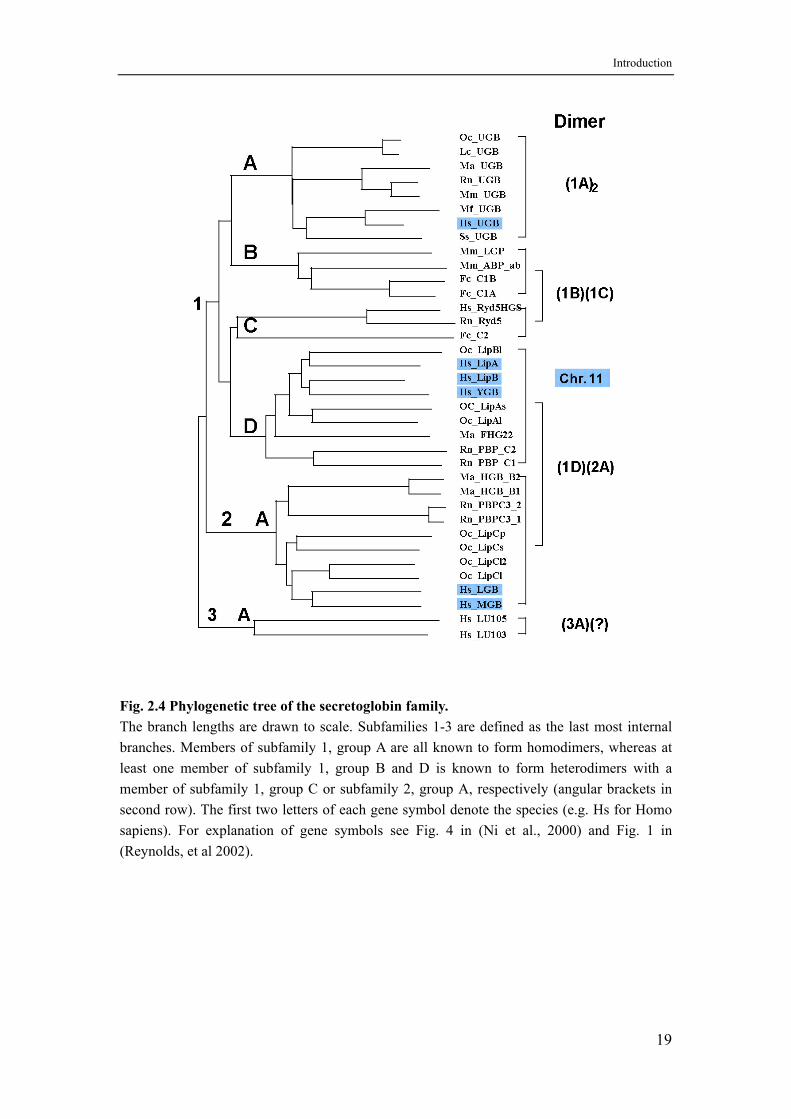
The secretoglobin family can be grouped into three subfamilies and six groups, as shown in Figure 2.4. All genes are composed of three exons with intron positions strongly conserved. The encoded proteins are very similar in length and all except two contain two conserved cysteines, one close to the amino-terminus and the other close to the carboxy-terminus, and one conserved central lysine (Ni et al., 2000).
Subfamily 1, group A contains the orthologous uteroglobins from different species. Most members of this family have been described to form antiparallel homodimers via two intermolecular disulfide bonds between the two conserved cysteines. From X-ray crystallographical and NMR analyses it is known that a uteroglobin monomer consists of a bundle of four alpha-helices (Mornon et al., 1980 and Umland et al., 1994). Between the two antiparallel molecules of a homodimer a pocket is formed in which a number of small hydrophobic ligands like progesterone can be bound. The hydrophobic pocket is accessible through a channel that is closed by the two disulfide bonds in the oxidized form (Mornon et al., 1980). In addition, a central lysine, which is conserved in all family members, is described as a calcium binding site of the phospholipase A2 type for human uteroglobin (Lys 42 in uteroglobin) (Barnes et al.,
17
Introduction
1996). The same region within helix 3 had been shown to be responsible for the inhibitory activity of uteroglobin on soluble phospholipase A(2) (sPLA(2)). Therefore it was suggested that uteroglobin may inhibit sPLA(2) activity by binding and sequestering Ca2+, essential for sPLA(2) activation. Because the conserved lysine can also form an exposed salt bridge with an aspartate side chain in its vicinity, it had to be postulated that this salt bridge must be dissolved before binding of calcium to Lys 42 is enabled. Recently it was shown that recombinant wild-type uteroglobin does not bind Ca2+ unless it is expressed with a histidine-tag suggesting that the calcium binding ability of uteroglobin is only an artifact (Chowdhury et al., 2002).
From biochemical data it can be predicted that members of subfamily 1, group B form heterodimers with members of subfamily 1, group C and members of subfamily 1, group D will likely form heterodimers with members of subfamily 2, group A (Ni et al., 2000 and see Figure 2.4). Therefore, the heterodimer is the fundamental unit of secretoglobin quaternary structure. Interestingly, all subfamily 1, group B-D and subfamily 2 members contain a third conserved cysteine that is forming a third disulfide bridge between the heteromers. A few secretoglobins show another quaternary structure feature. Prostatic binding protein (PBP) for example is a well characterized non-covalent heterodimer of two covalent heterodimers C1/C3 and C2/C3 (Heyns et al., 1978). In the exorbital lacrimal gland C1 and C2 are absent, and C3 forms a heterodimer with a hitherto unknown secretoglobin called the lacrimal component, and two C3/lacrimal component heterodimers form again a heterotetramer (Vercaeren et al., 1996). A mammaglobin/lipophilin B heterodimer that is heterodimerizing with itself has been found in the human mammary gland (Carter et al., 2002; Colpitts et al., 2001).
Although all secretoglobin family proteins are classical secretory proteins, only a few members have been shown to be glycosylated. The precise N-glycosylation site within exon 2 is known for rat PBP C3 (Peeters et al., 1981) and cat Fel dI Chain 2 (Morgenstern et al., 1991). Glycosylation of mammaglobin has been observed, which is consistent with the two predicted N-linked glycosylation sites in its primary sequence (Carter et al., 2002).
18
Introduction
Fig. 2.4 Phylogenetic tree of the secretoglobin family. The branch lengths are drawn to scale. Subfamilies 1-3 are defined as the last most internal branches. Members of subfamily 1, group A are all known to form homodimers, whereas at least one member of subfamily 1, group B and D is known to form heterodimers with a member of subfamily 1, group C or subfamily 2, group A, respectively (angular brackets in second row). The first two letters of each gene symbol denote the species (e.g. Hs for Homo sapiens). For explanation of gene symbols see Fig. 4 in (Ni et al., 2000) and Fig. 1 in (Reynolds, et al 2002).
19
Introduction
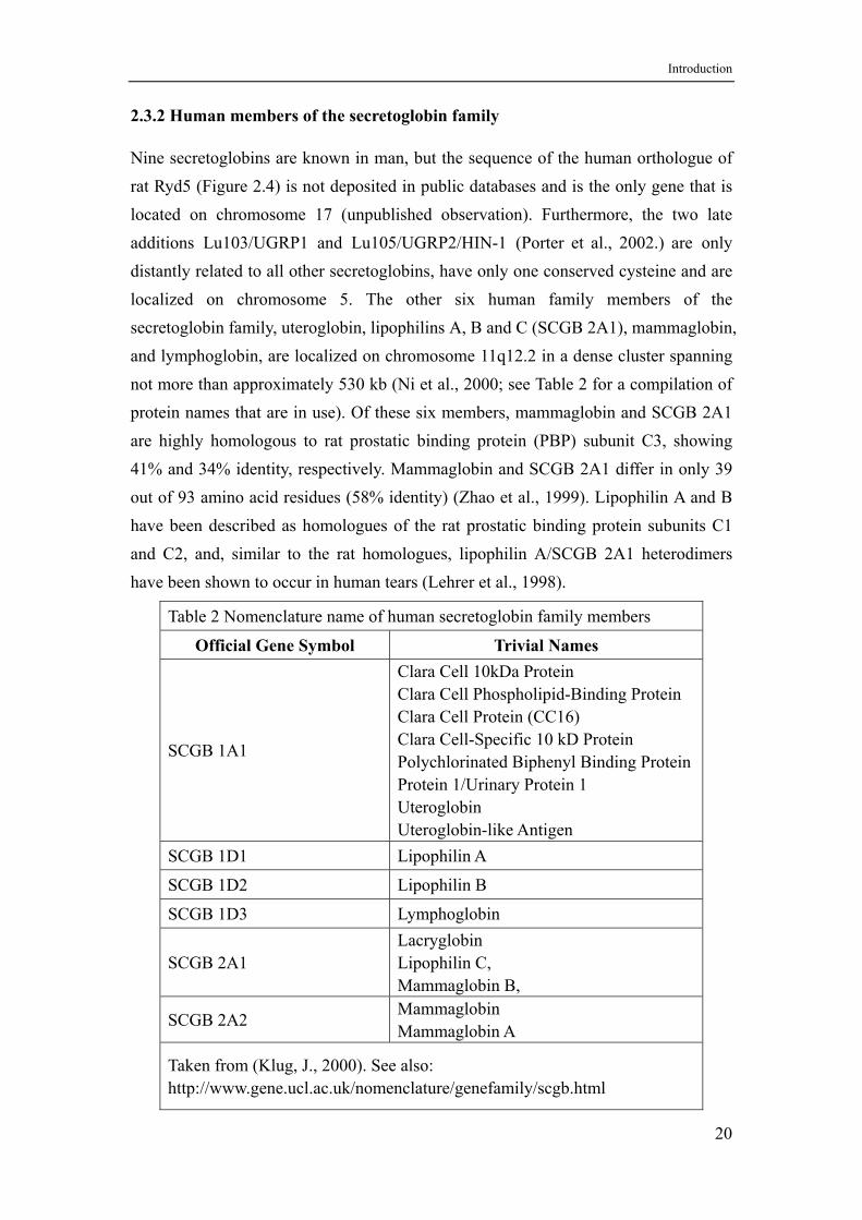
2.3.2 Human members of the secretoglobin family
Nine secretoglobins are known in man, but the sequence of the human orthologue of rat Ryd5 (Figure 2.4) is not deposited in public databases and is the only gene that is located on chromosome 17 (unpublished observation). Furthermore, the two late additions Lu103/UGRP1 and Lu105/UGRP2/HIN-1 (Porter et al., 2002.) are only distantly related to all other secretoglobins, have only one conserved cysteine and are localized on chromosome 5. The other six human family members of the secretoglobin family, uteroglobin, lipophilins A, B and C (SCGB 2A1), mammaglobin, and lymphoglobin, are localized on chromosome 11q12.2 in a dense cluster spanning not more than approximately 530 kb (Ni et al., 2000; see Table 2 for a compilation of protein names that are in use). Of these six members, mammaglobin and SCGB 2A1 are highly homologous to rat prostatic binding protein (PBP) subunit C3, showing 41% and 34% identity, respectively. Mammaglobin and SCGB 2A1 differ in only 39 out of 93 amino acid residues (58% identity) (Zhao et al., 1999). Lipophilin A and B have been described as homologues of the rat prostatic binding protein subunits C1 and C2, and, similar to the rat homologues, lipophilin A/SCGB 2A1 heterodimers have been shown to occur in human tears (Lehrer et al., 1998).
Table 2 Nomenclature name of human secretoglobin family members
Official Gene Symbol Trivial Names
SCGB 1A1
Clara Cell 10kDa Protein Clara Cell Phospholipid-Binding Protein Clara Cell Protein (CC16) Clara Cell-Specific 10 kD Protein Polychlorinated Biphenyl Binding Protein Protein 1/Urinary Protein 1 Uteroglobin Uteroglobin-like Antigen
SCGB 1D1 Lipophilin A
SCGB 1D2 Lipophilin B
SCGB 1D3 Lymphoglobin
SCGB 2A1 Lacryglobin Lipophilin C, Mammaglobin B,
SCGB 2A2 Mammaglobin Mammaglobin A
Taken from (Klug, J., 2000). See also: http://www.gene.ucl.ac.uk/nomenclature/genefamily/scgb.html
20
Introduction
As long as a generally accepted physiological function is not known for any secretoglobin, the expression pattern of each member is a decisive characteristic, and many proteins have been named accordingly: uteroglobin – uterus, mammaglobin – mammary gland, prostatein – prostate, lacryglobin – lacrimal gland. Uteroglobin expression is prominent in the prostate, testis, ovary, mammary gland, and salivary gland (Ni et al., 2000). Lipophilin A expression is found in lacrimal gland, thymus, and low level expression occurs in testis, kidney and ovary. Lipophilin B is found to be moderately expressed in heart, skeletal muscle, kidney and pancreas, whereas lipophilin C (SCGB 2A1) is strongly expressed in lacrymal gland, pancreas, prostate, testis, and ovary, but only weakly in thymus (Zhao et al., 1999). Mammaglobin is strongly and almost exclusively expressed in the mammary gland. One study found weak expression in prostate tissue (Ni et al., 2000). Mammaglobin has been used to evaluate primary, metastatic and occult breast cancer, and provides a new tool for breast cancer diagnosis and patient management (Watson et al., 1999; Fanger et al., 2002; Zehentner et al., 2002, and references therein). Lymphoglobin is expressed only in spleen and peripheral blood lymphocytes and is thus the only secretoglobin that is specifically expressed in lymphocytes (Ni et al., 2000).
Although each secretoglobin has a peculiar expression pattern, the expression patterns of all family members are overlapping and limited to a characteristic set of epithelial tissues separating the body interior from the exterior world. Most common are expression in lung, male and female genital organs like testis, prostate and uterus, as well as in mammary and salivary glands.
Although the homologies between secretoglobin family genes extend into regulatory regions, the expression pattern of each gene is remarkably distinct. Unlike other secretoglobins that are regulated by steroid hormones such as uteroglobin and PBP, mammaglobin expression is not induced by estrogens in estrogen receptor positive breast cancer cell lines such as MCF7 and T47D (Watson et al., 1998). Therefore, it will be interesting to explore which mechanisms govern tissue specific expression and how these mechanisms are modified between different secretoglobin genes.
21

Introduction
2.4 Aim of the Project
SCGB 2A1 (lacryglobin, lipophilin C, mammaglobin B) belongs to the secretoglobin family of small secretory proteins (formerly called uteroglobin/CC10/CCSP proteins). Though all members share many similarities – gene and protein structure (one structure known (Umland et al., 1994), the other five predicted to be very similar (Callebaut et al., 2000)), dimerization behaviour and genomic localization – the expression pattern of each gene is remarkably distinct. Of the six known human proteins only uteroglobin is well investigated in terms of gene regulation (Wolf et al., 1992). Therefore, it was intended to study the expression of one of the recently discovered secretoglobin genes. SCGB 2A1 seemed to be an interesting candidate because it is homologous to mammaglobin, that is almost exclusively expressed in the mammary gland independent of steroid hormones, and to subunit C3 of the rat prostatic binding protein that is under androgen control. Because SCGB 2A1 was already known to be expressed in the prostate it was first tested if SCGB 2A1 is controlled by androgens. For an immunohistochemical analysis of SCGB 2A1 expression in the prostate the histidine-tagged protein had to be expressed in E. coli and an antiserum produced in rabbits.
As a model system for all investigations the androgen responsive prostate cancer derived cell line LNCaP was used. In a systematic “top-to-bottom” approach it was intended to look first for DNase I hypersensitive sites within the SCGB 2A1 gene in the chromatin of LNCaP cells. These sites are highly indicative for regions functionally involved in gene regulation. In case of androgen regulation one could expect to find one or more androgen dependent DNase I hypersensitive sites involved in the opening of chromatin triggered by binding of the androgen receptor. After knowing the position(s) of DNase I hypersensitive site(s) the identified gene regions could be investigated more carefully by transfecting cognate promoter deletion- reporter gene constructs into LNCaP cells. By using computer analysis, DNase I footprinting and EMSA analyses potential transcription factor candidates should be identified. Finally, the functional relevance of binding of the identified transcription factors to their DNA elements for transcription of the SCGB 2A1 gene should be investigated by testing reporter gene constructs containing mutations in these DNA elements that were known to abrogate binding. Surprisingly, this rather descriptive approach led to an unexpected result concerning the mechanism of androgen induction of the SCGB 2A1 gene.
22
Materials and Methods
3. MATERIALS AND METHODS
3.1 Materials
3.1.1 Chemicals and equipment
Chemicals
Acrylamide/Bis-acrylamide Roth, Karlsruhe Agarose Gibco-BRL, Neu Isenburg Bacto-Tryptone Gibco-BRL, Neu Isenburg Bacto-yeast extract Gibco-BRL, Neu Isenburg Boric acid Merck, Darmstadt Bromophenol blue sodium salt Serva, Heidelberg Calcium chloride Merck, Darmstadt Chloroform Merck, Darmstadt Coomassie Brilliant Blue R250 Roth, Karlsruhe 2’-Deoxynucleoside 5’-triphosphates Gibco-BRL, Neu Isenburg Dihydrotestosterone (DHT) Sigma-Aldrich, Seelze Dimethyl sulfoxide (DMSO) Merck, Darmstadt Dipotassium hydrogenphosphate Merck, Darmstadt Disodiumhydrogen phosphate Merck, Darmstadt 1,4-Dithiothreitol Roth, Karlsruhe Ethanol Merck, Darmstadt Ethidiumbromide Roth, Karlsruhe Ethylene diaminetetraacetic acid disodium salt (EDTA) Merck, Darmstadt Ficoll 400 Sigma-Aldrich, Seelze Formamide Merck, Darmstadt Glacial acetic acid Merck, Darmstadt Glycerol Merck, Darmstadt Glycine Sigma-Aldrich, Seelze Guanidine hydrochloride Sigma-Aldrich, Seelze 4-(2-Hydroxyethyl)piperazine-1-ethanesulfonic acid (HEPES) Roth, Karlsruhe Igepal CA-630 (indistinguishable from NP-40) Sigma-Aldrich, Seelze Isopropylthio-β-D-galactoside (IPTG) Applichem, Darmstadt Leupeptin Sigma-Aldrich, Seelze
23
Materials and Methods
Magnesium chloride Merck, Darmstadt Manganese chloride Merck, Darmstadt Methanol Merck, Darmstadt Phenol/chloroform/isopropanol (25:24:1) Roth, Karlsruhe Phenylmethylsulfonyl fluoride (PMSF) Sigma-Aldrich, Seelze Poly (dI-dC) Sigma-Aldrich, Seelze Polyvinylpyrrolidone Sigma-Aldrich, Seelze Ponceau S Sigma-Aldrich, Seelze Potassium chloride Merck, Darmstadt Rotiphorese Gel 30, 40 Roth, Karlsruhe Sodium acetate Roth, Karlsruhe Sodium azide Merck, Darmstadt Sodium chloride Sigma-Aldrich, Seelze Sodium citrate Merck, Darmstadt Sodium dodecyl sulfate (SDS) Merck, Darmstadt N,N,N´,N´-Tetramethylethylenediamine (TEMED) Roth, Karlsruhe Tris(hydroxymethyl)aminomethane (Tris) Roth, Karlsruhe Triton X-100 Sigma-Aldrich, Seelze Urea Merck, Darmstadt Xylene cyanol FF Serva, Heidelberg
Cell Culture Media and Antibiotics
Ampicillin sodium salt Ratiopharm® Chloramphenicol Sigma, München DMEM medium (11965-092) Gibco-BRL, Karlsruhe FCS, trypsin, glutamine Gibco-BRL, Karlsruhe RPMI 1640 medium (72400-021) Gibco-BRL, Karlsruhe Penicillin/Streptomycin (15140-114) Gibco-BRL, Karlsruhe
Radioactive reagents
[α-32P] dCTP (370 MBq/ml) Amersham, Braunschweig [γ-32P] ATP (370 MBq/ml) Amersham, Braunschweig
24
Materials and Methods
Equipment
Agarose gel electrophoresis chambers Biorad, München AutoLumat 953 luminometer Berthold Technologies, Bad Wildbad Cell culture incubator BBD6220 Kendro, Hanau Clean bench HA2448GS Kendro, Hanau Geiger counter Mini 900 Mini Instruments, Burnham-on-Crouch Gel dryer model 583 Biorad, München
GeneAmp® PCR system 9700 Applied Bioystems, Darmstadt Inverted microscope (DMIL) Leica, Wetzlar Power supply units Biorad, München Phosphorimager Fuji FLA3000G and screens Raytest, Straubenhardt Semi-dry blot apparatus (Fastblot B33) Biometra, Göttingen Sorvall Superspeed refrigerated centrifuge Kendro, Hanau Szintillation counter Packard
Miscellaneous
Bio-Rad Protein Assay Biorad, München
Biodyne® B nylon membranes Pall, Dreieich DNA and protein size markers Roche, Mannheim Hybond ECL nitrocellulose membrane Amersham Biosciences, Freiburg Kodak BioMax X-ray film Integra Biosciences, Fernwald Sterile plastic ware for cell culture Greiner, Germany
3.1.2 Cell lines
HeLa: Established in 1951, the HeLa cell line was the first continuously cultured human epitheloid cell line from an adenocarcinoma of the cervix of a 31-year-old black female named Henrietta Lacks (Gey et al., 1952). HeLa cells are aneuploid and steroid hormone receptor negative. Cells were grown in DMEM medium supplemented with 10% FCS at 37°C with 5% CO2. Confluent cultures were split 1:4 to 1:6 using trypsin/EDTA.
LNCaP: LNCaP clone FGC was isolated in 1977 by J.S. Horoszewicz et al., from a needle aspiration biopsy of the left supraclavicular lymph node of a 50-year-old Caucasian male with confirmed diagnosis of metastatic prostate carcinoma (Murphy, 1980). The cells are expressing an androgen receptor point mutant which shows a broader steroid binding specificity than the wild type receptor (Veldscholte et al., 1992)
25
Materials and Methods
and are responsive to 5-alpha- dihydrotestosterone. The cells do not produce uniform monolayer, but grow in clusters which should be broken apart by repeated pipetting when subcultures are prepared. They attach only lightly to the substrate, do not become confluent and rapidly acidify the medium. Cells were grown in RPMI 1640 medium supplemented with 10% FCS at 37°C with 5% CO2. Subconfluent cultures were split 1:4 once a week using trypsin/EDTA (occasionally cells could be detached by tapping).
For storage cells were frozen with 70% medium, 20% FBS, 10% DMSO at about 2×10⁶ cells/ampoule. After thawing or trypsinization cells may need 1-2 days to become adherent again. Due to strong cell aggregation, it is difficult to perform an exact cell count. The cells should be allowed to incubate undisturbed for the first 24 hours after subculture.
3.1.3 Buffers and solutions
The following standard solutions were used:
Denhardt’s reagent: Denhardt’s reagent is ususlly made up as a 50× stock solution, which contains 1% (w/v) Ficoll 400, 1% (w/v) polyvinylpyrrolidone, 1% (w/v) bovine serum albumin in H2O, and is stored at -20°C after filtration.
Formamide loading buffer: 95% deionized formamide, 10mM EDTA (pH 8.0), 0.05% bromophenol blue, 0.05% xylene cyanol. The formamide is deionized by stirring on a magnetic stirrer with Dowex XG8 mixed bed resin for 1 hour and filtering it twice through Whatman No. 1 paper. Deionized formamide is stored in small aliquots at -70°C.
6× Gel-loading buffer: 0.25% (w/v) bromophenol blue, 30% (v/v) glycerol in H2O. PBS (pH 7.4): Dissolve 8 g of NaCl, 0.2 g of KCl, 1.44 g of Na2HPO4, and 0.24 g of
KH2PO4 in 800 mL of distilled H2O. Adjust the pH to 7.4 with HCl. Add H2O to 1 liter. Sterilize by autoclaving and store at room temperature. The final concentrations of the ingredients are 137 mM NaCl, 2.7 mM KCl, 4.3 mM Na2HPO4, 1.4 mM KH2PO4.
RNA Gel-loading Buffer: 95% deionized formamide, 0.025% (w/v) bromophenol blue, 0.025% (w/v) xylene cyanol FF, 5 mM EDTA (pH 8.0), 0.025% (w/v) SDS.
6× SDS gel-loading buffer: 280 mM Tris-Cl (pH 6.8), 12 % (v/v) SDS, 60 % (v/v) glycerol, 0.25% bromophenol blue.
20× SSC: Dissolve 175.3 g of NaCl and 88.2 g of sodium citrate in 800 ml of H2O. Adjust the pH to 7.0 with a few drops of concentrated HCl. Adjust the volume to 1 liter with H2O. Dispense into aliquots. Sterilize by autoclaving. The final
26
Materials and Methods
concentrations of the ingredients are 3.0 M NaCl and 0.3 M sodium citrate.
TAE (pH 8.0) electrophoresis buffer: Prepare a 50× stock solution in 1 liter of H2O: 242 g of Tris base, 57.1 mL of glacial acetic acid, 100 mL of 0.5 M EDTA (pH
8.0). The 1× working solution is 40 mM Tris-acetate/1 mM EDTA. TBE: Prepare a 5× stock solution in 1 liter of H2O: 54 g of Tris base, 27.5 g of boric
acid, 20 ml of 0.5 M EDTA (pH 8.0). The 0.5× working solution is 90mM Tris-borate/2 mM EDTA. The pH of the concentrated stock buffer should be approx. 8.3. Dilute the concentrated stock buffer just before use and make the gel solution and the electrophoresis buffer from the same concentrated stock solution.
Passing the 5× buffer stocks through a 0.22-µm filter can prevent or delay formation of precipitates.
TE8: 10mM Tris (pH 8.0), 1mM EDTA (pH 8.0). Sterilize by autoclaving and store the buffer at room temperature.
10× Tris-glycine SDS electrophoresis buffer: Prepare a 10× stock of electrophoresis buffer by dissolving 30.2 g of Tris base and 188 g of glycine in 800 mL of deionized H2O, then add 100 mL of a 10%(w/v) stock solution of electrophoresis
grade SDS and adjust the volume to 1000 mL with H2O. The 1× working solution is 250 mM glycine, 25 mM Tris-Cl, 0.1% SDS.
3.1.4 Enzymes and Antibodies
All restriction endonucleases, T4 DNA ligase and T4 DNA polymerase were supplied by Gibco Invitrogen, Karlsruhe. AmpliTaq DNA polymerase (Stoffel fragment) was supplied by Applied Bioystems, Darmstadt. PfuTurbo high fidelity DNA polymerase was supplied by Stratagene. Klenow fragment and DNase I was supplied by Roche, Mannheim.
Goat NF-Y antibody (catalogue number sc-7711) was obtained from Santa Cruz Biotechnology, Heidelberg. Rabbit polyclonal anti Sp1 and anti Sp3 antibodies (Hagen et al., 1994) were kindly provided by Guntram Suske, Universität Marburg. The Sp1 antibody is directed against the full length Sp1 protein, whereas the Sp3 antibody is directed against B-domain and C-terminus but not the A-domain (N-terminus). For the generation of a rabbit polyclonal anti SCGB 2A1 antibody, see chapter 3.2.15. The donkey anti rabbit IgG, HRP-linked whole antibody from
Amersham Biosciences, Freiburg, was used in combination with the ECL™ Plus detection system from Amersham Biosciences, Freiburg.
27
Materials and Methods
3.1.5 Oligonucleotides
The oligonucleotides used for PCR, cloning, subcloning and mutagenesis are listed below. Oligonucleotides were either synthesized on an Applied Biosystems 380A oligonucleotide synthesizer using deoxynucleoside phoshoramidite chemistry (Beaucage and Caruthers, 1981), or were supplied by MWG-Biotech, Ebersberg.
Oligonucleotide Sequence SCGB 2A1 UTR+50/KpnI-3′ 5′-GGCGGTACCTGTCTGTGTTCAGTCGTGC-3′
SCGB 2A1 -53/XhoI-5′ 5′-ATTCTCGAGAGGGACTAAGGTGCCTCCCT-3′
SCGB 2A1 -136/XhoI-5′ 5′-TACCTCGAGAGCACAGGCTGGCTGTGTTC-3′
SCGB 2A1 -382/XhoI-5′ 5′-GAACTCGAGGAAGAGGAGGAGACCAAGAG-3′
SCGB 2A1 -575/XhoI-5′ 5′-AACCTCGAGAACAACAGCAACAAAACCCC-3′
SCGB 2A1 -963/XhoI-5′ 5′-GTACTCGAGAATTAACTTAGTGTTGTAAT-3′
SCGB 2A1 –1314/XhoI-5′ 5′-TGGCTCGAGGGGCAAGACTCTGTCTCGAA-3′
SCGB 2A1 -1476/XhoI-5′ 5′-CCACTCGAGAGAAACCCCGTCTCTACTAA-3′
SCGB 2A1 –53/TK/XhoI-3′ 5′-TAGCTCGAGCCAGGAATGAGGCAATGTG-3′
SCGB 2A1 –28/TK/XhoI-3′ 5′-CTACTCGAGCCCCAGGGAGGCACCTTAG-3′
SCGB 2A1 HS Xba I-5′ 5′-CCTGAGCCAACTTTGTTTTTCT-3′
SCGB 2A1 HS Xba I-3′ 5′-GGTATGAGAAAGTGGTCATTGC-3′ SCGB 2A1 NF-Y mut 5′-AGTGTACACTCATGGGAACACAGCC-3′ SCGB 2A1 NF-1 mut 5′-GCAGTAATATTTTTATCCCTGA-3′ SCGB 2A1 dim-IR-GC box
distal mut 5′-TCCCCAGGGAGGCACCTTAGTCACTCAAAGGAAT-3′
SCGB 2A1 dim-IR-GC box proximal mut 5′-TCCCCATTGAGTCACCTTAGTCCCTCCCAGGAAT-3′
SCGB 2A1 dim-IR-GC box mut 5′-TCCCCATTGAGTCACCTTAGTCACTCAAAGGAAT-3′
28
Materials and Methods
3.1.6 Plasmids
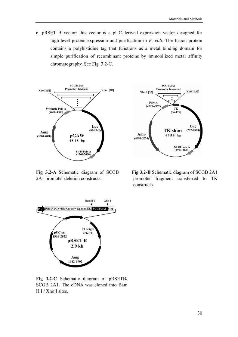
1. pGAW luciferase reporter vector: pGAW is a promoter probe vector in which a test promoter drives expression of the reporter gene luciferase. It was constructed by Braun & Suske (Braun and Suske, 1998), and is based on the commercial vector pGL3 (Promega, Mannheim). In pGAW the single BamHI site of pGL3 is destroyed by blunt end religation and the polylinker of pGL3 is replaced by a polylinker containing the recognition sequences for the restriction enzymes Pst I, EcoR I, EcoR V, Hind III, BamH I, Bgl II, Xho I, Sma I, Nhe I, Sac I and Kpn I. It was used for the SCGB 2A1 promoter deletion constructs (see chapter 3.2.7 and Fig. 3.2-A).
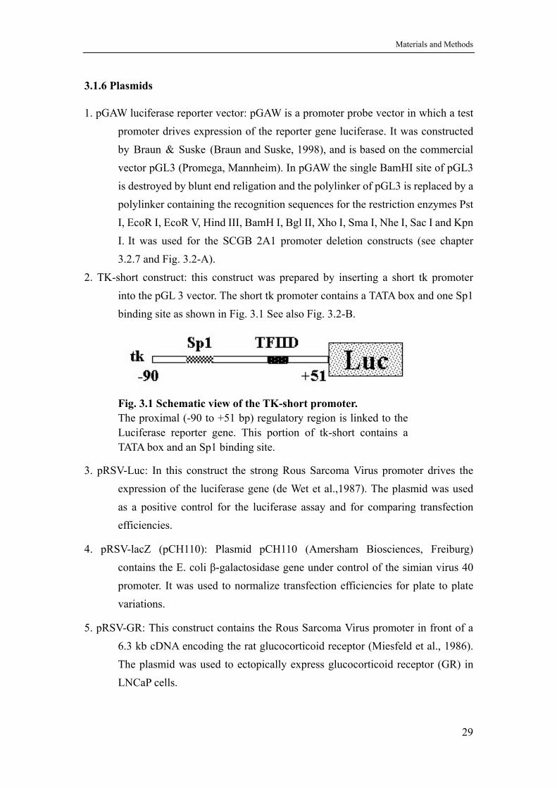
2. TK-short construct: this construct was prepared by inserting a short tk promoter into the pGL 3 vector. The short tk promoter contains a TATA box and one Sp1 binding site as shown in Fig. 3.1 See also Fig. 3.2-B.
Fig. 3.1 Schematic view of the TK-short promoter. The proximal (-90 to +51 bp) regulatory region is linked to the Luciferase reporter gene. This portion of tk-short contains a TATA box and an Sp1 binding site.
3. pRSV-Luc: In this construct the strong Rous Sarcoma Virus promoter drives the expression of the luciferase gene (de Wet et al.,1987). The plasmid was used as a positive control for the luciferase assay and for comparing transfection efficiencies.
4. pRSV-lacZ (pCH110): Plasmid pCH110 (Amersham Biosciences, Freiburg) contains the E. coli β-galactosidase gene under control of the simian virus 40 promoter. It was used to normalize transfection efficiencies for plate to plate variations.
5. pRSV-GR: This construct contains the Rous Sarcoma Virus promoter in front of a 6.3 kb cDNA encoding the rat glucocorticoid receptor (Miesfeld et al., 1986). The plasmid was used to ectopically express glucocorticoid receptor (GR) in LNCaP cells.
29
Materials and Methods
6. pRSET B vector: this vector is a pUC-derived expression vector designed for high-level protein expression and purification in E. coli. The fusion protein contains a polyhistidine tag that functions as a metal binding domain for simple purification of recombinant proteins by immobilized metal affinity chromatography. See Fig. 3.2-C.
.
Fig 3.2-A Schematic diagram of SCGB 2A1 promoter deletion constructs.
Fig 3.2-B Schematic diagram of SCGB 2A1 promoter fragment transferred to TK constructs.
Fig 3.2-C Schematic diagram of pRSETB/ SCGB 2A1. The cDNA was cloned into Bam H I / Xho I sites.
30
Materials and Methods
3.2 Methods
3.2.1 Cell culture and preparation of charcoal treated FCS
HeLa cells were cultured as monolayers in DMEM with 10% FCS, 2 mM L-glutamine, penicillin (100U/mL) and streptomycin (100µg/mL). LNCaP cells were maintained as monolayers in RPMI 1640 medium, supplemented as described above. Cells were grown at 37°C in 5% CO2. The cells were passaged with trypsin/EDTA at 80% confluency. LNCaP Cells were grown in RPMI 1640 medium supplemented with 10% FCS at 37°C with 5% CO2. Subconfluent cultures were split 1:4 once a week using trypsin/EDTA (occasionally cells could be detached by tapping). For storage cells were
frozen with 70% medium, 20% FBS, 10% DMSO at about 2×10⁶ cells/ampoule. After thawing or trypsinization cells may need 1-2 days to become adherent again. Due to strong cell aggregation, it is difficult to perform an exact cell count. The cells should be allowed to incubate undisturbed for the first 24 hours after subculture. Before transfection, cells were grown 24 hours in DMEM supplemented with 10% charcoal treated FCS.
To prepare charcoal stripped FCS, 25 g activated charcoal (Sigma) was coated with 2.5 g dextran (200,000 MW, Sigma) in 100 mL 0.01 M Tris-Cl (pH 7.4) buffer. The suspension was shaken at 4°C overnight. The dextran-coated charcoal was spun down at 14,000 G for 10 minutes at 4 °C and added to 500 ml of FCS. After shaking at room temperature for 2 h (or at 56°C for 30 min) the charcoal was pelleted by centrifugation at 14,000 G for 10 minutes at 4°C. The FCS supernatant was removed. Fresh charcoal (25 g) was added to the serum and shaken at room temperature for another 2 hours. After spinning down the charcoal at 14,000 G for 10 minutes at 4°C
temperature the serum was filtered through 0.45 µm and stored at –20°C. Before use treated FCS was filtered through 0.2 µm filters.
3.2.2 Purification of nucleic acids
3.2.2.1 Preparation of high molecular weight DNA from cultured cells
DNA extraction was performed essentially as described (Sambrook and Russell, 2001). Briefly, after washing cells grown to confluency twice with PBS, 3ml of TE
buffer per 5×10⁷ cells were added. After 10 min incubation at room temperature, a cell scraper was used to harvest the cells. Per mL of cell suspension 10 mL of cell lysis buffer were added. After 1 hour incubation at 37°C, proteinase K (20 mg/mL) was
31
Materials and Methods
added to a final concentration of 100 µg/mL and incubation was continued at 56°C overnight. Finally, the solution was extracted with phenol/ chloroform/isoamylalcohol (25:24:1), and DNA was ethanol precipitated, dissolved in TE and stored at 4°C.
Cell lysis buffer: 10 mM Tris-Cl, pH 8.0; 0.1 M EDTA, pH 8.0; 0.5% (w/v) SDS, 20 µg/mL DNase-free pancreatic RNase.
3.2.2.2 Preparation of total RNA from cultured cells
Total RNA was extracted from LNCaP and HeLa cells using the monophasic lysis reagent Trizol (Invitrogen, Karlsruhe), according to the manufacturer’s instructions. Cells are lysed in a solution of guanidine isothiocyanate and phenol. Addition of chloroform generates a second (organic) phase into which DNA and proteins are extracted, leaving RNA in the aqueous supernatant. RNA in the aqueous phase was precipitated with isopropyl alcohol and washed with 75% ethanol. The RNA pellet was dissolved in RNase-free water and stored at -70°C.
3.2.3 Gel electrophoresis
3.2.3.1 DNA agarose gel electrophoresis
2% to 0.5% agarose gels were routinely used to separate DNA fragments in a size range of 100 to 10, 000 bp (Sambrook and Russell, 2001). The appropriate amount of
agarose was dissolved in 1× TAE buffer (see chapter 3.1.3) by boiling for a few minutes in a microwave oven. When the gel solution has cooled down to some 60°C ethidium bromide was added to a final concentration of 0.5 µg/mL. The clear solution was then poured into a gel mold using a suitable comb for generating the sample wells and allowed to harden for some 30-45 min. The gel was mounted in the
electrophoresis chamber which was filled with 1× TAE running buffer until the gel was just submersed. DNA samples and a suitable size standard were mixed with 0.2
volume of 6× loading buffer and applied to the wells. A voltage of 2-10V/cm was applied until the bromophenol blue and xylene cyanol FF dyes had migrated an appropriate distance through the gel. After completion of electrophoresis the gel was examined on a 305 nm UV transilluminator and photographed using a gel documentation system (Intas, Göttingen).
3.2.3.2 SDS polyacrylamide gel electrophoresis and Coomassie Blue staining
Discontinuous SDS polyacrylamide gel electrophoresis (Davis, 1964 and Ornstein,
32
Materials and Methods
1964) was performed in a vertical system in order to analyze SCGB 2A1 expression in E. coli or to detect the native protein in tear fluid. The denatured polypeptides bind SDS and become negatively charged. Because the amount of SDS bound is usually proportional to the molecular weight of the polypeptide and is independent of its sequence, the mobility of protein-SDS complexes in polyacrylamide gels is inverse proportional to the size of the protein. By using markers of known size it is therefore possible to estimate the molecular weight of a protein.
SDS polyacrylamide gel electrophoresis was carried out in a discontinuous gel system consisting of an upper stacking gel, a lower resolving gel and an electrophoresis buffer with different pH and ionic strength than the gel buffers. The sample and the stacking gel contain Tris-Cl (pH 6.8), both buffer reservoirs contain Tris-glycine (pH 8.3), and the resolving gel contains Tris-Cl (pH 8.8). All components of the system contain 0.1% SDS (Laemmli, 1970). The fast chloride ions in the sample and stacking gel form the leading edge of a moving ion boundary, and the trailing edge is composed of slow glycine molecules. Between both edges of the moving boundary is a zone of lower conductivity and steeper voltage gradient, which sweeps the polypeptides of the sample and deposits them on the surface of the resolving gel. There the higher pH of 8.3 favors the ionization of glycine, so that the charged glycine molecules are moving fast through the stacked polypeptides and travel through the resolving gel immediately behind the chloride ions. Freed from the moving boundary the SDS-polypeptide complexes move through the resolving gel in a zone of uniform voltage and pH and are separated to size by sieving.
For most purposes a 15 or 18% resolving gel was prepared. The gel solution was poured into the assembled gel mold between two glass plates separated by 1 mm thick spacers leaving some 1 cm space for the stacking gel. The gel surface was overlaid with n-butanol in order to prevent inhibition of polymerization by oxygen. After polymerization was complete (30 min) the stacking gel (always 3%) was poured on top of the resolving gel, and the comb was inserted.
Samples were prepared in 1× SDS gel-loading buffer by means of a 6× concentrated stock solution. After having added 5% (v/v) β-mercaptoethanol or 10% (v/v) 1 M DTT all samples were boiled for 3 min to denature the proteins. After polymerization of the stacking gel (30 min) the comb was removed and the gel mounted in the electrophoresis chamber. Both electrode reservoirs were filled with SDS electrophoresis buffer, the wells were cleaned and samples loaded. Electrophoresis was performed at 25 mA constant power until the bromophenol blue
33
Materials and Methods
dye had reached the bottom of the gel. Coomassie Brilliant Blue is an aminotriarylmethane that forms strong but not
covalent complexes with proteins. The uptake of dye is approximately proportional to the amount of protein. Two forms of the dye, R-250 and G-250, are available. The SDS gel was immersed for several hours in a filtered methanol:H2O:acetic acid solution (500:400:100 mL) that contained 0.25 g Coomassie Brilliant Blue R-250 per 100 mL. Subsequently, the gel was destained in the methanol/acetic acid solution that had to be changed 4 to 6 times. The stained gel was photographed with a gel documentation system.
Alternatively, an improved staining procedure was applied which utilizes the colloidal properties of Coomassie Brilliant Blue (Neuhoff et al., 1988). This method is based on addition of 20% v/v methanol and higher concentrations of ammonium sulfate to the staining solution previously described. A commercial formulation with Coomassie Brilliant Blue G-250 was used (Roth, Karlsruhe). The gel was incubated in the colloidal Coomassie staining solution that was prepared according to the instructions of the supplier for a few hours on a tumbling shaker. The gel was briefly washed in 25% (v/v) methanol and dried or photographed as described above.
Coomassie blue staining solution: Dissolve 0.25 g of Coomassie Brilliant Blue R-250 in 90 mL of methanol:H2O (1:1, v/v) and 10 mL of glacial acetic acid. Filter the solution through a Whatman No. 1 filter to remove any particulate matter. Store at room temperature.
3.2.4 Northern blotting analysis
3.2.4.1 Electrophoresis of glyoxylated RNA through agarose gels
Separation of RNAs according to size is the first step in northern blotting and hybridization. Glyoxal was used to denature the RNA, ethidium bromide to stain it, and agarose gel electrophoresis to separate the resulting glyoxal-RNA-ethidium adducts (McMaster G and Carmichael, 1977).
10 µL RNA (up to 30 µg) were mixed with 10 µL of glyoxal reaction mixture and incubated for 60 min at 55°C. 2 µL of RNA gel-loading buffer (see chapter 3.1.3) were added to the glyoxylated RNA samples immediately before loading into the
wells of a 1.5% agarose gel (see chapter 3.2.3.1) prepared in 1× BPTE electrophoresis buffer (Sambrook and Russell, 2001), Electrophoresis was carried out at 5V/cm in
1× BPTE electrophoresis buffer that obviates the need for recirculation (Burnett,
34
Materials and Methods
1997).
10× BPTE Electrophoresis buffer: The 10× buffer was made by adding 3 g PIPES (free acid), 6 g Bis-Tris (free base), and 2 mL 0.5 M EDTA pH 8.0 to 90 mL of distilled H2O, treating the solution with DEPC (final concentration 0.1%) for 1 hour at 37°C, followed by autoclaving. The final pH of the
10× buffer is approx. 6.5 and the final concentrations of the ingredients are 100 mM PIPES, 300 mM Bis-Tris, 10 mM EDTA.
Glyoxal reaction mixture: 6 mL DMSO (HPLC grade, stored in aliquots at
–20°C), 2 mL deionized glyoxal, 1.2 mL 10× BPTE electrophoresis buffer, 0.6 mL 80% glycerol in H2O, 0.2 mL ethidium bromide solution (10 mg/mL in H2O). To deionize the glyoxal an equal volume mixed bed ion-exchange resin (Bio-Rad AG-510-X8) was added. After stirring for 30 min the resin was removed by filtration. When the pH of the glyoxal was still below 5.5 stirring with a fresh volume of resin was repeated. When the pH of the glyoxal was >5.5 aliquots were stored at –20°C.
DEPC-treated H2O: 10% (v/v) DEPC in ethanol was diluted in H2O to 0.1% (v/v) final DEPC concentration. The DEPC-containing water was incubated at 37°C overnight and autoclaved the next day.
3.2.4.2 Transfer and fixation of denatured RNA to membranes
The separated glyoxylated RNAs in the agarose gel were transferred to a positively charged nylon membrane by upward capillary transfer with 0.01 N NaOH/3 M NaCl for one hour (Thomas, 1980). During transfer the glyoxal groups are hydrolyzed due
to the alkaline pH. The membrane was washed for 5 min in 6× SSC (see chapter 3.1.3) at room temperature, dried on air for a few minutes and then irradiated at 254 nm for 105 seconds at 1.5 J/cm2 in order to fix the RNA to the membrane.
3.2.4.3 Northern hybridization
Northern hybridizations were carried out at high stringency in hybridization buffer
(6× SSC, 2× Denhardt’s reagent, 0.1% SDS, and 100 µg/mL denatured salmon sperm DNA). Initially, the membrane was prehybridized for 2 hours at 68°C in 0.1 mL of prehybridization solution per cm2 of membrane in a sealed plastic bag. A
double-stranded DNA probe was labeled with [α-32P] dCTP using the MegaprimeTM DNA labeling kit (Amersham Biosciences, Freiburg) to high specific activity
35
Materials and Methods
(>5×10⁸ cpm/µg) following the instructions of the supplier. The labeled probe was denatured by heating for 5 minutes at 100°C, chilled on ice and added directly to the
prehybridization solution (1×10⁶ cpm/mL). Incubation was continued at 68°C for 12-16 hours.
The membrane was removed from the plastic bag and transferred into 100-200 mL
of 1× SSC, 0.1% SDS at room temperature. After gentle agitation on a platform shaker for 10 min the membrane was transferred to 100-200 mL of 0.5× SSC, 0.1% SDS prewarmed to 68°C in a water bath. After 10 min the fluid was replaced by fresh solution and the washing was repeated again for a total of three washes at 68°C. The membrane was briefly dried on air for a few minutes. The still damp membrane was wrapped in plastic foil and exposed to a PhosphorImager screen (Fuji, Raytest) overnight or to X-ray film (BioMax, Kodak) in a cassette equipped with intensifying screens at –80°C for a minimum of 24 hours.
3.2.5 Southern blotting analysis
3.2.5.1 Electrophoresis of DNA through agarose gels
Purified genomic DNA was digested with restriction enzymes. At the end of the digestion DNA fragments were ethanol precipitated and dissolved in TE (pH 8.0) buffer. To 15 µg DNA in 25 µL TE buffer 5 µL of 6× gel-loading buffer were added (see chapter 3.1.3). DNA fragments were separated by electrophoresis through a 0.7% agarose gel prepared in 1× TAE electrophoresis buffer (<1 V/cm). After electrophoresis was complete, the gel was stained with 0.1µg/mL ethidium bromide and photographed with a gel documentation system.
3.2.5.2 Transfer and fixation of denatured DNA to membranes
After fractionating the DNA by gel electrophoresis, the DNA was denatured by soaking the gel in several gel volumes of alkaline transfer buffer for 15 minutes at room temperature with constant but gentle agitation on a rotary platform. The solution was changed and soaking of the gel was continued for a further 20 minutes. The denatured DNA was then transferred to a positively charged nylon membrane by upward capillary transfer with alkaline transfer buffer over about 18 hours. The membrane was neutralized in neutralization buffer for 15 minutes at room temperature. Because alkaline transfer results in covalent attachment of DNA to positively charged nylon membranes, it is not necessary to irradiate the membrane with UV in order to
36
Materials and Methods
fix the DNA to the membrane.
Alkaline transfer buffer: 0.4 M NaOH, 1 M NaCl. Neutralization buffer: 0.5 M Tris-Cl, pH 7.2 with 1 M NaCl.
3.2.5.3 Southern hybridization
Southern hybridizations were carried out at high stringency in phosphate-SDS hybridization buffer. Initially, the membrane was prehybridized for 2 hours at 68°C in 0.1 mL of prehybridization solution per cm2 of membrane in a sealed plastic bag. A
double-stranded DNA probe was labeled with [α-32P]dCTP using the MegaprimeTM DNA labeling kit (see chapter 3.2.4.3). The labeled probe was denatured by heating for 5 minutes at 100°C, chilled on ice and added directly to the prehybridization
solution (1×10⁶ cpm/mL). Incubation was continued at 68°C for 12-16 hours. The membrane was removed from the plastic bag and transferred into 100-200 mL
of phosphate-SDS washing solution. After gentle agitation on a platform shaker for 10 min the membrane was transferred to fresh washing buffer prewarmed to 68°C in a water bath. After 10 min the fluid was replaced by fresh solution and the washing was repeated again for a total of three washes at 68°C. The membrane was briefly dried on air for a few minutes. The still damp membrane was wrapped in plastic foil and exposed to a PhosphorImager screen and then to X-ray film as described in chapter 3.2.4.3.
Phosphate-SDS hybridization buffer: 0.5 M sodium phosphate (pH 7.2), 7% SDS, 1mM EDTA and 0.2 mg/mL denatured salmon sperm DNA. 0.5 M phosphate buffer is 134 g of Na2HPO4•7H2O, 4 mL of 85% H3PO4 (concentrated phosphoric acid), H2O to 1 liter.
Phosphate-SDS washing solution: 40 mM sodium phosphate, 0.1% SDS.
3.2.6 Mapping of DNase I hypersensitive sites
Treatment of nuclei with limited amounts of DNase I was used to reveal sites in
chromatin that are hypersensitive to the nuclease (Stalder et al., 1980). About 4×10⁷ cells were harvested into ice cold PBS and washed twice with 25 mL of ice-cold PBS without calcium and magnesium salts. The cells were spun down by centrifugation of the suspension at 1,000 G for 5 min at room temperature and resuspended in 5 mL cell lysis buffer. The suspension was homogenized by squeezing the lysed cells five time out of a 10 mL pipet held hard on the bottom of a 50 mL centrifuge tube. A 10 µL
37
Materials and Methods
aliquot of the cell lysate was mixed with an equal volume of 0.4% Trypan Blue dye,
and the solution was examined under a microscope equipped with a 20× objective. Lysed cells and nuclei take up the dye and appear blue, whereas unlysed cells are impermeable to the dye and remain translucent. The incubation was continued on ice until >80% of cells were lysed. The cell lysate was adjusted to 30 mL with cell lysis buffer and the nuclei were spun down at 1500 rpm at room temperature for 5 min. The nuclear pellet was resuspended in 1 mL nuclei digestion buffer.
A series of dilutions of the standard 10× DNase I solution (20, 40 and 60 µg/mL) was set up in nuclei digestion buffer and stored on ice until used. For the mapping, 250 µL aliquots of nuclei were transfered to a series of 2 mL tubes and digested with DNase I for 3 minutes at 22°C in a waterbath by adding 2.5 µL 100 mM CaCl2. The final DNase I concentration was 2, 4 and 6 µg/mL for three digests. The reaction was stopped by adding 250 µL proteinase K digestion buffer, and incubation was continued for 16 hours at 50°C with rotation. After three gentle and careful extractions of the digestion mixtures with phenol/chloroform the DNA was precipitated with the addition of 3 volumes of ice-cold ethanol. After 30 minutes on ice the DNA precipitates were collected by centrifugation at 3000g for 15 min at 4°C. The supernatant was decanted and the last drops of ethanol drained from the tubes on a paper towel. 200 µL of TE were added to each tube and the DNA was allowed to redissolve under rotation overnight. The concentration of the resuspended DNA was determined by measuring A260. The DNA samples were digested with the appropriate restriction enzyme, and restricted fragments were separated by agarose gel electrophoresis. Southern blotting and hybridization were performed as described in chapter 3.2.5 (Cockerill, 2000). The 380 bp DNA probe used for indirect endlabeling of the target fragment was amplified from LNCaP genomic DNA by standard PCR.
Cell lysis buffer: 60 mM KCl, 15 mM MgCl2, 10 mM Tris, pH 7.4, 300 mM sucrose, 0.1 mM EGTA, and 0.1 mM Pefabloc (Roth, Karlsruhe).
Nuclei digestion buffer: 60 mM KCl, 15 mM NaCl, 5 mM MgCl2, 10 mM Tris, pH 7.4, 300 mM sucrose, 0.1 mM EGTA.
Proteinase K digestion buffer: 100 mM Tris-HCl, pH 8.0, 40 mM EDTA, 2% SDS, 0.2 mg/mL proteinase K.
3.2.7 Cloning and subcloning
SCGB 2A1 promoter fragments were amplified from the P1 clone ICRFP700J1347Q6 that contains four secretoglobin genes (Ni et al., 2000). The upstream PCR primer
38
Materials and Methods
defined the upstream truncation point and provided an Xho I site for cloning (see chapter 3.1.5). The downstream primer contained the SCGB 2A1 promoter sequence from +50 to +32 followed by a Kpn I recognition site used for cloning (see 3.1.5). The PCR was carried out for 30 cycles at 94°C for 40 sec, 57°C for 40 sec and 72°C for 30 sec, followed by a final extension at 72°C for 5 min. Amplified PCR fragments were purified by using the Qiaquick PCR purification kit (QIAGEN, Hilden), and restricted with XhoI and Kpn I and ligated into pGAW. The vector was digested with Xho I and Kpn I, alkaline phosphatase treated and agarose gel purified using the GFXTM PCR DNA and gel band purification kit (Amersham Biosciences, Freiburg). The ligation reaction was carried out with 20 ng insert fragment, 100 ng vector and 0.1U T4 DNA
Ligase in 20 µL 1× Ligase reaction buffer for 14 h at 16°C. Ligation reactions were transformed into competent E. coli DH5α (see chapter 3.2.9). The sequences of all constructs were checked (Seqlab, Göttingen) across the promoter/luciferase border using the Glprimer2 (Promega, Mannheim).
Plasmid DNA for transfections was prepared from transformed E. coli DH5α using Nucleobond PC 500 columns (Macherey & Nagel, Düren). The quality of all plasmids (ratio supercoiled vs. nicked better than 70:30) was checked by agarose gelelectrophoresis before transfection.
For subcloning of promoter fragments into the TK vector (See chapter 3.1.6), Xho I fragments were ligated into Xho I linearized vector. When the inserts were oligonucleotides containing the sequence from –59 to –28, the vector could not be dephosphorylated. The direction of the inserts was confirmed by sequencing.
3.2.8 PCR-mediated mutagenesis
Point mutations were introduced at specific sites by producing two overlapping mutant fragments via PCR (left and right arms) that were joined together by PCR directed homologous recombination (Klug et al., 1991).
In a first PCR the left arm was generated from a suitable wild type promoter deletion construct, using as upstream primer the one which was also used to define the truncation point of this deletion construct (see chapter 3.1.5) and which contained an Xho I site for cloning. The downstream primer contained the desired mutation(s) (see Fig. 3.3) and, hence, is called the mutation primer (see chapter 3.1.5).
The right arm was also generated by PCR from the same wild type construct. The upstream primer was complementary to the mutation primer used for the left arm (see chapter 3.1.5) and the downstream primer was the downstream primer SCGB 2A1
39
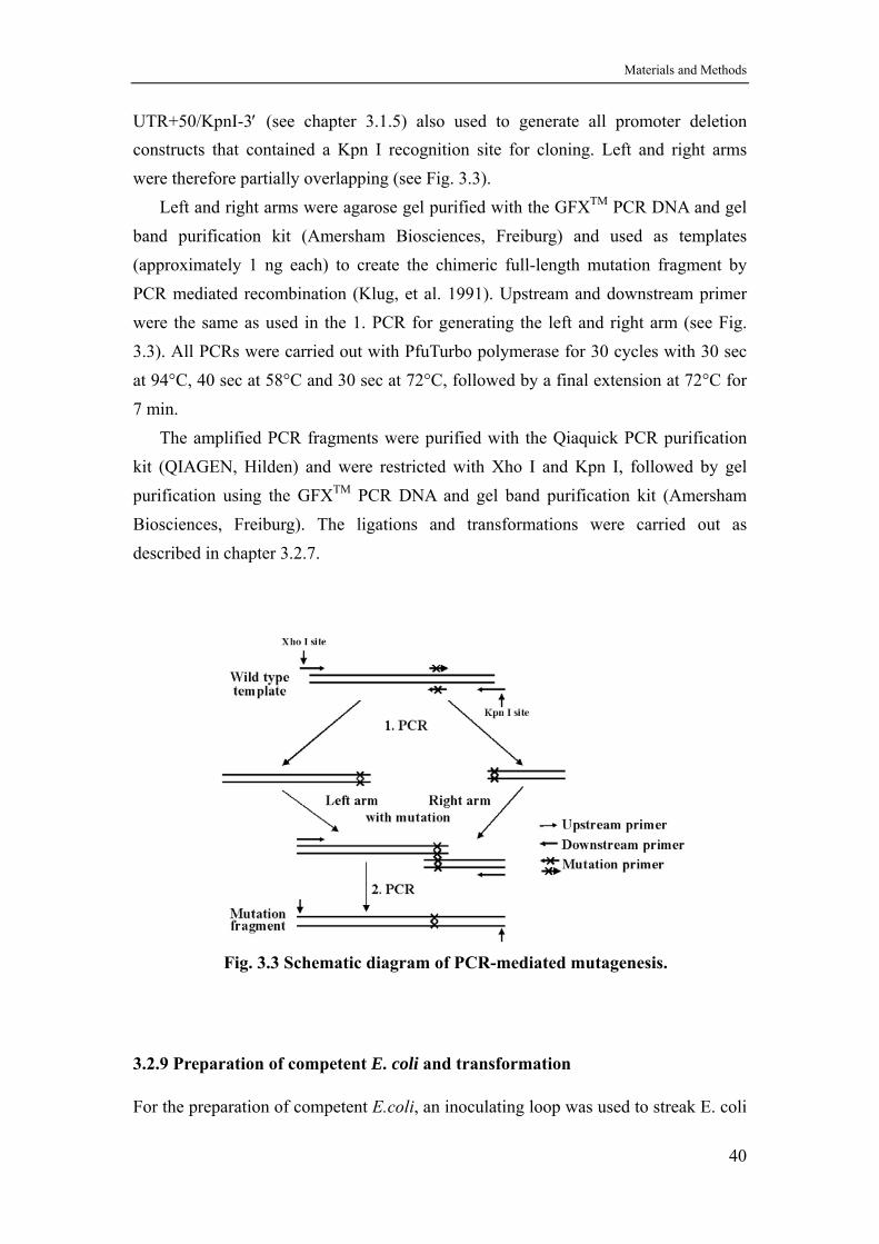
Materials and Methods
UTR+50/KpnI-3′ (see chapter 3.1.5) also used to generate all promoter deletion constructs that contained a Kpn I recognition site for cloning. Left and right arms were therefore partially overlapping (see Fig. 3.3).
Left and right arms were agarose gel purified with the GFXTM PCR DNA and gel band purification kit (Amersham Biosciences, Freiburg) and used as templates (approximately 1 ng each) to create the chimeric full-length mutation fragment by PCR mediated recombination (Klug, et al. 1991). Upstream and downstream primer were the same as used in the 1. PCR for generating the left and right arm (see Fig. 3.3). All PCRs were carried out with PfuTurbo polymerase for 30 cycles with 30 sec at 94°C, 40 sec at 58°C and 30 sec at 72°C, followed by a final extension at 72°C for 7 min.
The amplified PCR fragments were purified with the Qiaquick PCR purification kit (QIAGEN, Hilden) and were restricted with Xho I and Kpn I, followed by gel purification using the GFXTM PCR DNA and gel band purification kit (Amersham Biosciences, Freiburg). The ligations and transformations were carried out as described in chapter 3.2.7.
Fig. 3.3 Schematic diagram of PCR-mediated mutagenesis.
3.2.9 Preparation of competent E. coli and transformation
For the preparation of competent E.coli, an inoculating loop was used to streak E. coli
40
Materials and Methods
DH 5α directly from a frozen stock onto an SOB agar plate. The plate was incubated for 16 hours at 37°C. One colony was picked and grown in 5 mL LB medium overnight. The next day a 250 mL SOB culture containing 20 mM MgSO4 was inoculated with 1 mL overnight culture. The cells were grown for 2.5-3.0 hours at 37°C, under monitoring the growth of the culture every 20 minutes.
When the culture had reached an OD600 = 0.5±0.1, the cells were harvested by centrifugation at 3,000 G for 15 min at 4°C. The medium supernatant was decanted, and the tubes were kept in an inverted position for 1 minute to allow the last traces of medium to drain away. The cells were resuspended in 100 mL TB buffer and stored on ice for 10 minutes. The cells were collected again at 3,000 G for 15 minutes at 4°C. The buffer was decanted and the tubes were again put in an inverted position for 1 minute to allow the last traces of buffer to drain away. The cells were collected in one 50 mL Falcon tube by resuspending finally in 15 mL TB buffer. After gently mixing the buffer with 1050 µL DMSO (7% v/v), the mixture was kept on ice for 10 minutes. 200 µL aliquots were dispensed into 1.5 mL chilled, sterile microfuge tubes. Competent cells were snap frozen immediately by immersing the tightly closed tubes in liquid nitrogen. The competent cells were stored at -70°C until needed.
For transformations no more than 25 ng of plasmid in a volume not exceeding 3 µL was used for 50 µL competent cells. The tubes were swirled gently to mix the DNA and bacteria. The tubes were stored on ice for 30 minutes. The tubes were transferred to a heatblock preheated to 42°C. After exactly 90 seconds the tubes were put on ice again. After 1-2 minutes cooling 200 µL of SOC medium were added to each tube. Incubation of the tubes for 45 minutes in a shaking incubator allowed the bacteria to recover and to establish antibiotic resistance. 50 µL transformed competent cells were plated onto 90 mm agar LB plates containing the appropriate antibiotic (usually 50µg/ml ampicillin). The plates were stored at room temperature until the liquid had been absorbed. The plates were inverted and incubated at 37°C overnight. Colonies were screened by plasmid mini preparations and diagnostic restriction enzyme analysis and/or by direct DNA sequence analysis.
TB buffer: 10mM HEPES, 55 mM MnCl2, 15 mM CaCl2, 250 mM KCl, before adding MnCl2 adjust pH to 6.7 with KOH.
41
Materials and Methods
3.2.10 Transfections and reporter gene assays
3.2.10.1 Transfection of LNCaP and HeLa cells with the calcium phosphate method
Calcium phosphate forms an insoluble precipitate with DNA which attaches to the cell surface and is internalized by cells through endocytosis (Jordan et al., 1996).
24 hours before transfection, exponentially growing LNCaP or HeLa cells were
harvested by trypsination and replated at a density of 1×10⁵ to 4×10⁵ cells/cm2 in Falcon 50 mL tissue culture flasks in RPMI 1640 or DMEM medium, respectively, supplemented with 10% charcoal treated FCS (4,5 ml medium per flask). The cultures were incubated for 20-24 hours at 37°C in a humidified incubator with an atmosphere of 5% CO2. RPMI medium was changed for DMEM one hour before transfection because RPMI 1640 forms a granular precipitate with calcium phosphate that is not internalized efficiently by the cells.
The calcium phosphate-DNA coprecipitate was prepared as follows: 5 µg of construct DNA and 0.5 µg pRSV-β-gal (pCH110, see chapter 3.1.5, internal standard) were added into 250 µL of 250 mM CaCl2 in a sterile 5 mL plastic tube. One volume
of this 2× calcium-DNA solution was mixed with an equal volume of 2× HeBS solution at room temperature by immediately tapping the tube. After exactly 1 minute the calcium phosphate-DNA suspension was added to the plates (into the 4.5 mL medium above the cell monolayer). The flask was gently rocked to mix medium and suspension which then turns yellow-orange and becomes turbid. This step was carried out as quickly as possible because the efficiency of transfection declines rapidly once the DNA precipitate is formed (Jodan et al., 1996).
100 mM chloroquine diphosphate was diluted 1:1,000 directly into the medium after the addition of the calcium phosphate-DNA coprecipitate. The concentration of chloroquine added to the growth medium and the time of treatment are limited by the sensitivity of the cells to the toxic effect of the drug. The optimal treatment time of chloroquine for LNCaP and HeLa cells was empirically determined to be 4 hours. After treatment with DNA and chloroquine the medium was removed by aspiration, and the monolayer was washed once with PBS (see chapter 3.1.3). 1.5 mL of 15%
glycerol in 1×HeBS were added to each flask and left on the cells for exactly 2 min which was the time tested to be optimal for LNCaP and HeLa cells. The glycerol containing medium was removed by aspiration, and the cells were washed with PBS (see chapter 3.1.3). 5 mL of prewarmed complete growth medium were added with or without hormones. The cells were incubated again at 37°C in a humidified incubator
42
Materials and Methods
with an atmosphere of 5% CO2.
2× HEPES-buffered saline (HeBS): 21 mM HEPES, 0.7 mM Na2HPO4, 137mM NaCl, 5 mM KCl, 6 mM dextrose, pH 7.1 (very critical).
3.2.10.2 Luciferase assay
To assay the transfected cells for transient expression of the introduced luciferase gene the cells were harvested 48 hours after transfection. The cells were washed three times at room temperature with PBS without calcium and magnesium salts. PBS was added and removed gently because LNCaP cells can be easily displaced from the dish by vigorous pipetting. 350 µL of ice-cold cell lysis buffer was then added to the flasks. The buffer was distributed gently and the lysed cells were scraped from the dish using a rubber policeman. Cell lysates were collected in 1.5-mL microfuge tubes and cleared by centrifugation at 13,000 g for 5 min at 4°C.
The luciferase assay was performed by adding 100 µL of cell lysate into individual luminometer tubes containing 360 µL of luciferase assay buffer at room temperature. Light was measured with an AutoLumat 953 luminometer (Berthold, Bad Wildbad) by injecting 100 µL of 0.2 mM luciferin solution into the sample tubes and measuring light output over 10 sec (Brasier et al., 1989).
Cell lysis buffer: 1%(v/v) Triton X-100, 25 mM glycylglycine, pH 7.8, 15 mM MgSO4, 4 mM EGTA and 1 mM DTT.
Luciferase assay buffer: 25 mM glycylglycine, pH 7.8, 15 mM potassium phosphate, pH 7.8, 15 mM MgSO4, 4 mM EGTA, 2 mM ATP and 1 mM DTT.
3.2.10.3 β-Galactosidase assay
For the β-galactosidase assay, 40 µL of cell lysate were incubated with 60 µL of 4 mg/mL ONPG sodium phosphate solution and 250 µL buffer Z. After 1 hour incubation at 37°C the reaction was stopped by adding 100 µL 1 M Na2CO3, and the optical density of the solutions was measured at a wavelength of 420 nm in a spectrophotometer. Controls contained 40 µL of cell extract from mock-transfected cells. In addition, the positive controls should include 1 µL of a commercial β-galactosidase solution (50 units/mL). One unit of β-galactosidase is defined as the amount of enzyme that will hydrolyze 1 µM of ONPG substrate in 1 minute at 37°C (Young et al., 1993).
The concentration of total protein in the lysate was determined by the Bradford
43
Materials and Methods
assay (Bradford, 1976), using the Protein-Assay reagent (BioRad, München). Reagent background RLU were subtracted from sample RLU values and normalized for protein concentration. β-galactosidase results were used to normalize for plate-to-plate variations in transfection efficiency.
Buffer Z: 60 mM Na2HPO4, 40 mM NaHPO4, 10 mM KCl, 1 mM MgSO4, 50 mM β-Mercaptoethanol, pH 7.8.
Chloroquine (100 mM): Dissolve 52 mg of chloroquine diphosphate in 1 mL of
deionized distilled H2O. Sterilize the solution by passing it through a 0.22 µm filter; store the filtrate in foil-wrapped tubes at -20°C.
3.2.11 Preparation of nuclear extracts
Nuclear extracts of LNCaP and HeLa cells were prepared according to Andrews and
Faller, 1991, with some modifications. 5×10⁷ cells were harvested and collected by centrifugation at 250 G for 10 minutes at room temperature. The cells were rinsed several times with PBS (see chapter 3.1.3) without calcium and magnesium salts. The cell suspension was then transferred to a 2 mL microfuge tube. Cells were pelleted again and resuspended in 1 mL buffer A by flicking the tubes. After 10 minutes swelling on ice, the suspension was vortexed 10 seconds. Samples were centrifuged for 10 seconds and the supernatant was discarded. The pellet was resuspended in buffer C and incubated on ice for 20 minutes. Cellular debris was removed by centrifugation at maximum speed for 5 minutes at 4°C, and the supernatant was dialyzed twice for 2 hours each in buffer D. The supernatant was divided into aliquots of 100-200 µL which were snap-frozen in liquid nitrogen and stored at -70°C. Before freezing the protein concentration was determined using the Bradford method (see 3.2.10.3).
Buffer A: 10 mM HEPES-KOH, pH 7.9, 1.5 mM MgCl2, 10 mM KCl, 0.5 mM DTT, 0.2 mM PMSF.
Buffer C: 20 mM HEPES-KOH, pH 7.9, 25% glycerol, 420 mM NaCl, 1.5 mM MgCl2, 0.2 mM EDTA, 0.5 mM DTT, 0.2 mM PMSF.
Buffer D: 20 mM HEPES-KOH, pH 7.9, 20% glycerol, 100 mM KCl, 0.2 mM EDTA, 0.5 mM DTT and 0.5 mM PMSF.
44
Materials and Methods
3.2.12 DNase I footprinting
3.2.12.1 End-labeling of the DNA probe
A 32P-end-labeled 432 bp Xho I/Kpn I fragment from pGAW (-382) Luc which contains the sequence from -382 to +50 of the SCGB 2A1 promoter was used for the DNase I footprinting experiments.
For labeling of the lower strand, 6 µg pGAW (-382) Luc construct were digested
with Xho I for 40 min at 37°C in 20 µL restriction enzyme buffer. The 3′-recessed end was labeled by using Klenow fragment and filling in with dATP, dTTP, dGTP and
[α-32P]dCTP. The restriction enzyme and Klenow fragment were inactivated for 10 min at 70°C before digestion with the second restriction enzyme Kpn I was started. After 40 min digestion, the sample was loaded onto a 5% native polyacrylamide gel.
The gel was run in 1×TBE buffer at 10 V/cm until the bromophenol blue dye had reached the bottom of the gel.
For labeling of the upper strand, the pGAW (-382) Luc construct was digested
with Kpn I for 40 min at 37°C in 20 µL restriction enzyme buffer. The 3′-protruding ends were labeled with T4 DNA ploymerase in the presence of [α-32P]dCTP. T4 DNA polymerase rapidly digests 3′-protruding termini and then continues to remove nucleotides until a G residue (in this case) is encountered on the template strand. when a complementary radiolabeled C residue is incorporated into the DNA fragment. The reaction was stopped by heating to 70°C for 10 min. After 40 min digestion with the second restriction enzyme Xho I, the sample was loaded onto a 5% native
polyacrylamide gel and the gel was run in 1×TBE buffer at 10 V/cm until the bromophenol blue dye had reached the bottom of the gel.
After electrophoresis the gel plates were separated and the one with the gel attached was wrapped in plastic foil. Four filter paper snippets were laid asymmetrically on the wrapped gel, and a few microliter of radioactive solution were applied to each of them. After covering and fixing the filter snippets with adhesive film the gel was exposed to Kodak BioMax X-ray film (Integra Biosciences, Fernwald) for 2 min in order to locate the labeled fragment. A clean sharp scalpel was used to cut out the segment of the gel containing the band of interest. The gel slice was transferred to a microfuge tube and soaked in 400 µL TE (pH 8.0) overnight. The DNA probe extracted from the gel slice was ethanol precipitated and then dissolved in TE (pH 8.0) buffer.
45
Materials and Methods
3.2.12.2 Maxam-Gilbert sequencing reactions
Maxam-Gilbert G-reaction was performed and used as high-resolution size markers.
About 20×104 cpm of labeled fragment were diluted in 200 µL TE buffer and 1 µL dimethylsulfate (DMS, Sigma,) was added. After 3 min the reaction was stopped by adding 50 µL DMS stop solution. The nucleic acids were precipitated with 750 µL ethanol for 15 minutes at -70°C and collected by centrifugation at maximum speed for 15 minutes at 4°C in a microfuge. The pellets were rinsed with 1 mL of 70% ethanol, centrifuged again, and dried in the vacuum-concentrator to remove last traces of ethanol.
The base-modified DNA was resuspended in 100 µL of 10% (v/v) piperidine by vortexing. The tube was incubated for 30 minutes at 90°C, and sealed with a safety cap to prevent the lid from popping open during heating. Afterwards the piperidine was evaporated to dryness in a vacuum concentrator. The DNA was dissolved in 20 µL of H2O and evaporated to dryness once again. The amount of radioactivity remaining in the tube was measured by Cerenkov counting. The DNA fragments were dissolved in formamide loading buffer (see chapter 3.1.3) at about 5,000 cpm/µL buffer. Before loading onto the sequencing gel the tubes were heated at 90°C for 5 minutes to denature the DNA and then chilled on ice (Maxam and Gilbert, 1977).
DMS stop solution: 1.5 M sodium acetate (pH 7.0), 1 M β-mercaptoethanol, 250 µg/mL yeast tRNA.
3.2.12.3 DNase I digestion
About 7×10⁴ cpm of labeled fragment were incubated with 30 µg nuclear extract protein in the presence of 2 µg poly (dI-dC) in a 25 µL reaction volume in 1× binding buffer. The nuclear extract was preincubated with the unspecific competitor poly (dI-dC) for 1 minute to decrease nonspecific binding of proteins to the radiolabeled DNA fragment. The labeled SCGB 2A1 promoter fragment was subsequently added to the mixture and incubation was continued for 20 minutes at room temperature. The final Mg2+ and Ca2+ concentration was adjusted to 5 mM and 1mM, respectively, by adding 3 µL of a Mg2+/Ca2+ solution.
2 µL of a DNase I dilution (Roche, Mannheim, dissolved in 1× binding buffer) containing 20, 40 or 60 ng DNase I, were added to the binding reaction and incubated for exactly 1 minute at 20°C. The reactions were stopped by adding 100 µL of DNase I stop buffer containing proteinase K. After 40 min digestion at 45°C samples
46
Materials and Methods
were extracted twice with phenol/chloroform. The aqueous phases were transferred to fresh microfuge tubes, and nucleic acids were precipitated with 2.5 volumes of ethanol. The ethanolic solutions were chilled for 15 minutes at -70°C, and precipitates were collected by centrifugation at maximum speed for 15 minutes at 4°C. The pellets were rinsed with 1 mL of 70% ethanol, centrifuged again and dried in the vacuum concentrator.
The DNA pellets were dissolved in 5-10 µL of formamide loading buffer (see chapter 3.1.3) by vigorous vortexing and denatured by heating to 90°C for 3-5 minutes. Samples were loaded onto a 6% polyacrylamide sequencing gel that was prerun for at least 30 minutes before loading. About 20,000 cpm/sample were loaded, followed by the sequence ladder and control samples. The gel was run at sufficient constant power to maintain a gel temperature of 50°C until the bromophenol blue dye had reached the bottom of the gel. After separating the glass plates, the gel was transferred onto Whatman 3 MM paper and dried under vacuum at 80°C on a gel dryer (BioRad, München) for approx. 1 hour. The dried gel was exposed to a phosphorimage screen for 12 hours and analyzed on a Fuji PhosphorImager FLA3000G (Raytest, Straubenhardt).
2× Binding buffer: 20 mM HEPES-KOH, pH 7.9 (critical), 17% glycerol, 100 mM KCl, 0.2 mM EDTA and 0.4 mM DTT.
Mg2+/Ca2+ solution: 50 mM MgCl2, 10 mM CaCl2. DNase I stop buffer: 20 mM Tris, pH 7.6, 20 mM EDTA, 0.5% SDS, 100 ng/ mL
glycogen, 100 µg/mL proteinase K. 6% Polyacrylamide sequencing gel: 15 mL 40% acrylamide/bis (19:1) stock
solution, 20 mL 5×TBE, 42 g urea, 29.7 mL H2O.
3.2.13 Oligonucleotides and EMSA
A pair of complementary single-stranded oligonucleotides was heated to 95°C for 5 min in 100 µL of annealing buffer in a water bath and cooled to room temperature over 2 hours. The final concentration of the double-stranded oligonucleotide was 50 ng/µL. About 50 ng double stranded DNA was labeled by filling in the ends with
[α-32P] dCTP and Klenow fragment. Labeled double-stranded DNA was purified over a NickTM column (Amersham Biosciences, Freiburg) according to the instructions provided by the manufacturer.
For electrophoretic mobility shift assays 3 µg nuclear extract were preincubated
with 0.5 µg unspecific competitor poly (dI-dC) in 1× binding buffer (see chapter
47
Materials and Methods
3.2.12.3) for 10 min on ice. Subsequently about 2×10⁴ cpm of labeled double-stranded oligonucleotide were added to yield a final volume of 20 µL, and incubation was continued at room temperature for another 15 min. The competition experiments were performed by preincubating the extracts with 50 ng of unlabeled oligonucleotide (approx. 100 fold molar excess). Samples were loaded on a 4% native polyacrylamide EMSA gel (acrylamide/bisacrylamide ratio 39:1).
The gel was run in 0.5×TBE buffer at 160-200 V and 20 mA for 2 hours. After electrophoresis was complete the gel plates were separated, the gel was transferred to a piece of blotting paper and dried for approx. 1 hour at 80°C under vacuum in a gel dryer (BioRad, München). The dried gel was exposed to a phosphorimage screen for 2 hours and analyzed on a Fuji PhosphorImager FLA3000G (Raytest, Straubenhardt).
Annealing buffer: 10mM Tris-HCl, pH 8.0, 1 mM EDTA, 30 mM KCl.
3.2.14 Protein expression and purification
3.2.14.1 Preparation of the expression construct
The cDNA for SCGB 2A1 cloned into the Eco RI (5′) and Xho I (3′) sites of pBS(SK) (Stratagene, Amsterdam) was kindly provided by Jian Ni, Human Genome Sciences, Rockville, Maryland, U.S.A. In order to generate a bacterial expression construct in pRSETB (Invitrogen, Karlsruhe), the cDNA in pBS(SK) was amplified with the
upstream primer 5′-caaggatccgGGCTGCAAACTCCTGGAG-3′ and the T7 primer 5′-TAATACGACTCACTATAGGG-3′ as downstream primer. The upstream primer contained a Bam HI site for subcloning (underlined) followed by the coding sequence of SCGB 2A1 starting with GGC for glycin 21 of the pro-protein. The cycle conditions for a standard PCR with Goldstar Taq polymerase (Eurogentec, Seraing, Belgium) were: 2 min initial denaturation at 96°C, 30 cycles of denaturation for 1 min at 95°C, annealing for 30 sec at 52°C, extension for 30 sec at 73°C and a final extension for 5 min at 73°C. The 450 bp fragment was ethanol precipitated in the presence of ammonium acetate, digested with Bam HI/ Xho I and purified by agarose gel electrophoresis. The 410 bp restriction fragment was recovered from the agarose gel by cutting out the piece of agarose containing the fragment, freezing and thawing the piece of agarose three times and centrifuging it at 12, 000 G at 4°C for 30 min. The concentration of the restriction fragment in the supernatant was estimated by agarose gel electrophoresis, and some 20 ng were used in a standard ligation reaction with 100 ng of Bam HI and Xho I restricted and phosphatase treated pRSETB.
48
Materials and Methods
Recombinant clones were picked, and the sequence was verified using the T7 primer as sequencing primer (Seqlab, Göttingen). SCGB 2A1 is expressed as a fusion protein with a six histidine tag at the amino terminus followed by the Xpress epitope (trademark of Invitrogen) and an enterokinase cleavage site thus adding 33 amino acids to the 75 amino acid SCGB 2A1 sequence (see Fig. 3.2-C). The apparent molecular weight of the fusion protein is 12.4 kDa.
3.2.14.2 Expression of SCGB 2A1 in E. coli
Positive E. coli strain BL21 (DE3) clones containing the expression construct were selected from ampicillin resistant transformants and inoculated in 1 mL LB medium. To optimize expression conditions, 5 mL of LB medium containing 50 µg/mL ampicillin were inoculated with 50 µL of overnight culture. The cultures were incubated for >2 hours at 37°C in a shaking incubator until cells reached mid-log phase (OD600=0.8).
1 mL of each uninduced culture (zero-time aliquot) was transferred to microfuge tubes. The tubes were centrifuged at maximum speed for 1 minute at room temperature in a microfuge immediately. The supernatants were removed by
aspiration. The pellets were resuspended in 100 µL of 1× SDS gel-loading buffer. The remainder of each culture was induced by adding IPTG to a final concentration of 1 mM and incubation was continued at 37°C with aeration. At various time points during the induction period (e.g., 0.5, 1, 2 and 3 hours), 1 mL of each culture was transfered to a microfuge tube, the OD600 was measured in a spectrophotometer, and each pellet was resuspended like the zero-time aliquot. The samples were heated to 100°C for 3 minutes. All tubes were centrifuged at maximum speed for 1 minute at room temperature in a microfuge, and stored on ice until the samples were collected and ready for loading on a gel. The samples were warmed to room temperature and 0.15 OD600 units (of original culture) were loaded onto an 18% SDS-polyacrylamide gel. The gel was run at 8-15 V/cm until the bromophenol blue reached the bottom of the resolving gel. The gel was stained with Coomassie Brilliant Blue as described in 3.2.3.2. As the results showed (Fig. 4.13), best expression conditions were obtained with 3 hours of induction using 0.5 mM IPTG at 37°C.
For large scale expression, 5 mL of an overnight culture was inoculated into 500 mL LB (supplemented with 100 µg/mL ampicillin) in a 2 L Erlenmeyer flask and incubated at 37°C to an optical density of OD600=0.8. Expression was induced by adding solid IPTG to a final concentration of 0.5 mM (60 mg for 500 mL). Incubation
49
Materials and Methods
continued at 37°C for 3 h, when the cells were harvested by centrifugation (GSA rotor, 5,000 rpm, 10min, RT) and lysed in 6 mL of 6M guanidine-HCl pH 8.0 by shaking overnight at 4°C. The debris was removed by centrifugation (SS34 rotor, 18,000 rpm, 5 min, 4°C) and the supernatant was transferred to a new tube.
3.2.14.3 Purification of histidine-tagged SCGB 2A1 protein
2 mL of Ni2+-NTA-agarose resin (Qiagen, Hilden) were filled into a BioRad polyprep column. The resin was packed under gravity flow. The column was washed with 10 mL water; charged with Ni2+ by applying 8 mL 0.1 M NiSO4; washed twice with 10 mL water; equilibrated with 10 mL 6M guanidine-HCl pH 8.0; loaded with the bacterial lysate supernatant (see chapter 3.2.14.2); washed with 10 mL 6M guanidine-HCl pH 8.0; and washed with 10 mL 6M guanidine-HCl pH 6.0. The histidine-tagged SCGB 2A1 protein was eluted with 7 mL 6M guanidine-HCl pH 5.0. All media were applied to the column by means of a peristaltic pump with the flow rate adjusted to approx. 1ml/min.
In order to renature the protein, guanidine was removed by dialysis against 500 mL dialysis buffer. After 2 hours of dialysis at 4°C, dialysis buffer was exchanged for fresh one and dialysis continued for another 3 h. Precipitated protein (precipitation occurs in the second half) is removed by centrifugation (SS34 rotor, 13,000 rpm, 5 min, 4°C). After Bradford protein assay (see chapter 3.2.10.3) the cleared solution was divided into 200 µL aliquots and frozen in liquid nitrogen. The total yield of purified protein was approximately 5mg per liter of bacterial culture. A sample was analysed on an 18% SDS-PAGE stained with Coomassie blue.
Dialysis buffer: 10 mM HEPES pH7.8, 50 mM KCl, 1 mM DTE, 8.5% glycerol and 0.1 mM Pefabloc (Roth, Karlsruhe).
3.2.15 Antibody preparation
One New Zealand rabbit (approx. 3 kg) was immunized with recombinant His-tagged SCGB 2A1 produced in E. coli (see 3.2.14). Before the immunization protocol was started some 5-10 mL of preimmune serum was collected. 175 µg of protein in 1 mL dialysis buffer (10 mM HEPES pH7.8, 50 mM KCl, 1 mM DTE, 8.5% glycerol and 0.1 mM Pefabloc) were mixed with the same volume of GERBU Adjuvant 100 (Fa. GERBU, Gaiberg) and injected under the rabbit’s back skin at four different locations (first immunization). After four weeks a boost was performed with 80 µg of protein
50
Materials and Methods
mixed with 1 mL of GERBU Adjuvant 100. A second boost was performed with the same amount of protein six weeks after starting the immunization protocol. Ten days after the second boost, a blood sample was drawn, and the clotted blood was stored at 4˚C overnight. Serum was removed from the clot by centrifugation at 10,000 G for 10 min at 4˚C and tested in a Western blot against recombinant and native SCGB 2A1 from tears. When the test was positive the rabbit was sacrificed and all serum collected. Sodium azide was added to 0.02 % (w/v), and the serum was stored in aliquots at –20˚C.
3.2.16 Western blotting
Proteins were separated in an 18% SDS polyacrylamide gel and electro-transferred to a nitrocellulose membrane (ECL nitrocellulose membrane, Amersham Biosciences, Freiburg) at 140mA for 90 min using a semi-dry blot apparatus (Fastblot B33, Biometra, Göttingen) according to the instructions provided by the manufacturer. The membrane was blocked with 5% (w/v) skimmed milk powder (Becton Dickinson) in PBS for 2 hours and incubated overnight at 4°C with the rabbit anti-SCGB 2A1 antibody (see chapter 3.2.15) diluted 1:1,000 in PBS containing 5% (w/v) skimmed milk powder. After washing twice in PBS, the membrane was incubated for 1 hour at room temperature with an HRP-conjugated anti-rabbit IgG antibody diluted 1:10,000 in PBS containing 5% (w/v) skimmed milk powder. After washing twice in PBS, the membrane was incubated with ECL Detection Reagent (1:1 mixture (v/v) of Reagent 1 and Reagent 2) for 60 sec (Amersham Biosciences, Freiburg). The membrane was wrapped in plastic foil and exposed to Kodak BioMax X-rayfilm for 1–15 min (Integra Biosciences, Fernwald).
3.2.17 Immunostaining
Tissue samples from a prostate carcinoma (PC), benign prostate hyperplasia (BPH), fetal prostate tissue (1, 3 and 6 months of gestation) and prostate tissue from an adolescent subject, were either fixed in Bouin’s solution or in 4% formaldehyde. After fixation the tissue was embedded in paraffin at 65°C for 4 hours. 5 µm sections were used for the immunostainings and were kindly provided by Gerhard Aumüller, Universität Marburg.
In order to remove paraffin, praraffin embedded tissue sections were washed three times in xylene for 5 min, three times in 100% ethanol for 3 min, twice in 96%
51
Materials and Methods
ethanol for 3 min, once in 90% ethanol for 3 min, once in 80% ethanol for 3 min, once in 70% ethanol for 3 min, and once in 50% ethanol for 3 min.
After a final wash with distilled water for 3 min, the sections were digested with 20µg/mL Proteinase K in PBS for a time that was empirically determined to be optimal (usually 15-30 min). After digestion, slides were washed twice in PBS and incubated with the rabbit anti-SCGB 2A1 antibody (1:300 diluted in PBS, see chapter 3.2.15) at 25°C for 120 min. Negative controls were performed with preimmune serum. Slides were washed in PBS five times and a Cy 3-labeled anti-rabbit secondary antibody (Dianova, Hamburg. 1:400 diluted in PBS) was added at room temperature for 45 min. Slides were washed four times in PBS and mounted with Gelvatol (Burkard Scientific, Uxbridge, UK). The results were analyzed under a fluorescence microscope equipped with camera (Leica, DMLB).
For staining cells in tissue culture, adherent cells were prepared by growing on a coverslip, and then fixed and permeabilized in acetone/methanol (1:1, v/v) for 2 min at room temperature. The samples were air-dried without a PBS wash. Once cells were fixed and permeabilized, the same steps were performed as in tissue staining.
52
Results
4. RESULTS
4.1 SCGB 2A1 is expressed in the prostate
In northern blot analyses using poly (A)+ RNA from multiple human tissues, SCGB 2A1 was shown to be strongly expressed in pancreas, prostate, testis and ovary (Ni et al., 2000). More tissues were examined in an RT-PCR study (Zhao et al., 1999). This sensitive assay confirmed strong expression in prostate, testis and ovary and also showed strong expression of SCGB 2A1 in mammary gland, uterus, trachea, kidney, thymus and salivary gland. In order to find a suitable cell culture model for studying expression at the molecular level the cell type that is actively transcribing SCGB 2A1 in the prostate was identified by in situ hybridization.
4.1.1 SCGB 2A1 expression is localized to the glandular epithelium
The prostate consists of 30-50 branched tuboalveolar glands. Their discharging ducts are polymorphic and can be voluminous or tight and branched. The glandular epithelium is variable and can be simple or stratified columnar as well as cuboidal and transitional and is made up of three epithelial cell types, secretory main cells, basal cells and a small number of neuroendocrine cells. Paraffin embedded sections of a normal prostate were hybridized in situ with 35S labeled sense and antisense RNA probes generated from the SCGB 2A1 cDNA inserted into pBluescript SK. For microscopic analysis autoradiograms were developed as shown in Fig. 4.1. The antisense probe yielded a strong hybridization signal in the prostate epithelium that showed the typical glandular folds (Fig. 4.1.A). Hybridization with the sense probe resulted in background staining only (Fig. 4.1.B). Bright field illumination in combination with Hematoxylin staining confirmed that the hybridization signal resides in the epithelial layer (Fig. 4.1.C).
4.1.2 Expression of SCGB 2A1 in LNCaP cells is induced by androgen
Because SCGB 2A1 expression was localized to the glandular epithelium, the prostate expression was analyzed in the well-established and characterized cell line LNCaP. The LNCaP cell line was established in 1977 by J.S. Horoszewicz et al., from a left supraclavicular lymph node of a 50-year-old Caucasian male with confirmed diagnosis of metastatic prostate carcinoma (Horoszewicz et al., 1980; Murphy, 1980). The LNCaP cell line is androgen responsive and, thus, enables the investigation of androgen inducibility of SCGB 2A1 expression.
53

Results
A
B
C
Fig. 4.1 Localization of SCGB 2A1 expression in the prostate. A, Darkfield micrograph showing in situ hybridization with a 35S-labeled antisense probe. The glandular epithelial cells are strongly labelled. B, Darkfield micrograph showing in situ hybridization with a 35S-labeled sense probe. Note the background staining only. C, Brightfield micrograph showing in situ hybridization with a 35S-labeled antisense probe together with histochemical hematoxylin staining. Epithelial cells appear light blue, stromal cells dark blue. The majority of silver grains is found in and around epithelial cells.
54
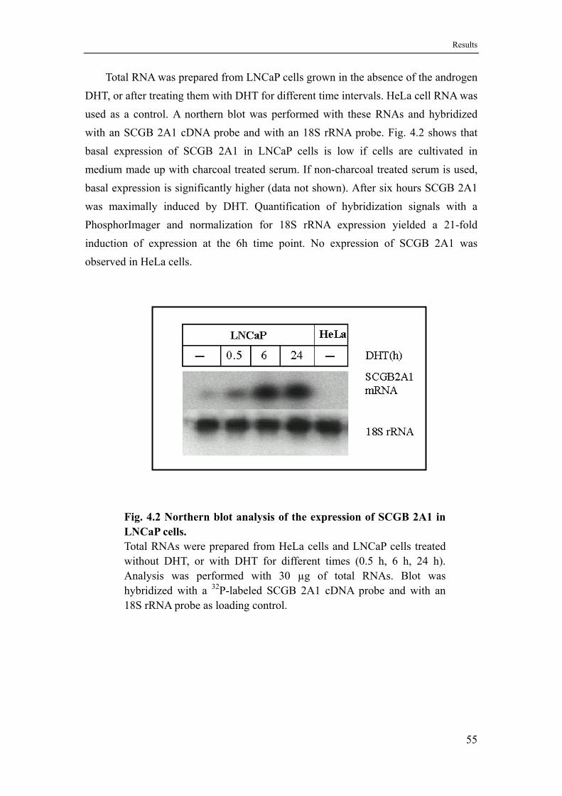
Results
Total RNA was prepared from LNCaP cells grown in the absence of the androgen DHT, or after treating them with DHT for different time intervals. HeLa cell RNA was used as a control. A northern blot was performed with these RNAs and hybridized with an SCGB 2A1 cDNA probe and with an 18S rRNA probe. Fig. 4.2 shows that basal expression of SCGB 2A1 in LNCaP cells is low if cells are cultivated in medium made up with charcoal treated serum. If non-charcoal treated serum is used, basal expression is significantly higher (data not shown). After six hours SCGB 2A1 was maximally induced by DHT. Quantification of hybridization signals with a PhosphorImager and normalization for 18S rRNA expression yielded a 21-fold induction of expression at the 6h time point. No expression of SCGB 2A1 was observed in HeLa cells.
Fig. 4.2 Northern blot analysis of the expression of SCGB 2A1 in LNCaP cells. Total RNAs were prepared from HeLa cells and LNCaP cells treated without DHT, or with DHT for different times (0.5 h, 6 h, 24 h). Analysis was performed with 30 µg of total RNAs. Blot was hybridized with a 32P-labeled SCGB 2A1 cDNA probe and with an 18S rRNA probe as loading control.
55
Results
4.2 A hormone dependent chromatin alteration occurs in the SCGB 2A1 promoter
SCGB 2A1 expression is androgen dependent in LNCaP cells. But sequence analysis of the promoter could not reveal any classical androgen responsive element. For direct genomic action of the androgen receptor a few AR specific AREs have been described (Claessens et al., 2001), but such a specific ARE could also not be identified. Therefore, in a directed approach, DNase I hypersensitive (DH) sites were mapped in order to identify functionally relevant promoter elements. DH sites represent local perturbations of the nucleosome array in nuclei rendering them accessible to nucleases or DNA-binding factors in general.
Nuclei were isolated from LNCaP cells grown in the presence or absence of DHT. Nuclei from HeLa cells and naked DNA were used as controls. Nuclei were treated with increasing amounts of DNase I. For naked DNA one tenth of the amounts used for nuclei was employed. Purified DNA was digested with the restriction enzyme Xba I, generating a 11 kb genomic fragment spanning from -3 kb to +8 kb of the SCGB 2A1 gene as indicated in Fig. 4.3-B. Restricted DNA samples were separated on an
agarose gel, and analyzed by Southern blotting with a 380 bp probe located at the 5′ end of the Xba I fragment, from -2455 to -2176 upstream of the transcription start site (Fig. 4.3-B and chapter 3.2.6).
As shown in Fig. 4.3-A, one prominent hypersensitive site (HS) was detected in the proximal promoter of the SCGB 2A1 gene at approximately ~100 bp. This HS was hormone-dependent because it only appeared in samples from LNCaP cells that were treated for 6 or 24 hours with DHT, but it was absent in samples from untreated cells or cells that were treated with DHT for only one hour. The HS also appeared to be specific, because it could not be detected in naked DNA and in nuclei from HeLa cells. In another experiment (data not shown), DNA purified from DNase I treated nuclei was digested with Xho I, producing a restriction fragment of about 10 kb from +1.7 kb upstream of the SCGB 2A1 gene start point of transcription to some 8 kb
downstream. The hybridzation probe was placed at the 3′ end of this fragment in order to detect the HS from downstream. Both mapping results confirmed a strong DNase I hypersensitive site in the proximal promoter region of the SCGB 2A1 gene after androgen treatment.
56
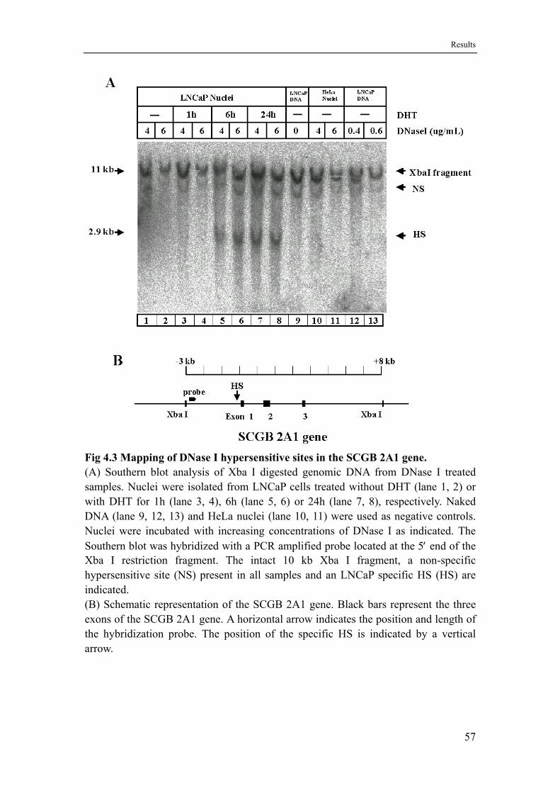
Results
Fig 4.3 Mapping of DNase I hypersensitive sites in the SCGB 2A1 gene. (A) Southern blot analysis of Xba I digested genomic DNA from DNase I treated samples. Nuclei were isolated from LNCaP cells treated without DHT (lane 1, 2) or with DHT for 1h (lane 3, 4), 6h (lane 5, 6) or 24h (lane 7, 8), respectively. Naked DNA (lane 9, 12, 13) and HeLa nuclei (lane 10, 11) were used as negative controls. Nuclei were incubated with increasing concentrations of DNase I as indicated. The Southern blot was hybridized with a PCR amplified probe located at the 5′ end of the Xba I restriction fragment. The intact 10 kb Xba I fragment, a non-specific hypersensitive site (NS) present in all samples and an LNCaP specific HS (HS) are indicated. (B) Schematic representation of the SCGB 2A1 gene. Black bars represent the three exons of the SCGB 2A1 gene. A horizontal arrow indicates the position and length of the hybridization probe. The position of the specific HS is indicated by a vertical arrow.
57
Results
4.3 The proximal promoter (-136/+50) of the SCGB 2A1 gene is sufficient for both constitutive and androgen-induced expression
The mapping of DNase I hypersensitive sites in the SCGB 2A1 gene indicated that only the gene’s promoter is responsible for the regulation by androgens. In order to confirm this result and to more precisely delineate the androgen responsive region promoter deletions were cloned in front of the luciferase reporter gene (Fig. 3.2-A and Fig. 4.4). The different promoter fragments were amplified from a human P1 clone that was shown to contain at least three genes of the secretoglobin gene cluster, among them SCGB 2A1 (Ni et al., 2000; SCGB 2A1 is called LGB in this reference). The primers were also used to artificially introduce restriction sites for cloning (Xho I
at the 5′-end and KpnI at the 3′-end; see also chapter 3.2.7 of Materials and Methods). As deletion endpoints -53, -136, -382, -575, -963, -1314 and -1476 bp, respectively, were chosen. All constructs were sequenced from the luciferase gene into the insert for verification of at least some 400 bp. Transient transfections into LNCaP cells were performed by the calcium phosphate DNA co-precipitation method. All results were corrected for plate to plate variations in transfection efficiency by normalization for β-galactosidase activity obtained by cotransfecting a constant amount of an RSV-β-Gal expression construct.
The transient transfection results are shown in Fig. 4.4, upper panel. For all constructs the basal level of transcription is low and varies only by a factor of two. After addition of the androgen DHT the longer promoter constructs with endpoints from -1476 to -575 are induced approximately 11-fold. When the region between -575 and -382 is deleted transcription is induced by a factor of 23 indicating the loss of a negative element. Deleting further down to -136 bp reduces the induction level to 15-fold suggesting that one or more positive acting elements are eliminated. But only when the proximal promoter from -136 to -53 bp was removed hormone regulation was abolished. These transfection results identified the proximal promoter as androgen responsive region, independently confirming the results obtained from the DNase I HS mapping experiment. When the same constructs were transfected into HeLa cells, no hormone responsiveness was observed (data not shown).
In order to test if the identified hormone responsive region can be functionally transferred to a heterologous promoter, the SCGB 2A1 promoter fragment starting at -136 and terminating at -53 was cloned in front of the Herpes simplex virus thymidine kinase promoter directing expression of a reporter gene (HSV-tk-Luc). Surprisingly, this construct is not androgen responsive in LNCaP cells (Fig. 4.4, lower panel) like
58
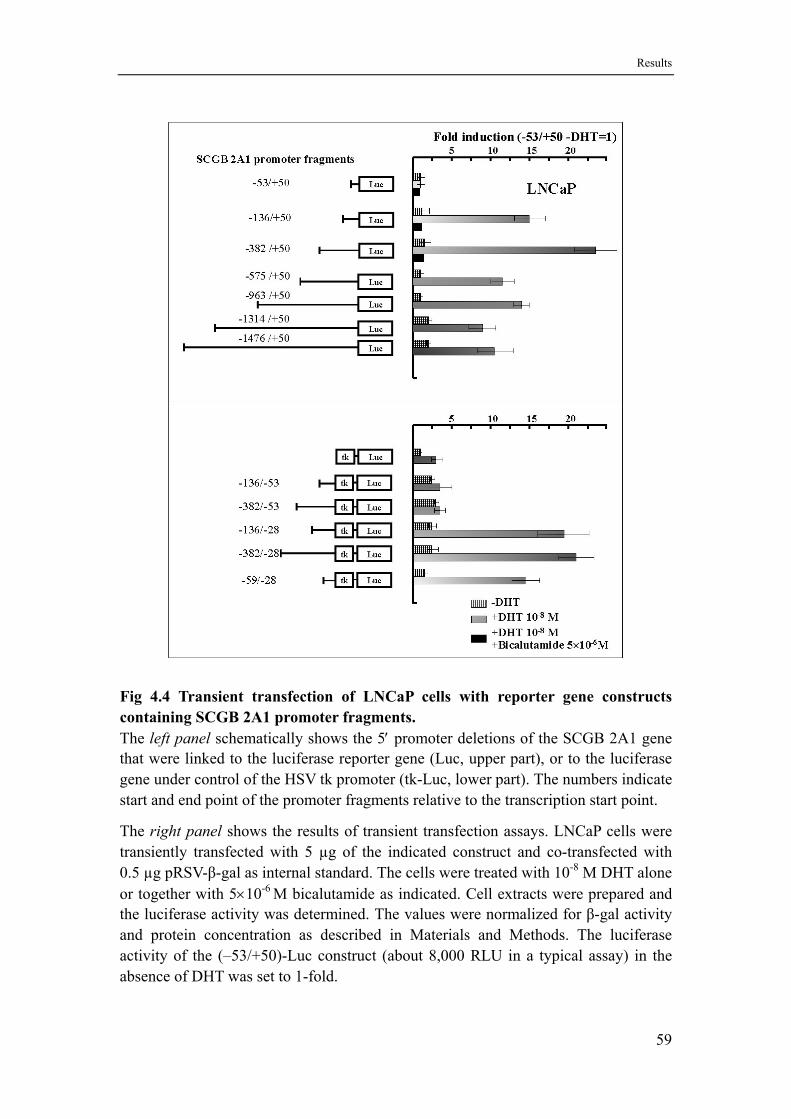
Results
Fig 4.4 Transient transfection of LNCaP cells with reporter gene constructs containing SCGB 2A1 promoter fragments. The left panel schematically shows the 5′ promoter deletions of the SCGB 2A1 gene that were linked to the luciferase reporter gene (Luc, upper part), or to the luciferase gene under control of the HSV tk promoter (tk-Luc, lower part). The numbers indicate start and end point of the promoter fragments relative to the transcription start point.
The right panel shows the results of transient transfection assays. LNCaP cells were transiently transfected with 5 µg of the indicated construct and co-transfected with 0.5 µg pRSV-β-gal as internal standard. The cells were treated with 10-8 M DHT alone or together with 5×10⁻⁶ M bicalutamide as indicated. Cell extracts were prepared and the luciferase activity was determined. The values were normalized for β-gal activity and protein concentration as described in Materials and Methods. The luciferase activity of the (–53/+50)-Luc construct (about 8,000 RLU in a typical assay) in the absence of DHT was set to 1-fold.
59
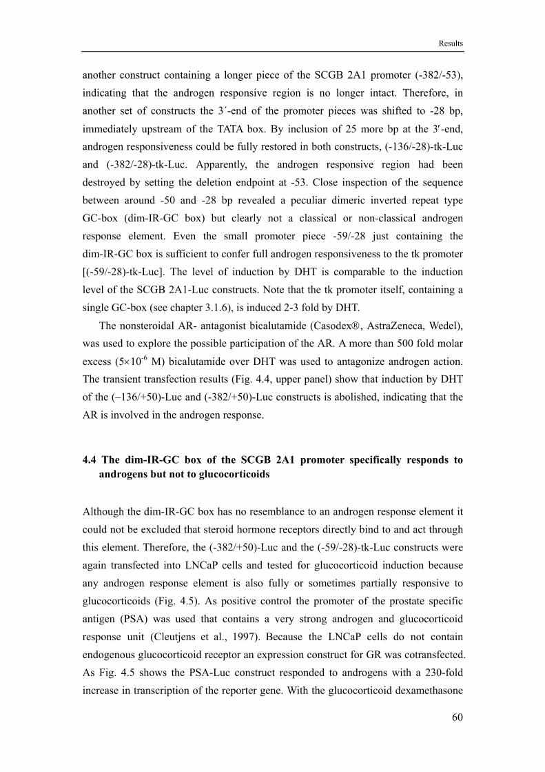
Results
another construct containing a longer piece of the SCGB 2A1 promoter (-382/-53), indicating that the androgen responsive region is no longer intact. Therefore, in another set of constructs the 3´-end of the promoter pieces was shifted to -28 bp,
immediately upstream of the TATA box. By inclusion of 25 more bp at the 3′-end, androgen responsiveness could be fully restored in both constructs, (-136/-28)-tk-Luc and (-382/-28)-tk-Luc. Apparently, the androgen responsive region had been destroyed by setting the deletion endpoint at -53. Close inspection of the sequence between around -50 and -28 bp revealed a peculiar dimeric inverted repeat type GC-box (dim-IR-GC box) but clearly not a classical or non-classical androgen response element. Even the small promoter piece -59/-28 just containing the dim-IR-GC box is sufficient to confer full androgen responsiveness to the tk promoter [(-59/-28)-tk-Luc]. The level of induction by DHT is comparable to the induction level of the SCGB 2A1-Luc constructs. Note that the tk promoter itself, containing a single GC-box (see chapter 3.1.6), is induced 2-3 fold by DHT.
The nonsteroidal AR- antagonist bicalutamide (Casodex®, AstraZeneca, Wedel), was used to explore the possible participation of the AR. A more than 500 fold molar
excess (5×10⁻⁶ M) bicalutamide over DHT was used to antagonize androgen action. The transient transfection results (Fig. 4.4, upper panel) show that induction by DHT of the (–136/+50)-Luc and (-382/+50)-Luc constructs is abolished, indicating that the AR is involved in the androgen response.
4.4 The dim-IR-GC box of the SCGB 2A1 promoter specifically responds to androgens but not to glucocorticoids
Although the dim-IR-GC box has no resemblance to an androgen response element it could not be excluded that steroid hormone receptors directly bind to and act through this element. Therefore, the (-382/+50)-Luc and the (-59/-28)-tk-Luc constructs were again transfected into LNCaP cells and tested for glucocorticoid induction because any androgen response element is also fully or sometimes partially responsive to glucocorticoids (Fig. 4.5). As positive control the promoter of the prostate specific antigen (PSA) was used that contains a very strong androgen and glucocorticoid response unit (Cleutjens et al., 1997). Because the LNCaP cells do not contain endogenous glucocorticoid receptor an expression construct for GR was cotransfected. As Fig. 4.5 shows the PSA-Luc construct responded to androgens with a 230-fold increase in transcription of the reporter gene. With the glucocorticoid dexamethasone
60
Results
the induction was still 110-fold. This represents the typical response pattern of a steroid hormone response unit and also demonstrated that the GR expression construct was able to express functional glucocorticoid receptor. Contrary to the results obtained with the PSA-Luc construct the SCGB 2A1 constructs (-382/+50)-Luc and (-59/-28)-tk-Luc did not at all respond to dexamethasone whereas they did respond to the androgen DHT as previously shown. Thus the dim-IR-GC box functions androgen specific.
Fig 4.5 The SCGB 2A1 promoter does not respond to glucocorticoids. 5 µg SCGB 2A1 (-382/+50)-Luc, (-59/-28)-tk-Luc and PSA-Luc constructs were transfected into LNCaP cells. 0.5 µg pRSV-GR were cotransfected as indicated. 0.5 µg pRSV-β-gal were always cotransfected as internal standard. The cells were treated with 10-8 M DHT or 10-8 M DEX as indicated at the bottom. The fold induction of luciferase activity is shown after normalization for β-gal activity and protein amount.
61
Results
4.5 DNase I footprints with nuclear extracts from LNCaP and HeLa cells identify three functional elements
The transfection experiments with promoter deletion/reporter gene constructs showed that in the proximal promoter of the SCGB 2A1 gene at least two binding sites for positively acting transcription factors are located. In order to identify their positions and cognate regulatory factor candidates, DNase I footprinting experiments were performed with nuclear extracts from LNCaP cells grown in the presence or absence of DHT and from HeLa cells. Both strands of the -382/+50 Xho I/Kpn I promoter fragment were 3´-end labeled before DNase I footprinting reactions were set up (Figs. 4.6 and 4.7). In the lower strand (Fig. 4.6) two strong footprints (from-186 to -155 and -136 to -109) were detected. The cognate binding proteins are of a ubiquitous nature because the same footprints are produced with HeLa nuclear extract. Another weak but LNCaP-specific footprint starts at position -36 and ends at an upstream position that could not be clearly identified (somewhere before -76). This weak footprint overlaps with the previously identified dim-IR-GC box.
On the upper strand (Fig. 4.7) two strong footprints (from -178 to -156 and -129 to -104) were identified with LNCaP and HeLa nuclear extracts that were largely overlapping with the strong footprints of the lower strand. In the region of the dim-IR-GC box (-59 to -28) again no clear footprint could be detected, but in the vicinity of position -21 and within the TATA box an unusual and LNCaP-specific hypersensitive site is apparent (shown by an asterisk).
None of the footprints was androgen dependent. The footprinted region -178 to -156 is missing in the (-136/+50)-Luc reporter construct that showed an approximately one third lower activity than the (-382/+50)-Luc construct that includes this element (Fig. 4.6).
A search for transcription factor binding sites in the SCGB 2A1 promotor using the Transfac database and the programme MatInspector (http://transfac.gbf.de/ TRANSFAC/ or http://www.gene-regulation.com) clearly revealed the transcription factor NF-Y as the prime candidate being responsible for the footprint from -129 to -104 (upper strand) and -136 to -109 (lower strand) that overlaps a canonical CCAAT-box. For the element -178 to -156 (upper strand) and -186 to -155 (lower strand), Transfac came up with no clear candidate, but the similarity of the footprint core (TTGGA in the lower strand) with the nuclear factor 1 core sequence (TTGGC) suggested to test NF1 for causing this footprint.
62
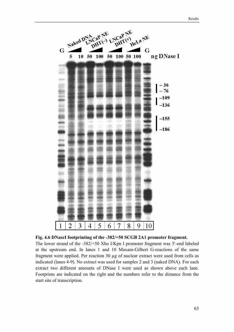
Results
Fig. 4.6 DNaseI footprinting of the -382/+50 SCGB 2A1 promoter fragment. The lower strand of the -382/+50 Xho I/Kpn I promoter fragment was 3′-end labeled at the upstream end. In lanes 1 and 10 Maxam-Gilbert G-reactions of the same fragment were applied. Per reaction 30 µg of nuclear extract were used from cells as indicated (lanes 4-9). No extract was used for samples 2 and 3 (naked DNA). For each extract two different amounts of DNase I were used as shown above each lane. Footprints are indicated on the right and the numbers refer to the distance from the start site of transcription.
63
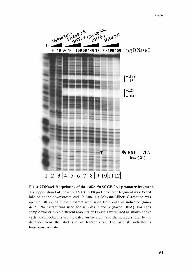
Results
Fig. 4.7 DNaseI footprinting of the -382/+50 SCGB 2A1 promoter fragment. The upper strand of the -382/+50 Xho I/Kpn I promoter fragment was 3′-end labeled at the downstream end. In lane 1 a Maxam-Gilbert G-reaction was applied. 30 µg of nuclear extract were used from cells as indicated (lanes 4-12). No extract was used for samples 2 and 3 (naked DNA). For each sample two or three different amounts of DNase I were used as shown above each lane. Footprints are indicated on the right, and the numbers refer to the distance from the start site of transcription. The asterisk indicates a hypersensitive site.
64
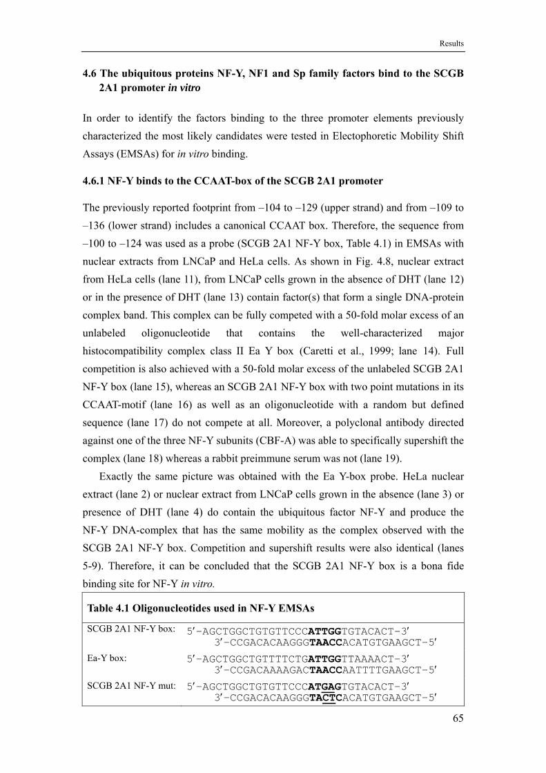
Results
4.6 The ubiquitous proteins NF-Y, NF1 and Sp family factors bind to the SCGB 2A1 promoter in vitro
In order to identify the factors binding to the three promoter elements previously characterized the most likely candidates were tested in Electophoretic Mobility Shift Assays (EMSAs) for in vitro binding.
4.6.1 NF-Y binds to the CCAAT-box of the SCGB 2A1 promoter
The previously reported footprint from –104 to –129 (upper strand) and from –109 to –136 (lower strand) includes a canonical CCAAT box. Therefore, the sequence from –100 to –124 was used as a probe (SCGB 2A1 NF-Y box, Table 4.1) in EMSAs with nuclear extracts from LNCaP and HeLa cells. As shown in Fig. 4.8, nuclear extract from HeLa cells (lane 11), from LNCaP cells grown in the absence of DHT (lane 12) or in the presence of DHT (lane 13) contain factor(s) that form a single DNA-protein complex band. This complex can be fully competed with a 50-fold molar excess of an unlabeled oligonucleotide that contains the well-characterized major histocompatibility complex class II Ea Y box (Caretti et al., 1999; lane 14). Full competition is also achieved with a 50-fold molar excess of the unlabeled SCGB 2A1 NF-Y box (lane 15), whereas an SCGB 2A1 NF-Y box with two point mutations in its CCAAT-motif (lane 16) as well as an oligonucleotide with a random but defined sequence (lane 17) do not compete at all. Moreover, a polyclonal antibody directed against one of the three NF-Y subunits (CBF-A) was able to specifically supershift the complex (lane 18) whereas a rabbit preimmune serum was not (lane 19).
Exactly the same picture was obtained with the Ea Y-box probe. HeLa nuclear extract (lane 2) or nuclear extract from LNCaP cells grown in the absence (lane 3) or presence of DHT (lane 4) do contain the ubiquitous factor NF-Y and produce the NF-Y DNA-complex that has the same mobility as the complex observed with the SCGB 2A1 NF-Y box. Competition and supershift results were also identical (lanes 5-9). Therefore, it can be concluded that the SCGB 2A1 NF-Y box is a bona fide binding site for NF-Y in vitro.
Table 4.1 Oligonucleotides used in NF-Y EMSAs
SCGB 2A1 NF-Y box: 5′-AGCTGGCTGTGTTCCC ′ ATTGGTGTACACT-33′-CCGACACAAGGGTAACCACATGTGAAGCT-5′
Ea-Y box: 5′-AGCTGGCTGTTTTCTG ′ ATTGGTTAAAACT-33′-CCGACAAAAGACTAACCAATTTTGAAGCT-5′
SCGB 2A1 NF-Y mut: 5′-AGCTGGCTGTGTTCCCATGAGTGTACACT-33′-CCGACACAAGGGTA
′ CTCACATGTGAAGCT-5′
65
Results
Fig. 4.8 NF-Y is binding to the SCGB 2A1 promoter in vitro. An EMSA was performed with 4 µg of nuclear extract from HeLa and LNCaP cells grown in the presence (+) or absence (LNCaP) of DHT and the Ea-Y box (lanes 1-9) and the putative SCGB 2A1 NF-Y box (lanes 10-19) as probe. Competitions with unlabeled oligonucleotides were as follows: no competitor (lanes 1-4 and 10-13); Ea Y-box (lanes 5 and 14); 2A1 NF-Y box (lanes 6 and 15); 2A1 NF-Y mut (lanes 7 and 16); an oligonucleotide of random sequence (lanes 8 and 17). NF-Y antibody is used in lanes 9 and 18 for supershifts. The positions of free probe, NF-Y/DNA complex and supershifted complex are indicated on the right.
66
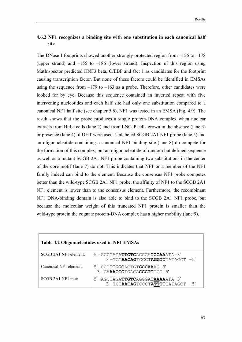
Results
4.6.2 NF1 recognizes a binding site with one substitution in each canonical half site
The DNase I footprints showed another strongly protected region from –156 to –178 (upper strand) and –155 to –186 (lower strand). Inspection of this region using MatInspector predicted HNF3 beta, C/EBP and Oct 1 as candidates for the footprint causing transcription factor. But none of these factors could be identified in EMSAs using the sequence from –179 to –163 as a probe. Therefore, other candidates were looked for by eye. Because this sequence contained an inverted repeat with five intervening nucleotides and each half site had only one substitution compared to a canonical NF1 half site (see chapter 5.6), NF1 was tested in an EMSA (Fig. 4.9). The result shows that the probe produces a single protein-DNA complex when nuclear extracts from HeLa cells (lane 2) and from LNCaP cells grown in the absence (lane 3) or presence (lane 4) of DHT were used. Unlabeled SCGB 2A1 NF1 probe (lane 5) and an oligonucleotide containing a canonical NF1 binding site (lane 8) do compete for the formation of this complex, but an oligonucleotide of random but defined sequence as well as a mutant SCGB 2A1 NF1 probe containing two substitutions in the center of the core motif (lane 7) do not. This indicates that NF1 or a member of the NF1 family indeed can bind to the element. Because the consensus NF1 probe competes better than the wild-type SCGB 2A1 NF1 probe, the affinity of NF1 to the SCGB 2A1 NF1 element is lower than to the consensus element. Furthermore, the recombinant NF1 DNA-binding domain is also able to bind to the SCGB 2A1 NF1 probe, but because the molecular weight of this truncated NF1 protein is smaller than the wild-type protein the cognate protein-DNA complex has a higher mobility (lane 9).
Table 4.2 Oligonucleotides used in NF1 EMSAs
SCGB 2A1 NF1 element: 5′-AGCTAGA AGGGA ATA-3′ TTGTC TCCAA3′-TCTAACAGTCCCTAGGTTTATAGCT -5′
Canonical NF1 element: 5′-CCTTTGGCACTGTGCCAAAG-3′ 3′-GAAACCGTGACACGGTTTCC-5′
SCGB 2A1 NF1 mut: 5′-AGCTAGATTGTCAGGGATAAAAATA-3′ 3′-TCTAACAGTCCCTATTTTTATAGCT -5′
67
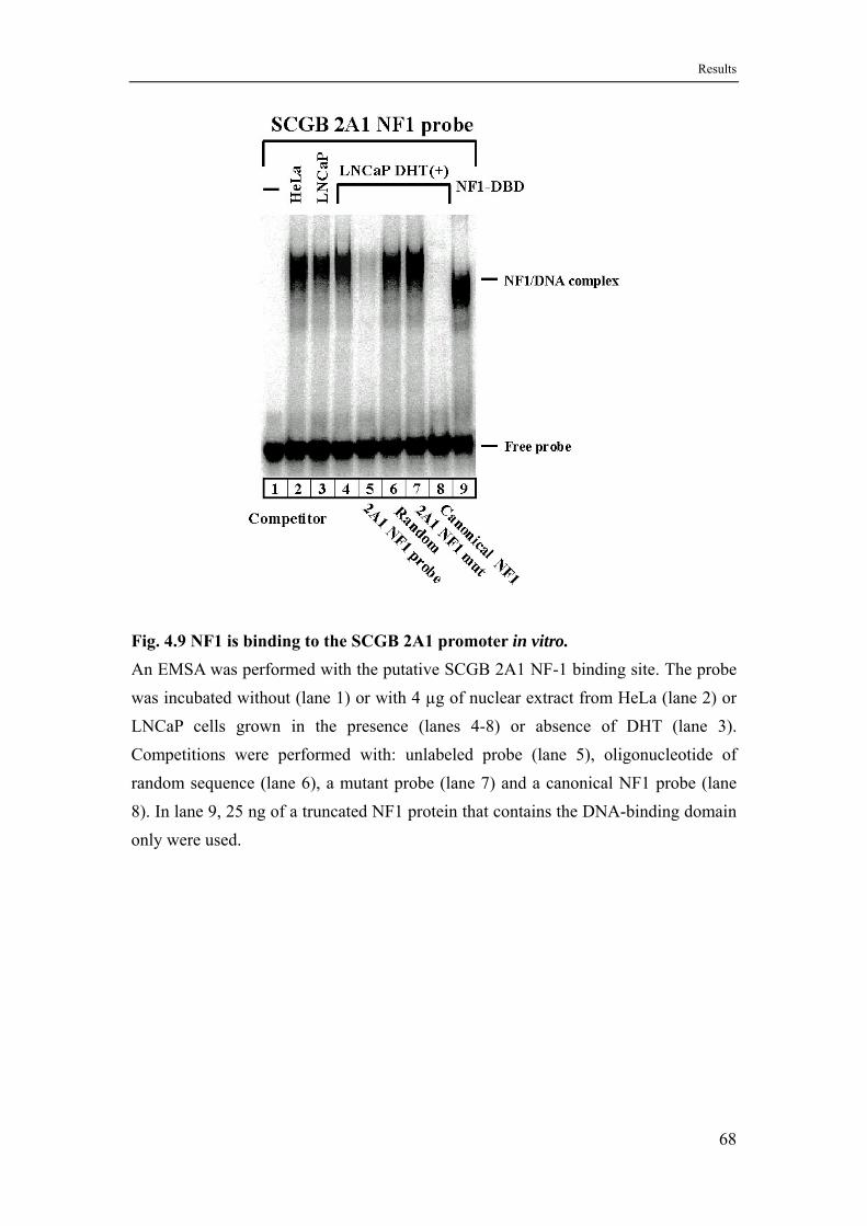
Results
Fig. 4.9 NF1 is binding to the SCGB 2A1 promoter in vitro. An EMSA was performed with the putative SCGB 2A1 NF-1 binding site. The probe was incubated without (lane 1) or with 4 µg of nuclear extract from HeLa (lane 2) or LNCaP cells grown in the presence (lanes 4-8) or absence of DHT (lane 3). Competitions were performed with: unlabeled probe (lane 5), oligonucleotide of random sequence (lane 6), a mutant probe (lane 7) and a canonical NF1 probe (lane 8). In lane 9, 25 ng of a truncated NF1 protein that contains the DNA-binding domain only were used.
68
Results
4.6.3 Sp family proteins recognize a dimeric inverted repeat type GC box
Initially when promoter fragments terminating at upstream position –53 were transferred to the tk promoter androgen responsiveness could not be restored in gene transfer experiments with LNCaP cells (Fig. 4.4). Therefore, new constructs were tested with an SCGB 2A1 promoter endpoint at –28, immediately upstream of the TATA box, which were indeed able to confer androgen responsiveness to the tk promoter. This result strongly indicated that the region from somewhere around –53 up to –28 is responsible for androgen sensitivity. Close inspection revealed an extended inverted repeat, with each half site of the repeat having homology to the GC box. Accordingly, this complex element was called a dimeric inverted repeat GC box (dim-IR-GC box). Surprisingly, it appeared as if ubiquitous factors were responsible for mediating the hormone response. In an EMSA it was investigated if Sp family proteins indeed can bind to the dim-IR-GC box in vitro (Fig. 4.10). With nuclear extracts from HeLa or LNCaP cells grown in the presence or absence of DHT the typical protein-DNA complex pattern of the Sp family, consisting of three major bands, appears when either a consensus GC box (lanes 10-13) (Suske, 1999) or the SCGB 2A1 dim-IR-GC box (lanes 1-4) is used as a probe. When an oligonucleotide probe of random but defined sequence is used in 50-fold molar excess no competition is observed (lanes 5 and 14) with either probe. But the unlabeled GC box fully competes with the formation of Sp-DNA complexes on the dim-IR-GC box (lane 6), and the unlabeled dim-IR-GC box strongly competes with the formation of Sp-DNA complexes on the GC box (lane 15). Direct competitor comparisons (not shown) indicated that the affinity of Sp factors to the dim-IR-GC box is lower than to the consensus GC box.
The use of antibodies directed against the two most common ubiquitous Sp factors Sp1 and Sp3 (a gift of Guntram Suske, University of Marburg) unequivocally showed that the observed DNA-protein complexes do contain the two proteins. Rabbit anti Sp1 antiserum is able to supershift the upper complex band of the Sp1/Sp3-DNA double band (lanes 7 and 16). Likewise rabbit anti Sp3 antiserum was able to supershift the lower complex band of the mentioned double band (lanes 8 and 17) as well as a complex band of lower mobility. The latter one is due to short Sp3 isoforms that originate from internal start codons (Kennett et al., 1997; Suske, 1999). Both antisera together supershift all oberserved major complex bands (lanes 9 and 18). All supershifts are more complete with the dim-IR-GC-box than with the consensus GC
69
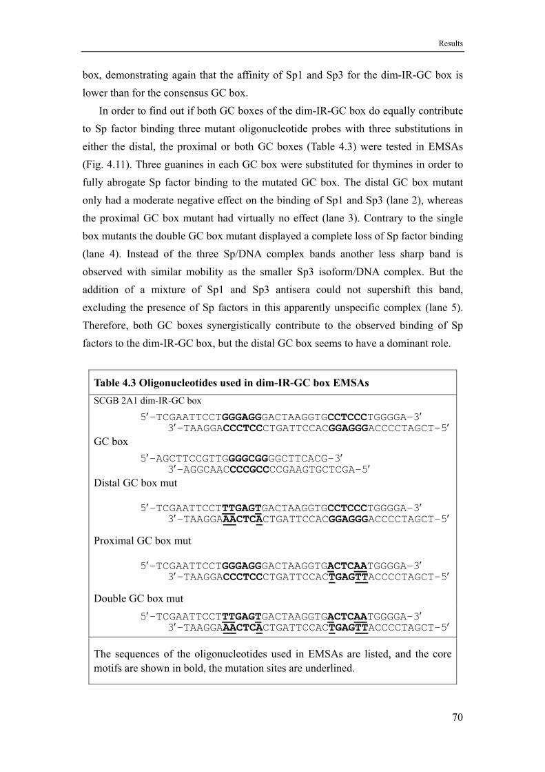
Results
box, demonstrating again that the affinity of Sp1 and Sp3 for the dim-IR-GC box is lower than for the consensus GC box.
In order to find out if both GC boxes of the dim-IR-GC box do equally contribute to Sp factor binding three mutant oligonucleotide probes with three substitutions in either the distal, the proximal or both GC boxes (Table 4.3) were tested in EMSAs (Fig. 4.11). Three guanines in each GC box were substituted for thymines in order to fully abrogate Sp factor binding to the mutated GC box. The distal GC box mutant only had a moderate negative effect on the binding of Sp1 and Sp3 (lane 2), whereas the proximal GC box mutant had virtually no effect (lane 3). Contrary to the single box mutants the double GC box mutant displayed a complete loss of Sp factor binding (lane 4). Instead of the three Sp/DNA complex bands another less sharp band is observed with similar mobility as the smaller Sp3 isoform/DNA complex. But the addition of a mixture of Sp1 and Sp3 antisera could not supershift this band, excluding the presence of Sp factors in this apparently unspecific complex (lane 5). Therefore, both GC boxes synergistically contribute to the observed binding of Sp factors to the dim-IR-GC box, but the distal GC box seems to have a dominant role.
Table 4.3 Oligonucleotides used in dim-IR-GC box EMSAs
SCGB 2A1 dim-IR-GC box 5′-TCGAATTCCT ′ GGGAGGGACTAAGGTGCCTCCCTGGGGA-3
3′-TAAGGACCCTCCCTGATTCCACGGAGGGACCCCTAGCT-5′ GC box 5′-AGCTTCCGTTG ′ GGGCGGGGCTTCACG-3
3′-AGGCAACCCCGCCCCGAAGTGCTCGA-5′ Distal GC box mut
5′-TCGAATTCCTTTGAGTGACTAAGGTGCCTCCCTGGGGA-33′-TAAGGA
′ AACTCACTGATTCCACGGAGGGACCCCTAGCT-5′
Proximal GC box mut
5′-TCGAATTCCTGGGAGGGACTAAGGTGACTCAATGGGGA-33′-TAAGGACCCTCCCTGATTCCAC
′ TGAGTTACCCCTAGCT-5′
Double GC box mut 5′-TCGAATTCCTTTGAGTGACTAAGGTGACTCAATGGGGA-3
3′-TAAGGA′
AACTCACTGATTCCACTGAGTTACCCCTAGCT-5′
The sequences of the oligonucleotides used in EMSAs are listed, and the core motifs are shown in bold, the mutation sites are underlined.
70
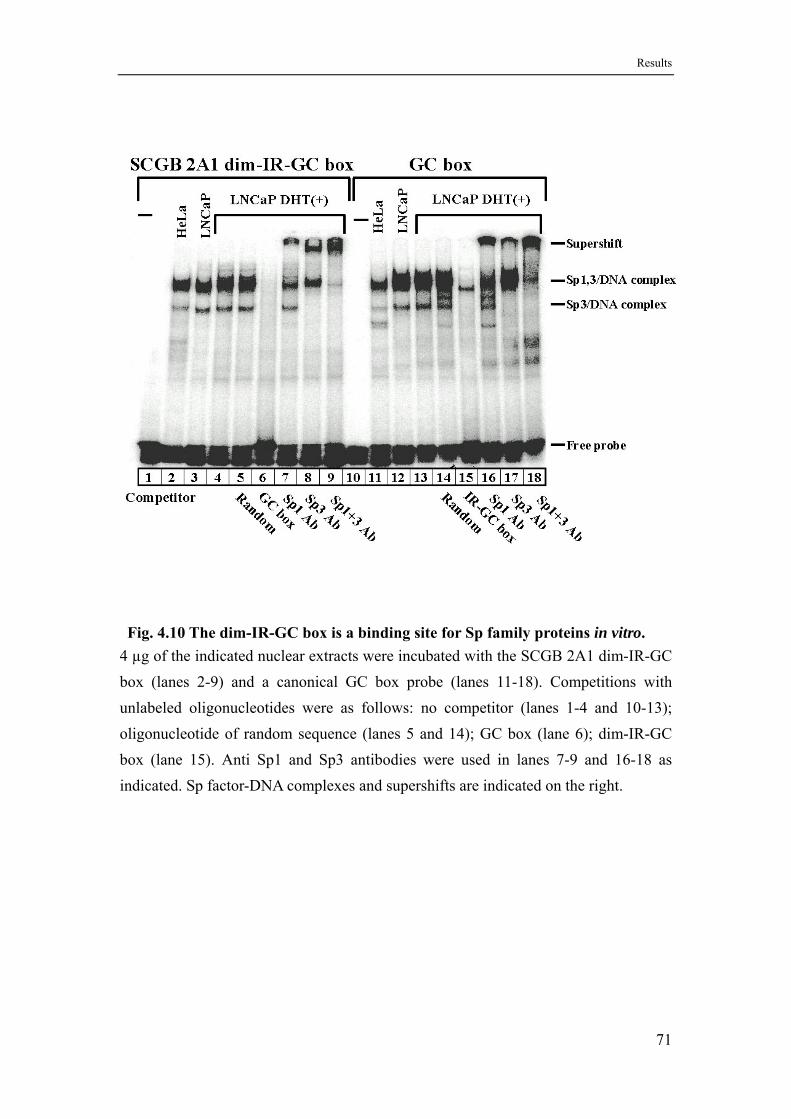
Results
Fig. 4.10 The dim-IR-GC box is a binding site for Sp family proteins in vitro. 4 µg of the indicated nuclear extracts were incubated with the SCGB 2A1 dim-IR-GC box (lanes 2-9) and a canonical GC box probe (lanes 11-18). Competitions with unlabeled oligonucleotides were as follows: no competitor (lanes 1-4 and 10-13); oligonucleotide of random sequence (lanes 5 and 14); GC box (lane 6); dim-IR-GC box (lane 15). Anti Sp1 and Sp3 antibodies were used in lanes 7-9 and 16-18 as indicated. Sp factor-DNA complexes and supershifts are indicated on the right.
71
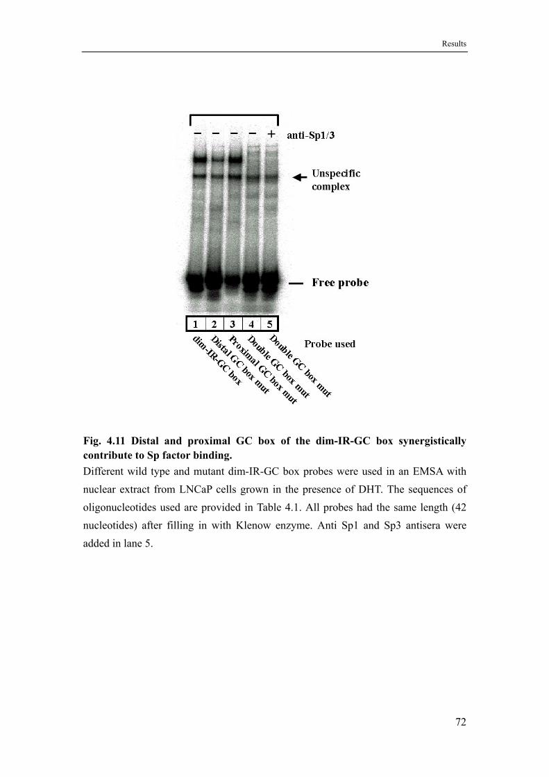
Results
Fig. 4.11 Distal and proximal GC box of the dim-IR-GC box synergistically contribute to Sp factor binding. Different wild type and mutant dim-IR-GC box probes were used in an EMSA with nuclear extract from LNCaP cells grown in the presence of DHT. The sequences of oligonucleotides used are provided in Table 4.1. All probes had the same length (42 nucleotides) after filling in with Klenow enzyme. Anti Sp1 and Sp3 antisera were added in lane 5.
72
Results
4.7 NF-Y is functionally important for basal promoter activity whereas binding of NF1 participates in and binding of Sp factors mediates the androgen response
Previously Electrophoretic Mobility Shift Assays showed that NF-Y, NF1 and Sp factors do bind to cognate elements of the SCGB 2A1 promoter in vitro. In order to demonstrate that the binding of these factors is also functionally relevant, mutations were introduced into each factor binding site. Promoter fragments carrying those mutations were cloned in front of the luciferase reporter gene and transfected into LNCaP prostate cells (Fig. 4.12).
For NF-Y, which was shown to bind to the promoter’s CCAAT box, the CCAAT core sequence was mutated to CTCAT. An oligonucleotide containing this mutated sequence was no longer able to compete for the NF-Y/CCAAT box complex (Fig. 4.8, lanes 7 and 16). The same two point mutations introduced into the SCGB 2A1 promoter terminating at –136 lead to an approximately 30% decrease in reporter gene activity under basal conditions (no hormone added) as well as under induced conditions (DHT added). The same result is obtained when the mutations were introduced into the promoter terminating at –382 (Fig. 4.12). Therefore, NF-Y seems to be generally required for promoter activity but not for hormone induction.
For NF1 the core of the identified binding site was mutated from TTGGA to TTTTA in the context of the promoter terminating at -382. These two point mutations also abrogate NF1 binding as shown by an EMSA (Fig. 4.9) in which an oligonucleotide containing the mutated sequence was no longer able to compete for the NF1/DNA complex (lane 7). Abrogation of NF1 binding leads to a some 40% decrease in induced transcription but to a slight increase in basal transcription (Fig. 4.12). Thus, NF1 seems to be involved in transmitting the hormone response. The activities of the –136/+50 wild type promoter and the –382/+50 promoter containing the NF1 mutations at around position –175 are very similar. This indicates that the SCGB 2A1 promoter from –136 up to –382 does contain only one functionally important positive element which is the NF1 binding site.
In order to investigate the functional importance of the dim-IR-GC box, the same mutations that were used in investigating DNA-binding in EMSAs (Table 4.3) were introduced into the promoter extending from –382 to +50 (Fig. 4.12). Mutation of the distal GC box, which has a moderate effect on Sp factor binding (Fig. 4.11, lane 2), has a dramatic effect on hormone induced transcription of the reporter gene (Fig. 4.12) leading to a 75% decrease in expression (down to 6-fold induction upon addition of
73
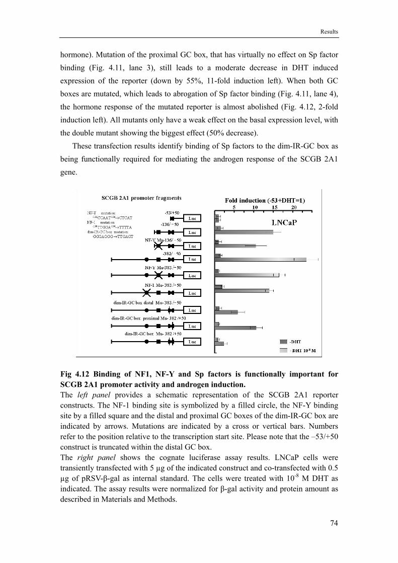
Results
hormone). Mutation of the proximal GC box, that has virtually no effect on Sp factor binding (Fig. 4.11, lane 3), still leads to a moderate decrease in DHT induced expression of the reporter (down by 55%, 11-fold induction left). When both GC boxes are mutated, which leads to abrogation of Sp factor binding (Fig. 4.11, lane 4), the hormone response of the mutated reporter is almost abolished (Fig. 4.12, 2-fold induction left). All mutants only have a weak effect on the basal expression level, with the double mutant showing the biggest effect (50% decrease).
These transfection results identify binding of Sp factors to the dim-IR-GC box as being functionally required for mediating the androgen response of the SCGB 2A1 gene.
Fig 4.12 Binding of NF1, NF-Y and Sp factors is functionally important for SCGB 2A1 promoter activity and androgen induction. The left panel provides a schematic representation of the SCGB 2A1 reporter constructs. The NF-1 binding site is symbolized by a filled circle, the NF-Y binding site by a filled square and the distal and proximal GC boxes of the dim-IR-GC box are indicated by arrows. Mutations are indicated by a cross or vertical bars. Numbers refer to the position relative to the transcription start site. Please note that the –53/+50 construct is truncated within the distal GC box. The right panel shows the cognate luciferase assay results. LNCaP cells were transiently transfected with 5 µg of the indicated construct and co-transfected with 0.5 µg of pRSV-β-gal as internal standard. The cells were treated with 10-8 M DHT as indicated. The assay results were normalized for β-gal activity and protein amount as described in Materials and Methods.
74
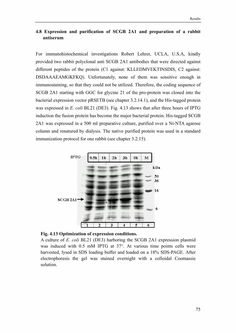
Results
4.8 Expression and purification of SCGB 2A1 and preparation of a rabbit antiserum
For immunohistochemical investigations Robert Lehrer, UCLA, U.S.A, kindly provided two rabbit polyclonal anti SCGB 2A1 antibodies that were directed against different peptides of the protein (C1 against: KLLEDMVEKTINSDIS, C2 against: DSDAAAEAMGKFKQ). Unfortunately, none of them was sensitive enough in immunostaining, so that they could not be utilized. Therefore, the coding sequence of SCGB 2A1 starting with GGC for glycine 21 of the pro-protein was cloned into the bacterial expression vector pRSETB (see chapter 3.2.14.1), and the His-tagged protein was expressed in E. coli BL21 (DE3). Fig. 4.13 shows that after three hours of IPTG induction the fusion protein has become the major bacterial protein. His-tagged SCGB 2A1 was expressed in a 500 ml preparative culture, purified over a Ni-NTA agarose column and renatured by dialysis. The native purified protein was used in a standard immunization protocol for one rabbit (see chapter 3.2.15).
Fig. 4.13 Optimization of expression conditions. A culture of E. coli BL21 (DE3) harboring the SCGB 2A1 expression plasmid was induced with 0.5 mM IPTG at 37°. At various time points cells were harvested, lysed in SDS loading buffer and loaded on a 18% SDS-PAGE. After electrophoresis the gel was stained overnight with a colloidal Coomassie solution.
75
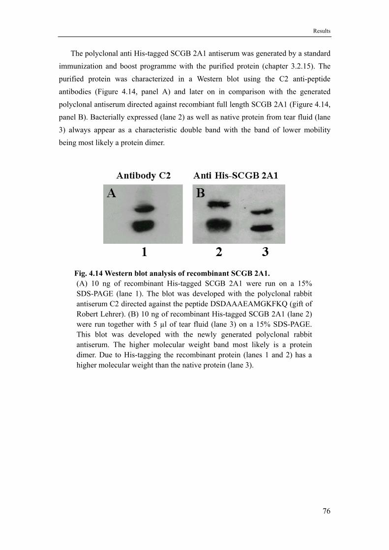
Results
The polyclonal anti His-tagged SCGB 2A1 antiserum was generated by a standard immunization and boost programme with the purified protein (chapter 3.2.15). The purified protein was characterized in a Western blot using the C2 anti-peptide antibodies (Figure 4.14, panel A) and later on in comparison with the generated polyclonal antiserum directed against recombiant full length SCGB 2A1 (Figure 4.14, panel B). Bacterially expressed (lane 2) as well as native protein from tear fluid (lane 3) always appear as a characteristic double band with the band of lower mobility being most likely a protein dimer.
Fig. 4.14 Western blot analysis of recombinant SCGB 2A1. (A) 10 ng of recombinant His-tagged SCGB 2A1 were run on a 15% SDS-PAGE (lane 1). The blot was developed with the polyclonal rabbit antiserum C2 directed against the peptide DSDAAAEAMGKFKQ (gift of Robert Lehrer). (B) 10 ng of recombinant His-tagged SCGB 2A1 (lane 2) were run together with 5 µl of tear fluid (lane 3) on a 15% SDS-PAGE. This blot was developed with the newly generated polyclonal rabbit antiserum. The higher molecular weight band most likely is a protein dimer. Due to His-tagging the recombinant protein (lanes 1 and 2) has a higher molecular weight than the native protein (lane 3).
76
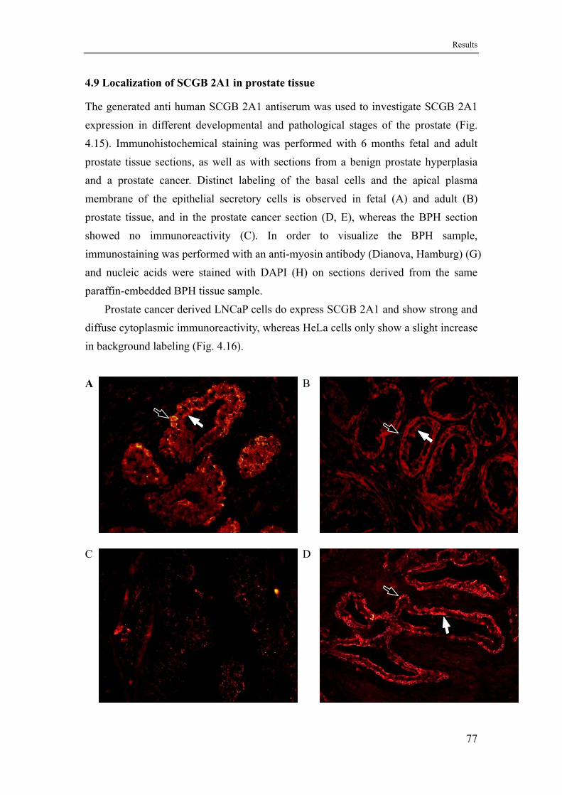
Results
4.9 Localization of SCGB 2A1 in prostate tissue
The generated anti human SCGB 2A1 antiserum was used to investigate SCGB 2A1 expression in different developmental and pathological stages of the prostate (Fig. 4.15). Immunohistochemical staining was performed with 6 months fetal and adult prostate tissue sections, as well as with sections from a benign prostate hyperplasia and a prostate cancer. Distinct labeling of the basal cells and the apical plasma membrane of the epithelial secretory cells is observed in fetal (A) and adult (B) prostate tissue, and in the prostate cancer section (D, E), whereas the BPH section showed no immunoreactivity (C). In order to visualize the BPH sample, immunostaining was performed with an anti-myosin antibody (Dianova, Hamburg) (G) and nucleic acids were stained with DAPI (H) on sections derived from the same paraffin-embedded BPH tissue sample.
Prostate cancer derived LNCaP cells do express SCGB 2A1 and show strong and diffuse cytoplasmic immunoreactivity, whereas HeLa cells only show a slight increase in background labeling (Fig. 4.16).
A B
C D
77
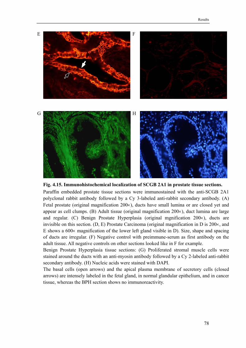
Results
E F
G H
Fig. 4.15. Immunohistochemical localization of SCGB 2A1 in prostate tissue sections. Paraffin embedded prostate tissue sections were immunostained with the anti-SCGB 2A1 polyclonal rabbit antibody followed by a Cy 3-labeled anti-rabbit secondary antibody. (A) Fetal prostate (original magnification 200×), ducts have small lumina or are closed yet and appear as cell clumps. (B) Adult tissue (original magnification 200×), duct lumina are large and regular. (C) Benign Prostate Hyperplasia (original mgnification 200×), ducts are invisible on this section. (D, E) Prostate Carcinoma (original magnification in D is 200×, and E shows a 600× magnification of the lower left gland visible in D). Size, shape and spacing of ducts are irregular. (F) Negative control with preimmune-serum as first antibody on the adult tissue. All negative controls on other sections looked like in F for example. Benign Prostate Hyperplasia tissue sections: (G) Proliferated stromal muscle cells were stained around the ducts with an anti-myosin antibody followed by a Cy 2-labeled anti-rabbit secondary antibody. (H) Nucleic acids were stained with DAPI. The basal cells (open arrows) and the apical plasma membrane of secretory cells (closed arrows) are intensely labeled in the fetal gland, in normal glandular epithelium, and in cancer tissue, whereas the BPH section shows no immunoreactivity.
78
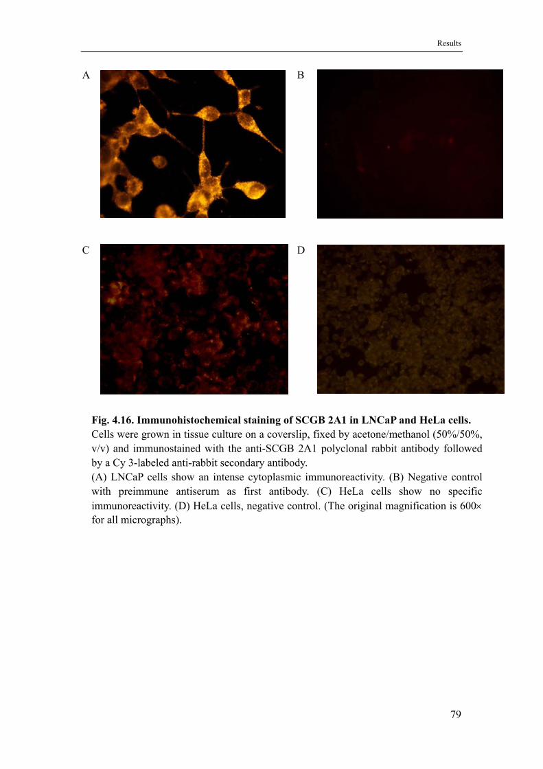
Results
A
B
C D
Fig. 4.16. Immunohistochemical staining of SCGB 2A1 in LNCaP and HeLa cells. Cells were grown in tissue culture on a coverslip, fixed by acetone/methanol (50%/50%, v/v) and immunostained with the anti-SCGB 2A1 polyclonal rabbit antibody followed by a Cy 3-labeled anti-rabbit secondary antibody. (A) LNCaP cells show an intense cytoplasmic immunoreactivity. (B) Negative control with preimmune antiserum as first antibody. (C) HeLa cells show no specific immunoreactivity. (D) HeLa cells, negative control. (The original magnification is 600× for all micrographs).
79
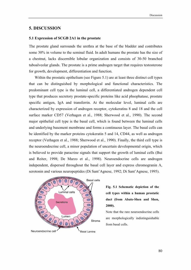
Discussion
5. DISCUSSION
5.1 Expression of SCGB 2A1 in the prostate
The prostate gland surrounds the urethra at the base of the bladder and contributes some 30% in volume to the seminal fluid. In adult humans the prostate has the size of a chestnut, lacks discernible lobular organization and consists of 30-50 branched tuboalveolar glands. The prostate is a prime androgen target that requires testosterone for growth, development, differentiation and function.
Within the prostatic epithelium (see Figure 5.1) are at least three distinct cell types that can be distinguished by morphological and functional characteristics. The predominant cell type is the luminal cell, a differentiated androgen dependent cell type that produces secretory prostate-specific proteins like acid phsophatase, prostate specific antigen, IgA and transferrin. At the molecular level, luminal cells are characterized by expression of androgen receptor, cytokeratins 8 and 18 and the cell surface marker CD57 (Verhagen et al., 1988; Sherwood et al., 1990). The second major epithelial cell type is the basal cell, which is found between the luminal cells and underlying basement membrane and forms a continuous layer. The basal cells can be identified by the marker proteins cytokeratin 5 and 14, CD44, as well as androgen receptor (Verhagen et al., 1988; Sherwood et al., 1990). Finally, the third cell type is the neuroendocrine cell, a minor population of uncertain developmental origin, which is believed to provide paracrine signals that support the growth of luminal cells (Bui and Reiter, 1998; De Marzo et al., 1998). Neuroendocrine cells are androgen independent, dispersed throughout the basal cell layer and express chromogranin A, serotonin and various neuropeptides (Di Sant’Agnese, 1992; Di Sant’Agnese, 1995).
Fig. 5.1 Schematic depiction of the
cell types within a human prostatic
duct (from Abate-Shen and Shen,
2000).
Note that the rare neuroendocrine cells
are morphologically indistinguishable
from basal cells.
80
Discussion
Based on observations that some transient populations of prostatic epithelial cells having both basal and luminal characteristics (Verhagen et al., 1992; Bonkhoff et al., 1994), it was proposed that there exists a stem cell reservoir within the prostatic epithelium. This stem cell compartment represents a subpopulation of androgen independent basal cells. These stem cells would give rise to a transiently proliferating compartment composed of pluripotent androgen responsive cells that in turn generate the prostatic basal cells, differentiated luminal cells and possibly also the neuroendocrine cells (Bonkhoff and Remberger, 1996; De Marzo et al., 1998). But some other studies support the hypothesis that the neuroendocrine cells are derived from a second prostatic stem cell lineage, different from the urogenital sinus lineage (Aumüller et al., 1999; Aumüller et al., 2001). This dual stem cell compartment theory would overcome a number of findings that are difficult to explain by a single stem cell compartment, such as constant and low number of neuroendocrine cells, their asymmetric distribution within the gland, and their androgen insensitivity.
In the prostate expression of SCGB 2A1 has been localized to the epithelial cells lining the secretory ducts by in situ hybridization (see chapter 4.1.1). Immunostaining experiments further localized SCGB 2A1 expression to both the glandular luminal cells and the basal cells (see chapter 4.9). Expression in basal cells is an unusual finding because only the luminal cells have been shown to express the prostate-specific secretory proteins investigated so far. But one study could show that basal cells isolated by differential cell sorting can produce prostatic secretory proteins when cocultured with stromal cells (Liu et al., 1997). Due to the fact that the basal cells are also androgen sensitive it would be no surprise to find some proteins that are secreted by the luminal cells also being expressed by the basal cells. It would be interesting now to immunostain SCGB 2A1 in combination with other basal and luminal cell specific markers in order to confirm this point.
In prostate tissue sections the expression of SCGB 2A1 was observed in different specimens including fetal gland, normal adult gland and prostate cancer, whereas the benign prostate hyperplasia (BPH) is negative (see chapter 4.9).
Unlike the adult prostate, the fetal prostate is under the influence of maternal placental hormones, as well as of the hormones produced by the fetus itself. Beginning in week 10 of gestation the prostate begins to develop under the action of fetal androgens. SCGB 2A1 protein is already detectable in the 6-month fetal prostate similar to other differentiation markers like estrogen receptor beta and PSA (Adams et al., 2002).
81
Discussion
In benign prostatic hyperplasia (BPH), proliferation of stromal and activated smooth muscle cells plays an important role in the induction and progression of the disease. In figure 4.15 (G), it can be seen that the fibromuscular stroma invades the basal cell layer and the zone of secretory epithelial cells which confirms that the tissue slide showing no reactivity with SCGB 2A1 antiserum (Fig. 4.15 (C)) was indeed a BPH tissue slide. Genes that are expressed in the prostate can be sorted into two groups. They are either expressed in normal prostate tissue, BPH as well as prostate cancer or are expressed in normal prostate tissue, only weekly expressed in BPH and not at all expressed in prostate cancer, so that there is a correlation with differentiation status. But in case of SCGB 2A1 expression is observed in normal and cancer tissue whereas expression in BPH is lacking. If this result can be confirmed with a larger number of BPH samples, SCGB 2A1 would have a potential for becoming a diagnostic marker in the differentiation of BPH versus prostate cancer.
Whereas in many cancers differentiation markers of the originating cell type are no longer detectable, in the very slow growing prostate cancer typical marker proteins continue to be expressed. The paradigm of such a marker is PSA that is similarly expressed in normal prostate tissue, BPH and cancer tissue and is the main test in the management of prostate cancer (Gelmini et al., 2003). Because PSA concentrations relate to age, prostate size, and the presence of prostate cancer, but can also be raised after ejaculation, riding a bike, prostate biopsy, surgery, or prostatitis, the validity of PSA as diagnostic marker is much under discussion (Frankel et al., 2003). Like PSA SCGB 2A1 is also expressed in prostate cancer, and due to its different mode of regulation by androgens it might be interesting to compare its expression in androgen dependent versus androgen independent cancers.
5.2 SCGB 2A1 is a new target gene for androgens in the prostate with a different kinetics of mRNA induction than the gene encoding prostate specific antigen
When LNCaP cells are treated with 10-8 M DHT over 24 hours expression of steady state SCGB 2A1 mRNA levels increase slowly. Compared to a low but detectable basal expression in the absence of androgens, the maximum SCGB 2A1 mRNA level in the presence of DHT (21-fold increase over basal) is obtained after six hours and stays that high for at least 24 hours (Fig. 4.1). This response identifies SCGB 2A1 as a new class of androgen target gene in the prostate. Although many androgen-regulated genes have been identified, for most of them only limited information on the mechanism of regulation is available. In a few genes functional androgen response
82

Discussion
elements (AREs) mediating a direct genomic action of the androgen receptor have been characterized. Among the genes that have been studied in most detail are the rat prostatic binding protein subunit C3(1) (Claessens et al., 1990) and the human prostate specific antigen (PSA, also called kallikrein 3, hKLK3) gene (Cleutjens et al. 1996; Cleutjens et al., 1997; Riegman et al., 1991). Because the PSA gene has also been investigated extensively in LNCaP cells, the promoter of this gene was used here as an example of direct genomic control of the androgen receptor. The PSA promoter contains two functionally active androgen response regions, an enhancer region at –4.2 kb and a promoter region in the proximal 400 bp (Cleutjens et al., 1996; Cleutjens et al., 1997). The latter only provides a low level of androgen regulation (4-6 fold induction) dependent on the presence of two AREs, ARE-I at –170 bp and ARE-II at –394 bp. ARE-III, presumably together with other still unknown elements in the enhancer for full androgen regulation (some 3000-fold induction). ARE-I and ARE-III closely resemble the ARE consensus sequence, whereas ARE-II is a low affinity AR binding site deviating considerably from the consensus sequence. In the absence of androgen, PSA mRNA is expressed at a low basal level in LNCaP cells when analyzed by Northern blotting with total RNA (Wolf, D.A. et al., 1992). But in the presence of 3.3 nM synthetic androgen mibolerone, the PSA mRNA level rises continuously within 4-72 hours. In the presence of 1 nM synthetic androgen R1881, mRNA levels reached the maximum level already after 24 hours (Riegman et al., 1991). Another example for a gene that is under direct genomic control of the androgen receptor in the prostate is the human prostate-specific glandular kallikrein-1 (hKLK2). hKLK2 mRNA is not detectable in LNCaP cells in the absence of hormone when analyzed by Northern blotting (Young et al., 1992). But in the presence of 3 nM mibolerone the steady state hKLK2 mRNA level rises continuously, very similar to the PSA message, from 5-24 hours. Compared to PSA and hKLK2 the kinetics of SCGB 2A1 induction is different because the steady state mRNA level in the presence of androgen is rising faster and reaches its maximum already after 6 hours. The steady state mRNA level is mainly determined by the rate of transcription initiation and the mRNAs half life. In case of PSA androgens strongly induce transcription initiation in less than 3 hours as determined by nuclear run-on assays. Run-on acitivity decreases already after 24 hours whereas steady state levels continue to increase up to 72 hours. This was explained by showing that the PSA mRNA half life is considerably increased in the presence of hormone (Wolf, D.A. et al., 1992). Therefore, the comparatively fast androgen response seen in the SCGB 2A1 Northern could be explained by a short
83
Discussion
mRNA half life in the absence of hormone combined with a strong and fast androgen effect on mRNA stability.
5.3 The kinetics of androgen induction of a DNase I hypersensitive site in the SCGB 2A1 promoter indicates an indirect non-genomic hormone response
The control regions in a gene can often be localized by mapping DNase I hypersensitive sites (DH sites). Genes contain discrete sequences of DNA that are sensitive to DNase I cleavage in chromatin, and the pattern of these so-called hypersensitive sites changes depending on the activation state of the gene (Weintraub and Groudine, 1976). By comparing the pattern of hypersensitive sites obtained by cleavage of a non-induced gene with that obtained when the gene is actively transcribed or induced, regions that participate in gene regulation can be identified. A major strength of this approach is that it makes no a priori assumptions about the locations of control sequences and allows a quick investigation of several kb of DNA for potential regulatory sequences.
During interphase of the eukaryotic cell cycle, the bulk of DNA is assembled into highly folded nucleoprotein filaments. This is even true for euchromatic regions of the genome which are actively undergoing RNA transcription, DNA replication, DNA repair or recombination. Today we know that these processes can function efficiently in the chromatin environment due to the actions of two classes of highly conserved chromatin remodelling enzymes. The first class includes enzymes that covalently modify the nucleosomal histones (e.g. by acetylation, phosphorylation, methylation and ADP-ribosylation) and, at least in the case of histone acetylation by histone acetyl transferases (HATs), these covalent modifications can destabilize the folding of nucleosomal arrays and promote RNA transcription (Fischle et al., 2003) The second class of enzymes is composed of multi-subunit complexes (like SWI/SNF etc.) that use the energy of ATP hydrolysis to disrupt histone-DNA interactions and thus modify chromatin structure (Peterson, 2000). Independent of the exact mechanism that leads to a change in chromatin structure, mapping of DNase I hypersensitive sites is ideally suited to identify potential regulatory regions in an unknown target gene as a first step in promoter analysis.
There are in general two possibilities to map hypersensitive sites. The most popular one, that was also used here, involves the isolation of nuclei from cells or tissue of interest followed by incubation of the intact nuclei with varying amounts of DNase I (Cockerill, 2000). Alternatively cells of interest can be permeabilized and
84
Discussion
then treated directly with DNase I without prior isolation of nuclei (Aasland and Stewart, 1999).
Having shown that SCGB 2A1 is expressed androgen-dependent in LNCaP cells, DH sites were mapped in the absence and presence of hormone. Within a 10 kb Xba I fragment encompassing the SCGB 2A1 gene including 3 kb upstream of exon 1 and 3.3 kb downstream of exon 3 an androgen dependent DH site appeared in the proximal promoter at position –100 bp +/- 50 bp. A time course experiment revealed that it takes six hours of hormone treatment until the DH site becomes maximally accessible for DNase I. Compared to other steroid hormone dependent DH sites in other genes this androgen response in the SCGB 2A1 gene appears rather delayed. By using chromatin immunoprecipitation it was shown that androgen induces robust recruitment of androgen receptor, members of the p160 coactivator family, CREB-binding protein/p300 and RNA polymerase II specifically to the enhancer of the PSA gene within 30 min (Louie et al., 2003; Jia et al., 2003). Therefore it appears that a promoter which is under direct genomic control of the androgen receptor or a different steroid hormone receptor does respond to a hormone signal in less than 60 min when DH sites, run on transcription (Adom et al., 1991) or receptor recruitment are measured. But the kinetics of DH site induction by androgen in the SCGB 2A1 promoter does not fit into this picture because after one hour of induction with DHT no hypersensitivity could be observed. This observation led to the hypothesis that its androgen response could be mounted by a non-genomic, non-ARE-mediated indirect mechanism.
5.4 Promoter deletion analyses identify a dimeric inverted repeat GC box as the “androgen responsive element”
Reporter gene constructs that were under the control of SCGB 2A1 promoter fragments terminating between –1476 and –575 were induced approximately 11-fold by the androgen DHT in LNCaP cells (Fig. 4.3). When the promoter was shortened to position – 382 the induction level was roughly doubled to 23-fold indicating the loss of a negatively acting element. It is known that eukaryotic transcription is regulated not only by activators but also by repressors. Many eukaryotic genes are subject to both repression and activation. Most eukaryotic repressors are also modular proteins. Similar to activators, they usually contain a single DNA-binding domain, one or a few repression domains, and can control transcription when they are bound at sites hundreds to thousands of base pairs from a start site.
85
Discussion
Further shortening the promoter to position –136 again reduced the induction level to 15-fold indicating that a positively acting element is located between –382 and –136. Because the low basal transcription level is also somewhat reduced when the two constructs (–382/+50)-Luc and (–136/+50)-Luc are compared, it is not clear from these experiments if this element has a positive effect on both basal and induced or mainly on induced transcription.
When the promoter fragment (–53/+50) was tested in transfections with LNCaP cells androgen induction was abolished. Therefore, androgen responsiveness is located in the fragment –136 to –53 which coincides with the position of the androgen inducible DNase I hypersensitive site (see Fig. 4.4, upper panel).
In order to evaluate the contribution of the androgen receptor in androgen induction bicalutamide (Casodex, ICI 176334) was used as a specific inhibitor of the androgen receptor in LNCaP cells (Veldscholte et al., 1992a). This compound is the most widely used competitive AR antagonist and was shown to stabilize AR association with cytosolic heat shock protein complexes in LNCaP cells preventing nuclear translocation (Veldscholte et al., 1992b). In a recent study using prostate cancer tissue it was shown that bicalutamide-liganded AR can translocate into the nucleus. Moreover, specific DNA binding by the bicalutamide-liganded AR was demonstrated in vivo using a VP16-AR fusion protein and was confirmed by chromatin immunoprecipitation. Nonetheless, bicalutamide could not stimulate interactions between the AR N and C termini or recruitment of steroid receptor coactivator proteins. Apparently bicalutamide stimulates the assembly of a transcriptionally inactive AR on DNA (Masiello et al., 2002). Because bicalutamide was able to abrogate DHT induced reporter gene expression in LNCaP cells transfected with the two constructs (–382/+50)-Luc and (-136/+50)-Luc it can be concluded that the androgen recpetor is required for mediating the hormone response.
In order to investigate if the androgen responsive region acts as an autonomous element, the fragment (–136/–53) was cloned in front of the Herpes simplex virus thymidine kinase promoter (HSV-tk) and tested in gene transfer experiments (see chapter 4.3, lower panel). The tk promoter used in these experiments terminated at position -90 and contained one GC box and the TATA box and was slightly inducible by androgens by a factor of 2-3 fold. Because the resultant (–136/-53)-tk-Luc construct as well as the (–382/-53)-tk-Luc construct were not more inducible than the tk basic construct itself, the length of the SCGB 2A1 promoter fragment was extended another 25 bp up to position –28 in order to exclude that the “androgen responsive
86
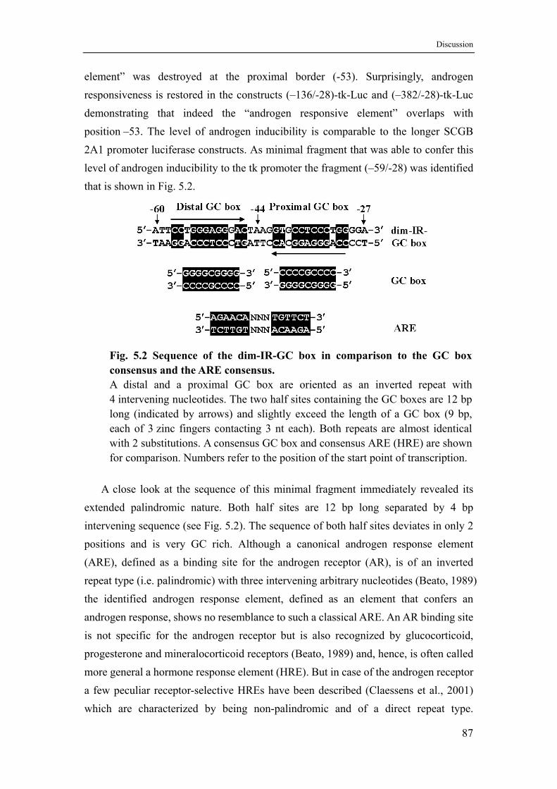
Discussion
element” was destroyed at the proximal border (-53). Surprisingly, androgen responsiveness is restored in the constructs (–136/-28)-tk-Luc and (–382/-28)-tk-Luc demonstrating that indeed the “androgen responsive element” overlaps with position –53. The level of androgen inducibility is comparable to the longer SCGB 2A1 promoter luciferase constructs. As minimal fragment that was able to confer this level of androgen inducibility to the tk promoter the fragment (–59/-28) was identified that is shown in Fig. 5.2.
Fig. 5.2 Sequence of the dim-IR-GC box in comparison to the GC box consensus and the ARE consensus. A distal and a proximal GC box are oriented as an inverted repeat with 4 intervening nucleotides. The two half sites containing the GC boxes are 12 bp long (indicated by arrows) and slightly exceed the length of a GC box (9 bp, each of 3 zinc fingers contacting 3 nt each). Both repeats are almost identical with 2 substitutions. A consensus GC box and consensus ARE (HRE) are shown for comparison. Numbers refer to the position of the start point of transcription.
A close look at the sequence of this minimal fragment immediately revealed its extended palindromic nature. Both half sites are 12 bp long separated by 4 bp intervening sequence (see Fig. 5.2). The sequence of both half sites deviates in only 2 positions and is very GC rich. Although a canonical androgen response element (ARE), defined as a binding site for the androgen receptor (AR), is of an inverted repeat type (i.e. palindromic) with three intervening arbitrary nucleotides (Beato, 1989) the identified androgen response element, defined as an element that confers an androgen response, shows no resemblance to such a classical ARE. An AR binding site is not specific for the androgen receptor but is also recognized by glucocorticoid, progesterone and mineralocorticoid receptors (Beato, 1989) and, hence, is often called more general a hormone response element (HRE). But in case of the androgen receptor a few peculiar receptor-selective HREs have been described (Claessens et al., 2001) which are characterized by being non-palindromic and of a direct repeat type.
87
Discussion
Therefore, from sequence comparisons it is very unlikely that the SCGB 2A1 ARE is a binding site for the androgen receptor. Instead, it’s GC content and the GGACCC core motif of each half site immediately suggested that both half sites are GC boxes and, thus, binding sites for Sp family transcription factors (Suske, 1999). Hence, the SCGB ARE was named a dimeric inverted repeat GC box (dim-IR-GC box).
5.5 Unresponsiveness of the dim-IR-GC box towards glucocorticoids excludes the presence of a cryptic HRE
Although there was no indication whatsoever that the dim-IR-GC box is or contains a hormone responsive element, defined as a binding site for androgen, glucocorticoid, progesterone or mineralocorticoid receptors, the possibility remained that a cryptic binding site does exist. Therefore, the androgen and glucocorticoid responses of the SCGB 2A1 and PSA promoters were compared in LNCaP cells (see chapter 4.4). The PSA promoter contains two functionally active androgen response regions (see chapter 5.2), which contain multiple HREs that have been shown to be responsive to androgens and glucocorticoids alike (Cleutjens et al., 1997). Because LNCaP cells do not express the glucocorticoid receptor (Schuurmans et al., 1988) it had to be ectopically expressed from a cotransfected GR expression construct. As shown before, the PSA promoter responds strongly to the androgen DHT and the glucocorticoid dexamethasone. This demonstrates that the GR expression construct as well as the whole experimental setup were functioning as expected. Contrary to the PSA promoter the SCGB 2A1 promoter did not respond to DEX at all even if the GR was expressed. Instead there was a slight repression of basal activity in the presence of DEX and GR. This result shows that a cryptic but functional HRE does not exist in the SCGB 2A1 promoter because an androgen-specific HRE with 100% androgen selectivity has not been described yet.
5.6 NF-Y is functionally important for full promoter activity
DNase I footprinting of the –382/+50 SCGB 2A1 promoter fragment revealed two strong footprints at positions –186/-155 and –104/-136. Because these footprints were not only observed with nuclear extracts from DHT induced or uninduced LNCaP cells but also with HeLa nuclear extract they are likely caused by ubiquitous proteins. Sequence analysis in combination with a presumably ubiquitous nature of the footprinted proteins led to the hypothesis that the distal footprint was due to binding of nuclear factor 1 (NF1) and the proximal footprint due to binding of nuclear factor
88
Discussion
Y (NF-Y). This hypothesis was confirmed in EMSAs demonstrating in vitro that LNCaP and HeLa nuclear extract proteins recognize a NF1 binding site at around –167 and a CAAT-box at around –110 within the center of the footprinted regions. Transfection of reporter gene constructs with binding site mutants that were shown in EMSAs to abrogate DNA-binding showed that NF-Y is functionally important for basal promoter activity whereas NF1 also participates in mediating the androgen response.
The CCAAT motif is one of the most common DNA-binding elements present in the promoters of about 30% of all mammalian genes as a nucleotide comparison of 502 unrelated RNA polymerase II promoters implies (Bucher, 1990). The motif is most often found between 80 and 100 bp upstream of the transcription start site, and the sequences around the CCAAT box are highly conserved when orthologous genes are compared. Similarly, the SCGB 2A1 CCAAT box is located at position –110 but orthologous genes have not been characterized in their promoter regions yet.
NF–Y, also called CCAAT-binding factor (CBF), is a unique DNA-binding protein that interacts with the CCAAT motif that is also called a Y-box. The affinity of NF-Y binding to DNA varies depending on the sequences flanking CCAAT. High-affinity CBF-binding sites have been defined by PCR-mediated random binding site selection and yielded the consensus 5'-[T/C][A/G][A/G] CCAAT CA-3' (Bi et al., 1997). A survey of 178 NF-Y binding CCAAT boxes confirmed the absolute requirement for the 5 nt CCAAT core and also pointed to specific flanking sequences - C, Pu, Pu on the 5'-side and C/G, A/G, G, A/C, G on the 3'-side - required for efficient binding (Mantovani, 1998). The flanking sequences of the SCGB 2A1 CCAAT box (5'-ACA-CCAAT-GGGAA-3') show a very good match with the flanking sequence consensus on the 3' side and a poor match (one out of three) on the 5' side. Together with the results obtained from EMSA competition experiments with the high affinity mouse MHC II Ea Y–box (see chapter 4.6.1) one can conclude that the SCGB 2A1 CCAAT motif also possesses a comparably high affinity for NF-Y.
It is known that several proteins can bind to CCAAT motifs. But among them, only NF-Y requires a very high degree of sequence conservation. Other CCAAT-binding proteins include CTF/NF1 and CCAAT/enhancer binding protein (C/EBP), but their binding sites often do not contain the complete CCAAT core. Hence, NF-Y can be distinguished from other CCAAT binding proteins on the basis of DNA sequence requirements (Maity and Crombrugghe, 1998).
Promotors with CCAAT motifs can be divided into three groups based on the
89
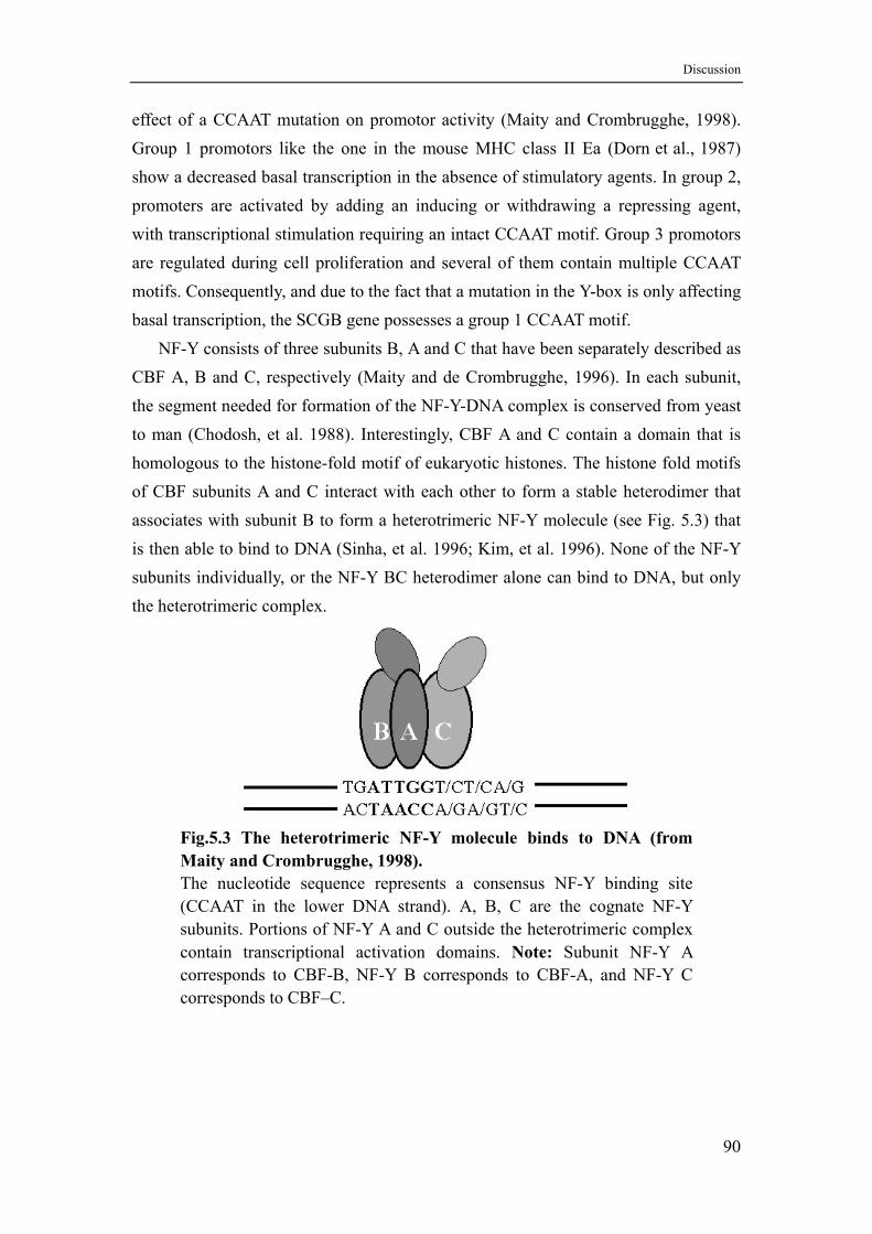
Discussion
effect of a CCAAT mutation on promotor activity (Maity and Crombrugghe, 1998). Group 1 promotors like the one in the mouse MHC class II Ea (Dorn et al., 1987) show a decreased basal transcription in the absence of stimulatory agents. In group 2, promoters are activated by adding an inducing or withdrawing a repressing agent, with transcriptional stimulation requiring an intact CCAAT motif. Group 3 promotors are regulated during cell proliferation and several of them contain multiple CCAAT motifs. Consequently, and due to the fact that a mutation in the Y-box is only affecting basal transcription, the SCGB gene possesses a group 1 CCAAT motif.
NF-Y consists of three subunits B, A and C that have been separately described as CBF A, B and C, respectively (Maity and de Crombrugghe, 1996). In each subunit, the segment needed for formation of the NF-Y-DNA complex is conserved from yeast to man (Chodosh, et al. 1988). Interestingly, CBF A and C contain a domain that is homologous to the histone-fold motif of eukaryotic histones. The histone fold motifs of CBF subunits A and C interact with each other to form a stable heterodimer that associates with subunit B to form a heterotrimeric NF-Y molecule (see Fig. 5.3) that is then able to bind to DNA (Sinha, et al. 1996; Kim, et al. 1996). None of the NF-Y subunits individually, or the NF-Y BC heterodimer alone can bind to DNA, but only the heterotrimeric complex.
Fig.5.3 The heterotrimeric NF-Y molecule binds to DNA (from Maity and Crombrugghe, 1998). The nucleotide sequence represents a consensus NF-Y binding site (CCAAT in the lower DNA strand). A, B, C are the cognate NF-Y subunits. Portions of NF-Y A and C outside the heterotrimeric complex contain transcriptional activation domains. Note: Subunit NF-Y A corresponds to CBF-B, NF-Y B corresponds to CBF-A, and NF-Y C corresponds to CBF–C.
90
Discussion
Although this heterotrimeric complex is the functional NF-Y entity, differential expression of subunit B under specific conditions has been observed in several cell lines that also resulted in an alteration of NF-Y dependent promoter activity. This cell specific alteration of subunit B synthesis results in variable NF-Y activity in different cells, which suggest that, although NF-Y is a ubiquitous transcription factor, differential expression of NF-Y subunits can occur during growth and differentiation of individual cell lineages (Maity and de Crombrugghe, 1998). In some promoters, the Y box may be a target for regulation. In embryonic tissues, another protein, called the CCAAT-displacement protein (CDP), binds to CCAAT boxes, preventing NF-Y from recognizing them (Kim et al., 1997).
One way through which NF-Y might regulate transcription is by cooperative interactions with other sequence specific transcription factors. The liver-specific serum albumin promoter for example contains a NF-Y binding site that is adjacent to a strong binding site for C/EBP. NF-Y and C/EBP activate transcription synergistically in liver nuclear extracts in vitro. However, this transcriptional synergism is not observed when the NF-Y binding site is moved 10 bp away from the C/EBP site. Thus, the precise positioning of C/EBP and NF-Y binding sites in the albumin promoter is required for optimal formation of a stable transcription complex (Milos and Zaret, 1992).
Another example is the MHC class II Ea promoter, which contains a stereospecific alignment of a so called X box and a Y box. A mutation in the Y box, abrogating NF-Y binding, also abolishes in vivo binding of transcription factors recognizing the X box. But vice versa, mutations in the X box have only little effect on NF-Y binding in vivo. These and other results indicate that NF-Y has a crucial role in establishing proper promoter occupancy that may be related to its histone fold motifs (Wright et al., 1994; Linhoff et al., 1997). The requirement for a precise location of NF-Y binding sites might also explain why the CCAAT motif is found in the –80 region.
5.7 NF1 participates in the androgen response
The NF1 family of DNA binding proteins has been implicated in the transcriptional activation of many eukaryotic genes that are ubiquitously expressed, or hormonally, nutritionally and developmentally regulated. The NF1 family is composed of four members in vertebrates including NF1-A, NF1-B, NF1-C and NF1-X (Richard, 2000). Transcripts of each of the four NF1 genes are differentially spliced, yielding as many as nine distinct proteins from a single gene. Products of the four NF1 genes differ in
91
Discussion
their abilities to either activate or repress transcription. Although binding sites for NF1 proteins within the promoters of several tissue-specific genes have been shown to be essential for their expression, it is unclear which NF1 gene functions in specific tissues during development. The finding that NF1 binding sites are functional in both DNA replication and gene expression was one of the earliest indications that the same protein could be important in both processes (Jones et al., 1987).
NF1 was originally identified as a host-encoded protein required for efficient initiation of adenovirus replication in vitro (Nagata et al., 1982) and was later shown to be involved in the transcriptional regulation of a large number of cellular genes. NF1 proteins bind to DNA as homodimers and heterodimers, all of which appear to recognize the “consensus binding” site TTGGC(N5)GCCAA with the same apparent affinity. NF1 can also specifically bind to individual half sites (TTGGC or GCCAA) although with somewhat reduced affinity (Meisterernst et al., 1988).
Experimental analyses as well as computer predictions suggest that NF1 does not require a defined spacing between both DNA half sites (Roulet et al., 2000). Increasing the spacer length from 5 to 6 or 7 bp has relatively mild effects on DNA binding, whereas the deletion of only one base pair does not allow simultaneous interaction of two NF1 molecules with both half sites. Due to this flexible DNA binding mode the sequence specificity of this protein cannot be represented by a simple consensus sequence or weight matrix.
All deviations from the fully palindromic DNA site decrease NF1 binding. The most deleterious substitutions are found at position 3, whereas single or double mutations located at the downstream border of the core TTGGC show little effect (Roulet et al., 2000). Results from testing a series of DNA binding sequence variants argue against a binding model in which any substitution leads to the loss of only one specific amino-acid base-pair interaction. Roulet et al. showed that a generalized profile of the NF1 binding sequence can be used to accurately predict the binding affinity. The sequence of the identified NF1 binding site in the SCGB2A1 promoter is TTGTC(N5)TCCAA and contains one base substitution in each half site separated by a 5 bp spacer. Using the Roulet-profile the predicted score of this sequence is 73 that compares very well with predicted scores and experimental values for other well characterized binding sites in natural promoters like in the alpha-globin (predicted 73, experimental 87) and c-myc (63/65) genes or the HIV-LTR (70/73) (Roulet et al., 2000).
92
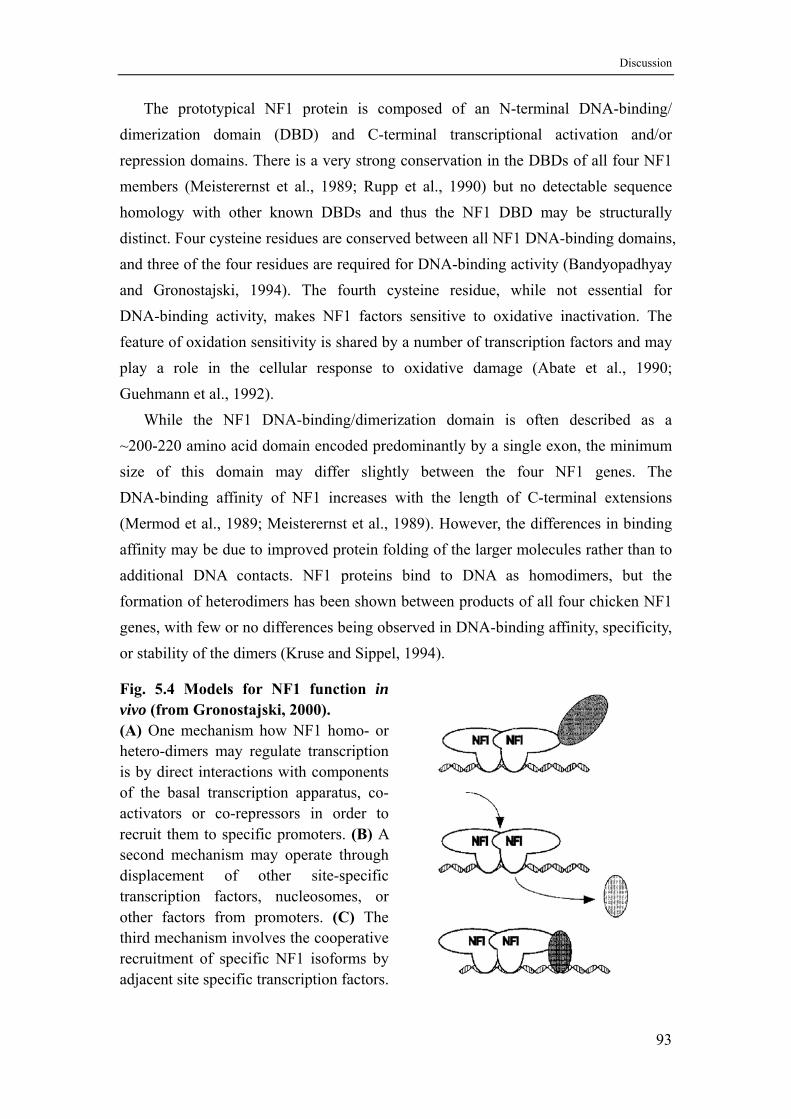
Discussion
The prototypical NF1 protein is composed of an N-terminal DNA-binding/ dimerization domain (DBD) and C-terminal transcriptional activation and/or repression domains. There is a very strong conservation in the DBDs of all four NF1 members (Meisterernst et al., 1989; Rupp et al., 1990) but no detectable sequence homology with other known DBDs and thus the NF1 DBD may be structurally distinct. Four cysteine residues are conserved between all NF1 DNA-binding domains, and three of the four residues are required for DNA-binding activity (Bandyopadhyay and Gronostajski, 1994). The fourth cysteine residue, while not essential for DNA-binding activity, makes NF1 factors sensitive to oxidative inactivation. The feature of oxidation sensitivity is shared by a number of transcription factors and may play a role in the cellular response to oxidative damage (Abate et al., 1990; Guehmann et al., 1992).
While the NF1 DNA-binding/dimerization domain is often described as a ~200-220 amino acid domain encoded predominantly by a single exon, the minimum size of this domain may differ slightly between the four NF1 genes. The DNA-binding affinity of NF1 increases with the length of C-terminal extensions (Mermod et al., 1989; Meisterernst et al., 1989). However, the differences in binding affinity may be due to improved protein folding of the larger molecules rather than to additional DNA contacts. NF1 proteins bind to DNA as homodimers, but the formation of heterodimers has been shown between products of all four chicken NF1 genes, with few or no differences being observed in DNA-binding affinity, specificity, or stability of the dimers (Kruse and Sippel, 1994).
Fig. 5.4 Models for NF1 function in vivo (from Gronostajski, 2000). (A) One mechanism how NF1 homo- or hetero-dimers may regulate transcription is by direct interactions with components of the basal transcription apparatus, co- activators or co-repressors in order to recruit them to specific promoters. (B) A second mechanism may operate through displacement of other site-specific transcription factors, nucleosomes, or other factors from promoters. (C) The third mechanism involves the cooperative recruitment of specific NF1 isoforms by adjacent site specific transcription factors.
93
Discussion
NF1 factors activate transcription through multiple mechanisms as shown in Fig. 5.3 (Gronostajski, 2000). The best-studied mechanism involves a direct interaction with basal transcription factors. The largest isoform, NF1-C, has a C-terminal ~100 amino acid proline rich domain (Mermod et al., 1989) that contains a single copy of the heptapeptide repeat occurring in the C-terminal domain of RNA polmerase II (CTD) (Meisterernst et al., 1989). When linked to heterologous DNA-binding domains this 100 residue proline-rich domain stimulates transcription five to ten fold in mammalian cells (Mermod et al., 1989; Seipel et al., 1992) whereas its deletion drastically reduces transactivation (Chaudhry et al., 1998). This domain has been shown to interact in vitro with human TFIIB (Kim and Roeder, 1994) and yeast TBP (Xiao et al., 1994). Deletion of the CTD repeat abolishes both the interaction with TFIIB and transactivation in yeast. C-terminal regions of NF1-C outside this proline-rich domain increase transcription approximately three fold, suggesting that the two regions cooperate in activating transcription (Mermod et al., 1989). The other three NF1 genes also encode proteins with proline rich C-termini, but none of them contains a perfect match to the CTD repeat. Thus it is unclear whether the mechanism of activation is the same for all human NF1 members.
A second mechanism proposes the displacement of repressive histones, either by direct competition for DNA binding or by interactions of histones with the proline rich transactivation domain of NF1-C. Such a mechanism may be of particular importance for promoters known to contain positioned nucleosomes, such as the mouse mammary tumor virus (MMTV) promoter (Chaudhry et al., 1999). It is also likely that specific interactions between NF1 and various coactivator proteins play a role in transcriptional activation. One specific coactivator interacting with the proline-rich domain is TAFII55, which also interacts with a number of other transcription factors including Sp1, YY1, USF and HIV TAT (Chiang and Roeder, 1995). Interactions of NF1 with other factors such as pirin (Wendler et al., 1997) and Ski protein (Tarapore et al., 1997) have also been observed.
NF1 proteins and their cognate binding sites have been shown to affect the expression of genes regulated by a number of signal transduction pathways, including those controlled by insulin (Cooke and Lane, 1999), camp (Chu et al., 1991), and steroid hormones (Garlatti et al., 1996; Chaudhry et al., 1999). It is difficult to determine whether the effects of some hormones/growth factors on NF1 proteins are direct or indirect, since the expression of NF1 proteins can be affected by the growth and differentiation state of the cell. In case of the MMTV promoter the response to
94
Discussion
steroid hormones is markedly decreased by mutation of a NF1 binding site immediately downstream of a hormone response element, suggesting a functional synergism between receptor and NF1 (Chavez and Beato, 1997). The DNA binding domain of NF1 is already sufficient for enabling such a synergism with the glucocorticoid receptor which led to the model of NF1 being an architectural factor that keeps the chromatin open as soon as chromatin remodelling preceding transcription has been initiated by steroid hormone receptors (Di Croce et al., 1999).
5.8 Sp family factors confer androgen regulation of the SCGB 2A1 gene
DNase I footprinting of the –382/+50 SCGB 2A1 promoter fragment also revealed a weak footprint immediately upstream of the TATA box. The borders could not be unequivocally determined, but its main characteristic is the absence of a block of pyrimidines at around position –60 in the lower strand and a hypersensitive thymidine (-21) within the TATA box in the upper strand. These features are only observed with nuclear extracts from DHT treated or untreated LNCaP cells but not or significantly weaker with nuclear extract from HeLa cells (see chapter 4.5).
Within these limits the “androgen response element”, meaning the element that is responsible for induction of the gene by androgens, could be identified between –59 and –28 as the dim-IR-GC box. When the dim-IR-GC box was placed in front of the minimal thymidine kinase promoter, consisting of a TATA box and one GC box, transcription of a reporter gene could be induced by the androgen DHT but not at all by the glucocorticoid dexamethasone (see chapter 4.4). But if the “androgen response element” of the SCGB 2 A1 promoter would represent or contain a binding site for the androgen receptor, responsiveness towards glucocorticoids has to be expected because a typical steroid hormone response element for androgens is also responsive for glucocorticoids, progestins and mineralocortiocids (Cleutjens et al., 1997; Beato and Klug, 2000; Ham et al., 1988).
So far no androgen receptor binding site has been described that is completely unresponsive towards glucocorticoids. A few elements show selective but not specific induction by the androgen receptor when compared to the glucocorticoid receptor. Among them are the two AREs in the rat probasin promoter as well as the functional intronic ARE in the rat C3(1) gene. All three elements still show 1.6- to 7-fold residual induction by glucocorticoids in transfection experiments (Claessens et al., 2001). These androgen-selective elements can be viewed as a direct repeat of the
5′-TGTTCT-3′ half site with three intervening nucleotides. Therefore, Claessens et al.
95
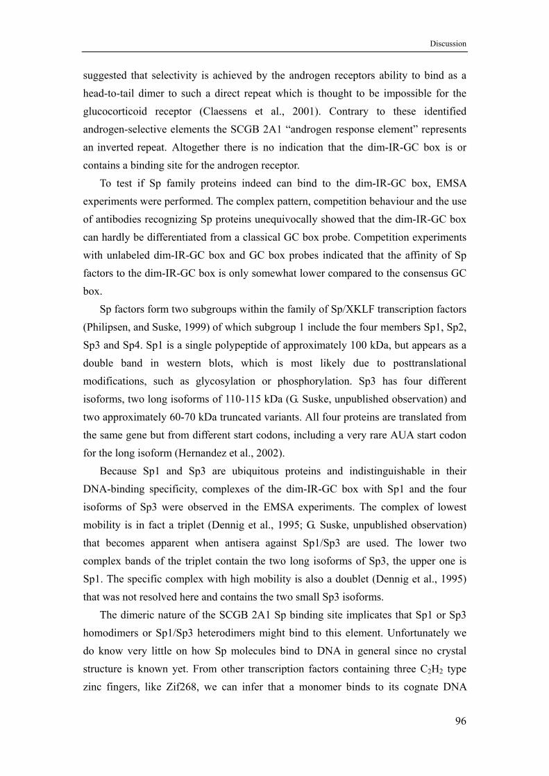
Discussion
suggested that selectivity is achieved by the androgen receptors ability to bind as a head-to-tail dimer to such a direct repeat which is thought to be impossible for the glucocorticoid receptor (Claessens et al., 2001). Contrary to these identified androgen-selective elements the SCGB 2A1 “androgen response element” represents an inverted repeat. Altogether there is no indication that the dim-IR-GC box is or contains a binding site for the androgen receptor.
To test if Sp family proteins indeed can bind to the dim-IR-GC box, EMSA experiments were performed. The complex pattern, competition behaviour and the use of antibodies recognizing Sp proteins unequivocally showed that the dim-IR-GC box can hardly be differentiated from a classical GC box probe. Competition experiments with unlabeled dim-IR-GC box and GC box probes indicated that the affinity of Sp factors to the dim-IR-GC box is only somewhat lower compared to the consensus GC box.
Sp factors form two subgroups within the family of Sp/XKLF transcription factors (Philipsen, and Suske, 1999) of which subgroup 1 include the four members Sp1, Sp2, Sp3 and Sp4. Sp1 is a single polypeptide of approximately 100 kDa, but appears as a double band in western blots, which is most likely due to posttranslational modifications, such as glycosylation or phosphorylation. Sp3 has four different isoforms, two long isoforms of 110-115 kDa (G. Suske, unpublished observation) and two approximately 60-70 kDa truncated variants. All four proteins are translated from the same gene but from different start codons, including a very rare AUA start codon for the long isoform (Hernandez et al., 2002).
Because Sp1 and Sp3 are ubiquitous proteins and indistinguishable in their DNA-binding specificity, complexes of the dim-IR-GC box with Sp1 and the four isoforms of Sp3 were observed in the EMSA experiments. The complex of lowest mobility is in fact a triplet (Dennig et al., 1995; G. Suske, unpublished observation) that becomes apparent when antisera against Sp1/Sp3 are used. The lower two complex bands of the triplet contain the two long isoforms of Sp3, the upper one is Sp1. The specific complex with high mobility is also a doublet (Dennig et al., 1995) that was not resolved here and contains the two small Sp3 isoforms.
The dimeric nature of the SCGB 2A1 Sp binding site implicates that Sp1 or Sp3 homodimers or Sp1/Sp3 heterodimers might bind to this element. Unfortunately we do know very little on how Sp molecules bind to DNA in general since no crystal structure is known yet. From other transcription factors containing three C2H2 type zinc fingers, like Zif268, we can infer that a monomer binds to its cognate DNA
96
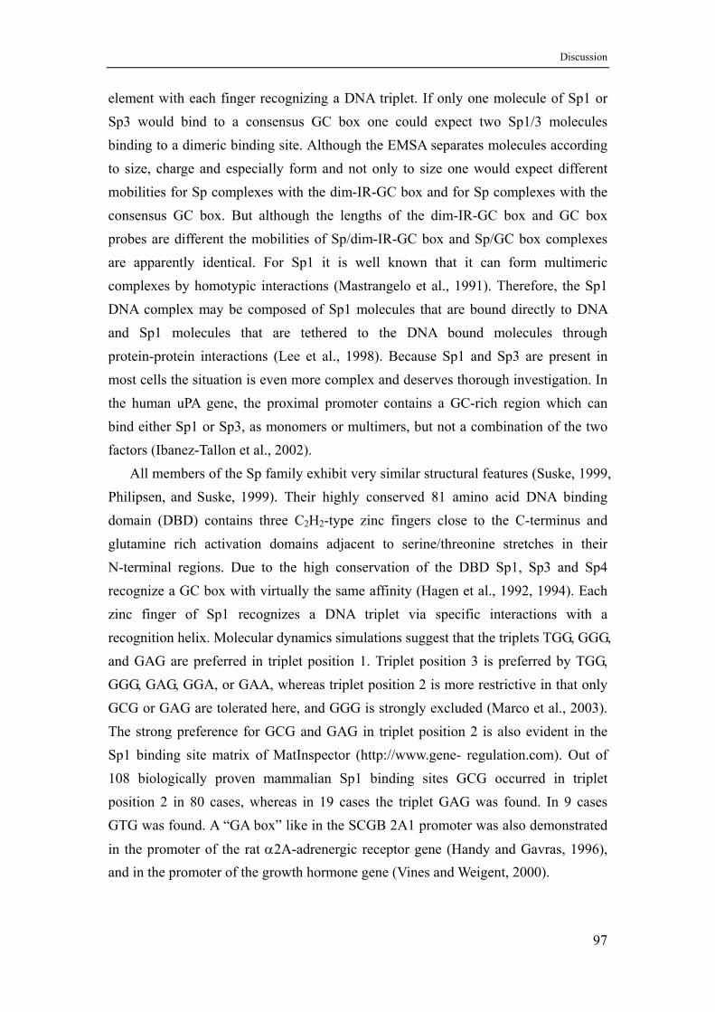
Discussion
element with each finger recognizing a DNA triplet. If only one molecule of Sp1 or Sp3 would bind to a consensus GC box one could expect two Sp1/3 molecules binding to a dimeric binding site. Although the EMSA separates molecules according to size, charge and especially form and not only to size one would expect different mobilities for Sp complexes with the dim-IR-GC box and for Sp complexes with the consensus GC box. But although the lengths of the dim-IR-GC box and GC box probes are different the mobilities of Sp/dim-IR-GC box and Sp/GC box complexes are apparently identical. For Sp1 it is well known that it can form multimeric complexes by homotypic interactions (Mastrangelo et al., 1991). Therefore, the Sp1 DNA complex may be composed of Sp1 molecules that are bound directly to DNA and Sp1 molecules that are tethered to the DNA bound molecules through protein-protein interactions (Lee et al., 1998). Because Sp1 and Sp3 are present in most cells the situation is even more complex and deserves thorough investigation. In the human uPA gene, the proximal promoter contains a GC-rich region which can bind either Sp1 or Sp3, as monomers or multimers, but not a combination of the two factors (Ibanez-Tallon et al., 2002).
All members of the Sp family exhibit very similar structural features (Suske, 1999, Philipsen, and Suske, 1999). Their highly conserved 81 amino acid DNA binding domain (DBD) contains three C2H2-type zinc fingers close to the C-terminus and glutamine rich activation domains adjacent to serine/threonine stretches in their N-terminal regions. Due to the high conservation of the DBD Sp1, Sp3 and Sp4 recognize a GC box with virtually the same affinity (Hagen et al., 1992, 1994). Each zinc finger of Sp1 recognizes a DNA triplet via specific interactions with a recognition helix. Molecular dynamics simulations suggest that the triplets TGG, GGG, and GAG are preferred in triplet position 1. Triplet position 3 is preferred by TGG, GGG, GAG, GGA, or GAA, whereas triplet position 2 is more restrictive in that only GCG or GAG are tolerated here, and GGG is strongly excluded (Marco et al., 2003). The strong preference for GCG and GAG in triplet position 2 is also evident in the Sp1 binding site matrix of MatInspector (http://www.gene- regulation.com). Out of 108 biologically proven mammalian Sp1 binding sites GCG occurred in triplet position 2 in 80 cases, whereas in 19 cases the triplet GAG was found. In 9 cases GTG was found. A “GA box” like in the SCGB 2A1 promoter was also demonstrated
in the promoter of the rat α2A-adrenergic receptor gene (Handy and Gavras, 1996), and in the promoter of the growth hormone gene (Vines and Weigent, 2000).
97
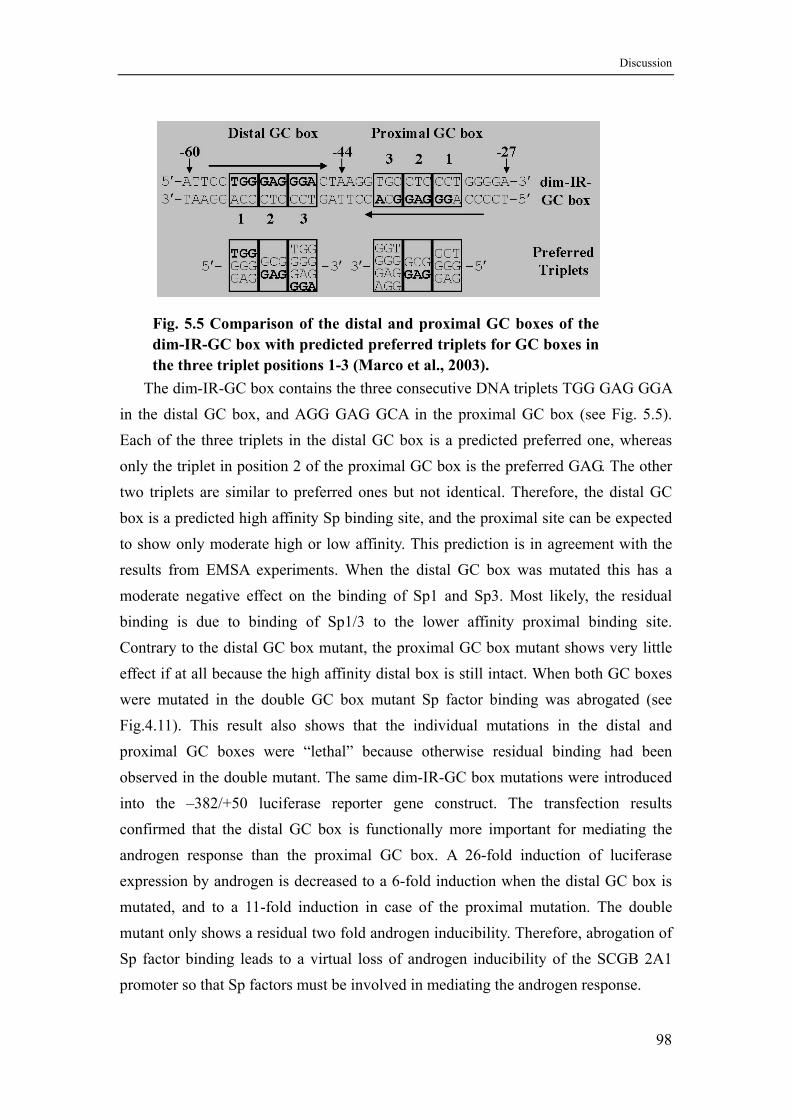
Discussion
Fig. 5.5 Comparison of the distal and proximal GC boxes of the dim-IR-GC box with predicted preferred triplets for GC boxes in the three triplet positions 1-3 (Marco et al., 2003).
The dim-IR-GC box contains the three consecutive DNA triplets TGG GAG GGA in the distal GC box, and AGG GAG GCA in the proximal GC box (see Fig. 5.5). Each of the three triplets in the distal GC box is a predicted preferred one, whereas only the triplet in position 2 of the proximal GC box is the preferred GAG. The other two triplets are similar to preferred ones but not identical. Therefore, the distal GC box is a predicted high affinity Sp binding site, and the proximal site can be expected to show only moderate high or low affinity. This prediction is in agreement with the results from EMSA experiments. When the distal GC box was mutated this has a moderate negative effect on the binding of Sp1 and Sp3. Most likely, the residual binding is due to binding of Sp1/3 to the lower affinity proximal binding site. Contrary to the distal GC box mutant, the proximal GC box mutant shows very little effect if at all because the high affinity distal box is still intact. When both GC boxes were mutated in the double GC box mutant Sp factor binding was abrogated (see Fig.4.11). This result also shows that the individual mutations in the distal and proximal GC boxes were “lethal” because otherwise residual binding had been observed in the double mutant. The same dim-IR-GC box mutations were introduced into the –382/+50 luciferase reporter gene construct. The transfection results confirmed that the distal GC box is functionally more important for mediating the androgen response than the proximal GC box. A 26-fold induction of luciferase expression by androgen is decreased to a 6-fold induction when the distal GC box is mutated, and to a 11-fold induction in case of the proximal mutation. The double mutant only shows a residual two fold androgen inducibility. Therefore, abrogation of Sp factor binding leads to a virtual loss of androgen inducibility of the SCGB 2A1 promoter so that Sp factors must be involved in mediating the androgen response.
98

Discussion
5.9 How can Sp 1/3 indirectly mediate androgen induction of the SCGB 2A1 gene?
Several recent studies have shown that some hormone-responsive genes either do not contain a corresponding hormone responsive element within their promoters or only a non-fuctional half site. Instead the hormone response is mediated via binding sites for Sp family transcription factors. For example, in the cathepsin D gene a composite GC-box/half site ERE element is required for estrogen induction (Krishnan et al., 1994). If the GC-box part is mutated and binding of Sp factors thus abrogated, estrogen induction is lost. A similar element responsible for estrogen induction was identified in the gene encoding heat shock protein 27 (Porter et al., 1996). Mutation of the ERE half site did not affect estrogen induction in transfection experiments. Moreover, Sp1 and ER were shown to physically interact, and the DNA binding domain of the ER is not required for the functional synergism of both factors (Porter et al., 1997). In both cases a ternary complex of Sp1, ER and the GC-box/half site ERE probe have been shown in electrophoretic mobility shift assays. Likewise
estrogen induction of the retinoic acid receptor α1 gene is functionally dependent on the integrity of two Sp binding sites (Sun et al., 1998). Again hormone induction is dependent on the presence of the estrogen receptor, but its DNA binding domain is not required. Another example is the induction of ubiquitin (UbC) expression by glucocorticoids in muscle cells mediated by Sp1 and MEK1, neither the rat nor human UbC promoter sequences contain a consensus glucocorticoid-response element (Marinovic, et al., 2002). One of the functionally required GC boxes in the UbC promoter is also located immediately upstream of the TATA box like the dim-IR-GC box element in the SCGB 2A1 gene.
In some other HRE-containing genes the hormone response is modulated by Sp factors. In the promoter of the rabbit uteroglobin gene, for example, an adjacent Sp binding site is an essential part of a composite estrogen-responsive unit. The estrogen induced recruitment of Sp1, but not of the related transcription factor Sp3, mediates estrogen activation (Scholz et al., 1998). The mouse vas deferens protein (MVDP) promoter is androgen responsive and contains an ARE. Transient transfection assays revealed that site-directed mutations in the NF1 and Sp1 binding elements strongly reduced (NF1) or abolished (Sp1) androgen induced expression (Darne et al., 1997).
A physical interaction of AR and Sp factors would be a straightforward explanation for the involvement of Sp factors in mediating a hormone response, either through a genomic route with the help of an HRE or non-genomic via protein-protein
99
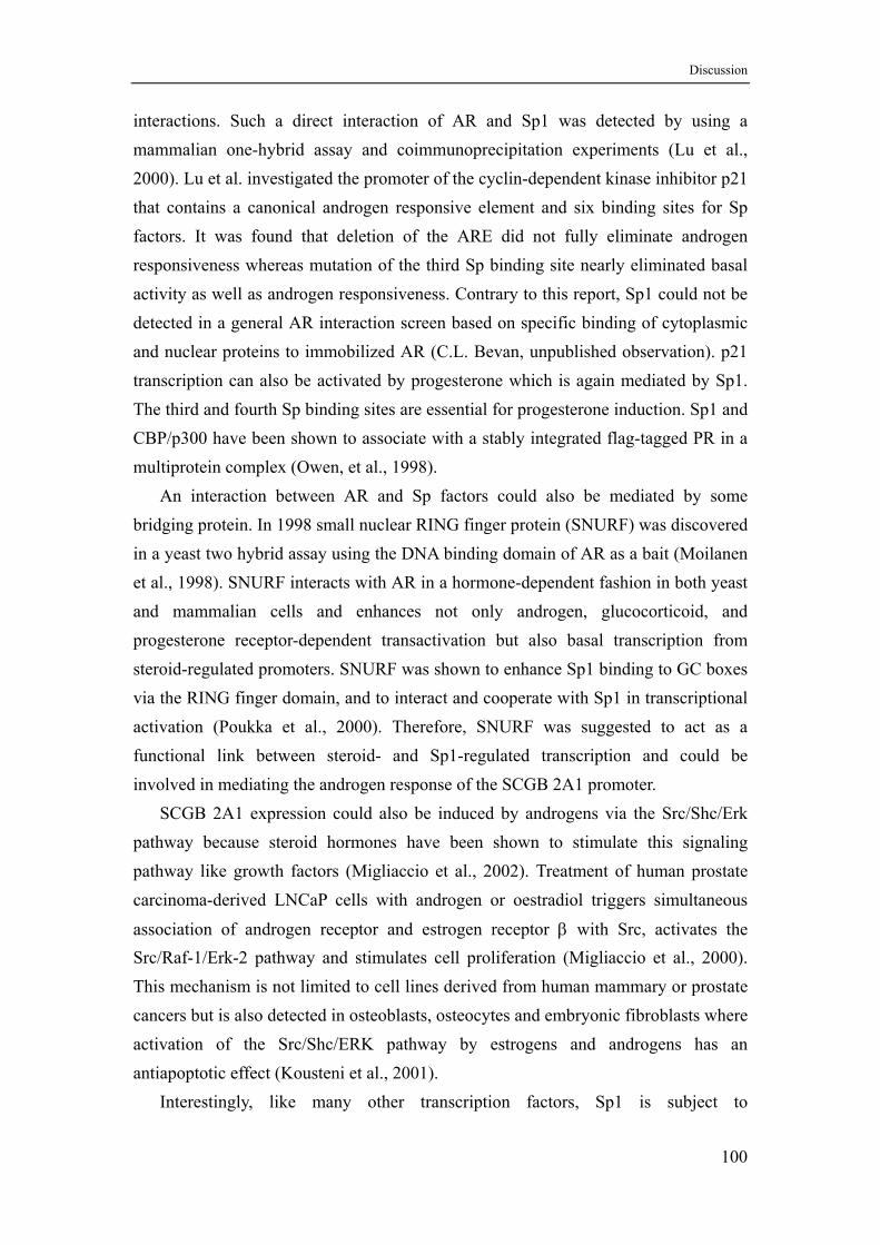
Discussion
interactions. Such a direct interaction of AR and Sp1 was detected by using a mammalian one-hybrid assay and coimmunoprecipitation experiments (Lu et al., 2000). Lu et al. investigated the promoter of the cyclin-dependent kinase inhibitor p21 that contains a canonical androgen responsive element and six binding sites for Sp factors. It was found that deletion of the ARE did not fully eliminate androgen responsiveness whereas mutation of the third Sp binding site nearly eliminated basal activity as well as androgen responsiveness. Contrary to this report, Sp1 could not be detected in a general AR interaction screen based on specific binding of cytoplasmic and nuclear proteins to immobilized AR (C.L. Bevan, unpublished observation). p21 transcription can also be activated by progesterone which is again mediated by Sp1. The third and fourth Sp binding sites are essential for progesterone induction. Sp1 and CBP/p300 have been shown to associate with a stably integrated flag-tagged PR in a multiprotein complex (Owen, et al., 1998).
An interaction between AR and Sp factors could also be mediated by some bridging protein. In 1998 small nuclear RING finger protein (SNURF) was discovered in a yeast two hybrid assay using the DNA binding domain of AR as a bait (Moilanen et al., 1998). SNURF interacts with AR in a hormone-dependent fashion in both yeast and mammalian cells and enhances not only androgen, glucocorticoid, and progesterone receptor-dependent transactivation but also basal transcription from steroid-regulated promoters. SNURF was shown to enhance Sp1 binding to GC boxes via the RING finger domain, and to interact and cooperate with Sp1 in transcriptional activation (Poukka et al., 2000). Therefore, SNURF was suggested to act as a functional link between steroid- and Sp1-regulated transcription and could be involved in mediating the androgen response of the SCGB 2A1 promoter.
SCGB 2A1 expression could also be induced by androgens via the Src/Shc/Erk pathway because steroid hormones have been shown to stimulate this signaling pathway like growth factors (Migliaccio et al., 2002). Treatment of human prostate carcinoma-derived LNCaP cells with androgen or oestradiol triggers simultaneous
association of androgen receptor and estrogen receptor β with Src, activates the Src/Raf-1/Erk-2 pathway and stimulates cell proliferation (Migliaccio et al., 2000). This mechanism is not limited to cell lines derived from human mammary or prostate cancers but is also detected in osteoblasts, osteocytes and embryonic fibroblasts where activation of the Src/Shc/ERK pathway by estrogens and androgens has an antiapoptotic effect (Kousteni et al., 2001).
Interestingly, like many other transcription factors, Sp1 is subject to
100
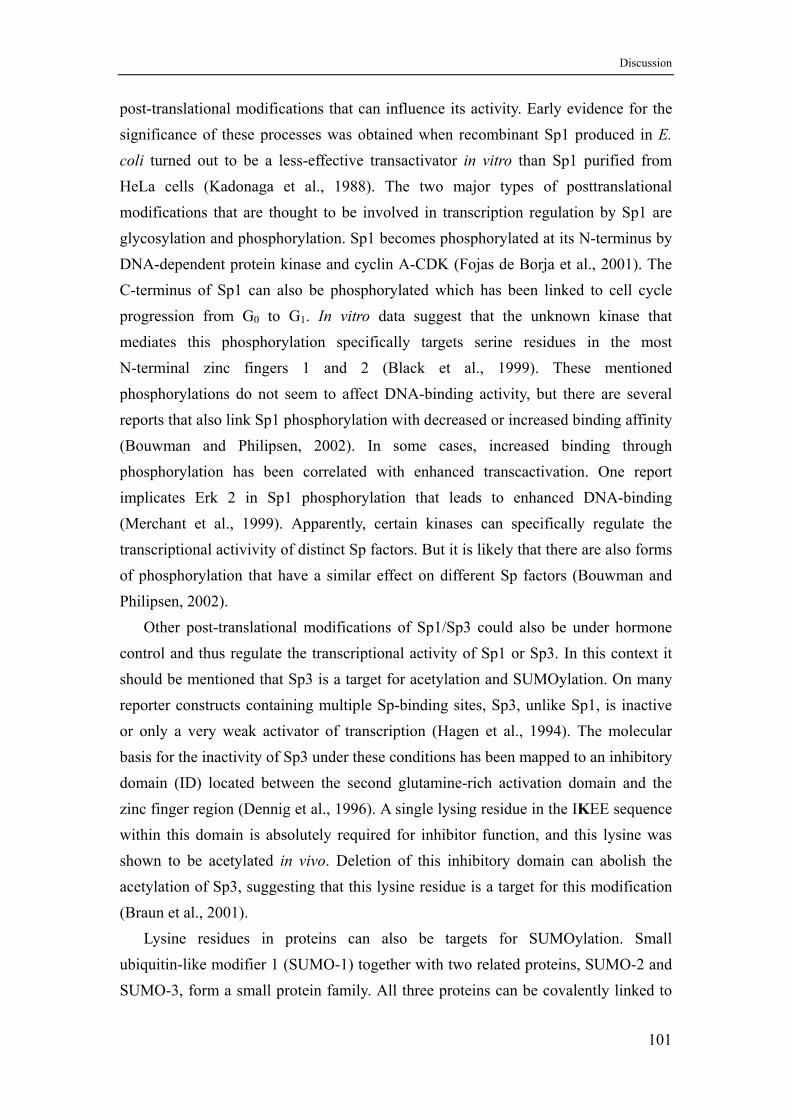
Discussion
post-translational modifications that can influence its activity. Early evidence for the significance of these processes was obtained when recombinant Sp1 produced in E. coli turned out to be a less-effective transactivator in vitro than Sp1 purified from HeLa cells (Kadonaga et al., 1988). The two major types of posttranslational modifications that are thought to be involved in transcription regulation by Sp1 are glycosylation and phosphorylation. Sp1 becomes phosphorylated at its N-terminus by DNA-dependent protein kinase and cyclin A-CDK (Fojas de Borja et al., 2001). The C-terminus of Sp1 can also be phosphorylated which has been linked to cell cycle progression from G0 to G1. In vitro data suggest that the unknown kinase that mediates this phosphorylation specifically targets serine residues in the most N-terminal zinc fingers 1 and 2 (Black et al., 1999). These mentioned phosphorylations do not seem to affect DNA-binding activity, but there are several reports that also link Sp1 phosphorylation with decreased or increased binding affinity (Bouwman and Philipsen, 2002). In some cases, increased binding through phosphorylation has been correlated with enhanced transcactivation. One report implicates Erk 2 in Sp1 phosphorylation that leads to enhanced DNA-binding (Merchant et al., 1999). Apparently, certain kinases can specifically regulate the transcriptional activivity of distinct Sp factors. But it is likely that there are also forms of phosphorylation that have a similar effect on different Sp factors (Bouwman and Philipsen, 2002).
Other post-translational modifications of Sp1/Sp3 could also be under hormone control and thus regulate the transcriptional activity of Sp1 or Sp3. In this context it should be mentioned that Sp3 is a target for acetylation and SUMOylation. On many reporter constructs containing multiple Sp-binding sites, Sp3, unlike Sp1, is inactive or only a very weak activator of transcription (Hagen et al., 1994). The molecular basis for the inactivity of Sp3 under these conditions has been mapped to an inhibitory domain (ID) located between the second glutamine-rich activation domain and the zinc finger region (Dennig et al., 1996). A single lysing residue in the IKEE sequence within this domain is absolutely required for inhibitor function, and this lysine was shown to be acetylated in vivo. Deletion of this inhibitory domain can abolish the acetylation of Sp3, suggesting that this lysine residue is a target for this modification (Braun et al., 2001).
Lysine residues in proteins can also be targets for SUMOylation. Small ubiquitin-like modifier 1 (SUMO-1) together with two related proteins, SUMO-2 and SUMO-3, form a small protein family. All three proteins can be covalently linked to
101
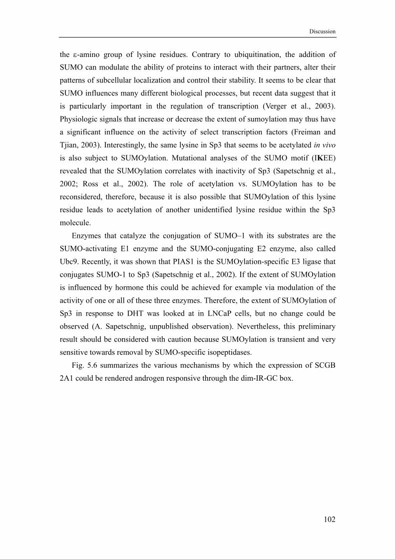
Discussion
the ε-amino group of lysine residues. Contrary to ubiquitination, the addition of SUMO can modulate the ability of proteins to interact with their partners, alter their patterns of subcellular localization and control their stability. It seems to be clear that SUMO influences many different biological processes, but recent data suggest that it is particularly important in the regulation of transcription (Verger et al., 2003). Physiologic signals that increase or decrease the extent of sumoylation may thus have a significant influence on the activity of select transcription factors (Freiman and Tjian, 2003). Interestingly, the same lysine in Sp3 that seems to be acetylated in vivo is also subject to SUMOylation. Mutational analyses of the SUMO motif (IKEE) revealed that the SUMOylation correlates with inactivity of Sp3 (Sapetschnig et al., 2002; Ross et al., 2002). The role of acetylation vs. SUMOylation has to be reconsidered, therefore, because it is also possible that SUMOylation of this lysine residue leads to acetylation of another unidentified lysine residue within the Sp3 molecule.
Enzymes that catalyze the conjugation of SUMO–1 with its substrates are the SUMO-activating E1 enzyme and the SUMO-conjugating E2 enzyme, also called Ubc9. Recently, it was shown that PIAS1 is the SUMOylation-specific E3 ligase that conjugates SUMO-1 to Sp3 (Sapetschnig et al., 2002). If the extent of SUMOylation is influenced by hormone this could be achieved for example via modulation of the activity of one or all of these three enzymes. Therefore, the extent of SUMOylation of Sp3 in response to DHT was looked at in LNCaP cells, but no change could be observed (A. Sapetschnig, unpublished observation). Nevertheless, this preliminary result should be considered with caution because SUMOylation is transient and very sensitive towards removal by SUMO-specific isopeptidases.
Fig. 5.6 summarizes the various mechanisms by which the expression of SCGB 2A1 could be rendered androgen responsive through the dim-IR-GC box.
102
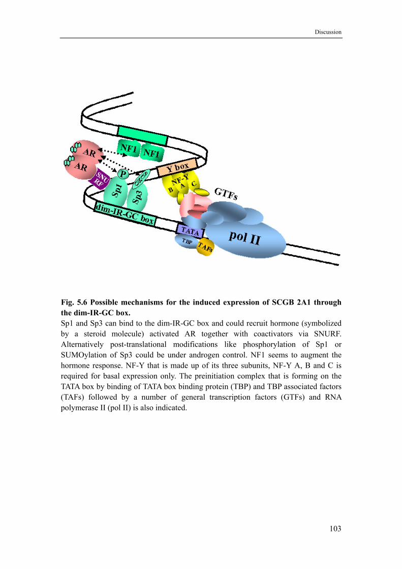
Discussion
Fig. 5.6 Possible mechanisms for the induced expression of SCGB 2A1 through the dim-IR-GC box. Sp1 and Sp3 can bind to the dim-IR-GC box and could recruit hormone (symbolized by a steroid molecule) activated AR together with coactivators via SNURF. Alternatively post-translational modifications like phosphorylation of Sp1 or SUMOylation of Sp3 could be under androgen control. NF1 seems to augment the hormone response. NF-Y that is made up of its three subunits, NF-Y A, B and C is required for basal expression only. The preinitiation complex that is forming on the TATA box by binding of TATA box binding protein (TBP) and TBP associated factors (TAFs) followed by a number of general transcription factors (GTFs) and RNA polymerase II (pol II) is also indicated.
103
Summary
6. Summary
The secretoglobins belong to a minority of proteins with unclear physiological function. In the past research in the field was dominated by work on the family founder member uteroglobin and its orthologs. This was mainly due to the fact that some 20 to 30 years ago uteroglobin served as a first model for a steroid hormone binding protein, as the steroid hormone receptors had not been cloned yet. Later on it served as a model gene to study gene regulation by steroid hormone receptors. Many new members of the secretoglobin family were identified in the past few years but remained poorly investigated in terms of gene regulation.
Studying one of the new family members was thought to be promising in finding new clues concerning physiological function of the secretoglobins in general. Many family members are expressed in the genital tract of males and females where they might play a role in reproduction. One of the new members, lipophilin C (= lacryglobin, mammaglobin B, secretoglobin SCGB 2A1) was first shown to be expressed by the lacrimal glands in the form of a heterodimer with lipophilin A, another secretoglobin that is present in tears. Due to functional similarities of lacrimal and prostate glands it was not surprising to find SCGB 2A1 also being expressed in the prostate. Because expression was found to be under androgen control, gene regulation was investigated using the androgen responsive human prostate cancer cell line LNCaP. Relevant regulatory regions were identified in the genes chromatin by mapping DNase I hypersensitive sites before and after androgen induction. Only one prominent hypersensitive site appeared in the proximal promoter after 6 hours of androgen induction indicating an indirect response. Sequence analysis did not reveal any canonical or non-canonical androgen response element(s). DNase I footprinting identified two factors binding to the proximal promoter region that were found to be the ubiquitous transcription factors nuclear factor I (NF1) and CAAT-box transcription factor NF-Y. Analysis of promoter deletions using luciferase reporter constructs showed that 136 bp of the promoter still containing the NF-Y binding site are sufficient to confer androgen responsiveness to the reporter gene or the heterogenous HSV-tk promoter. Analyses of promoter constructs with mutations in the NF1 and NF-Y binding sites demonstrated that both factors significantly contribute to the basal activity, and that NF1 helps to mediate the androgen response. Eventually androgen responsiveness could be traced down to a dimeric inverted repeat GC box immediately upstream of the TATA box that is a binding site for Sp family transcription factors.
104
Summary
Therefore, androgen responsiveness in the SCGB 2A1 gene is mediated by an indirect mechanism that requires the androgen receptor but operates through a binding site for ubiquitous transcription factors of the Sp family, notably the well known factors Sp1 and Sp3. This response is androgen-specific because glucocorticoids in conjunction with the cotransfected glucocorticoid receptor were not able to activate SCGB 2A1 expression in LNCaP cells. Thus, SCGB 2A1 was unexpectedly identified as a target gene for a non-ARE mediated action of the androgen receptor. Future investigations will focus on the exact mechanism of this indirect hormone response.
105
References
7. References
Aasland, R., Stewart, A. F., 1999. Analysis of DNaseI hypersensitive sites in chromatin by cleavage in permeabilized cells. Methods Mol. Biol. 119, 355-362.
Abate, C., Patel, L., Rauscher, F.J.I., Curran, T., 1990. Redox regulation of Fos and Jun DNA-binding activity in vitro. Science 249, 1157-1161.
Abate-Shen, C., Shen, M.M., 2000. Molecular genetics of prostate cancer. Genes Dev. 14, 2410-2434.
Adams, J.Y., Leav, I., Lau, K.M., Ho, S.M., Pflueger, S. M., 2002. Expression of estrogen receptor beta in the fetal, neonatal, and prepubertal human prostate. Prostate 52, 69-81.
Adler, A.J., Scheller, A., Hoffman, Y., Robins, D.M., 1991. Multiple components of a complex androgen-dependent enhancer. Mol. Endocrinol. 5, 1587-96.
Adom, J., Carr, K. D., Gouilleux, F., Marsaud, V., Richard-Foy, H., 1991. Chromatin structure of hormono-dependent promoters. J. Steroid Biochem. Mol. Biol. 40, 325-332.
Andrews, N.C., Faller, D.V., 1991. A rapid micropreparation technique for extraction of DNA-binding proteins from limiting numbers of mammalian cells. Nucleic Acids Res. 19, 2499.
Antequera, F., Bird, A., 1993. CpG islands. DNA Methylation: Molecular Biology and Biological Significance. Birkhäuser Verlag, pp. 169-185.
Aronica, S.M., Kraus, W.L., Katzenellenbogen, B.S., 1994. Estrogen action via the cAMP signalling pathway - stimulation of adenylate cyclase and cAMP regulated gene transcription. Proc. Nat. Acad. Sci. USA 91, 8517-8521.
Aumüller, G., Leonhardt, M., Janssen, M., Konrad, L., Bjartell, A., Abrahamsson, P.A., 1999. Neurogenic origin of human prostate endocrine cells. Urology 53, 1041-1048.
Aumüller, G., Leonhardt, M., Renneberg, H., von Rahden, B., Bjartell, A., Abrahamsson, P.A., 2001. Semiquantitative morphology of human prostatic development and regional distribution of prostatic neuroendocrine cells. Prostate 46, 108-115.
Baldi, E., Krausz, C., Luconi, M., Bonaccorsi, L., Maggi, M., Forti, G., 1995. Actions of progesterone on human sperm - a model of non-genomic effects of steroids. J. Steroid Biochem. Mol. Biol. 53, 199-203.
Ballare, C., Uhrig, M., Bechtold, T., Sancho, E., Di Domenico, M., Migliaccio, A., Auricchio, F., Beato, M., 2003. Two domains of the progesterone receptor interact with the estrogen receptor and are required for progesterone activation of the c-Src/Erk pathway in mammalian cells. Mol. Cell. Biol. 23, 1994-2008.
106
References
Bandyopadhyay, S., Gronostajski, R.M., 1994. Identification of a conserved oxidation-sensitive cysteine residue in the NFI family of DNA-binding proteins. J. Biol. Chem. 269, 29 949-29 955.
Barnes, H.J., Nordlund-Möller, L., Nord, M., Gustafsson, J.Å., Lund, J., Gillner, M., 1996. Structural basis for calcium binding by uteroglobins. J. Mol. Biol. 256, 392-404.
Beato, M., 1989. Gene regulation by steroid hormones. Cell 56, 335-344.
Beato, M., Eisfeld, K., 1997. Transcription factor access to chromatin. Nucleic Acids Res. 25, 3559-3563.
Beato, M., Herrlich, P., Schütz, G., 1995. Steroid hormone receptors: many actors in search of a plot. Cell 83, 851-857.
Beato, M., Klug, J., 2000. Steroid hormone receptors: an update. Human Reproduction Update 6, 225-236.
Beato, M., Sanchez-Pacheco, A., 1996. Interaction of steroid hormone receptors with the transcription initiation complex. Endocr. Rev. 17, 587-609.
Beaucage, S. L., Caruthers, M. H., 1981. Deoxynucleoside phoshoramidites - a new class of key intermediates for deoxypolynucleotide synthesis. Tetrahedron Lett. 22, 1859-1862.
Beier, H.M., 1968. Uteroglobin: A hormone-sensitive endometrial protein involved in blastocyst development. Biochim. Biophys. Acta. 160, 289-291.
Benten, W.P.M., Lieberherr, M., Sekeris, C.E., Wunderlich, F., 1997. Testosterone induces Ca2+ influx via non-genomic surface receptors in activated T cells. FEBS Lett. 407, 211-214.
Berg, J., 1989. DNA-binding specificity of steroid receptors. Cell 57, 1065-1068.
Bi, W., Wu, L., Coustry, F., de Crombrugghe, B., Maity, S. N., 1997. DNA binding specificity of the CCAAT-binding factor CBF/NF-Y. J. Biol. Chem. 272, 26562-26572.
Black, A. R., Jensen, D., Lin, S. Y., Azizkhan, J. C., 1999. Growth/cell cycle regulation of Sp1 phosphorylation. J. Biol. Chem. 274, 1207-1215.
Bonkhoff, H. Remberger, K., 1996. Differentiation pathways and histogenetic aspects of normal and abnormal prostatic growth: A stem cell model. Prostate 28, 98-106.
Bonkhoff, H., Stein, U., Remberger, K., 1994. Multidirectional differentiation in the normal hyperplastic and neoplastic human prostate: Simultaneous demonstration of cell-specific epithelial markers. Hum. Pathol. 25, 42-46.
Bradford, M. M., 1976. A rapid and sensitive method for the quantitation of microgram quantities of protein utilizing the principle of protein-dye binding. Anal. Biochem. 72, 248-254.
107
References
Brasier A.R., Tate J.E., Habener J.F., 1989. Optimized use of firefly luciferase assay as a reporter gene in mammalian cell lines. BioTechniques 7,1116-1122.
Braun, H., Suske, G., 1998. Combinatorial action of HNF3 and Sp family transcription factors in the activation of the rabbit uteroglobin/CC10 promoter. J. Biol. Chem. 273, 9821-9828.
Braun, H., Koop, R., Ertmer, A., Nacht, S., Suske, G., 2001. Transcription factor Sp3 is regulated by acetylation. Nucleic Acids Res. 29, 4994-5000.
Bouwman, P., Philipsen, S., 2002. Regulation of the activity of Sp1-related transcription factors. Mol. Cell. Endocrinol. 195, 27-38.
Brown, C.J., Goss, S.J., Lubahn, D.B., Joseph, D.R., Wilson, E.M., French, F.S., Willard, H.F., 1989. Androgen receptor locus on the human X chromosome: regional localization to Xq11-12 and description of a DNA polymorphism. Am. J. Hum. Genet. 44, 264-269.
Bubulya, A, Wise, S.C., Shen, X.Q., Burmeister, L.A., Shemshedini, L., 1996. c-Jun can mediate androgen receptor-induced transactivation. J. Biol. Chem. 271, 24583-24589.
Bucher, P., 1990. Weight matrix descriptions of four eukaryotic RNA polymerase II promoter elements derived from 502 unrelated promoter sequences. J. Mol. Biol. 212, 563-578.
Bui, M. Reiter, R.E., 1998. Stem cell genes in androgen-independent prostate cancer. Cancer Metastasis Rev. 17, 391-399.
Burnett W.V., 1997. Northern blotting of RNA denatured in glyoxal without buffer recirculation. BioTechniques 22, 668-671.
Cadepond, F., Schweizer-Groyer, G., Segard-Maurel, I., Jibard, N., Hollenberg, S.M., Giguere, V., Evans, R.M., Baulieu, E.E., 1991. Heat shock protein 90 as a critical factor in maintaining glucocorticosteroid receptor in a nonfunctional state. J. Biol. Chem. 226, 5834-5841.
Callebaut, I., Poupon, A., Bally, R., Demaret, J.P., Housset, D., Delettre, J., Hossenlopp, P., Mornon, J.P., 2000. The uteroglobin fold. Ann. N. Y. Acad. Sci. 923, 90-112.
Caretti, G., Motta, M. C., Mantovani, R., 1999. NF-Y associates with H3-H4 tetramers and octamers by multiple mechanisms. Mol. Cell. Biol. 19, 8591-8603.
Carter, D., Douglass, J.F., Cornellison, C.D., Retter, M.W., Johnson, J.C., Bennington, A.A., Fleming, T.P., Reed, S.G., Houghton, R.L., Diamond, D.L., Vedvick, T.S., 2002. Purification and characterization of the mammaglobin/lipophilin B complex, a promising diagnostic marker for breast cancer. Biochemistry 41, 6714-6722.
108
References
Chaudhry, A.Z., Vitullo, A.D., Gronostajski, R.M., 1998. Nuclear factor I (NFI) isoforms differentially activate simple versus complex NFI-responsive promoters. J. Biol. Chem. 273, 18 538-18 546.
Chaudhry, A.Z., Vitullo, A.D., Gronostajski, R.M., 1999. Nuclear factor I-mediated repression of the mouse mammary tumor virus promoter is abrogated by the coactivators p300/CBP and SRC-1. J. Biol. Chem. 274, 7072-7081.
Chavez, S. Beato, M., 1997. Nucleosome-mediated synergism between transcription factors on the mouse mammary tumor virus promoter. Proc. Natl. Acad. Sci. USA 94, 2885-2890.
Chen, J.D., Evans, R.M., 1995. A transcriptional co-repressor that interacts with nuclear hormone receptors. Nature 377, 454-457.
Chen, S., Wang, J., Yu, G., Liu, W., Pearce, D., 1997. Androgen and glucocorticoid receptor heterodimer formation. A possible mechanism for mutual inhibition of transcriptional activity. J. Biol. Chem. 272, 14 087-14 092.
Chiang, C.M., Roeder, R.G., 1995. Cloning of an intrinsic human TFIID subunit that interacts with multiple transcriptional activators. Science 267, 531-536.
Chodosh, L.A., Baldwin, A.S., Carthew, R.W., Sharp, P.A., 1988. Human CCAAT-binding proteins have heterologous subunits. Cell 53, 11-24.
Chodosh, L.A., Olesen, J., Hahn, S., Baldwin, A.S., Guarente, L., Sharp, P.A., 1988. A yeast and a human CCAAT-binding protein have heterologous subunits that are functionally interchangeable. Cell 53, 25-35.
Chowdhury, B., Mantile-Selvaggi, G., Miele, L., Cordella-Miele, E., Zhang, Z., Mukherjee, A.B., 2002. Lys 43 and Asp 46 in alpha-helix 3 of uteroglobin are essential for its phospholipase A(2) inhibitory activity. Biochem. Biophys. Res. Commun. 295, 877-883.
Chu, H.M., Fischer, W.H., Osborne, T.F., Comb, M.J., 1991. NF-I proteins from brain interact with the proenkephalin cAMP inducible enhancer. Nucleic Acids Res. 19, 2721-2728.
Claessens, F., Rushmere, N. K., Davies, P., Celis, L., Peeters, B., Rombauts, W. A., 1990. Sequence-specific binding of androgen-receptor complexes to prostatic binding protein genes. Mol. Cell. Endocrinol. 74, 203-212
Claessens, F., Verrijdt, G., Schoenmakers, E., Haelens, A., Peeters, B., Verhoeven, G., Rombauts, W., 2001. Selective DNA binding by the androgen receptor as a mechanism for hormone-specific gene regulation. J. Steroid Biochem. Mol. Biol. 76, 23-30.
109
References
Cleutjens, K. B., van Eekelen, C. C., van der Korput, H. A., Brinkmann, A. O., Trapman, J., 1996. Two androgen response regions cooperate in steroid hormone regulated activity of the prostate-specific antigen promoter. J. Biol. Chem. 271, 6379-6388.
Cleutjens, K. B., van der Korput, H. A., van Eekelen, C. C., van Rooij, H. C., Faber, P. W., Trapman, J., 1997. An androgen response element in a far upstream enhancer region is essential for high, androgen-regulated activity of the prostate-specific antigen promoter. Mol. Endocrinol. 11, 148-161.
Cleutjens, C. B., Steketee, K., van Eekelen, C. C., van der Korput, J. A., Brinkmann, A. O., Trapman, J., 1997. Both androgen receptor and glucocorticoid receptor are able to induce prostate-specific antigen expression, but differ in their growth- stimulating properties of LNCaP cells. Endocrinology 138, 5293-5300.
Cockerill, P.N., 2000. Identification of DNaseI hypersensitive sites within nuclei. Methods Mol. Biol. 130, 29-46.
Colpitts, T.L., Billing-Medel, P., Friedman, P., Granados, E.N., Hayden, M., Hodges, S., Menhart, N., Roberts, L., Russell, J., Stroupe, S.D., 2001. Mammaglobin is found in breast tissue as a complex with BU101. Biochemistry 40, 11048-11059.
Cooke, D.W., Lane, M.D., 1999. The transcription factor nuclear factor I mediates repression of the GLUT4 promoter by insulin. J. Biol. Chem. 274, 12 917-12 924.
Croston, G.E., Kadonaga, J.T., 1993. Role of chromatin structure in the regulation of transcription by RNA polymerase II. Curr. Opin. Cell Biol. 5, 417-423.
Culig, Z., Hobisch, A., Cronauer, M.V., Hittmair, A., Radmayr, C., Bartsch, G., Klocker, H., 1995. Activation of androgen receptor by polypeptide growth factors and cellular regulators. World J. Urol. 13, 285-289.
Darne, C.H., Morel, L., Claessens, F., Manin, M., Fabre, S., Veyssiere, G., Rombauts, W., Jean, C.L., 1997. Ubiquitous transcription factors NF1 and Sp1 are involved in the androgen activation of the mouse vas deferens protein promoter. Mol. Cell Endocrinol. 132, 13-23.
Davis, B. J., 1964. Disc Electrophoresis - II: Method and application to human serum proteins. Ann. New York Acad. Sci. 121, 404-427.
De Marzo, A.M., Nelson, W.G., Meeker, A.K., Coffey, D.S., 1998. Stem cell features of benign and malignant prostate epithelial cells. J. Urol. 160, 2381-2392.
De Wet, J. R., Wood, K. V., DeLuca, M., Helinski, D. R., Subramani, S., 1987. Firefly luciferase gene: Structure and expression in mammalian cells. Mol. Cell. Biol. 7, 725-737.
Dennig, J., Beato, M., Suske, G., 1996. An inhibitor domain in Sp3 regulates its glutamine-rich activation domains. EMBO J. 15, 5659-5667.
110
References
Dennig, J., Hagen, G., Beato, M., Suske, G., 1995. Members of the Sp transcription factor family control transcription from the uteroglobin promoter. J. Biol. Chem. 270, 12 737-12 744.
Di Croce, L., Koop, R., Venditti, P., Westphal, H.M., Nightingale, K.P., Corona, D.F., Becker, P.B., Beato, M., 1999. Two-step synergism between progesterone receptor and the DNA binding domain of nuclear factor 1 on MMTV minichromosomes. Mol. Cell 4, 45-54.
Di Sant’Agnese P, 1992. Neuroendocrine differentiation in carcinoma of the prostate. Diagnostic, prognostic and therapeutic implications. Cancer 70, 254-268.
Di Sant’Agnese P, 1995. Neuroendocrine differentiation in prostatic carcinoma. Recent findings and new concepts. Cancer 75, 1850-1859.
Dorn, A., Bollekens, J., Staub, A., Benoist, C., Mathis, D., 1987. A multiplicity of CCAAT box-binding proteins. Cell 50, 863-872.
Eisfeld, K., Candau, R., Truss, M., Beato, M., 1997. Binding of NF1 to the MMTV promoter in nucleosomes: influence of rotational phasing, translational positioning and histone H1. Nucleic Acids Res. 25, 3733-3742.
Ewing, L.L., Zirkin, B., 1983. Leydig cell structure and steroidogenic function. Recent Prog. Horm. Res. 39, 599-635.
Fanger, G.R., Houghton, R.L., Retter, M.W., Hendrickson, R.C., Babcook, J., Dillon, D.C., Durham, M.D., Reynolds, L.D., Johnson, J.C., Carter, D., Fleming, T.P., Roche, P.C., Persing, D.H., Reed, S.G., 2002. Detection of mammaglobin in the sera of patients with breast cancer. Tumour Biol. 23, 212-221.
Fischle, W., Wang, Y., Allis, C. D., 2003. Histone and chromatin cross-talk. Curr. Opin. Cell. Biol. 15, 172-183.
Fojas de Borja, P., Collins, N. K., Du, P., Azizkhan-Clifford, J., Mudryj, M., 2001. Cyclin A–CDK phosphorylates Sp1 and enhances Sp1-mediated transcription. EMBO J. 20, 5737-5747.
Frankel, S., Smith, G.D., Donovan, J., Neal, D., 2003. Screening for prostate cancer. Lancet 361, 1122-1128.
Freiman, R. N., Tjian, R., 2003. Regulating the regulators: lysine modifications make their mark. Cell 112, 11-17.
Gandini, O., Kohno, H., Curtis, S., Korach, K.S., 1997. Two transcription activation functions in the amino terminus of the mouse esterogen receptor that are affected by the carboxy terminus. Steroids 62, 508-515.
Garlatti, M., Aggerbeck, M., Bouguet, J., Barouki, R., 1996. Contribution of a nuclear factor 1 binding site to the glucocorticoid regulation of the cytosolic aspartate aminotransferase gene promoter. J. Biol. Chem. 271, 32 629-32 634.
111
References
Gelmini, S., Tricarico, C., Petrone, L., Forti, G., Amorosi, A., Dedola, G. L., Serio, M., Pazzagli, M., Orlando, C., 2003. Real-time RT-PCR for the measurement of prostate-specific antigen mRNA expression in benign hyperplasia and adenocarcinoma of prostate. Clin. Chem. Lab. Med. 41, 261-265.
Gey, G. O., Coffman, W. D., Kubicek, M. T., 1952. Tissue culture studies of the proliferative capacity of cervical carcinoma and normal epithelium. Cancer Res. 12, 264-265.
Gorczynska, E., Handelsman, D.J., 1995. Androgens rapidly increase the cytosolic calcium concentration in Sertoli cells. Endocrinology 136, 2052-2059.
Gronostajski, R.M., 2000. Roles of the NFI/CTF gene family in transcription and development. Gene 249, 31-35.
Guehmann, S., Vorbrueggen, G., Kalkbrenner, F., Moelling, K., 1992. Reduction of a conserved Cys is essential for Myb DNA-binding. Nucleic Acids Res. 20, 2279-2286.
Guiochon-Mantel, A., Lescop, P., Christinmaitre, S., 1991. Nucleocytoplasmic shuttling of the progesterone receptor. EMBO J. 10, 3851-3859.
Hagen, G., Müller, S., Beato, M., Suske, G., 1992. Cloning by recognition site screening of two novel GT box binding proteins: a family of Sp1 related genes. Nucleic Acids Res. 20, 5519-5525.
Hagen, G., Müller, S., Beato, M., Suske, G., 1994. Sp1-mediated transcriptional activation is repressed by Sp3. EMBO J. 13, 3843-3851.
Ham, J., Thomson, A., Needham, M., Webb, P., Parker, M., 1988. Characterization of response elements for androgens, glucocorticoids and progestins in mouse mammary tumour virus. Nucleic Acids Res. 16, 5263-5276.
Handy, D.E., Gavras, H., 1996. Evidence for cell-specific regulation of transcription of the rat α2A-adrenergic receptor gene. Hypertension 27, 1018-1024.
Hennig, W., 1999. Heterochromatin. Chromosoma 108, 1-9.
Hernandez, E.M., Johnson, A., Notario, V., Chen, A., Richert, J.R., 2002. AUA as a translation initiation site in vitro for the human transcription factor Sp3. J. Biochem. Mol. Biol. 35, 273-282.
Heyns, W., Peeters, B., Mous, J., Rombauts, W., De Moor, P., 1978. Purification and characterization of prostatic binding protein and its subunits. Eur. J. Biochem. 89, 181-186.
Horoszewicz, J.S., Leong, S.S., Chu, T.M., Wajsman, Z.L., Friedman, M., Papsidero, L., Kim, U., Chai, L.S., Kakati, S., Arya, S.K., Sandberg, A.A., 1980. The LNCaP cell line - a new model for studies on human prostatic carcinoma. Prog. Clin. Biol. Res. 37, 115-132.
112
References
Hörlein, A., Näär, A.M., Heinzel, T., 1995. Ligand-independent repression by the thyroid hormone receptor mediated by a nuclear receptor co-repressor. Nature 377, 397-404.
Ibanez-Tallon, I., Ferrai, C., Longobardi, E., Facetti, I., Blasi, F., Crippa, M.P., 2002. Binding of Sp1 to the proximal promoter links constitutive expression of the human uPA gene and invasive potential of PC3 cells. Blood 100, 3325-3332.
Ibarrola, I., Ogiza, K., Marino, A., Macarulla, J.M., Trueba, M., 1991. Steroid hormone specifically binds to rat kidney plasma membrane. J. Bioenerg. Biomembr. 23, 919-926.
Ikonen, T., Palvimo, JJ., Kallio, P.J., Reinikainen, P., Jänne, O.A., 1994. Stimulation of androgen regulated transactivation by modulators of protein phosphorylation. Endocrinology 135, 1359-1366.
Jenster, G., van der Korput, J.A., van Vroonhoven, C., van der Kwast, T.H., Trapman, J., Brinkmann, A.O., 1991. Domains of the human androgen receptor involved in steroid binding, transcriptional activation and subcellular localization. Mol. Endocrinol. 5, 1396-1404.
Jenster, G., Trapman, J., Brinkmann, A.O., 1993. Nuclear import of the human androgen receptor. Biochem. J. 293, 761-768.
Jia, L., Kim, J., Shen, H., Clark, P. E., Tilley, W. D., Coetzee, G. A., 2003. Androgen receptor activity at the prostate specific antigen locus: steroidal and non-steroidal mechanisms. Mol. Cancer Res. 1, 385-392.
Jones, K., Kadonaga, J., Rosenfeld, P., Kelly, T., Tjian, R., 1987. A cellular DNA-binding protein that activates eukaryotic transcription and DNA replication. Cell 48, 79-89.
Jordan M., Schallhorn A., Wurm F.W., 1996. Transfecting mammalian cells: Optimization of critical parameters affecting calcium-phosphate precipitate formation. Nucleic Acids Res. 24,596-601.
Kadonaga, J. T., Courey, A. J., Ladika, J., Tjian, R., 1988. Distinct regions of Sp1 modulate DNA binding and transcriptional activation. Science 242, 1566-1570.
Kamei, Y., Xu, L., Heinzel, T., 1996. A CBP integrator complex mediates transcriptional activation and AP-1 inhibition by nuclear receptors. Cell 85, 403-414.
Kennett, S. B., Udvadiaand, A. J., and Horowitz, J. M., 1997. Sp3 encodes multiple proteins that differ in their capacity to stimulate or repress transcription. NAR 25, 3110-3117.
Klug, J., Wolf, M., Beato, M., 1991. Creating chimeric molecules by PCRdirected homologous DNA recombination. Nucleic Acids Res. 19, 2793.
113
References
Klug, J., Beier, H.M., Bernard, A., Chilton, B.S., Fleming, T.P., Lehrer, R.I., Miele, L., Pattabiraman, N., Singh, G. 2000. Uteroglobin/Clara cell 10-kDa family of proteins: nomenclature committee report. Ann. N. Y. Acad. Sci. 923, 348-354.
Kim, E.C., Lau, J.S., Rawlings, S., Lee, A.S., 1997. Positive and negative regulation of the human thymidine kinase promoter mediated by CCAAT binding transcription factors NF-Y/CBF, dbpA, and CDP/cut. Cell Growth Differ. 8, 1329-1338.
Kim, I.S., Sinha, S., de Crombrugghe, B., Maity, S.N., 1996. Determination of functional domains in the C subunit of the CCAAT-binding factor (CBF) necessary for formation of a CBF-DNA complex: CBF-B interacts simultaneously with both the CBF-A and CBF-C subunits to form a heterotrimeric CBF molecule. Mol Cell Biol. 16, 4003-4013.
Kim, T.K., Roeder, R.G., 1994. Proline-rich activator CTF1 targets the TFIIB assembly step during transcriptional activation. Proc. Natl. Acad. Sci. USA 91, 4170-4174.
Kingsley, C., Winoto A., 1992. Cloning of GT box-binding proteins: a novel Sp1 mutigene family regulating T-cell receptor gene expression. Mol. Cell. Biol. 12, 4251-4261.
Kornberg, R.D., 1996. RNA polymerase II transcriptional control. Trends Biochem. Sci. 21, 325-326.
Kousteni, S., Bellido, T., Plotkin, L.I., O'Brien, C.A., Bodenner, D.L., Han, L., Han, K., DiGregorio, G.B., Katzenellenbogen, J.A., Katzenellenbogen, B.S., Roberson, P.K., Weinstein, R.S., Jilka, R.L., Manolagas, S.C., 2001. Nongenotropic, sex-nonspecific signaling through the estrogen or androgen receptors: dissociation from transcriptional activity. Cell 104, 719-730.
Krishnan, R.S., Daniel Jr., J.C., 1967. "Blastokinin": inducer and regulator of blastocyst development in the rabbit uterus. Science 158, 490-492
Krishnan, V., Wang, X., Safe, S., 1994. Estrogen receptor-Sp1 complexes mediate estrogen-induced cathepsin D gene expression in MCF-7 human breast cancer cells. J. Biol. Chem. 269, 15 912-15 917.
Kruse, U., Sippel, A.E., 1994. Transcription factor nuclear factor I proteins form stable homo- and heterodimers. FEBS Lett. 348, 46-50.
Kumar, A.P., Butler, A.P. 1997. Transcription factor Sp3 antagonizes activation of the ornithine decarboxylase promoter by Sp1. Nucleic Acids Res. 25, 2012-2019.
Labrie, F., Luu-The, V., Lin, S.X., Labrie, C., Simard, J., Breton, R., Belanger, A., 1997. The key role of 17β-hydroxysteroid dehydrogenases in sex steroid biology. Steroids 62, 148-158.
114
References
Laemmli, U. K., 1970. Cleavage of structural proteins during the assembly of the head of bacteriophage T4. Nature 227, 680-685.
Langley, E., Zhou, Z., Wilson, E.M., 1995. Evidence for an anti-parallel orientation of the ligand-activated human androgen receptor dimer. J. Biol. Chem. 270, 29 983-29 990.
Lee, J.S., Lee, C.H., Chung, J.H., 1998. Studying the recruitment of Sp1 to the β-globin promoter with an in vivo method: protein position identification with nuclease tail (PIN*POINT). Proc. Natl. Acad. Sci. USA 95, 969-974.
Lehrer, R.I., Xu, G., Abduragimov, A., Dinh, N.N., Qu, X.D., Martin, D., Glasgow, B.J., 1998. Lipophilin, a novel heterodimeric protein of human tears. FEBS Lett. 432, 163-167.
Lewin, B., 2000. Genes VII. Oxford University Press, Oxford.
Liang, Y., Robinson, D.F., Dennig, J., Suske, G., Fahl, W.E., 1996. Transcriptional regulation of the SIS/PDGF-B gene in human osteosarcoma cells by the Sp family of transcription factors. J. Biol. Chem. 271, 11 792- 11 797.
Liu, A.Y., True, L.D., LaTray, L., Nelson, P.S., Ellis, W.J., Vessella, R.L., Lange, P.H., Hood, L., van den Engh, G., 1997. Cell-cell interaction in prostate gene regulation and cytodifferentiation. Proc. Natl. Acad. Sci. USA 94, 10705-10710.
Linhoff, M.W., Wright, K.L., Ting, J.P., 1997. CCAAT-binding factor NF-Y and RFX are required for in vivo assembly of a nucleoprotein complex that spans 250 base pairs: the invariant chain promoter as a model. Mol. Cell. Biol. 17, 4589-96.
Lodish, H., Baltimore, D., Berk, A., Zipursky, S.L. Matsudaira, P., Darnell, J., 1995. Molecular Cell Biology. Third edition. WH Freeman and Company, New York.
Louie, M. C., Yang, H. Q., Ma, A. H., Xu, W., Zou, J. X., Kung, H. J., Chen, H. W., 2003. Androgen-induced recruitment of RNA polymerase II to a nuclear receptor-p160 coactivator complex. Proc. Natl. Acad. Sci. 100, 2226-2230.
Lu, S., Jenster, G., Epner, D.E., 2000. Androgen induction of cyclin-dependent kinase inhibitor p21 gene: role of androgen receptor and transcription factor Sp1 complex. Mol. Endocrinol. 14, 753-760.
Lu, H., Zawel, L., Fisher, L., Egly, J.M., Reinberg, D., 1992. Human general transcription factor IIH phosphorylates the C-terminal domain of RNA polymerase II. Nature 358, 641-645.
Lubahn, D.B., Brown, T.R., Simental, J.A., Higgs, H.N., Migeon, C.J., Wilson, E.M., French, F.S., 1989. Sequence of the intron/exon junctions of the coding region of the human androgen receptor gene and identification of a point mutation in a family with complete androgen insensitivity. Proc. Natl. Acad. Sci. USA 86, 9534-9538.
115
References
Maity, S.N., De Crombrugghe, B., 1996. Purification, characterization, and role of CCAAT-binding factor in transcription. Methods Enzymol. 273, 217-232.
Maity, S.N., de Crombrugghe, B., 1998. Role of the CCAAT-binding protein CBF/NF-Y in transcription. Trends Biochem Sci. 23, 174-178.
Mantovani, R., 1998. A survey of 178 NF-Y binding CCAAT boxes. Nucleic Acids Res. 26, 1135-1143.
Marco, E., Gracia-Nieto, R., Gago, F., 2003. Assessment by Molecular Dynamics simulations of the structural determinants of DNA-binding specificity for transcription factor Sp1. J. Mol. Biol. 328, 9-32
Marinovic, A.C., Zheng, B., Mitch, W.E., Price, S.R., 2002. Ubiquitin (UbC) expression in muscle cells is increased by glucocorticoids through a mechanism involving Sp1 and MEK1. J. Biol. Chem. 277, 16673-16681.
Masiello, D., Cheng, S., Bubley, G.J., Lu, M.L., Balk, S.P., 2002. Bicalutamide functions as an androgen receptor antagonist by assembly of a transcriptionally inactive receptor. J. Biol. Chem. 277, 26321-26326.
Mastrangelo, I.A., Courey, A.J., Wall, J.S., Jackson, S.P., Hough, P.V.C., 1991. DNA looping and Sp1 multimer links: A mechanism for transcriptional synergism and enhancement. Proc. Natl. Acad. Sci. USA 88, 5670-5674.
Maxam A.M., Gilbert W., 1977. A new method for sequencing DNA. Proc. Natl. Acad. Sci. USA 74,560-564.
McClure, W.R., 1985. Mechanism and control of transcription initiation in prokaryotes. Annu. Rev. Biochem. 54, 171-204.
McEwan, I.J., Gustafsson, J.Å, 1997. Interaction of the human androgen receptor transactivation function with the general transcription factor TFIIF. Proc. Natl. Acad. Sci. USA 94, 8485-8490.
McMaster G., Carmichael G.G., 1977. Analysis of single- and double-stranded nucleic acids on polyacrylamide and agarose gels by using glyoxal and acridine orange. Proc. Natl. Acad. Sci. USA 74, 4835-4838
Meisterernst, M., Gander, I., Rogge, L., Winnacker, E.L., 1988. A quantitative analysis of nuclear factor I/DNA interactions. Nucleic Acids Res. 16, 4419-4435.
Meisterernst, M., Rogge, L., Foeckler, R., KaraghiosoV, M., Winnacker, E.L., 1989. Structural and functional organization of a porcine gene coding for nuclear factor I. Biochemistry 28, 8191-8200.
Merchant, J.L., Du, M., Todisco, A., 1999. Sp1 phosphorylation by Erk 2 stimulates DNA binding. Biochem. Biophys. Res. Commun. 254, 454-461.
116
References
Mermod, N.O, Neill, E., Kelly, T., Tjian, R., 1989. The proline-rich transcriptional activator of CTF/NF1 is distinct from the replication and DNA binding domain. Cell 58, 741-753.
Miesfeld, R., Rusconi, S., Godowski, P.J., Maler, B.A., Okret, S., Wikstrom, A.C., Gustafsson, J.A., and Yamamoto, K.R., 1986. Genetic complementation of a glucocorticoid receptor deficiency by expression of cloned receptor cDNA. Cell 46, 389-399.
Migliaccio, A., Castoria, G., Di Domenico, M., de Falco, A., Bilancio, A., Lombardi, M., Barone, M. V., Ametrano, D., Zannini, M. S., Abbondanza, C., and Auricchio, F. 2000. Steroid-induced androgen receptor-oestradiol receptor beta-Src complex triggers prostate cancer cell proliferation. EMBO J. 19, 5406-5417.
Migliaccio, A., Castoria, G., Di Domenico, M., de Falco, A., Bilancio, A., Lombardi, M., Bottero, D., Varricchio, L., Nanayakkara, M., Rotondi, A., Auricchio, F., 2002. Sex steroid hormones act as growth factors. J. Steroid Biochem. Mol. Biol. 83, 31-35.
Milos, P.M., Zaret, K.S., 1992. A ubiquitous factor is required for C/EBP-related proteins to form stable transcription complexes on an albumin promoter segment in vitro. Genes Dev. 6, 991-1004.
Misseyanni, A., Klug, J., Suske, G., Beato, M., 1991. Multiple components of a complex androgen-dependent enhancer. Mol. Endocrinol. 5, 1587-1596.
Misseyanni, A., Klug, J., Suske, G., Beato, M., 1991. Novel upstream elements and the TATA-box region mediate preferential transcription from the uteroglobin promoter in endometrial cells. Nucleic Acids Res. 19, 2849-2859.
Moilanen, A.M., Poukka, H., Karvonen, U., Hakli, M., Janne, O.A., Palvimo, J.J., 1998. Identification of a novel RING finger protein as a coregulator in steroid receptor-mediated gene transcription. Mol. Cell. Biol. 18, 5128-5139.
Morgenstern, J.P., Griffith, I.J., Brauer, A.W., Rogers, B.L., Bond, J.F., Chapman, M.D., Kuo, M.C., 1991. Amino acid sequence of Fel dI, the major allergen of the domestic cat: protein sequence analysis and cDNA cloning. Proc. Natl. Acad. Sci. USA 88, 9690-9694.
Mornon, J.P., Fridlansky, F., Bally, R., Milgrom, E., 1980. X-ray crystallographic analysis of a progesterone-binding protein: the C2221 crystal form of oxidized uteroglobin at 2.2 Å resolution. J. Mol. Biol. 137, 415-429.
Murphy, G.P., 1980 Models for prostate cancer. Liss, New York.
Nakagama, H., Heinrich, G., Pelletier, J., Housman, D.E., 1995. Sequence and structural requirements for high-affinity DNA binding by the WT1 gene product. Mol. Cell. Biol. 15, 1489-1498.
117
References
Nagata, K., Guggenheimer, R.A., Enomoto, T., Lichy, J.H., Hurwitz, J., 1982. Adenovirus DNA replication in vitro: identification of a host factor that stimulates synthesis of the preterminal protein-dCMP complex. Proc. Natl. Acad. Sci. USA 79, 6438-6442.
Nazareth, L.V., Weigel, N.L., 1996. Activation of the human androgen receptor through a protein kinase A signaling pathway. J. Biol. Chem. 271, 19 900-19 907.
Neuhoff, V., Arold, N., Taube, D., Ehrhardt, W., 1988. Improved staining of proteins in polyacrylamide gels including isoelectric focusing gels with clear background at nanogram sensitivity using Coomassie Brilliant Blue G-250 and R-250. Electrophoresis 9, 255-262.
Ni, J., Kalff-Suske, M., Gentz, R., Schageman, J., Beato, M. Klug, J., 2000. All human genes of the uteroglobin family are localized on chromosome 11q12.2 and form a dense cluster. Ann. N.Y. Acad. Sci. 923, 25-42.
Onate, S.A., Tsai, S.Y., Tsai, M.J., 1995. Sequence and characterization of a coactivator for the steroid hormone receptor superfamily. Science 270, 1354-1375.
Ornstein, L., 1964. Disc Electrophoresis - I: Background and Theory. Ann. New York Acad. Sci. 121, 321-349.
Owen, G.I., Richer, J.K., Tung, L., Takimoto, G., Horwitz, K.B., 1998. Progesterone regulates transcription of the p21WAF1 cyclin dependent kinase inhibitor gene through Sp1 and CBP/p300. J. Biol. Chem. 273, 10696-10701.
Palvimo, J.J., Reinikainen, P., Ikonen, T., Kallio, P.J., Moilanen, A., Jänne, O.A., 1996. Mutual transcription interference between RelA and androgen receptor. J. Biol. Chem. 271, 24 151-24 156.
Peeters, B., Rombauts, W., Mous, J., Heyns, W., 1981. Structural studies on rat prostatic binding protein. The primary structure of its glycosylated component C3. Eur. J. Biochem. 115, 115-121.
Peterson, C. L. 2000. ATP-dependent chromatin remodeling: going mobile. FEBS Lett. 476, 68-72.
Philipsen, S., Suske, G., 1999. A tale of three fingers: the family of mammalian Sp/XKLF transcription factors. Nucleic Acids Res. 27, 2991-3000.
Ponta, H., Cato, A.C.B., Herrlich, P., 1992. Interference of pathway specific transcription factors. Biochem. Biophys. Acta.1129, 255-261.
Porter, W., Wang, F., Wang, W., Duan, R., Safe, S., 1996. Role of estrogen receptor/Sp1 complexes in estrogen-induced heat shock protein 27 gene expression. Mol. Endocrinol. 10, 1371-1378.
118
References
Porter, W., Saville, B., Hoivik, D., Safe, S., 1997. Functional synergy between the transcription factor Sp1 and the estrogen receptor. Mol. Endocrinol. 11, 1569-1580.
Porter, D., Lahti-Domenici, J., Torres-Arzayus, M., Chin, L., Polyak, K., 2002. Expression of high in normal-1 (HIN-1) and uteroglobin related protein-1 (UGRP-1) in adult and developing tissues. Mech. Dev. 114, 201-204.
Poukka, H., Karvonen, U., Yoshikawa, N., Tanaka, H., Palvimo, J. J., Jänne, O. A., 2000. The RING finger protein SNURF modulates nuclear trafficking of the androgen receptor. J. Cell. Sci. 113, 2991-3001.
Pratt, W.B., Toft, D.O., 1997. Steroid receptor interactions with heat shock protein and immunophilin chaperones. Endocrine Rev. 18, 306-360.
Razin, A., Cedar, H., 1991. DNA methylation and gene expression. Microbiol. 55, 451-458.
Renaud, J., Rochel, N., Ruff, M., Vivat, V., Chambon, P., Gronemeyer, H., Moras, D., 1995. Crystal structure of the RAR-γ ligand-binding domain bound to all-trans retinoic acid. Nature 378, 681-689.
Rennie, P.S., Bruchovsky, N., Leco, K.J., Sheppard, P.C., McQueen, S.A., Cheng, H., Snoek, R., Hamel, A., Bock, M.E., MacDonald, B.S., Nickel, B.E., Chang, C., Liao, S., Cattini, P.A., Matusk, R.J., 1993. Characterization of two cis-acting DNA elements involved in the androgen regulation of the probasin gene. Mol. Endocrinol. 7, 23-36.
Reynolds, S.D., Reynolds, P.R., Pryhuber, G.S., Finder, J.D., Stripp, B.R., 2002. Secretoglobins SCGB3A1 and SCGB3A2 define secretory cell subsets in mouse and human airways. Am. J. Respir. Crit. Care Med. 166, 1498-1509.
Richard, M.G., 2000. Roles of the NFI/CTF gene family in transcription and development. Gene 249, 31-45.
Riegman, P.H., Vleitstra, R.J., van der Korput, J.A., Brinkmann, A.O., Trapman,J., 1991. The promoter of the prostate-specific antigen gene contains a functional androgen responsive element. Mol. Endocrinol. 5, 1921-1930.
Roulet, E., Bucher, P., Schneider, R., Wingender, E., Dusserre, Y., Werner, T., Mermod, N., 2000. Experimental analysis and computer prediction of CTF/NFI transcription factor DNA binding sites. J. Mol. Biol. 297, 833-848.
Ross, S., Best, J. L., Zon, L. I., Gill, G., 2002. SUMO-1 modification represses Sp3 transcriptional activation and modulates its subnuclear localization. Mol. Cell. 10, 831-842.
119
References
Rupp, R., Kruse, U., Multhaup, G., Gobel, U., Beyreuther, K., Sippel, A., 1990. Chicken NFI/TGGCA proteins are encoded by at least three independent genes: NFI-A, NFI-B and NFI-C with homologues in mammalian genomes. Nucleic Acids Res. 18, 2607-2616.
Sambrook, J., Russell, D.W., 2001 Molecular Cloning: A Laboratory Manual, 3nd edition. Cold Spring Harbor: Cold Spring Harbor Laboratory Press.
Sapetschnig, A., Rischitor, G., Braun, H., Doll, A., Schergaut, M., Melchior, F., Suske, G., 2002. Transcription factor Sp3 is silenced through SUMO modification by PIAS1. EMBO J. 21, 5206-5215.
Sato, N., Sadar, M.D., Bruchovsky, N., Saatcioglu, F., Rennie, P.S., Sato, S., Lange, P.H., Gleave, M.E., 1997. Androgenic induction of prostate-specific antigen gene is repressed by protein-protein interaction between the androgen receptor and AP-1/c-Jun in the human prostate cancer cell line LNCaP. J. Biol. Chem. 272, 17485-17494.
Scholz, A., Truss, M., Beato, M., 1998. Hormone-induced recruitment of Sp1 mediates estrogen activation of the rabbit uteroglobin gene in endometrial epithelium. J. Biol. Chem. 273, 4360-4366.
Scholz, A., Truss, M., Beato, M., 1999. Hormone-dependent recruitment of NF-Y to the uteroglobin gene enhancer associated with chromatin remodeling in rabbit endometrial epithelium. J. Biol. Chem. 274, 4017-4926.
Schuurmans, A. L., Bolt, J., Voorhorst, M. M., Blankenstein, R. A., Mulder, E., 1988. Regulation of growth and epidermal growth factor receptor levels of LNCaP prostate tumor cells by different steroids. Int. J. Cancer 42, 917-922
Seipel, K., Georgiev, O., Schaffner, W., 1992. Different activation domains stimulate transcription from remote (‘enhancer’) and proximal (‘promoter’) positions. EMBO J. 11, 4961-4968.
Sherwood, E.R., Berg, L.A., Mitchell, N.J., McNeal, J.E., Kozlowski, J.M., Lee, C. 1990. Differential cytokeratin expression in normal, hyperplastic and malignant epithelial cells from human prostate. J. Urol. 143, 167-171.
Sinha, S., Kim, I.S., Sohn, K.Y., de Crombrugghe, B., Maity, S.N., 1996. Three classes of mutations in the A subunit of the CCAAT-binding factor CBF delineate functional domains involved in the three-step assembly of the CBF-DNA complex. Mol. Cell. Biol. 16, 328-337.
Smith, D.W., Toft, D.O., 1993. Steroid receptors and their associated proteins. Mol. Endocrinol. 7, 4-11.
Stalder, J., Larsen, A., Engel, J.D., Dolan, M., Groudine, M., Weintraub, H., 1980. Tissue-specific DNA cleavages in the globin chromatin domain introduced by DNase I. Cell 20, 451-460.
120
References
Steinsapir, J., Socci, R., Reinach, P., 1991. Effects of androgen on intercellular calcium of LNCaP cells. Biochem. Biophys. Res. Comm. 179, 90-96.
Sternglanz, R., 1996. Histone acetylation: a gateway to transcriptional activation. Trends Biochem. Sci. 21, 357-358.
Sun, G., Porter, W., Safe, S., 1998. Estrogen-induced retinoic acid receptor alpha1 gene expression: role of estrogen receptor-Sp1 complex. Mol. Endocrinol. 12, 882-890.
Sun, Z.W., Allis, C.D., 2002. Ubiquitination of histone H2B regulates H3 methylation and gene silencing in yeast. Nature 418, 104-108.
Supp, D.M., Witte, D.P., Branford, W.W., Smith, E.P., Potter, S.S., 1996. Molecular cloning and analysis of two subunits of the human TFIID complex: hTAFII130 and hTAFII100. Proc. Natl. Acad. Sci. USA 93, 13611-13616.
Suske, G., 1999. The Sp-family of transcription factors. Gene 238, 291-300.
Tarapore, P., Richmond, C., Zheng, G., Cohen, S.B., Kelder, B., Kopchick, J., Kruse, U., Sippel, A.E., Colmenares, C., Stavnezer, E., 1997. DNA binding and transcriptional activation by the Ski oncoprotein mediated by interaction with NFI. Nucleic Acids Res. 25, 3895-3903.
Thomas, P. S., 1980. Hybridization of denatured RNA and small DNA fragments transferred to nitrocellulose. Proc. Natl. Acad. Sci. USA 77, 5201-5205.
Umland, T.C., Swaminathan, S., Singh, G., Warty, V., Furey, W., Pletcher, J., Sax, M., 1994. Structure of a human Clara cell phospholipid-binding protein-ligand complex at 1.9 Å resolution. Nat. Struct. Biol. 1, 538-545.
Veldscholte, J., Berrevoets, C.A., Ris-Stalpers, C., Kuiper, G.G., Jenster, G., Trapman, J., Brinkmann, A.O., Mulder, E., 1992a. The androgen receptor in LNCaP cells contains a mutation in the ligand binding domain which affects steroid binding characteristics and response to antiandrogens. J. Steroid. Biochem. Mol. Biol. 41, 665-669.
Veldscholte, J., Berrevoets, C.A., Brinkmann, A.O., Grootegoed, J.A., Mulder, E., 1992b. Anti-androgens and the mutated androgen receptor of LNCaP cells: differential effects on binding affinity, heat-shock protein interaction, and transcription activation. Biochemistry 31, 2393-2399.
Vercaeren, I., Vanaken, H., Devos, A., Peeters, B., Verhoeven, G., Heyns, W., 1996. Androgens transcriptionally regulate the expression of cystatin-related protein and the C3 component of prostatic binding protein in rat ventral prostate and lacrimal gland. Endocrinology 137, 4713-4720.
Verger, A., Perdomo, J., Crossley, M., 2003. Modification with SUMO. EMBO Rep. 4, 137-142.
121
References
Verhagen, A.P., Aalders, T.W., Ramaekers, F.C., Debruyne, F.M., Schalken, J.A., 1988. Differential expression of keratins in the basal and luminal compartments of rat prostatic epithelium during degeneration and regeneration. Prostate 13, 25-38.
Verhagen, A.P., Ramaekers, F.C., Aalders, T.W., Schaafsma, H.E., Debruyne, F.M., Schalken, J.A., 1992. Colocalizatioin of basal and luminal cell-type cytokeratins in human prostate cancer. Cancer Res. 52, 6182-6187.
Verrijzer, C.P., Chen, J.L., Yokomori, K., Tjian, R., 1995. Binding of TAFs to core elements directs promoter selectivity by RNA polymerase II. Cell 81, 1115-1125.
Verrijzer, C.P., Tjian, R., 1996. TAFs mediate transcriptional activation and promoter selectivity. Trends Biochem. Sci. 11, 287-289.
Vines C.R., Weigent D.A., 2000. Identification of SP3 as a negative regulatory transcription factor in the monocyte expression of growth hormone. Endocrinology 141, 938-946.
Watson, M.A., Darrow, C., Zimonjic, D.B., Popescu, N.C., Fleming, T.P., 1998. Structure and transcriptional regulation of the human mammaglobin gene, a breast cancer associated member of the uteroglobin gene family localized to chromosome 11q13. Oncogene 16, 817-824.
Watson, M.A., Dintzis, S., Darrow, C.M., Voss, L.E., DiPersio, J., Jensen, R., Fleming, T.P., 1999. Mammaglobin expression in primary, metastatic, and occult breast cancer. Cancer Res. 59, 3028-3031.
Wehling, M., Ulsenheimer, A., Schneider, M., Neylon, C., Christ, M., 1994. Rapid effects of aldosterone on free intracellular calcium in vascular smooth muscle and endothelial cells subcellular localization of calcium elevations by single cell imaging. Biochem. Biophys. Res. Comm. 204, 475-481.
Weintraub, H., Groudine, M., 1976. Chromosomal subunits in active genes have an altered conformation. Science 193, 848-856.
Wendler, W., Kremmer, E., Forster, R., Winnacker, E.L., 1997. Identification of pirin, a novel highly conserved nuclear protein. J. Biol. Chem. 272, 8482-8489.
Wolf, D.A., Schulz, P., Fittler, F., 1992. Transcriptional regulation of prostate kallikrein-like genes by androgen. Mol. Endocrinol. 6, 753-762.
Wolf, M., Klug, J., Hackenberg, R., Gessler, M., Grzeschik, K.H., Beato, M., Suske, G., 1992. Human CC10, the homologue of rabbit uteroglobin: Genomic cloning, chromosomal localization and expression in endometrial cells. Hum. Mol. Genet. 1, 371-378.
Wolffe, A.P., 1999. Chromatin: Structure and Function. The third edition. Academic Press. San Diego.
122
References
Wright, K.L., Vilen, B.J., Itoh-Lindstrom, Y., Moore, T.L., Li, G., Criscitiello, M., Cogswell, P., Clarke, J.B., Ting, J.P., 1994. CCAAT box binding protein NF-Y facilitates in vivo recruitment of upstream DNA binding transcription factors. EMBO J. 13, 4042-4053.
Wurtz, J., Bourguet, W., Renaud, J., Vivat, V., Chambon, P., Moras, D., Gronemeyer, H., 1996. A canonical structure for the ligand-binding domain of nuclear receptors. Nature Struct. Biol. 3, 87-94.
Xiao, H., Lis, J.T., Xiao, H., Greenblatt, J., Friesen, J.D., 1994. The upstream activator CTF/NF1 and RNA polymerase II share a common element involved in transcriptional activation. Nucleic Acids Res. 22, 1966-1973.
Young, C. Y., Andrews, P. E., Montgomery, B. T., Tindall, D. J., 1992. Tissue-specific and hormonal regulation of human prostate-specific glandular kallikrein. Biochemistry 31, 818-824.
Young, D.C., Kingsley, S.D., Ryan, K.A., Dutko, F.J., 1993. Selective inactivation of eukaryotic -galactosidase in assays for inhibitors of HIV-1 TAT using bacterial -galactosidase as a reporter enzyme. Anal. Biochem. 215, 24-30.
Young, R.A., 1991. RNA polymerase II. Annu. Rev. Biochem. 60, 689-715.
Yu, C.Y., Motamed, K., Chen, J., Bailey, A.D., Shen, C.K., 1991. The CACC box upstream of human embryonic epsilon globin gene binds Sp1 and is a functional promoter element in vitro and in vivo. J. Biol. Chem. 266, 8907-8915.
Zawel, L., Reinberg, D., 1993. Initiation of transcription by RNA polymerase II: a multi-step process. Prog. Nucleic. Acid. Res. Mol. Biol. 44, 67-108.
Zehentner, B.K., Dillon, D.C., Jiang, Y., Xu, J., Bennington, A., Molesh, D.A., Zhang, X., Reed, S.G., Persing, D., Houghton, R.L., 2002. Application of a multigene reverse transcription-PCR assay for detection of mammaglobin and complementary transcribed genes in breast cancer lymph nodes. Clin. Chem. 48, 1225-1231.
Zhao, C., Nguyen, T., Yusifov, T., Glasgow, B.J., Lehrer, R.I., 1999. Lipophilins: human peptides homologous to rat prostatein. Biochem. Biophys. Res. Comm. 256, 147-155.
Zhou, Z, Kemppainen, J.A., Wilson, E.M., 1995. Identification of three proline-directed phosphorylation sites in the human androgen receptor. Mol. Endocrinol. 9, 605-615.
123
List of Academic Teachers
8. List of Academic Teachers
My academic teachers in Marburg: Herr Prof. Dr. Gerhard Aumüller, Herr Prof. Dr. Guntram Suske, Herr PD Dr. Hannes Westphal, Herr Dr. Jörg Klug, Herr Prof. Dr. Jürgen Seitz, Herr Prof. Dr. Klaus Michael Heeg, Herr PD Dr. Lutz Konrad, Herr Prof. Dr. Miguel Beato, Frau Prof. Dr. Monika Löffler, Herr Prof. Dr. Walter Krause.
My academic teachers in Giessen: Herr Prof. Dr. Andreas Meinhardt, Herrn Prof. Dr. Bernd Hoffmann, Frau Dr. Chistiane Pfarrer, Frau PD Dr. vet. Elvira Hinsch, Herr Prof. Dr. Klaus-Dieter Hinsch, Herr PD Dr. Klaus Steger, Herr Prof. Dr. Martin Bergmann, Herr Prof. Dr. Dr. h.c. Rudolf Leiser, Herr PD Dr. Uwe Lang, Herr Prof. Dr. Dr. Wolf-Bernhard Schill, Herr Prof. Dr. Wolfgang Weidner.
124

Acknowledgements
9. Acknowledgements
Firstly, I would like to give my special thanks to my mentor, Herrn Dr. Jörg Klug, for all his intellectual and practical help, and introducing me into Academia here in Marburg. I believe that his seriousness, tolerance and humor will ensure my professional development in the future. My deep gratitude goes to Herr Prof. Dr. Miguel Beato for his effective and foreseeing instructions. Special thanks also go to Frau Dr. Carina Cardalda, Frau Dr. Cecilia Ballare, Herrn Dr. Ronald Koop, Herrn Dr. Guillermo Vicent and Herrn Dr. Hannes Westphal, for their warmhearted help and guidance. I would like to extend my sincere thanks to Herrn Prof. Dr. Guntram Suske, Frau Dr. Alexandra Sapetschnig and Frau Iris Rohner for their generous support, which was important for my work. I would particularly like to thank Frau Karin Theis and Herrn Bernhard Gross for their friendship and help, which was very important to my integration here in IMT. For the provision of prostate tissue sections and technical instructions, I would like to thank Herrn Prof. Dr. Gerhard Aumüller and Herrn Dr. Song Jian, Institut für Anatomie und Zellbiologie, Philipps-Universität Marburg. Finally, I gratefully acknowledge Herrn Prof. Dr. med. Klaus-Dieter Hinsch and Frau PD Dr. med. vet. Elvira Hinsch, Zentrum für Dermatologie und Andrologie, Justus-Liebig-Universität Giessen, for their help in funding the project.
125
Curriculum Vitae
10. Curriculum Vitae
Persönliche Daten
Name Fei Xiao Anschrift Geschwister-Scholl-Str. 3/109
35039 Marburg e-mail: [email protected]
Geburtsdatum und -ort 3. Mai 1975 in XuZhou, Provinz Jiangsu, Volksrepublik China Familienstand ledig
Schulbildung
Grundschule und Weiterführende Schule
XuZhou, Provinz Jiangsu, China, September 1980 - Juli 1992
der Humanmedizin im Department of Medicine an der Nanjing Traditional Chinese Medicine University, 09/1992 - 07/1997 Abschluß mit dem Bachelor Degree, Juli 1997
Praktisches Jahr am Lehrkrankenhaus der Nanjing
Traditional Chinese Medicine University, 1996
Studium
Aufnahmeprüfung zum Masters-Studiengang am Peking Union Medical College, Sommer 1997 Abschluß mit dem Masters Degree, Februar 2000 Titel der Masters-Arbeit: „Studies on Determination of Homocysteine and Apolipoprotein and their Applications in Diagnosis for Cardiovascular Disease“ am National Center for Clinical Laboratory, China (Prof. Dr. Jian Guo).
Berufserfahrung Teilzeitstelle als Forschungsassistent im Department of Biochemistry, Nanjing Traditional Chinese Medicine University, Frühjahr 1995
Promotion März 2000 - Juni 2003
Experimentelle Untersuchungen zum Thema:
„The Gene Encoding Human SCGB 2A1 is under Indirect Androgen Control Operating through an Sp Family Binding Site in Prostate Cells“ am Institut für Molekularbiologie und Tumorforschung der Universität Marburg (Dr. Jörg Klug/Prof. Dr. M. Beato).
Marburg, Sommer 2003
126
Anhang
11. Ehrenwörtliche Erklärung Ich erkläre ehrenwörtlich, dass ich die dem Fachbereich Medizin Marburg zur Promotionsprüfung eingereichte Arbeit mit dem Titel „The Gene Encoding Human SCGB 2A1 is under Indirect Androgen Control Operating through an Sp Family Binding Site in Prostate Cells“ am Institut für Molekularbiologie und Tumorforschung der Philipps-Universität Marburg unter Leitung von Prof. Dr. M. Beato und Dr. Jörg Klug ohne sonstige Hilfe selbst durchgeführt und bei der Abfassung der Arbeit keine anderen als die in der Dissertation angeführten Hilfsmittel benutzt habe. Ich habe bisher an keinem in- oder ausländischen Medizinischen Fachbereich ein Gesuch um Zulassung zur Promotion eingereicht, noch die vorliegende oder eine andere Arbeit als Dissertation vorgelegt. Marburg, den 15.07.2003
127