Embed Size (px)
Citation preview
Kidney International, Vol. 61 (2002), pp. 570–578
Vascular endothelial growth factor production and regulationin human peritoneal mesothelial cells
SONJA MANDL-WEBER, CLEMENS D. COHEN, BETTINA HASLINGER, MATTHIAS KRETZLER
and THOMAS SITTER
Nephrologisches Zentrum, Medizinische Klinik und Poliklinik-Innenstadt, Klinikum der Universitat Munchen,Munich, Germany
Vascular endothelial growth factor production and regulation Long-term peritoneal dialysis (PD) is associated within human peritoneal mesothelial cells. severe structural and functional alterations in the perito-
Background. Vascular endothelial growth factor (VEGF) neal membrane [1]. From a morphological point of view,was recently found in peritoneal effluents of peritoneal dialysis
various diabetiform alterations have been described, in-(PD) patients. It was suggested that human peritoneal mesothe-cluding loss of mesothelial cells, interstitial fibrosis, vas-lial cells (HMC) contribute to the intraperitoneal production of
VEGF, which may augment vascular permeability, vasodilation cular wall thickening and increased angiogenesis [2, 3].and neoangiogenesis in the peritoneal membrane. The present From a functional point of view, ultrafiltration failure isstudy was designed to assess the influence of proinflammatory the most frequent transport abnormality in long termcytokines, thrombin, d-glucose and glycated albumin in the
PD, often leading to technical dropout [4]. It has beenregulation of VEGF synthesis in primary HMC cultures.shown that a decrease of ultrafiltration with time on PDMethods. VEGF antigen concentrations were measured inwas mainly associated with increased transport of lowthe cell supernatant by ELISA and VEGF mRNA expression
was evaluated by real time RT-PCR. molecular weight solutes [5], suggesting an augmentationResults. Incubation of HMC with interleukin-1� (IL-1�; 10 of the vascular surface area within the peritoneal mem-
to 100 U/mL), tumor necrosis factor-� (TNF-�; 500 to 1000 brane. In addition, a significant increase in vascularU/mL) or thrombin (1 to 10 U/mL) resulted in a time (24
density has been demonstrated in the peritoneum ofto 72 hours) and concentration dependent increase in VEGFlong-term PD patients [2, 3]. Within this framework, thesynthesis. In contrast, d-glucose (30 to 90 mmol/L), which isvascular endothelial growth factor (VEGF) might playcommonly used as an osmotic agent in peritoneal dialysis, was
not able to up-regulate VEGF expression. High glucose levels an important role in the modification of peritoneal mem-even decreased VEGF production. However, exposure of brane characteristics. VEGF increases vascular perme-HMC to Amadori-modified glycated albumin, which is gener- ability [6], induces vasodilation by stimulating nitric ox-ated in the peritoneal cavity in the presence of glucose-based
ide synthase [7], and is a powerful angiogenic factor [8].dialysis solutions, resulted in a dose and time dependent in-The results from several studies provide clear evidencecrease in VEGF mRNA expression and antigen secretion.
Conclusions. These results demonstrate, to our knowledge for the local production of VEGF in peritoneal endothe-for the first time, that glycated serum albumin, not glucose, lial and mesothelial cells. VEGF concentration in theincreases VEGF production in HMC. HMC play an important dialysate was higher than expected by transport fromrole as a source of intraperitoneal VEGF synthesis, and VEGF the circulation only by diffusion [9]. In accordance, im-expression also is up-regulated in the presence of proinflam-
munostaining studies have recently demonstrated an up-matory cytokines and thrombin. Additionally, these resultsregulation of VEGF expression in peritoneal endothelialconfirm clinical data that the continuous exposure of the perito-
neal membrane to glucose-based dialysis solutions is an impor- and mesothelial cells [10, 11].tant stimulus for VEGF expression. However, it is not glucose The expression of VEGF can be up-regulated by isch-per se, but nonenzymatic glycation products like glycated albu- emia [12], high glucose concentrations [13, 14], and vari-min that up-regulate VEGF expression.
ous cytokines [15, 16]. However, few data are availableabout the regulation of VEGF synthesis in human perito-neal mesothelial cells (HMC). Glucose degradation prod-Key words: mesothelial cells, peritoneal dialysis, d-glucose, glycated
albumin, thrombin, cytokines. ucts and cytokines increase VEGF expression in HMC[11, 17] and it has been speculated that advanced glyca-Received for publication November 27, 2000tion end products may promote the expression of VEGFand in revised form August 28, 2001
Accepted for publication September 10, 2001 in peritoneal cells [10].We assessed the possible role of proinflammatory cyto- 2002 by the International Society of Nephrology
570
Mandl-Weber et al: VEGF in human mesothelial cells 571
kines [tumor necrosis factor-� (TNF�), interleukin-1� mesothelial cells as assessed by their uniform cobble-stone appearance at confluence, by the absence of von(IL-1�)], thrombin, d-glucose and glycated albumin in
the regulation of VEGF expression in HMC. Thrombin Willebrand factor and the uniform positive staining forcytokeratins 8 and 18 and for vimentin [21]. For thewas chosen because we recently demonstrated a state of
hypercoagulation in the peritoneal cavity of PD patients experiments, confluent cultures were used at the secondor third passage, and cells were always refed 48 hourswith permanent generation of thrombin [18, 19]. In anal-
ogy to endothelial cells, we hypothesized that high glu- before the experiment with M199 or glucose free Leibo-vitz (L-15)-medium, supplemented with 2% (vol/vol) hu-cose concentrations also may induce VEGF expression
in HMC. Interestingly, we demonstrate that not glucose, man serum and antibiotics. Confluent cells have beendemonstrated to be in a non-proliferative state underbut early glycated albumin, thrombin and proinflamma-
tory cytokines enhance VEGF production in HMC. We these conditions [22]. Incubation of cells with doses oftested compounds for up to 24 hours did not have anypropose that besides endothelial cells, HMC play an
important role as a reservoir of intraperitoneal VEGF significant effect on cell viability as tested by vital cellstaining with acridine orange and ethidium bromide andsynthesis, which is increased by proinflammatory condi-
tions and by the generation of advanced glycation end by LDH release (cell viability exceeded 95%). Condi-tioned media were obtained by incubating cells in 2 cm2products (AGEs).dishes at 37�C with 0.5 mL serum-free medium (M199or L-15 medium) containing the appropriate concentra-
METHODStion of the test compound. Conditioned media were cen-
Materials trifuged for five minutes at 2000 � g to remove cells andcellular debris, and samples were frozen at �20�C untilMedium M199, Leibovitz (L-15)-medium and new-
born calf serum were obtained from Gibco BRL (Eg- use.genstein, Germany), tissue culture plates were from
Spent pooled dialysateCostar (Cambridge, MA, USA). Human serum was pre-pared from freshly collected blood of healthy donors Samples of drained dialysate were obtained from con-
senting PD patients (N � 5) without peritonitis afterand stored at �20�C. Fibronectin from human serum,trypsin and IL-1�, and human TNF� were purchased overnight dwell-time. The effluents were transferred to
sterile tubes, immediately centrifuged (4000 � g for 15from Boehringer (Mannheim, Germany). Collagenasetype II was from Worthington (Freehold, NY, USA). min) and filtered through a 0.2-�m pore sized filter. After
the composition of each sample was measured (albuminMonoclonal antibodies against cytokeratins 8 and 18 andagainst vimentin were a gift from Dr. G. van Muijen 86 � 21 mg/dL; glucose 491 � 23 mg/dL; pH 7.4 � 0.2,
N � 5), the effluents were pooled and stored at �20�C(University of Nijmegen, The Netherlands). D-glucose,glycated human albumin (GHSA) and human serum al- until use. Dianeal 1.36% (Baxter, McGaw Park, IL,
USA) adjusted to pH 7.4 was used as fresh dialysate.bumin were purchased from Sigma Chemicals (Deisen-hofen, Germany). GHSA contained 1 to 5 mol fructo-
Assaysamine per mole albumin and was produced as describedby Baynes, Thorpe and Murtiashaw [20]. The GHSA The assay of VEGF antigen was performed by Quan-
tikine human VEGF immunoassay from R&D Systemspreparation did not contain measurable AGEs as dem-onstrated by fluorescence assays (from 360 to 600 nm). (Minneapolis, MN, USA). This assay recognizes the solu-
ble isoforms VEGF121 and VEGF165. Diluted aliquots ofThrombin was obtained from Behring (Marburg, Ger-many). the cell supernatants were assayed without prior purifi-
cation.Cell culture experiments
RNA isolation and real-time quantitative RT-PCRHuman mesothelial cells were isolated from the omen-tal tissue of consenting patients undergoing elective sur- Total RNA was extracted from cells as described by
Chomczynski and Sacchi [23]. RNA samples were dis-gery as described previously [21]. Cells were grown infibronectin-coated dishes in M199 medium supplemen- solved in H2O. The RNA concentration in each sample
was determined spectrophotometrically. Samples of 2 �gted with 25 mmol/L Hepes (pH 7.3), 2 mmol/L glutamine,10% (vol/vol) human serum (HS), 10% (vol/vol) new- total RNA underwent oligo-dT primed reverse transcrip-
tion for one hour at 40�C using a modified Molony-born calf serum (heat-inactivated), penicillin (100 U/mL)and streptomycin (100 �g/mL) at 37�C under 5% CO2/ murine leukemia virus (MMLV) reverse transcriptase
(Superscript, Life Technologies, Karlsruhe, Germany).95% air atmosphere. The medium was replaced everytwo to three days. Subcultures were obtained by trypsin/ Real-time quantitative reverse transcription-polymer-
ase chain reaction (RT-PCR) was performed on a Taq-ethylenediaminetetraacetic acid (EDTA) treatment at asplit ratio of 1:3. Cells from omental tissue were pure Man ABI 7700 Sequence Detection System (PE Bio-
Mandl-Weber et al: VEGF in human mesothelial cells572
systems, Weiterstadt, Germany) using heat-activated experiments were performed. The increase in VEGFTaqDNA polymerase (Amplitaq Gold, PE Weiterstadt, protein after stimulation with both cytokines was paral-Germany). Thermal cycler conditions contained holds leled by significant changes in mRNA expression afterfor two minutes at 50�C and ten minutes at 95�C, followed 8 and 24 hours (Fig. 1D).by 45 cycles of 15 seconds at 95�C and one minute at
Effect of thrombin on VEGF production and mRNA60�C. Message expression was calculated following theexpression in HMC��Ct procedure [24]. Glyceraldehyde-3-phosphate de-
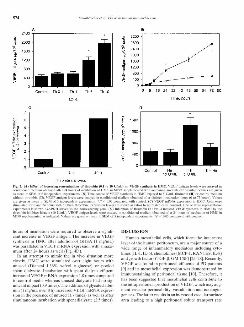
hydrogenase (GAPDH) served as the reference house- Our prior study demonstrated that there is a high ratekeeping gene. The amplification efficiencies of the target of thrombin-induced fibrin formation in the peritonealand reference were shown to be approximately equal cavity of PD patients. To assess the effect of thrombinwith a slope of log input amount to �Ct 0.1 (here on VEGF synthesis, confluent HMC were incubated with0.031). Controls consisting of H2O or samples that were various concentrations of thrombin (0.1 to 10 U/mL)not reverse transcribed were negative for target and ref- over 24 hours. Figure 2A demonstrates that the additionerence. of thrombin resulted in a dose-dependent increase in
Following oligonucleotide primers (300 nmol/L) and VEGF antigen levels in the cell supernatant, as it wasprobe (100 nmol/L) were used for human VEGF (gb statistically significant at a concentration of 5 U/mL (P NM-003376; bp 53-127): sense, 5-GCCTTGCTGCTCTA 0.05). VEGF antigen was increased almost 2.5-fold afterCCTCCAC-3, antisense, 5-ATGATTCTGCCCTCCTC a 24 hour incubation in the presence of 5 U/mL thrombinCTTCT-3; internal fluorescence labeled probe (FAM), compared to unstimulated conditions (1208 � 135 pg/5-AAGTGGTCCCAGGCTGCACCCAT-3, detecting 105 cells vs. 484 � 60 pg/105 cells, N � 6). Time-courseall known VEGF isoforms. Commercially available, pre- production of VEGF following HMC stimulation bydeveloped TaqMan reagents were used for GAPDH. thrombin (5 U/mL) is demonstrated in Figure 2B. ThePrimers and probes were obtained from PE Biosystems. antigen increase in HMC after addition of 5 U/mL
thrombin was paralleled by significant changes in VEGFStatistical analysis of the datamRNA expression after 8 and 24 hours (Fig. 2C).
Data are given as mean � SEM. Statistical analysis The specificity of the thrombin effect was tested bywas performed using the Wilcoxon matched-pairs signed the addition of hirudin, a naturally occurring inhibitor ofrank test for non-parametric data. A P value 0.05 was thrombin that blocks both the proteolytic and membraneconsidered to indicate statistically significant differences. receptor-binding functions of thrombin. When HMC
treated with thrombin (5 U/mL) were simultaneously in-RESULTS cubated with hirudin (10 U/mL), a significant inhibition
of the increase in VEGF synthesis was obtained (Fig. 2D).Effect of cytokines on VEGF production and mRNAexpression in HMC
Effect of elevated d-glucose concentrations on VEGFTo investigate whether inflammatory mediators have a production and mRNA expression in HMC
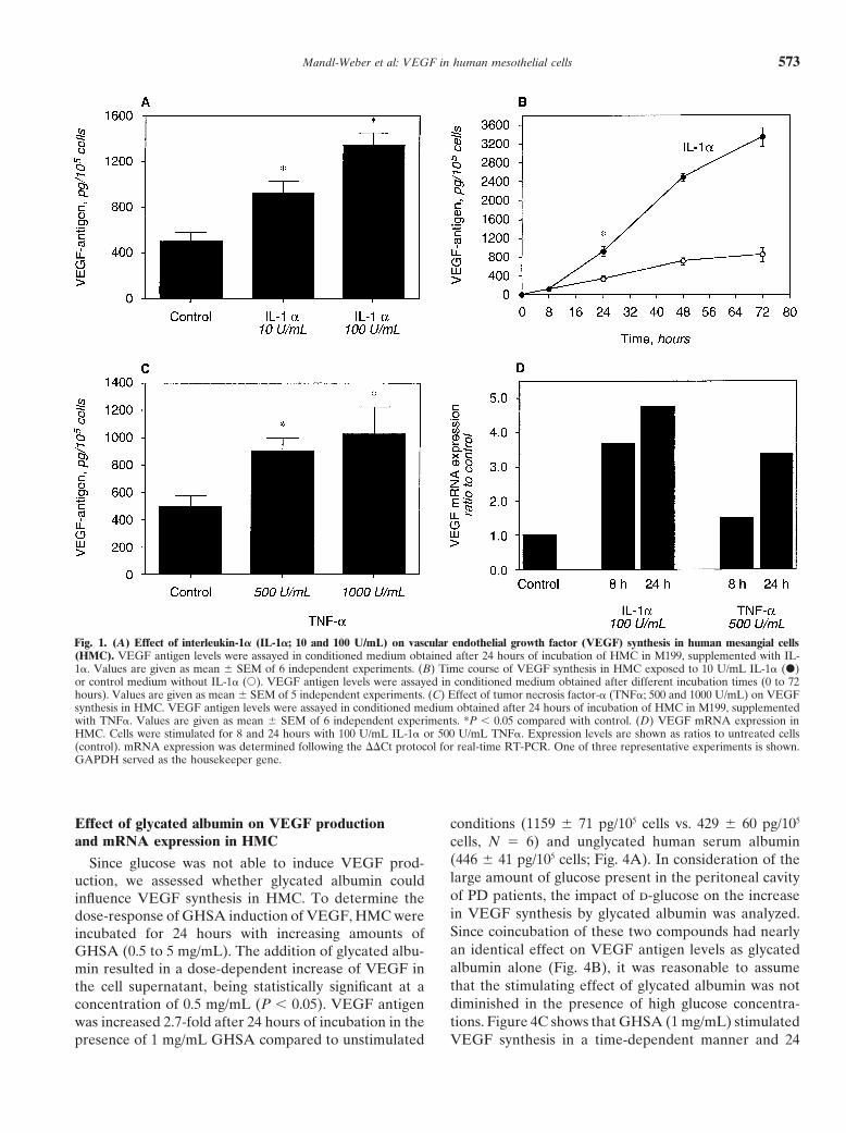
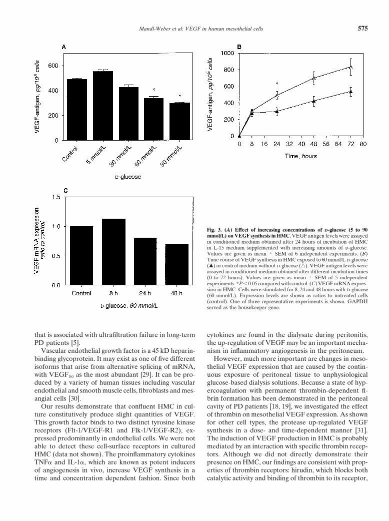
stimulating effect on VEGF synthesis in HMC, confluentTo investigate the effect of d-glucose, which is com-cells were incubated with IL-1� (10 and 100 U/mL) and
monly used as an osmotic agent in peritoneal dialysis onTNF� (500 and 1000 U/mL). As shown in Figure 1A,VEGF synthesis in HMC, confluent cells were incubatedthe addition of IL-1� resulted in a dose-dependent in-with various concentrations (5 to 90 mmol/L) of d-glu-crease in VEGF antigen levels in the cell supernatant.cose over 24 hours. The glucose concentration from 30VEGF antigen was increased twofold after 24 hour incu-to 90 mmol/L corresponds to the range we found in PDbation with 10 U/mL IL-1� and was enhanced approxi-patients intraperitoneally during a dwell time of 15 tomately threefold in the presence of 100 U/mL IL-1�. A240 minutes after instillation of a 2.27% (126 mmol/L)time dependent increase of VEGF production is demon-d-glucose dialysis solution (data not shown). As shownstrated in Figure 1B. VEGF levels significantly rose inin Figure 3A, the addition of d-glucose was not able toresponse to IL-1� (10 U/mL) within 24 hours and in-up-regulate VEGF production. In contrast, high glucosecreased continuously over 72 hours. Also, the addition ofconcentrations (60 to 90 mmol/L) even decreased VEGFTNF� (500 and 1000 U/mL) resulted in a concentrationsynthesis with diminished levels over 72 hours (Fig. 3B).dependent increase in VEGF antigen concentration inCulturing HMC over five days under high glucose condi-the cell supernatant after 24 hours (Fig. 1C). However,tions (30 to 90 mmol/L) was not able to stimulate VEGFthe up-regulation of VEGF by this cytokine was weakerexpression (data not shown). Parallel to a drop in VEGFthan in the presence of IL-1�.antigen levels, mRNA expression was also slightly dimin-To determine whether the up-regulation in VEGFished in the presence of d-glucose (60 mmol/L) after 24production induced by IL-1� and TNF� was associated
by an increase in mRNA expression, real-time RT-PCR and 48 hours (Fig. 3C).
Mandl-Weber et al: VEGF in human mesothelial cells 573
Fig. 1. (A) Effect of interleukin-1� (IL-1�; 10 and 100 U/mL) on vascular endothelial growth factor (VEGF) synthesis in human mesangial cells(HMC). VEGF antigen levels were assayed in conditioned medium obtained after 24 hours of incubation of HMC in M199, supplemented with IL-1�. Values are given as mean � SEM of 6 independent experiments. (B) Time course of VEGF synthesis in HMC exposed to 10 U/mL IL-1� (�)or control medium without IL-1� (�). VEGF antigen levels were assayed in conditioned medium obtained after different incubation times (0 to 72hours). Values are given as mean � SEM of 5 independent experiments. (C) Effect of tumor necrosis factor-� (TNF�; 500 and 1000 U/mL) on VEGFsynthesis in HMC. VEGF antigen levels were assayed in conditioned medium obtained after 24 hours of incubation of HMC in M199, supplementedwith TNF�. Values are given as mean � SEM of 6 independent experiments. *P 0.05 compared with control. (D) VEGF mRNA expression inHMC. Cells were stimulated for 8 and 24 hours with 100 U/mL IL-1� or 500 U/mL TNF�. Expression levels are shown as ratios to untreated cells(control). mRNA expression was determined following the ��Ct protocol for real-time RT-PCR. One of three representative experiments is shown.GAPDH served as the housekeeper gene.
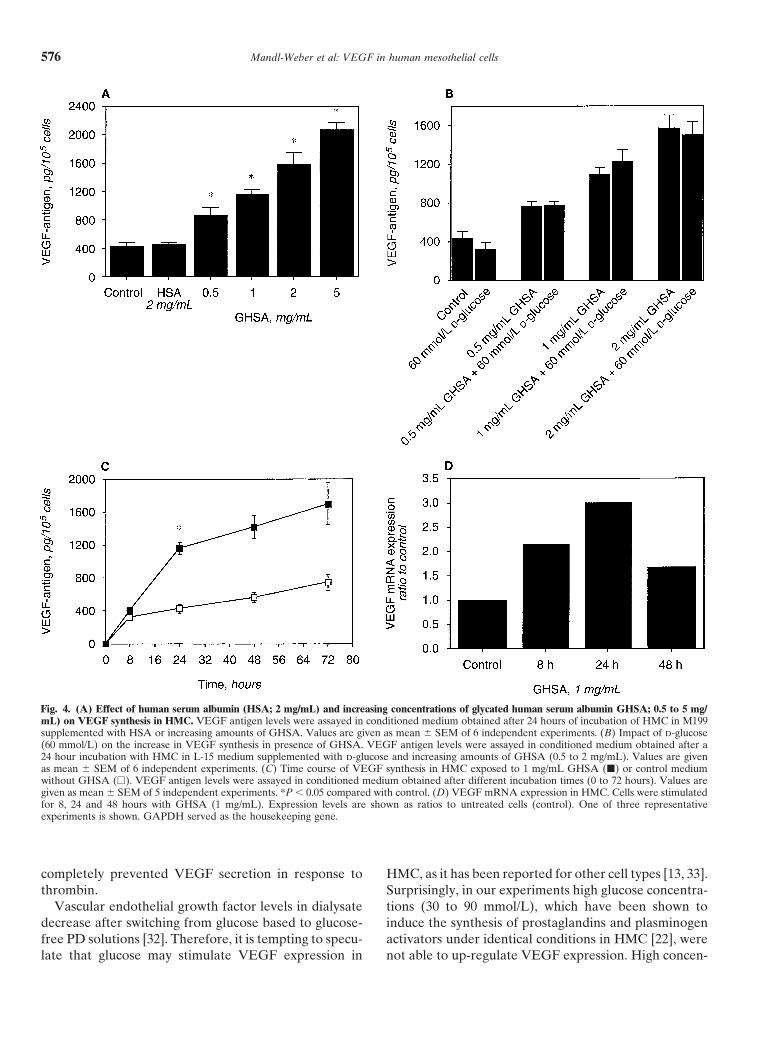
Effect of glycated albumin on VEGF production conditions (1159 � 71 pg/105 cells vs. 429 � 60 pg/105
and mRNA expression in HMC cells, N � 6) and unglycated human serum albumin(446 � 41 pg/105 cells; Fig. 4A). In consideration of theSince glucose was not able to induce VEGF prod-large amount of glucose present in the peritoneal cavityuction, we assessed whether glycated albumin couldof PD patients, the impact of d-glucose on the increaseinfluence VEGF synthesis in HMC. To determine thein VEGF synthesis by glycated albumin was analyzed.dose-response of GHSA induction of VEGF, HMC wereSince coincubation of these two compounds had nearlyincubated for 24 hours with increasing amounts ofan identical effect on VEGF antigen levels as glycatedGHSA (0.5 to 5 mg/mL). The addition of glycated albu-albumin alone (Fig. 4B), it was reasonable to assumemin resulted in a dose-dependent increase of VEGF inthat the stimulating effect of glycated albumin was notthe cell supernatant, being statistically significant at adiminished in the presence of high glucose concentra-concentration of 0.5 mg/mL (P 0.05). VEGF antigentions. Figure 4C shows that GHSA (1 mg/mL) stimulatedwas increased 2.7-fold after 24 hours of incubation in the
presence of 1 mg/mL GHSA compared to unstimulated VEGF synthesis in a time-dependent manner and 24
Mandl-Weber et al: VEGF in human mesothelial cells574
Fig. 2. (A) Effect of increasing concentrations of thrombin (0.1 to 10 U/mL) on VEGF synthesis in HMC. VEGF antigen levels were assayed inconditioned medium obtained after 24 hours of incubation of HMC in M199, supplemented with increasing amounts of thrombin. Values are givenas mean � SEM of 6 independent experiments. (B) Time course of VEGF synthesis in HMC exposed to 5 U/mL thrombin (�) or control mediumwithout thrombin (�). VEGF antigen levels were assayed in conditioned medium obtained after different incubation times (0 to 72 hours). Valuesare given as mean � SEM of 5 independent experiments. *P 0.05 compared with control. (C) VEGF mRNA expression in HMC. Cells werestimulated for 8 and 24 hours with 5 U/mL thrombin. Expression levels are shown as ratios to untreated cells (control). One of three representativeexperiments is shown. GAPDH served as the housekeeping gene. (D) Inhibition of thrombin (5 U/mL) induced VEGF synthesis in HMC by thethrombin inhibitor hirudin (10 U/mL). VEGF antigen levels were assayed in conditioned medium obtained after 24 hours of incubation of HMC inM199 supplemented as indicated. Values are given as mean � SEM of 5 independent experiments. *P 0.05 compared with control.
hours of incubation were required to observe a signifi- DISCUSSIONcant increase in VEGF antigen. The increase in VEGF Human mesothelial cells, which form the innermostsynthesis in HMC after addition of GHSA (1 mg/mL) layer of the human peritoneum, are a major source of awas paralleled in VEGF mRNA expression with a maxi- wide range of inflammatory mediators including cyto-mum after 24 hours as well (Fig. 4D). kines (IL-1, IL-6), chemokines (MCP-1, RANTES, IL-8)
In an attempt to mimic the in vivo situation moreand growth factors (TGF-�, GM-CSF) [25–28]. Recently,closely, HMC were stimulated over eight hours withVEGF was found in peritoneal effluents of PD patientsunused (Dianeal 1.36% wt/vol d-glucose) or pooled[9] and its mesothelial expression was demonstrated byspent dialysate. Incubation with spent dialysis effluentimmunostaining of peritoneal tissue [10]. Therefore, itincreased VEGF mRNA expression 1.8 times comparedhas been suggested that mesothelial cells contribute toto control media whereas unused dialysate had no sig-the intraperitoneal production of VEGF, which may aug-nificant impact (0.9 times). The addition of glycated albu-ment vascular permeability, vasodilation and neoangio-min (1 mg/mL over 8 h) increased VEGF mRNA expres-genesis. The latter results in an increased vascular surfacesion in the presence of unused (1.7 times) as well as after
simultaneous incubation with spent dialysate (2.5 times). area leading to a high peritoneal solute transport rate
Mandl-Weber et al: VEGF in human mesothelial cells 575
Fig. 3. (A) Effect of increasing concentrations of D-glucose (5 to 90mmol/L) on VEGF synthesis in HMC. VEGF antigen levels were assayedin conditioned medium obtained after 24 hours of incubation of HMCin L-15 medium supplemented with increasing amounts of d-glucose.Values are given as mean � SEM of 6 independent experiments. (B)Time course of VEGF synthesis in HMC exposed to 60 mmol/L d-glucose(�) or control medium without d-glucose (�). VEGF antigen levels wereassayed in conditioned medium obtained after different incubation times(0 to 72 hours). Values are given as mean � SEM of 5 independentexperiments. *P 0.05 compared with control. (C) VEGF mRNA expres-sion in HMC. Cells were stimulated for 8, 24 and 48 hours with d-glucose(60 mmol/L). Expression levels are shown as ratios to untreated cells(control). One of three representative experiments is shown. GAPDHserved as the housekeeper gene.
that is associated with ultrafiltration failure in long-term cytokines are found in the dialysate during peritonitis,the up-regulation of VEGF may be an important mecha-PD patients [5].
Vascular endothelial growth factor is a 45 kD heparin- nism in inflammatory angiogenesis in the peritoneum.However, much more important are changes in meso-binding glycoprotein. It may exist as one of five different
isoforms that arise from alternative splicing of mRNA, thelial VEGF expression that are caused by the contin-uous exposure of peritoneal tissue to unphysiologicalwith VEGF165 as the most abundant [29]. It can be pro-
duced by a variety of human tissues including vascular glucose-based dialysis solutions. Because a state of hyp-ercoagulation with permanent thrombin-dependent fi-endothelial and smooth muscle cells, fibroblasts and mes-
angial cells [30]. brin formation has been demonstrated in the peritonealcavity of PD patients [18, 19], we investigated the effectOur results demonstrate that confluent HMC in cul-
ture constitutively produce slight quantities of VEGF. of thrombin on mesothelial VEGF expression. As shownfor other cell types, the protease up-regulated VEGFThis growth factor binds to two distinct tyrosine kinase
receptors (Flt-1/VEGF-R1 and Flk-1/VEGF-R2), ex- synthesis in a dose- and time-dependent manner [31].The induction of VEGF production in HMC is probablypressed predominantly in endothelial cells. We were not
able to detect these cell-surface receptors in cultured mediated by an interaction with specific thrombin recep-tors. Although we did not directly demonstrate theirHMC (data not shown). The proinflammatory cytokines
TNF� and IL-1�, which are known as potent inducers presence on HMC, our findings are consistent with prop-erties of thrombin receptors: hirudin, which blocks bothof angiogenesis in vivo, increase VEGF synthesis in a
time and concentration dependent fashion. Since both catalytic activity and binding of thrombin to its receptor,
Mandl-Weber et al: VEGF in human mesothelial cells576
Fig. 4. (A) Effect of human serum albumin (HSA; 2 mg/mL) and increasing concentrations of glycated human serum albumin GHSA; 0.5 to 5 mg/mL) on VEGF synthesis in HMC. VEGF antigen levels were assayed in conditioned medium obtained after 24 hours of incubation of HMC in M199supplemented with HSA or increasing amounts of GHSA. Values are given as mean � SEM of 6 independent experiments. (B) Impact of d-glucose(60 mmol/L) on the increase in VEGF synthesis in presence of GHSA. VEGF antigen levels were assayed in conditioned medium obtained after a24 hour incubation with HMC in L-15 medium supplemented with d-glucose and increasing amounts of GHSA (0.5 to 2 mg/mL). Values are givenas mean � SEM of 6 independent experiments. (C) Time course of VEGF synthesis in HMC exposed to 1 mg/mL GHSA (�) or control mediumwithout GHSA (�). VEGF antigen levels were assayed in conditioned medium obtained after different incubation times (0 to 72 hours). Values aregiven as mean � SEM of 5 independent experiments. *P 0.05 compared with control. (D) VEGF mRNA expression in HMC. Cells were stimulatedfor 8, 24 and 48 hours with GHSA (1 mg/mL). Expression levels are shown as ratios to untreated cells (control). One of three representativeexperiments is shown. GAPDH served as the housekeeping gene.
completely prevented VEGF secretion in response to HMC, as it has been reported for other cell types [13, 33].Surprisingly, in our experiments high glucose concentra-thrombin.
Vascular endothelial growth factor levels in dialysate tions (30 to 90 mmol/L), which have been shown toinduce the synthesis of prostaglandins and plasminogendecrease after switching from glucose based to glucose-
free PD solutions [32]. Therefore, it is tempting to specu- activators under identical conditions in HMC [22], werenot able to up-regulate VEGF expression. High concen-late that glucose may stimulate VEGF expression in
Mandl-Weber et al: VEGF in human mesothelial cells 577
angiogenesis in the peritoneal membrane. Perit Dial Int 20(Suppltrations of d-glucose even decreased the synthesis of2):S19–25, 2000
VEGF. The lack of an increase in VEGF in the presence 3. Mateijsen MA, van der Wal AC, Hendriks PM, et al: Vascularand interstitial changes in the peritoneum of CAPD patients withof glucose is in accordance to the study of Selgas et alperitoneal sclerosis. Perit Dial Int 19:517–525, 1999[34], who demonstrates that the ex vivo VEGF produc-
4. Davies SJ, Phillips L, Griffiths AM, et al: What really happens totion by HMC from PD patients is not correlated with people on long-term peritoneal dialysis? Kidney Int 54:2207–2217,
1998cumulative glucose overload during the whole PD pe-5. Davies SJ, Bryan J, Phillips L, Russell GI: Longitudinal changesriod.
in peritoneal kinetics: The effects of peritoneal dialysis and perito-In an effort to test the hypothesis that chronic expo- nitis. Nephrol Dial Transplant 11:498–506, 1996
6. Senger DR, Brown LF, Claffey KP, Dvorak HF: Vascular per-sure to glucose may initiate other factors that modulatemeability factor, tumor angiogenesis and stroma generation. Inva-VEGF production, we investigated the effect of glycatedsion Metastasis 14:385–394, 1994
serum albumin on VEGF expression. Serum albumin is 7. Horowitz JR, Rivard A, van der Zee R, et al: Vascular endothelialgrowth factor/vascular permeability factor produces nitric oxide-the most abundant protein in the peritoneal cavity anddependent hypotension. Evidence for a maintenance role in quies-a major target of nonenzymatic glycation in the presencecent adult endothelium. Arterioscler Thromb Vasc Biol 17:2793–
of glucose [35, 36]. Early glycation products of albumin 2800, 19978. Leung DW, Cachianes G, Kuang WJ: Vascular endothelialin an Amadori configuration are generated in the perito-
growth factor is a secreted angiogenic mitogen. Science 246:1306–neal cavity of PD patients, and the presence of Amadori1309, 1989
albumin is found in the dialysate as well as in the meso- 9. Zweers MM, de Waart DR, Smit W, et al: Growth factors VEGFand TGF-beta1 in peritoneal dialysis. J Lab Clin Med 134:124–132,thelial layer of PD patients [37]. Our experiments dem-1999onstrate, to our knowledge for the first time, that expo-
10. Combet S, Miyata T, Moulin P et al: Vascular proliferation andsure of HMC to Amadori-modified glycated albumin enhanced expression of endothelial nitric oxide synthase in human
peritoneum exposed to long-term peritoneal dialysis. J Am Socresulted in a concentration- and time-dependent increaseNephrol 11:717–728, 2000in VEGF mRNA expression and antigen secretion. In 11. Inagi R, Miyata T, Yamamoto T, et al: Glucose degradation prod-
accordance with this observation, it has been reported uct methylglyoxal enhances the production of vascular endothelialgrowth factor in peritoneal cells: Role in the functional and mor-that glucose degradation products induce VEGF expres-phological alterations of peritoneal membranes in peritoneal dial-sion in mesothelial cells [11] and a correlation between ysis. FEBS Lett 463:260–264, 1999
AGE accumulation and VEGF expression in the perito- 12. Stein I, Neeman M, Shweiki D, et al: Stabilization of vascularendothelial growth factor mRNA by hypoxia and hypoglycemianeum was found in peritoneal biopsies [10].and coregulation with other ischemia-induced genes. Mol Cell BiolIn summary, our findings support the concept that 15:5363–5368, 1995
locally produced and released VEGF is involved in peri- 13. Kim NH, Jung HH, Cha DR, Choi DS: Expression of vascularendothelial growth factor in response to high glucose in rat mesan-toneal neoangiogenesis, which is an important patho-gial cells. J Endocrinol 165:617–624, 2000genic factor in ultrafiltration failure in long term PD. It 14. Sone H, Kawakami Y, Okuda Y, et al: Vascular endothelial growth
is tempting to speculate that besides endothelial cells, factor is induced by long-term high glucose concentration and up-regulated by acute glucose deprivation in cultured bovine retinalthe mesothelium contributes to the intraperitoneal syn-pigmented epithelial cells. Biochem Biophys Res Commun 221:thesis of VEGF. Consistent with observations in other 193–198, 1996
cell types we could demonstrate that VEGF expression 15. Imaizumi T, Itaya H, Nasu S, et al: Expression of vascular endothe-lial growth factor in human umbilical vein endothelial cells stimu-in HMC is up-regulated in presence of proinflammatorylated with interleukin-1alpha—an autocrine regulation of angio-cytokines and thrombin. Our results also confirm clinical genesis and inflammatory reactions. Thromb Haemost 83:949–955,
data that the continuous exposure of the peritoneal mem- 200016. Perez-Ruiz M, Ros J, Morales-Ruiz M, et al: Vascular endothelialbrane to glucose-based dialysis solutions is an important
growth factor production in peritoneal macrophages of cirrhoticstimulus for VEGF expression. However, it is not glucose patients: Regulation by cytokines and bacterial lipopolysaccharide.per se, but nonenzymatic glycation products like glycated Hepatology 29:1057–1063, 1999
17. Jayne DG, Perry SL, Morrison E, et al: Activated mesothelial cellsalbumin, that up-regulate VEGF expression.produce heparin-binding growth factors: Implications for tumormetastases. Br J Cancer 82:1233–1238, 2000
ACKNOWLEDGMENT 18. Sitter T, Spannagl M, Schiffl H, et al: Imbalance between intra-peritoneal coagulation and fibrinolysis during peritonitis of CAPD
This work was supported by a grant from the Else Kroner-Fresenius- patients: The role of mesothelial cells. Nephrol Dial TransplantStiftung to T.S. 10:677–683, 1995
19. Goedde M, Sitter T, Schiffl H, et al: Coagulation- and fibrinolysis-Reprint requests to PD Dr. med. Thomas Sitter, Medizinische Klinik, related antigens in plasma and dialysate of CAPD patients. Perit
Innenstadt Klinikum, Universitat Munchen, Ziemssenstraße 1, D-80336 Dial Int 17:162–166, 1997Munchen, Germany. 20. Baynes JW, Thorpe SR, Murtiashaw MH: Nonenzymatic glucosy-E-mail: [email protected] lation of lysine residues in albumin. Methods Enzymol 106:88–98,
198421. van Hinsbergh VW, Kooistra T, Scheffer MA, et al: Character-REFERENCES
ization and fibrinolytic properties of human omental tissue meso-thelial cells. Comparison with endothelial cells. Blood 75:1490–1. Krediet RT: The peritoneal membrane in chronic peritoneal dial-
ysis. Kidney Int 55:341–356, 1999 1497, 199022. Sitter T, Haslinger B, Mandl S, et al: High glucose increases2. Krediet RT, Zweers MM, van der Wal AC, Struijk DG: Neo-
Mandl-Weber et al: VEGF in human mesothelial cells578
prostaglandin E2 synthesis in human peritoneal mesothelial cells: 30. Dvorak HF, Brown LF, Detmar M, Dvorak AM: Vascular perme-ability factor/vascular endothelial growth factor, microvascular hyp-Role of hyperosmolarity. J Am Soc Nephrol 9:2005–2112, 1998erpermeability, and angiogenesis. Am J Pathol 146:1029–1039, 199523. Chomczynski P, Sacchi N: Single-step method of RNA isolation by
31. Maragoudakis ME, Tsopanoglou NE, Andriopoulou P, Mara-acid guanidinium thiocyanate-phenol-chloroform extraction. Analgoudakis MM: Effects of thrombin/thrombosis in angiogenesis andBiochem 162:156–159, 1987tumor progression. Matrix Biol 19:345–351, 200024. Fink L, Seeger W, Ermert L: et al: Real-time RT-PCR after laser-
32. Zweers MM SD, Struijk DG, Smit W, Krediet RT: Vascularassisted cell picking. Nat Med 4:1329–1333, 1998endothelial growth factor in peritoneal dialysis: A longitudinal fol-25. Lanfrancone L, Boraschi D, Ghiara P, et al: Human peritoneallow-up. J Lab Clin Med 137:125–132, 2001mesothelial cells produce many cytokines (granulocyte colony-stim-
33. Williams B, Gallacher B, Patel H, Orme C: Glucose-inducedulating factor [CSF], granulocyte-monocyte-CSF, macrophage-CSF,protein kinase C activation regulates vascular permeability factorinterleukin-1 [IL-1], and IL-6) and are activated and stimulated tomRNA expression and peptide production by human vasculargrow by IL-1. Blood 80:2835–2842, 1992 smooth muscle cells in vitro. Diabetes 46:1497–1503, 199726. Topley N, Mackenzie RK, Williams JD: Macrophages and meso- 34. Selgas R, del Peso G, Bajo MA, et al: Spontaneous VEGF produc-
thelial cells in bacterial peritonitis. Immunobiol 195:563–573, 1996 tion by cultured peritoneal mesothelial cells from patients on perito-27. Visser CE, Tekstra J, Brouwer-Steenbergen JJ, et al: Chemokines neal dialysis. Perit Dial Int 20:798–801, 2000
produced by mesothelial cells: huGRO-alpha, IP-10, MCP-1 and 35. Dulaney JT, Hatch FE Jr: Peritoneal dialysis and loss of proteins:RANTES. Clin Exp Immunol 112:270–275, 1998 A review. Kidney Int 26:253–262, 1984
28. Witowski J, Jorres A, Coles GA, et al: Superinduction of IL-6 36. Lamb E, Cattell WR, Dawnay A: Glycated albumin in serum andsynthesis in human peritoneal mesothelial cells is related to the dialysate of patients on continuous ambulatory peritoneal dialysis.induction and stabilization of IL-6 mRNA. Kidney Int 50:1212–1223, Clin Sci 84:619–626, 19931996 37. Posthuma N, ter Wee PM, Niessen H, et al: Amadori albumin
29. Ferrara N: Molecular and biological properties of vascular endothe- and advanced glycation end-product formation in peritoneal dialysisusing icodextrin. Perit Dial Int 21:43–51, 2001lial growth factor. J Mol Med 77:527–543, 1999