Embed Size (px)
Citation preview
CASE
HOUSTON SOCIETY OF CLINICAL PATHOLOGISTS
1964 SEM INAR
Rl!SPIRATORY SYSTEM, GASTROINTESTINAL SYSTE.M, GENITOURINARY SYSTEM AND BONE
D I ~ N 0 S E S
1. Thymoma, mixed type (reticuloepithelial- lymphocyti c) invading l ung :parenchyma.
2. Malignant pheochromocytoma with metastasis to rib.
3. Lav grade chondrosarcoma of la.rynx.
4. Seminoma, :primary in anterior mediastinum.
5· Extraosseous osteogenic sarcoma.
6. Synovial sarcoma involving soft tissue of inguinal region with extension i nto femoral vein.
7. Mixed tumor of saliyary gland type, primary in bronchus.
8 . Bronchogenic carcinoma, l arge cell undifferentiated.
9· Mineral o-il granuloma invoLving l ung parenchyma.
10. Lymphosarcoma of lower esophagus (peraesophageal) .
11. Solid carcinoma of thyroid with amyloid stroma. Massive cervical and mediastinal lymph nodal metastasis .
L2. Osteitis fibrosa "brown tumor" of hyperparathyroidism.
13. Well differentiated islet cell carcinoma of head of pancreas. Findings compatible with Zoll inger-ELlison syndrome.
14. Embryonal rhabdomyosarcoma.
15. Pedunculated adenoma of Brunner' s glands of duodenum.
16. Benign mesenchymoma of kidney.
...... , , !! ! ..;
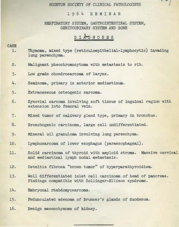
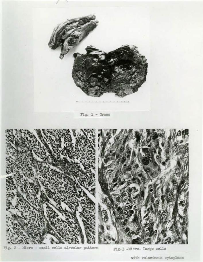
Fig . 1 - Gross
. 2 - Low po\ler Fig. 3 - High power
\.
v
HoustoQfSOciety of Clinical Pathologists Apri1 25, 1964
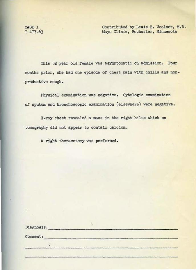
Seminar CASE l T- 477- 63
At surgical e xploration, a tumor mass was found to arise in the ant er ior mediastinum adherent to and merging into the righ-t upper l obe of t he l ung and pericardium. A right upper lobectomy was performed . Gross examination of the specimen revealed a tumor mass (6 x 6 x ~ em.) c1osely app11ed to and invading the apical portion of the lobe . On cut section, invasion of lung parenchyme. was apparent end projections of tumor had i nvaded a l arge bronchus extending centrally almost to the h ilus. (Fig. 1).
Sections of t he tumor reveal an intimate mixture of large pale epithel ial or reticular cells and small cells resembling lymphocyte s in approximately equal proportions . (Fig. 2) . The growth pattern shows these cel l s to be arranged in sheets forming rounded lobules separated by fibrous trabeculae . The tumor is seen to involve lung parenchyma and extend into a large peripheral bronchus . The large reticular cells, which vary somewhat in s ize, show indistinct cell outlines and have rather pale vesicular nuclei. These large cells do not appear anaplastic in that there is a dist i nct l ac k of abnormal chromatin clumping, nucleoli are not prominent and mit oses are diffi cult to f ind. The small cells resemble mature lymphocytes . The tumor thus appears to be "lymphoepit he lial" in its histologic pattern . (Fig. 3) . T"ne differential diagnosis lies bet"Ween a primary peripheral lung carcinoma of "lymphoepithelial" architecture extending into the mediastinum, a malignant lymphoma of "mixed" type, and a mediastinal thymoma inva\li ng lung parenchyma. The latter concept is strongly favored by reason of (1) a growth pattern consisting of large lobules of tumor separated by fibrous t rabeculae . (2) Intimate mixture of two types of cells such as is commonly seen in thymoma . (3) A lack of anapl asia (mitotic figures, large nucleoli, etc. ) in the large reticular cells. Certain commonly observed features of thymoma including microcyst formation, peri vascular spaces , vascular lymphocytic cuffing and epithelial palisading are not observed in these sections. The absence of these , however, does not r ule out a di agnosis of thymoma. This tumor i s markedly invasive. Such invasive quali t i es are characteristic of thymoma and do not correlate wit h any variation in histologic pattern of such tumors . Malignant lymphoma and lymphoepithelioma, either metastatic or primary, are excluded in that the tumor l a cks evidence of cellular anaplasia. One is left with a diagnosis of thymoma invading lung substance.
DIAGNOSIS : Thymoma, mixed type (reticuloepithelial- lympbocytic) invading l ung parenchyma.
References:
1. P. E. llernatz, M.D., E. G. Harrison, M.D. and O. T . Clagett, M.D. , rtThylnoma: A Clinicopathologic Stud,y," Journal of Thoracic and CardiOvascular Surgery, Vol. 42, No . 4, pages 424-444, October, 1961 .
2 . Lattes, R. , and Jonas, 8 . : "The Pathological and Clinical Features in Eighty Cases of Thymoma," llull. New York Acad . ~led . 33: 145-147, l957.
Follow up: Recent case.
/
Fig. l - Gro&&
Fig. 2 - Micro - omall cells alveolar pattern Pig. 3 -Micro- Large cella
vith voluminous cytoplasCI
Houston Societ y of Clinical Pathologists Seminar April 25, 1964
This patient was explored for an indeterminate abdominal mass on the right side with apparent metastasis to the right tenth rib. The surgeon encountered a huge ret r operi toneal tumor situated just above the right ki dney. The kidney itself was not involved and could be separated readily from tbe tumor. 'I'i:J.e right adrenal gland was incorporated in the surface of the mass and the surgeon felt that the tU!l:Or was of adrenal origin. The tumor was movable and readily di ssected away from adjacent t i ssues. By extending the incision, the right tenth rib was resected along •ith i ntercostal muscles and periosteum from the two adjacent ribs.
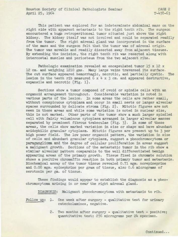
Pathol ogic examination revealed an encapsulated tumor 15 x 12 x 12 em. and weighi ng 1210 gms. Many large veins t r aversed its surf ace The cut surface appeared hemorrhagic , necrotic, and partially cystic. The lesion in the t enth rib measured 6 x 4 x 3 em. and appeared destructive, expansile and necrotic (Fig. 1) .
Sections show a tumor composed of ovoid or spindle cel ls wi th an organoid arrangement throughout. Considerable variation is noted in various parts of the lesion . In some areas the cells are rather small, wi thout conspicuous cytoplasm and occur in small nests or l arger alveolar spaces surrounded by delicate stroma (Fig. 2) . Mitotic figures are not seen in these areas and while some variation is noted in nuclear size, this is not marked. Other parts of the tumor show a much larger spindled cell with fairly voluminous cytopl asm arranged in larger alveolar masses separ ated by prominent f ibrous t rabeculae (Fig. 3) . In some of these areas, the cel ls show marked variation in size or shape and have abundant acidophilic granular cytoplasm. Mitotic figures are present up t o 3 per high power field. The low power organoid pattern, the variation in size of cells and abundant granular cytoplasm, suggest a pheochromocytoma or paraganglioma and the degree of cellular proliferation in areas suggest a malignant growth. Sections oi' the metastatic tumor in the rib show a simi l ar alveolar pattern comparable to the well di fferentiated beni gn appear i ng areas of the primary growth . Tissue f ixed in chromate sol ution shows a positive chromaffin rea~ion in both pr imary tumor and met astasi s . Biochemical assay of the tumor t i ssue revealed 0. 71 mgm. norepineprine and 0 .08 mgm. epinephrine per gram of tissue , also 0 .6 micrograms of s erotonin per glll· of tissue.
These findings would appear to establish the diagnosis as a pheochromocytoma ar i s ing in or near t he right adrenal gland.
DIAGNOSIS: 1-!alignant pheochromocytoma ~<ith metastasis to rib.
Follow up: 1 . One week after surgery - qualitative test for urinary catecholamines, negative .
2. Two months after surgery - qualitative test - positi ve; quantitative test : 276 micrograms per 24 specimen.
Continued . ..
Houston Society or Clinical Pathologists Seminar April 25, 1964
CASE 2 T-478-63
References :
3. Six months af'ter surgery - qualitative test - positive; quantitative test : 24llo micrograms per 24 hour specimen.
4. Six months af'ter surgery: X-ray of the sternum shows a lytic defect 2 em. in diameter in the distal portion of sternum, apparently metastasi s, also area of destruction involving left innominate bone and left ilium in region of acetabulum. Blood pressure 130/72. Irradi ation therapy given without obvious benefit.
l. Russell P. Sherwin, M. D., Histopathology of Pheochromocytoma, Cancer, 12: 861-877, September-October, 1959.
2 . Jesse L. Bollman, M.D., Eunice V. Flock, Fn.D., Grace M. Roth, Fb .D. , and Walter F. Kvale, M. D. , Catecbolamines in Patients with Pheochromocytoma, Journal of Laboratory and Clinical Medicine, 56: 506- 519, October, 1960.
3· Alexander M. Minno, M. D. , Warren A. Bennett, N.D. and Walter F. Kvale, M.D. , Pheochromocytoma, A Study of 15 Cases Diagnosed at Aut opsy, New England Journal of Medicine, 251 : 959-965, December, 1954.
1+ . James T . Priestley, M.D. ; Walter F. Kvale, M.D.; and Ray w. Gifford, Jr. , M.D., Pheochromocytoma, Archives of Surgery, 86: 778-789, 1-'.a.y' 1963.
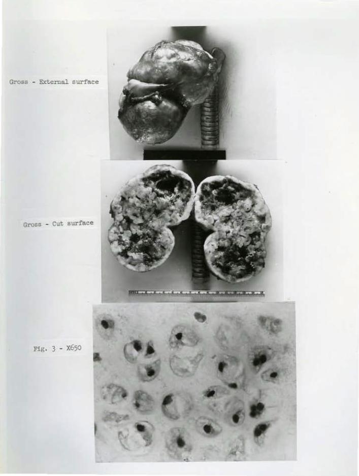
Gross - External 6 urface
Gross - cut surface
Fig. 3 - X650
Houston Soci ety of Clinical Pathol ogists Seminar Apr il 25, 1964
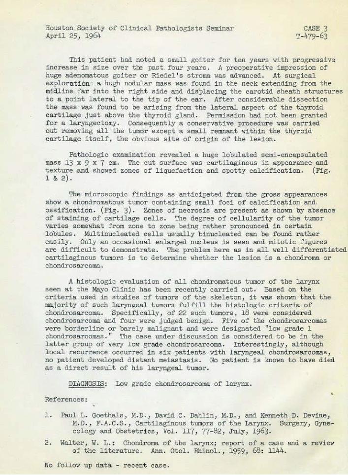
CASE 3 T-479.-63
This patient hsd not-ed a, small goit e r for ten ye.ars wi '\;h progressive i ncrease in size over tl:e past. four years. A preoperative impression of huge a.deno~~tous goiter or Riedel ' s str oma. was advanced. At surgi cal exploration~ a. hugh nodular mass was found. i n the neck extending from the midline far i nto the right side and displacing t he carotid sheath struct ur es to ·a poi nt l ateral to t he t i p of the· ear. After considerable dissection t he mass was found t o be arising from the lateral aspect of t he t hyroi d cartilage just above the thyroid gland . Permission had not been granted for a. laryngectomy. Consequently a conser vative procedure· was carri ed out removing all the tumor except a small remnant within the thyroid cartilage its~lf , the obvious site ·of origin of the lesion.
Pathologic examination revea.leci a huge lobulated semi- encapsulated mass 13 .x 9 .x 7 em. The cut surface. was cartilaginous in appearance and texture and showed zones of liquefaction and spot ty cal cifi cation. (Fig. 1 & 2).
The micr oscopic f indi ngs as ant i ci pated from the gross appearances show a chondromatous t umor containing small foci of calcification and. ossificat i on. (Fig. 3). Zones of necros i s are present as shown by absence of staining of cartilage cells. The degree of cell\llarity of t he tumor varies somewhat :rrom zone to zone bej.hg rather pronounced in certain lobules. l~ltinucleated cells usually binucleated can be found r ather easily. Only an occasional enlarged nucleus is seen and mitoti c figures a;re difficult t o demonstr ate . The pr oblem her e as in all we·ll di fferentiat ed car tilaginous tumor s i s to determine whether t he lesion i s a chonciroma or chondrosarcoma..
A histologic eva luat ion of all chonciromatous tumor of the l arynx seen at t he Mayo CLinic has been recently carri ed out . Based on the· cri ter i a used in studies of t umors of the skel eton, i t was shown t hat t he majority of such laryngeal t umor s fulfill t he histol ogic criteri a of chondrosarcoma. Specifically, of 22 such tumors, 18 were considered chond.l:·osar coma and f our were judged benign. Five of the chondr osarcomas ••er e border line or barel y malignant and ••ere des.i gnated "low grade 1 choncirosarcoma.s." The case under discussion i s considered to be in t he latter group of very l ow grade chondrosarcoma. Int er estingly, although local recurrence occur:r.ed i n s i x patients wi t h laryngeal chondrosarcomas, no patient developed di stant met astasis·. No patient is known to have died as a direct r esult of his lar yngeal tumor.
DIAGNOSlS: Low grade choncirosarcoma or larynx.
References :
l. Paul L. Goet hals, M. D. , David C. Dahlin, M. D., and Kenneth D. DevL'le., M.D. ·' F'. A.C.S., Cartilaginous tumors of the Larynx. Surgery, Gynecology and Obstetr i cs, Vol. 117, 77-82, July, 1963.
2. ~falter, H. L· : Chondroma of the l arynx; report of a case and a r eview of the literature. Ann. Otol. Rhinal., 1959, 68: 1144.
No follow up data - recent case.
Fig. l
Fig. 2 - X200
Houston Society o~ Clinical Pathologist s Seminar April 2.5) 1964
'-'
CASE. 4 T-480-63
At eyploratio.n, the surgeon encoun.ter ed a hard, apparently rnalignant tumor in the left anterior mediasti.num ov~Tlying the arch of tb~ aorta and t he pulmonary vessels. After considerable difficulty the tumor mass wa s r e moved a l ong with a portion of adjacent peri cardium, left pleura and supeTfici al portion of l eft upper lobe of lung. Pathologic· examination r evealed a circumscribed partia lly encapsulated tumor (8 x 6 x 6 em.) . The cut Scurface i s yellowi sh tan in col or end homogeneous except for small irregular areas of necrosis. (Fig . 1) .
Sectiol)s sho-w a uniform histologic pattern throughout all parts of the· tumor.. lt i s compos.ed of rather lar ge r ounded cell s 1 r egular in size and shape 1 arranged in nests or larger solid masses separated by a delicate conpective tis sue strqma. . The latt er is infiltrated, by l ymphocytes - thi s being slight in some areas and rather marked fu others . (Fig. 2). The l arge cells are polygonal in shape with a moderate amount of pale· or finely granular cytoplasm; the nuclei appear round, contain a moderately coarse chromatin network and occasiona l l y a prominent nucleol us . Mi toti c figures are easily found . The histologic appearance of the tumor is markedly s imilar if not identical t o t hat seen in many primary testicular seminomas. The find i ngs at surgery, absence of testicular enlargement , and s ubsequent clin.i ca l course, however , sugges.t a pr i mary tumor of the anteri or medi a stipum a nd not a met ast atic t esticular neoplasm.
Th e differential diagnosis in this cas e might in·clude (1) reti culum c·ell sarcoma, primaTY i n mediastinum; ( 2) thymoma "i mitati ng" a s eminoma; (3) met astatic lympho- epitheli oma, or (4) pri mary mediastina l s.eminoma. The arrangement of the tumor i n varying sized nests of cells •Nithin a lymphoid stroma is· not characteri&tic of reticulum cell sarcoma. a.s observed in l ymph nodes .e l s ewhere and on this basis , I think a primary l ymphoma. can be ·excluded . Regar ding the possibilit y of thymoma, the t umor in t his case may or may not have or iginated wi thin the thymuS gland. Such a point o~ ori gin if proven does not mean that t he t umor must be cal led a: "thymoma" s .ince we have observed teratomas containing hair and ot her s tructures compl etf!lY e,nclosed •Nithin a lobe of the thymus gland. Our current h istologic classifi cati on of t hymomas 1 with or without clinical myasthenia gravis , does not include .a variant resembling any test icular neoplasm. The r esemblance of this tumor to naso- pharyngeal l ymphoepithelioma is not s·ufficient ly s t r i king t o suggest a metastasis from such a source . We are l eft wit.h a tumor in t he anterior mediastinum, apparent ly pri mary i n t his s i t e, whi ch bea rs a stri king resembla nce t o seminoma of test i s. The c·oncept of a primary seminoma in t his s i te should b e regarded in the s a me l ight as a pr imary terat oma or chorio-carcil;loma. both of" which are well recognized phenomena in this region.
~IAGNE>SIS : Semino!!18, prilllary in ant eri or medi ast inum.
Ref er ences :
l. Friedman, N.B. : The comparative Morphogenesis of Extragenital and Gonadal Teratoi d Tumors, Cancer 4: 262-276.
Cont inued ...
Houston Society of Clinical Pathologists Seminar April 25, 1964
CASE 4 T-!Klo-63
2 . Woolner, L. B. , Jamplis, R. w. 1 and Kirklin, J , w.: Seminoma (Germinoma) Apparently Primary in the Anterior ~!ediastinum, New England J, Med. 252 : 653-657, 1955 .
3. Samuel L. Kountz, M.D. , J ohn E. Connolly, M.D., and Roy Cohn, M.D., Seminoma-like (or Seminomatous) Tumors of the Anterior ~!ediastinum. Journal of Thoracic and Cardiovascular Surgery, Vol. 5, Number 3, March, 1963, pages 289-301.
Follow up information :
August, 1958
( September , 1959
May, 196o
October, 196o
February, 1961
April, 1961
July, 1961
March, 1962
December, 1962
March, 1963
October, 1963
Initial surgery followed by postoperative Co. 6o therapy - 300 r in air to medillostinum.
Pain in back - metastasis noted in sacrum. Co. 6o -3600 r in air given with good response .
Left sacroil.iac pain - co. 6o- 4200 r .
Recurrence of pain in lower ribs - x-ray of thoracic apparently negative - 2400 r in air to thoracic spine.
Co. 60 - 2500 r. to right i lium.
HN2 given with subsequent relief of pain.
Co . 6o - 5250 r to left anterior chest wall .
Destructive lesion anterior end 8th left rib. Biopsy - metastatic seminoma. Co . 6o therapy to rib -2400 r in air .
Feeling well, metastatic lesion in right mid-lung field . Co . 60 to right chest.
X-ray of ches t: Mass shadow seen December, 1962 has disappeared.
Last Visit - feels well, plays golf, bowls on team, appetite good, weight stable. X-ray of cheat: 2 metastatic nodules in left base. Lumbar spine and pelvis: No new lesions noted. Rx: HN2.
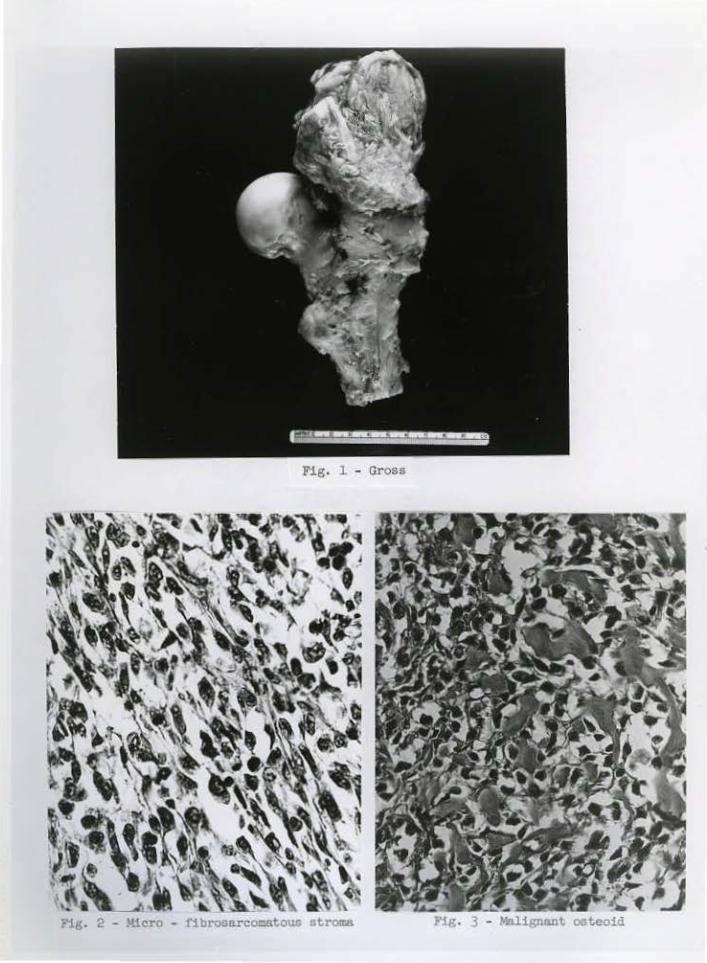
Fig. 1 - Gross
Pig. 3 - Mol.ignant ooteoid
Houston Society o"f Clinical Pathol ogist s Seminar April 25, ~964
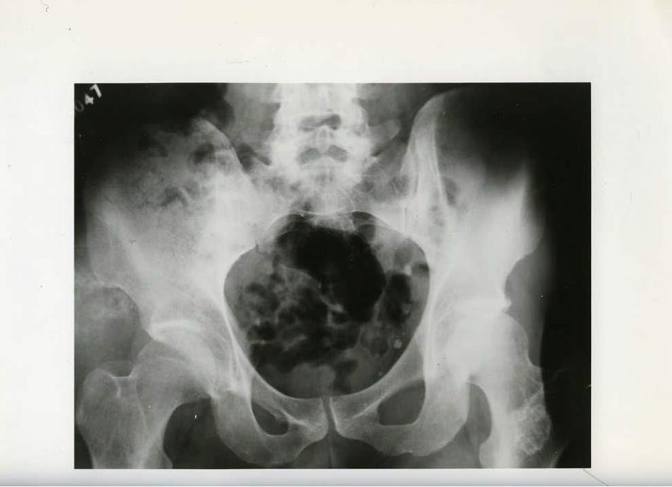
CASE 5 T-574- 63
A right hindquarter amputat ion was per -formed on t his patient following biopsy of a tumor mass in the r egion of t he r ight hip . Ori pathologic examination the tumor was fauna to be within striated muscl eJmeasured 10 x 8 x 7 em. and~hile firmly attached t o t he greater trochanter, showed no evidence of involvement of bone cortex. (Fig. 1).
Sect ions of the tumor show consi derable variation in histologic architecture , I n some areas the pi cture i s that of an extremely anaplastic sarcoma containing a scattering o1' mult inucleated giant cells of osteoclastic type. Here t he tumor cells are large , with round or oval nuclei and rather scanty cytoplasm. Mitoses are 1'r equent . In other zon~sJ t he tumor cells are spindled and show varying amounts of collagen production resembling immature or more ~~turated f ibr osarcoma. (F~g. 2). Small areas of osteoid production by tumor cells are apparent (Fig. 3) as are rather broad sheets o"f ca:rt; i l agi nous transformati on. Necrosis and hemorrhage are pr ominent in some parts of the lesion. At t he periphery. of the tumor, a small a r ea of maturat ion into well formed bone is noted .
In vi ew of t he history of trauma i n this case and the presence .of a so"ft tissue tumor mass, the possibil i t y of myosi t i s ossi f icans was immediately suggested. Since the lesi on was in soft t i s sues and did not appear to arise from the bone , t he differential .diagnosis is obviously between soft t issue osteogenic sarcoma or mal i gnant mesenchymoma and myositis ossificans. A number of points can be l isted iri favor of a soft tissue sarcoma with cartil age and bone formation and against myositis ossificans. Marked nuc·lear anaplasi a i s noted throughout the tumor and t hi s degree of nuclear change s t r ongly suggests sarcoma. In addition, there are .areas o.f necrosis within the neoplastic tissue. The tumor appears invasive at the margins and there i s no orderly e:onal mat uration as in myositis ossificans. By and large} there i s no maturat ion of the ost eoid throughout t he tumor.
Parosteal osteogenic sarcoma i s excluded in this c.ase in that the tumor .does not involve the adjacent bone. Since the differentiation observed 1n the t umor invol ves only carti lage and osteoid} it would appear ina~propriate to use t he term "malignant mesenchymoma" for the tumor.
DIAGNOS.IS.: Extraosseous osteogenic· sarcoma
References:
1. "Osteogenic Sarcoma of the Extraskeletal Soft Tissues 1 " Gerald Fine} M.D. and Arthur Purdy Stout, M.D. , Cancer, Vol. 9J No. 5, September} October, 195.6J. pages 1027-1043.
Follow up i n formation: Two years .after surgery patient developed massive pleural .ef:t'usion on the right. X-ray of chest after t horacentesis reveal·ed nodular shadows on the r i ght , l at erally near t he chest wall. Needle biopsy of a nodule showed metastati c mali gnancy compatibl e with osteogenic sarcoma. Nitr ogen mustard was i nsti l l ed. The pati ent succumbed in September} 1963 , two and one half year s after amputation .
'"~. 0~
Fig. 1 - Gross
PROXIML
Fig. 2 - Micro - spindled cell sareom component Fig. 3 - Micro - glandular component
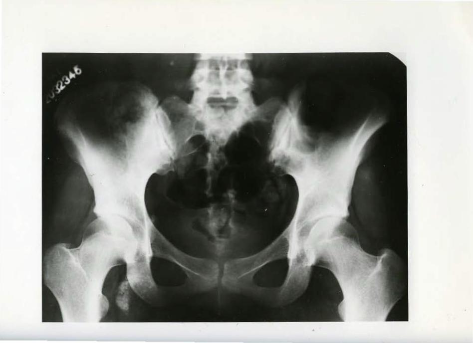
Bouston S.oci ety of Cl inical Pathologists Seminar CASE 6 April 25, 1964 16o9-55, 1616-55, T-575-63
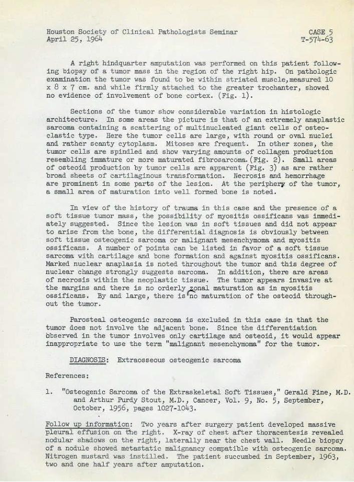
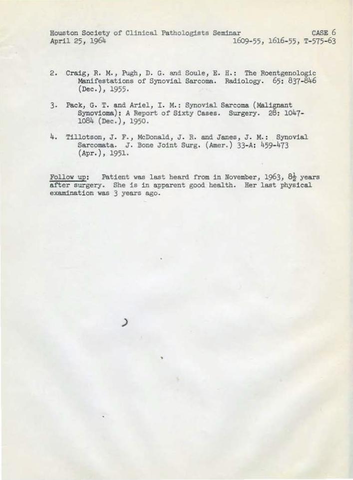
An exploratory operation 'ilas performed on this patient, the incision being over t he femoral vessels just distal to the inguinal l i gament . A tumor mass ~as found medial to the vessels with gross extension of the tumor int o t he f emoral vei n. The mass was r emoved along with a segment of femoral vein. Extending the incision cephalad, the sur geon noted that the tumor mass in the vein apparently ext ended approximat ely one i nch above the l igation. A hindquarter amputat ion was carried out 2 days later. Residual calcifying t UD!Or was present occluding the distal 2.5 em. of f emoral vein. (Fig. 1). No r esi dual tumor was found in the postoperat ive tumor site and all regional lymph nodes were negati ve for tumor .
Sect ions reveal a spindle cell neoplasm a port i on of which i s shown t o be within the l umen of a large vein. Focal calcification is prominent in some sect ions. Two tissue patterns are observed: (1) Fibroblastic zones, composed of rather small hyperchr omatic spi ndle cel ls showing some mitotic activi ty and arranged in intertwining f ascicles . This component is highl y suggestiye of fibrosarcoma. (Fig. 2). (2) Int erspersed with these zones a r e areas made up .of somewhat larger., less hyperchromatic cellS, l oosel y arranged or forming small clefts or spaces. The cuboidal cells l i ning some of these spaces pr ovide a di stinct gland-like or pseudoacinar pat tern. (Fi g . 3) . I n areas a distinct papillary configuration is provided by small t ongues of tumor tissue projecting i nto large spaces . The centers of many of t hese projections have undergone hyaline degeneration and focal calci fication i s present i n some of the papillae. Possibly only two i nterpret a t ions can be considered here: (1) synovial sarcoma with i ntravascular extension and extensive calci fication, (2) angiosarcoma involving femoral vein and adjacent soft tissue.
The possibi lity q7'< angiosarcoma is suggested .by t he large spaces within the tull!or into which project papill~J.ry proliferations cover ed by plump cells of possible endotheli al character. Favoring the interpretat ion of synovi a l sar co.ma are t he br oad :f'ibrosarcomatous zones combined with areas of paler staining cells with a pseudoglandular pattern. I do not t hink there is convinci ng evidence of differentiation towards vascular endothelial spaces wi thi n the neoplasm and my interpretatiop . favors a d iagnosi s of synovial sarcoma. The presence of calcificat ·ion withi n the tumor i s characteristic of this neoplasm.
DIAG~OSIS; Synovial sarcoma i nvolving soft tissue of ingui nal r egion ;dth extension into femoral vein.
Re:f'e·rences:
1 . Cr ocker, D. W. and Stout , A. P.: Synovial Sarcoma in Children. Cancer .· J.2: ll23- ll33 (Nov. -Dec .), 1959·
·Continued •..
Houston Society of Clinical Pathologists Seminar CASE 6 April 25, 1964 1609-55, 1616-55, T-575-63
2 . Craig, R. t4. , Pugh, D. G. a'ld Soule, E. H.: The Roentgenologi c Manifestations of Synovial Sarcoma. Radiol ogy. 65 : 837-846 (Dec.) , 1955.
3· Pack, G. T. and Ariel, I . M. : Synovial Sarcoma (Malignant Synovioma) : A Report of Sixty Cases . Surgery. 28: 1047-lo84 (Dec.), 1950.
4. Tillotson, J . F., McDonald, J . R. and Janes , J . !~ .: Synovial Sarcomata. J . Bone J oint Surg. (Amer. ) 33-A: 459-473 (Apr. ), 1951.
Follow uu: Patient was l ast heard from in November, 1963, Bt years after surgery. She is 1n apperent good health. .Her last physical exem1netion was 3 years ago.
)
'
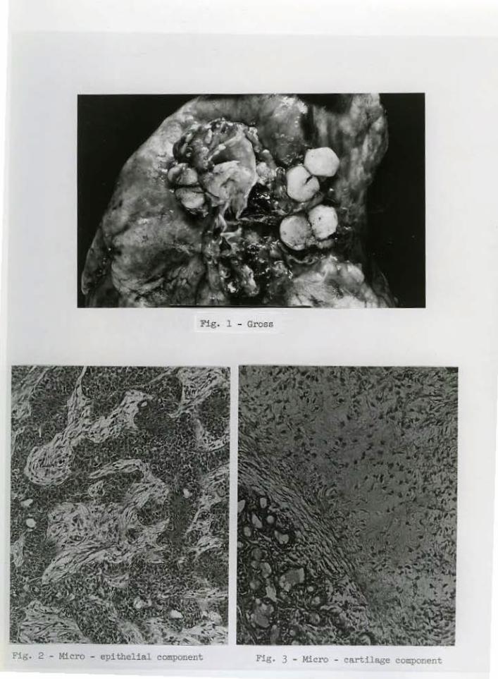
Fig. 1 - Gross
Pig. 2 - Micro - epithelial component Fig. 3 - Micro - cnrtiloge component

(
Houston Society of Clinical Pathologists Seminar April 25, 1964
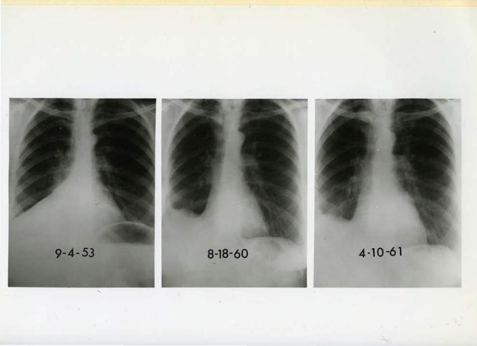
CASE 7 T-625-63, T- 626-63
This patient, on original l:lronchoscop:(c examination in 1953, was found to have a l ar ge t umor fill ing. the lumen of the r i ght l ower lobe bronchus. It bled easily on manipulation and had the gross appearance
·of a br onchial adenoma. At surgical expl oration, the tumor was noted to ar i se in t he bronchus intermediu.s and to ext end thr ough the br onchi al and i nto the hilus a distance of 2 em. Tne polypoid portion measured 1 x 5 em. A right middle and l owex· l obectomy was carried out. At t he second o~ration , in 1961, a: recurrent tumor mass was found in t he region of the stump of the previously resected lower lobe bronchus . The surgeon described a t .umor mass lying post er iorly against the chest wall cl ose t o the vertebrae , and extending through the parietal pleura. In adclit ion, t here were two recurrent nodules of tumor in the region of t he azygos vein and one on the di aphr agm.
Pathologic examination (1961 ·specimen) revealed a pol ypoid t1,1111or mas s within t he lumen of the resected bronchus (2 x 1.5 x 1 .5 em. ) and i n addition multiple nodules of tumor aggregating 5 x 2 x 2 em. were noted on the external surface of bronchus in the hilar region, as well as multiple (3) implants as described above. (Fig. 1).
Hi s t ol ogic sections of t he 1953 specimen a nd the 1961 recurrence ar e essent ially simil ar. The tumor consi sts of two el ements: (1) a prominent epitheli al component the cells of which are arranged in a wi de variet y of patterns (2) a connective tissue eleme~t whi ch i s l ess prominent and consists largely of somewhat myxoid, spindle and stellate cell areas in wr..ich chondroid met apl asia can be focally demonstrated. The tumor i n i t s varied structural patterns thus appears t o fulfill all requi r ements for a diagnosis of mixed t umor of saUvary gland type . The point of origin is apparently the mucus gl ands of the bronchial wall . Careful examination of the epithelial component reveals that the cells are small, regular· i n shape and s i ze, and t he overall appearance is that of a benign neoplasm. Mit otic figures are difficult to demonst rat e. Low power archit ectural pat terns include anastomosing or interlacing epithelial strands, well formed tubular and glandular structures , and more cellular solid sheets of epithelial cells corresponding to th~ well known patter n of cellular mixed tumor of salivary glands. (Fig . 2) . Foci of squamous metaplasia are readily demonstr ated within the epithelial component and in ar eas large clear or slightly granular cells in the tumor are strongly reminisc·e~t of the serous cells of bronchial mucosa. Myxoid areas composed of stellate and spindle cells are present to a lesser degree t han i n some mixed tumors of sali vary glands, but chondroid metaplasia can be demonstrated in these areas in both the primary and recurrent tumor. (Fig. 3). The benign appearance of the epithel i al component, the br oad range of pattern within the tumor and the presence of a connective element with newl y f ormed cartilage indicate to me that the tumor is identi cal vith mi xed tumor of
Continued . ..
Houston Society of CUni cal Pathologists Seminar April 25, 1964
CASE 7 T-625-63, T-626-63
salivary gland type. Support for t his concept is pr ovided by the slow rate of growth of t he t umor combined with a recurr ence in t he f orm of multipl e nodules eight year s after primary r esect i on .
n ifferent ial diagnoses in t hi s case i nclude bronchial adenoma of carc i noid type and t he ratqer uncommon w~co-epidermoid tumor of sali vary gland type ari sing i n t he bronchus. The l atter possibili t y was giyen some consideration on i ni tial s tudy of t he tumor but I bel i eve i t can be excl uded r eadi l y on t he basis of t he chondroid metaplasia of the stroma.
niAGNOSIS: 1•!i.Xed t umor of sal i vary glaoii. t ype , prima..ry in bronchus.
Refer ence :
W. s. Payne , M.D., R. S . Fontana, M. D. , L. B. Wool ner , M.D., Bronchi al Tumors Originati ng from Mucous Gl ands : Current Cl assifi cation and Unusual Manif estation , Medical Clinics of North Ameri ca, VoL liB, July, 1964 ( i n press) .
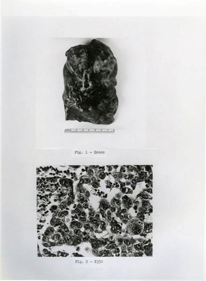
Fig. l - Gross
Fig. 2 - X350
Hous t on Society of Clinical Pathologists Seminar April 25, 1964
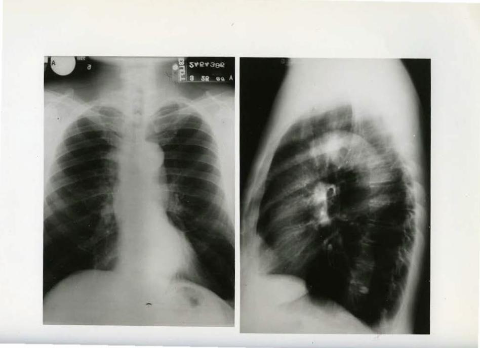
CASE 8 T-627-63
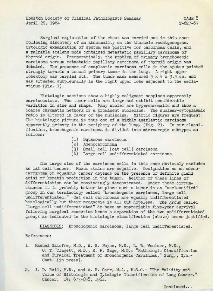
Surgical exploration of the chest was carried out in this cas·e. following diScovery of an .abnormality on the thoracic r oent genogr am. Cytol ogic examination of sputum was positive for carci noma cells, and a pal pable sca lene node contained metas tatic papillary carcinoma of thyroid ori gin. Preoperatively, the problem of prill'.ary bronchogenic carcinoma versus metastati c papillary carcinoma of t hyroid origin was deba ted. The presence of anaplastic carcinoma cells in the sputum pointed str ongly towards a second primary tumor in the lung. A r i ght upper lobectomy was carried out. The tumor !MSS measured 5 x 4 x 3·5 em. and was s ituated subpleurally in t he right upper lobe adjacent to the mediastinum. ·(Fig. 1).
Hi s t ologi c secti ons show a highly malignant neoplasm apparent ly carcinomatous. The tumor cells are large and exhibit consider .able variation in size and shape . 14any nuclei are hyperchromatic and sho~ a coarse chromat in network or a prominent nucleolus . The nuclear--cytoplasmic ratio is altered in favor of the nucleolus. Mitotic f i gure s are frequent . The histologic picture is thus one of a highly anaplastic carcinoma apparently primary in the periphery of the lung. (Fig. 2). In our classification, bronchogenic carcinoma is diVided into microscopi c subtypes as follows :
(1) (2) (3) ( 4)
Squamous carcinoma Adenocarcinoma Small cell (oat cell) carcinoma Lar ge cell undifferentiated ca r cinoma
The large siz-e of the carcinoma cells in this case obViously exclud~s an oat cell cancer. Mucus stains are negative . Designation as an adenocarcinoma or' squamous cancer depends on the presence of definite gland acini or kerati .n production in the tumor. Neither of these l in.es of differentiation can be convinci ngly demonstrated. Under these circums tances it is probably better to· place such a tumor i n an "unclassified" group in our terminology called "bronchogenic carci noma, l arge cell undifferentiated." Oat cell carcinomas are equally undifferentiated hisologically but their prognosis is all but hopeless . The group called "large cell undifferentiated" do have an appreciabl e five-ye.ar surVival following surgical resection hence a separ~tion of the two undifferentiated groups as indic·ated in the histologic classification (above) seems· justified.
;DIAGNOSIS: Bronchogenic carcinoma, large cell undifferent iated.
References :
1. Manuel Galofre, M.D., w. s. Payne, 14.D. 1 L. B. Woolner, M.D., 0. T. "Claget t, M.D. , R. P-·. Gage, I•!. D.: "Pathologic Classi :f.ication and Surgical Treatment of Bronchogenic Carcinoma," Surg., Gyn.Obst. (in press) .
2 . J , D. :Reid, M.D .. , and A. H. Carr, !.f.A. , B. S.C .: "The Validity and Va,l\le of Histologic and Cytologic Classification of Lung Cancer. " Cancer. 14: 673-698, 1961.
Continued •. .
Houst.on Society o£ Clinical Pathologists Seminar April 25, 1964
CASE 8 T-627-63
Follow un: At the t ime of chest surgery, two enlarged hilar or media·st.inal lYIJ!Ph nodes were found to contain papillary carcinoma of thy:roid origin. Subsequent surgicaL exploration of thyroid gl:and revealed an occult papillary carc inoma (1 em. in diamet er) i n the lei't lobe near the isthmus. Subt otal thyroidectomy was car ried out .
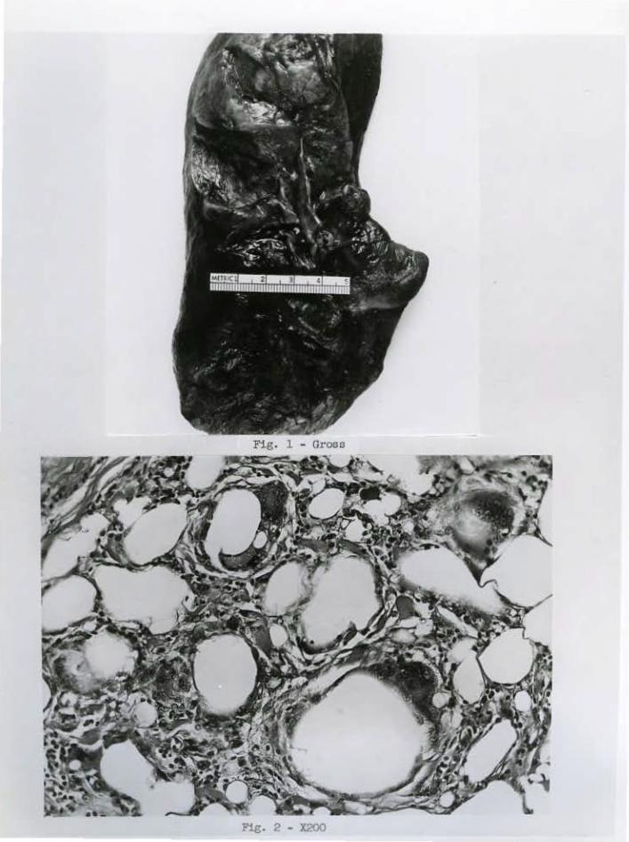
Fig. 1 - Gross
Fig. 2 - X200
Houston Society of Clinical Pathologists Seminar April 25, 1964
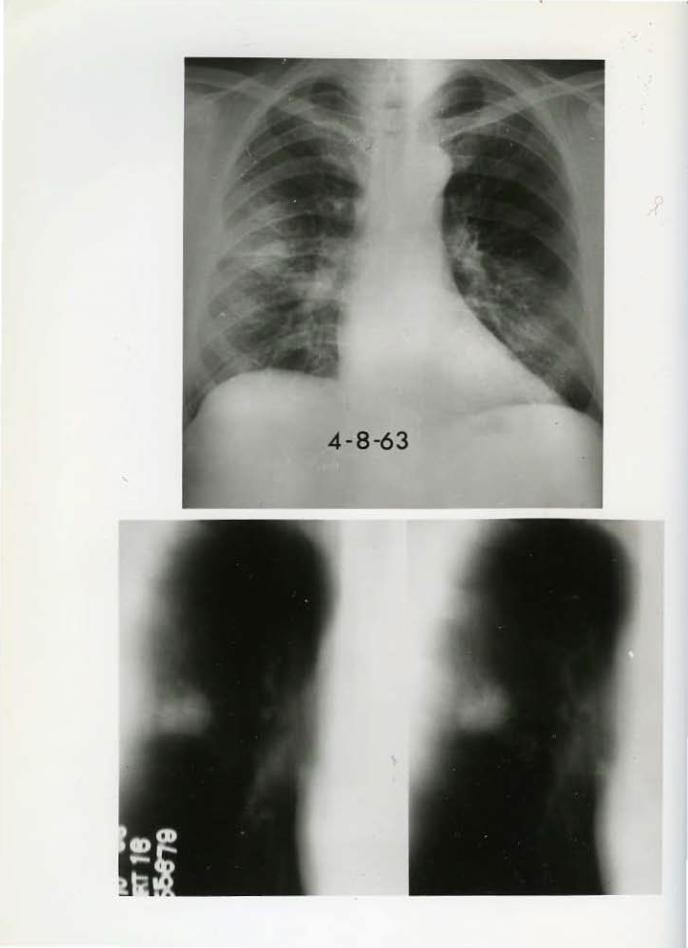
C.ASE 9 T-628-63
This patient was operated on f or an i ndeterminat e lesion of t he right lung knmm t o be present for one year . On gross inspection, t he· s urgeon f ound ~ mass in the ant erior segment of t he upper lobe adjacent to the middl e lobe. It had all tqe gross characteri stics of cancer i ncluding puckering of the overlying pleura. There was no evidence of met ast atic spread to ,egional nodes. A right upper l obectomy was carried ou1; .
Pat hol ogic examination revealed .a consolidated area involving pr edominantly the anteri or segment of the lobe (8 x 4.5 x 3 em.) with focal puckering of the overlying visceral pleura (Fi g . 1). Sections of the "tumor" stained with hematoxyl in-eosin show a cha~·acteristic and diagnostic honey combed patt ern of small and lar ge spaces , apparently empty. Tnese spaces are associated wi t h varying amoun.ts of rea9ti ve fibrosis) thin and delicate in some areas, coarser and more cellular in others. Mult inucl eated giant cells of foreign body type are readily recogni zabl e at the margin of many of the clear spaces; in others t he giant cells are . so flattened and e·longated as to be scar cely recognizable CFig . 2) . Frozen sections of the lesion stai ned for fat gave a pale orange yellow color wit h .Sudan I V and stains for mineral oil using Sudan Bl ack B a pa l e lavender. Chemical analysis of the wet t i ssue was carried out. The consol idated pulmonary parenchyma was shown to contain 6~ mineral oil by weight . A diagnosis of mi neral oil granuloma was t hus establ ished. No recent history of mineral oil ingestion or use of oily nose drops could be establ ished. However, on furt her questioning, t be patient, now 66, believed that hi s mother had given him mineral oil in his boyhood.
DIAGNOSIS : Mineral oil g ranuloma i nvolving l ung parenchyma
References:
J ampolis , Robert W. , M. D. , McDonaJ.d, John R. , 1·1. D. , and Clagett , o. Theron, M.lJ. ''International Abstracts of Surgery," Vol. 97, No. 2, pages 105- 119, August, 1953 .
L--- Fig. 1 _ Gross
Houston Society of Clinical Pathol ogists Seminar April 25, 1964
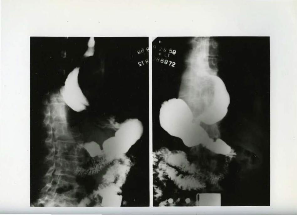
CASE 10 T-629-63
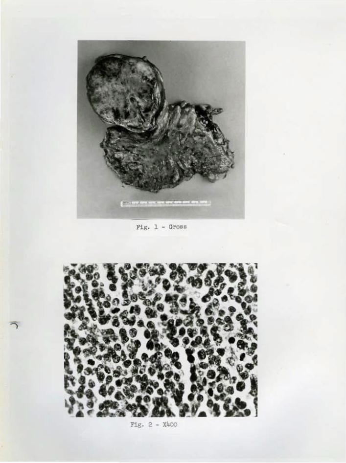
This pat i ent was explored for ar. indeterminate lesion of the l ower mediastinum. The surgeon described a large s mooth tumor mass deep in t he mediastinum, anterior to the aorta and posterior t o the heart . I t was apparently encapsulated a nd could be- mobilized readily ·out it could not be separated -easily from the esophagus and cardia of the stomach .. The tumor grossly resembled a sarcoma and the surgeon felt t hat t he lesion was probab+y a l eiomyosarcoma of lower esophagus. The upper end of the stomach and l ower esophagus were mobilized and widel y r esec.ted. Gro,ss examination revealed an apparently encapsulated tumor (10 x 9 x 7 em.) adhe·rent to the- outer aspect o:!' th_e l ower esophageal ••all (Fig. 1 ).
Sections of the . tumor show a uniform or "monotonous" pattern of tumor cells of the l ymphoi d ser ies probably bast consi dered s omewhat i mmature lymphocytes. The nuclear s tructure shows rather finely divided chromatin but without t h e prominent nucleoli a ssociated with l ymphobl asts (Fig . 2) . The t umor cells appear too small to be des ignated reti culum cells. Mit oses are readily found in the tumor . There i s no normal lymph node structure such as germ center .or follicle formation or eVidence of open sinuses. No pl asma cel l or eosinophilic infil tration i s noted.
The differential diagnosis ~<ould appear to include (1) m_ed iastinal angiofol l icula r lymph node hyperplasia,(2) primary l ymphosarcoma of lo••er outer wall of -esophagus, possiblrt origi nating in a paraesophe:geal l~~h node. (3) Lymphosarcoma of cardia of stomach extending into lower mediast i num. Angiofollicu~ar l ymph node h~~erplasia i s a t erm used to describe a benign, tumor - l i ke enl argement of lymph nodes . The tumor s are most ly but not invariably f ound in t he chest especially in t he mediastinum. They pr esent as s ol itary masses, 5 to 16 em. in d iameter. Micr oscopically> there are 2 predominant f eatures: Hyperplasia of lymphoid follicles i n which is s een ma.rked capillary proliferat ion wit h endothelial hyperplasia. The lymphoi d follicles usually show pale central reticular cel l s and associated- ramifYing capillar i es.
The ab sence of any lymphoid follicles i n the case at hand w-ould s eem to exclude angiofollicular hyperpl asia in the di fferential diagnosi s. The his tologic evidence i s strongly in favor of a malignant tumor of lymphoid origin, perhaps best des-cribed by the term lymphosarcoma. Anatomic evidence points to· a paraeso~hageal origin rather than gastr ic with involvement of the adventitia or outer aspect of t he muscle wall only.
D:r:AGI>IOSIS: Lymphosarcoma of l ower esophagus (paraesophagea:l)
FolJ.o,'w \lP: No postoperat ive irradiation given. November, 1963, 4 1/3 year s ai'ter surgery. Feels of recurrence o f metastasis.
Patient last s e.en well - no evidence
Mediastinal Nodes
F":ig. 1 - Gross
Right Radical Neck Dissectio~
Fig. 3 -Micro - X300
Houston Society of Clinical Pathol ogists Seminar April 25, 1964
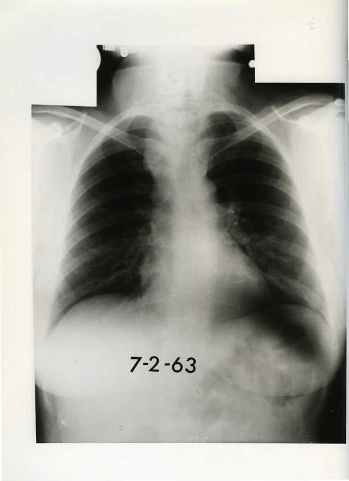
.CASE 11 T-640-63
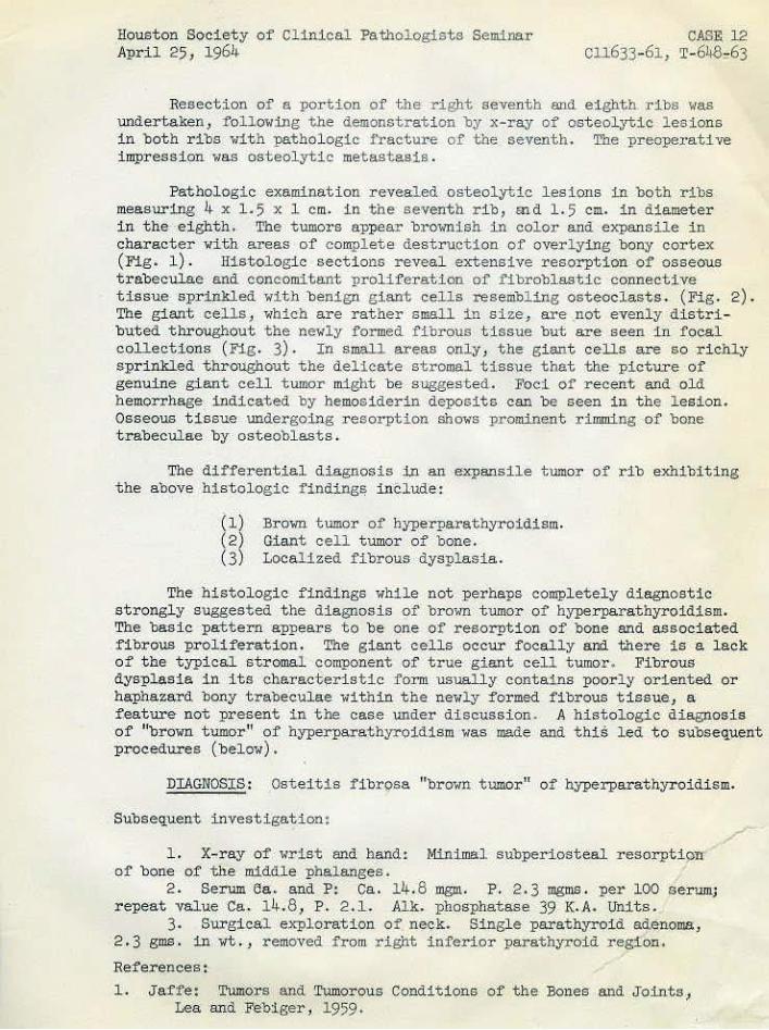
The . operation performed o.n thi s "oman consisted of a total right l obect.omy, a partial left l obectomy with removal of isthmus , r ight r adi cal neck di ssection and removal of invol ved lymph nodes from pretr acheal region, anterior mediastinum, r i g)lt tracheo-esophageal gToove and r i ghtposterior superior mediastinum. The pr imary t umor in the right lobe of the t hyroid had been partially removed previously.
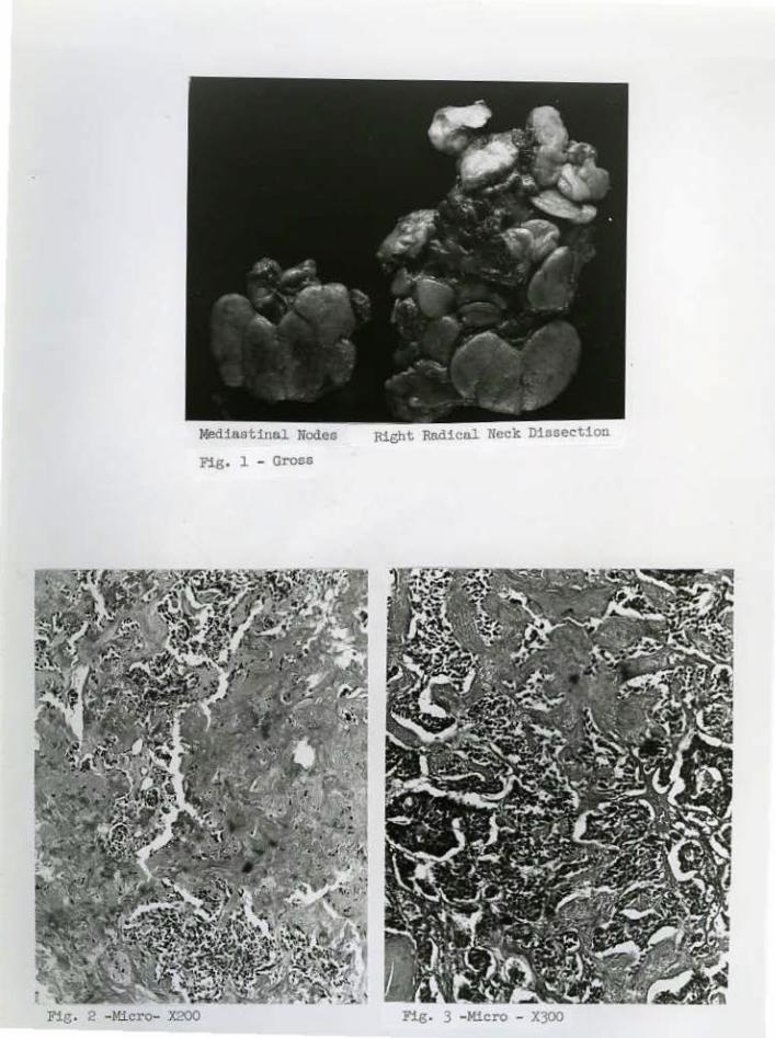
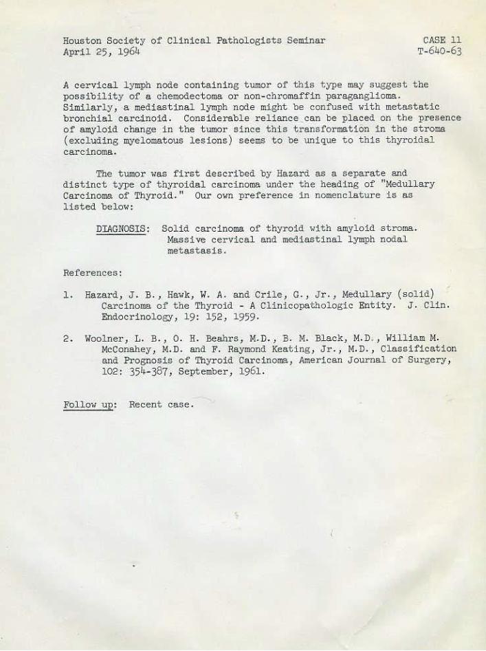
Pat hologic examination r evealed a small focus of residual tumor in the remnant of the right lobe and a normal left lobe and isthmus. T'ne lymph node involvement in this case "as ' massive. Several of the' tremendousl y enl arged pretr acheal and tracheo-esophageal groove nodes measured up to. 5 em. in diSJJleter. The mass of nodal tissue in the mediastinum was of sufficient bulk t o displace the trachea to the left and appear on x-ray of t he .chest as a: ••idenin:g of the superior mediastinum. (Fi g. 1).
Hist ol ogic sections reveal a tumor composed of rather small cells wi th round or oval nuclei and scanty cytopl asm. The tumor c·el ls are separated int o varying sized compar tment s by a r at her prominent fibr ous stroma (Fig. 2) . In some areas, t he s t roma is acellular and appears hya!ine in structure; i n others, an amyl oid trans~ormation i s suggest ed on H & E sections and special staine ar.e positive for this substance. T'ne amyl oi d change in the st ro.ma is spotty in distribution both in the primary t umor and met ast at ic deposits . It is noteworthy that in some foci the amyloid stroma appear s to repl ace or crowd out the t umor cells almost complete:Ly resulting i n a hi ghly character i stic low po••er pattern (Fig. 3). In addition t o transf ormation of the large stromal trabeculae, smal l foci of amyloid-like mat erial ~zy appear wit hin tumor nests them-· selves sometimes gi ving a pseudo adenoid cystic appearance. In areas devoid of such changes, the tumor cel ls are seen to occur in sheets in a str uct ureless or ' 'solid" pattern. No' papillary processes or follic l e formation are· observed. Although the cel1s are typically round, a few areas show some· spindling of the .nuclei.
In some eY~mp1es of this thyroidal tumor , a few small folli cles cont a ini ng coll oid are seen . These appear to represent r esidual normal fol licles surrounded by tumor rather than true follic1e formati on or colloid production by the tumor. While undifferenti ated i n structure·, t he tumor does not exhibit eVidence of ra]>id gro••th. As a rule mitotic figures are r ather difficult to find. When presented with a primar y thyroi dal tumor of thi s type, t he pathologi s t should be guided to t he correct diagnosis by such features as (l) circumscr ibed but unencapsulated gross t umor. (2) Small round or Spindl e shaped cells growing in "solid" sheets ••i t.hout any J?81?i llary or f olli cular differ entiat i on . (3) Absence of evidence of rapid growth such as is seen in anaplastic carcinoma (4) varying degrees or hyaline a:nd/or amyloi d tra:nsformation in the stroma.
Continued ••.
Houston Soci~ty of Clinical Pathologists Seminar April 25 , 1964
CASE 11 T-640-63
A cervical l ymph node containi ng tumor of this type may suggest the possibility of a chemodectoma or non-chromaffin paraganglioma. Simil arly, a mediastinal lymph node might be confused with metastatic br onchial carc;J,no·i d . Considerable reliance can be placed on the presence of amyloi d change in the tumor since this transformation i n the stroma (excluding myelomat ous les ions) seems to be unique t o this thyroidal carcinoma.
The tumor was fir s t described by Hazard as a separate and distinct type of thyroidal carcinoma under t he heading of "t•!edullary Carcinoma of Thyroid." Our o••n preference in nomenclature is as l i sted below :
DIAGNOSIS: Soli d carcinoma of thyroid wit h amyloid stroma. 1-la.ssi ve cervical and mediast inal l ymph nodal metastasi s.
References:
1. Hazard, J . B., Hawk, W. A. and Crile , G. , J r., Hedullary (solid) Carcinoma of the Thyroid - A Clinicopathologic Entit y . J, Clin. Endocrino~ogy , 19: 152 , 1959.
2 . Woolner 1 L. B., 0. H. Beahrs, !4. D. , B. !4. Black, M.D. 1 Willi am H. McConahey, l•l. D. and F. Raymond Keating, J r ., M. D., Classifi cation and Prognosis of Thyr oi d Carcinoma, American Journal of Surgery, 102: 35!f-387 , Se:pt ember, 1961.
Follow up: Recent case .
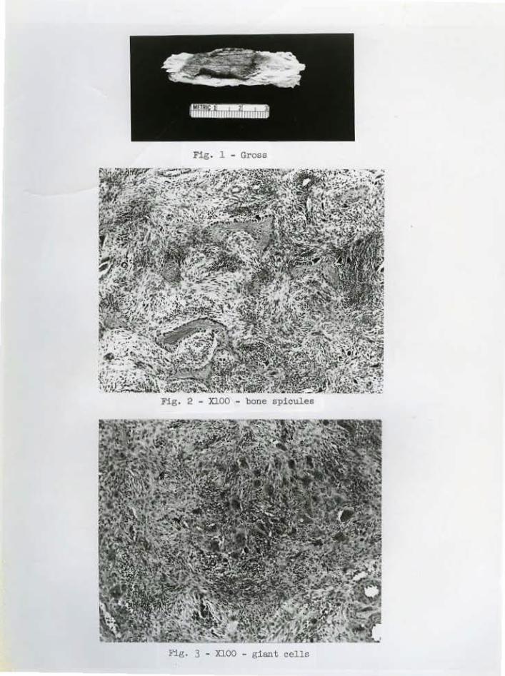
Fig. 1 - Gross
Fig. 2 - XlOO - bone spicules
Fig. 3 - XlOO - giant cells
Houston Society of Clinical Patho~ogists Seminar April 25, ~961!.
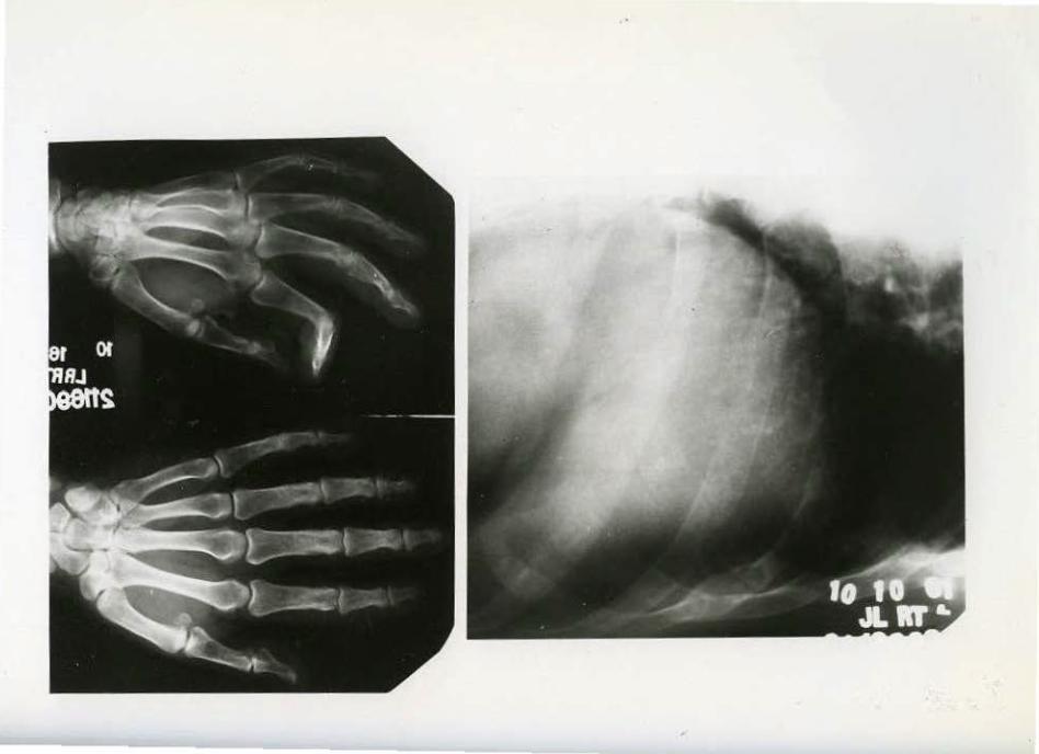
CASE. 12 Cl.1633-61, T-648::.63
Jlesection of a portion of the right seventh and. eighth ribs was undertaken, :following the demonstrati on by x-ray of ost eol ytic lesions in bot h r ibs w;Lt h pathol ogic fracture of the aeventh. The precp.erati ve impr ession was osteo~ytic metastasis .
Pathologic examinati on revealed osteolytic lesions in both ribs measuring 4 x 1. 5 x 1 em. in the seventh r ib, m d 1. 5 em. in diamete·r in the ·e i ghth. The t umors appear brownish in color and e xpansile i n character with areas of compl ete dest ruction of overl y!ng bony cortex (Fig. 1) . Histo~ogic sections reveal extensive resorption of osseous trabec~e and conc·omitant nrolif er ation of fibroblastic connecti ve tissue sprinkled with benisn giant cell.s resembling osteoclasts . (Fig. 2). The gi ant cells, which are rather small in size, are .not evenly distribute<i throughout. the newly formed fibrous tissue but are seen in focal c ollections (Fig. 3). In small areas only, the gi ant cells are s o rich~ spr inkled throUghout t he delicate stromal tissue that the picture of genuine giant cell tumor might be suggested. Foci of recent and .ol d hemorrhage· indicated by hemos iderin depos i t s can be seen in the l esion. Osseous t ;Lssue . undergoing resorption shows ])Tominent rimming <>f bone trabe.culae by osteoblast s .
The differential diagnosis in an e'xpansile tumor of rib exhibiting the above h i stologic finding~ 1nclude :
(1) Br own tumor of hyperparathyroidism. (2) Giant cell tumor of bone. (3) Localized fibrous dysplasia.
The histologi c findings while not perhaps completely diagnostic strongly suggested the diagnosi s of bro'Nn tumor of hyperparathyroidism. The basic patt ern appears to be one o.f re<:~orption of bone and associated fibrous proliferati on. The giant cells occur focally and there is a l ack o f t he typi cal stromal component of true giant cell t umor, Fibrous dysplasia in its characteristic form usually contai ns poorly ori,ented or ha1>ha zard bony t r abeculae ;;i t h in t he newly formed fibrous t issue., a feat ure not present in the case under dis cussion . A hi stologic diagnos;is of "brown t umor" of hyperpara thyroidism was made and this led to subsequent pr-ocedures ( b elm; ) .
DIAGNOSIS: Os t e itis fibrosa "brown t umor" of hyperpara:th:y-roidi sm.
Subs.equent investigation!
1. X-ray of· 't-lris t and hand: Minimal subperios teal reaorpti)ln of bone of the middl e phalanges.
2 . Serum ea. and P: Ca. 14. 8 mgm. -p·. 2 . 3 lllo"1llS . per 100 s .erum; repeat value Ca. 14.8, P. 2.1. Alk. phosphatase 39. K.A. Units.
3. Surg;lca:l exp.J.otati on of neck. S.ingl e parat hyroi d adenoma, 2.3 gms. in wt . , removed from right inferior parathyroid region.
Ref erences:
l. Jaffe: Tumors and Tumorous Conditions of the Bones AJld .Joints, Lea and Feb i ger , 1959·
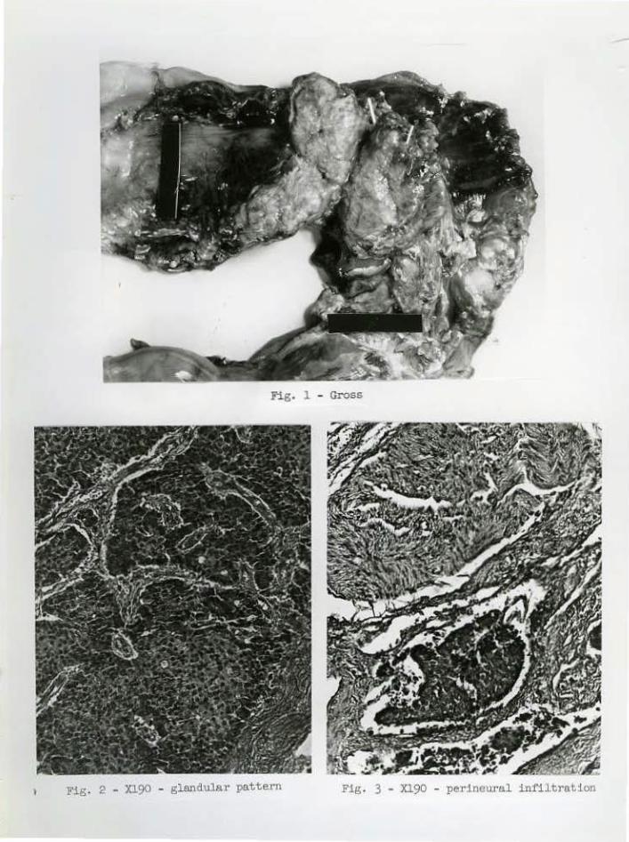
Fig. 1 - Gross
Fig. 2 - J0.90 - glandulaJ· pattern Fig. 3 - Xl90 - perineural infiltrntion
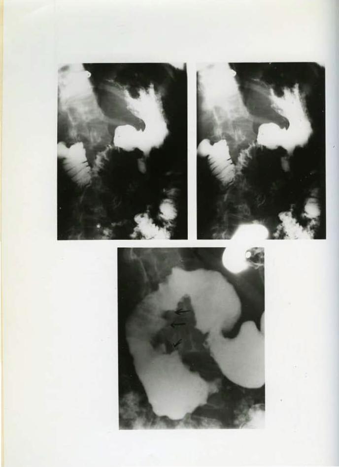
Houston Societ y of Clinical Pathologists Seminar CASE 13 April 25, 1964 X279-59, .x11096-61, Xl07-59, T-647-63
F'olloying a prolonged period of s teatorrhea, vomiting and abdominal pain, this patient underwent surgical explorat ion. A tumor of the pancreas was stxongly suspected preoperat-ively on the basis of an abnoTDJality of t he second part of the duodenum seen by roentgenogram. Fol lowing needle biopsy of the head of the pancreas, a Whipple procedure vas carried ·out .
The surgical specimen consisted of the distal 10 em. of stomach, entire duodenum and upper 15 em. of jejunum, head of pancr-eas and terminal portion of common duct. Pathologic examination showed a poorly encapsulated tumor of the he!);d of the pancreas, 6 ell!. in diameter. (Fig. 1). Multiple de·ep chronic inflammatory ulceTs 0 . 5 to 1. 5 car. in diameter wer e ·pr esent in the duod·enum di s tal to the §>mpulla of Vater , also multiple superficial mucosal ulcers .of j.ejunum up to l em.. in dia!lleter .
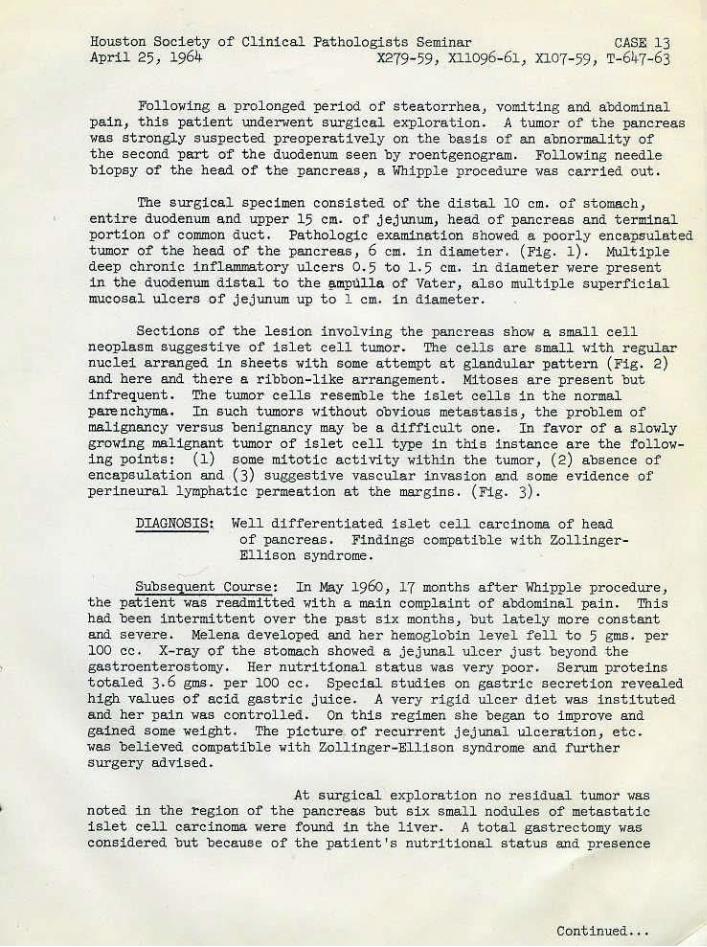
Sections of the lesion invol ving t he pancreas show a small cell neoplasm suggesti ve of islet cell tumor . The cells are small with regular nuclei arranged in shee·ts with some attempt at glandular pattern (Fig. 2) and here and there a ribbon-like· arrangement. Mitoses are present put infrequent. The tumo~cells resemble the islet cells in the nor mal parenchyma. In such tumors without obvious metastas i s , the problem of malignancy versus benignancy may be a difficult one. I n favor of a slowly gr-owing malignant tumor of islet cell type in this instance are the following points: (1) some· mitotic activity wi thin the tumor, (2) absence of encapsulation and (3) suggestive vascular invasion and some evidence of perineural l ymphatic permeation at t he margins. (Fig. 3) .
DIAG~OSIS~ Well differenti ated islet cell carcinoma of head of pancreas. Findings compatible with ZollingerEllison syndTome.
Subsequent Course: In May 1960, 17 months after Whippl e procedur~, the pa:tient was r eadtidtted with a main C!>mplaint of abdominal pai n. Thi s had been intermittent over the past s.ix months, but lately more constant and severe . Melena d.eveloped and her hemoglobin lev.el fell to 5 gms. per 100 c·c. X-ray of the stomach showed a jejunal ulcer j ust beyond the gastroenterostom,y. Her nutri t :!onal status was very poor. Serum proteins totaled 3. 6 gms. per 100 cc. Special s t udies on gastric secret ion revealed high values of acid gastri c juiee. A very rigid ulce·r d;l:et was i nstit uted and her pain was control led. On this regimen s he began t o improve and gained some wei ght . The picture of r .ecurrent jejunal ulceration, etc. was believed compatible wi th Zollinger-Ellison syndrome and further surger y advised. · ·
Jlt surgical explor ation no residual tumor was not ed in the region of the pancreas but six s mall nodules of metastati c islet c·eu carcinoma w.ere f ound in the liver. A total gastrectomy was co):!sidered but because of the patient 's nut ritional status and presence
Continued .. .
Jlouston S·o~i~ty of Clinical Pathol ogists Seminar Cl!SE 13 April 25~ 1964 X279-59, Xll096-61, XJ.W-59, T-647·-63
of li V!!r metastas'is, the surgeon .,.lected to d<:> a parti al gastrectomy and b.i latera.l ·vagotomy. Pathol ogic findings i n the resect~d spe~imen showed a subacute perforating ulcer 2 em. in d iamet er involving the anastompsis and di stal to this in the jej unum a subacute inflammatory perforating peptic ulcer l em. in di amE)tE)r . Biopsy specimen f:t'om liver revealed metast atic islet cell tumor .
The pati~nt did well for eight months following surgery, then developed further attacks of abdominal pain. She di ed of an oven;helming gas bacillus infe ction shortly ther eaft er .
.AUTOPSY FINDINGS~ 1. Large gastric ulcer on gastric side of stroma '.rith perforation onto wall of tr;msverse colon.
2. :Recurrent nodUle of i slet o;.eU t .umor 5 ~m. in diameter surrounding porta-l vein.
3. Multiple me·t ast at ic nodules up to 2 m. in l iver.
.4. Septicemia: due t o gas bacillus i nfection.
Refe:I'ences~
1. K .. A. Huisenga, Goodrich, and William H. J. Summerskill; Peptic Ulcer with Islet Cell Tumor; A :Reappraisal, Am. Journal of l<!edicine
(in press).
2 . L . o. Underdahl, M.n·. , Lewis B. lfoolner, M. D. , and R. Marden Black, M.D.: Multiple Endocrine .Adenomas: Report of e'ight cases in vhich t he Par athyroids, Pituitary and Pancreatic Islets were Involved. The Journal o f C:lini pal Endocrinology and loletabolism, Vol. XIII, No. 1 , January, 1953, pages 20-47.
Fig . 1 - X200
Fig. 2
x6oo - spinclled cells
Fig. 3
~ x6oo - giant cells
,
Houston Society of Clinical Pathologists Seminar April 25 , 1964
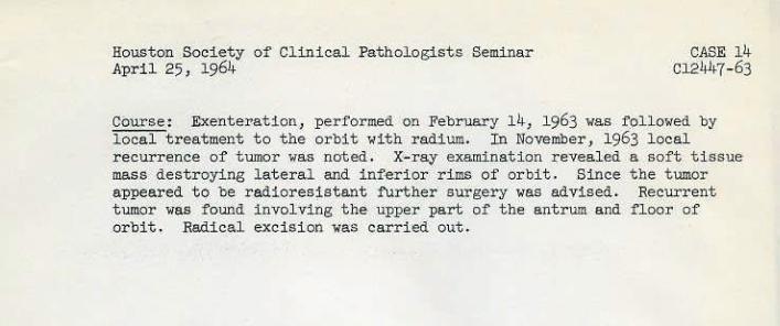
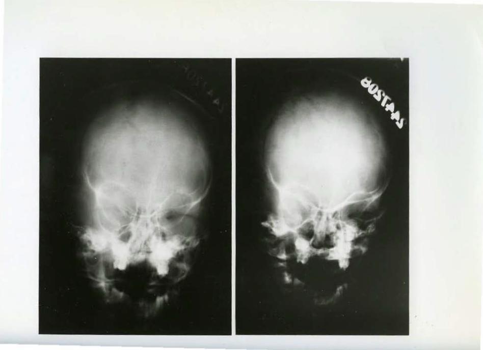
CASE 14 Cl2447-63
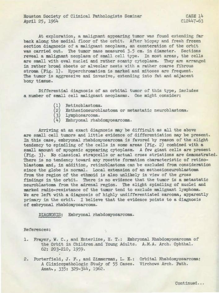
At expl oration, a malignant appearing tumor was found extending far back along the medial floor o~ the orbit. After biopsy and fiesh frozen section diagnosis of a malignant neoplasm, an exenteration of the orbit was carried out. The t umor mass measured 3·5 em. in d~meter. Sections reyeal a I!!Blignant neoplasm of small cell type. In most areas, the cells are small with oval nuclei and rather scanty cytoplasm. They a:re arranged in rather broad sheets or alveolar nests with a rather coarse fibrous stroma {Fig. l). Hyperchromat ism is marked and lllitoses are fiequent. ~e tumor is aggressive and invasive, extending into fat and adjacent bony tias.ue .
Differential diagnosis of an orbital tumor o~ this type, includes a number of small cell malignant neoplasms . One might consider:
Retinoblastoma. Esthesioneuroblastoma or metastatic neuroblastoma. Lymphosarcoma. Embryonal rhabdomyosarcoma.
ArriVing at an exact diagnosis may be difficult as all the above are small cell tumors and little evidence o~ differentiation may be present. In this case, embryonal rhabdomyosarcoma is favored by reason of the slight tendency to spindling of the cells in some areas (Fig. 2) combined with a small amount of myogenic appearing cytoplasm. A few giant cells are present {Fig. 3). No cl.assical strapcells or actual cross striations are demonstrated. There is no tendency toward any rosette formation characteristic of retinoblastoma a:nd, in addition, retinoblastoma can be excluded fiom consideration since the globe is normal. Local extension of an esthesioneurob~astoma 1':rom the region of the ethmoid is also unlikely in view of the gross findings in t_he orbit . There is no evidence that the tumor is a metastatic neuroblastoma fiom the adrenal region. ~e slight spind~ing of nuc~ei and marked radio- resistance of the tumor tend to exclude malignant lymphoma. We are lett with a di~nosis of highly ~differentiated sarcoma, apparently primary in the orbit. I believe that the evidence point s t o a diagnosis of embryonal. rhabdomyosarcoma.
DIAGNOSIS! Embryonal. rhabdomyosarcoma .
References:
1. :Frayer, W. C., and Enterl.ine, H. T. : Embryonal Rhabdomyosarcoma of the Orbi t in Children,and Young Adults . A.M.A. ~reb. Ophthal. 62: 203-2l0, 1959·
2 . Porterfield, J, F., and Zimmerman, L. E.: A Clinicopathologic Study of 55 Cases. Anat,, 335: 329-344, 1962.
Orbital Rhabdomyosarcoma: Virchows Arch. Path.
Continued .. .
Houston Society of Clinical Pathologi sts Seminar April 25, 1964
CASE 14 Cl24h7-63
Course: Exenteration, performed on February 14, 1963 was follo'•ed by l ocal t reatment to t he orbit with radium. In November , 1963 local reclll'rence of tumor was noted. X-ray examination revea.l ed a soft t i ssue· mass destroying lateral and inf~rior rims. of orbit. Since the tumor appeared to b e radiores~stant fUrther surgery was advised . Recurrent tumor was found i nvolving the· upper part of the antrum and f l oor of orbit. Radical excis ion was carri ·ed out.
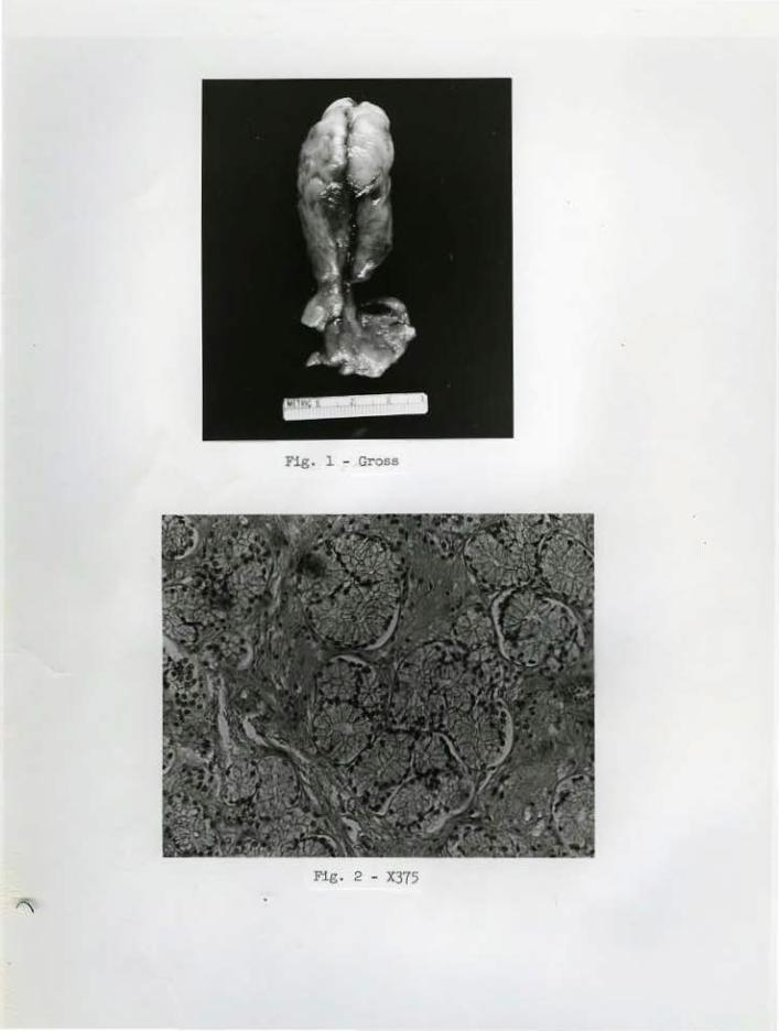
Fig. l. - Gross
Fig . . 2 - X375
Houston Society o~ Clini cal Pathologists Seminar April 25, 1964
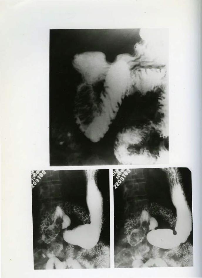
CASE 15 C2315-61
The resected tumor mass measured 6 em. in length and 2.4 em. in diameter. It was ~eduncu1at~d and attached to the duodenal wall by a pedicle 2 em. in length. The external surface was smooth. The cut ,s.urface r.eveal a solid tumor divided by narrow septa into lobul es up to 1 em. in diameter. (Fig. 1).
Sections of the tumor show well differentiated glandular tissue t hroughout (Fig. 2). The gland acini are composed of large cells with voluminous cl ear or slightly gland).llar cytoplasm and small regular basally pli'ICed nucl ei. The architecture is somewhat lobular with rather delicate interlobular septums. Proliferating f i brous tissue separates individual gland acini. The entire tumor is ensheathed in normal duodenal mucosa and at the region of the stalk, the tumor acini merge with. and are indistinguishable from the normally p l aced Brunner 1 s glands. This ·evidence plus tre absence of cellul ar atypia, mitoses or invasive qualities l ead me to believe that the tumor represents a rare primary duodenal tumor, namely adenoma of Brunner 1 s glands .
Perhaps as many as 75 examples of small t umors or hyperplastic nodules of Brunner's glands (usually 1 em or less in diameter) have been desc·ribed in the literature. In addition, ten larger tumors of this type have been reported. Most large tumo:r;s have caused symptoms of gastroi ntestinal bleeding, obstruction or intussus ception. Rare reports of malignant tumors arising in Brunner's glands have been p resented but the· evidence is not convincing.
DIAGNOSIS: Pedunculated adenoma of' Br tumer 's glands of duodenum.
Reference:
Lloyd Silyerman, M.D., John M. Waugh, M.D., Kenneth A. Huizenga, M.D., Edgar G. Harrison, Jr., M.D.: Large Adenomatous Polyp of Brunner'a Glands, Am. J, of Clin. Pathology, Vol. 36, No. 5, pages 438-443, November , 1961
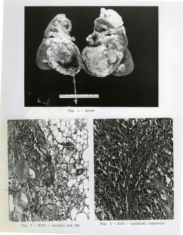
Fig. 1 - Gl'OSS
hg. 2 - Xl50 - ve~sels and fat
f . ' .,. ' . ' ''' 1 ' ' • -1. '· . }: ",. l ' ~, ' ·, ' ... ~ • . - ., \ • '\. ..... ~ • • :<..-
";\~I ' ' • •fa\ . • C . • .. - 1' . .. I ' .. • '... . ... -~ .... -- . .'4\ ' ' f! f ~ - ;,. ' • •
.· l t·,,. :;• .. I , , -~' ~I. l-\ >~,l-., ' ". ' ' ' ... . , ' ''\ ' 1 ' ' .. ~ ' ~.:_--.: ':..:-~ ,_._ -" . " : :-"'t ... . 'V' . . ~..,, , ' <. : .. t;: ' "t."\· '' v t , •. ~, ... _ t ···'l', . .., ·- '· . 1~. - •• •• • • " .......... ' .J · '<: ,;, , ...•. -. '} ' .. -· __ ,_, .,_
' ,- . ' ,.o I ' I (. ,. ~- ..... _ ~"·~ , . I. " ,·., '. i~<l!"~- -, .. ~_--r· -· ... ~ ·.• . '· .. ··:'! -~~ 'l
. ,;~ ~. l •' . . .. ; .:0. ' I ' I 'f- ' . ' ' ' T ~. ' ' '\ ,U,. ' t, .. :' ' . I . ~ .. ·-· " . I ._1 ~ ,i I ·.. • . . . ,... • . ·,. .... ~,.,.- r_-~ ,,,·. ,·• -··.).<
. · .~ .. ·,. ~ ' ' 1··~ ... , .. ! ~
"
,, ' ' ' ,,. '( '. i . '!r ~ •• " •• ~·--:' .J' \..~ .. ,. Q . \
··A··· I 'I '• . "'··-" ·"~-',~ :'· ·.-·.·· '-~_\,,;;_ ~.·
"· ~ .. '(•! • ) ' ( .· ).t! 'I-' ' I f'i , • ' ._ . , •' ' ·.' . 'I ' I ~ : 1;!1 1•-¥ - ) ' ' ' \ ·, l ' • • - - ;... t. I • • I> f • • • . . . . -.. ~.~ ... .~ "'. I· ... ~-·· ,-.... ~~
·. '• ·' ~ .- ' i ~ ' "' ' . .. " ~\~) -• ,-~~ l ., '. ' ' •' 1·' l '""- r-.1 ; ,. ;~.-~· .. ,. ' -~, ~- ~ ,
' • ' 1 ' • ' •• -1· ' \ ' , . .. ~, ; W r ,_.' V: • ~ :'.:.P ~~ ' \\ I• ' \ l · 1 f,_ . · ;,_.• ~.. , , ,
' \. ' \ ,I ' ' ' ' ~'\ \ ' f. • ·7' \ • • 4 ' ~ I ' ' ,, ~~ , , f\\!
' it·'~- ·., · -~~ • 1 \ \ r ,-.. r 't/~ \ ,~: ~~,~~ -.~ '\ ' " . 'j' ! ' '\ • "" 't-.. ~: ~ . I"! . • ' ,, . . •
.. ' ' ' , f • • . r ...., .. .,., ..... . ~~. ~ ' .. ' \. '" • '·' ~-- - --·\ !• ";r ,<J.
Fig. 3 - Xl50 - spindled component
Houston Society of C~inica~ Patho~ogista Seminar April 25, ~964
CASE 16 M9664-63
The surgeon found the right kidney to contain a tumor mass which had the consistency of a hypernephroma. It was situated in the lower ~~~ediu portion of the kidney and projected toward the lower pole. The ureter was deviated medially by the tumor.
Patho~ogic examination revealed a kidney weighing 560 gms. Arising in the region of the pelvis and invading the lower portion of the kidney almost to the capsule, there is a semi- encapsulated tumor measuring 10 x 8 x 6 em. (Fig . . 1). A moderate degree of hydronephrosis has resulted from distortion of ureter by t he tumor.
Sections show that the tumor is composed of a mixture of well differentiated mesenchymal tissues predominantly adult fat, smooth muscle and fairly ~arge vascular spaces. (Fig. 2) . The walls of the vascular spaces are or varying thickness and are composed of concentrically arranged spindle cells apparently surooth muscle . Many of these artery-like structures show hyuine degeneration of the wall. The bulk of the extravascular tissue is composed of adult fat cells intimately miXed with smooth muscle. The latter occurs in irregular sized foci or bundles interdigitating with fat and someti.mes closely surrounding arteria~ spaces. No mitoses are seen in this spind~e cell component of the tumor. (Fig. 3) . Areas of foam ce~ and multinucleated giant cells representing breakdown of fatty tissue are noted . Sections at the junction of tumor with kidney cortex show an irregular boundary with apparent destructi on of kidney substance .
Thi s tumor, composed of benign appearing mesenchyma tissues, has been noted particularly in or in the region of t he kidney although rather similar tumors are seen elsewhere. Various names have been suggested including angiolipoleiomyoma, capsuloma of kidney, hamartottl6, and benign mesenchymoms. The latter designation, suggested by Dr. Stout, has much to recommend it.
DIAGNOSIS: Benign ~esenchymoms of kidney.
References:
1 . Duane N. Tweeddale, M.D. , Clyde J . Dawe , M. D. , John R. McDonal d, M.D., and Or mond s. Culp, M. D.: Angiol ipoleiomyoma of the Kidney." Cancer, Vol. 8, 764-770, 1955 .
2. MarkS . LeBer, M.D., Arthur Purdy Stout, M.D.: Beni gn Mesench;mc·ti!IU in Children. Cancer, Vol. 15, 598-6o5, Jan. -June, 1962.
)
}012. ~ 1>C.1 J... ..H... I
<,-Cf: loose lt. # tt.. s. 1964
SEMINAR
HOUSTON SOCIETY OF CLINICAL PATHOLOGISTS
RESPIRATORY SYSTEM, GASTROINTESTINAL SYSTEM, GENITOURINARY SYSTEM AND BONE
Presented by
r~m::::::::sa~m~s SlaiiiEils m m::::::::se~m m~e,
LEWIS B. WOOLNER, M.D. HAROLD G. JACOBSON, M.D.
I PROFESSOR OF PATHOLOGY CHIEF, DIVISION OF DIAGNOSTIC ~9
MAYO FOUNDATION GRADUATE SCHOOL ~ RADIOLOGY
11 OF MEDICINE MONTEFIORE HOSPITAL e
l ROCHESTER, MINNESOTA BRONX, NEW YORK J m::::::::sm~m~mr;;;;;;;;;:;;u!l m~m m m::::::::sm mr====lm m
Houston, T •xos
----------------~--~-'A""':p":ril 25, 1964
•
HOUSTON SOCIE.'l'Y OF CLINICAL PATHOLOGISTS
1964 SEMINAR
RESPIRATORY SYSTEM, GASTROINTESTINAL SYSTEM, GENITOURINARY SYSTEM AND BONE
Jointly sponsored with
Texas Radiological Society and Houston Radiological Society
April 25, 1964
9 :00 A.H. - 4:00 P . M.
at
Jesse Jones ~ledical Library Bldg. -- Auditorium 1133 M. D. Anderson Blvd .
Texas ~2dical Center Houston, Texas
Supported by
ST. LUKE'S EPISCOPAL HOSPITAL and the Al-IERICAN CANCER SOCIETY
LEWIS B. WOOLNER, M.D. Professor of Pathology
Mayo Foundation Graduate School of Medicine
Rochester, Minnesota
Conducted by
HAROLD O. JACOBSON, M.D. Chief, Division of
Diagnostic Radiology Montefiore Hospital
Bronx, trew York
DIAGNOSES
~
~ ~>~sw J »o..J, fer Cnf(." -t t.k£""5
You are asked to mail your diagnoses in advance of t he meeting in order to allow ampl e time for t abulations . Si gnature is not required. Regi strat ion for the meeti ng is not required . The meeting -will start pr omptly at 9 :00 A . ~!.
2 M il, L.It.U~( Ml: ·------~~~--~----~--~-----------------------
3 ·------~~~~~~~---~-~-~ -u ~~)~-----------4. ______ s_~_M_• _~•_M_~---~---· ------'-,u-~~--------------------------
5· ______ ~ ________________ ~~--~~ ~Q~T~"--· --~_''---~~-~~~~~--------
6. _______ ~_,_,_v_•_n~-----'-'-~-~-A~1 __ c ___ M~~-=·~~~--~~-----------~
7 ·---------------~~~~~---------------~~~---------)
8. ________________ ~---------------------------
9 Mll lc.l!tV Oil.- &rl41tJUU) MA ·-----------~------~~-------------------------------------
13·----~~--~~--~~~~---------------------'f' 14 .. __________________________________________ ___
15 . ______ __:___ ______ ;__...;,:.;,; ____ ___; ____ ..;_;__ __________ _
16·----------'~------~~o_v __ ~--~~1 ______________ __
Please mail to: William T. Hi ll, M.D. Associate Pathologist St. Luke ' s Episcopal Hospital The Texas Medical Center Houston 1 Te.xas 77025
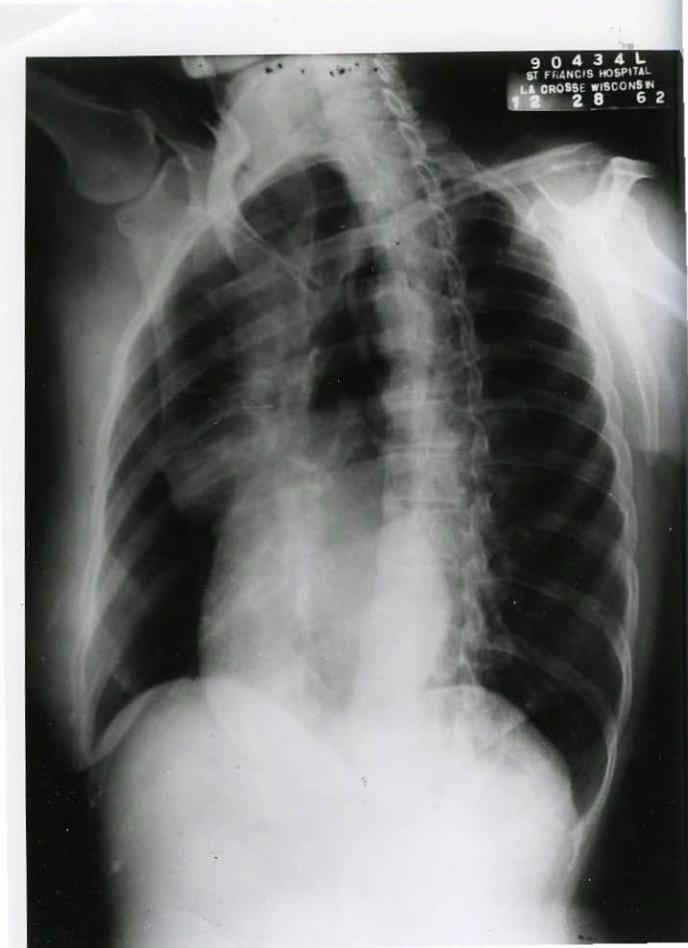
CASE l T 477-63
Contributed by Lewis B. Woolner, M.D. Mayo Clinic, Rochester, Minnesot a
This 52 year old f emale was asymptouatic on admission . Four
months prior, she had one episode of chest pain With chills and non-
productive cough.
Physical examination was negati ve . Cytologic examinati on
of sputum and bronchoscopic examir.at:l:on (elsewhere) were negative.
X-ray chest revealed a mass i n t he right hilus which on
tomography d.id not appear to contain calcium.
A right thoracotomy was performed .
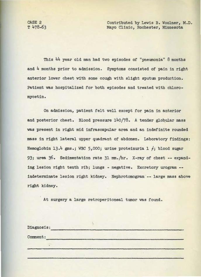
CASE 2 T 478-63
Contributed by Lewis B. Woolner, M.D. Mayo Clinic, Rochester, Minnesota
This 44 year old IDBn had tvo episodes of "pnewnonia" 8 months
and 4 months prior to admission . Symptoms consist ed of pain in r ight
anterior lower cheat with some cough with slight sputum production .
Patient was hospitalized t or both episodes and treated with chloro-
mycetin.
On admission, patient felt well except for pain in anterior
and posteri or chest, Blood pressure l4o/78. A tender globular IDBSS
was present in ri.ght mid intrascapular area and an indefinite rounded
mass in right later al upper quadrant of abdomen. Laboratory findings:
Hemoglobin 13 . 4 gJnS . ; WBC 51 000; urine proteinuria l I; blood sugar
93; urea 36 . Sedimentation rate 31 =./hr. X-ray of chest -- expand-
ing lesion right tenth rib; lungs - negative. Excretory urogram - -
indeterminate lesion right kidney, Nephrotomogrem -- large mass above
right kidney.
At surgery a large retroperitoneal tumor was found.
Diagnosis: _________________________________________________ __
Comment: ___________________________________________________ __
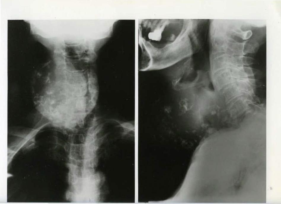
CASE 3 T 479-63
Coatributed by LeWis B. Woolner, M.D. Mayo Clinic, Rochester, Minnesota
This 73 year old woman first noted a small goiter 10 years
ago. This began to grow larger about 4 years ago vith recent slight
dyspnea and dysphagia.
Physical Examinati on: A huge, atony hard goiter i nvolving
right lobe and isthmus . Estimated weight, 500 grams. Euthyroid.
X-ray of neck -- 10 em. round, soft tissue mass in the anterior r i ght
side ot neck. The mass contains a large amount of irregular calcifi-
cation . Mass presumed to be a goit er. Trachea displaced to left and
posteriorly.
At surgery, a huge mase (13 x 9 x 7 em. , weight 430 grams) was
removed from right side of neck.
· Diagnosis: ________________________________________________ _
Comment : __________________________________________________ _
GASE 4 T 48o-63
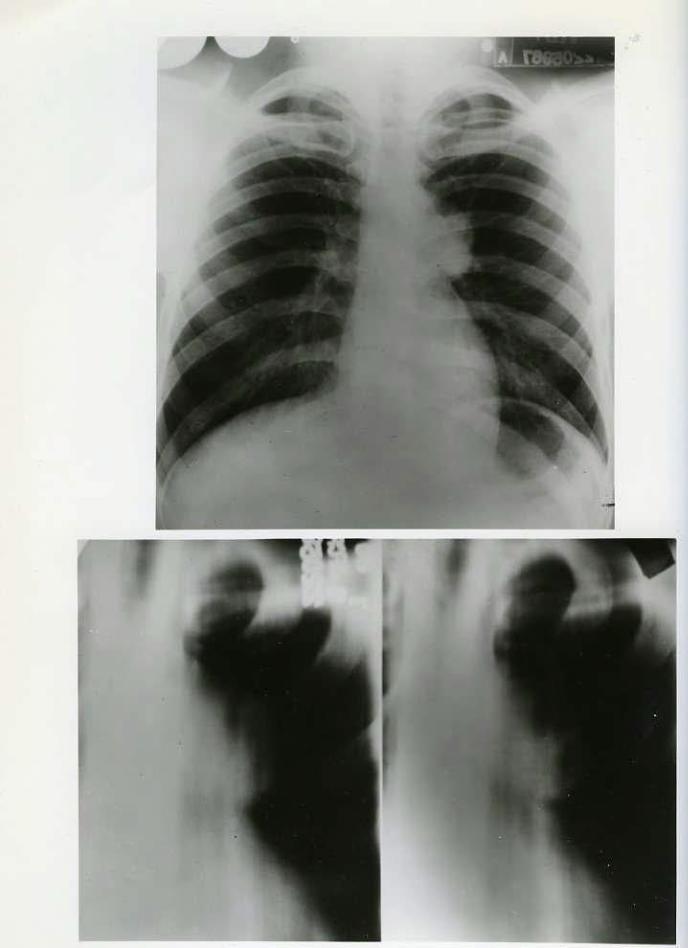
Contributed by Lewis B, Woolner, M.D. Mayo Clinic, Rochester, Minnesota
This 23 year old male was referred for treatment following
discovery of an abnormality in roentgenogram of. cheat.
In the past 2 years he had 3 episodes lasting a few days to
2 weeks of fever, pleuritic pain and cough with aching in left
shoulder and arm.
On admission he was asymptomatic. Laboratory findings were
essentially normal. Chest x-ray showed a large mass anterior to the
left hi lus apparently extending from the anterior mediastinum.
Following a sternal splitting procedure, a mass was found
overlying the arch of aorta and the pulmonary vessels.
I
Diagnosis=----------------~------------------------------
Comment: ----------------------------------------
CASE 5 T 574-63
Contributed by Levie B. Wool~er , M.D. Mayo Cli nic, Ro~ester, Minnesota
This 47 year old male comp.lait:ed of achii1g and stiffness of'
right calf and thigh of 8 months duration . Ten months prior to
admission, the patient fell from a step ladder falling 10 feet and
striking his right hip against ba.sement stairs.
Physical examir~tion on admissi on revealed a hard non-tender
mass bulging t he lateral a spect of the right hip and extending from
the right anterior superior iliac spine to the sub-trochanteric region.
X-ray examination revealed a large partially calcti'ied mass adjacent tv
the right hip laterally.
SurgicaJ exploration revealed a soft tissue mass, 10 x 8 x
7 em. , i nvolvin3 m~.<scle, attached to the greater trochanter but with
no involvement of bor.e .
Di agnosis: ________________________________________________ _
Comment: __________________________________________________ _
CASE 6 1609-55, 1616-55, T 575-63
Contributed by Levis B. Woolner, M.D. Mayo Clinic , Rochester, Minnesota
This 17 year old white female came to the Clinic because of pain in her right thigh.
Six years ago, she had been hospitalized and given penicillin injection for "inflamnation above right knee cap ."
Nine months ago she began to limp on her right leg and shortly thereafter began to notice an ache in her right anterior thigh vhile at rest. X-rays by her local doctor revealed "calcium deposits in hip."
For the past month, patient has noted pain in right thigh while walking. She stated that for the past year the right l eg was larger than the left.
Physical examination revealed tenderness over the right femoral triangle with 'palpable femoral vein and dilated s~erficial veins of the thigh . Circumference of the right thigh was 5 em. greater than the left .
Laboratory Findings: Leukocyte count 7,900/ml . ; differential count: neutrophile 6~; band forms lli; lymphocytes 13i; monocytes 3'f,; eosinophile 5i; erythrocytic sedimentation rate 50 m.jhr. X-ray examination shoved calcific collection in soft tissues of right inguinalfemoral region . No continuity Vith bone .
At surgery a mass was found in the hip region, deep to the femoral vein, vith extension into the vein lumen.
Diagnosis: _____________________ ~-------------------------------------
C~nt=-------------------------------------------------
8-18-60 4·10-61
CASE 7 T-625 - T-626
Contributed by Lewis B. Woolner, l~.D. ~myo Clinic, Rochester, Minnesota
This 50 year old female was fir st seen in September, 1953 for evaluation of "atelectasis" of right lower lobe. She had had an attack of pneumonia in May, 1953 for which she was treated with antibiotics. She did not make a rapid recovery and continued to have productive cough (l/ 4 cup sputum daily) with occasionally a pin.k tinge to sputum but no frank hemoptysis . One 'Week prior to admission, an x- ray of the chest 'Was taken and she was then referred for evaluation.
On admission, in September 1:953, she was found to have d~lness and depressed breath sounds at right base posteriorly. X-rays.
Second Admission: Apri l, 1961. X-ray .
Diagnosis: ______________________________________________________ __
Comment : ____________________________________________________ ___
CASE 8 T-627-63
Contributed by Lewis B. Woolner, M.D. Mayo Clinic, Rochester, Minnesota
This 61 year old physician came to the Clinic in March, 1963 following examination and study elsewhere for fever, malaise, muscular aching an.d some cough dating bsck to approximately December, 1962. Persistent low grade fever and elevated.white count suggested infect ion. Stool and blood cultures and smears of sputum for acid fast bacilli (e.w . ) were negative . Prior chest x-ray (March, 1962) was negative .
On admissi on, patient had few pulmonary symptoms other than chronic cough. He had slight dyspnea on moderate exertion) sligh~ weight loss and continued fever up to 100°F. Smoking history: 1-2 packs per day for 20-30 years. X-ray of chest.
A biopsy of right scalene node was carried out which showed metastatic papil lary carcinoma with psammoma bodies consi stent with a primary in ·thyroid gland.
Diagnosis: ________________________________________________ __
Comment: ____________________________________________________ ___
4-8-63
CASE 9 T-628- 63
Contributed by Lewis B. Woolner, M.D. Mayo Clinic, Rochester, lo!innesota
This 66 year old white male, undergoing routine check- up, was found to have an indeterminate lesi on in the right lung. He had no symptoms referable to the chest. There was no history of exposure to dusts or i ngest i on of mineral oil. Tomograms of the lesion revealed no calcification, Compari son of chest x-ray with film taken one year prior revealed littl e change in size of the lesion.
Laboratory Examination: Cytologic examinati on of sputum -negative for carcinoma cells, Smears and cultures :for tubercle bacilli -- negative. Skin tests: Tuberculin P. P. D. - single strength Positive (++); histoplasmin- negative; coccidioidin negati ve; F.B.I. - 4.6 mcg. /100 mJ. . serum. Urinalysi s -normal , WBC 6,700. Sedimentation rate 40 mm. /hr.
Diagnosis : _____________________________________________________________________________ ___
Comment:---------------------------------------------------------------------------------
CASE 10 T-629-63
Contributed by Lewis B. Woolner, M.D. Mayo Clinic, Rochester, Minnesota
This 69 year year old male complained or 25 :pound weight loss over the past rive months . He noted occasional mild dysphagia with solid foods sticking momentarily in lower esophagus. Re also had a cough productive of 1-2 oz. or sputum per day and containing brownish material (blood?) on two occas~ons.
On esophagoscopy, rather marked pulsation was noted within he~al sac on the left side. The mucosa or lover esophagus and hernial sac were markedly injected but there was no eVidence or ulceration. Bronchoscopy was essentially negati ve.
Diagnosis: ____________________________________________________ ___
Comment:---------------------------------------------------------
7-2-63
CASE ll T-64o~63
J
Contributed by Lewis B. 'Woolner, M.D. Mayo Clinic, Rochester, Minnesota
This 43 year old female consulted her local physician after noting a lump in the right side of her neck of one month's duration. A subtotal right lobectomy was carried out. The resected tissue contained an unencapsulated tumor mass . She was referred for consideration of her problem and for further treatment if necessary.
On examination t here was much induration in t he region of the healed incision in the n·eck, making i t difficult to palpate remainder of thyroid gland. The right deep jugular lymph nodes were mas sively enlarged. X-ray of the chest.
A surgical exploration wa.s performed.
Diagnosis: ____________________________________________________ _
Comment: ____________________________ ~----------------------
CASE 12 Cll633-61 T-648-63
Contributed by Lewis B. Woolner, M.D. Mayo Clinic, Rochester, Minnesota
This 61 year old white male was first seen in 1956 at which time he COIIIPlained of dyspepsia and pain in right upper quadrant . Be was found to have a non-functioning gallbladder. Cholecystectomy revealed a chronic contracted cholecystitis with a single gall stone. After surgery he contil.nued to have episodes of right chest wall pain over lower ribs. On his second admission in -1961, he had noted continuous pain in the right lower rib margin for the previous two weeks. '
X-ray of chest revealed osteolytic lesions of right seventh and eighth ribs with pathologic fracture of seventh rib . Orographic studies revealed nothing to suggest a malignant renal lesion.
Laboratory Data: _Hel!IOglobin 14.0 gms; urinalysis - normal; Sedimentation rate 9 mm.jhr.
Surgical resection of the osteolytic lesions of seventh and eighth ribs were carried out.
Diagnosis: ______________________________________________________ _
Comment: ____________________________________________________ __
'
CASE 13 X279- 59, Xll096-61 Xl07- 59, T-647-63
Contributed by Lewis B. W'ool.ner, M.D. Mayo Cl1n1c , Rochester, Minnesota
This 45 year old female was admitted 1n Januar-,t, 1959 with a history of fatigue, veigllt loss {15 lbs . ), abdominal bloating and chronic diarrhea vi th steatorrhea dating back to June, 1957 . In January, 1958, she developed attacks of severe generalized abdominal pain which persisted' for a bout two mont hs . A diagnosis of sprue vas made in June, 1958, and patient vas treated vith diet and bed rest. She improved some\lhat duri ng the next few months but in December, 1958 became wor se with more vomiting, numerous bulky frothy stools per day and episodes of epi gastric pain, especially at night.
Laboratory findings on admission : lJrinalysis - normal . Hemoglobin 13.6 gms; WBC 6900; Sedimentation rate 22 mm./br. Serwn calcium 8 .1 ; Serum amylase 4,000 units; Serum lipase 1 . 4 units .
X- ray f indings : Chest - negative . Flat plate of abdomen -no evidence of pancreatic calcification; stomach and duodenum -abnormalit y o:f' the second porti on of the duodenum suggesting a tumor of the pancreas .
On 1-15- 59 an abdominal ex:ploration was performed.
Diagnosis: __________________________________________________ __
Comment: __________________________________________________ _
CASE lt. Cl2~7-63
Contributed by Lewi s B. Woolner, M.D. Mayo Cl.inic 1 Rochester, Minnesota
This 19 month old white male child was first seen i n J anuary, 1.963 with nondpainful proptosis of l.eft eye . Swelling of the lower lid was firs t noted by hi s mot her in early January with pro~ession over one month . Examinat i on r e veal.ed proptosis, conjunct ival i njection, upward displacement of eye and a palpable fir m mass along the i n ferior margi n of the orbit. X-ray findings : Chest - negative; h ead - nega t ive; sinuses - unsatisfactor y.
Orbital expl oration was performed .
Nine months later, x- ray of orbit .
Diagnosis : ________________________________________________ __
Co~t: ________________________________________________ _
CASE 15 02315-61
Contributed by Lewis B. Woolner, M.D . l:laYO Clinic, Rochester, Minnesota
This 56 year old female compl ai ned of palpitation weakness, ~ faintness and occasional black stools of two months duration. She
had been hospitalized elsewhere and t reated for anemia wi th liver injections, Vitamin B12, and severa.J. bl ood t r ansfusions. At time of admission> she complained only of continued passage of ~rk stools. Roentgenogram ·of the stomach and duodenum gave evidence of a deformity of t he duodena). cap. The patient was treated for a suspected duodenal ulcer. She returned 1~ years later complaining of weakness and head.aches. Hemoglobin values were 7 . 4 gm. Following re- examination of the stoma~h by roentgenogram, surgery was performed.
Diagnosis: ____________ ~-----------------------------------------Comment: ______________________________________________________ ___
CASE 16 M9664-63
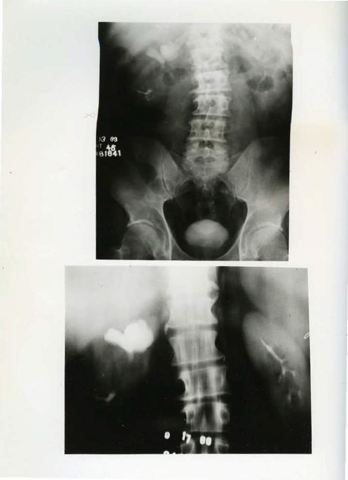
Contributed by Lewis B. Woolner, M.D. Mayo Clinic, Rochester , Minnesota
This 55 year old mal e was admitted in December, 1963 with a history of recurring upper gastroint est inal bleeding sin·ce 1951. On several ,occasions he had what appeared t o be massive hemorrhages ~c.companied by mild shock, tarry stools and a l ow l evel of hemo·globin. He was practically free ot gastric distress between acute episodes . He had been treated for duodenal ulcer but w~thout x-ray confirmation. The patient has had known hypertension since 1954, ranging f r om 196/100 to 26o/ll0. His last episode of :melena was one month ago.
On admission he complained of weakness on exertion and some abdominal distress. X- ray examinati on : Chest - normal; gallbladder -poorly functioning wit h stones; stomach - duodenal ul cer.
X-ray of kidneys.
Definitive surgery was performed.
Diagnosis:------------------~-------------------------------comment: ____________________________________________________ _


![Wanderheft 021 Usedom [1964]](https://img.pdfslide.org/doc/110x75/56d6bd711a28ab30168e0306/wanderheft-021-usedom-1964.jpg)