Embed Size (px)
Citation preview
The American Journal of Pathology, Vol. 184, No. 5, May 2014
ANIMAL MODELS
Gastrointestinal Pathology in Juvenile and AdultCFTR-Knockout FerretsXingshen Sun,* Alicia K. Olivier,y Yaling Yi,* Christopher E. Pope,zx Hillary S. Hayden,x Bo Liang,* Hongshu Sui,*Weihong Zhou,* Kyle R. Hager,x Yulong Zhang,* Xiaoming Liu,* Ziying Yan,* John T. Fisher,* Nicholas W. Keiser,* Yi Song,*Scott R. Tyler,* J. Adam Goeken,y Joann M. Kinyon,{ Matthew C. Radey,x Danielle Fligg,{ Xiaoyan Wang,* Weiliang Xie,*Thomas J. Lynch,* Paul M. Kaminsky,* Mitchell J. Brittnacher,x Samuel I. Miller,xk** Kalpaj Parekh,yy David K. Meyerholz,y
Lucas R. Hoffman,zx Timothy Frana,{ Zoe A. Stewart,zz and John F. Engelhardt*
ajp.amjpathol.org
From the Departments of Anatomy and Cell Biology,* Pathology,y Cardiothoracic Surgery,yy and Surgery,zz University of Iowa, Iowa City, Iowa; theDepartments of Pediatrics,z Microbiology,x Genome Sciences,k and Medicine,** University of Washington, Seattle, Washington; and the Department ofVeterinary Diagnostic & Production Animal Medicine,{ College of Veterinary Medicine, Iowa State University, Ames, Iowa
Accepted for publication
C
P
h
January 30, 2014.
Address correspondence toJohn F. Engelhardt, Ph.D.,Room 1-111 BSB, Departmentof Anatomy and Cell Biology,College of Medicine, Universityof Iowa, 51 Newton Rd., IowaCity, IA 52242. E-mail: [email protected].
opyright ª 2014 American Society for Inve
ublished by Elsevier Inc. All rights reserved
ttp://dx.doi.org/10.1016/j.ajpath.2014.01.035
Cystic fibrosis (CF) is a multiorgan disease caused by loss of a functional cystic fibrosis transmembraneconductance regulator (CFTR) chloride channel in many epithelia of the body. Here we report thepathology observed in the gastrointestinal organs of juvenile to adult CFTR-knockout ferrets. CFgastrointestinal manifestations included gastric ulceration, intestinal bacterial overgrowth with villousatrophy, and rectal prolapse. Metagenomic phylogenetic analysis of fecal microbiota by deepsequencing revealed considerable genotype-independent microbial diversity between animals, with themajority of taxa overlapping between CF and non-CF pairs. CF hepatic manifestations were variable, butincluded steatosis, necrosis, biliary hyperplasia, and biliary fibrosis. Gallbladder cystic mucosal hy-perplasia was commonly found in 67% of CF animals. The majority of CF animals (85%) had pancreaticabnormalities, including extensive fibrosis, loss of exocrine pancreas, and islet disorganization.Interestingly, 2 of 13 CF animals retained predominantly normal pancreatic histology (84% to 94%) attime of death. Fecal elastase-1 levels from these CF animals were similar to non-CF controls, whereas allother CF animals evaluated were pancreatic insufficient (<2 mg elastase-1 per gram of feces). Thesefindings suggest that genetic factors likely influence the extent of exocrine pancreas disease in CFferrets and have implications for the etiology of pancreatic sufficiency in CF patients. In summary, thesestudies demonstrate that the CF ferret model develops gastrointestinal pathology similar to CF patients.(Am J Pathol 2014, 184: 1309e1322; http://dx.doi.org/10.1016/j.ajpath.2014.01.035)
Supported by NIH grants DK096518 and HL108902 (J.F.E.), K08DK092284 (Z.A.S.), K08 HL114725 (K.P.), P30 DK089507 (B.R. Ram-sey), K02 HL105543 (L.R.H.), P30 DK054759 (J.F.E.), the Cystic FibrosisFoundation, and the Roy J. Carver Chair in Molecular Medicine (J.F.E.).
X.S. and A.K.O. contributed equally to this work.Disclosures: None declared.
Cystic fibrosis (CF) is the most common life-threatening,autosomal recessive, genetic disorder among Caucasians,occurring in approximately 1 in 3500 births. Defects in thecystic fibrosis transmembrane conductance regulator (CFTR)gene that disrupt function of this chloride channel cause ab-normalities in electrolyte and fluid movement across manyepithelia of the body, leading to viscous, poorly hydrated,secretions.1 Although chronic bacterial infections in the lungare the most significant cause of mortality in CF, pathology inmultiple other organs contributes to the progression of dis-ease and overall health of CF patients. These organs includethe intestine, pancreas, liver, and gallbladder for whichclinical and/or histological disease is seen in CF patients at
stigative Pathology.
.
frequencies of 10% to 90%.1e4 In the current study, weevaluated gastrointestinal disease in juvenile and adultCFTR-knockout ferrets.
Mouse models of CF have been critical to our under-standing of CFTR function in several organs, however, CFmice fail to develop spontaneous disease in the lung andpancreas, and have relatively minor disease of the liver and
Sun et al
gallbladder.5,6 The recent creation of new CF pig and ferretmodels has provided the field with new tools to dissect CFdisease pathophysiology and the factors that influence dis-ease severity in CF patients.7,8 Comparisons between CFmouse, pig, and ferret models have clearly demonstratedthat species-specific differences in organ physiology andCFTR biology influence the extent of pathology in majororgans affected in CF.9,10 For example, all newborn CFmodels have intestinal pathology that manifests as meco-nium ileus at birth in the case of CF pigs (100% of ani-mals)11 and CF ferrets (75% of animals),12 or as intestinalobstruction at weaning in the case of CF mice.5 By contrast,pancreatic phenotypes at birth are highly variable betweenspecies with disease being most severe in CF pigs,11 lesssevere in CF ferrets,12,13 and absent in CF mice.5 Similarly,CF pigs demonstrate histopathology in the gallbladder andliver at birth,11 whereas disease in these organs is relativeminor in newborn CF ferrets12,13 and CF adult mice.5,6
Elucidating the differences in the severity of CF gastroin-testinal disease at birth and in disease progression betweenthe various species may aid in dissecting genetic and envi-ronmental factors that influence gastrointestinal diseaseseverity in CF patients.
Here, we report the phenotype of gastrointestinal organs(pancreas, liver, gallbladder, stomach, and intestine) in olderCF animals reared on antibiotics until 6 months of age, orthe time at which they were euthanized due to severity ofdisease. We have also evaluated the extent of bacterialovergrowth in the CF intestine and the types of bacterialflora found in the intestine of non-CF and CF animals. Ourfindings demonstrate that juvenile and adult CF ferretsnaturally acquire gastrointestinal disease at frequenciessimilar to that observed in CF patients. Of great interest, asmall subset of CF animals was pancreatic sufficient frombirth, implicating modifier genes that can compensate forthe loss of CFTR in the exocrine pancreas. These studiessuggest that the CF ferret model could be useful for testingtherapies aimed at gastrointestinal organs and dissectinghow genetic variation influences disease in CF.
Materials and Methods
Rearing of CF Ferrets
The previously described CFTR exon-10 disrupted ferretmodel was used for all studies.8 The colony of CFTRþ/�
ferrets was backcrossed from sable to both albino and cin-namon coat colors to increase genetic diversity. Heterozy-gous matings were performed at Marshall Farms (NorthRose, NY) and pregnant jills were shipped to the Universityof Iowa at 21 to 28 days gestation. After birth, kits wererapidly genotyped as previously described.13 CF kits thatpassed meconium were paired with a non-CF animal, andgenerally all other kits were removed from the litter. CF andnon-CF kits were reared as previously described.14 Each CFanimal was paired with a non-CF sibling control and treated
1310
identically in terms of feeding, antibiotics, oral laxative, andpancreatic enzyme supplementation. This approach wasused to control the variable clinical care of each CF animals(ie, antibiotics to control lung infection and oral laxatives tocontrol gut obstruction). All kits were reared from birth on20 mg/kg metronidazole s.c. (2� daily) and 4.0 mg/kgpiperacillin-tazobactam s.c. (2� daily), with each dose in100 mL of saline. If weight gain decreased in CF animalsover a 12- to 18-hour period, both the non-CF control andCF animals were placed on 10 mg/kg enrofloxacin s.c. (2�daily). If weight gain decreased a second time, the non-CFcontrol and CF animals were placed on 30 mg/kg cefazolins.c. (2� daily). This antibiotic protocol was required becauseCF ferrets are highly susceptible to lung infections duringthe early neonatal period.14 Porcine pancreatic enzymes(Viokase-V; Neogen Corporation, Lexington, KY) supple-mentation was initiated at approximately 20 to 30 days whenkits were transitioned to artificial nipple feeding. When an-imals reached 21 weeks of age, the dose of antibiotics wasreduced by 25% per week until the animals were free fromantibiotics at 6 months of age.
Histology, Immunohistochemistry, and Morphometry
Standard histopathology analysis was performed on paraffinsections from formalin-fixed tissues. The tissues werecollected at the time of euthanasia (clinical death) and imme-diately placed in 10% neutral buffered formalin for at least72 hours. Tissues were then paraffin-embedded, sectioned(4 mm) and stained with H&E or periodic acid-Schiff. Histo-pathological examination was performed by a veterinarypathologist, and age-matched CFTR�/� and CFTRþ/þ;þ/�
controls were used. Formalin-fixed, paraffin-embedded pan-creata were stained by immunohistochemistry for insulin andglucagon (both from MP Biomedicals, Santa Ana, CA) aspreviously described.13 Slides for morphometric assessmentwere scanned with an Aperio ScanScope CS (Buffalo Grove,IL) and images analyzed with Image-Pro Premier softwareversion 9.0 (Media Cybernetics, Rockville, MD). Morphom-etry was performed to determine the percent normal pancreasin two pancreatic sufficient and two pancreatic insufficient CFanimals. For the CF animals at approximately 250 days of age,four sections from different areas of the pancreas were eval-uated because the entire pancreas could not fit onto one slide.For CF animals at 19 days of age, three sections of the entirepancreas (100 mm apart) were evaluated for morphometrybecause the entire pancreas fit onto one slide. Normal tissuewas defined by an experienced veterinary pathologist aslacking inflammation and structural changes associated withCF pancreata.
Human CF and Non-CF Pancreatic Tissue
Formalin-fixed human pancreatic tissues were obtainedfrom the National Disease Research Interchange (Philadel-phia, PA). Tissue samples were collected from three non-CF
ajp.amjpathol.org - The American Journal of Pathology
GI Pathology in Adult CF Ferrets
brain dead donors patients: after head trauma, traumaticinjury, and traumatic brain injury. Six CF patient sampleswere evaluated: three diagnosed with CF-related diabetesand three that were not diagnosed with CF-related diabetes.
Fecal EL-1 Assays
Fecal elastase-1 (EL-1) assays were performed using a canineEL-1 ELISA kit (ScheBo Biotech AG, Giessen, Germany)according to the manufacturer’s instructions. Before ELISA,fecal material was first processed using an E1 Quick-Prep-Canine extraction kit (ScheBo Biotech AG) according to themanufacturer’s instructions. This fecal EL-1 assay detectsferret EL-1, but does not cross-react with porcine EL-1 inreconstitution assays performed with Viokase-V pancreaticenzyme (Neogen Corporation).
Bacteriology of Intestinal Samples
At the time of necropsy, a portion of the duodenum and ileum(containing fecal material) was homogenized in sterile salineand a portion was plated directly onto blood agar, Mac-Conkey agar, colistin and naladixic acid agar, colistin andnaladixic acid reducible agar, and chocolate agar in a four-quadrant streak pattern. Cultures were incubated aerobically
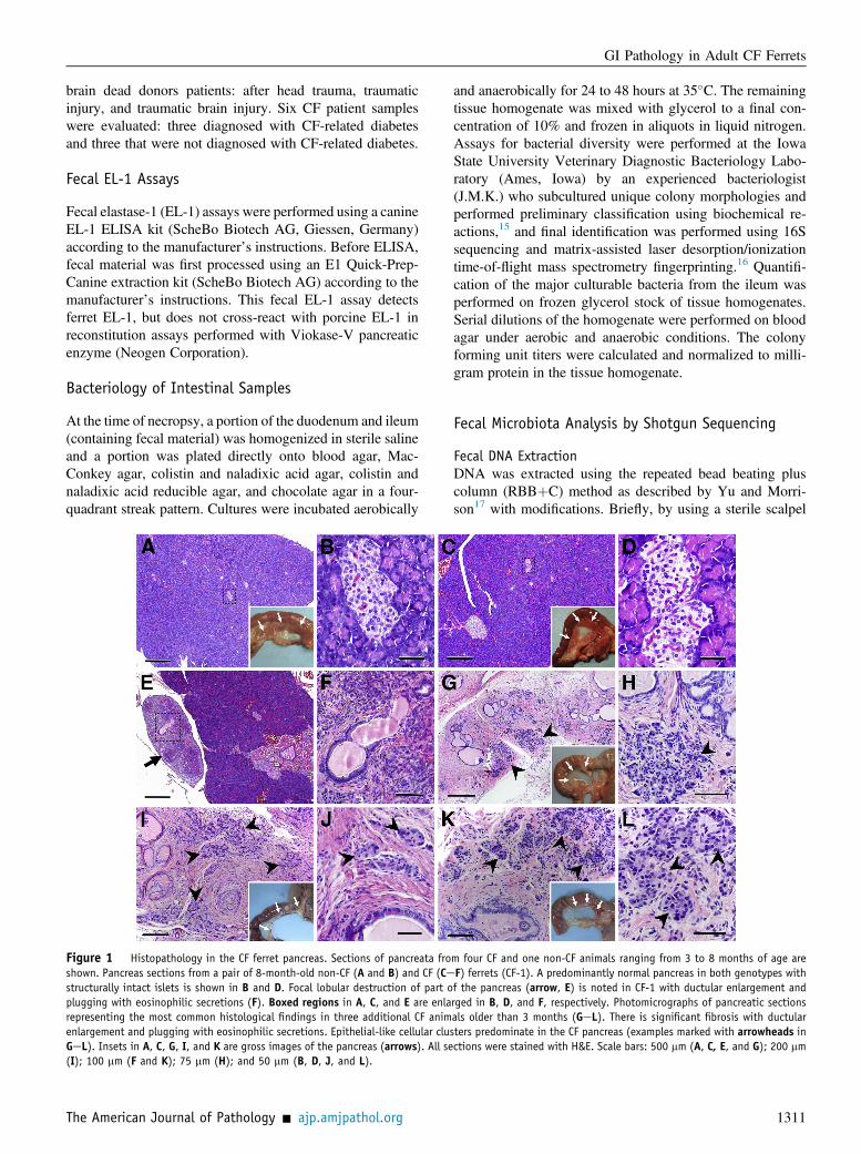
Figure 1 Histopathology in the CF ferret pancreas. Sections of pancreata froshown. Pancreas sections from a pair of 8-month-old non-CF (A and B) and CF (Cestructurally intact islets is shown in B and D. Focal lobular destruction of part oplugging with eosinophilic secretions (F). Boxed regions in A, C, and E are enlarepresenting the most common histological findings in three additional CF animenlargement and plugging with eosinophilic secretions. Epithelial-like cellular cluGeL). Insets in A, C, G, I, and K are gross images of the pancreas (arrows). All se(I); 100 mm (F and K); 75 mm (H); and 50 mm (B, D, J, and L).
The American Journal of Pathology - ajp.amjpathol.org
and anaerobically for 24 to 48 hours at 35�C. The remainingtissue homogenate was mixed with glycerol to a final con-centration of 10% and frozen in aliquots in liquid nitrogen.Assays for bacterial diversity were performed at the IowaState University Veterinary Diagnostic Bacteriology Labo-ratory (Ames, Iowa) by an experienced bacteriologist(J.M.K.) who subcultured unique colony morphologies andperformed preliminary classification using biochemical re-actions,15 and final identification was performed using 16Ssequencing and matrix-assisted laser desorption/ionizationtime-of-flight mass spectrometry fingerprinting.16 Quantifi-cation of the major culturable bacteria from the ileum wasperformed on frozen glycerol stock of tissue homogenates.Serial dilutions of the homogenate were performed on bloodagar under aerobic and anaerobic conditions. The colonyforming unit titers were calculated and normalized to milli-gram protein in the tissue homogenate.
Fecal Microbiota Analysis by Shotgun Sequencing
Fecal DNA ExtractionDNA was extracted using the repeated bead beating pluscolumn (RBBþC) method as described by Yu and Morri-son17 with modifications. Briefly, by using a sterile scalpel
m four CF and one non-CF animals ranging from 3 to 8 months of age areF) ferrets (CF-1). A predominantly normal pancreas in both genotypes withf the pancreas (arrow, E) is noted in CF-1 with ductular enlargement andrged in B, D, and F, respectively. Photomicrographs of pancreatic sectionsals older than 3 months (GeL). There is significant fibrosis with ductularsters predominate in the CF pancreas (examples marked with arrowheads inctions were stained with H&E. Scale bars: 500 mm (A, C, E, and G); 200 mm
1311
Sun et al
blade, 250 mg of frozen fecal sample was transferred into a2 mL sterile tube containing 1 mL of lysis buffer (500 mmol/LNaCl, 50 mmol/L Tris-HCl, pH 8.0, 50 mmol/L EDTA, 4%sodium dodecyl sulfate, and 0.5 g of sterile DNA-free silica/zirconia beads [BioSpec, Bartlesville, OK]). Subsequentextraction was performed as described.17 The resulting DNAsamples were stored at �80�C until sequenced.
Illumina Sequencing and Microbiota Analysis UsingMetagenomic Phylogenetic AnalysisWell-established methods for ultra-high throughputsequencing were used to identify members of the bacterialcommunity in whole fecal samples from three pairs of CF andnon-CF ferrets, as described for human fecal samples.18
Briefly, sequencing libraries were made from DNA pre-parations using Illumina’s Nextera technology and weresequenced on the MiSeq platform. There were 15 to 20million paired-end reads (150 nucleotides long) generated persample, according to the manufacturer’s standards (Illumina
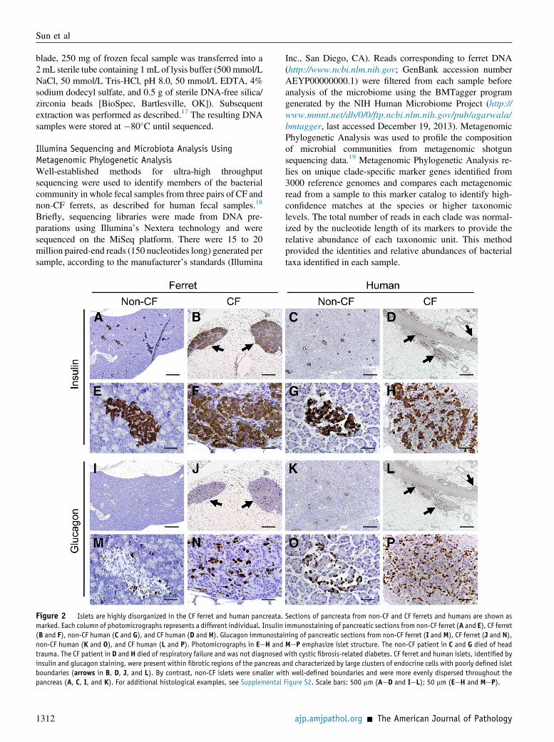
Figure 2 Islets are highly disorganized in the CF ferret and human pancreata.marked. Each column of photomicrographs represents a different individual. Insulin(B and F), non-CF human (C and G), and CF human (D and H). Glucagon immunostanon-CF human (K and O), and CF human (L and P). Photomicrographs in EeH andtrauma. The CF patient in D and H died of respiratory failure and was not diagnosedinsulin and glucagon staining, were present within fibrotic regions of the pancreas aboundaries (arrows in B, D, J, and L). By contrast, non-CF islets were smaller wipancreas (A, C, I, and K). For additional histological examples, see Supplemental
1312
Inc., San Diego, CA). Reads corresponding to ferret DNA(http://www.ncbi.nlm.nih.gov; GenBank accession numberAEYP00000000.1) were filtered from each sample beforeanalysis of the microbiome using the BMTagger programgenerated by the NIH Human Microbiome Project (http://www.mmnt.net/db/0/0/ftp.ncbi.nlm.nih.gov/pub/agarwala/bmtagger, last accessed December 19, 2013). MetagenomicPhylogenetic Analysis was used to profile the compositionof microbial communities from metagenomic shotgunsequencing data.19 Metagenomic Phylogenetic Analysis re-lies on unique clade-specific marker genes identified from3000 reference genomes and compares each metagenomicread from a sample to this marker catalog to identify high-confidence matches at the species or higher taxonomiclevels. The total number of reads in each clade was normal-ized by the nucleotide length of its markers to provide therelative abundance of each taxonomic unit. This methodprovided the identities and relative abundances of bacterialtaxa identified in each sample.
Sections of pancreata from non-CF and CF ferrets and humans are shown asimmunostaining of pancreatic sections from non-CF ferret (A and E), CF ferretining of pancreatic sections from non-CF ferret (I and M), CF ferret (J and N),MeP emphasize islet structure. The non-CF patient in C and G died of headwith cystic fibrosis-related diabetes. CF ferret and human islets, identified bynd characterized by large clusters of endocrine cells with poorly defined isletth well-defined boundaries and were more evenly dispersed throughout theFigure S2. Scale bars: 500 mm (AeD and IeL); 50 mm (EeH and MeP).
ajp.amjpathol.org - The American Journal of Pathology
GI Pathology in Adult CF Ferrets
Study Approval
This study was performed according to protocols approvedby the Institutional Animal Care and Use Committee (Uni-versity of Iowa). Human pancreatic tissues were obtainedfrom the National Human Tissue Resource Centerwith IRBapproval (University of Iowa).
Results
Significant Pancreatic Remodeling Occurs in MostJuvenile and Adult CF Ferrets
Exocrine pancreatic insufficiency significantly impacts thehealth and nutrition of approximately 85% to 90% of CFpatients.20,21 Both exocrine and endocrine pancreatic diseasecontribute to malnutrition and the development of diabetes inCF patients.21,22We recently reported that juvenile CF ferretsdevelop CF-related diabetes with many of the phenotypesobserved in CF patients.13 These studies reported a signifi-cant loss (approximately 50%) in islet mass of CF ferretswithin the first month of age. In the current study, we eval-uated the histopathology of the pancreas in older CF and non-CF animals (Figure 1). Eighty-five percent (11 of 13) of CFanimals had significant loss of the exocrine pancreas withassociated fibrosis, ductal proliferation, and plugging ofintralobular ducts (Figure 1, GeL). Interestingly, two CFanimals (CF-1 and CF-12) had pancreata that were largelyhistologically normal (Figure 1, AeD, and Supplemental
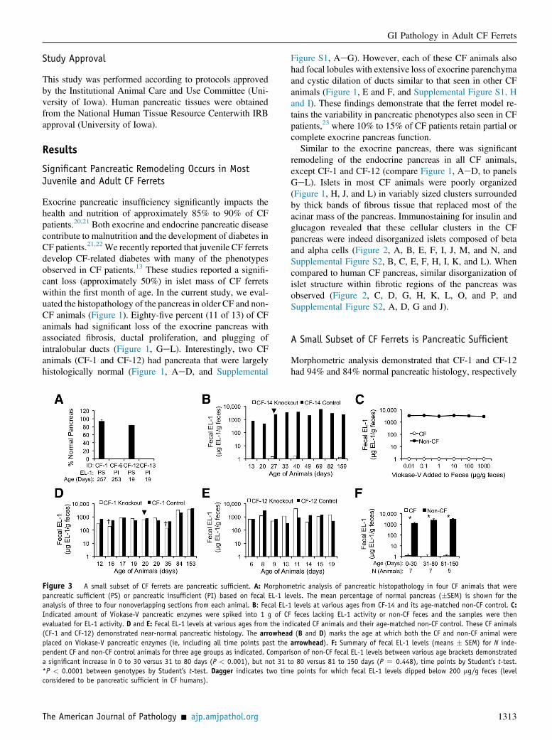
Figure 3 A small subset of CF ferrets are pancreatic sufficient. A: Morphompancreatic sufficient (PS) or pancreatic insufficient (PI) based on fecal EL-1 leanalysis of three to four nonoverlapping sections from each animal. B: Fecal EL-1Indicated amount of Viokase-V pancreatic enzymes were spiked into 1 g of CFevaluated for EL-1 activity. D and E: Fecal EL-1 levels at various ages from the ind(CF-1 and CF-12) demonstrated near-normal pancreatic histology. The arrowheaplaced on Viokase-V pancreatic enzymes (ie, including all time points past thependent CF and non-CF control animals for three age groups as indicated. Compara significant increase in 0 to 30 versus 31 to 80 days (P < 0.001), but not 31 t*P < 0.0001 between genotypes by Student’s t-test. Dagger indicates two timconsidered to be pancreatic sufficient in CF humans).
The American Journal of Pathology - ajp.amjpathol.org
Figure S1, AeG). However, each of these CF animals alsohad focal lobules with extensive loss of exocrine parenchymaand cystic dilation of ducts similar to that seen in other CFanimals (Figure 1, E and F, and Supplemental Figure S1, Hand I). These findings demonstrate that the ferret model re-tains the variability in pancreatic phenotypes also seen in CFpatients,23 where 10% to 15% of CF patients retain partial orcomplete exocrine pancreas function.
Similar to the exocrine pancreas, there was significantremodeling of the endocrine pancreas in all CF animals,except CF-1 and CF-12 (compare Figure 1, AeD, to panelsGeL). Islets in most CF animals were poorly organized(Figure 1, H, J, and L) in variably sized clusters surroundedby thick bands of fibrous tissue that replaced most of theacinar mass of the pancreas. Immunostaining for insulin andglucagon revealed that these cellular clusters in the CFpancreas were indeed disorganized islets composed of betaand alpha cells (Figure 2, A, B, E, F, I, J, M, and N, andSupplemental Figure S2, B, C, E, F, H, I, K, and L). Whencompared to human CF pancreas, similar disorganization ofislet structure within fibrotic regions of the pancreas wasobserved (Figure 2, C, D, G, H, K, L, O, and P, andSupplemental Figure S2, A, D, G and J).
A Small Subset of CF Ferrets is Pancreatic Sufficient
Morphometric analysis demonstrated that CF-1 and CF-12had 94% and 84% normal pancreatic histology, respectively
etric analysis of pancreatic histopathology in four CF animals that werevels. The mean percentage of normal pancreas (�SEM) is shown for thelevels at various ages from CF-14 and its age-matched non-CF control. C:feces lacking EL-1 activity or non-CF feces and the samples were thenicated CF animals and their age-matched non-CF control. These CF animalsd (B and D) marks the age at which both the CF and non-CF animal werearrowhead). F: Summary of fecal EL-1 levels (means � SEM) for N inde-ison of non-CF fecal EL-1 levels between various age brackets demonstratedo 80 versus 81 to 150 days (P Z 0.448), time points by Student’s t-test.e points for which fecal EL-1 levels dipped below 200 mg/g feces (level
1313
Sun et al
(Figure 3A). The finding that 15% of the CF ferretsanalyzed had largely normal pancreatic histology raised thepossibility that these CF animals were pancreatic sufficientfrom birth. To this end, we evaluated fecal elastase-1 (EL-1)levels in samples collected from CF animals with abnormaland largely normal pancreatic morphology at the time ofdeath. All CF ferrets, with the exception of CF-1 and CF-12,had fecal EL-1 levels <2 mg EL-1/g feces at all time pointsevaluated (Figure 3, B and F), regardless of whether animalswere fed oral porcine pancreatic enzymes (Viokase-V)(Figure 3B). This level of fecal EL-1 in CF animals was>1000-fold lower than non-CF controls. Reconstitutionexperiments spiking various amounts of Viokase-V into CFfeces lacking immunoreactive EL-1, or non-CF feces con-taining significant immunoreactive EL-1, did not alter EL-1ELISA results (Figure 3C). In contrast to most CF animals,the two CF animals with largely normal pancreatic mor-phology (CF-1 and CF-12) had levels of fecal EL-1 that didnot differ from non-CF control littermates (Figure 3, D andE). These findings demonstrate that a small subset of CFferrets is pancreatic sufficient. Although 2 of 13 CF animalsevaluated in this study were pancreatic sufficient, the overall
1314
prevalence of this pancreatic sufficient phenotype in the CFcolony is likely much less frequent. This conclusion is basedon the finding that CF-1 and CF-12 had growth curves frombirth that were similar to non-CF controls and the fact thefrequency of this observation in our rearing experience isapproximately 1% to 2%.
Gallbladder Abnormalities Are More Common than LiverAbnormalities in Older CF Ferrets
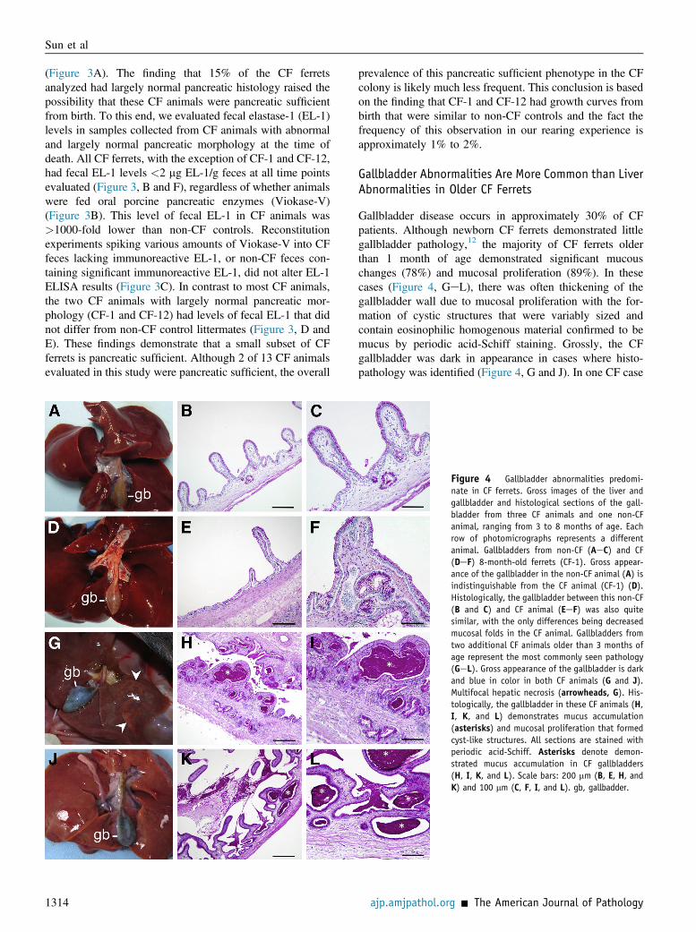
Gallbladder disease occurs in approximately 30% of CFpatients. Although newborn CF ferrets demonstrated littlegallbladder pathology,12 the majority of CF ferrets olderthan 1 month of age demonstrated significant mucouschanges (78%) and mucosal proliferation (89%). In thesecases (Figure 4, GeL), there was often thickening of thegallbladder wall due to mucosal proliferation with the for-mation of cystic structures that were variably sized andcontain eosinophilic homogenous material confirmed to bemucus by periodic acid-Schiff staining. Grossly, the CFgallbladder was dark in appearance in cases where histo-pathology was identified (Figure 4, G and J). In one CF case
Figure 4 Gallbladder abnormalities predomi-nate in CF ferrets. Gross images of the liver andgallbladder and histological sections of the gall-bladder from three CF animals and one non-CFanimal, ranging from 3 to 8 months of age. Eachrow of photomicrographs represents a differentanimal. Gallbladders from non-CF (AeC) and CF(DeF) 8-month-old ferrets (CF-1). Gross appear-ance of the gallbladder in the non-CF animal (A) isindistinguishable from the CF animal (CF-1) (D).Histologically, the gallbladder between this non-CF(B and C) and CF animal (EeF) was also quitesimilar, with the only differences being decreasedmucosal folds in the CF animal. Gallbladders fromtwo additional CF animals older than 3 months ofage represent the most commonly seen pathology(GeL). Gross appearance of the gallbladder is darkand blue in color in both CF animals (G and J).Multifocal hepatic necrosis (arrowheads, G). His-tologically, the gallbladder in these CF animals (H,I, K, and L) demonstrates mucus accumulation(asterisks) and mucosal proliferation that formedcyst-like structures. All sections are stained withperiodic acid-Schiff. Asterisks denote demon-strated mucus accumulation in CF gallbladders(H, I, K, and L). Scale bars: 200 mm (B, E, H, andK) and 100 mm (C, F, I, and L). gb, gallbadder.
ajp.amjpathol.org - The American Journal of Pathology
GI Pathology in Adult CF Ferrets
(CF-1), the animal with a predominantly normal pancreas,the gallbladder was grossly and histologically very similarto the non-CF control (Figure 4, AeF). However, even inthis case there were fewer mucosal folds and evidence formultifocal cystic structures (Figure 4, E and F). Although noclear association between pancreatic sufficiency and gall-bladder disease in CF patients has been noted, it is inter-esting that the older CF animal that appeared pancreaticsufficient also had a fairly normal gallbladder. There wereno gallbladder abnormalities observed in CF animals thatwere less than 1-month-old (Table 1).
Liver abnormalities are not uncommon in CF patients andare characterized by biliary cirrhosis (25%), hepatic stea-tosis (30%), and clinical cirrhosis (5%).2,24,25 Althoughnewborn CF ferrets demonstrated consistent abnormalitiesin liver enzymes and bile plugging at birth,12,13 histopath-ological abnormalities in older CF ferrets were highly var-iable. The majority of both non-CF (75%) and CF (67%)animals demonstrated minor-to-moderate portal lymphoidaggregates (Figure 5, AeD and F). Biliary ducts weregenerally unremarkable except for rare luminal cell debrisand rare lymphocytes within the epithelium. There were 3of 10 CF animals older than 1 month of age that demon-strated hepatic pathology not observed in non-CF controls(Table 1). These included multifocal mid-zonal necrosis(Figures 4G and 5E) in CF-4 and lipid vacuoles consistentwith steatosis (Figures 5, G and H, and 4J) in CF-3 andCF-10. The pattern of hepatic necrosis in CF-4 is atypical ofhuman CF liver disease. The cause of the necrosis is un-known, however, sepsis can induce hepaticmidzonal necrosis
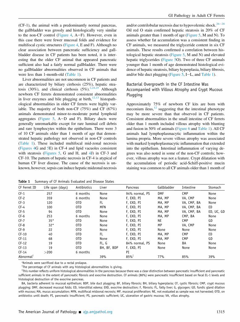
Table 1 Summary of CF Animals Evaluated and Disease States
CF Ferret ID Life span (days) Antibiotics Liver
CF-1 257 6 months NoneCF-2 359 6 months NoneCF-3 120 OTD FLCF-4 100 OTD MFNCF-5 94 OTD NoneCF-6 253 6 months NoneCF-7 35* OTD NoneCF-8 32* OTD NoneCF-9 22 OTD NoneCF-10 40 OTD FLCF-11 68 OTD NoneCF-12 19 OTD FL, GCF-13 19 OTD BH, BF, BDPCF-14 >200 6 monthsAbnormaly 39%
*Animals were sacrificed due to a rectal prolapse.yThe percentage of CF animals with any histological abnormalities is giving.zThis number reflects uniform histological abnormalities in the pancreas because
sufficient animals in the extent of pancreatic fibrosis and exocrine destruction. CFhistological destruction of the exocrine pancreas.BA, bacteria adherent to mucosal epithelium; BDP, bile duct plugging; BF, bil
plugging; DMF, decreased mucosal folds; ED, interstitial edema; EXD, exocrine dewith mucous; MA, mucus accumulation; MFN, multifocal midzonal necrosis; MP, muantibiotics until death; PI, pancreatic insufficient; PS, pancreatic sufficient; UC,
The American Journal of Pathology - ajp.amjpathol.org
and/or centrilobular necrosis due to hypovolemic shock.26e28
Oil red O stain confirmed hepatic steatosis in 20% of CFanimals greater than 1 month of age (Figure 5, M and N). Toassess whether fat accumulation was a consistent finding inCF animals, we measured the triglyceride content in six CFanimals. These results confirmed a correlation between his-tological hepatic steatosis (Figure 5, M and N) and elevatedhepatic triglycerides (Figure 5O). Two of three CF animalsyounger than 1 month of age demonstrated histological evi-dence of hepatic steatosis, biliary hyperplasia, biliary fibrosis,and/or bile duct plugging (Figure 5, IeL, and Table 1).
Bacterial Overgrowth in the CF Intestine WasAccompanied with Villous Atrophy and Crypt MucousPlugging
Approximately 75% of newborn CF kits are born withmeconium ileus,12 suggesting that the intestinal phenotypemay be more severe than that observed in CF patients.Consistent abnormalities in the small intestine of CF ferretsolder than 1 month included villous atrophy with bluntingand fusion in 50% of animals (Figure 6 and Table 1). All CFanimals had lymphoplasmacytic inflammation within thelamina propria. More severe villous atrophy was associatedwith marked lymphoplasmacytic inflammation that extendedinto the epithelium. Intestinal inflammation of varying de-grees was also noted in some of the non-CF controls; how-ever, villous atrophy was not a feature. Crypt dilatation withthe accumulation of periodic acid-Schiff-positive mucinstaining was common to all CF animals older than 1 month of
Pancreas Gallbladder Intestine Stomach
94% normal, PS DMF CMP NoneF, EXD, PI MA, MP VA, CMP NoneF, EXD, PI MA, MP VA, CMP, BA NoneF, EXD, PI MA, MP VA, CMP, BA EDF, EXD, PI MA, MP VA, CMP, BA ED, UC, GDF, EXD, PI MA, MP CMP, BA NoneF, EXD, PI NE CMP GDF, EXD, PI MP VA, CMP NoneF, EXD, PI None None NoneF, EXD, PI MA, MP CMP EDF, EXD, PI MA, MP CMP GD84% normal, PS None BA NoneF, EXD, PI None None NonePI85%z 77% 85% 39%
there was a clear distinction between pancreatic insufficient and pancreaticanimals (86%) were pancreatic insufficient based on fecal EL-1 levels and
iary fibrosis; BH, biliary hyperplasia; CF, cystic fibrosis; CMP, crypt mucousstruction; F, fibrosis; FL, fatty liver; G, glycogen; GD, fundic gland dilationcosal proliferation; NE, not evaluated as sample was not harvested; OTD, onulceration of gastric mucosa; VA, villus atrophy.
1315
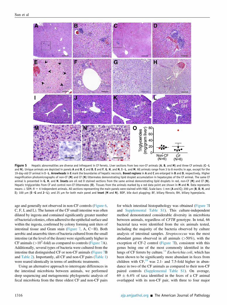
Figure 5 Hepatic abnormalities are diverse and infrequent in CF ferrets. Liver sections from two non-CF animals (A, B, and M) and three CF animals (CeLand N). Unique animals are depicted in panels A and B, C and D, E and F, G, H, and N, IeL, and M. All animals range from 3 to 8 months in age, except for the19-day-old CF animal in IeL. Arrowheads in E mark the boundaries of hepatic necrosis. Boxed regions in A and C are enlarged in B and D, respectively. Highermagnification photomicrographs of non-CF (M) and CF (N) littermates demonstrating lipid droplet accumulation in hepatocytes of the CF animal. The same CFanimal is presented in G, H, and N. Insets are oil red O stained sections from the same animal demonstrating lipid droplets in red, non-CF (M) and CF (N).Hepatic triglycerides from CF and control non-CF littermates (O). Tissues from the animals marked by a red data point are shown in M and N. Data representsmeans � SEM; NZ 6 independent animals. All sections representing the main panels were stained with H&E. Scale bars: 1 mm (A and C); 200 mm (B, D, H, andI); 100 mm (EeG and JeL); and 25 mm for both main panel and inset (M and N). BDP, bile duct plugging; BF, biliary fibrosis; BH, biliary hyperplasia.
Sun et al
age and generally not observed in non-CF controls (Figure 6,C, F, I, and L). The lumen of the CF small intestine was oftendilated by ingesta and contained significantly greater numberof bacterial colonies, often adhered to the epithelial surface andwithin the ingesta, confirmed by colony forming unit titers ofintestinal tissue and Gram stain (Figure 7, A, CeH). Bothaerobic and anaerobic titers of bacteria cultured from the smallintestine (at the level of the ileum) were significantly higher inCF animals (>103-fold) as compared to controls (Figure 7A).Additionally, several types of bacteria were cultured from theintestine that distinguished CF or non-CF animals (Figure 7Band Table 2). Importantly, all CF and non-CF pairs (Table 1)were reared identically in terms of antibiotic treatments.
Using an alternative approach to interrogate differences inthe intestinal microbiota between animals, we performeddeep sequencing and metagenomic phylogenetic analysis offecal microbiota from the three oldest CF and non-CF pairs
1316
for which intestinal histopathology was obtained (Figure 7Iand Supplemental Table S1). This culture-independentmethod demonstrated considerable diversity in microbiotabetween animals, regardless of CFTR genotype. In total, 66bacterial taxa were identified from the six animals tested,including the majority of the bacteria observed by cultureanalysis of intestinal samples. Streptococcus was the mostabundant genus observed in all animals (>50%), with theexception of CF-2 control (Figure 7I), consistent with thisgenus being one of the most commonly identified in thelungs of CF ferrets by culture.14 Escherichia coli, which hasbeen shown to be significantly more abundant in feces fromchildren with CF,18 was 2.1- and 7.5-fold higher in abun-dance in two of the CF animals as compared to their non-CFpaired controls (Supplemental Table S1). On average,69 � 6.4% of taxa identified in the feces of a CF animaloverlapped with its non-CF pair, with three to four major
ajp.amjpathol.org - The American Journal of Pathology
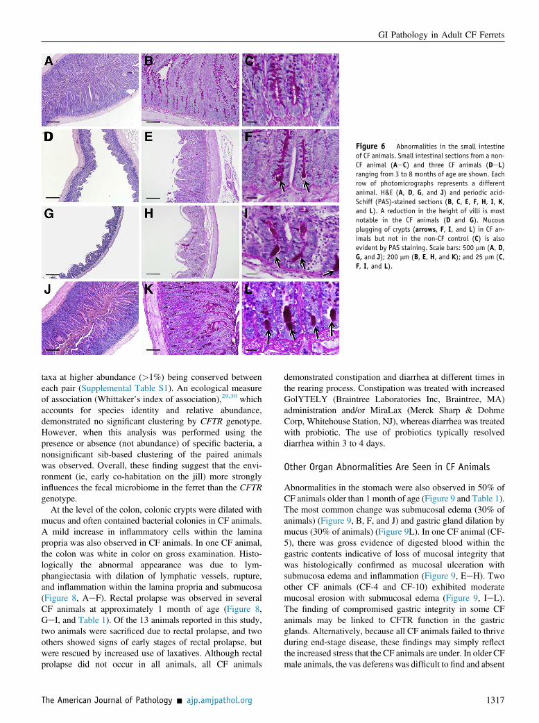
Figure 6 Abnormalities in the small intestineof CF animals. Small intestinal sections from a non-CF animal (AeC) and three CF animals (DeL)ranging from 3 to 8 months of age are shown. Eachrow of photomicrographs represents a differentanimal. H&E (A, D, G, and J) and periodic acid-Schiff (PAS)-stained sections (B, C, E, F, H, I, K,and L). A reduction in the height of villi is mostnotable in the CF animals (D and G). Mucousplugging of crypts (arrows, F, I, and L) in CF an-imals but not in the non-CF control (C) is alsoevident by PAS staining. Scale bars: 500 mm (A, D,G, and J); 200 mm (B, E, H, and K); and 25 mm (C,F, I, and L).
GI Pathology in Adult CF Ferrets
taxa at higher abundance (>1%) being conserved betweeneach pair (Supplemental Table S1). An ecological measureof association (Whittaker’s index of association),29,30 whichaccounts for species identity and relative abundance,demonstrated no significant clustering by CFTR genotype.However, when this analysis was performed using thepresence or absence (not abundance) of specific bacteria, anonsignificant sib-based clustering of the paired animalswas observed. Overall, these finding suggest that the envi-ronment (ie, early co-habitation on the jill) more stronglyinfluences the fecal microbiome in the ferret than the CFTRgenotype.
At the level of the colon, colonic crypts were dilated withmucus and often contained bacterial colonies in CF animals.A mild increase in inflammatory cells within the laminapropria was also observed in CF animals. In one CF animal,the colon was white in color on gross examination. Histo-logically the abnormal appearance was due to lym-phangiectasia with dilation of lymphatic vessels, rupture,and inflammation within the lamina propria and submucosa(Figure 8, AeF). Rectal prolapse was observed in severalCF animals at approximately 1 month of age (Figure 8,GeI, and Table 1). Of the 13 animals reported in this study,two animals were sacrificed due to rectal prolapse, and twoothers showed signs of early stages of rectal prolapse, butwere rescued by increased use of laxatives. Although rectalprolapse did not occur in all animals, all CF animals
The American Journal of Pathology - ajp.amjpathol.org
demonstrated constipation and diarrhea at different times inthe rearing process. Constipation was treated with increasedGolYTELY (Braintree Laboratories Inc, Braintree, MA)administration and/or MiraLax (Merck Sharp & DohmeCorp, Whitehouse Station, NJ), whereas diarrhea was treatedwith probiotic. The use of probiotics typically resolveddiarrhea within 3 to 4 days.
Other Organ Abnormalities Are Seen in CF Animals
Abnormalities in the stomach were also observed in 50% ofCF animals older than 1 month of age (Figure 9 and Table 1).The most common change was submucosal edema (30% ofanimals) (Figure 9, B, F, and J) and gastric gland dilation bymucus (30% of animals) (Figure 9L). In one CF animal (CF-5), there was gross evidence of digested blood within thegastric contents indicative of loss of mucosal integrity thatwas histologically confirmed as mucosal ulceration withsubmucosa edema and inflammation (Figure 9, EeH). Twoother CF animals (CF-4 and CF-10) exhibited moderatemucosal erosion with submucosal edema (Figure 9, IeL).The finding of compromised gastric integrity in some CFanimals may be linked to CFTR function in the gastricglands. Alternatively, because all CF animals failed to thriveduring end-stage disease, these findings may simply reflectthe increased stress that the CF animals are under. In older CFmale animals, the vas deferens was difficult to find and absent
1317
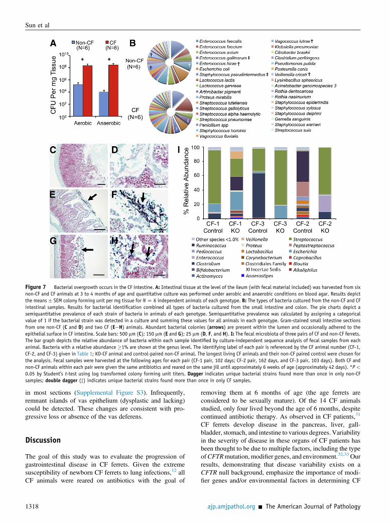
Figure 7 Bacterial overgrowth occurs in the CF intestine. A: Intestinal tissue at the level of the ileum (with fecal material included) was harvested from sixnon-CF and CF animals at 3 to 4 months of age and quantitative culture was performed under aerobic and anaerobic conditions on blood agar. Results depictthe means� SEM colony forming unit per mg tissue for NZ 6 independent animals of each genotype. B: The types of bacteria cultured from the non-CF and CFintestinal samples. Results for bacterial identification combined all types of bacteria cultured from the small intestine and colon. The pie charts depict asemiquantitative prevalence of each strain of bacteria in animals of each genotype. Semiquantitative prevalence was calculated by assigning a categoricalvalue of 1 if the bacterial strain was detected in a culture and summing these values for all animals in each genotype. Gram-stained small intestine sectionsfrom one non-CF (C and D) and two CF (EeH) animals. Abundant bacterial colonies (arrows) are present within the lumen and occasionally adhered to theepithelial surface in CF intestine. Scale bars: 500 mm (C); 150 mm (E and G); 25 mm (D, F, and H). I: The fecal microbiota of three pairs of CF and non-CF ferrets.The bar graph depicts the relative abundance of bacteria within each sample identified by culture-independent sequence analysis of fecal samples from eachanimal. Bacteria with a relative abundance �1% are shown at the genus level. The identifying label of each pair is referenced by the CF animal number (CF-1,CF-2, and CF-3) given in Table 1; KO-CF animal and control-paired non-CF animal. The longest living CF animals and their non-CF paired control were chosen forthe analysis. Fecal samples were harvested at the following ages for each pair (CF-1 pair, 102 days; CF-2 pair, 162 days, and CF-3 pair, 103 days). Both CF andnon-CF animals within each pair were given the same antibiotics and reared on the same jill until approximately 6 weeks of age (approximately 42 days). *P <
0.05 by Student’s t-test using log transformed colony forming unit titers. Dagger indicates unique bacterial strains found more than once in only non-CFsamples; double dagger (z) indicates unique bacterial strains found more than once in only CF samples.
Sun et al
in most sections (Supplemental Figure S3). Infrequently,remnant islands of vas epithelium (dysplastic and lacking)could be detected. These changes are consistent with pro-gressive loss or absence of the vas deferens.
Discussion
The goal of this study was to evaluate the progression ofgastrointestinal disease in CF ferrets. Given the extremesusceptibility of newborn CF ferrets to lung infections,12 allCF animals were reared on antibiotics with the goal of
1318
removing them at 6 months of age (the age ferrets areconsidered to be sexually mature). Of the 14 CF animalsstudied, only four lived beyond the age of 6 months, despitecontinued antibiotic therapy. As observed in CF patients,31
CF ferrets develop disease in the pancreas, liver, gall-bladder, stomach, and intestine to various degrees. Variabilityin the severity of disease in these organs of CF patients hasbeen thought to be due to multiple factors, including the typeofCFTRmutation, modifier genes, and environment.32,33 Ourresults, demonstrating that disease variability exists on aCFTR null background, emphasize the importance of modi-fier genes and/or environmental factors in determining CF
ajp.amjpathol.org - The American Journal of Pathology
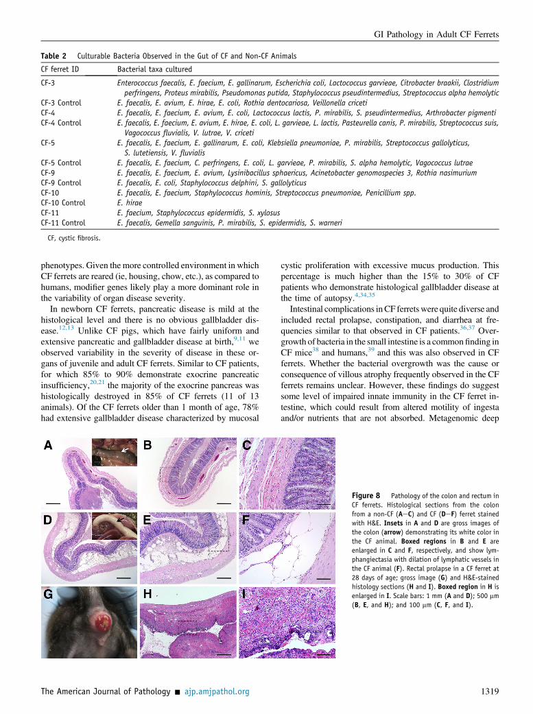
Table 2 Culturable Bacteria Observed in the Gut of CF and Non-CF Animals
CF ferret ID Bacterial taxa cultured
CF-3 Enterococcus faecalis, E. faecium, E. gallinarum, Escherichia coli, Lactococcus garvieae, Citrobacter braakii, Clostridiumperfringens, Proteus mirabilis, Pseudomonas putida, Staphylococcus pseudintermedius, Streptococcus alpha hemolytic
CF-3 Control E. faecalis, E. avium, E. hirae, E. coli, Rothia dentocariosa, Veillonella cricetiCF-4 E. faecalis, E. faecium, E. avium, E. coli, Lactococcus lactis, P. mirabilis, S. pseudintermedius, Arthrobacter pigmentiCF-4 Control E. faecalis, E. faecium, E. avium, E. hirae, E. coli, L. garvieae, L. lactis, Pasteurella canis, P. mirabilis, Streptococcus suis,
Vagococcus fluvialis, V. lutrae, V. cricetiCF-5 E. faecalis, E. faecium, E. gallinarum, E. coli, Klebsiella pneumoniae, P. mirabilis, Streptococcus gallolyticus,
S. lutetiensis, V. fluvialisCF-5 Control E. faecalis, E. faecium, C. perfringens, E. coli, L. garvieae, P. mirabilis, S. alpha hemolytic, Vagococcus lutraeCF-9 E. faecalis, E. faecium, E. avium, Lysinibacillus sphaericus, Acinetobacter genomospecies 3, Rothia nasimuriumCF-9 Control E. faecalis, E. coli, Staphylococcus delphini, S. gallolyticusCF-10 E. faecalis, E. faecium, Staphylococcus hominis, Streptococcus pneumoniae, Penicillium spp.CF-10 Control E. hiraeCF-11 E. faecium, Staphylococcus epidermidis, S. xylosusCF-11 Control E. faecalis, Gemella sanguinis, P. mirabilis, S. epidermidis, S. warneri
CF, cystic fibrosis.
GI Pathology in Adult CF Ferrets
phenotypes. Given the more controlled environment in whichCF ferrets are reared (ie, housing, chow, etc.), as compared tohumans, modifier genes likely play a more dominant role inthe variability of organ disease severity.
In newborn CF ferrets, pancreatic disease is mild at thehistological level and there is no obvious gallbladder dis-ease.12,13 Unlike CF pigs, which have fairly uniform andextensive pancreatic and gallbladder disease at birth,9,11 weobserved variability in the severity of disease in these or-gans of juvenile and adult CF ferrets. Similar to CF patients,for which 85% to 90% demonstrate exocrine pancreaticinsufficiency,20,21 the majority of the exocrine pancreas washistologically destroyed in 85% of CF ferrets (11 of 13animals). Of the CF ferrets older than 1 month of age, 78%had extensive gallbladder disease characterized by mucosal
The American Journal of Pathology - ajp.amjpathol.org
cystic proliferation with excessive mucus production. Thispercentage is much higher than the 15% to 30% of CFpatients who demonstrate histological gallbladder disease atthe time of autopsy.4,34,35
Intestinal complications inCF ferretswere quite diverse andincluded rectal prolapse, constipation, and diarrhea at fre-quencies similar to that observed in CF patients.36,37 Over-growth of bacteria in the small intestine is a commonfinding inCF mice38 and humans,39 and this was also observed in CFferrets. Whether the bacterial overgrowth was the cause orconsequence of villous atrophy frequently observed in the CFferrets remains unclear. However, these findings do suggestsome level of impaired innate immunity in the CF ferret in-testine, which could result from altered motility of ingestaand/or nutrients that are not absorbed. Metagenomic deep
Figure 8 Pathology of the colon and rectum inCF ferrets. Histological sections from the colonfrom a non-CF (AeC) and CF (DeF) ferret stainedwith H&E. Insets in A and D are gross images ofthe colon (arrow) demonstrating its white color inthe CF animal. Boxed regions in B and E areenlarged in C and F, respectively, and show lym-phangiectasia with dilation of lymphatic vessels inthe CF animal (F). Rectal prolapse in a CF ferret at28 days of age; gross image (G) and H&E-stainedhistology sections (H and I). Boxed region in H isenlarged in I. Scale bars: 1 mm (A and D); 500 mm(B, E, and H); and 100 mm (C, F, and I).
1319
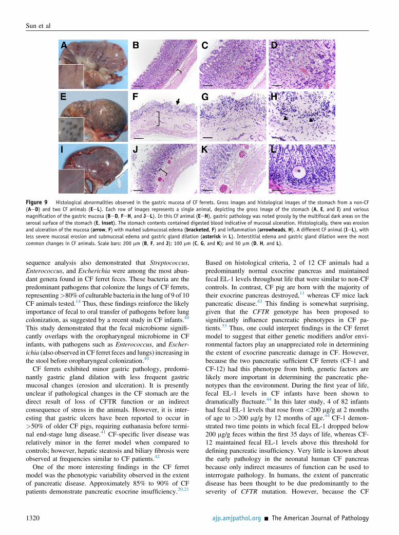
Figure 9 Histological abnormalities observed in the gastric mucosa of CF ferrets. Gross images and histological images of the stomach from a non-CF(AeD) and two CF animals (EeL). Each row of images represents a single animal, depicting the gross image of the stomach (A, E, and I) and variousmagnification of the gastric mucosa (BeD, FeH, and JeL). In this CF animal (EeH), gastric pathology was noted grossly by the multifocal dark areas on theserosal surface of the stomach (E, inset). The stomach contents contained digested blood indicative of mucosal ulceration. Histologically, there was erosionand ulceration of the mucosa (arrow, F) with marked submucosal edema (bracketed, F) and inflammation (arrowheads, H). A different CF animal (IeL), withless severe mucosal erosion and submucosal edema and gastric gland dilation (asterisk in L). Interstitial edema and gastric gland dilation were the mostcommon changes in CF animals. Scale bars: 200 mm (B, F, and J); 100 mm (C, G, and K); and 50 mm (D, H, and L).
Sun et al
sequence analysis also demonstrated that Streptococcus,Enterococcus, and Escherichia were among the most abun-dant genera found in CF ferret feces. These bacteria are thepredominant pathogens that colonize the lungs of CF ferrets,representing>80%of culturable bacteria in the lung of 9 of 10CF animals tested.14 Thus, these findings reinforce the likelyimportance of fecal to oral transfer of pathogens before lungcolonization, as suggested by a recent study in CF infants.40
This study demonstrated that the fecal microbiome signifi-cantly overlaps with the oropharyngeal microbiome in CFinfants, with pathogens such as Enterococcus, and Escher-ichia (also observed in CF ferret feces and lungs) increasing inthe stool before oropharyngeal colonization.40
CF ferrets exhibited minor gastric pathology, predomi-nantly gastric gland dilation with less frequent gastricmucosal changes (erosion and ulceration). It is presentlyunclear if pathological changes in the CF stomach are thedirect result of loss of CFTR function or an indirectconsequence of stress in the animals. However, it is inter-esting that gastric ulcers have been reported to occur in>50% of older CF pigs, requiring euthanasia before termi-nal end-stage lung disease.41 CF-specific liver disease wasrelatively minor in the ferret model when compared tocontrols; however, hepatic steatosis and biliary fibrosis wereobserved at frequencies similar to CF patients.42
One of the more interesting findings in the CF ferretmodel was the phenotypic variability observed in the extentof pancreatic disease. Approximately 85% to 90% of CFpatients demonstrate pancreatic exocrine insufficiency.20,21
1320
Based on histological criteria, 2 of 12 CF animals had apredominantly normal exocrine pancreas and maintainedfecal EL-1 levels throughout life that were similar to non-CFcontrols. In contrast, CF pig are born with the majority oftheir exocrine pancreas destroyed,11 whereas CF mice lackpancreatic disease.43 This finding is somewhat surprising,given that the CFTR genotype has been proposed tosignificantly influence pancreatic phenotypes in CF pa-tients.33 Thus, one could interpret findings in the CF ferretmodel to suggest that either genetic modifiers and/or envi-ronmental factors play an unappreciated role in determiningthe extent of exocrine pancreatic damage in CF. However,because the two pancreatic sufficient CF ferrets (CF-1 andCF-12) had this phenotype from birth, genetic factors arelikely more important in determining the pancreatic phe-notypes than the environment. During the first year of life,fecal EL-1 levels in CF infants have been shown todramatically fluctuate.44 In this later study, 4 of 82 infantshad fecal EL-1 levels that rose from <200 mg/g at 2 monthsof age to >200 mg/g by 12 months of age.44 CF-1 demon-strated two time points in which fecal EL-1 dropped below200 mg/g feces within the first 35 days of life, whereas CF-12 maintained fecal EL-1 levels above this threshold fordefining pancreatic insufficiency. Very little is known aboutthe early pathology in the neonatal human CF pancreasbecause only indirect measures of function can be used tointerrogate pathology. In humans, the extent of pancreaticdisease has been thought to be due predominantly to theseverity of CFTR mutation. However, because the CF
ajp.amjpathol.org - The American Journal of Pathology
GI Pathology in Adult CF Ferrets
ferrets studied were deficient in CFTR, our findings suggestthat modifier genes are the most likely explanation for thepancreatic sufficiency from birth in CF-1 and CF-12. Suchmodifiers may include alternative chloride or bicarbonatechannels that compensate for the lack of CFTR. If thesefactors could be identified and manipulated, they could be ofsignificant value to CF patients.
One other finding about ferret fecal EL-1 levels is worthnoting. There was a significant (P < 0.001) increase in fecalEL-1 levels between 1 to 2 months of age in wild-type controlanimals, and this increase also occurred in CF-1. This is thewindow in which ferrets are weaned and may account for theincrease. It is presently unclear if similar changes occur inhumans at weaning, however, the fecal EL-1 levels in juve-nile ferrets are three to four times higher than typically foundin humans.45,46 Thus, this change may be species-specific.CF ferrets have been shown to have glucose excursions>200 mg/dL during the 1- to 2-month period when weaningoccurs,13 and it is possible that a decline in endocrine func-tion during this time window is linked to increased pressureon the pancreas to produce pancreatic enzyme.
In summary, our findings demonstrate that the lack ofCFTR function leads to gastrointestinal disease in juvenileand adult ferrets in a similar fashion to humans. Interest-ingly, CF ferrets demonstrated phenotypic variability in theextent of disease in the liver, pancreas, and gallbladdersimilar to that observed in CF patients. The CF ferret modelmay be useful in determining the genetic and environmentalinfluences responsible for disease variability in CF patients,and offer an important modality for testing therapies tar-geting pancreatic, gallbladder, intestinal, and liver disease.
Acknowledgment
We thank Robyn L. Marsh for performing the clusteringanalyses for ecological associations in the fecal microbiomedeep sequence data.
Supplemental Data
Supplemental material for this article can be found athttp://dx.doi.org/10.1016/j.ajpath.2014.01.035.
References
1. Rowe SM, Miller S, Sorscher EJ: Cystic fibrosis. N Engl J Med 2005,352:1992e2001
2. Kopelman H: Cystic fibrosis. 6. Gastrointestinal and nutritional as-pects. Thorax 1991, 46:261e267
3. Robertson MB, Choe KA, Joseph PM: Review of the abdominalmanifestations of cystic fibrosis in the adult patient. Radiographics2006, 26:679e690
4. Oppenheimer EH, Esterly JR: Pathology of cystic fibrosis review of theliterature and comparison with 146 autopsied cases. Perspect PediatrPathol 1975, 2:241e278
5. Grubb BR, Boucher RC: Pathophysiology of gene-targeted mousemodels for cystic fibrosis. Physiol Rev 1999, 79:S193eS214
The American Journal of Pathology - ajp.amjpathol.org
6. Wilke M, Buijs-Offerman RM, Aarbiou J, Colledge WH,Sheppard DN, Touqui L, Bot A, Jorna H, de Jonge HR, Scholte BJ:Mouse models of cystic fibrosis: phenotypic analysis and researchapplications. J Cyst Fibros 2011, 10(Suppl 2):S152eS171
7. Rogers CS, Hao Y, Rokhlina T, Samuel M, Stoltz DA, Li Y, Petroff E,Vermeer DW, Kabel AC, Yan Z, Spate L, Wax D, Murphy CN,Rieke A, Whitworth K, Linville ML, Korte SW, Engelhardt JF,Welsh MJ, Prather RS: Production of CFTR-null and CFTR-DeltaF508heterozygous pigs by adeno-associated virus-mediated gene targetingand somatic cell nuclear transfer. J Clin Invest 2008, 118:1571e1577
8. Sun X, Yan Z, Yi Y, Li Z, Lei D, Rogers CS, Chen J, Zhang Y,Welsh MJ, Leno GH, Engelhardt JF: Adeno-associated virus-targeteddisruption of the CFTR gene in cloned ferrets. J Clin Invest 2008,118:1578e1583
9. Keiser NW, Engelhardt JF: New animal models of cystic fibrosis: whatare they teaching us? Curr Opin Pulm Med 2011, 17:478e483
10. Fisher JT, Zhang Y, Engelhardt JF: Comparative biology of cysticfibrosis animal models. Methods Mol Biol 2011, 742:311e334
11. Meyerholz DK, Stoltz DA, Pezzulo AA, Welsh MJ: Pathology ofgastrointestinal organs in a porcine model of cystic fibrosis. Am JPathol 2010, 176:1377e1389
12. Sun X, Sui H, Fisher JT, Yan Z, Liu X, Cho HJ, Joo NS, Zhang Y,Zhou W, Yi Y, Kinyon JM, Lei-Butters DC, Griffin MA, Naumann P,Luo M, Ascher J, Wang K, Frana T, Wine JJ, Meyerholz DK,Engelhardt JF: Disease phenotype of a ferret CFTR-knockout model ofcystic fibrosis. J Clin Invest 2010, 120:3149e3160
13. Olivier AK, Yi Y, Sun X, Sui H, Liang B, Hu S, Xie W, Fisher JT,Keiser NW, Lei D, Zhou W, Yan Z, Li G, Evans TI, Meyerholz DK,Wang K, Stewart ZA, Norris AW, Engelhardt JF: Abnormal endocrinepancreas function at birth in cystic fibrosis ferrets. J Clin Invest 2012,122:3755e3768
14. Sun X, Olivier AK, Liang B, Yi Y, Sui H, Evans TI, Zhang Y,Zhou W, Tyler SR, Fisher JT, Keiser NW, Liu X, Yan Z, Song Y,Goeken JA, Kinyon JM, Fligg D, Wang X, Xie W, Lynch TJ,Kaminsky PM, Stewart ZA, Pope RM, Frana T, Meyerholz DK,Parekh K, Engelhardt JF: Lung phenotype of juvenile and adult CFTR-knockout ferrets. Am J Respir Cell Mol Biol 2013, (In Press)
15. Quinn PJ: Clinical veterinary microbiology. Edited by PJ Quinn,ME Carter, BK Markey, GR Carter. London, Wolfe, 1994, pp 648
16. Haigh J, Degun A, Eydmann M, Millar M, Wilks M: Improved per-formance of bacterium and yeast identification by a commercialmatrix-assisted laser desorption ionization-time of flight mass spec-trometry system in the clinical microbiology laboratory. J ClinMicrobiol 2011, 49:3441
17. Yu Z, Morrison M: Improved extraction of PCR-quality communityDNA from digesta and fecal samples. Biotechniques 2004, 36:808e812
18. Hoffman LR, Pope CE, Hayden HS, Heltshe S, Levy R, McNamara S,Jacobs MA, Rohmer L, Radey M, Ramsey BW, Brittnacher MJ,Borenstein E, Miller SI: Escherichia coli dysbiosis correlates with gastro-intestinal dysfunction in children with cystic fibrosis. Clin Infect Dis 2013
19. Segata N, Waldron L, Ballarini A, Narasimhan V, Jousson O,Huttenhower C: Metagenomic microbial community profiling usingunique clade-specific marker genes. Nat Methods 2012, 9:811e814
20. Baker SS, Borowitz D, Baker RD: Pancreatic exocrine function inpatients with cystic fibrosis. Curr Gastroenterol Rep 2005, 7:227e233
21. Kalnins D, Durie PR, Pencharz P: Nutritional management of cysticfibrosis patients. Curr Opin Clin Nutr Metab Care 2007, 10:348e354
22. Stecenko AA, Moran A: Update on cystic fibrosis-related diabetes.Curr Opin Pulm Med 2010, 16:611e615
23. Zielenski J: Genotype and phenotype in cystic fibrosis. Respiration2000, 67:117e133
24. Lindblad A, Glaumann H, Strandvik B: Natural history of liver diseasein cystic fibrosis. Hepatology 1999, 30:1151e1158
25. Moyer K, Balistreri W: Hepatobiliary disease in patients with cysticfibrosis. Curr Opin Gastroenterol 2009, 25:272e278
26. Irie H, Mori W: Fatal hepatic necrosis after shock. Acta Pathol Jpn1986, 36:363e374
1321
Sun et al
27. Caruana JA Jr, Montes M, Camara DS, Ummer A, Potmesil SH,Gage AA: Functional and histopathologic changes in the liver duringsepsis. Surg Gynecol Obstet 1982, 154:653e656
28. Rose R, Banerjee A, Ramaiah SK: Calpain inhibition attenuates iNOSproduction and midzonal hepatic necrosis in a repeat dose model ofendotoxemia in rats. Toxicol Pathol 2006, 34:785e794
29. Somerfield PJ, Clarke RK: Inverse analysis in non-parametric multi-variate analyses: distinguishing groups of associated species whichcovary coherently across samples. J Exp Mar Biol Ecol 2013, 449:261e273
30. Whittaker RH: A study of summer foliage insect communities in theGreat Smoky Mountains. Ecol Monogr 1952, 22:1e44
31. Welsh MJ, Ramsey BW, Accurso F, Cutting GR. The metabolic andmolecular basis of inherited disease. Edited by CR Scriver. New York,McGraw-Hill, 2001
32. Bush A: Cystic fibrosis in the 21st century. Edited by A Bush,EWFW Alton, JC Davies, U Griesenbach, A Jaffe. New York, Karger,2006
33. Coffey MJ, Ooi CY. Pancreatitis in cystic fibrosis and CFTR-relateddisorder. Edited by L Rodrigo. Croatia; Rijeka, InTech, 2012, pp 76e90
34. Esterly JR, Oppenheimer EH: Observations in cystic fibrosis of thepancreas. I.Thegallbladder.Bull JohnsHopkinsHosp1962, 110:247e255
35. Chaudry G, Navarro OM, Levine DS, Oudjhane K: Abdominal mani-festations of cysticfibrosis in children. Pediatr Radiol 2006, 36:233e240
36. Stern RC, Izant RJ Jr, Boat TF, Wood RE, Matthews LW,Doershuk CF: Treatment and prognosis of rectal prolapse in cysticfibrosis. Gastroenterology 1982, 82:707e710
37. Weber AM, Roy CC: Bile acid metabolism in children with cysticfibrosis. Acta Paediatr Scand Suppl 1985, 317:9e15
38. Norkina O, Burnett TG, De Lisle RC: Bacterial overgrowth in thecystic fibrosis transmembrane conductance regulator null mouse smallintestine. Infect Immun 2004, 72:6040e6049
1322
39. Fridge JL, Conrad C, Gerson L, Castillo RO, Cox K: Risk factors forsmall bowel bacterial overgrowth in cystic fibrosis. J Pediatr Gastro-enterol Nutr 2007, 44:212e218
40. Madan JC, Koestler DC, Stanton BA, Davidson L, Moulton LA,Housman ML, Moore JH, Guill MF, Morrison HG, Sogin ML,Hampton TH, Karagas MR, Palumbo PE, Foster JA, Hibberd PL,O’Toole GA: Serial analysis of the gut and respiratory microbiome incystic fibrosis in infancy: interaction between intestinal and respiratorytracts and impact of nutritional exposures. MBio 2012, 3:pii:e00251e12
41. Stoltz DA, Meyerholz DK, Pezzulo AA, Ramachandran S, Rogan MP,Davis GJ, Hanfland RA, Wohlford-Lenane C, Dohrn CL, Bartlett JA,Nelson GA 4th, Chang EH, Taft PJ, Ludwig PS, Estin M, Hornick EE,Launspach JL, Samuel M, Rokhlina T, Karp PH, Ostedgaard LS,Uc A, Starner TD, Horswill AR, Brogden KA, Prather RS, Richter SS,Shilyansky J, McCray PB Jr, Zabner J, Welsh MJ: Cystic fibrosis pigsdevelop lung disease and exhibit defective bacterial eradication atbirth. Sci Transl Med 2010, 2:29ra31
42. Feranchak AP, Sokol RJ: Cholangiocyte biology and cystic fibrosisliver disease. Semin Liver Dis 2001, 21:471e488
43. Guilbault C, Saeed Z, Downey GP, Radzioch D: Cystic fibrosis mousemodels. Am J Respir Cell Mol Biol 2007, 36:1e7
44. O’Sullivan BP, Baker D, Leung KG, Reed G, Baker SS, Borowitz D:Evolution of pancreatic function during the first year in infants withcystic fibrosis. J Pediatr 2013, 162:808e812.e1
45. Herzig KH, Purhonen AK, Rasanen KM, Idziak J, Juvonen P,Phillps R, Walkowiak J: Fecal pancreatic elastase-1 levels in olderindividuals without known gastrointestinal diseases or diabetes melli-tus. BMC Geriatr 2011, 11:4
46. Wali PD, Loveridge-Lenza B, He Z, Horvath K: Comparison of fecalelastase-1 and pancreatic function testing in children. J Pediatr Gas-troenterol Nutr 2012, 54:277e280
ajp.amjpathol.org - The American Journal of Pathology