Embed Size (px)
Citation preview
ORIGINAL PAPER
Atxn2 Knockout and CAG42-Knock-in Cerebellum ShowsSimilarly Dysregulated Expression in CalciumHomeostasis Pathway
Melanie Vanessa Halbach1& Suzana Gispert1 & Tanja Stehning1 & Ewa Damrath1
&
Michael Walter2 & Georg Auburger1
Published online: 11 February 2016# The Author(s) 2016. This article is published with open access at Springerlink.com
Abstract Spinocerebellar ataxia type 2 (SCA2) is an autoso-mal dominantly inherited neurodegenerative disorder withpreferential affection of Purkinje neurons, which are knownas integrators of calcium currents. The expansion of apolyglutamine (polyQ) domain in the RNA-binding proteinataxin-2 (ATXN2) is responsible for this disease, but the caus-al roles of deficient ATXN2 functions versus aggregation tox-icity are still under debate. Here, we studied mouse mutantswith Atxn2 knockout (KO) regarding their cerebellar globaltranscriptome by microarray and RT-qPCR, in comparisonwith data from Atxn2-CAG42-knock-in (KIN) mouse cerebel-lum. Global expression downregulations involved lipid andgrowth signaling pathways in good agreement with previousdata. As a novel effect, downregulations of key factors incalcium homeostasis pathways (the transcription factorRora, transporters Itpr1 and Atp2a2, as well as regulatorInpp5a) were observed in the KO cerebellum, and some of
them also occurred subtly early in KIN cerebellum. TheITPR1 protein levels were depleted from soluble fractions ofcerebellum in bothmutants, but accumulated in its membrane-a s s o c i a t e d f o r m o n l y i n t h e S CA 2 m o d e l .Coimmunoprecipitation demonstrated no association ofITPR1 with Q42-expanded or with wild-type ATXN2. Thesefindings provide evidence that the physiological functions andprotein interactions of ATXN2 are relevant for calcium-mediated excitation of Purkinje cells as well as for ATXN2-triggered neurotoxicity. These insights may help to understandpathogenesis and tissue specificity in SCA2 and other polyQataxias like SCA1, where inositol regulation of calcium fluxand RORalpha play a role.
Keywords Atxn2 . Itpr1 . Rora . Calcium . Homeostasis .
Cerebellum . Signaling
Introduction
Spinocerebellar ataxias (SCAs) comprise a group of autoso-mal dominantly inherited disorders that are characterized bymassive neurodegeneration preferentially in the cerebellumbut also in other areas like spinal cord and brainstem [1].The main shared symptom of these neurodegenerative pro-cesses is motor incoordination (gait, stance, and limb ataxia)[2, 3]. Additionally, there are non-motor symptoms that arecharacteristic for SCA subgroups like dementia or maculardegeneration. Thus, the genetic basis and the initial pheno-types of diverse SCAs are heterogeneous, but their clinicaland pathological features converge at later stages [4]. An im-portant shared feature and a determinant of patients’ motorincoordination is the early vulnerability of cerebellarPurkinje neurons, which are known to function via integrationof glutamatergic inputs like climbing fibers and parallel fibers
Melanie Vanessa Halbach and Suzana Gispert are joint first authors
Electronic supplementary material The online version of this article(doi:10.1007/s12311-016-0762-4) contains supplementary material,which is available to authorized users.
* Georg [email protected]
Melanie Vanessa [email protected]
1 Experimental Neurology, Department of Neurology, GoetheUniversity Medical School, Building 89, 3rd floor, Theodor SternKai 7, 60590 Frankfurt am Main, Germany
2 Institute for Medical Genetics, Eberhard-Karls-University ofTuebingen, 72076 Tuebingen, Germany
Cerebellum (2017) 16:68–81DOI 10.1007/s12311-016-0762-4
[5]. Indeed, some emerging pathogenic pathways are sharedamong the SCAs and seem to involve the glutamate calcium-mediated excitation at the dendritic spines of cerebellarPurkinje neurons [6, 7]. However, for none of the SCAs, thepreliminary insights into molecular pathogenesis have led toneuroprotective therapy.
The overall prevalence of SCAs is 5–7 cases per 100,000people, with SCA2 being the second most prevalent variant[8]. SCA2 is caused by a CAG triplet repeat expansion in theATXN2 gene and, therefore, belongs to the group of CAGtriplet repeat disorders like SCA1, SCA3, SCA6, SCA7,SCA17, Huntington’s Disease (HD), Dentatorubralpallidoluysian atrophy (DRPLA), and Spinal and bulbar mus-cle atrophy (SBMA) [9, 10]. In comparison to other SCAs, theSCA2 phenotype is characterized by saccadic slowing,hyporeflexia, myoclonus/fasciculations and postural/actiontremor in early disease stages [11–13], Parkinsonian signs inlater disease stages [14, 15], and prominent precerebellar/reticulotegmental/cranial nuclei/midbrain/thalamic/somato-sensory/motoneuron degeneration apart from the classicolivo-ponto-cerebellar atrophy (OPCA) upon autopsy[16–21]. In 90 % of the human population, the ATXN2 CAGrepeat on chromosome 12q24 encodes a polyQ domain ofabout 22–23 glutamines encoded by the sequence (CAG)8-CAA-(CAG)4-CAA-(CAG)8or9 [22, 23]. Individuals with anexpansion>Q32 have a high risk to develop SCA2. The agesof onset and of death are negatively correlated with the CAGrepeat size [24]. Furthermore, intermediate length CAG re-peats (27–31 units) often with remaining CAA interruptionshave been shown to be a risk factor for amyotrophic lateralsclerosis (ALS), frontotemporal dementia (FTLD), progres-sive supranuclear palsy, and autosomal dominantParkinson’s disease (PD) [25–31].
Physiologically, the ATXN2 protein shows abundant ex-pression in many cell populations throughout the organism,such as cerebellar Purkinje neurons, hippocampal pyramidalneurons, or liver hepatocytes [32]. In cervical cancer HeLacells, ATXN2 was identified as a protein with direct RNAbinding [33]. It has been described as a cytoplasmic proteinassociating with the rough endoplasmic reticulum (ER) andcolocalizing with ribosomes [34]. ATXN2 is involved in theregulation of global RNA processing and ribosomal transla-tion by binding via its PAM2 motif to PABPC1 and via itsLSM/LSMAD domains directly to RNA [35, 36].Furthermore, a role in stress response exists, with ATXN2relocalizing to the RNA quality control machinery in stressgranules together with the seeding factor TIA-1 [37]. Finally, asmall number of studies implicated ATXN2 also in trophicsignaling, cytoskeletal reorganization, or nuclear transcription[38–41].
It is clear that polyglutamine (polyQ) diseases involve neu-rotoxicity due to a gain-of-function of the mutant proteins, butcontroversy exists about the pathogenic roles of fibril
formation and aggregation as well as the contributions of apartial loss-of-function [42–45]. In SCA2 mouse models, thepolyQ expansion of ATXN2 leads to sequestration of its phys-iological interaction partner poly(A)-binding protein C1(PABPC1) into insolubility [46]. This should affect globalmRNA turnover and protein synthesis in many neuron popu-lations but cannot stand alone to explain the selective neuro-degeneration pattern of SCA2. The genetic ablation of Atxn2in mice does not lead to a neurodegenerative process withweight loss such as SCA2, but instead to obesity, lipid anom-alies, and insulin resistance [47, 48]. Given that ATXN2 bindsto the poly(A)-tail of mRNAs, we decided to perform an un-biased global survey of the cerebellar transcriptome in Atxn2KO mice and then to test if the particularly strong expressiondysregulations are mirrored in our previously characterizedSCA2 mouse model with knock-in (KIN mice) of a CAG42expansion into the Atxn2 gene [46]. This approach allows usto discern loss-of-function and gain-of-function effects onmRNAs by ATXN2 mutations. We observed a prominent in-teresting dysregulation in calcium homeostasis pathways, val-idated them independently at the RNA and protein level, andassessed these effects in different brain regions and at variousages. Mechanistically, a progressive accumulation of ITPR1in the relatively insoluble tissue fraction occurred selectivelyin the SCA2 mouse model, but the expected sequestration ofthis calcium release protein by endogenous amounts of Q42-ATXN2 was not detectable in coimmunoprecipitation andcolocalization studies.
Material and Methods
Animals
Generation and characterization of Atxn2 KO and Atxn2-CAG42-KIN mice has been described formerly [46, 47]. ForKOmice, deletion of Atxn2 exon 1 was achieved through Cre-Lox recombination and confirmed by quantitative real-timereverse transcriptase PCR (RT-qPCR) or by Western blot inthe mice and all tissues under study. For KIN mice, the singleCAG typical for the murine sequence of Atxn2 exon 1 wasexpanded to 42 CAGs by homologous recombination. Micewere backcrossed from mixed 129/Ola (KO) and 129Sv/Pas ×C57BL/6 (KIN) background into C57BL/6 for morethan eight generations. Animals were housed in individuallyventilated cages with fixed light cycle under routine healthmonitoring at the FELASA-certified Central Animal Facility(ZFE) of the Goethe UniversityMedical School, Frankfurt amMain. They were fed ad libitum and bred in heterozygousmatings. For dissection, mice were sacrificed by cervical dis-location. Subsequently, cerebella were removed and frozenimmediately in liquid nitrogen. Tissue was stored at −80 °Cuntil further use. All procedures were in accordance with the
Cerebellum (2017) 16:68–81 69
German Animal Welfare Act, the Council Directive of 24November 1986 (86/609/EWG) with Annex II, and theETS123 (European Convention for the Protection ofVertebrate Animals).
Genotyping
Tail biopsies were used for genotyping and DNAwas isolatedusing Proteinase K (Ambion) treatment and ethanol precipita-tion. For Atxn2 KO PCR, 50 ng of DNA, 16 μl Pink Juice[125 μM Cresol Red Sodium Salt (Sigma Aldrich), 12.5 %10× PCR buffer with 15 mM MgCl2 (Applied Biosystems),250 μM dNTPs (Thermo Scientific), 25 % sucrose], 1 μl ofthe forward (5′-TTG CCC CTT CTT GAG ACT GG-3′) andeach of the two reverse primers (5′-GTA GAA CTG GGT GATGGGGT-3′ and 5′-TGAGTAGCA AAAGCA AGGCC-3′), aswell as 0.1 μl Taq Polymerase (AmpliTaq® DNA Polymerase,Applied Biosystems) were used. PCR conditions were as fol-lows: initial denaturation at 95 °C for 3 min, 35 cycles of94 °C for 30 s, 57 °C for 30 s, and 72 °C for 50 s, plus a finalelongation step of 7 min at 72 °C. Predicted band sizes are443 bp for the wild-type (WT) and 239 bp for the KO allele.
For Atxn2-CAG42-KIN PCR, 50 ng of DNA, 16.25-μlH2O, 2.5 μl 10× Buffer, 4 μl dNTPs (both Takara Bio Inc.,Japan), 0.5 μl of forward (5′-TGA GTT GAC TCC ACA GGGAGG TGA GC-3′) and 0.5 μl of reverse (5′-CCA TCT CGCCAG CCC GTA AGA TTC-3′) primers, as well as 0.25 μl ofLA Taq polymerase (Takara Bio Inc., Japan) were used. Thefollowing PCR conditions were applied: 3 min initial denatur-ation at 94 °C, followed by 30 cycles of 94 °C for 15 s, and68 °C for 4 min, as well as a final elongation step of 9 min at68 °C. The predicted length of theWTand KIN alleles are 793and 984 bp, respectively.
Global Transcriptome Profiling
After dissection, cerebellar tissue from 6-week- and 6-month-old Atxn2 KO and WT animals (4 Atxn2+/+ vs. 4 Atxn2−/− foreach age) was sent to MFT Services (Tübingen, Germany).RNAwas extracted and its quality was verified. Then, 100 ngof total RNAwas amplified, labeled, and biotinylated with theGeneChip HT 3′IVT Express Kit (Affymetrix, Santa Clara,CA, USA). From the labeled and fragmented cRNA, 15 μgwas hybridized with the GeneChip HT Mouse Genome 4302.0 Array Plates (Affymetrix) and then washed, stained, andscanned automatically in a GeneTitan instrument(Affymetrix). The microarray chips can detect more than 39,000 transcripts covering 34,000 genes. Hybridization artifactsand proper grid alignment were controlled by visual inspec-tion of the scanned images. Raw data were obtained withAGCC 3.0 software (Affymetrix) and stored in CEL files.The software platform R 2.14.0 and Bioconductor (www.bioconductor.org) were used for further analysis starting
with background correction of the complete expressioninformation and Robust Multichip Average (RMA) normali-zation. F-statistics was applied (empirical Bayes model) andthe resulting P values were further corrected for multiple test-ing using the BBenjamini-Hochberg^ method. After this cor-rection, transcripts with P values <0.05 were considered assignificantly dysregulated. The complete microarray tran-scriptome results were deposited in the public database GEO(http://www.ncbi.nlm.nih.gov/geo/query/acc.cgi?acc=GSE55177).
Unbiased Transcriptome Bioinformatics
STRING database interaction analysis The factors fromTable 1 were loaded via the Multiple Names entry (http://string.embl.de/). Automatized visualization of connectionsbetween calcium regulators was kept. Remaining randomfactors were manually grouped by function.
Gene Set Enrichment AnalysisGene symbols and M-valuesof microarray data from 12-week-old cerebellum were sub-jected to nonspecific filtering, and the remaining genes wereanalyzed using Gene Set Enrichment Analysis (GSEA) andthe Java-based version GSEA-P [49, 50]. For each compari-son, the probe IDs were ranked according to the t test statistic.Probe IDs were collapsed to gene symbols. For duplicate en-tries, the maximum value was used. Permutations were per-formed on gene sets due to the low number of biologicalreplicates. We used the c2 (online pathway databases,PubMed publications, expert of domain knowledge) genesetsfrom the MSigDB database (v4.0, May 2013, http://www.broadinstitute.org/gsea/msigdb/index.jsp) to analyze the datasets.
RNA Isolation and Expression Analysis
For expression analysis, RNA was extracted from cerebellartissue (25 mg) of the relevant mice with TRIzol® reagent(Invitrogen) according to the manufacturers’ protocol.Remaining DNA was digested with DNase I AmplificationGrade (Invitrogen) before cDNA synthesis. Reverse transcrip-tion was performed with SuperScript III ReverseTranscriptase (Invitrogen) and expression levels were mea-sured by RT-qPCR with the StepOnePlus Real-Time PCRSystem (Applied Biosystems). Therefore, cDNA from 25-ngRNA, 10 μl of FastStart Universal Probe Master (Rox) Mix(Roche), and 1 μl of one of the following TaqMan Assays(Applied Biosystems) were used for each reaction: Atp2a2(Mm01201431_m1), Inpp5a (Mm00805812_m1), Itpr1(Mm00439907_m1), Rora (Mm01173766_m1), as well asTbp (Mm00446973_m1) and Hprt1 (Mm00446968_m1) asendogenous controls. PCR conditions were 50 °C for 2 min,followed by 10 min at 95 °C and 40 cycles of 95 °C for 15 s
70 Cerebellum (2017) 16:68–81
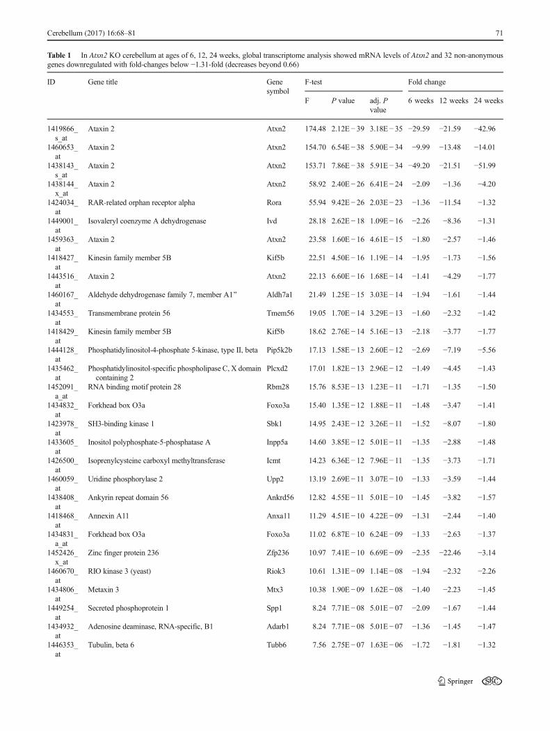
Table 1 In Atxn2 KO cerebellum at ages of 6, 12, 24 weeks, global transcriptome analysis showed mRNA levels of Atxn2 and 32 non-anonymousgenes downregulated with fold-changes below −1.31-fold (decreases beyond 0.66)
ID Gene title Genesymbol
F-test Fold change
F P value adj. Pvalue
6 weeks 12 weeks 24 weeks
1419866_s_at
Ataxin 2 Atxn2 174.48 2.12E− 39 3.18E− 35 −29.59 −21.59 −42.96
1460653_at
Ataxin 2 Atxn2 154.70 6.54E− 38 5.90E− 34 −9.99 −13.48 −14.01
1438143_s_at
Ataxin 2 Atxn2 153.71 7.86E− 38 5.91E− 34 −49.20 −21.51 −51.99
1438144_x_at
Ataxin 2 Atxn2 58.92 2.40E− 26 6.41E− 24 −2.09 −1.36 −4.20
1424034_at
RAR-related orphan receptor alpha Rora 55.94 9.42E− 26 2.03E− 23 −1.36 −11.54 −1.32
1449001_at
Isovaleryl coenzyme A dehydrogenase Ivd 28.18 2.62E− 18 1.09E− 16 −2.26 −8.36 −1.31
1459363_at
Ataxin 2 Atxn2 23.58 1.60E− 16 4.61E− 15 −1.80 −2.57 −1.46
1418427_at
Kinesin family member 5B Kif5b 22.51 4.50E − 16 1.19E− 14 −1.95 −1.73 −1.56
1443516_at
Ataxin 2 Atxn2 22.13 6.60E− 16 1.68E− 14 −1.41 −4.29 −1.77
1460167_at
Aldehyde dehydrogenase family 7, member A1^ Aldh7a1 21.49 1.25E− 15 3.03E− 14 −1.94 −1.61 −1.44
1434553_at
Transmembrane protein 56 Tmem56 19.05 1.70E− 14 3.29E− 13 −1.60 −2.32 −1.42
1418429_at
Kinesin family member 5B Kif5b 18.62 2.76E − 14 5.16E− 13 −2.18 −3.77 −1.77
1444128_at
Phosphatidylinositol-4-phosphate 5-kinase, type II, beta Pip5k2b 17.13 1.58E− 13 2.60E− 12 −2.69 −7.19 −5.56
1435462_at
Phosphatidylinositol-specific phospholipase C, X domaincontaining 2
Plcxd2 17.01 1.82E− 13 2.96E− 12 −1.49 −4.45 −1.43
1452091_a_at
RNA binding motif protein 28 Rbm28 15.76 8.53E− 13 1.23E− 11 −1.71 −1.35 −1.50
1434832_at
Forkhead box O3a Foxo3a 15.40 1.35E− 12 1.88E− 11 −1.48 −3.47 −1.41
1423978_at
SH3-binding kinase 1 Sbk1 14.95 2.43E− 12 3.26E− 11 −1.52 −8.07 −1.80
1433605_at
Inositol polyphosphate-5-phosphatase A Inpp5a 14.60 3.85E − 12 5.01E− 11 −1.35 −2.88 −1.48
1426500_at
Isoprenylcysteine carboxyl methyltransferase Icmt 14.23 6.36E − 12 7.96E− 11 −1.35 −3.73 −1.71
1460059_at
Uridine phosphorylase 2 Upp2 13.19 2.69E− 11 3.07E− 10 −1.33 −3.59 −1.44
1438408_at
Ankyrin repeat domain 56 Ankrd56 12.82 4.55E− 11 5.01E− 10 −1.45 −3.82 −1.57
1418468_at
Annexin A11 Anxa11 11.29 4.51E− 10 4.22E− 09 −1.31 −2.44 −1.40
1434831_a_at
Forkhead box O3a Foxo3a 11.02 6.87E− 10 6.24E− 09 −1.33 −2.63 −1.37
1452426_x_at
Zinc finger protein 236 Zfp236 10.97 7.41E− 10 6.69E− 09 −2.35 −22.46 −3.14
1460670_at
RIO kinase 3 (yeast) Riok3 10.61 1.31E− 09 1.14E− 08 −1.94 −2.32 −2.26
1434806_at
Metaxin 3 Mtx3 10.38 1.90E− 09 1.62E− 08 −1.40 −2.23 −1.45
1449254_at
Secreted phosphoprotein 1 Spp1 8.24 7.71E− 08 5.01E− 07 −2.09 −1.67 −1.44
1434932_at
Adenosine deaminase, RNA-specific, B1 Adarb1 8.24 7.71E− 08 5.01E− 07 −1.36 −1.45 −1.47
1446353_at
Tubulin, beta 6 Tubb6 7.56 2.75E− 07 1.63E− 06 −1.72 −1.81 −1.32
Cerebellum (2017) 16:68–81 71
and 60 °C for 60 s. Gene expression data was analyzed usingthe 2−ΔΔCt method [51]. Additionally, mean Ct values fromTbp andHprt1were averaged to avoid false positive results inone of the housekeeping genes. Then, Ct values from therespective transcript were normalized to Tbp+Hprt and tothe average of the WT values.
Protein Extraction and Quantitative Immunoblotting
For SDS polyacrylamide gel electrophoresis (PAGE), immu-noblotting, and quantitative densitometry, protein was extract-ed from 25 mg cerebellar tissue of either 6-month-old Atxn2KO and WT or 18-month-old Atxn2-CAG42-KIN and WTmice. The tissue was homogenized in 10 vol. RIPA buffer(50 mM Tris–HCl (pH 8.0); 150 mM NaCl; 1 mM EDTA;1 mM EGTA; 1 % Igepal CA-630 (Sigma); 0.5 % sodiumdeoxycholate; 0.1 % SDS; 1 mM PMSF; Complete ProteaseInhibitor Cocktail (Roche)) with a motor pestle and incubatedon ice for 15min. Samples were then centrifuged for 20min at4 °C and 16,000×g and the supernatant was transferred into anew tube and kept on ice until further processing (RIPA frac-tion). Using sonification, the remaining pellet was dissolved in½ vol. 2× SDS buffer (137 mM Tris–HCl (pH 6.8); 4 % SDS;20 % glycerol; Complete Protease Inhibitor Cocktail(Roche)), centrifuged for 10 min at 16,000×g and the remain-ing supernatant was separated as SDS fraction. For proteinconcentration determination, the BCA protein assay kit
(Interchim, France) was applied and the received values werenormalized to the respective buffers. Samples were boiledwith 2× loading buffer (25 % stacking gel buffer (0.5 MTris, 0.4 % SDS; pH 6.8), 20 % Glycerol, 4 % SDS, 5 % β-Mercaptoethanol, and 0.05 % Bromophenol blue) at 95 °C for5 min to denature proteins. Subsequently, 20 μg of each pro-tein lysate was loaded onto a 7 % polyacrylamide gel and afterelectrophoresis transferred to a nitrocellulose membrane.Blocking was performed in 5 % slim-milk powder in PBSTfor 1 h. Primary antibodies were used at the following dilu-tions: INPP5A (MyBiosource, MBS716862, 1:1000) andITPR1 (Abcam, ab5804, 1:1000 and Millipore ABS55,1:1000). Fluorescently tagged secondary antibodies were used(LI-COR Odyssey Infrared Imaging, goat anti-mouse IRDye800CW and goat anti-rabbit IRDye 680RD, both 1:15,000),and proteins were detected using a LI-COR Odyssey InfraredImaging System. Densitometry was performed with ImageJsoftware and protein values were normalized to β-ACTINlevels using EXCEL.
Coimmunoprecipitation
For coimmunoprecipitation (Co-IP) studies, cerebellumfrom 8-week-old Atxn2-CAG42-KIN and Atxn2 KO andWT mice was homogenized in 1:10 w/v NP40 buffer (20mM Tris–HCl pH 8.0; 137 mM NaCl; 1 % Glycerol;0.1 % Igepal CA-630 (Sigma); 2 mM EDTA; Complete
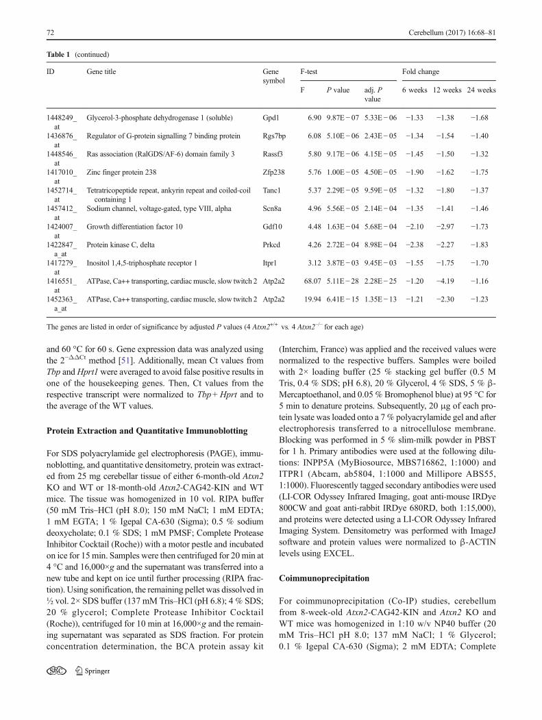
Table 1 (continued)
ID Gene title Genesymbol
F-test Fold change
F P value adj. Pvalue
6 weeks 12 weeks 24 weeks
1448249_at
Glycerol-3-phosphate dehydrogenase 1 (soluble) Gpd1 6.90 9.87E− 07 5.33E− 06 −1.33 −1.38 −1.68
1436876_at
Regulator of G-protein signalling 7 binding protein Rgs7bp 6.08 5.10E − 06 2.43E− 05 −1.34 −1.54 −1.40
1448546_at
Ras association (RalGDS/AF-6) domain family 3 Rassf3 5.80 9.17E− 06 4.15E− 05 −1.45 −1.50 −1.32
1417010_at
Zinc finger protein 238 Zfp238 5.76 1.00E− 05 4.50E− 05 −1.90 −1.62 −1.75
1452714_at
Tetratricopeptide repeat, ankyrin repeat and coiled-coilcontaining 1
Tanc1 5.37 2.29E− 05 9.59E− 05 −1.32 −1.80 −1.37
1457412_at
Sodium channel, voltage-gated, type VIII, alpha Scn8a 4.96 5.56E− 05 2.14E− 04 −1.35 −1.41 −1.46
1424007_at
Growth differentiation factor 10 Gdf10 4.48 1.63E− 04 5.68E− 04 −2.10 −2.97 −1.73
1422847_a_at
Protein kinase C, delta Prkcd 4.26 2.72E− 04 8.98E− 04 −2.38 −2.27 −1.83
1417279_at
Inositol 1,4,5-triphosphate receptor 1 Itpr1 3.12 3.87E− 03 9.45E− 03 −1.55 −1.75 −1.70
1416551_at
ATPase, Ca++ transporting, cardiacmuscle, slow twitch 2 Atp2a2 68.07 5.11E− 28 2.28E− 25 −1.20 −4.19 −1.16
1452363_a_at
ATPase, Ca++ transporting, cardiacmuscle, slow twitch 2 Atp2a2 19.94 6.41E − 15 1.35E− 13 −1.21 −2.30 −1.23
The genes are listed in order of significance by adjusted P values (4 Atxn2+/+ vs. 4 Atxn2−/− for each age)
72 Cerebellum (2017) 16:68–81
Protease Inhibitor Cocktail (Roche)) using a motor pestleand incubated for 15 min on ice. Subsequently, sampleswere centrifuged for 20 min at 16,000×g and 4 °C andsupernatant was transferred into a new tube. Prior to theimmunoprecipitation step, 20 μl of Protein A agarosebeads (Santa Cruz) were washed twice with lysis buffer(120 mM NaCl; 0.1 % Triton X 100; 50 mM Tris–HCl pH7.5) for 5 min. A centrifugation step of 1 min at 2300×gfollowed before the supernatant was discarded. To de-crease the adhesiveness of the beads, they were incubatedfor 1 h at room temperature (RT) on a rotating wheeltogether with 1 ml of blocking buffer (0.2 % NaCl;0.1 % gelatin; 0.05 % NaN3; 50 mM Tris; 0.1 %Triton). At the same time, 250 μg of cerebellar proteinextract was preincubated with the respective pulling anti-bodies against ATXN2 (50 μl/sample, custom-made) orITPR1 (10 μl/sample, Abcam ab5804) for 2 h at 4 °Con a rotating wheel. Beads were then centrifuged for1 min at 2300×g and blocking buffer was removed beforethe preincubated protein extract was added. Subsequently,samples were incubated at 4 °C on a rotating wheelovernight and centrifuged for 1 min at 2300×g. Thesupernatant was removed and samples were washedthree times with 1 ml of lysis buffer and centrifugedagain for 1 min at 2300×g . After discarding thesupernatant, samples were boiled for 5 min at 95 °Cwith 25 μl loading buffer and loaded onto the 8 % SDSgel. The immunoblot detection of ATXN2 was carried outwith the commercial rabbit polyclonal IgG antibody fromProteinTech (catalog no. 21776-1-AP) at titer 1:500,employing the IRdye680 donkey anti-rabbit (1:15,000 atRT for 1 h) as secondary antibody.
Immunohistochemical Staining
Paraffin-embedded slices were used after rehydration in adescending ethanol series. Between incubation steps,slices were stored or washed in Tris/HCl buffer pH 7.6.For antigen retrieval, slices were autoclaved (BiocareMedical) in Bull’s Eye Decloaker (1:20). The followingconditions were applied: 125 °C for 30 s and 90 °C for10 s. Slides were subsequently cooled down and washed.Slices were incubated in 100 % methanol, 30 % H2O2,and Tris/HCl pH 7.6 (1:1:8) for 30 min in a wet chamberfor background reduction. After another washing step,they were blocked in 2.5 μl Triton-X-100, 18.2 mg DL-Lysine, and 998 μl 5 % Tris–BSA for 30 min. Then, theslices were incubated with the first antibodies (anti-ATXN2, BD Biosciences, 611378, 1:50 and anti-ITPR1,Abcam, ab5804, 1:800) overnight. After another washingstep, incubation with the secondary fluorescently labeledantibodies followed for 6 h (Cy3 and Cy2, Dianova, 711-225-152 and 715-165-150, 1:1000). Finally, slices were
mounted with DAKO fluorescent mounting medium.Microscopic pictures were taken with a Nikon confocalmicroscope Eclipse 90i and a ×60 magnification.
Statistical Analysis
Data analysis was conducted with GraphPad Prism softwareversion 4.03 (2005) using Student’s t test. Error bars indicateSEM. Significant P values (<0.05) were marked as follows:p<0.05*, p<0.01**, p<0.001***. A trend (T) was notedwhen 0.05<P<0.1.
Results
Microarray Transcriptome Profile of Atxn2 KOCerebellum Shows Dysregulation of Several FactorsInvolved in Calcium Homeostasis Pathways
Attempting to gain deeper insight into the function of theRNA-binding protein ATXN2 and the cerebellar conse-quences of its deficiency, we performed global tran-scriptome analysis of Atxn2 KO mice. Cerebella from 6-week-, 3-month-, and 6-month-old animals, in each casefour wild-type (WT, Atxn2+/+) versus four homozygousknockout (KO, Atxn2−/−) mice, were examined formRNA level changes using a total of 24 Affymetrix oli-gonucleotide microarray chips. The data confirmed theabsence of Atxn2 mRNA at six spots for all three ageswith changes ranging from −51.99-fold to −1.36-fold(Table 1), with variance probably due to technical impre-cisions in independent assessments. Apart from Atxn2, theanalysis of significant downregulations with consistencyacross ages revealed a list of 126 transcript coding for 109genes. Table 1 shows the 32 non-anonymous genes withchanges stronger than −1.31-fold (corresponding to a de-crease to 0.66, or log2-fold-changes/M-values ≤ −0.39)among them, ordered by significance of the adjusted Pvalue. Interaction analysis at the String Heidelberg data-base highlighted the calcium homeostasis pathway to beaffected (Supp. Fig. 1), but multiple factors of RNA pro-cessing (Riok3, Rbm28, Adarb1), bioenergetics (Aldh7a1,Gpd1), cell adhesion (Tanc1, Tmem56), growth (Icmt,Gdf10, Foxo3, Rassf3, Sbk1), and lipid signaling(Pip4k2b, Plcxd2, Rgs7bp) were also present in this shortlist. Further bioinformatics assessment of the completetranscriptome of 12-week-old cerebella by Gene SetEnrichment Analysis (GSEA) at the Broad Institute serveralso revealed significant downregulations in the KEGGadherens junction pathway of cell adhesion, theBIOCARTA PPARA pathway of lipid signaling, theBIOCARTA EGF pathway of growth signaling, and theBIOCARTA Creb pathway of nuclear response to
Cerebellum (2017) 16:68–81 73
extracellular triggers with Camk2b (calcium/calmodulin-dependent protein kinase II beta) as the strongest down-regulation (Suppl. Fig. 2–5). These results are in goodagreement with previous observations of phenotypic ef-fects in the lipid metabolism, insulin, and EGF signaling[38, 39, 47]. The most significant change after Atxn2 wasobserved for Rora as a transcription factor that regulatesseveral genes involved in calcium homeostasis besideshaving other effects (Table 1), Inpp5a and Itpr1 as tworegulators of calcium flux and signaling besides otherfunctions, as well as Prkcd and Anxa11 as calcium-regulated targets were present among the top 32 genes,and age-consistent significant downregulations (−1.2-fold) were observed at two individual microarray spotsfor the calcium flux regulator Atp2a2 (Table 1, bottom),as a possible correlate of altered calcium signal transmis-sion at Purkinje neuron dendrites. Thus, this unbiasedanalysis of cerebellar transcriptome dysregulations withconsistency through three ages advanced previous knowl-edge on altered RNA processing/bioenergetics/cell adhe-sion/growth/lipid signaling and documented novel promi-nent downregulations of mRNAs for proteins involved incalcium homeostasis and signaling that are caused byAtxn2 loss.
Downregulation of Factors Involved in CalciumHomeostasis Pathways is Confirmed Independentlyby qPCR
To validate the results of the transcriptome analysis and toinvestigate even younger ages as well as other brain re-gions in Atxn2 KO homozygous and heterozygous ani-mals, an approach by RT-qPCR was used. The downreg-ulation of Atp2a2, Inpp5a, Itpr1, and Rora mRNA levelsin cerebellum of 6-month-old Atxn2−/−animals was thusconfirmed by an independent technique (Table 2; n ≥ 4Atxn2+/+ vs. ≥3 Atxn2−/−). Fold changes were similar tothose found by the microarray profiling, apart from Itpr1that showed a much smaller downregulation.
To test the temporal dynamics of these effects, 6-week-old Atxn2−/− mice were studied (n ≥ 4 Atxn2+/+ vs. ≥4Atxn2−/−). To further analyze the effects independently inanimals with reduced mutation load, heterozygous(Atxn2+/−) mice at the age of 6 weeks (n≥ 4 Atxn2+/+ vs.4 Atxn2+/−) were analyzed. Finally, in order to test theearliest appearance of these effects in animals with re-duced mutation load, heterozygous pups at ~3 days ofage (n = 4 Atxn2+/+ vs. 4 Atxn2+/−) were assessed. Asshown in Table 2, consistent results for all these condi-tions were observed, with significant downregulations ofsimilar strength for Atp2a2 and Inpp5a, as well asdownregulations of weaker strength for Rora. These ob-servations indicate that changes in calcium homeostasis
pathways are present very early even in heterozygousAtxn2 KO cerebellum.
Atxn2-CAG42-KIN Mice Show Subtle Transcript LevelChanges in Calcium Homeostasis Pathways
Aware that RORA has been shown to be an important modi-fier of the cerebellar ataxia in SCA1 mouse models [52], weextended the analysis to the cerebellum of SCA2 mousemodels. The Atxn2-CAG42-KIN (Atxn2CAG42/CAG42) mousecerebellum at an age of 18 months was assessed by RT-qPCR.At this age, the Atxn2-CAG42-KIN mice show the first be-havioral deficits and visible aggregates of the ATXN2 proteinin cerebellar Purkinje neurons [46]. In contrast to KO mice atthis age, only Atp2a2 and Itpr1 were slightly but significantlydownregulated in homozygous Atxn2-CAG42 KIN micewhile Inpp5a and Rora did not show any change at all (n≥4Atxn2+/+ vs. ≥4 Atxn2CAG42/CAG42) (Table 3).
To again document the temporal dynamics, the transcrip-tion analysis was extended to the cerebellum of 6-month- and6-week-old Atxn2-CAG42-KIN mice. While Atp2a2 andItpr1 were significantly downregulated (n≥4 Atxn2+/+ vs. ≥3
Table 2 Atp2a2, Inpp5a, Itpr1, and Rora were confirmed to bedownregulated in Atxn2 KO mice using RT-qPCR
Gene symbol WT vs. HOM KO Cbll 6 months
Atp2a2 −1.13-fold*Inpp5a −1.30-fold**Itpr1 −1.05-fold*Rora −1.42-fold**Gene symbol WT vs. HOM KO Cbll 6 weeks
Atp2a2 −1.24-fold*Inpp5a −1.39-fold**Itpr1 −1.05-fold n.s.
Rora −1.11-fold**Gene symbol WT vs. HET KO Cbll 6 weeks
Atp2a2 −1.11-fold*Inpp5a −1.35-fold**Itpr1 −1.07-fold n.s.
Rora −1.15-fold***Gene symbol WT vs. HET KO Cbll 3 days
Atp2a2 −1.17-fold**Inpp5a −1.19-fold**Itpr1 +1.04-fold n.s.
Rora −1.14-fold T
Downregulation of the transcripts was detected by qPCR in cerebellum.Atp2a2 and Inpp5a showed the first but Rora the strongest downregula-tion. Significant changes are marked with asterisks while trends areindicated with BT^ (n ≥ 4 Atxn2+/+ vs. n ≥ 3 Atxn2−/− )
T statistical trend with P value between 0.05 and 0.10, n.s. non-signifi-cant, Cbll Cerebellum
*P< 0.05; **P< 0.01; ***P< 0.001
74 Cerebellum (2017) 16:68–81
Atxn2CAG42/CAG42) at 6 months of age with similar strength(about -1.15-fold) as at 18 months of age, only Itpr1 wasdownregulated at 6 weeks of age (n ≥ 4 Atxn2+/+ vs. ≥4Atxn2CAG42/CAG42) (-1.12-fold). These data indicate an unex-pected preferential and early affection of Itpr1 mRNA levelsin the SCA2 model cerebellum, which is not observed in theAtxn2 KO tissue.
Dysregulated Protein Levels in Calcium HomeostasisPathways in Atxn2 KO and Atxn2-CAG42-KIN Mice
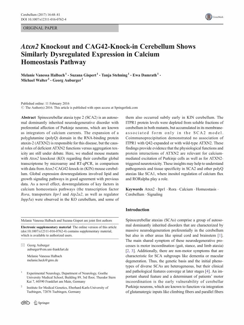
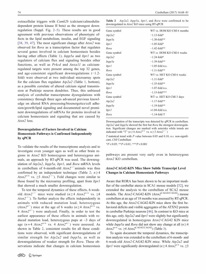
Despite the modest fold changes of the expressiondysregulations observed, we attempted to validate the corre-sponding protein levels. In view of the limited availability ofspecific antibodies, only INPP5A and ITPR1 levels wereassessed via quantitative immunoblots in cerebellar tissuefrom 6-month-old mice (8 Atxn2+/+ vs. ≥7 Atxn2−/−). In theRIPA-soluble fraction (Fig. 1a) where mostly cytosolic factorswill be detected, both proteins were significantly downregu-lated (fold changes −1.21-fold for INPP5A and −1.61-fold forITPR1). In the SDS-soluble fraction (Fig. 1b), wheremembrane-associated proteins such as cortical actin or alsoaggregating proteins may be solubilized, only INPP5Ashowed significant downregulation (−1.31-fold). These datademonstrate that the relatively subtle transcript dysregulationsare indeed resulting in substantially altered abundance of the
corresponding proteins, making an impairment of calciumflux regulation in Atxn2KOmouse cerebellum more credible.
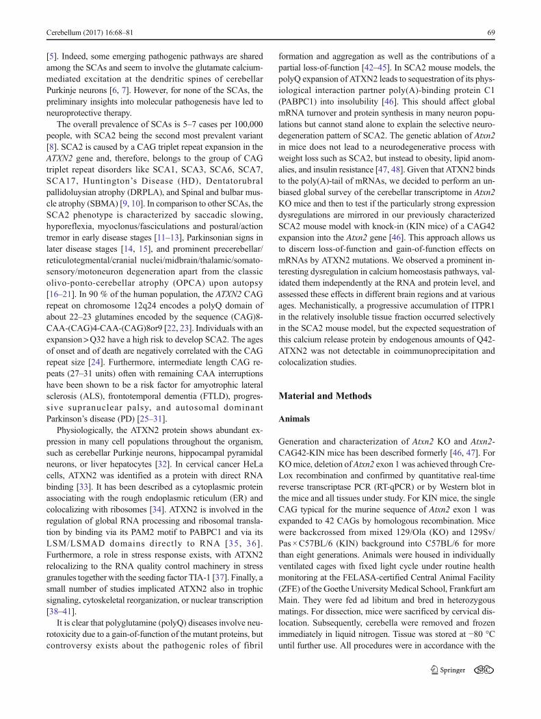
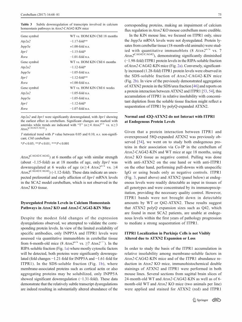
In the KIN mouse line, we focused on ITPR1 only, sincethe Inpp5a mRNA levels were not dysregulated. Protein ly-sates from cerebellar tissue (18-month-old animals) were stud-ied with quantitative immunoblots (8 Atxn2+/+ vs. 7Atxn2CAG42/CAG42), demonstrating significantly diminished(−1.98-fold) ITPR1 protein levels in the RIPA-soluble fractionof Atxn2-CAG42-KINmice (Fig. 2a). Conversely, significant-ly increased (1.28-fold) ITPR1 protein levels were observed inthe SDS-soluble fraction of Atxn2-CAG42-KIN mice(Fig. 2b). In view of the previously demonstrated aggregationof ATXN2 protein in the SDS/urea fraction [46] and reports ona protein interaction between ATXN2 and ITPR1 [53, 54], thisaccumulation of ITPR1 in relative insolubility with concomi-tant depletion from the soluble tissue fraction might reflect asequestration of ITPR1 by polyQ-expanded ATXN2.
Normal and 42Q-ATXN2 do not Interact with ITPR1at Endogenous Protein Levels
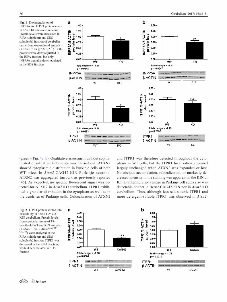
Given that a protein interaction between ITPR1 andoverexpressed 58Q-expanded ATXN2 was previously ob-served [54], we went on to study both endogenous pro-teins in their association via Co-IP in the cerebellum ofAtxn2-CAG42-KIN and WT mice at age 18 months, usingAtxn2 KO tissue as negative control. Pulling was donewith anti-ATXN2 on the one hand or with anti-ITPR1on the other hand, performing pull-downs with unspecificIgG or using beads only as negative controls. ITPR1(Fig. 3, panel above) and ATXN2 (panel below) at endog-enous levels were readily detectable as input in tissues ofall genotypes and were concentrated by its immunoprecip-itation, providing the necessary quality control. However,ITPR1 bands were not brought down in detectableamounts by WT or Q42-ATXN2. These results suggestthat ATXN2 polyQ expansion sizes such as Q42, whichare found in most SCA2 patients, are unable at endoge-nous levels within the first years of pathology progressionto mediate a strong sequestration of ITPR1.
ITPR1 Localization in Purkinje Cells is not VisiblyAltered due to ATXN2 Expansion or Loss
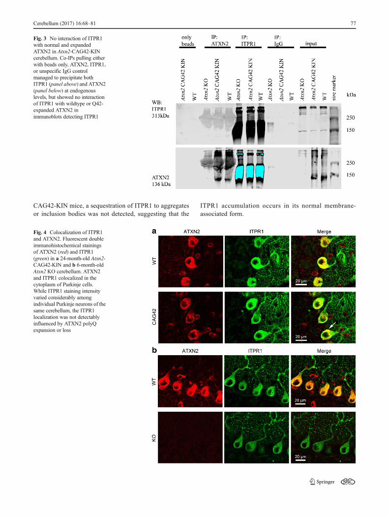
In order to study the basis of the ITPR1 accumulation inrelative insolubility among membrane-soluble factors inAtxn2-CAG42-KIN mice and of the ITPR1 abundance re-duction in Atxn2 KO mice, immunohistochemical doublestainings of ATXN2 and ITPR1 were performed in bothmouse lines. Several sections from sagittal brain slices of24-month-old WT and Atxn2-CAG42-KIN as well as of 6-month-old WT and Atxn2 KO mice (two animals per line)were applied and stained for ATXN2 (red) and ITPR1
Table 3 Subtle downregulation of transcripts involved in calciumhomeostasis pathways in Atxn2-CAG42-KIN mice
Gene symbol WT vs. HOM KIN Cbll 18 months
Atp2a2 −1.17-fold**Inpp5a +1.00-fold n.s.
Itpr1 −1.15-fold*Rora −1.01-fold n.s.Gene symbol WT vs. HOM KIN Cbll 6 months
Atp2a2 −1.12-fold*Inpp5a −1.05-fold n.s.Itpr1 −1.12-fold**Rora +1.00-fold n.s.
Gene symbol WT vs. HOM KIN Cbll 6 weeks
Atp2a2 −1.05-fold n.s.Inpp5a −1.05-fold n.s.Itpr1 −1.12-fold*Rora −1.07-fold n.s.
Atp2a2 and Itpr1 were significantly downregulated, with Itpr1 showingthe earliest effect in cerebellum. Significant changes are marked withasterisks while trends are indicated with BT^ (n ≥ 4 Atxn2+/+ vs. n ≥ 3Atxn2CAG42/CAG42 )
T statistical trend with P value between 0.05 and 0.10, n.s. non-signifi-cant, Cbll cerebellum
*P< 0.05; **P< 0.01; ***P< 0.001
Cerebellum (2017) 16:68–81 75
(green) (Fig. 4a, b). Qualitative assessment without sophis-ticated quantitative techniques was carried out. ATXN2showed cytoplasmic distribution in Purkinje cells of bothWT mice. In Atxn2-CAG42-KIN Purkinje neurons,ATXN2 was aggregated (arrow), as previously reported[46]. As expected, no specific fluorescent signal was de-tected for ATXN2 in Atxn2 KO cerebellum. ITPR1 exhib-ited a granular distribution in the cytoplasm as well as inthe dendrites of Purkinje cells. Colocalization of ATXN2
and ITPR1 was therefore detected throughout the cyto-plasm in WT cells, but the ITPR1 localization appearedlargely unchanged when ATXN2 was expanded or lost.No obvious accumulation, relocalization, or markedly de-creased intensity in the staining was apparent in the KIN orKO. Furthermore, no change in Purkinje cell soma size wasdetectable neither in Atxn2-CAG42-KIN nor in Atxn2 KOcerebellum. Thus, although less salt-soluble ITPR1 andmore detergent-soluble ITPR1 was observed in Atxn2-
Fig. 1 Downregulation ofINPP5A and ITPR1 protein levelsin Atxn2 KO mouse cerebellum.Protein levels were measured inRIPA-soluble (a) and SDS-soluble (b) fraction of cerebellartissue from 6-month-old animals(8 Atxn2+/+ vs. ≥7 Atxn2−/−). Bothproteins were downregulated inthe RIPA fraction, but onlyINPP5Awas also downregulatedin the SDS fraction
Fig. 2 ITPR1 protein shifted intoinsolubility in Atxn2-CAG42-KIN cerebellum. Protein levelsfrom cerebellar tissue of 18-month-old WT and KIN animals(8 Atxn2+/+ vs. 7 Atxn2CAG42/CAG42) were analyzed in theRIPA-soluble (a) and SDS-soluble (b) fraction. ITPR1 wasdecreased in the RIPA fraction,while it accumulated in SDSfraction
76 Cerebellum (2017) 16:68–81
CAG42-KIN mice, a sequestration of ITPR1 to aggregatesor inclusion bodies was not detected, suggesting that the
ITPR1 accumulation occurs in its normal membrane-associated form.
Fig. 3 No interaction of ITPR1with normal and expandedATXN2 in Atxn2-CAG42-KINcerebellum. Co-IPs pulling eitherwith beads only, ATXN2, ITPR1,or unspecific IgG controlmanaged to precipitate bothITPR1 (panel above) and ATXN2(panel below) at endogenouslevels, but showed no interactionof ITPR1 with wildtype or Q42-expanded ATXN2 inimmunoblots detecting ITPR1
Fig. 4 Colocalization of ITPR1and ATXN2. Fluorescent doubleimmunohistochemical stainingsof ATXN2 (red) and ITPR1(green) in a 24-month-old Atxn2-CAG42-KIN and b 6-month-oldAtxn2 KO cerebellum. ATXN2and ITPR1 colocalized in thecytoplasm of Purkinje cells.While ITPR1 staining intensityvaried considerably amongindividual Purkinje neurons of thesame cerebellum, the ITPR1localization was not detectablyinfluenced by ATXN2 polyQexpansion or loss
Cerebellum (2017) 16:68–81 77
Discussion
Our global transcriptome survey of Atxn2 KO cerebellum de-fined significant downregulations of key molecules in severalpathways where previous studies had demonstrated phenotyp-ic changes, namely extracellular growth/adhesion signals vialipid and transcription changes. As a prominent novel effectwith selectivity for cerebellum, the mRNA levels of calciumhomeostasis pathway components Atp2a2, Inpp5a, Itpr1, andparticularly Rora showed varying decreases. This insight mayprovide parameters to measure how the loss of physiologicalfunction of ATXN2 affects the integration of calcium signalsin the dendritic trees of Purkinje dendrites. It is also useful tounderstand how the physiological function of ATXN2 is al-tered by polyQ expansions in SCA2. Thus, our data for thefirst time show clearly significant dysregulations in theabundance of several factors involved in calcium homeostasispathways, in the cerebellum of a Q42-knock-in mouse modelof SCA2, but these changes are mild even at old age and areprobably not the sole basis for the marked pathology alreadymanifest. These factors define a pathway of Purkinje neuronsignaling. Climbing fiber and parallel fiber afferents excitePurkinje cell dendrites via glutamate receptors, resulting inthe hydrolysis of phosphatidylinositol bisphosphate (PIP2) in-to diacylglycerol (DG) and inositol 1,4,5-trisphosphate(InsP3). InsP3 then binds ITPR1 which thereupon releasesCa2+ from the endoplasmic reticulum into the cytosol. Thestimulator InsP3 itself can be hydrolyzed by inositolpolyphosphate-5-phosphatase (INPP5A) producing InsP2 thatis unable to activate the InsP3 receptor. This mechanism in-hibits further stimulation of calcium release [53]. TheSERCA2 protein, encoded by Atp2a2, counteracts ITPR1 bytranslocating Ca2+ back into the endoplasmic reticulum lumen[55]. Rora is of specific interest as a transcription factor thatregulates several genes involved in calcium homeostasis andsignaling like Calb1 (encoding Calbindin), Grm1 (encodingthe metabotropic glutamate receptor, which influences calci-um signaling through excitatory synaptic neurotransmission),and Itpr1 [56], among other effects. Overall, calcium-mediated excitation influences the duration, direction, extent,and type of synaptic plasticity, thus underlying thepathomechanism of diverse neurological diseases [57].
The calcium signaling pathway has been implicated in thepathogenesis of several spinocerebellar ataxias. Inositol 1,4,5-trisphosphate receptor 1 (ITPR1) is mutated in SCA15 andwas implicated early on also in other human andmurine SCAs[58–60]. A downregulation of Itpr1 and Inpp5a expression atthe transcript level was already reported for several other SCAmouse models: Sca1[82Q] transgenic mice [52, 61], Sca1−/−
and Sca1154Q/+ mice [62], as well as Sca3[Q79] transgenic(only Itpr1) [63] and Sca7266Q/+ mice (only Inpp5a) [64]. Adownregulation of Atp2a2was shown in Sca1[82Q] transgen-ic [52, 61], Sca1−/− and Sca1154Q/+ [62] mouse models but also
in a mouse mutant called staggerer [52, 56]. The staggerermice have a spontaneous mutation of the Rora gene, resultingin complete loss of RORA function and congenital ataxia.Similar to the genes mentioned above, Rora was shown tobe dysregulated in Sca1[Q82] transgenic [52] andSca3[69Q] transgenic mice [65]. Additionally, an increase inRora level (by Tip60 loss) can delay cerebellar degeneration inthe Sca1[Q82] transgenic mouse model [66]. Thus, these al-terations appear to be common to various SCAvariants as partof a downstream pathway of cerebellar pathogenesis and maybe useful as molecular read-outs of presymptomatic or clini-cally manifest ataxia.
It is interesting to meditate about the varying mechanismshow this calcium pathway can be affected by different SCAdisease proteins. An interaction with the transcription factorRORAwas demonstrated for the nuclear factor Ataxin-1 [52].In contrast, Ataxin-2 is a cytoplasmic protein with concentra-tion at the rough endoplasmic reticulum. There, itsoverexpressed 58Q-expanded variant was observed to bindto ITPR1 [7, 54, 67]. Although these studies found the over-expression of INPP5A by adeno-associated virus over17 weeks and until 10 months to alleviate the Purkinje neuronphenotypes and motor incoordination in mice with stabletransgenic overexpression of a human CAG58-ATXN2cDNA, our data in the GAG42-knock-in mouse model ofSCA2 show no significant upregulation of the Inpp5a tran-script, but instead consistent downregulations of the Atp2a2mRNA to occur at ages before and after the manifestation ofmotor incoordination. It is impossible to predict the net effectof these changes in abundance of antagonistic calcium modu-lators without future functional studies of electrophysiologyand calcium imaging in knock-in mice. The mild changes atthe RNA level translate into substantial changes at the proteinlevel: INPP5A protein is downregulated in the KO cerebel-lum. Furthermore, ITPR1 is markedly altered in its levels andsolubility both by ATXN2-Q42 expansion and by ATXN2deficiency. Given that a sequestration of ITPR1 into insolubil-ity through protein interaction with expanded ATXN2 couldnot be substantiated in our coimmunoprecipitation andcolocalization studies, the likeliest explanation would predictthat the reduced amounts of Itpr1 mRNA result in reducedamounts of newly synthesized ITRP1 at ribosomes, solublein the RIPA fraction, but the cells accumulate ITPR1 in its ERmembrane-associated form, which is represented by the SDSfraction. This might be a compensatory stabilization effort ofthe cells to maintain calcium homeostasis. The previous ob-servation of protein interactions between ITPR1 and ATXN2by Liu and coworkers [54] may be explained by the longerpolyQ expansion in ATXN2, by its overexpression, and bydifferent experimental conditions with a more stringent lysisbuffer.
An alternative pathomechanism instead of protein interac-tion is also conceivable. Expanded ATXN2 could associate
78 Cerebellum (2017) 16:68–81
with the Itpr1 mRNA and influence its translation efficiency.Recently, it was demonstrated that ATXN2 is involved in thestability of mRNA and protein expression by binding to AU-rich sequences in the 3′UTR [68]. These authors furthermorefound that a depletion of ATXN2 resulted in decreased mRNAstability and protein levels of specific targets. A polyQ expan-sion of ATXN2 did not abolish its function in this process butdecreased its efficiency. In our analysis, the downregulationsof Atp2a2, Inpp5a, Itpr1, and Rora mRNA levels in microar-ray transcriptomics and in RT-qPCR was specific for cerebel-lum in KO mice and was robustly detectable from 3 days to6 months of age, even upon partial loss-of-function. Atp2a2and Inpp5a transcript level changes were the first to be detect-ed in KO cerebellum, even in heterozygous tissue.Conversely, in KIN cerebellum, the downregulation of Itpr1levels was the most pronounced and earliest detectablechange, while Atp2a2 was significantly downregulated fromage 6 months onward. Thus, in Atxn2 KO mice, the loss ofATXN2 may result in decreased transcript and protein levelsdue to a decreased mRNA stability, while in Atxn2-CAG42-KIN mice, the mRNA stability may be partially decreased,compensatory efforts may stabilize the protein through mod-ulation of its degradation speed, and additional pathology mayoccur in later disease stages or in stronger expansions due toATXN2 accumulation/aggregation/sequestration.
The accumulation of ITPR1 in the membrane fraction inassociation with polyQ-expanded ATXN2 would affect Ca2+
release, a feature that could be tested in young mice but iscumbersome to evaluate in aged brain tissues. This effortseems hard to justify, given that SCA2 involves not only thecerebellum but progresses into a multisystem-atrophy of thenervous system, and that our data suggest the calcium homeo-stasis pathway alteration not to be prominent in tissues likemidbrain and motoneurons (data not shown). It will be partic-ularly important to determine if the effects of ATXN2 polyQexpansions on the motor neuron degeneration in amyotrophiclateral sclerosis has shared pathogenesis pathways.
Our present findings clarify the position of ATXN2 effectsin the emerging shared molecular pathways of spinocerebellarataxias. Previous data had suggested a role of ATXN2 inSCA1, given that double mutant Drosophila melanogasterflies showed increased dAtx2 levels to enhance Ataxin-1[82Q]-induced neurodegeneration, while decreased dAtx2levels suppress the neurotoxicity [69]. Furthermore, ATXN2appears to have also a role in SCA3, given that dAtx2 activityhastens the onset of nuclear inclusions associated with SCA3[70]. The dependence of ITPR1, INPP5A, ATP2A2, andRORA levels on ATXN2 mutations provides proof-of-principle of the existence of shared molecular changes earlyin the course of SCA2, SCA1, and SCA3.
Further assessment of additional Atxn2-KO mouse tissuesby global deep proteomics substantiated the downregulationof iso-valeryl dehydrogenase (IVD), which had shown the
second strongest transcript downregulation in this microarraytranscriptomics analysis. The IVD protein reduction had nom-inal significance in cerebellum and showed also actual signif-icance after multiple testing correction in liver tissue, where adecrease to 7 % of WT levels was observed in quantitativeimmunoblots. This effect was part of a systematic downregu-lation of amino acids and fatty acids pathways, which isthought to affect excitation and growth signals [71]. Giventhat deep proteomics is not sensitive toward quantitativechanges of factors with very low abundance like transcriptionfactors, and that tissue extractions even with 8 % SDS maysolubilize nuclear proteins only partially, it is not surprisingthat RORalpha changes were not detected by this approach.
Acknowledgments We are grateful for funding by the DFG (AU96/11-1). Our thanks also go to the staff at the animal facilities (ZFE of theUniversity hospital Frankfurt and mfd Wendelsheim). We like to thankBeatrice Kern, Neurophysiology, Goethe University Medical School fortaking the microscopic pictures.
Compliance with Ethical Standards
Conflict of Interest The authors declare no conflict of interest.
Open Access This article is distributed under the terms of the CreativeCommons At t r ibut ion 4 .0 In te rna t ional License (h t tp : / /creativecommons.org/licenses/by/4.0/), which permits unrestricted use,distribution, and reproduction in any medium, provided you giveappropriate credit to the original author(s) and the source, provide a linkto the Creative Commons license, and indicate if changes were made.
References
1. Rub U, Schols L, Paulson H, Auburger G, Kermer P, Jen JC, et al.Clinical features, neurogenetics and neuropathology of thepolyglutamine spinocerebellar ataxias type 1, 2, 3, 6 and 7. ProgNeurobiol. 2013;104:38–66. doi:10.1016/j.pneurobio.2013.01.001.
2. Fujioka S, Sundal C, Wszolek ZK. Autosomal dominant cerebellarataxia type III: a review of the phenotypic and genotypic character-istics. Orphanet J Rare Dis. 2013;8:14. doi:10.1186/1750-1172-8-14.
3. Whaley NR, Fujioka S, Wszolek ZK. Autosomal dominant cere-bellar ataxia type I: a review of the phenotypic and genotypic char-acteristics. Orphanet J Rare Dis. 2011;6:33. doi:10.1186/1750-1172-6-33.
4. Koeppen AH. The pathogenesis of spinocerebellar ataxia.Cerebellum. 2005;4:62–73. doi:10.1080/14734220510007950.
5. Empson RM, Knopfel T. Functional integration of calcium regula-tory mechanisms at Purkinje neuron synapses. Cerebellum.2012;11:640–50. doi:10.1007/s12311-010-0185-6.
6. Carlson KM, Andresen JM, Orr HT. Emerging pathogenic path-ways in the spinocerebellar ataxias. Curr Opin Genet Dev.2009;19:247–53. doi:10.1016/j.gde.2009.02.009.
7. Kasumu A, Bezprozvanny I. Deranged calcium signaling inPurkinje cells and pathogenesis in spinocerebellar ataxia 2(SCA2) and other ataxias. Cerebellum. 2012;11:630–9. doi:10.1007/s12311-010-0182-9.
Cerebellum (2017) 16:68–81 79
8. Velazquez-Perez L, Rodriguez-Labrada R, Garcia-Rodriguez JC,Almaguer-Mederos LE, Cruz-Marino T, Laffita-Mesa JM. A com-prehensive review of spinocerebellar ataxia type 2 in Cuba.Cerebellum. 2011;10:184–98. doi:10.1007/s12311-011-0265-2.
9. Bauer PO, Nukina N. The pathogenic mechanisms ofpolyglutamine diseases and current therapeutic strategies. JNeurochem. 2009;110:1737–65. doi:10.1111/j.1471-4159.2009.06302.x.
10. Orr HT. Cell biology of spinocerebellar ataxia. J Cell Biol.2012;197:167–77. doi:10.1083/jcb.201105092.
11. Lastres-Becker I, Rub U, Auburger G. Spinocerebellar ataxia 2(SCA2). Cerebellum. 2008;7:115–24. doi:10.1007/s12311-008-0019-y.
12. Orozco Diaz G, Nodarse Fleites A, Cordoves Sagaz R, Auburger G.Autosomal dominant cerebellar ataxia: clinical analysis of 263 pa-tients from a homogeneous population in Holguin, Cuba.Neurology. 1990;40:1369–75.
13. Velazquez-Perez L, Rodriguez-Labrada R, Canales-Ochoa N,Montero JM, Sanchez-Cruz G, Aguilera-Rodriguez R, et al.Progression of early features of spinocerebellar ataxia type 2 inindividuals at risk: a longitudinal study. Lancet Neurol. 2014;13:482–9. doi:10.1016/S1474-4422(14)70027-4.
14. Freund HJ, Barnikol UB, Nolte D, Treuer H, Auburger G, Tass PA,et al. Subthalamic-thalamic DBS in a case with spinocerebellarataxia type 2 and severe tremor-A unusual clinical benefit. MovDisord. 2007;22:732–5. doi:10.1002/mds.21338.
15. Boesch SM, Donnemiller E, Muller J, Seppi K, Weirich-SchwaigerH, Poewe W, et al. Abnormalities of dopaminergic neurotransmis-sion in SCA2: a combined 123I-betaCIT and 123I-IBZM SPECTstudy. Mov Disord. 2004;19:1320–5. doi:10.1002/mds.20159.
16. Rub U, Burk K, Schols L, Brunt ER, de Vos RA, Diaz GO, et al.Damage to the reticulotegmental nucleus of the pons inspinocerebellar ataxia type 1, 2, and 3. Neurology. 2004;63:1258–63.
17. Rub U, Gierga K, Brunt ER, de Vos RA, Bauer M, Schols L, et al.Spinocerebellar ataxias types 2 and 3: degeneration of the pre-cerebellar nuclei isolates the three phylogenetically defined regionsof the cerebellum. J Neural Transm. 2005;112:1523–45. doi:10.1007/s00702-005-0287-3.
18. Rub U, Schultz C, Del Tredici K, Gierga K, Reifenberger G, de VosRA, et al. Anatomically based guidelines for systematic investiga-tion of the central somatosensory system and their application to aspinocerebellar ataxia type 2 (SCA2) patient. Neuropathol ApplNeurobiol. 2003;29:418–33.
19. Rub U, Del Turco D, Del Tredici K, de Vos RA, Brunt ER,Reifenberger G, et al. Thalamic involvement in a spinocerebellarataxia type 2 (SCA2) and a spinocerebellar ataxia type 3 (SCA3)patient, and its clinical relevance. Brain. 2003;126:2257–72. doi:10.1093/brain/awg234.
20. Gierga K, Burk K, Bauer M, Orozco Diaz G, Auburger G, SchultzC, et al. Involvement of the cranial nerves and their nuclei inspinocerebellar ataxia type 2 (SCA2). Acta Neuropathol.2005;109:617–31. doi:10.1007/s00401-005-1014-8.
21. Estrada R, Galarraga J, Orozco G, Nodarse A, Auburger G.Spinocerebellar ataxia 2 (SCA2): morphometric analyses in 11 au-topsies. Acta Neuropathol. 1999;97:306–10.
22. Pulst SM, Nechiporuk A, Nechiporuk T, Gispert S, Chen XN,Lopes-Cendes I, et al. Moderate expansion of a normally biallelictrinucleotide repeat in spinocerebellar ataxia type 2. Nat Genet.1996;14:269–76. doi:10.1038/ng1196-269.
23. Hernandez A, Magarino C, Gispert S, Santos N, Lunkes A, OrozcoG, et al. Genetic mapping of the spinocerebellar ataxia 2 (SCA2)locus on chromosome 12q23-q24.1. Genomics. 1995;25:433–5.
24. Auburger GW. Spinocerebellar ataxia type 2. Handb Clin Neurol.2012;103:423–36. doi:10.1016/B978-0-444-51892-7.00026-7.
25. Charles P, Camuzat A, Benammar N, Sellal F, Destee A, BonnetAM, et al. Are interrupted SCA2 CAG repeat expansions responsi-ble for parkinsonism? Neurology. 2007;69:1970–5. doi:10.1212/01.wnl.0000269323.21969.db.
26. Elden AC, Kim HJ, Hart MP, Chen-Plotkin AS, Johnson BS, FangX, et al. Ataxin-2 intermediate-length polyglutamine expansionsare associated with increased risk for ALS. Nature. 2010;466:1069–75. doi:10.1038/nature09320.
27. Gispert S, Kurz A,Waibel S, Bauer P, Liepelt I, Geisen C, et al. Themodulation of amyotrophic lateral sclerosis risk by ataxin-2 inter-mediate polyglutamine expansions is a specific effect. NeurobiolDis. 2012;45:356–61. doi:10.1016/j.nbd.2011.08.021.
28. Gwinn-Hardy K, Chen JY, Liu HC, Liu TY, Boss M, Seltzer W, etal. Spinocerebellar ataxia type 2 with parkinsonism in ethnicChinese. Neurology. 2000;55:800–5.
29. Ross OA, Rutherford NJ, Baker M, Soto-Ortolaza AI, CarrasquilloMM, DeJesus-Hernandez M, et al. Ataxin-2 repeat-length variationand neurodegeneration. Hum Mol Genet. 2011;20:3207–12.10.1093/hmg/ddr227.
30. Yamashita C, Tomiyama H, Funayama M, Inamizu S, Ando M, LiY, et al. The evaluation of polyglutamine repeats in autosomal dom-inant Parkinson’s disease. Neurobiol Aging. 2014. doi:10.1016/j.neurobiolaging.2014.01.022.
31. Lee T, Li YR, Ingre C, Weber M, Grehl T, Gredal O, et al. Ataxin-2intermediate-length polyglutamine expansions in European ALSpatients. Hum Mol Genet. 2011;20:1697–700. doi:10.1093/hmg/ddr045.
32. Huynh DP, Del Bigio MR, Ho DH, Pulst SM. Expression of ataxin-2 in brains from normal individuals and patients with Alzheimer’sdisease and spinocerebellar ataxia 2. Ann Neurol. 1999;45:232–41.
33. Castello A, Fischer B, Eichelbaum K, Horos R, Beckmann BM,Strein C, et al. Insights into RNA biology from an atlas of mam-malian mRNA-binding proteins. Cell. 2012;149:1393–406. doi:10.1016/j.cell.2012.04.031.
34. van de Loo S, Eich F, Nonis D, Auburger G, Nowock J. Ataxin-2associates with rough endoplasmic reticulum. Exp Neurol.2009;215:110–8. doi:10.1016/j.expneurol.2008.09.020.
35. Ralser M, Albrecht M, Nonhoff U, Lengauer T, Lehrach H,Krobitsch S. An integrative approach to gain insights into the cel-lular function of human ataxin-2. J Mol Biol. 2005;346:203–14.doi:10.1016/j.jmb.2004.11.024.
36. Satterfield TF, Pallanck LJ. Ataxin-2 and its Drosophila homolog,ATX2, physically assemble with polyribosomes. Hum Mol Genet.2006;15:2523–32. doi:10.1093/hmg/ddl173.
37. Nonhoff U, RalserM,Welzel F, Piccini I, Balzereit D, YaspoML, etal. Ataxin-2 interacts with the DEAD/H-box RNA helicase DDX6and interferes with P-bodies and stress granules. Mol Biol Cell.2007;18:1385–96. doi:10.1091/mbc.E06-12-1120.
38. Drost J, Nonis D, Eich F, Leske O, Damrath E, Brunt ER, et al.Ataxin-2 modulates the levels of Grb2 and SRC but not ras signal-ing. J Mol Neurosci. 2013;51:68–81. doi:10.1007/s12031-012-9949-4.
39. Nonis D, Schmidt MH, van de Loo S, Eich F, Dikic I, Nowock J, etal. Ataxin-2 associates with the endocytosis complex and affectsEGF receptor trafficking. Cell Signal. 2008;20:1725–39. doi:10.1016/j.cellsig.2008.05.018.
40. Hallen L, Klein H, Stoschek C, Wehrmeyer S, Nonhoff U, RalserM, et al. TheKRAB-containing zinc-finger transcriptional regulatorZBRK1 activates SCA2 gene transcription through direct interac-tion with its gene product, ataxin-2. HumMol Genet. 2011;20:104–14. doi:10.1093/hmg/ddq436.
41. Satterfield TF, Jackson SM, Pallanck LJ. A Drosophila homolog ofthe polyglutamine disease gene SCA2 is a dosage-sensitive regula-tor of actin filament formation. Genetics. 2002;162:1687–702.
80 Cerebellum (2017) 16:68–81
42. Zoghbi HY, Orr HT. Pathogenic mechanisms of a polyglutamine-mediated neurodegenerative disease, spinocerebellar ataxia type 1.J Biol Chem. 2009;284:7425–9. doi:10.1074/jbc.R800041200.
43. Zoghbi HY, Orr HT. Polyglutamine diseases: protein cleavage andaggregation. Curr Opin Neurobiol. 1999;9:566–70. doi:10.1016/S0959-4388(99)00013-6.
44. Michalik A, Van Broeckhoven C. Pathogenesis of polyglutaminedisorders: aggregation revisited. HumMol Genet 2003: 12 Spec No2:R173-86. doi 10.1093/hmg/ddg295.
45. Costa Mdo C, Paulson HL. Toward understanding Machado-Joseph disease. Prog Neurobiol. 2012;97:239–57. doi:10.1016/j.pneurobio.2011.11.006.
46. Damrath E, Heck MV, Gispert S, Azizov M, Nowock J, Seifried C,et al. ATXN2-CAG42 sequesters PABPC1 into insolubility andinduces FBXW8 in cerebellum of old ataxic knock-in mice. PLoSGenet. 2012;8, e1002920. doi:10.1371/journal.pgen.1002920.
47. Lastres-Becker I, Brodesser S, Lutjohann D, Azizov M, BuchmannJ, Hintermann E, et al. Insulin receptor and lipid metabolism pa-thology in ataxin-2 knock-out mice. Hum Mol Genet. 2008;17:1465–81. doi:10.1093/hmg/ddn035.
48. Auburger G, Gispert S, Lahut S, Omur O, Damrath E, Heck M, etal. 12q24 locus association with type 1 diabetes: SH2B3 orATXN2? World J Diabetes. 2014;5:316–27. doi:10.4239/wjd.v5.i3.316.
49. Subramanian A, Tamayo P, Mootha VK, Mukherjee S, Ebert BL,Gillette MA, et al. Gene set enrichment analysis: a knowledge-based approach for interpreting genome-wide expression profiles.Proc Natl Acad Sci U S A. 2005;102:15545–50. doi:10.1073/pnas.0506580102.
50. Subramanian A, Kuehn H, Gould J, Tamayo P, Mesirov JP. GSEA-P: a desktop application for gene set enrichment analysis.Bioinformatics. 2007;23:3251–3. doi:10.1093/bioinformatics/btm369.
51. Livak KJ, Schmittgen TD. Analysis of relative gene expression datausing real-time quantitative PCR and the 2(-Delta Delta C(T))Method. Methods. 2001;25:402–8. doi:10.1006/meth.2001.1262.
52. Serra HG, Duvick L, Zu T, Carlson K, Stevens S, JorgensenN, et al.RORalpha-mediated Purkinje cell development determines diseaseseverity in adult SCA1 mice. Cell. 2006;127:697–708. doi:10.1016/j.cell.2006.09.036.
53. Kasumu AW, Liang X, Egorova P, Vorontsova D, Bezprozvanny I.Chronic suppression of inositol 1,4,5-triphosphate receptor-mediated calcium signaling in cerebellar purkinje cells alleviatespathological phenotype in spinocerebellar ataxia 2 mice. JNeurosci. 2012;32:12786–96. doi:10.1523/JNEUROSCI.1643-12.2012.
54. Liu J, Tang TS, Tu H, Nelson O, Herndon E, Huynh DP, et al.Deranged calcium signaling and neurodegeneration inspinocerebellar ataxia type 2. J Neurosci. 2009;29:9148–62. doi:10.1523/JNEUROSCI.0660-09.2009.
55. BerridgeMJ, Lipp P, BootmanMD. The versatility and universalityof calcium signalling. Nat Rev Mol Cell Biol. 2000;1:11–21. doi:10.1038/35036035.
56. Gold DA, Baek SH, Schork NJ, Rose DW, Larsen DD, Sachs BD,et al. RORalpha coordinates reciprocal signaling in cerebellar de-velopment through sonic hedgehog and calcium-dependent path-ways. Neuron. 2003;40:1119–31.
57. Maggio N, Vlachos A. Synaptic plasticity at the interface of healthand disease: new insights on the role of endoplasmic reticulum
intracellular calcium stores. Neuroscience. 2014;281C:135–46.doi:10.1016/j.neuroscience.2014.09.041.
58. Desaiah D, Vig PJ, Subramony SH, Currier RD. Inositol 1,4,5-t r i s p ho sph a t e r e c ep t o r s a nd p r o t e i n k i n a s e C i nolivopontocerebellar atrophy. Brain Res. 1991;552:36–40.
59. Maeda N, Niinobe M, Mikoshiba K. A cerebellar Purkinje cellmarker P400 protein is an inositol 1,4,5-trisphosphate (InsP3) re-ceptor protein. Purification and characterization of InsP3 receptorcomplex. Embo J. 1990;9:61–7.
60. Mikoshiba K, Huchet M, Changeux JP. Biochemical and immuno-logical studies on the P400 protein, a protein characteristic of thePurkinje cell from mouse and rat cerebellum. Dev Neurosci.1979;2:254–75.
61. Lin X, Antalffy B, Kang D, Orr HT, Zoghbi HY. Polyglutamineexpansion down-regulates specific neuronal genes before patholog-ic changes in SCA1. Nat Neurosci. 2000;3:157–63. doi:10.1038/72101.
62. Crespo-Barreto J, Fryer JD, Shaw CA, Orr HT, Zoghbi HY. Partialloss of ataxin-1 function contributes to transcriptional dysregulationin spinocerebellar ataxia type 1 pathogenesis. PLoS Genet. 2010;6,e1001021. doi:10.1371/journal.pgen.1001021.
63. Chou A-H, Yeh T-H, Ouyang P, Chen Y-L, Chen S-Y, Wang H-L.Polyglutamine-expanded ataxin-3 causes cerebellar dysfunction ofSCA3 transgenic mice by inducing transcriptional dysregulation.Neurobiol Dis. 2008;31:89–101. doi:10.1016/j.nbd.2008.03.011.
64. Gatchel JR,Watase K, Thaller C, Carson JP, Jafar-Nejad P, ShawC,et al. The insulin-like growth factor pathway is altered inspinocerebellar ataxia type 1 and type 7. Proc Natl Acad Sci U SA. 2008;105:1291–6. doi:10.1073/pnas.0711257105.
65. Konno A, Shuvaev AN, Miyake N, Miyake K, Iizuka A, MatsuuraS, et al. Mutant ataxin-3 with an abnormally expandedpolyglutamine chain disrupts dendritic development and metabo-tropic glutamate receptor signaling in mouse cerebellar Purkinjecells. Cerebellum. 2014;13:29–41. doi:10.1007/s12311-013-0516-5.
66. Gehrking KM, Andresen JM, Duvick L, Lough J, Zoghbi HY, OrrHT. Partial loss of Tip60 slows mid-stage neurodegeneration in aspinocerebellar ataxia type 1 (SCA1) mouse model. Hum MolGenet. 2011;20:2204–12. doi:10.1093/hmg/ddr108.
67. Hansen ST, Meera P, Otis TS, Pulst SM. Changes in Purkinje cellfiring and gene expression precede behavioral pathology in a mousemodel of SCA2. Hum Mol Genet. 2013;22:271–83. doi:10.1093/hmg/dds427.
68. Yokoshi M, Li Q, Yamamoto M, Okada H, Suzuki Y, Kawahara Y.Direct binding of Ataxin-2 to distinct elements in 3′UTRs promotesmRNA stability and protein expression. Mol Cell. 2014;55:186–98.doi:10.1016/j.molcel.2014.05.022.
69. Al-Ramahi I, Perez AM, Lim J, Zhang M, Sorensen R, de Haro M,et al. dAtaxin-2 mediates expanded Ataxin-1-induced neurodegen-eration in a Drosophila model of SCA1. PLoS Genet. 2007;3, e234.doi:10.1371/journal.pgen.0030234.
70. Lessing D, Bonini NM. Polyglutamine genes interact to modulatethe severity and progression of neurodegeneration in Drosophila.PLoS Biol. 2008;6, e29. doi:10.1371/journal.pbio.0060029.
71. Meierhofer D, Halbach MV, Sen NE, Gispert S, Auburger G.Atxn2-Knock-Out mice show branched chain amino acids and fattyacids pathway alterations. Mol. Cell. Proteomics. 2016 (in press).
Cerebellum (2017) 16:68–81 81