Embed Size (px)
Citation preview
BULLETIN 2/2002
Nr. 48 August 2002
Schweizerische Gesellschaft für Strahlenbiologie und Medizinische Physik
Société Suisse de Radiobiologie et de Physique Médicale
Società Svizzera di Radiobiologia e di Fisica Medica
Swiss Society of Radiobiology and Medical Physics Member of the European Federation of Organisations for Medical Physics (EFOMP) and the International Organization for Medical Physics (IOMP)
BULLETIN Nr. 48 (August 2002)
Inhalt • Editorial / Impressum 2 • President's Letter 3 • Programm der Jahrestagung 2002 5 • Rubrik: Bestrahlungstechnik Radiotherapie 6 • Zum Lesen empfohlen 7 • Report of the Working Group: Quality Control in External Beam Therapy 8 • Statusbericht der Arbeitsgruppe PMP 9 • Report on the European CMS-Focus User Group Meeting 11 • Siemens Report on Image guided Radiotherapy 12 • Statistik der SRO: Übersicht Radio-Onkologien in der Schweiz 2001 16 • Rezension: Kompendium für Strahlenschutz – Sachverständige 18 • Stelleninserate 19 • Personalia 20 • Pressespiegel 21 • Tagungskalender 29 • Hinweise für Autoren 31
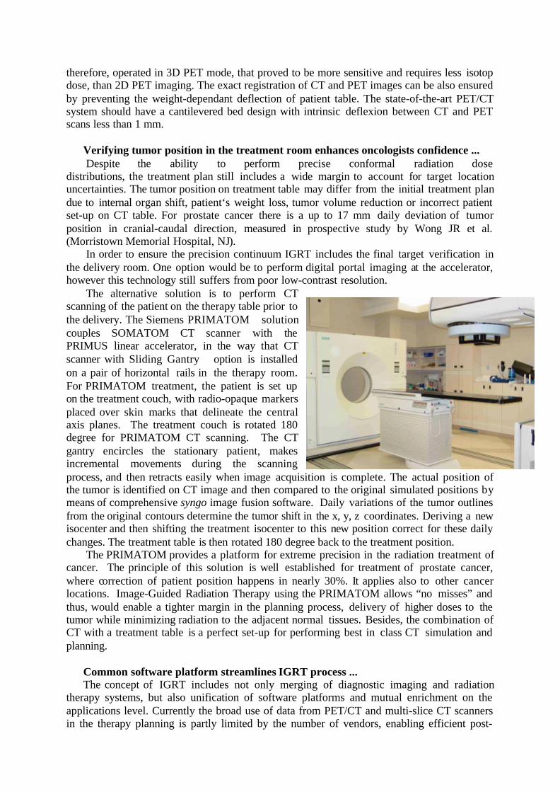
Titelbild: The Siemens PRIMATOM solution couples SOMATOM CT scanner with the PRIMUS linear accelerator, in the way that CT scanner with Sliding Gantry option is in-stalled on a pair of horizontal rails in the therapy room.
Editorial Schon wieder sind 4 Monate vorbei und die neueste Ausgabe des SGSMP-Bulletins liegt vor Ihnen. Diese beginnt mit einem teilweise etwas provokativ gehaltenem Vorwort unseres Prä-sidenten zum Thema IMRT – gerne dürfen Sie darauf mit einem Leserbrief antworten! Wir sind gespannt auf Ihre Reaktionen.
Als weitere Neuigkeit präsentiert Ihnen Roman Menz eine neue Rubrik „Bestrahlungs-technik Radiotherapie“. Darin wird ein konkreter, vielleicht etwas spezieller Fall aus dem Klinikalltag vorgestellt. Auch hierauf dürfen Sie gerne mit der Einreichung eines Kommen-tars oder einem weiteren Beitrag nach Ihrem Geschmack reagieren.
Neben diversen weiteren Beiträgen enthält diese Bulletin-Ausgabe auch eine Vorstellung von IGRT (Image-Guided Radiation Therapy) aus der Sicht von Siemens Medical Solutions. Und im reichhaltigen Pressespiegel erfahren Sie unter anderem auch einige Neuigkeiten zum ersten privaten Ärztehaus für Radio-Onkologie in der Schweiz in Allschwil (BL).
Wir hoffen, dass Ihnen das Lesen des einen oder anderen Artikels Freude bereitet! Mit spät-sommerlichen Grüssen Werner Roser
Redaktionsschluss für das Bulletin Nr. 49 (3/02): 30. November 2002
Impressum Herausgeber: Schweizerische Gesellschaft für Strahlenbiologie und Medizinische Physik (SGSMP/SSRPM/SSRFM) Redaktion: Dr. Roman Menz Dr. Werner Roser Radio-Onkologie Abt. für Technik & Koordination Kantonsspital Winterthur Paul Scherrer Institut 8401 Winterthur 5232 Villigen PSI Tel. 052 266 2648 Tel. 056 310 3514 Fax 052 266 4514 Fax 056 310 3383 [email protected] [email protected] Sekretariat der SGSMP: Dr. R. Mini, Klinik für Radio-Onkologie, Abt. für Med. Strahlenphysik, Inselspital, 3010 Bern, Tel.: 031 / 632 84 31 Fax: 031 / 632 26 76, E-mail: [email protected] Autoren dieser Ausgabe: L. André, H. Blattmann, Ch. v. Briel, W. Burkard, N. Lomax,
R. Menz, H.-P. Rutz, U. Schneider, B. Schnekenburger, W.W. Seelentag, Fa. Siemens
2
President's Letter
Multi-Leaf-Collimator, Intensity Modulated Radio-Therapy, Electronic Portal Imaging
Device, Virtual Simulator, … - if you are planning to bring your radiotherapy clinic up to the "standard", these features are a "must"! Are they really? Could it be that all they are improv-ing are your clinic's profile (and health costs, of course), but not the result - whether measured in patients' survival or reduced side effects?
Let's look at IMRT as an example: a new technology resulting in high expectations - but
is there any proof around yet? Should IMRT not be restricted to a few centres doing random-ised studies first, before spreading the technique to all radiotherapy clinics - or is the im-provement so obvious, that a randomised trial would not be ethical? It would clearly not be acceptable to start a randomised study for deep seated tumours with linacs in one, and 300 kV X-ray machines in the other arm - but isn't IMRT more in the situation of pion therapy a few years ago: a new technology started with high expectations - but at a few centres only; several years later pion therapy was abandoned as the small (though existing) improvements did not justify the huge expenses. How "obvious" should the improvement be in order to render it ethically acceptable not to request proof by randomised trials - and who is taking the deci-sion? Was there ever a randomised study showing the superiority of CT based planning as compared to just doing ap-pa with midline dose? I guess it would at least today be difficult to justify such a study.
Which brings us to another point: under which circumstances will it be possible in the
first place to prove that any given therapy is better than the alternative? 5-year survival rates for early diagnosed prostate cancer are now in excess of 90%: this makes it difficult to im-prove to an extent which can be proven statistically. Our second chance are side effects: will it be possible to reduce them by a statistically significant extent? Whilst reduced acute side ef-fects might be observed on a much shorter time scale than cure rates - for final conclusions one still has to wait for the outcome to make sure reduced acute side effects are not counter-balanced by reduced cure rates (much less likely by increased late morbidity). Sure, there are cancers where the chances to show improved results are better - but what are the incidence rates of these cancers? Due to their small absolute numbers it would be better to have just a few centres investigating the potential improvement, rather than all of us trying to do so. Whilst I agree that many of these questions fall into the medical domain, I feel that also physi-cists supporting the acquisition of IMRT should be prepared to discuss these issues.
When discussing the introduction of IMRT into the clinic, the most common explanation
for not already practising the technique are the complex physics (planning, dosimetry) and quality assurance issues. Sure - these are important parts in the treatment chain, and it is time consuming to put them into clinical use - but we have a fair idea how to do it ! Have the more "medical" issues been discussed to the same extent? Very basic questions like "what is the aim?" - should we go for increased dose (hoping for improved cure rates) or reduced side ef-fects? The answer will likely differ for different diagnoses. Are there agreed upon guidelines for target definition? Looking back to the planning intercomparisons organised by SGSMP and SASRO several years ago, doubts come to mind. How should dose distributions be evalu-ated? With "conventional" techniques there are only a limited number of degrees of freedom (like wedges or beam weights), and consequently it is comparatively simple to determine which dose distribution is the best. With IMRT there is an almost unlimited number of pa-rameters to be fitted: will our present tools be sufficient to decide? Just an example: tighter margins will in many cases also lead to greater dose variation across organs-at-risk - has this
3
partial volume effect been understood well enough to base a decision about the optimal treat-ment plan on it? Is our patient positioning stable and reproducible enough to allow better spar-ing of organs at risk by tighter margins - or will organ movement spoil the exercise in most locations? Should we develop image guided therapy to a routine tool before we can try to make IMRT (e.g. of the prostate) a routine procedure? Another problem: the increased beam-on times during IMRT will (largely due to head transmission) result in increased whole body doses; do we know enough of the radiation biology of these low whole body doses to balance this effect and the potentially better dose distribution in the target area? These are just a few questions - and more could be asked.
Whenever a society is confronted with such a complex situation, the usual reaction is to
form a committee or working group. Should the SGSMP set up a working group on the physi-cal aspects of IMRT - or will the problems be sufficiently similar to problems dealt with by our German colleagues in the DGMP "Arbeitskreis 24"? (http://www.uke.uni-hamburg.de/kliniken/radiologie/strahlen/forschung/akimrt/Startseite.htm) Would it be effi-cient to set up a working group with our medical colleagues, i.e. within SASRO, to discuss all these issues in a wider context? I would like to encourage you to ventilate this with your col-leagues at your home institutions - so we have a basis for discussion and decision.
Let me conclude with a statement which will hopefully avoid any misunderstandings. I'm not against investigating IMRT- on the contrary: this is a promising new technology, which should be investigated thoroughly (also in Switzerland), as it has many potential benefits to offer. I feel strongly, however, that it should be investigated with clear aims and agreed upon tools in mind - not just because it's available. If too many people dabble with IMRT there is also a risk of negative outcome, which might be due to sub-optimal implementation of the technique - but likely would be blamed on the principle.
How do you feel about IMRT? "Letters to the Editor" are not yet a tradition in our Bulle-
tin - but all traditions need to be started at some stage ☺. All readers are invited to comment on the above, and send their opinions on IMRT, the advantages and/or problems they envis-age, to the Bulletin editors. Deadline for the next issue is end of November.
S.Webb: The future of photon external-beam radiotherapy: the dream and the reality. Physica Medica 17/4 (2001) 207-215. R.J.Schulz, A.R.Kagan: On the role of intensity-modulated radiation therapy in radiation oncology. Medical Physics 29/7 (2002) 1473-1482
4
Swiss Society of Radiobiology and Medical Physics
Annual General Meeting Paul Scherrer Institute PSI, November 6, 2002
Program
Welcome 11:00 – 11:15
Scientific Session Is there a Future for Radiobiology? Pruschy Martin 11:15 – 11:40
Coffee 11:40 – 11:55
Synchrotron Radiation for Imaging M. Stampanoni 11 55 – 12:20
Synchrotron Radiation for Rad.Biol./Therapy H. Blattmann 12:20 – 12:45
Lunch (Oase / Sandwich for WGMP) 12:45 – 13:00
Administrative Sessions / Visit of SLS Working Group Med. Phys. (Valley) 13:00 – 14:00
Visit to the SLS 14:00 – 15:00
Membership Assembly 15:00 – 17:30
End of Meeting
Hans Blattmann, PSI
5
Rubrik: Bestrahlungstechnik Radiotherapie In dieser Rubrik, welche ich in dieser Ausgabe des Bulletins eröffnen möchte, geht es darum, für einen konkreten, vielleicht etwas speziellen Fall aus dem Klinikalltag der Radio-Onkologie die verwendete Bestrahlungstechnik kurz vorzustellen. Diese Rubrik soll Sie motivieren, entweder Kommentare dazu abzugeben oder aber besser noch, eigene Techniken aus Ihrem Klinikalltag vorzustellen. Falls diese Idee Anklang findet, werden wir 1-2 Fälle in weiteren Ausgaben des Bulletins veröffentlichen. Concommitant Boost – Bestrahlung für ein Tonsillenkarzinom Konzept: 20 x 1.8 Gy grossvolumig inkl. zervik. Lymphknoten, dazu conkommitierender Bo-ost mit 9 x 1.5 Gy, total 49.5 Gy; danach Myelonschonung mit 8 x 1.8 Gy mit Elektronen & Photonen, total 63.9 Gy; ausserdem 25 x 2 Gy auf supraclav. Lymphknoten. Bestrahlungstechnik für cc Boost: 5 - Felder - Technik, wobei 1 Feld cranio-caudal durchge-hend sowie je 2 Halbfelder für cranialen resp. caudalen Teilbereich aneinandergesetzt. Mit dem durchgehenden Feld wird ein 'hot match' vermieden. Die mögliche Ueberdosierung beim Matching zweier Halbfelder wurde mittels Filmdosimetrie gemessen und ist < 5 %. Die Halbfelder sind sowohl cranial als auch caudal schräg opponierend gewählt, aber die Ein-strahlrichtungen sind auf Grund der speziellen Form des Zielvolumens verschieden, deshalb die Aufteilung in cranialen und caudalen Teil und somit die Abweichung von der sonst gängi-gen 2-3 - Felder - Technik. Das durchgehende Feld trägt zu ca. 30 % der Dosis bei, während die opponierenden Felder in etwa gleich bewichtet sind. Da das Myelon bei den Halbfeldern ausgeblockt ist, erhält es ca. 30 % der Dosis (4 Gy), was unter Berücksichtigung der gesamten Bestrahlungsserie tolerabel ist. Normiert wird auf ein Referenzpunkt im cranialen oder caudalen Teil. Die Bewichtung erfolgt durch Vorgabe der Monitoreinheiten. Die unabhängige Kontrollrechnung erfolgt an zwei Referenzpunkten, jeweils in der Mittel-ebene des cranialen sowie des caudalen Teils und zwar invers, d.h. durch Vorgabe der Moni-toreinheiten und Ueberprüfung der Dosis am Referenzpunkt.
6
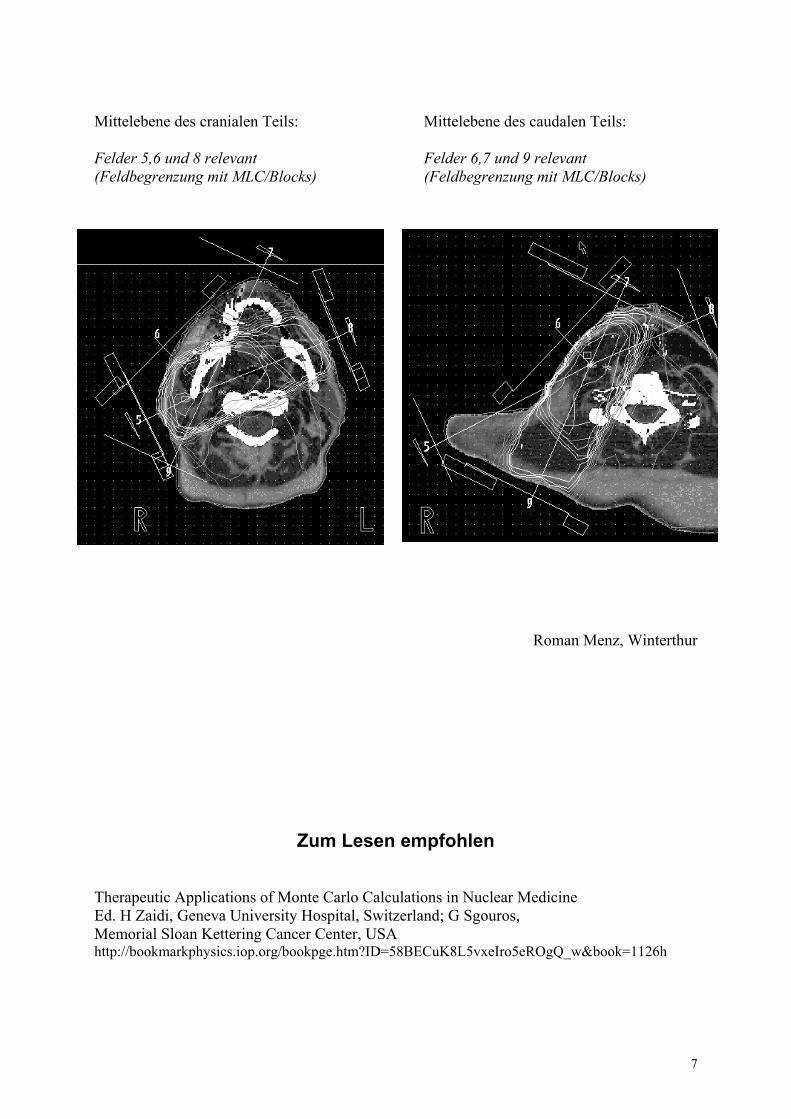
Mittelebene des cranialen Teils: Mittelebene des caudalen Teils: Felder 5,6 und 8 relevant Felder 6,7 und 9 relevant (Feldbegrenzung mit MLC/Blocks) (Feldbegrenzung mit MLC/Blocks)
Roman Menz, Winterthur
Zum Lesen empfohlen Therapeutic Applications of Monte Carlo Calculations in Nuclear Medicine Ed. H Zaidi, Geneva University Hospital, Switzerland; G Sgouros, Memorial Sloan Kettering Cancer Center, USA http://bookmarkphysics.iop.org/bookpge.htm?ID=58BECuK8L5vxeIro5eROgQ_w&book=1126h
7
Report of the working group: Quality Control in External Beam Radiotherapy
The working group is continuing its activities in 2002, with the intention of producing up-dated recommendations to superceed those of the Recommendations No 1 of the SSRMP, which were last revised in 1992. The last meeting was held in June 2002. As the recommendations proposed by this working group are intended to become part of the new “Beschleunigerverordnung” of the BAG, the activities of the group are coordinated with BAG. The following list shows our working schedule with those tasks already completed and those tasks still open. Our aim is to finish the report this year. Introduction complete Linear Accelerator Units
Mechanical Checks complete Check of optical SSD-distance-indicators complete Zero position of the angle scales complete Scale of vertical table movement complete Table latitudinal and longitudinal scales complete Table top deflection under load complete Light /radiation field coincidence complete Mechanical isocentre check complete Radiation isocentre check complete Laser alignment complete Field size indicators complete
Radiation Checks complete X-Rays complete Electrons complete
MultiLeaf Collimator open Dynamic Wedges complete
Dynamic wedge factors complete Interrupted dynamic wedge exposures complete Dynamic wedge profiles complete Dynamic wedge factor variation with gantry angle complete
Special treatment techniques complete Linac safety checks complete
Room entrance interlock complete Audio video monitor complete Emergency off switches complete
Touchguards complete Deadman switches complete Accessory (tray and wedge) interlocks complete Radiation leakage and contamination complete
Cobalt 60 Units
Mechanical checks complete Check of optical SSD-distance-indicators complete Zero position of the angle scales complete Scale of vertical table movement complete Table latitudinal and longitudinal scales complete Table top deflection under load complete
8
Position of isocentre complete Light / radiation field coincidence complete Field size indicators complete Rotation speed of the gantry complete
Skip-scan technique complete Precision of collimator position at opposing gantry angles complete Radiation checks
Beam output: definitive calibration and routine constancy check complete Output constancy with gantry angle complete Precision of timers complete Shutter reaction time complete Output factors complete Tray transmission factors complete Wedge factors complete Wedge factors and constancy with gantry angle complete Field flatness and symmetry complete
Flatness and symmetry at different gantry positions complete Wipe test complete Head leakage complete Low and Medium Energy Units
Low Energy Units (up to 100 kV) open Medium Energy Units (100-300 kV) open
Tables: frequencies and tolerances part. compl. We are grateful to all members of the working group, who are Luca and Antonella Cozzi, Florica Ionescu-Farca, Hans Roser and Philipp Trueb. Chair: Nicci Lomax, Uwe Schneider, Zürich Aus PMP soll der Schweizerische Berufsverband für Medizinphysi-
kerinnen und Medizinphysiker entstehen Wie an der letzten Hauptversammlung der SGSMP angekündigt, hat sich unter der Bezeich-nung PMP (Profession Medical Physics) eine Arbeitsgruppe gebildet, die eine Organisation vorbereitet, die in Zukunft die Standespolitik für das Fach Medizinphysik betreiben soll. Die nicht abschliessende Liste der Aufgaben für diese neue Organisation umfasst folgende Punkte:
• Formung des Berufbildes Medizin-Physik • Vertretung der berufsspezifischen Belange gegen aussen • Öffentlichkeitsarbeit für den Berufstand „Medizin-Physik“ • Einsatz für eine geeignete Grundausbildung zur Medizin-Physik • Definition der Fachanerkennung • Administrative Belange im Zusammenhang mit der Fachanerkennung • Einsatz für eine angemessene Stellung der Medizin-Physik in der Klinik • Erarbeitung von Modellen für die finanzielle Abgeltung der Tätigkeiten der Medizin-
Physik in der Klinik
9
• Einsatz für die Schaffung von Ausbildungsstellen • Kontakt mit Behörden zu Belangen des Berufstandes „Medizin-Physik“ • Verbindung zur Physikalischen Gesellschaft
Ein wichtiger Entscheid war auch die Frage der geeigneten Organisationsstruktur. Die Vor-stellungen gingen von einer permanenten Arbeitsgruppe innerhalb der SGSMP bis hin zum vollständig unabhängigen Verein. Eine Arbeitsgruppe wäre sowohl dem Vorstand wie auch der Hauptversammlung Rechenschaft schuldig, einem Gremium, in dem bei einer (nicht er-reichbaren) Vollbesetzung die aktiven MedizinphysikerInnen eine Minderheit darstellen. Ich vertrete die Meinung, wir sollten dem Vorbild der FMH folgen und die Standespolitik sowohl in der Ausführung wie auch in der Verantwortung vollständig den aktiven Medizinphysike-rInnen vorbehalten. Andererseits würde ein unabhängiger Verein fast notgedrungen zu einer Konkurrenzierung der SGSMP führen. Es ginge nicht nur um die Fragen, wer die Schweizeri-schen MedizinphysikerInnen in den internationalen Gremien vertritt, sondern auch, wer für unsere Behörden der Ansprechpartner im Bereich der Medizinphysik darstellt. Mit zwei ge-trennten Vereinen würden wir unserer Sache einen Bärendienst erweisen. Es kommt hinzu, dass der Berufsverband mindestens die organisatorischen Bereiche der Fachanerkennung SGSMP übernehmen muss. Eine Namensänderung würde vermutlich dazu führen, dass die Erwähnung der Fachanerkennung SGSMP bei nächster Gelegenheit aus der Strahlenschutz-Verordnung gestrichen würde. Ein Vorgang, den wir sowohl aus unserem Interesse wie auch im Interesse der sicheren Behandlung der Patienten vermeiden sollten. Es bleibt als dritter Weg die Schaffung einer unabhängigen Struktur innerhalb der SGSMP in Form eines Untervereins der SGSMP. Als Name haben wir: „Schweizerischer Berufsverband für Medizinphysikerinnen und Medizinphysiker“ (SBMP) gewählt. „Unterverein“ bedeutet nicht nur, dass die beiden Vereine manche Organe teilen können (z.B. Revisoren) und es in beiden Vorständen eine ex officio-Vertreter des anderen Vorstandes gibt, sondern auch, dass alle Mitglieder des SBMP auch Mitglieder im SGSMP sein müssen. Dies hat auch Nachteile, ist doch eine gemeinsame Struktur mit der Schweizerischen Gesellschaft für Biomedizinische Technik (SGBT) nicht möglich. Auf beidseitigen Wunsch hat eine Kontaktaufnahme mit die-ser Thematik stattgefunden. Bei den bestehenden Randbedingungen ist es aber schwierig, eine Lösung zu finden, die das gemeinsame Potential nutzt. Eine in den Statuten festgelegte Ab-sicht zur Zusammenarbeit mit den entsprechenden Gremien der SGBT kann, so hoffen wir, mindestens ein erster Schritt zu einem verbündeten Einsatz für unsere gemeinsame Sache darstellen. Wie geht es jetzt weiter? Am 22. August 2002 findet nochmals eine PMP-Arbeitssitzung statt. Dabei sollen vor allem die Statuten des neuen Vereins und die Anpassung der SGSMP-Statuten bereinigt werden. Diese Statuten-Entwürfe werden dann fristgerecht an alle SGSMP-Mitglieder verschickt. Am 6. November 2002, dem Tag der SGSMP-Hauptversammlung (am PSI in Villigen) soll vorerst in einer kurzen Sitzung der anwesenden aktiven Medizinphysike-rInnen geklärt werden, ob die von der Arbeitsgruppe gewählten Strukturen auf einen Konsens stossen. Anderenfalls würde der Antrag auf Statutenänderung noch vor der Hauptversamm-lung zurückgezogen. Die SGSMP-Hauptversammlung hat dann nur über die Änderung der SGSMP-Statuten zu befinden. Nimmt die Hauptversammlung diese Statuten-Änderung an, so steht der Gründungsversammlung des SBMP durch die aktiven Medizinphysiker nichts mehr im Wege. An dieser Gründungsversammlung müssten dann die SBMP-Statuten angenommen werden und die Organe des Verbandes (Vorstand, Präsident etc.) bestellt werden. Ich bin zu-versichtlich, dass wir diese Ziel erreichen.
Léon André
10
Report on the European CMS - Focus User’s Group Meeting from 31st of May to 1st of June 2002 at Lincoln County Hospital, GB Many users have been attending the CMS User’s Group Meeting in Lincoln (GB) this year. The meetings before have been organized in different countries throughout Europe to see and learn about the difference in treatment planning as well as treatment modalities and to encour-age discussions across the borders. The atmosphere in which this meeting took place was quite good. Ian Green, a Focus user for some years, from Lincoln County Hospital and his team organized this meeting locally. Ian Green spent as well a lot of time and effort to set up the User`s Group website which can be accessed on (www.irg.34sp.com/CMSEUG2002/Home.htm). Also very important is the fact that he will maintain this site in the future. Siegfried Kaufhold and Randy McPhee had addressed the general aspects of CMS in Europe and the world. CMS is doing very well according to the figures presented and they will con-tinue to remain worldwide leader in treatment planning systems. Jim Percy, who showed how FOCUS would develop in the various aspects of treatment plan-ning in the future, gave quite a number of presentations. A “hands on” show of the new Re-lease 4.0 and Focal Sim at different workstations gave a closer insight of what we will be see-ing in the near future. There are always things around treatment planning which can be improved, and therefore, a session to discuss previous and current issues was held. Quite a few of last year’s issues had been worked on and were discussed. The current issues were recorded by CMS and will be investigated. Given that there might be questions around physical problems, as well as hardware and appli-cations, Stefan Reinhardt and Edyta Bubula have been addressing customer support and new physics tools. Users who have already commissioned their planning system wished to have these tools some time before. A variety of user presentations ranging from the first experience in commissioning to the set up of IMRT techniques gave a good insight into the application of FOCUS. The accuracy of treatment planning has been studied with TLD measurements in a Alderson phantom and compared to a real clinical case. Another presentation showed different measurements in a water phantom with open and wedged beams which have been compared to treatment plan figures. Commissioning treatment machines is always an issue and was addressed by another talk. Beside optimisation of treatment plans, there is a need to derive better-defined target volumes. Employing different identification methods might help to reduce variation in tumour volume detection. A users meeting requires, of course, some social event. Given the setting of this historic city, Lincoln, a walking tour of the Bailgate area had been arranged. The well preserved and kept city centre, around one of the largest cathedrals in Europe, gave a good insight into previous centuries. This event had been finished with a nice dinner at the Knights Restaurant in Lin-coln. A walking tour of the newly built Radiotherapy Department gave an insight into a “state of the art” radiotherapy unit. Given that the numbers of European users are constantly increasing, there is a need to modify future meetings. Some of the users wished to attach the European user meetings to any one of the other large meetings, like ESTRO, in order to reduce travel expenses and time. In sum-mary – we had a very successful meeting with many discussion, presentations and new insight into future planning. Bruno Schnekenburger, Ph.D., European CMS User’s Group President
11
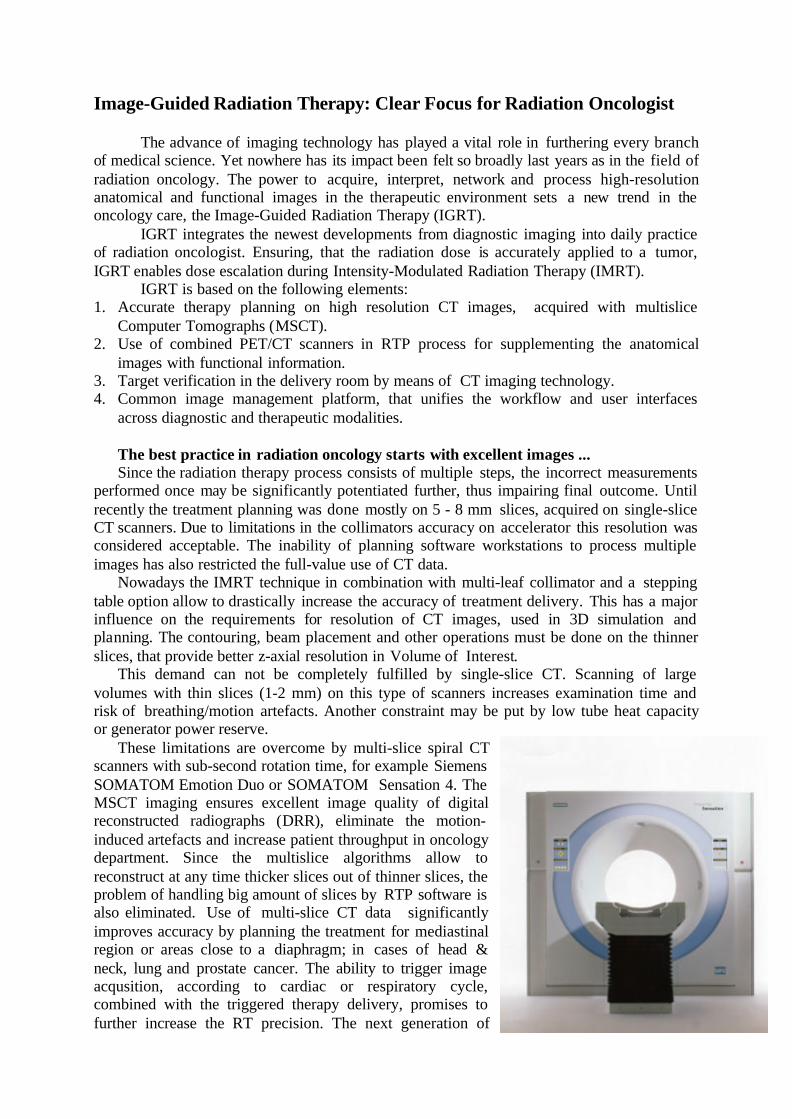
Image-Guided Radiation Therapy: Clear Focus for Radiation Oncologist
The advance of imaging technology has played a vital role in furthering every branchof medical science. Yet nowhere has its impact been felt so broadly last years as in the field ofradiation oncology. The power to acquire, interpret, network and process high-resolutionanatomical and functional images in the therapeutic environment sets a new trend in theoncology care, the Image-Guided Radiation Therapy (IGRT).
IGRT integrates the newest developments from diagnostic imaging into daily practiceof radiation oncologist. Ensuring, that the radiation dose is accurately applied to a tumor,IGRT enables dose escalation during Intensity-Modulated Radiation Therapy (IMRT).
IGRT is based on the following elements:1. Accurate therapy planning on high resolution CT images, acquired with multislice
Computer Tomographs (MSCT).2. Use of combined PET/CT scanners in RTP process for supplementing the anatomical
images with functional information.3. Target verification in the delivery room by means of CT imaging technology.4. Common image management platform, that unifies the workflow and user interfaces
across diagnostic and therapeutic modalities.
The best practice in radiation oncology starts with excellent images ...Since the radiation therapy process consists of multiple steps, the incorrect measurements
performed once may be significantly potentiated further, thus impairing final outcome. Untilrecently the treatment planning was done mostly on 5 - 8 mm slices, acquired on single-sliceCT scanners. Due to limitations in the collimators accuracy on accelerator this resolution wasconsidered acceptable. The inability of planning software workstations to process multipleimages has also restricted the full-value use of CT data.
Nowadays the IMRT technique in combination with multi-leaf collimator and a steppingtable option allow to drastically increase the accuracy of treatment delivery. This has a majorinfluence on the requirements for resolution of CT images, used in 3D simulation andplanning. The contouring, beam placement and other operations must be done on the thinnerslices, that provide better z-axial resolution in Volume of Interest.
This demand can not be completely fulfilled by single-slice CT. Scanning of largevolumes with thin slices (1-2 mm) on this type of scanners increases examination time andrisk of breathing/motion artefacts. Another constraint may be put by low tube heat capacityor generator power reserve.
These limitations are overcome by multi-slice spiral CTscanners with sub-second rotation time, for example SiemensSOMATOM Emotion Duo or SOMATOM Sensation 4. TheMSCT imaging ensures excellent image quality of digitalreconstructed radiographs (DRR), eliminate the motion-induced artefacts and increase patient throughput in oncologydepartment. Since the multislice algorithms allow toreconstruct at any time thicker slices out of thinner slices, theproblem of handling big amount of slices by RTP software isalso eliminated. Use of multi-slice CT data significantlyimproves accuracy by planning the treatment for mediastinalregion or areas close to a diaphragm; in cases of head &neck, lung and prostate cancer. The ability to trigger imageacqusition, according to cardiac or respiratory cycle,combined with the triggered therapy delivery, promises tofurther increase the RT precision. The next generation of
multislice scanners, e.g. SOMATOM Sensation 16 with 24-rows detector, enables the trueisotropic volume imaging while performing the routine scans with sub-millimeter resolutionin a few seconds.
Bridging the anatomy and function changes the world of radiation oncology ...CT images are used as a gold standard for radiation therapy planning. However, CT
imaging has limited precision in determining tumor spread in multilesional disease. It´s ofteninsufficient to distinguish viable tumor tissue from therapy-induced necrosis or scarring aftersurgery, thus impairing accurate measurement of tumor size and volume. Since visible tumorshrinkage occur lately during therapy, CT imaging has also limited accuracy in earlyassessment of tumor response. In the opposite, PET functional imaging is highly sensitive forcorrect staging of cancer, delineation of metabolic active tumor volume and monitoringtherapy course. According to results of German PET consensus conference (2000) the PETimaging changes cancer management in 18 %.
The lack of exact anatomic corellation in PET images, resulting in suboptimal diagnosticspecificity, forced the development of combined PET/CT systems. The ability to acquireintrinsically registered anatomical and functional images in a single exam was immediatelyrecognized by the oncological community as a breakthrough in cancer management. In nearlyall cancer locations, but especially in head&neck, mediastinal, abdominal and pelvis cancers,PET/CT shows increased diagnostic specificity and sensitivity. Besides PET/CT breaks thecliché on PET as a „time-consuming“ modality. Due to CT-based attenuation correction ofPET data, the PET/CT study lasts by 35% less than conventional PET exam. The use ofLutetium Oxyorthosilicate (LSO) detector crystal in Siemens, biograph LSO, PET/CTscanner, reduces whole body examinationtime to unprecedented 15 minutes.
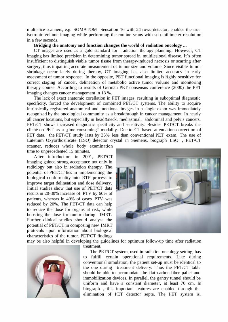
After introduction in 2001, PET/CTimaging gained strong acceptance not only inradiology but also in radiation therapy. Thepotential of PET/CT lies in implementing thebiological conformality into RTP process toimprove target delineation and dose delivery.Initial studies show that use of PET/CT dataresults in 20-30% increase of PTV by 60% ofpatients, whereas in 40% of cases PTV wasreduced by 20%. The PET/CT data can helpto reduce the dose for organs at risk, whileboosting the dose for tumor during IMRT.Further clinical studies should analyse thepotential of PET/CT in composing new IMRTprotocols upon information about biologicalcharacteristics of the tumor. PET/CT findingsmay be also helpful in developing the guidelines for optimum follow-up time after radiation
treatment.The PET/CT system, used in radiation oncology setting, has
to fulfill certain operational requirements. Like duringconventional simulation, the patient set-up must be identical tothe one during treatment delivery. Thus the PET/CT tableshould be able to accomodate the flat carbon-fiber pallet andimmobilization devices. In parallel, the gantry tunnel should beuniform and have a constant diameter, at least 70 cm. Inbiograph, this important features are enabled through theelimination of PET detector septa. The PET system is,
therefore, operated in 3D PET mode, that proved to be more sensitive and requires less isotopdose, than 2D PET imaging. The exact registration of CT and PET images can be also ensuredby preventing the weight-dependant deflection of patient table. The state-of-the-art PET/CTsystem should have a cantilevered bed design with intrinsic deflexion between CT and PETscans less than 1 mm.
Verifying tumor position in the treatment room enhances oncologists confidence ...Despite the ability to perform precise conformal radiation dose
distributions, the treatment plan still includes a wide margin to account for target locationuncertainties. The tumor position on treatment table may differ from the initial treatment plandue to internal organ shift, patient‘s weight loss, tumor volume reduction or incorrect patientset-up on CT table. For prostate cancer there is a up to 17 mm daily deviation of tumorposition in cranial-caudal direction, measured in prospective study by Wong JR et al.(Morristown Memorial Hospital, NJ).
In order to ensure the precision continuum IGRT includes the final target verification inthe delivery room. One option would be to perform digital portal imaging at the accelerator,however this technology still suffers from poor low-contrast resolution.
The alternative solution is to perform CTscanning of the patient on the therapy table prior tothe delivery. The Siemens PRIMATOM solutioncouples SOMATOM CT scanner with thePRIMUS linear accelerator, in the way that CTscanner with Sliding Gantry option is installedon a pair of horizontal rails in the therapy room.For PRIMATOM treatment, the patient is set upon the treatment couch, with radio-opaque markersplaced over skin marks that delineate the centralaxis planes. The treatment couch is rotated 180degree for PRIMATOM CT scanning. The CTgantry encircles the stationary patient, makesincremental movements during the scanningprocess, and then retracts easily when image acquisition is complete. The actual position ofthe tumor is identified on CT image and then compared to the original simulated positions bymeans of comprehensive syngo image fusion software. Daily variations of the tumor outlinesfrom the original contours determine the tumor shift in the x, y, z coordinates. Deriving a newisocenter and then shifting the treatment isocenter to this new position correct for these dailychanges. The treatment table is then rotated 180 degree back to the treatment position.
The PRIMATOM provides a platform for extreme precision in the radiation treatment ofcancer. The principle of this solution is well established for treatment of prostate cancer,where correction of patient position happens in nearly 30%. It applies also to other cancerlocations. Image-Guided Radiation Therapy using the PRIMATOM allows “no misses” andthus, would enable a tighter margin in the planning process, delivery of higher doses to thetumor while minimizing radiation to the adjacent normal tissues. Besides, the combination ofCT with a treatment table is a perfect set-up for performing best in class CT simulation andplanning.
Common software platform streamlines IGRT process ...The concept of IGRT includes not only merging of diagnostic imaging and radiation
therapy systems, but also unification of software platforms and mutual enrichment on theapplications level. Currently the broad use of data from PET/CT and multi-slice CT scannersin the therapy planning is partly limited by the number of vendors, enabling efficient post-
processing of large CT data sets, support of PET data and image fusion in RTP software. Thisfunctionality, commonly used in radiology, should inevitably become a standard in radiationoncology. In the past Siemens has undertaken successful efforts to implement a singleuniversal software platform, syngo, across its imaging modalities. Now, syngo is beingbrought by Siemens into radiation therapy department through a concept of specialist-dedicated oncology workplaces.
Syngo software architecture enables not only a commonuser interface across the different workstations in thedepartment, but optimizes the workflow by advancing the wayin which the images are managed, processed and transferred.Common platform allows also seamless transfer of relevantapplications from one workflow into another one. In theSiemens Virtual Simulation solution the user profits from userinterface, similar to SOMATOM CT and from a singledatabase for system-wide access to patient data. He is able toutilize applications like 3-D Reconstruction for an unlimitedmulti-slice CT data ranges and effectively process fusedimages to optimize disease management protocols.
The Image-Guided Radiation Therapy, the comprehensive integration of state-of-the-artdiagnostic modalities into oncology environment, will significantly enhance the capabilities ofradiation oncologist and provide him with an efficient tool to achieve better outcomes forcancer patients.
Mamin M.Siemens Medical Solutions
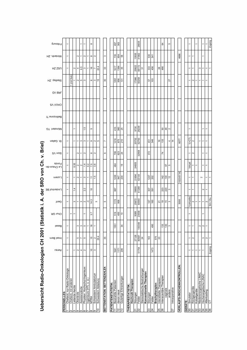
Ueb
ersi
cht R
adio
-Onk
olog
ien
CH
200
1 (S
tatis
tik i.
A. d
er S
RO
von
Ch.
v. B
riel)
Aarau
Insel Bern
Basel
Chur GR
Genf
Lindenhof BE
Luzern
La Chaux-de-Fonds
Sion VS
St. Gallen
Münsterl. TG
Bellinzona TI
CHUV VS
JRB VD
Stadtsp. ZH
USZ ZH
Winterth. ZH
Fribourg
PER
SON
ELLE
S:1
Che
farz
t, nu
r Rad
io-O
nkol
ogi e
11
11
11
11
11
11
2C
hefa
rzt,
mit
ande
ren
11
(mit
Nuk
)3
Leite
nde
Aerz
te1
13
1.4
10.
351
11
24
Obe
rärz
te2
53
12
22
12
42
5As
sist
enzä
rzte
47
33
12
13
56.
51
16
Phys
iker
, Hoc
hsch
ul-In
geni
eure
25
51
22.
23
1.4
23
1.5
33
21
7In
geni
eure
(HTL
o. ä
.)1
-31
0.2
11
18
MTR
A10
1518
3.7
14.3
1011
7.5
89
58
168
89
Schw
este
rn A
mbu
lato
rium
32
11.
53.
82
23
72
10Sc
hwes
tern
Abt
eilu
ng29
.45
1520
.4
BET
TEN
STA
TIO
N: B
ETTE
NZA
HLE
N35
56
730
333
PATI
ENTE
NST
ATI
STIK
:11
Best
rahl
te P
atie
nten
1641
1551
516
1172
887
1239
484
706
1216
415
1091
1617
735
497
12da
von
neue
1171
450
958
947
623
875
380
956
1384
654
383
13G
utar
tige
Erkr
anku
ngen
206
3410
234
1924
210
2513
135
58
THER
API
ESTA
TIST
IK:
Hoc
hvol
t-The
rapi
en21
Sitz
unge
n27
128
9389
2587
219
855
2169
424
805
2075
297
3017
386
2582
213
935
22Fe
lder
5175
480
587
1003
4823
558
6494
061
289
6314
432
908
5275
029
239
5025
869
172
4186
228
835
Ster
eota
ktis
che
Best
rahl
unge
n24
31K
onve
ntio
nelle
The
rapi
en23
Sitz
unge
n16
314
730
712
4737
245
157
333
530
24Fe
lder
1473
490
160
591
202
546
103
456
541
Bra
chyt
hera
pien
575
25In
ters
titie
lle T
hera
pien
2321
1628
26In
tera
cavi
täre
The
rapi
en13
318
223
142
7413
350
490
64
v
agin
ale
130
1522
013
087
48
a
nder
e
33
312
54
227
527
Intra
oper
ativ
e 14
VER
LAU
FS-/W
OC
HEN
KO
NTR
OLL
EN:
5000
1023
/571
960
7746
86
GER
ÄTE
:30
Sim
ulat
o r1
11
11(
virtu
elle
)1
1Vi
rtuel
11(
+CT)
11
11
131
Rön
tgen
gerä
te1
21
21
11
22
21
132
Koba
ltger
äte
11
11
133
Nie
dere
nerg
etis
che
LIN
AC1
21
11
334
Hoc
hene
rget
isch
e LI
NAC
13
21
12
11
11
11
11
35Af
terlo
adin
g2
12
11
11
11
11
136
CT
Zuga
ng1
Sim
Vitu
.1
1Zu
gang
5
2
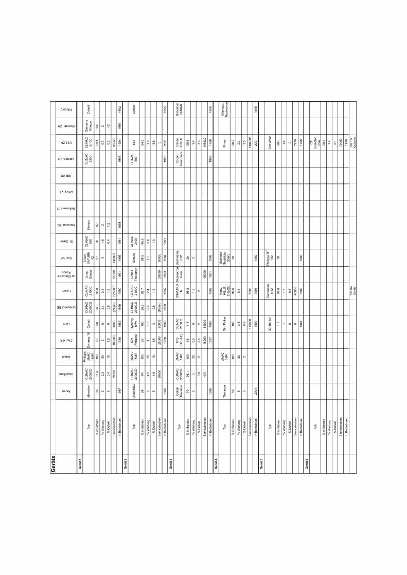
Ger
äte
Aarau
Insel Bern
Basel
Chur GR
Genf
Lindenhof BE
Luzern
La Chaux-de-Fonds
Sion VS
St. Gallen
Münsterl. TG
Bellinzona TI
CHUV VS
JRB VD
Stadtsp. ZH
USZ ZH
Winterth. ZH
Fribourg
Ger
ät 1
Typ
Mev
atro
nC
LIN
AC
2300
CD
Philip
ps
LIN
AC
18M
VD
ynar
ay 1
6C
obal
tC
LIN
AC
2300
CD
CLI
NAC
21
00C
Lina
c El
ecta
Lina
c SA
TUR
N
42
CLI
NAC
60
0Pr
imus
CLI
NAC
23
00C
LIN
AC
6/10
0Si
emen
s Pr
imus
Cob
alt
% in
Bet
rieb
9897
.410
080
6096
.895
.897
9897
99.1
133
% W
artu
ng2
2.2
202
02.
42.
43
1.6
20.
72
% D
efek
t5
0.4
101.
50
0.8
1.8
0.4
1.2
0.2
<2
Serv
icek
oste
n79
000
1000
0040
00(P
aket
)22
5000
*91
000
1400
0083
950
in B
etrie
b se
it19
9719
8819
8919
9619
9019
9119
9519
9119
9819
9519
8419
9919
92
Ger
ät 2
Typ
Lina
c BB
CC
LIN
AC
2300
CD
LIN
AC
18M
VSi
m
(Phi
lipps
)D
ynar
ay
6MV
CLI
NAC
23
00C
DC
LIN
AC
2100
CC
obal
t Th
erat
ron
Sim
ulix
CLI
NAC
21
00C
LIN
AC
600
6Ex
Clin
ac
% in
Bet
rieb
9894
100
2610
096
.895
.798
.596
.394
.5
% W
artu
ng2
4.2
201
1.5
2.4
2.4
1.5
2.4
1.6
% D
efek
t5
1.8
101.
63
0.8
1.9
1.3
3.9
Serv
icek
oste
n39
500
2500
080
000
(Pak
et)
2000
050
000
0
in B
etrie
b se
it19
8619
8819
8919
9919
9219
9319
9419
9119
9520
0119
95
Ger
ät 3
Typ
Cob
alt
Ther
aton
CLI
NAC
23
00C
DLI
NAC
6M
VTP
S (H
ELAX
)C
LIN
AC
2100
CXI
MAT
RO
NN
ucle
otro
n C
urie
Gam
mam
ed
12i
Cob
alt
Tera
troni
csC
linac
21
00 C
Sim
ulat
or
VAR
IAN
% in
Bet
rieb
7096
.110
065
115
96.8
2095
.3
% W
artu
ng2
320
0.5
51.
22
1.4
% D
efek
t0.
92
0.5
22
3.3
Serv
icek
oste
n40
116
000
8500
045
000
1465
30
in B
etrie
b se
it19
8819
9719
9319
9219
9119
9819
9319
9019
95
Ger
ät 4
Typ
Ther
apax
LIN
AC
5MV
Sim
Pic
ker
Konv
. H
ILLE
TH
2000
Siem
ens
Stab
ilipla
n 30
0kV
Prim
art
Afte
rload
. N
ucle
otro
n
% in
Bet
rieb
5010
010
099
.610
96.2
% W
artu
ng5
202.
50.
42.
3
% D
efek
t5
20.
91.
5
Serv
icek
oste
n11
5000
5500
1484
00*
in B
etrie
b se
it20
0119
9919
9719
8020
0119
95
Ger
ät 5
Typ
30-1
00 k
VG
amm
ame
d 12
iPh
ilipps
RT
100
Sim
ulat
or
% in
Bet
rieb
1.5
97.6
1098
.8
% W
artu
ng1
1.6
1.2
% D
efek
t0
0.8
0
Serv
icek
oste
n0
4000
079
70
in B
etrie
b se
it19
6719
9019
8019
95
Ger
ät 6
Typ
CT
Som
aton
Pl
us%
in B
etrie
b98
.5
% W
artu
ng1.
4
% D
efek
t0.
1
Serv
icek
oste
n70
000
in B
etrie
b se
it19
99*fü
r alle
G
erät
e*a
ls T
eil
Kauf
prei
s
Kompendium für Strahlenschutz-Sachverständige (Philipp R. Trueb, Ed.; Verlag Paul Haupt, Bern-Stuttgart-Wien, ISBN 3-258-06475-X, La-denpreis: 88 CHF/56 EUR)
Das vorliegende Kompendium richtet sich an alle Ärztinnen und Ärzte, die eine Bewilligung für den Umgang mit einer Röntgenanlage besitzen oder erwerben wollen. Es ist von verschie-denen Autoren mit grosser Lehrerfahrung äusserst didaktisch und mit viel Liebe zum Detail geschrieben und gestaltet worden. Sehr übersichtlich gliedern sich die Kapitel: „Röntgenstrah-lung und deren Einfluss auf Materie; Technik einer Röntgenaufnahme; Qualitätssicherung; Indikationsstellung; Bildqualität/Einstelltechnik; Strahlenbiologie; Strahlenschutz“ wie Perlen an einer Kette und lassen sich genüsslich lesen. Die Kapitel sind graphisch einheitlich aufein-ander abgestimmt und sind ausserdem durch sehr sorgfältig ausgewählte Tabellen und Bilder sehr gut und informativ ergänzt. Die Texte sind auf das wesentliche beschränkt und vermitteln ein Konzentrat, ohne zu ermüden.
Beispiele: “Ionisierende Strahlen sind somit alle hochenergetischen Strahlen, die in der Mate-rie bis in die atomare Struktur vordringen und diese durch Wechselwirkungen verändern.”; “Die Schwächung von Röntgenstrahlen beruht auf Absorption und Streuung.”; ”Der Fetus und der Embryo sind im allgemeinen noch empfindlicher als Kinder oder Erwachsene.”; ”Der Strahlenschutz beruht auf den drei Prinzipien: Rechtfertigung, Optimierung und Dosisgrenz-werte.”; “Es handelt sich um eine Poisson-Statistik.”
Sehr informativ und nützlich sind insbesondere die Tabellen zu Konstanzprüfung, Effektiver Dosis bei verschiedenen Untersuchungen, und der Anhang, der in drei Teile gegliedert ist: A zur Einstelltechnik, der mit vielen aufwendigen Farbfotos sehr praktisch gestaltet ist; B Indi-kationsstellung; C Literatur.
Einziges kleines Manko, welches wir identifizieren konnten, betrifft die bei der SUVA versi-cherten beruflich strahlenexponierten Personen. Dieselben sind nämlich nicht nur ständig zu überwachen und sie müssen ein Dosimeter tragen, wie im Kompendium richtig beschrieben, sie sind zusätzlich auch noch bezüglich der Berufstauglichkeit regelmässig ärztlich zu unter-suchen, worauf das Kompendium nicht hinweist. Detailliert geben zu diesem Thema die Un-terlagen der SUVA Auskunft, welche unentgeltlich bestellt werden können (Tel. 041 419 51 11 oder über http://www.suva.ch; Bestellnummer 2869/4.d). Dort kann ausserdem das Strah-lenschutzgesetz zusammengebunden mit der Strahlenschutzverordnung bestellt werden (1655.d), und für speziell interessierte ist auch noch ein Dossier zum Thema Strahlenunfall verfügbar (2869/21.d).
Insgesamt beurteilen wir dieses Kompendium aber als hervorragend, unterhaltsam (ja!) und fachdienlich, und es stellt damit ein äusserst willkommenes Nachschlagewerk dar. Last but not least, es bleibt zu hoffen, dass bald eine version française und eine versione italiana auf den Markt kommen werden, denn die Schweiz ist ja bekanntlich vielsprachig. And why not an English version, adapted for E.U. doctors?
Dr. med. Hans Peter Rutz & Dr. phil. Walter Burkard
Abteilung für Strahlenmedizin, Paul Scherrer Institut, 5232 Villigen PSI
18
Stelleninserate
Medizinphysiker(in) Veterinärmedizin
Seit 1997 behandeln wir Krebspatienten mit einer neuartigen Bestrahlungstechnik für tiefliegende Tumoren. Die Protonentherapie-Anlage, die wir dafür entwickelt haben, dient in einem gemeinsamen Programm mit der Veterinärmedizinischen Fakultät der Universität Zürich auch der Untersuchung spezieller Fragen an Tierpatienten. Am Tierspital steht dazu ein Linearbeschleuniger zur Verfügung.
Ihre Aufgaben • Betreuung des Linearbeschleunigers, Dosimetrie und Qualitätskontrolle sowie
Bestrahlungsplanung für Tiere am Tierspital für Photonen und Elektronen und am PSI für Protonen
• eigene wissenschaftliche Arbeiten auf dem Gebiet der vergleichenden Therapie-planung für Protonen, Photonen und Elektronen sowie Mitarbeit und Beratung bei wissenschaftlichen Arbeiten in der Radioonkologie der Veterinärmedizin
Ihr Profil • abgeschlossenes Physikstudium, Fachanerkennung in Medizinischer Physik oder
gleichwertiges Diplom (ev. in Ausbildung)
• ausgezeichnetes Teamverhalten und hohes Interesse an interdisziplinärer Arbeit
Die Arbeitsorte sind das PSI in Villigen und die Universität Zürich. Die Anstellung eignet sich auch für Teilzeitarbeit.
Weitere Auskünfte geben Ihnen gerne Herr Dr. H. Blattmann, Telefon 056 310 4095, EMail [email protected] oder Frau Prof. Dr. med. vet. B. Kaser-Hotz, Telefon 01 635 8449, EMail [email protected]. Wir freuen uns auf Ihre Bewerbung: PAUL SCHERRER INSTITUT, Personalabtei-lung, Elke Baumann, Kennziffer 2105, 5232 Villigen PSI Weitere Stellenangebote: www.psi.ch
19
UNIVERSITAETSSPITAL ZUERICH
Die Klinik für Radio-Onkologie
sucht mit Stellenantritt ab sofort oder nach Vereinbarung eine/einen
Medizin-Physiker / -Physikerin
Die Hauptaufgaben umfassen die Mitarbeit bei der Radiotherapie (Bestrahlungsplanung, Do-simetrie, Gerätekontrollen) mit Linearbeschleunigern, HDR-Afterloading- Brachytherapiege-rät und konventionellen Röntgengeräten sowie die Betreuung des Simulators, TPS und CT Scanners und der Klinik-Informationssysteme. Zu diesen vielseitigen Aufgaben gehört die Mitwirkung bei der Einführung der Intensitätsmodulationstechnik in die klinische Routine an einem zweiten Linearbeschleuniger. Grundlegende und angewandte Forschung wird aus-drücklich gefördert. Vorträge bei der Klinikinternen und -extern Fortbildung gehören eben-falls zum Aufgabenbereich. Wie suchen einen Physiker mit mehrjähriger Erfahrung, welcher über die Fachanerkennung SGSMP oder eine gleichwertige Ausbildung verfügt, sowie gute Informatikkenntnisse besitzt. Wichtig ist eine ausgeprägte Bereitschaft zur interdisziplinären Teamarbeit. Für weitere Informationen steht Ihnen Dr. J.B.Davis, Leiter Strahlenphysik, Radio-Onkologie, Universitätsspital Zürich, Tel. 01 / 255 34 62, E-mail: [email protected], gerne zur Verfügung. Schriftliche Bewerbungen sind an das Personalbüro 1, Schmelzbergstrasse 26, 8091 Zürich zu richten.
Personalia
Jorn Verwey, MSc., ist seit dem 1. März 2002 als Medizinphysiker in der Abt. für Strahlen-therapie am Paul Scherrer Institut (PSI) beschäftigt. Er war zuvor für das Projekt TULOC am PSI tätig. Dr. Werner Roser wechselte am 1. Mai 2002 innerhalb des PSIs von der Abt. für Strahlenme-dizin zur Abt. für Technik und Koordination. Er beschäftigt sich mit Fragen der Sicherheit und der Dokumentation im PROSCAN Projekt (Erweiterung der Protonentherapie am PSI). Frau Dr. med. Beate Timmermann ist seit dem 1. Juli 2002 in der Abt. für Strahlenmedizin am PSI als Radio-Onkologin tätig. Zuvor arbeitete sie in der Abt. für Radio-Onkologie an der Uniklinik in Tübingen.
20
Pressespiegel
Chernobyl Gets Glowing Reviews
CHERNOBYL, Ukraine May 11 -- Yuri Zayets pointed his binoculars toward a distant copse of birches and shouted excitedly from midway up the fire tower: "They're over there, grazing near the forest."
It had taken nearly two hours of driving through the unique radioactive wilderness born of the 1986 Chernobyl nuclear disaster to find them, but one of the world's few wild herds of rare Przewalski horses finally came into view.
"Stay here," Denis Vishnevsky, a zoologist with the Chernobyl Ecology Center, said after the group of official guides and a journalist piled out of their minibus to see the short but powerfully robust horses, introduced here in 1998 to eat what was supposedly "excess" vege-tation in the depopulated area. "They'll come to us." "Chernobyl safaris," mused Rima Kise-lytsia, a guide with Chernobylinterinform, the agency that shepherds all visitors to the "Zone of Alienation" around the now-decommissioned reactor, an area that once was home to 135,000 people. "It's a strange idea, but I like it."
Chernobyl tourism has been a hot topic in Ukraine since January, when a U.N. report urged Chernobyl communities to learn to live safely with radiation -- such as consuming only produce grown outside the zone. The report suggested specialized tourism as one of several possible ways to bring money into a region that has swallowed more than $100 billion in sub-sidies from Soviet, Ukrainian and international government funds since the nuclear accident 16 years ago.
Back in the town of Chernobyl, where the zone's administration manages the Rhode Is-land-sized no man's land around the destroyed reactor, one official said economic benefits of tourism will never be more than minor.
But he doesn't reject the idea outright. "The U.N. is 12 years too late," said Mykola Dmy-truk, deputy director of Chernobylinterinform, referring to technicians who have been coming to the zone for that long. "We've been allowing tours since 1994."
A few Kiev tourist agencies advertise Chernobyl excursions on their Web sites, but so far the zone administration doesn't actively promote the idea. "A great deal still isn't known," said Dmytruk, "and we warn everyone about the risks, even scientists."
The risks, though small, are real. And so is the desolation. But the aftermath of the acci-dent has created a misleading stereotype of the zone as a toxic wasteland, a nuclear desert devoid of life, and certainly not a place a sane person would want to visit.
In fact, by ending industrialization, deforestation, cultivation and other human intrusions, radiation has transformed the zone into one of Europe's largest wildlife habitats, a fascinating and at times beautiful wildness teeming with large animals such as moose, wolves, boar and deer. It now is home to 270 bird species, 31 of them endangered -- making the zone one of the few places in Europe to spot rarities such as black storks and booted eagles.
And traveling to Chernobyl may qualify as a kind of adventure tourism. The very knowl-edge of the buzzing background of radiation imbues even the prosaic act of walking down the street with an aura of excitement. It isn't the same adrenalin punch as bungee jumping in the Andes, but it is a palpable sensation -- like being surrounded by ghosts.
By law, no one can enter the zone without permission. But except for children under 17, the administration may give permission to pretty much anyone. The vast majority of the nearly 1,000 annual visitors are scientists, journalists, politicians and international nuclear officials, but the zone has hosted a handful of what Dmytruk calls "pure" tourists -- including three Japanese in 2000 -- and it can put together customized programs, such as safaris in
21
search of Przewalski horses, which some experts believe are the ancestors of all domestic horses but far more aggressive..
"If a group of Californians want to go bird-watching, we can organize that," Dmytruk said, adding, "so long as they know the difference between plutonium and potatoes."
Of course, Chernobyl isn't Club Med. But 16 years after the fourth reactor bloc spewed radiation around the globe, the risks are mostly manageable. About a quarter of the cesium and strontium have already decayed, and 95% of the remaining radioactive molecules are no longer in fallout that can get on or inside a visitor, but have sunk to a depth of about 5 inches in the soil.
From there, they have insinuated themselves into the food chain, making the zone's di-verse and abundant flora and fauna radioactive indeed. An antler shed recently by a Cherno-byl elk was stuffed with so much strontium that it cannot be allowed out of the zone. But three Przewalski foals born in the wild, though radioactive, have grown to adolescence with no visible effects.
Such radioactivity now has receded to the background. On an average day, a visitor might receive an extra radiation dose about equivalent to taking a two-hour plane trip, zone officials say.
That is, if the visitor follows the strict but simple safety rules: "Don't eat local food, stay on the pavement, and go only where your guide takes you," Dmytruk said.
It is almost impossible to smell fresher air in an urban setting than here in the town of Chernobyl, where the number of cars seen on a warm April day could be counted on one hand and songbirds frequently provide the only sound.
"It is one of the zone's many paradoxes, but because human activity is banned nearly eve-rywhere, the region is one of Ukraine's environmentally cleanest," Dmytruk said. "Except for radiation." Today, villages are slowly succumbing to encroaching forests. In the abandoned town of Pripyat, less than two miles from the nuclear reactor, empty black windows stare blindly from high-rise buildings at kindergartens littered with heartbreakingly small gas masks.
It may seem like an odd place for a rewarding tourism experience. But nowhere else can a visitor stand amid a herd of wild Przewalski horses like a character in Jean Auel's Ice Age novels, or watch a pair of rare white-tailed eagles circling above the ghostly high-rises of Pripyat, a moving monument to the devastating effects of technology gone awry and nature's near miraculous resilience and recovery. Quelle: Sandy Perle [[email protected]] An: [email protected] Am: Montag, 13. Mai 2002 17:31
Medaille mit zwei Seiten
Ein Eiweiss schützt Mäuse vor Krebs, lässt sie aber rascher altern
ni. Dass ein natürlich vorhandenes Eiweiss mit Namen p53 bei Tier und Mensch eine wich-tige Rolle in der Krebsentstehung spielt, ist schon länger bekannt. Nicht gewusst hat man bis jetzt, dass dieses Protein auch den Alterungsprozess beeinflussen kann. Das haben nun ameri-kanische Wissenschafter aus Houston im Gliedstaat Texas bei Mäusen nachgewiesen. Sie brachten dazu eine Genmutation ins Erbgut der Tiere ein, die zu einer Überfunktion von p53 führte.
22
Wie erwartet, erkrankten die genetisch veränderten Mäuse kaum an Krebs. Erstaunlicher-weise zeigten sie jedoch bereits früh in ihrem Leben die typischen Merkmale fortgeschritte-nen Alters: Im Gegensatz zu unbehandelten Kontrollmäusen litten sie nach 18 Monaten unter Gewichtsverlust und Muskelschwund, ihre Knochen waren osteoporotisch verändert, die Or-gane geschrumpft; zudem reagierten sie anfälliger auf Stress und starben früher als ihre nor-malen Gefährten – obwohl Letztere häufiger tödliche Tumoren entwickelten.
Das Protein p53 hat in Körperzellen wichtige Abwehrfunktionen inne. Sobald eine Zelle durch schädigende Einflüsse bedroht ist – etwa durch radioaktive Strahlung, Zytostatika oder krebsfördernde Gene (Onkogene) –, wird die Aktivität dieses Proteins gesteigert. Dadurch kommt es in der betroffenen Zelle zum Stillstand der Teilung, oder aber die Zelle wird gleich «entsorgt», indem sie dem programmierten Zelltod (Apoptose) zugeführt wird. Das Protein hilft auch bei der Reparatur von geschädigter Erbsubstanz. Alle diese Mechanismen sollen verhindern, dass Zellen entarten und schliesslich zu Tumoren werden.
Eine mögliche Erklärung dafür, dass zu viel p53 den Alterungsprozess beschleunigt, sehen die Wissenschafter darin, dass dieses schützende Eiweiss nicht nur Krebszellen in ihrem Wachstum hemmt, sondern möglicherweise auch die Teilung von Stammzellen beeinträchtigt, die bei der Regeneration von Körpergeweben nötig sind. Obwohl noch nicht klar ist, ob die Resultate dieser Mäuse-Versuche auf den Menschen übertragbar sind, ergeben sich daraus zahlreiche Fragen. Ist es etwa denkbar, dass künftige Anti-Aging-Medikamente, welche die Wirkung von p53 modulieren, dem Anwender zwar ein längeres Leben, aber auch vermehrt Tumoren bescheren? Und müssen Kinder und Jugendliche, die mit modernen Chemotherapien ihren Krebs besiegt haben, vielleicht in späteren Jahren mit frühzeitigen Alterserscheinungen rechnen?
Quelle: Nature 415, 26–27; 45–53 (2001). - NZZ vom 9.1.2002
Mit strahlenden Medikamenten gegen Krebs Ein Forschungsteam am Kantonsspital Basel entwickelt radioaktiv markierte Substanzen, die sich an Tumorzellen anlagern. In den Körper von Krebskranken injiziert, bestrahlen diese Arzneien den Krebs von innen. Die zukunftsträchtige Therapie steckt noch in der Entwick-lungsphase. Mit der Chirurgie, der Chemotherapie und der Bestrahlung des Körpers hat die Medizin drei Waffen gegen die Krankheit Krebs zur Verfügung. In Zukunft könnte eine vierte Strategie dazukommen: die Nuklearmedizin. Diese nutzt radioaktiv strahlende Medikamente, so ge-nannte Radiopharmaka, um die Tumoren von innen gezielt zu bestrahlen. Den Patientinnen und Patienten wird dazu ein radioaktiver Stoff gespritzt, der an Krebszellen andockt. Auf die-se Weise sammeln sich die Radiopharmaka beim Tumor und seinen Wucherungen (Metasta-sen) an. Mit einer speziellen Kamera, der so genannten Gammakamera, die röntgenbildähnli-che Bilder (Szintigramme) liefert, können die Tumore sichtbar gemacht werden. Helmut Mäcke, Professor für Radiologische Chemie am Institut für Nuklearmedizin des Kan-tonsspitals Basel, demonstriert, wie sich mit einer radioaktiven Substanz Krebsmetastasen im Körper aufspüren, sichtbar machen und teilweise auch zerstören lassen. Er klemmt mehrere Szintigramme an einen hinterleuchteten Plexiglaskasten. Der Patient leidet an einem Tumor, der bereits Metastasen gebildet hat, wie die Bilder zeigen: rund zwei Dutzend schwarze Fle-
23
Auch in Basel wurden verschiedenste Radiopharmaka entwickelt. Bereits an die 100 neue Radiopeptide haben die Forscher synthetisiert und an Zelllinien und im Tierversuch getestet. Dabei haben sie verschiedene Hormone mit unterschiedlichen radioaktiven Strahlern kombi-niert. Zudem testen sie auch so genannte trifunktionelle Radiopharmaka: Diese kutschieren neben Strahlern auch chemotherapeutische Substanzen direkt zum Tumor - um ihn so gleich doppelt zu bekämpfen. Von einem Durchbruch in der Krebstherapie sei man allerdings noch weit entfernt: «Wir sind erst ganz am Anfang einer neuen Therapie, die laufend verbessert werden muss!», sagen die Forschenden. So ist auch der 15-jährige krebskranke Junge nicht geheilt. Aber nach vier The-rapien hat sich sein Zustand stabilisiert: «Er kann weitgehend auf Morphium verzichten, hat eine Lehre begonnen, und in einem Brief hat er gar davon berichtet, dass er wieder schwim-men kann», sagt Helmut Mäcke. Von Ruth Jahn Quelle: Basler Zeitung vom 12. April 2002
Der Forum-Gast «Tschernobyl»: Ein trauriger Jahrestag! Alt Nationalrat Hansjürg Weder* 16 Jahre nach «Tschernobyl», der grössten indus-triellen Katastrophe der Menschheit (vom 26. 4. 1986), die nach Schätzung wirklich-keitsfremder Technokraten nur alle 10 000 Jahre einmal stattfinden dürfte, steht fest, dass die tödlich strahlende Radioaktivität noch immer auf den Feldern, Wäldern, Dör-fern und Städten der Ukraine, Weissrusslands und der Russischen Föderation liegt. Sie vergiftet noch immer Mensch, Tier und Umwelt. 18 Millionen Hektaren landwirtschaftlicher Fläche sind für immer verstrahlt. 26 Pro-zent der weissrussischen Wälder und der grösste Teil der Wiesen in den Flussniederun-gen des Pribjat, Dnjepr und Sosch liegen in der hoch radioaktiven Zone. Über 3600 Dörfer und Städte sind verseucht, mehr als zwei Millionen Menschen, darunter über eine halbe Million Kinder, leben heute in zum Teil stark verstrahlten Gebieten. Die Strahlung ist auf leisen Sohlen dahergekommen. Man roch sie nicht, man sah und hörte sie nicht. Sie war einfach da. Heute, morgen, übermorgen - bis in alle Ewigkeit. Die Wachhunde und der unsterbliche Tote! Anno 1990 besuchte ich Tschernobyl. Hier stand der unsterbliche Tote - ein strahlender Schrotthaufen. Als die Geigerzähler verrückt spielten, erinnerte ich mich an den ameri-kanischen Senator Gravel, der einmal sagte: «Es kann nur als Verbrechen gegen die Menschheit bezeichnet werden, wenn man Atommüll produziert, ohne zu wissen, wo man diesen noch nach Tausenden von Jahren tödlich strahlenden Stoff sicher lagern könnte. Wenn das kein Verbrechen ist, dann muss es wohl Wahnsinn sein. Aber Un-schuld ist es nicht, denn die Betreiber und Förderer dieser Politik sind oft genug auf ihr frevelhaftes Tun aufmerksam gemacht worden.» Mit flammenden Worten nahm er die kommenden Generationen in Schutz, die wir zu Wachhunden unseres Wohlstandmülls degradieren!
25
Tschernobyl gehört der ganzen Welt Der ukrainische Volksdeputierte Dr. Juriy Stscherbak, der seinerzeit vor der national-rätlichen Energiekommission in Bern referierte, sagte zum Schluss seines aufrüttelnden Votums: «Tschernobyl ist eine globale Katastrophe. Die Erfahrung Tschernobyl gehört nicht allein der Ukraine und Weissrussland, sie gehört der gesamten Menschheit. Wir müssen gemeinsam dafür arbeiten, dass die schrecklichen und tragischen Folgen von Tschernobyl so gering wie möglich bleiben. Ich wende mich an alle Länder mit dem Appell, uns zu helfen. Tschernobyl ist nach Seveso, Bhopal, Harrisburg und Schwei-zerhalle eine weitere Warnung, nicht nur in Bezug auf die Atomenergie, sondern auch auf alle andern immer komplexer werdenden technischen Grosssysteme, die nicht be-herrschbar und daher lebensbedrohend sind.» Eine Katastrophe jenseits aller Vorstellungen Die Buchautorin Swetlana Alexijewitsch schreibt: «Diese Katastrophe ist ein Ereignis, wofür wir noch kein System von Vorstellungen, noch keine Analogien und Erfahrun-gen haben, woran unsere Augen und Ohren noch nicht gewöhnt sind, wofür nicht ein-mal unser bisheriger Sprachschatz, unser ganzes inneres Instrumentarium ausreicht.» Die Autorin hat mit vielen Menschen gesprochen, mit Alten, die nicht wissen, was Ra-dioaktivität bedeutet und deshalb zurück in die «Zone» gegangen sind; mit todkranken Liquidatoren (etwa 700 000), die heute im Stich gelassen werden und hilflos dahinsie-chen; mit Kindern, die wissen, dass sie bald sterben müssen. Sie hat die Ohnmacht der Betroffenen erfahren, angesichts der Arroganz der Apparatschiks und der ausbleiben-den grosszügigen Hilfe aus dem «reichen» Westen. 16,2 Milliarden Franken - wer bezahlt? Die Kosten für die Stillegungs- und Entsorgungskosten belaufen sich in der Schweiz - nach Angaben der Stromwirtschaft - auf satte 16,2 Milliarden Franken. Für die A-Werke Mühleberg, Beznau I und II, Gösgen und Leibstadt werden die Kosten für die Stilllegung auf 2,5 Milliarden Franken geschätzt und sollen durch Zahlungen der Be-treiber in einen Fonds mit Nachschusspflicht und Solidarhaftung gedeckt sein. Aber die viel höheren Kosten von rund 13,7 Milliarden Franken für die Atommüll-Entsorgung sind damit nicht finanziert und drohen, an den gutgläubigen Schweizer Steuerzahlern hängen zu bleiben. Wie der Basler Nationalrat Ruedi Rechsteiner als Erster herausge-funden hat, sind diese Mittel gar nicht vorhanden. Die AKW-Betreiber haben bis heute erst 1,44 Milliarden Franken für die Stilllegungskosten und erst 0,94 Milliarden Franken in den Fonds für die Entsorgung einbezahlt. Wollte man den fehlenden Betrag in Kürze aufbringen, müsste der Strom ab Steckdose um etwa drei Rappen verteuert werden. Für diesen Betrag wird aber heute eine Kilowattstunde elektrischer Energie gehandelt! Kreide gefressen Billig sei der Atomstrom, pflegten die AKW-Betreiber lauthals zu sagen. Das pure Ge-genteil, ist richtig. Wem kommt bei diesem uneingehaltenen Versprechen nicht das Märchen von den sieben Geisslein in den Sinn? Nachdem der Wolf die Kreide gefres-sen hatte, klang seine Stimme fein und lieblich und die sieben dummen Geisslein - Bundesrat und Stromkunden - merkten gar nicht, mit wem sie es zu tun hatten; sie glaubten, es wäre die gute Mutter Geiss, dabei war es die abgebrühte Atomlobby. Vom
26
Bundesrat verlangen die AKW-Gegner, dass die Entsorgungskosten vollumfänglich von den AKW-Betreibern übernommen werden, wie das im Atomgesetz vorgeschrie-ben ist. Die Befürchtung aber bleibt, dass wir als Stromkunden in Kürze zur Kasse ge-beten werden. Der Wolf im Schafspelz wird sich ins geldgierige Pfötchen lachen. Katastrophe auch in der Schweiz möglich Das unermessliche und unbewältigte Leid, das in der Ukraine und in Weissrussland angerichtet wurde, ist in der Schweiz kaum mehr eine Schlagzeile wert. Die Langzeit-folgen sind verheerend. So sind 80 Prozent der 500 000 Kinder, die in den verstrahlten Gebieten Weissrusslands leben, krank. Als ob diese Katastrophe nicht existierte, hat der Bundesrat unter dem Druck der Atomlobby ein eigentliches Atomförderungsgesetz als «indirekten Gegenvorschlag» zu den Volksinitiativen «Strom ohne Atom» und «Mora-toriumPlus» verabschiedet. Die Atomkraftwerke sollen in Betrieb bleiben, solange sie sicher sind. Als ob das ein exakt zu bestimmender Zustand wäre. Die Schweiz soll da-mit zum Experimentierfeld für einen atomaren Freilandversuch werden. Eine Katastro-phe wie in Tschernobyl ist in der Schweiz - trotz gegenteiliger Behauptungen der Atomlobbyisten - nicht auszuschliessen. Denn je älter die Atomkraftwerke werden, desto grösser das Unfallrisiko. 4 200 000 000 000 Franken Eine Kernschmelzkatastrophe (GAU) würde die dichtbesiedelte Schweiz ruinieren. Das Bundesamt für Zivilschutz rechnet mit Schadenskosten von 4200 Milliarden Franken, was der unvorstellbaren Zahl 4 200 000 000 000 entspricht. Die AKW-Betreiber haften jedoch für höchstens eine Milliarde Franken Schadenskosten. Dafür zahlen sie eine lächerliche Versicherungsprämie von 0,058 Rappen pro Kilowattstunde Atomstrom. Dies kommt einer gigantischen Subventionierung des Atomrisikos gleich. Kommende Generationen schützen Ein Horrorszenario wie beschrieben darf in der Schweiz nie vorkommen. Der beste Schutz davor ist eine klare Strategie für den Ersatz der Atomkraftwerke. Mit den Volk-sinitiativen «Strom ohne Atom» und der Initiative «MoratoriumPlus» ist der Weg aus der atomaren Sackgasse vorgezeichnet. Den erfolgreichen Kampf für diese Initiativen sind wir uns, unseren Kindern und kommenden Generationen schuldig. * Hansjürg Weder, geb. 1928. 1983-1995 Basler Nationalrat. Wer den gebeutelten Kindern von Tschernobyl helfen möchte, richte seine Spende an: Green Cross, Schweiz, Zweierstrasse 106, 8036 Zürich, PC-Konto 80-576-7 (Tscher-nobyl-Kinder). Quelle: Basler Zeitung vom 25. April 2002
27
Erste private Radio-Onkologie in Allschwil
Das erste private Ärztehaus für die Bestrahlung von Krebskranken entsteht zurzeit in Allschwil. Allschwil/Basel. -eck. Noch ist es ein Bauplatz beim Ziegeleiareal. Gestern war Bau-beginn. Und bereits am 15. März 2003 wird hier an der Binningerstrasse 76 ein moder-nes Zentrum für Radio-Onkologie eröffnet. «Es ist das erste Ärztehaus für Radio-Onkologie in der Schweiz», sagt der Berner Architekt Hansrudolf Friedli. Die Architektur Friedli AG realisiert das Vorhaben als Generalunternehmer. Rund 10 Millionen Franken werden investiert. Das Haus wird über einen Computertomographeund ein Bestrahlungsgerät der neuesten Generation ver Privatinitiative Getragen wird das Vorhaben vom Basler Radio-Onkologen Beat Amsler, der zurzeit als Oberarzt und Stellvertreter des Chefs auf der Radio-Onkologie im Kantonsspital Basel arbeitet. Amslers Ärztehaus funktioniert als grosse Privatpraxis. In einer ersten Phase werde er etwa 7 Personen beschäftigen, in einer zweiten etwa 12, sagt Amsler gegen-über der BaZ. Unabhängig von einem Spital betriebene Radio-Onkologie-Häuser gebe es im Ausland bereits. Ein gewisses finanzielles Risiko gehe er zwar ein, meint Amsler. Doch sei er überzeugt, dass sein Zentrum einem Bedürfnis entspreche. Denn in den nächsten Jahren komme die Baby-Boom-Generation in jene Altersphase, in der ver-mehrt Tumore auftreten. Amsler verweist auch auf die langen Wartezeiten am Kan-tonsspital. Auch in Basel müssten Patienten 5 bis 7 Wochen warten, bis sie bestrahlt werden. Andreas Bitterlin, Informationschef des Basler Kantonsspitals spricht von 4 bis 5 Wochen. Medizinisch sei ein Ausbau zurzeit nicht nötig, wehrt Bitterlin den Vorwurf ab, dass das Kantonsspital die Entwicklung verschläft. Notfälle würden sofort behan-delt, die Wartenden «erhalten psychologische Hilfe». Konkurrenz für Basel? Sein Zentrum werde «eine gewisse Konkurrenz» für Basel sein, zumal 90 Prozent der Patienten ambulant bestrahlt werden können, sagt Amsler. Im Vergleich zu Basel (1600 Patienten pro Jahr) verfüge er aber nur über eine Kapazität für 250 Patienten. Das wer-de zu einer Verkürzung der Wartezeiten in Basel führen. Ähnlich sieht das auch Bitter-lin. Im künftigen Wettstreit mit Allschwil hebt er aber schon heute die «hohe Kompe-tenz auf universitärem Niveau» und das interdisziplinäre Team hervor. Amsler hinge-gen betont, dass in seinem Zentrum der Informationsaustausch mit den einweisenden Ärzten besser sein werde, als dies im Grossbetrieb möglich sei.
n fügen.
Quelle: Basler Zeitung vom 15. Mai 2002
28
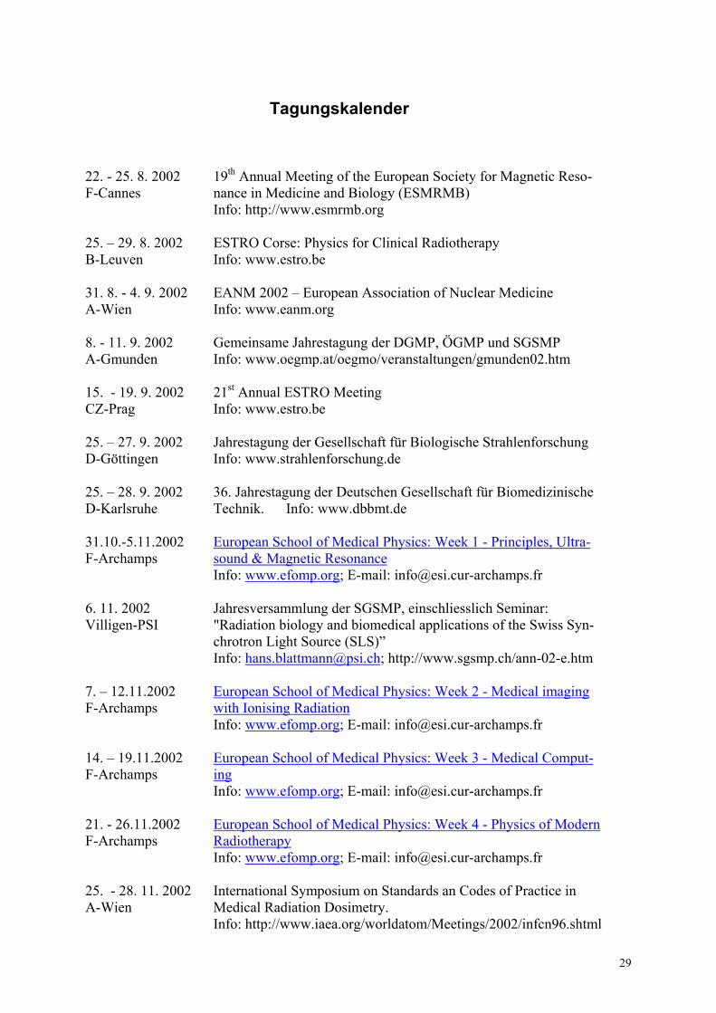
Tagungskalender
22. - 25. 8. 2002 F-Cannes
19th Annual Meeting of the European Society for Magnetic Reso-nance in Medicine and Biology (ESMRMB) Info: http://www.esmrmb.org
25. – 29. 8. 2002 B-Leuven
ESTRO Corse: Physics for Clinical Radiotherapy Info: www.estro.be
31. 8. - 4. 9. 2002 A-Wien
EANM 2002 – European Association of Nuclear Medicine Info: www.eanm.org
8. - 11. 9. 2002 A-Gmunden
Gemeinsame Jahrestagung der DGMP, ÖGMP und SGSMP Info: www.oegmp.at/oegmo/veranstaltungen/gmunden02.htm
15. - 19. 9. 2002 CZ-Prag
21st Annual ESTRO Meeting Info: www.estro.be
25. – 27. 9. 2002 D-Göttingen
Jahrestagung der Gesellschaft für Biologische Strahlenforschung Info: www.strahlenforschung.de
25. – 28. 9. 2002 D-Karlsruhe
36. Jahrestagung der Deutschen Gesellschaft für Biomedizinische Technik. Info: www.dbbmt.de
31.10.-5.11.2002 F-Archamps
European School of Medical Physics: Week 1 - Principles, Ultra-sound & Magnetic Resonance Info: www.efomp.org; E-mail: [email protected]
6. 11. 2002 Villigen-PSI
Jahresversammlung der SGSMP, einschliesslich Seminar: "Radiation biology and biomedical applications of the Swiss Syn-chrotron Light Source (SLS)” Info: [email protected]; http://www.sgsmp.ch/ann-02-e.htm
7. – 12.11.2002 F-Archamps
European School of Medical Physics: Week 2 - Medical imaging with Ionising Radiation Info: www.efomp.org; E-mail: [email protected]
14. – 19.11.2002 F-Archamps
European School of Medical Physics: Week 3 - Medical Comput-ing Info: www.efomp.org; E-mail: [email protected]
21. - 26.11.2002 F-Archamps
European School of Medical Physics: Week 4 - Physics of Modern Radiotherapy Info: www.efomp.org; E-mail: [email protected]
25. - 28. 11. 2002 A-Wien
International Symposium on Standards an Codes of Practice in Medical Radiation Dosimetry. Info: http://www.iaea.org/worldatom/Meetings/2002/infcn96.shtml
29
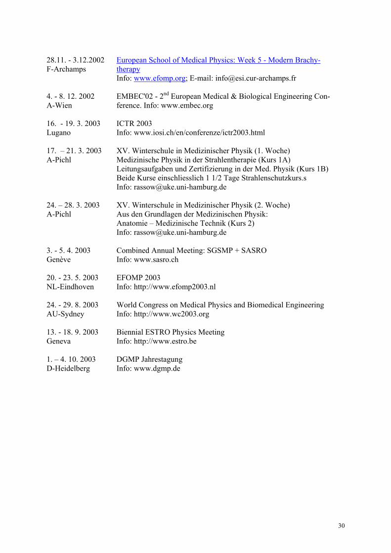
28.11. - 3.12.2002 F-Archamps
European School of Medical Physics: Week 5 - Modern Brachy-therapy Info: www.efomp.org; E-mail: [email protected]
4. - 8. 12. 2002 A-Wien
EMBEC'02 - 2nd European Medical & Biological Engineering Con-ference. Info: www.embec.org
16. - 19. 3. 2003 Lugano
ICTR 2003 Info: www.iosi.ch/en/conferenze/ictr2003.html
17. – 21. 3. 2003 A-Pichl
XV. Winterschule in Medizinischer Physik (1. Woche) Medizinische Physik in der Strahlentherapie (Kurs 1A) Leitungsaufgaben und Zertifizierung in der Med. Physik (Kurs 1B) Beide Kurse einschliesslich 1 1/2 Tage Strahlenschutzkurs.s Info: [email protected]
24. – 28. 3. 2003 A-Pichl
XV. Winterschule in Medizinischer Physik (2. Woche) Aus den Grundlagen der Medizinischen Physik: Anatomie – Medizinische Technik (Kurs 2) Info: [email protected]
3. - 5. 4. 2003 Genève
Combined Annual Meeting: SGSMP + SASRO Info: www.sasro.ch
20. - 23. 5. 2003 NL-Eindhoven
EFOMP 2003 Info: http://www.efomp2003.nl
24. - 29. 8. 2003 AU-Sydney
World Congress on Medical Physics and Biomedical Engineering Info: http://www.wc2003.org
13. - 18. 9. 2003 Geneva
Biennial ESTRO Physics Meeting Info: http://www.estro.be
1. – 4. 10. 2003 D-Heidelberg
DGMP Jahrestagung Info: www.dgmp.de
30
Hinweise für die Autoren Auch Sie sind aufgerufen, an der Gestaltung unseres Bulletins mitzuwirken. Erwünscht sind alle Beiträge, welche für die Mitglieder unserer Gesellschaft von Interesse sein könnten, z.B.
− Berichte von Tagungen, Arbeitsgruppentreffen, Seminaren usw. − Berichte über die Arbeit in verschiedenen Gremien und Kommissionen − Kurz gefasste Ergebnisse von Umfragen, Vergleichsmessungen etc. − Kurzporträts einzelner Institute (apparative Ausrüstung, Schwerpunkte der Arbeit usw.) − Bericht über nationale und internationale Empfehlungen − Kleine Mitteilungen − Hinweis auf Publikationen (Bücher, Zeitschriften) − Hinweis auf Veranstaltungen aller Art (Tagungen, Seminare...) − Lesenswerte Kurzartikel aus Zeitungen oder Zeitschriften (wenn möglich im Original) − Personalia (Ernennungen, Stellenwechsel usw. von Mitgliedern).
Damit unser Bulletin ein möglichst einheitliches Schriftbild aufweist, sind die Autoren der einzelnen Beiträge gebeten, folgende Richtlinien zu beachten. − Schrift wenn möglich Times New Roman oder ähnlich, Grösse 12 Punkt, Zeilenabstand 1. − Ränder links und rechts je 2.5 cm, oben und unten je 3.0 cm, Blocksatz. − Titel in Arial, 14 Punkt, fett, nicht unterstrichen, zentiert. Am einfachsten schicken Sie Ihr Dokument, als MS-Office 95/97 – Dokument abge-speichert, per E-mail an eine der im Impressum erwähnten Adressen der Redaktoren.
31
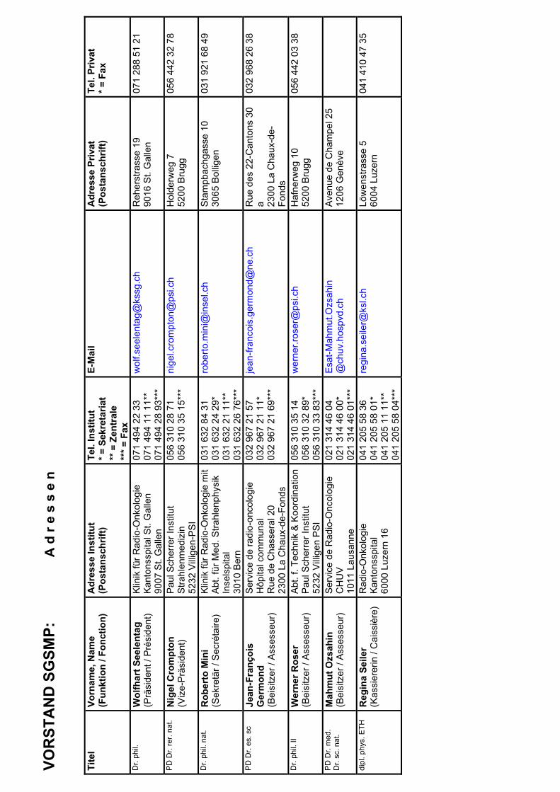
VOR
STA
ND
SG
SMP:
A
d r
e s
s e
n Ti
tel
Vo
rnam
e, N
ame
(Fun
ktio
n / F
onct
ion)
A
dres
se In
stitu
t (P
osta
nsch
rift)
Tel.
Inst
itut
* = S
ekre
taria
t **
= Z
entr
ale
*** =
Fax
E-M
ail
Adr
esse
Priv
at
Tel.
Priv
at
(Pos
tans
chrif
t) * =
Fax
Dr.
phil.
W
olfh
art S
eele
ntag
(P
räsi
dent
/ Pr
ésid
ent)
Klin
ik fü
r Rad
io-O
nkol
ogie
Ka
nton
sspi
tal S
t. G
alle
n 90
07 S
t. G
alle
n
071
494
22 3
3 07
1 49
4 11
11*
* 07
1 49
4 28
93*
**
wol
f.see
lent
ag@
kssg
.ch
Reh
erst
rass
e19
9016
St.
Gal
len
071
288
51 2
1
PD D
r. re
r. na
t. N
igel
Cro
mpt
on
(Viz
e-Pr
äsid
ent)
Paul
Sch
erre
r Ins
titut
St
rahl
enm
ediz
in
5232
Villi
gen-
PSI
056
310
28 7
1 05
6 31
0 35
15*
**
nige
l.cro
mpt
on@
psi.c
h
H
olde
rweg
752
00 B
rugg
05
6 44
2 32
78
Dr.
phil.
nat
. R
ober
to M
ini
(Sek
retä
r / S
ecré
taire
) Kl
inik
für R
adio
-Onk
olog
ie m
it Ab
t. fü
r Med
. Stra
hlen
phys
ik
Inse
lspi
tal
3010
Ber
n
031
632
84 3
1 03
1 63
2 24
29*
03
1 63
2 21
11*
* 03
1 63
2 26
76*
**
robe
rto.m
ini@
inse
l.ch
St
ampb
achg
asse
10
3065
Bol
ligen
031
921
68 4
9
PD D
r. es
. sc
Jean
-Fra
nçoi
s G
erm
ond
(Bei
sitz
er /
Asse
sseu
r)
Serv
ice
de ra
dio-
onco
logi
e H
ôpita
l com
mun
al
Rue
de
Cha
sser
al 2
0 23
00 L
a C
haux
-de-
Fond
s
032
967
21 5
7 03
2 96
7 21
11*
03
2 96
7 21
69*
**
jean
-fran
cois
.ger
mon
d@ne
.ch
R
ue d
es 2
2-C
anto
ns 3
0 a 23
00 L
a C
haux
-de-
Fond
s
032
968
26 3
8
Dr.
phil.
II
Wer
ner R
oser
(B
eisi
tzer
/ As
sess
eur)
Abt.
f. Te
chni
k &
Koor
dina
tion
Paul
Sch
erre
r Ins
titut
52
32 V
illige
n PS
I
056
310
35 1
4 05
6 31
0 32
89*
05
6 31
0 33
83*
**
wer
ner.r
oser
@ps
i.ch
Haf
nerw
eg 1
0 52
00 B
rugg
05
6 44
2 03
38
PD D
r. m
ed.
Dr.
sc. n
at.
Mah
mut
Ozs
ahin
(B
eisi
tzer
/ As
sess
eur)
Serv
ice
de R
adio
-Onc
olog
ie
CH
UV
1011
Lau
sann
e
021
314
46 0
4 02
1 31
4 46
00*
02
1 31
4 46
01*
**
Esat
-Mah
mut
.Ozs
ahin
@
chuv
.hos
pvd.
ch
Aven
ue d
e C
ham
pel 2
5 12
06 G
enèv
e
dipl
. phy
s. E
TH
Reg
ina
Seile
r (K
assi
erer
in /
Cai
ssiè
re)
Rad
io-O
nkol
ogie
Ka
nton
sspi
tal
6000
Luz
ern
16
041
205
58 3
6 04
1 20
5 58
01*
04
1 20
5 11
11*
* 04
1 20
5 58
04*
**
regi
na.s
eile
r@ks
l.ch
Löw
enst
rass
e5
6004
Luz
ern
041
410
47 3
5