Embed Size (px)
Citation preview
1
EPR-and Moessbauer Studies of Benzoyl-CoA Reductase*Matthias Boll‡*, Georg Fuchs§, Christian Meier¶, Alfred Trautwein¶, and David J. Lowe‡
Biological Chemistry Department, John Innes Centre, Colney, Norwich NR4 7UH, UK
Institut für Biologie II, Mikrobiologie, Universität Freiburg, Schänzlestr.1, D-79104 Freiburg
Institut für Physik, Medizinische Universität Lübeck, Ratzeburger Allee 160, D-2358 Lübeck
‡ John Innes Centre, Norwich
§ University of Freiburg
¶ University of Lübeck
Running title: Redox Clusters of Benzoyl-CoA Reduktase
This work was supported by grants from the EC BIOTECH program, the Deutsche
Forschungsgemeinschaft, the Fonds der Chemischen Industrie and the BBSRC.
* The costs of publication of this article were defrayed in part by the payment of page charges.
This article must therefore be hereby marked ‘advertisement’ in accordance with 18 U.S.C.
Section 1734 solely to indicate this fact.
*To whom correspondence should be addressed: Institut für Biologie II, Schänzlestr.1, D-79104
Freiburg, Germany
Tel: 44 7612032685. Fax: 44 7612032626. E-mail: [email protected].
Copyright 2000 by The American Society for Biochemistry and Molecular Biology, Inc.
JBC Papers in Press. Published on July 19, 2000 as Manuscript M001508200 by guest on A
ugust 15, 2019http://w
ww
.jbc.org/D
ownloaded from
2
Abbreviations:
BCR, benzoyl-CoA reductase; Fd, ferredoxin; AdoPP[NH]P, adenosine 5’-[β,γ-
imido]triphosphate; SHE, standard hydrogen electrode.
by guest on August 15, 2019
http://ww
w.jbc.org/
Dow
nloaded from
3
SUMMARY
Benzoyl-CoA reductase catalyzes the two-electron transfer from a reduced ferredoxin to the
aromatic ring of benzoyl-CoA; this reaction is coupled to stoichiometrical ATP-hydrolysis. A very
low reduction potential (< -1 V) is required for the first electron transfer to the aromatic ring. In
this work the nature of the redox centers of purified benzoyl-CoA reductase from Thauera
aromatica was studied by EPR- and Moessbauer spectroscopy. The results obtained indicated the
presence of three [4Fe-4S] clusters. Redox titration studies revealed that the reduction potentials
of all three clusters were below –500 mV. The previously reported S=7/2 state of the enzyme
during benzoyl-CoA independent ATPase activity [Boll, M., Albracht, S.J.P., and Fuchs, G., 1997
Eur. J. Biochem. 244, 840-851] was confirmed by Moessbauer spectroscopy. Inactivation by
oxygen was associated with the irreversible conversion of part of the [4Fe-4S] clusters to
[3Fe-4S] clusters. Acetylene stimulated the benzoyl-CoA independent ATPase activity and
induced novel EPR-signals with gav>2. The presence of simple cubane clusters in benzoyl-CoA
reductase as the sole redox-active metal centers demonstrates novel aspects of [4Fe-4S] clusters
since they adopt the role of elemental sodium or lithium which are used as electron donors in the
analogous chemical Birch reduction of aromatic rings.
by guest on August 15, 2019
http://ww
w.jbc.org/
Dow
nloaded from
4
INTRODUCTION
For many decades it appeared that the biological metabolism of aromatic compounds was
restricted to an aerobic metabolism. However, in recent years an increasing number of anaerobic
bacteria, which are able to oxidize various mononuclear aromatic compounds completely as their
sole source for energy and cell carbon, has been discovered. The strategy of these bacteria is the
conversion of the multitude of aromatic compounds to only a few key intermediates which
become dearomatized in a reductive enzymatic process (for recent reviews see 1,2).
Most of the known pathways lead to benzoyl-CoA which represents the substrate for the
dearomatizing enzyme benzoyl-CoA reductase (BCR). The biochemistry of this enzyme has only
been studied in the denitrifying bacterium Thauera aromatica [3]. It catalyzes the two-electron
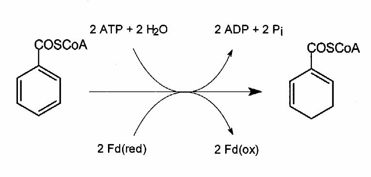
reduction of benzoyl-CoA to cyclohexa-1,5-diene-1-carbonyl-CoA (Fig.1). This two-electron
transfer from reduced ferredoxin to the aromatic ring is stoichiometrically coupled to the
hydrolysis of two molecules of MgATP to MgADP [3,4]. A radical mechanism, corresponding to
the Birch reduction, of alternate one-electron and one–proton transfer steps to the aromatic has
been proposed for the BCR reaction [5,6]. In this case a high redox barrier has to be overcome,
since the transfer of the first electron to a thioester of benzoic acid requires a potential of -1.9 V
[7].
The 163 kDa enzyme consists of four different subunits with molecular masses of 48, 45, 38 and
32 kDa containing 10-12 mol/mol enzyme of iron and acid-labile sulfur and substoichiometric
amounts (0.2-0.3 mol/mol enzyme) of FAD [3,4]. The flavin is assumed to bind unspecifically to
the two ATP binding sites of BCR. In a previous study the metal centers of BCR were
investigated by electron paramagnetic resonance (EPR-) spectroscopy [8]. In the dithionite-
reduced state, EPR signals of two interacting [4Fe-4S]+1 centers were detected; additional EPR-
signals could not clearly be assigned but were characteristic for [2Fe-2S]+1 clusters. BCR also
hydrolyses MgATP in the absence of a reducible substrate [3]. This uncoupled ATPase activity
was only observed in the dithionite-reduced and not in the thionine-oxidized state of the enzyme
by guest on August 15, 2019
http://ww
w.jbc.org/
Dow
nloaded from
5
[8]. Novel EPR signals were induced only in the steady state of this ATPase activity and were
assigned to an S=7/2 high spin system. In parallel, a shift from interacting to non-interacting
[4Fe-4S]+1 clusters was detected. These observations allowed the presentation of a first model for
the catalytic cycle of the BCR reaction. This model included an ATP-driven electron activation as
a result of conformational changes affecting a reduced, EPR-active iron-sulfur cluster [8].
In this work we investigated the EPR properties of BCR in more detail. These studies included
temperature- and redox potential-dependent studies. In addition we studied the effects of oxygen,
AdoPP[NH]P and acetylene on the EPR properties of BCR. We also investigated 57Fe-enriched
BCR by Moessbauer spectroscopy in the reduced and oxidized state in the absence and presence
of the individual substrates. The results obtained clarified the nature of the redox centers of BCR
which we identified as three [4Fe-4S] clusters. The ATP dependent switch from the S=1/2 to the
S=7/2 state was confirmed by Moessbauer spectroscopy and could clearly be assigned to a
[4Fe-4S] cluster.
by guest on August 15, 2019
http://ww
w.jbc.org/
Dow
nloaded from
6
EXPERIMENTAL PROCEDURES
Growth of bacterial cells
T. aromatica was isolated in our laboratory and has been deposited in the Deutsche Sammlung
von Mikroorgansimen (Braunschweig, Germany; DSM 6984) [9]. It was grown anoxically at
28°C in a mineral salt medium in a 200 l fermenter; 4-OH-benzoate and nitrate in a molar ratio of
1:3.5 served as sole sources of energy and cell carbon. Continuous feeding of the substrates, cell
harvesting, storage and preparation of cell extracts were carried out as described previously [10].
57Fe-enrichment of BCR was achieved by adding 57Fe2+ as sole source of iron to the medium. For
this purpose 175 mg of metallic 57Fe (95.7% enriched, Advanced Materials and Technology
Consulting, New York) was dissolved in 2.2 ml of 12 M HCl overnight at 80°C. After the metal
was completely dissolved it was added drop-wise to 100 ml of 0.6 M nitrilotriacetic acid pH 5.2
under continuous stirring. During this procedure the pH was held constant between 3-5 by adding
2 M NaOH. With this amount of 57Fe T. aromatica was grown in a 100 l batch culture under
continuous feeding of 4-OH-benzoate and nitrate to OD578 ~ 2.6 yielding 250 g of cells (wet
mass). Enrichment in 57Fe was estimated >90 % in purified BCR.
Protein purification, enzyme activity assay, purity control and sample storage
Purification of BCR from extracts of T. aromatica was performed under strictly anaerobic
conditions in a glove box under a N2/H2 atmosphere (95:5, by vol.) as described earlier [3]. The
procedure included three chromatographic steps including anion exchange chromatography on
DEAE-Sepharose (Pharmacia), chromatography on ceramic hydroxyapatite (BioRad) and gel
filtration using Sephadex G-75 (Pharmacia). Concentration of the protein samples was achieved
by centrifugation (8 000 x g) in Microsep Microconcentrators (exclusion limit 50 kDa). The
concentration of the enzyme was 175 mg ml-1 for Moessbauer studies or 15-50 mg ml-1 for EPR
spectroscopy. The purity of these enzyme preparations was >90 % as estimated by Coomassie
staining of SDS-PAGE gels. In previous experiments we have shown that an additional
chromatography step using a Mono Q anion exchange column (Pharmacia) increased the purity
up to virtual homogeneity but did not increase the specific activity of the enzyme indicating a
by guest on August 15, 2019
http://ww
w.jbc.org/
Dow
nloaded from
7
concomitant loss of activity. Since there was no difference in the EPR spectra between the
samples of 90 %-100 % purity the Mono Q chromatography step was omitted in routine enzyme
preparations. Enzyme activity was determined in a continuous spectrophotometric assay recording
the benzoyl-CoA- and MgATP-dependent oxidation of reduced methyl viologen at 730 nm at
37°C [3]. Purified BCR (250-300 mg) was obtained from 200 g of cells (wet mass) with specific
activities of 0.40-0.45 µmol benzoyl-CoA min -1 mg-1. Concentrated protein samples were stored
anaerobically in tubes sealed with gas-tight stoppers at -80°C for several months.
Effect of acetylene on BCR activity
The effect of acetylene on BCR activity was tested in the standard spectrophotometric assay
following the oxidation of reduced methyl viologen at 730 nm [3]. A saturated stock solution was
prepared at 20°C (diameter 2 cm) by flushing acetylene with an overpressure of 0.2 bar first
through wash bottle containing 10 mM dithionite and then through a stoppered 5 ml test tube
(2 cm diameter) containing 1 ml of assay buffer with 150 mM Mops/KOH, 10 mM MgCl2, pH
7.3. Aliquots of this acetylene stock solution (5-40 µl) were added to a standard enzyme assay
mixture.
To test the effect of acetylene on benzoyl-CoA-independent ATPase activity, ATP consumption
and ADP formation were discontinuously determined at 30°C. Samples (usually 100 µl) were
periodically retrieved from a 1 ml assay mixture in stoppered vials containing 150 mM
Mops/KOH pH 7.3, 5 mM sodium dithionite, 5 mM ATP, 10 mM MgCl2, 0.2-0.4 mg BCR and
16 µl-80 µl of the saturated acetylene solution. The volume of the gas phase was approximately
1.5 ml, the reaction was started by addition of ATP. The samples were directly analyzed by HPLC
using a MonoQ anion exchange column as described previously [7].
Sample preparation for EPR- and Moessbauer spectroscopy
All BCR samples for EPR- and Moessbauer spectroscopy were prepared in an anaerobic glove
box under a 100 % nitrogen atmosphere (<1.0 ppm O2). Prior to any sample preparation of BCR
excess of dithionite and corresponding oxidation products were removed by passing the
by guest on August 15, 2019
http://ww
w.jbc.org/
Dow
nloaded from
8
concentrated enzyme sample (0.1-1 mM; 0.5-1 ml) over a Biogel P-6 (Biorad) desalting column
(volume: 5 ml, diameter: 1 cm) which had been equilibrated with either a 100 mM Mops/KOH pH
7.3 or with 100 mM Tris/HCl buffer pH 8.0 both containing 10 mM MgCl2. If not otherwise
stated this dithionite free enzyme sample represented the starting material for all sample
preparations.
Reduction of the enzyme
Reduction of BCR was performed according to standard anaerobic procedures by adding sodium
dithionite from a freshly prepared stock solution (100 mM in 100 mM Mops/KOH pH 7.3) giving
a final 10-fold excess of this reductant compared to the enzyme. The concentration of these
dithionite solutions was checked from time to time by titration against ferricyanide, showing that
they were between 80-90 % of the nominal concentration. The pH of the enzyme solutions was
not affected by dithionite addition since the buffer concentration in the enzyme preparations was
10-100-fold higher than the final dithionite concentration (~ 1-10 mM). The reduced enzyme was
quickly transferred into EPR tubes or Moessbauer sample holders and then frozen either inside the
glove box on dry ice (Moessbauer-samples) or outside the glove box in an gas tight sealed EPR
tube in liquid nitrogen. When BCR was reduced with deazaflavin/light the enzyme was brought to
100 mM Tris/HCl buffer pH 8.0 using a desalting P6-column desalting which had been
equilibrated with this buffer. Deazaflavin was obtained as a kind gift from Prof. S. Ghisla
(University at Konstanz, Germany). The final concentration of deazaflavin in the sample was 20-
40 µM (for EPR samples) or 200 µM (for Moessbauer samples). The EPR samples were
illuminated in airtight sealed glass tubes (diameter: 1 cm) for 15 min with white light (250 W
halogen bulb) from both sides of the tube (distance: approximately 2-3 cm). Moessbauer samples
were illuminated for 15 min by installing the sample holders inside and the light source outside a
glove box consisting of transparent PVC (distance to the tube: 5-10 cm).
Effect of MgATP, benzoyl-CoA and AdoPP[NH]P
When the benzoyl-CoA-independent ATP hydrolyzing activity of dithionite-reduced BCR was
studied, 10 mM ATP and 20 mM MgCl2 for EPR spectroscopy and 25 mM MgATP and 40 mM
by guest on August 15, 2019
http://ww
w.jbc.org/
Dow
nloaded from
9
MgCl2 for Moessbauer spectroscopy were used. Since ADP is a competitive inhibitor of BCR (Ki
for MgADP is 1.1 mM, Km for ATP is 0.6 mM, [8]) a MgATP regenerating system was added
consisting of phosphoenolpyruvate (same concentration as ATP) and pyruvate kinase (Sigma,
activity in the assay was 10 times higher than BCR activity). Prior to freezing of the EPR tubes or
Moessbauer sample holders, BCR was incubated for less than 1 min to ensure that excess residual
MgATP was present. To test the effect of the inactivating compound AdoPP[NH]P on the EPR
and Moessbauer spectra, the enzyme was incubated for 10 min at room temperature with 0.05
mM (EPR-spectroscopy) or 0.2 mM (Moessbauer spectroscopy) of this compound in the
presence of 20 mM MgCl2.
Effect of acetylene
The effect of acetylene on the EPR spectrum of BCR was studied in the presence and absence of
MgATP. After addition of 60 µl of a saturated acetylene solution prepared at 20°C as described
above to 540 µl of a BCR solution (25 mg ml -1 in 100 mM Mops/KOH and 20 mM MgCl2 pH
7.3) the sample was incubated for 10 min at room temperature. Half of the solution was directly
transferred into an EPR tube and frozen whereas the other half of the solution was incubated for 2
min with 10 mM ATP for 1 min at room temperature and then frozen in an EPR tube.
Redox titration experiments of BCR
Dye-mediated redox titration of BCR was performed in a Miller-Howe anaerobic glove box under
a nitrogen atmosphere (<1 ppm O2). The enzyme/mediator mixture (2.5 ml) was dissolved in
100 mM Mops/KOH pH 7. The concentration of BCR was 100-150 µM. The mediator consisted
of methyl and benzyl viologens, neutral red, safranin O, phenosafranin, anthraquinone-2-sulfonate,
2-hydroxy-1,4-naphthoquinone, indigo disulfonate, resorufin, methylene blue, phenazine
methosulfate and N’N’N’N’-tetramethyl-p-phenylenediamine at a final concentration of 40 µM
each. The redox potential was adjusted with 10-100 mM sodium dithionite and potassium
ferricyanide prepared freshly and anaerobically in the same buffer as BCR. Potentials reported in
this paper are with reference to SHE and were obtained by using a potential of +243 mV vs SHE
for the saturated calomel electrode at 22°C. Mediator/enzyme mixtures with a stable potential
by guest on August 15, 2019
http://ww
w.jbc.org/
Dow
nloaded from
10
could be obtained from -543 mV to +100 mV. Stabilization (drift <1 mV/min) of the potentials
usually required 1-5 min under continuous stirring. Samples with defined redox potentials were
immediately frozen anaerobically in EPR tubes and stored in liquid nitrogen. Redox titrations
were normally performed in the oxidative direction but as a control for reversibility, some samples
were prepared by re-reduction with dithionite.
EPR spectroscopy
X-band EPR spectra were recorded on an updated Bruker 200D-SRC spectrometer. Low
temperature measurements were made using an Oxford Instruments ESR 900 cryostat modified to
take sample tubes of up to 4-mm internal diameter. Recording conditions are described in the
legends to the individual figures. Spin concentrations of ground-state transition EPR signals were
determined by comparison with a 1.00 mM copper sulfate sample in 11 mM sodium EDTA.
Moessbauer spectroscopy
57Fe Mössbauer spectra were recorded with a conventional constant acceleration spectrometer
using a 57Co source in a Rh matrix (1 GBq using the bath cryostat; 1.3 GBq using the magnet
cryostat). Measurements at 4.2 K and 77 K were performed with a bath cryostat (Oxford
Instruments) and a permanent magnet mounted outside the cryostat producing a field of 20 mT ⊥
γ. High-field measurements were performed with a cryostat equipped with a superconducting
magnet (Oxford Instruments). The spectra were analyzed assuming Lorentzian line shape; the
isomer shift is quoted relative to α-Fe at room temperature.
Other methods
Benzoyl-CoA was synthesized according to Schachter and Taggart [11]. Protein concentration
was determined by the Bradford method using bovine serum albumin as standard [12]. SDS
PAGE were performed as described by Laemmli [13]. Protein was visualized by Coomassie blue
staining [14].
by guest on August 15, 2019
http://ww
w.jbc.org/
Dow
nloaded from
11
RESULTS
1. Nature of the iron-sulfur centers
The nature of the iron-sulfur centers of BCR was studied by Moessbauer and EPR spectroscopy.
Metal analysis of BCR revealed the presence of 10-12 mol iron/mol enzyme, and
substoichiometric amounts of zinc. Earlier EPR studies [7] had indicated the presence of two
[4Fe-4S] and additional Fe-S clusters which were assigned to [2Fe-2S] clusters or to a novel
metal cluster with an unknown structure. In addition, as part of this work, similar EPR signals
typical of [3Fe-4S] clusters were detected in partially inactivated BCR preparations (data not
shown).
Moessbauer spectroscopy: oxidized BCR
For Moessbauer spectroscopy studies, BCR was purified from extracts of T. aromatica grown
anaerobically with 57Fe as sole source of iron. High 57Fe enrichment was achieved as estimated
from the resonance absorption effect of about 9 % for each subspectrum of the oxidized sample,
which corresponds to about 11 mM 57Fe (sample concentration was about 1 mM and isolated
benzoyl-CoA reductase samples were shown to contain 10-12 mol Fe/mol enzyme). The
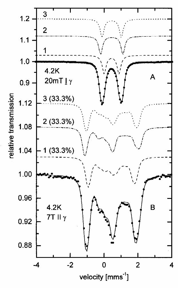
Moessbauer spectrum of thionine-oxidized BCR taken at 4.2 K in a small external field of 20 mT
perpendicular to the γ-beam shows a slightly asymmetric quadrupole doublet (Fig. 2A). For
reasons which will be discussed later the spectrum was analyzed using three subspectra of equal
area. The isomer shifts and quadrupole splittings so obtained (δ~0.45 mm s-1 and ∆EQ~1 mm s-1,
see Table 1) are typical of delocalized mixed-valence iron sites in tetrahedral sulfur coordination
and are characteristic of [4Fe-4S]2+clusters [15]. The slightly different quadrupole splittings of the
subspectra indicate structural differences between the three [4Fe-4S]2+ clusters which each
contain two delocalized mixed-valence iron-pairs with spins S12=9/2 and S34=9/2 with antiparallel
coupling yielding a total cluster spin of zero. Indeed, the spectrum recorded in a high external
field of 7T parallel to the γ beam (Fig. 2B) shows magnetic splitting only due to the external field
and thus reveals the diamagnetic nature of the clusters in the oxidized BCR sample.
by guest on August 15, 2019
http://ww
w.jbc.org/
Dow
nloaded from
12
These results allowed us to exclude the presence of [2Fe-2S] and [3Fe-4S] clusters since both
types of oxidized clusters ([2Fe-2S]2+ and [3Fe-4S]+) show subspectra with parameters typical for
Fe3+S4 (δ~0.25 mm s-1; ∆EQ~0.7 mm s-1), which are clearly absent in the Mössbauer spectra of
thionine oxidized BCR. Additionally [3Fe-4S]+ clusters would reveal paramagnetic features in a
high external field of 7T, whereas the oxidized sample contained clearly only diamagnetic species.
Moessbauer spectroscopy: reduced BCR
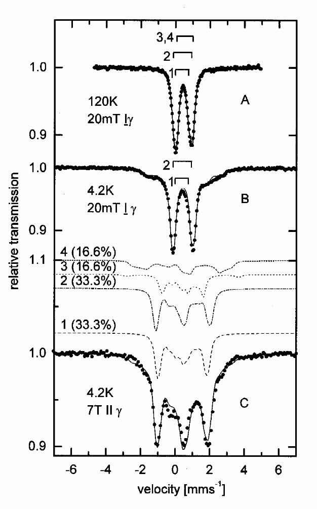
The Mössbauer spectrum of deazaflavin reduced BCR recorded at 120 K (Fig 3A) and 4.2 K (Fig
3B) in a small external field of 20 mT ⊥ γ is shown in Fig. 3A and 3B. The 4.2 K spectrum shows
the presence of a paramagnetic species and a quadrupole doublet, the latter with parameters (δ
~0.45 mms-1 and ∆EQ~1 mms-1, see Table 1) similar to those observed in the oxidized sample.
EPR investigations (see EPR section of this work) showed the presence of S=1/2 species and
therefore the paramagnetic species were analyzed by using the spin-Hamiltonian approximation
for a total cluster spin S=1/2 [16]. As starting parameters for the simulation parameters for [4Fe-
4S]1+ published in [15] were used. With this procedure 33% of the total area were attributed to a
[4Fe-4S]1+ cluster with parameters given in Table 1. The remaining 66% of the total area could be
simulated with two doublets whose parameters are similar to those (see Table 1) found for
components 1 and 2 in the thionine oxidized sample. The spectrum taken at 4.2 K in an high
external field of 7T || γ (Fig. 3C) was simulated with the same parameter set as the low-field data.
Subspectra 1 and 2 represent diamagnetic [4Fe-4S]2+ clusters as in thionine oxidized BCR. By
elevating the temperature to 120 K (Fig. 3a) the diamagnetic subspectra exhibit the isomer shifts δ
1=0.41mms-1, δ2=0.4mms-1 and the quadrupole splittings ∆Q1=1.04mms-1, ∆EQ2=0.76mms-1. The
spectrum of [4Fe-4S]1+ can be simulated with one quadrupole doublet with the parameters δ
=0.55mms-1, ∆EQ2=0.95mms-1, which are similar to those reported for the reduced ferredoxin
from C. pasteurianum [15].
EPR spectroscopy
Based on the results obtained from Moessbauer spectroscopy we reinvestigated the [4Fe-4S]
clusters of reduced BCR using temperature dependent and power saturation EPR studies. To
by guest on August 15, 2019
http://ww
w.jbc.org/
Dow
nloaded from
13
achieve a high degree of reduction, the dithionite used in previous studies was replaced by
deazaflavin/light as reductant. Our aim was a clear assignment of individual EPR-subspectra to
the three [4Fe-4S] clusters of BCR as identified by Moessbauer spectroscopy. For this purpose
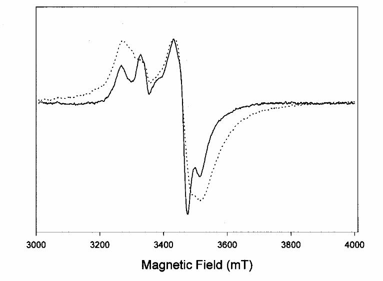
we first compared the EPR spectra of unlabeled with 57Fe-labeled enzyme. All EPR features
clearly broadened in the 57Fe spectrum as a result of unresolved hyperfine interaction of unpaired
electrons with the nucleus of 57Fe (I=1/2), (Fig. 4). This confirmed that high 57Fe enrichment was
achieved and that no other paramagnetic metal than iron dominated the EPR spectra of our
sample.
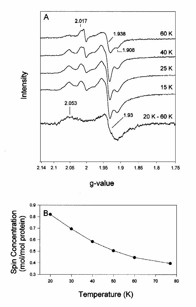
Further EPR studies were performed with unlabeled BCR. Unusually for [4Fe-4S] clusters, which
normally relax rapidly [17], an EPR signal from a [4Fe-4S]+1 cluster was observed up to 77 K. At
60 K a rhombic EPR spectrum characteristic of a single [4Fe-4S]+1 cluster with g-values at 2.017,
1.938, 1.908 was observed (Fig. 5A); this cluster is referred to cluster I. A minor EPR signal at
g=2.004 with less than 0.05 spin/mol protein was superimposed on this spectrum and indicated
the presence of a radical of unknown nature. At temperatures below 40 K additional features at
g=2.053 and around g=1.93 intensified indicating the rise of an EPR signal from an additional
[4Fe-4S]+1 cluster relaxing significantly faster than cluster I. At temperatures below 25 K all
features started to saturate simultaneously at 2 mW (Fig. 5A). A difference spectrum between the
spectra recorded at 20 K/ 0.2 mW and the spectrum taken at 60 K and 2 mW normalized for
temperature and microwave power is presented in Fig. 5A. The difference spectrum was rather
broad and axial and is assigned to cluster II with g-values at 2.053, 1.93, and 1.93. The
dependencies of the EPR signals of both clusters on temperature and microwave power were
highly similar resulting in only a weak difference spectrum. The total spin concentration in the
dithionite reduced sample was not higher than 0.82 mol/mol enzyme, indicating only a partial
reduction of the clusters. This was in agreement with the results obtained from Moessbauer
spectroscopy where the highest degree of reduction was 33 %. EPR signals from the putative
third [4Fe-4S] cluster of BCR, as expected from Moessbauer spectroscopy, could not be detected
in the deazaflavin-reduced sample indicating that it was not reducible by this system. The
temperature dependence of the total spin concentration between 20 K to 77 K fitted well to the
by guest on August 15, 2019
http://ww
w.jbc.org/
Dow
nloaded from
14
observation that cluster II relaxes faster than cluster I (Fig. 5B). After normalization for
temperature, the spin concentration approximately doubled from 0.42 (60 K) to 0.82 (20 K)
spins/enzyme. This increase is considered to be mainly a result of the increased intensity of the
cluster II EPR signal which is not detectable at temperatures above 40 K.
At temperatures below 15 K, broad unresolved features covering more than 200 mT started to
develop. These broad features also have been detected in a previous study and were ascribed to
two interacting 2[4Fe-4S] clusters [8]. Even at 4 K and 20 mW microwave power these features
could not be saturated [8]. It was concluded that in the some protein molecules the clusters
interacted magnetically, whereas others showed EPR features of single isolated [4Fe-4S]+1
species, indicating a conformational heterogeneity. Since spin integration of the broad signal
extending over 200 mT could not be carried out accurately it was impossible to give an exact
ratio of coupling/non-coupling clusters. An additional problem was that all clusters were at best
partially reduced, so it was not clear which of the [4Fe-4S] clusters contributed to this broad
signal. A rough estimate indicated that the spin concentration of the broad signals at 4 K was in
the same range as the total spin concentration detected at 25 K. Therefore, we estimate that
photoreduction with deazaflavin resulted in a 50 % reduction of all clusters with half of the
paramagnetic clusters interacting. This value is slightly higher than that in the samples used for
Moessbauer spectroscopy (33 %).
The results obtained from Moessbauer spectroscopy and the content of iron and acid-labile sulfur
(10-12 mol/mol enzyme) strongly indicated the presence of three [4Fe-4S] clusters but only two
clusters could be identified clearly by EPR spectroscopy in the deazaflavin reduced state, although
the broad signal of interacting clusters could not be assigned. In order to identify a third [4Fe-4S]
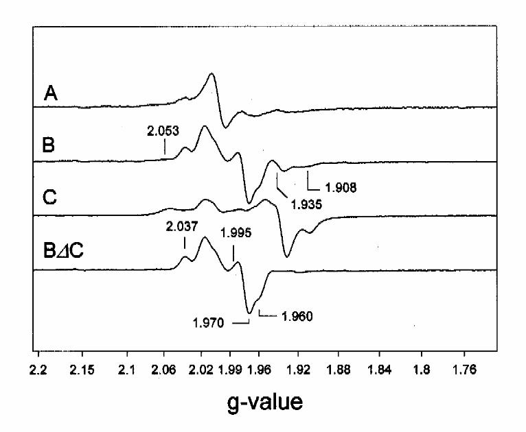
cluster by EPR spectroscopy, we studied BCR under steady state conditions of ATP-dependent
benzoyl-CoA reduction. In Fig. 6B a representative EPR-spectrum of BCR recorded at 40 K is
presented showing that clusters I and II are both more oxidized than in the dithionite-reduced
sample, as indicated by a 80 % decrease of signal intensity at g=2.05 and g=1.93,1.91. In parallel,
novel EPR features were observed in the g=2 region. After subtraction of the EPR spectrum of
by guest on August 15, 2019
http://ww
w.jbc.org/
Dow
nloaded from
15
dithionite-reduced BCR (x 0.2) from the one obtained in the steady state of substrate reduction,
an EPR signal was left with features at g=2.037, 1.995, 1.97 and 1.96 (Fig. 6 B∆C). These
features could not be ascribed to an isolated S=1/2 species, but instead are typical of an
interaction with a second magnetic site. With this assumption, the EPR-signal indicated the
presence of a [4Fe-4S] cluster with g-values ~2.03, 1.995 and 1.965 with splitting constants in the
range 2-3 mT. A decrease of the modulation amplitude to 0.3 mT did not resolve the signal
further (not shown). Its spin concentration amounted to less than 0.35 spins/enzyme indicating
that cluster III was reduced to approximately the same extent as the other two clusters in the
photoreduced sample. Optimal EPR conditions were 30 K and 2 mW. The nature of the second
magnet involved in the interaction with cluster III is not known. It is rather unlikely that this is
either cluster I or II since the 40 K difference spectrum in Fig. 6D was obtained easily, which
would not have been the case had one of these clusters been coupled magnetically with cluster III.
A hyperfine interaction with a nuclear spin, such as a proton, is an unlikely explanation since this
type of interaction usually results in splitting constants significantly below 2 mT, although this
cannot be ruled out absolutely. It is likely that the complex EPR spectrum of cluster III is due to a
dipole-dipole interaction with an additional unknown paramagnet, such as a radical species. At
temperatures higher than 77 K an isotropic radical signal remains (Fig. 6A). It is not clear whether
the decrease of spin concentration of the radical at lower temperatures was only due to saturation
or also to an interaction with cluster III.
2. Redox properties of the clusters
The natural electron donor for BCR is a reduced ferredoxin with two [4Fe-4S] clusters with
reduction potentials of -431 mV and –587 mV vs SHE [4,19]. The reduction potentials of the
iron-sulfur clusters of BCR therefore are expected to be below -400 mV. As discussed above,
Moessbauer studies indicated that only 1/3 of all clusters could be reduced by dithionite or
deazaflavin/light, indicating that the reduction potential of at least two clusters must be far below
-500 mV.
by guest on August 15, 2019
http://ww
w.jbc.org/
Dow
nloaded from
16
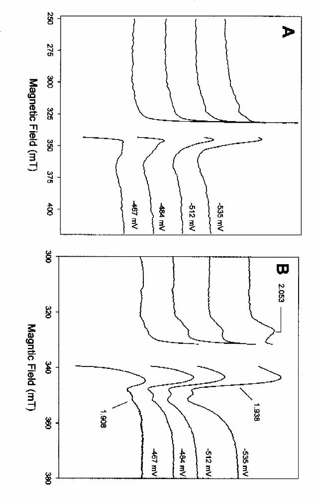
We have studied the redox potential dependence of the EPR spectra recorded at different
temperatures. Our goal was to investigate the redox potential-dependent rise of both the signals
of the interacting and the non-interacting clusters. At potentials more positive than -400 mV, only
weak EPR-signals were detected, originating from adventitious iron at g = 4.3, or in some batches
minor signals (spin concentration less than 0.3 mol/mol enzyme) of an oxidized [3Fe-4S]+1 cluster
(not shown). This EPR signal is assigned to a partial degradation of [4Fe-4S] to [3Fe-4S] clusters
and was similar to the spectra obtained when BCR was exposed to air, leading to an irreversible
inactivation of the enzyme (see also Fig. 10). In Fig. 7B samples recorded at 25 K at potentials
from –467 mV to –535 mV are shown. Usually the redox titration was performed in oxidative
direction by oxidation of dithionite-reduced enzyme with ferricyanide. At –467 mV the 25 K
spectrum displays features of cluster I (see 60 K spectrum in Fig. 5A) as indicated by the ratio of
signal amplitudes at g=1.93 to 1.908 and the typical rhombicity. When the potential was lowered
further, additional features appeared at g=2.053. In parallel, the shape in the g<2 region changed
as a result of an increase of intensity at g=1.91. These two features were typical for an increase of
spin concentration of cluster II which increasingly dominated the spectrum as the potential was
poised lower. Due to the strong radical signal of the mediators the spin concentration at -543 mV
and 25 K could only be estimated to be in the range of 0.4-0.6 mol/mol enzyme; the spin
concentration was lower than in photoreduced sample (0.82 mol/mol).
We also recorded EPR-spectra of the same samples at 4 K in order to investigate the redox
potential-dependent features of the EPR signals of the magnetically interacting clusters (Fig.7A).
At -467 mV, when the 25 K spectrum mainly displayed features of cluster I, a very poorly
resolved and broad signal around g=2 started to develop. In parallel to reduction of cluster II, the
EPR signal of the interacting [4Fe-4S]+ clusters also increased, indicating that these signals derive
from the magnetic interaction of cluster II with at least one of the other two clusters. However,
our results show that this interaction did not involve all reduced cluster II, since at higher
temperatures EPR signals typical of non-interacting clusters could be ascribed to isolated clusters
I and II. No sign of even a partial reduction of the putative cluster III was observed at potentials
down to -543 mV. However one has to take into account that the EPR signals ascribed to this
by guest on August 15, 2019
http://ww
w.jbc.org/
Dow
nloaded from
17
cluster (g=2.037, 1.97, 1.96) are quite narrow and close to those of the strong radical of the
redox dyes, so that they could be obscured by the latter. Additionally, cluster III might couple
with cluster II resulting in the broad signal observed at low temperatures. It was not possible to
determine accurately the spin concentration from the low temperature spectra.
Since none of the clusters was even half-reduced a fit to a Nernst curve was impossible. The
spectra shown in Fig.7B clearly imply that the reduction potential of cluster I and cluster II must
be lower than -500 mV. However due to only a partial reduction a fit to Nernst curve was
impossible.
3. Effect of MgATP and AdoPP[NH]P on the clusters
Reduced BCR catalyzes both a stoichiometric ATP hydrolysis coupled to benzoyl-CoA reduction,
and benzoyl-CoA-independent ATP hydrolysis [8]. In a previous EPR study three major effects
were observed under steady state conditions of substrate-independent ATPase activity. First, new
EPR signals were induced: an isotropic signal at g=5.15 accompanied by a minor one at g=12 [8].
These two signals were assigned to the transitions of the first excited state and ground state an
S=7/2 high spin system. Second, the broad signal at g=2 assigned to two interacting [4Fe-4S]
clusters sharpened up, indicating that the interaction between the clusters was diminished after
prolonged incubation with MgATP. Third, a concomitant loss of S=1/2 spins was observed in the
course of the experiment. In these studies the nature of the paramagnet with the S=7/2 high-spin
system was not identified. Moessbauer studies should indicate whether the switch from the S=1/2
to a S=7/2 high spin system occurred in one isolated cluster, or in two interacting [4Fe-4S]
clusters. Additionally, we wanted to clarify whether the two effects of MgATP described above
were the result of binding or hydrolysis of MgATP. For this purpose the non-hydrolyzable ATP
analog AdoPP[NH]P was used; this compound does not act as a competitive inhibitor but
inactivated BCR irreversibly in a time-dependent reaction [8].
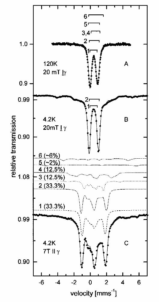
The Moessbauer spectra of BCR under steady state conditions of benzoyl-CoA-independent
ATPase activity are shown in Fig. 8. The spectra recorded at 4.2K at 20 mT ⊥ γ (Fig. 8B) and
by guest on August 15, 2019
http://ww
w.jbc.org/
Dow
nloaded from
18
7T || γ (Fig. 8C) exhibit an additional magnetic hyperfine pattern when compared to Fig.3. EPR
studies on an equivalent sample revealed the presence of a S=7/2 species [8]. We therefore
simulated this magnetic pattern using the spin-Hamiltonian approximation with S=7/2 and with D
= 4.3 cm-1 and E/D = 0.12 derived from EPR spectra [8]. In the spectrum taken at 120 K
(Fig. 8A) there is a slight shoulder at about 1.5 mm s-1 which can be assigned to the S=7/2 species
as it did not appear in the spectrum of deazaflavin/reduced reduced BCR (Fig. 3A). This line
originates from Fe2+ sites similar to those in the S=7/2 species of the selenium-substituted
2[4Fe-4Se]1+ clusters in the ferredoxin from Clostridium pasteurianum [20]. Therefore the same
pattern of electron localization as described in [20] was assumed, i.e. a S=7/2 species with two
subspectra in the ratio Fe2+:Fe3+ = 3:1. For the Fe3+ site values δ=0.39 mm s-1 and ∆EQ=1.17
mm s-1 taken from [20] were used. For the Fe2+ sites slightly different values (δ=0.58 mm s-1 and
∆EQ =1.55 mm s-1) were used in order to be consistent with the data taken at 120 K. The low-
and high-field spectra of Fig. 8B,C were fitted taking η, Ax, Ay, Az and the total area of the two
subspectra of the S=7/2 as free parameters, and using the same parameters as for the
deazaflavin/light-reduced sample for the [4Fe-4S]1+ (S=1/2) and [4Fe-4S]2+ clusters. The mean
values of the resulting A-Tensors of the S=7/2 species (Tab.1) are comparable with the theoretical
and the experimental mean values given in [20]. Again 66% of the spectral area represent
diamagnetic [4Fe-4S]2+ clusters, 25 % belong to paramagnetic (S=1/2) [4Fe-4S]1+ and 8% can be
assigned to [4Fe-4S]1+ clusters with spin S=7/2. The fact that the ratio of diamagnetic clusters to
paramagnetic clusters is 2:1 for two different preparations strongly indicates that BCR contains
three discrete [4Fe-4S] clusters.
In order to distinguish between the effects of binding and hydrolysis, EPR-spectra of BCR were
recorded at 4 K and 0.2 mW in the presence of MgATP and AdoPP[NH]P (Fig. 9). Under these
conditions it was certain that the fast relaxing signals of the interacting [4Fe-4S] clusters were not
saturated. In the steady state of benzoyl-CoA-independent ATPase activity, the broad EPR signal
of the interacting [4Fe-4S] clusters sharpened up (indicated by arrow 2, Fig. 9A,B). In parallel the
typical isotropic signal at g=5.15 of the first excited state transition of an S=7/2 system was
observed (arrow 1 in Fig. 9B). The accompanying signal at g=12, which results from a ground
by guest on August 15, 2019
http://ww
w.jbc.org/
Dow
nloaded from
19
state transition of the S=7/2 system [8], represents only a minor contribution to the total spin and
therefore was only detectable at very high enzyme concentrations (not shown). When ATP (10
mM) was replaced by AdoPP[NH]P virtually no signal of an S=7/2 spin system was observed
(Fig. 9C). However, in the same way as with MgATP, the broad EPR-signal of the interacting
[4Fe-4S] clusters changed to sharper signals of non-interacting clusters. We therefore conclude
that binding of the nucleotide eliminates the interaction between two [4Fe-4S] clusters, whereas
hydrolysis of ATP induces the switch from S=1/2 to S=7/2 of a [4Fe-4S] cluster. The latter could
not be observed in the presence of the non-hydrolyzable ATP analog AdoPP[NH]P. No
significant changes were induced by ATP or by AdoPP[NH]P in the EPR signals of the non-
interacting [4Fe-4S] clusters recorded at higher temperatures.
4. Effect of oxygen
BCR is highly sensitive to oxygen, with a half life in the absence of reducing agents of less than 30
s [3]. We investigated this irreversible inactivation by recording EPR spectra of BCR after
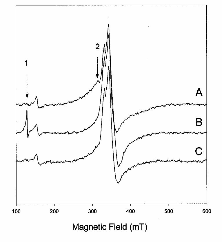
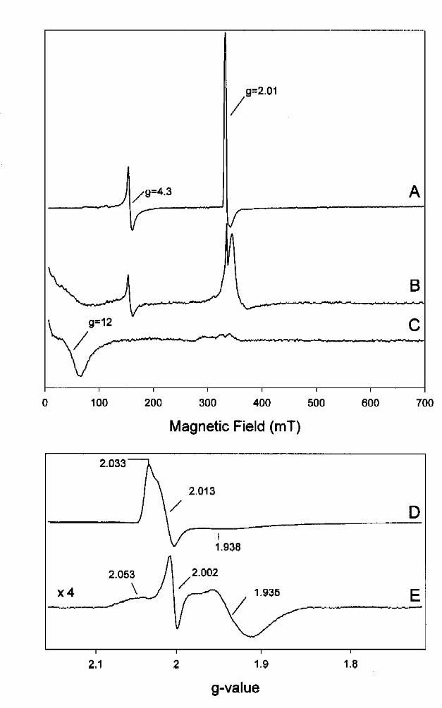
exposure to air and after re-reduction of the enzyme with dithionite. In Figure 10A the full scale
4.8 K spectrum of air-oxidized BCR displays two main features at g=4.3 and g=2.01. The former
signal was assigned to adventitiously bound tetrahedrally coordinated Fe3+. It was clearly
increased compared to an intact mildly thionine-oxidized sample (not shown), indicating the
partial release of iron atoms from the [4Fe-4S] clusters as a result of degradation. Note, that the
optimal temperature for this signal was between 20-30 K and therefore it was highly saturated
under the conditions of Fig. 10. The second axial EPR signal was typical of an oxidized [3Fe-4S]+
cluster with g~2.013 and was not saturated at 4.8 K and 2 mW microwave power. An EPR
spectrum displaying the g=2 region of the axial spectrum of the [3Fe-4S]+ cluster is shown in
more detail in Fig.10D. In addition to the sharper features at g=2.033 and g=2.013, a weak and
broad signal was observed at g=1.938. Note that a radical signal at g=2.00 also contributed to the
spectrum shown. The reduction potential of the [3Fe-4S]+1/0 cluster was between –100 mV and
-150 mV as estimated from EPR spectra recorded at potentials between +100 mV to –300 mV
(not shown). The rise of [3Fe-4S]+1 EPR signals after incubation on air was an indication of the
degradation of one or more [4Fe-4S] clusters to [3Fe-4S] clusters. However, the spin
by guest on August 15, 2019
http://ww
w.jbc.org/
Dow
nloaded from
20
concentration of this signal was less then 1 mol/mol enzyme indicating that 5 min incubation
resulted only in a partial cluster degradation. A full scan 4 K EPR-spectrum of the enzyme sample
which had been anaerobically re-reduced by excess of dithionite after exposure to air is presented
in Fig. 10B. The EPR-signal of the [3Fe-4S]+1 signal completely disappeared as a result of its
reduction to a [3Fe-4S]0 cluster. Additionally, a broad signal around g=12 was detected
characteristic of an S=1 state of a [3Fe-4S]0 cluster. This signal sharpened up when the EPR
spectrum was recorded with a parallel mode cavity confirming the presence of an integer spin
system. (Fig. 10C). Anaerobic incubation of air-treated BCR in the presence of excess dithionite
and Fe2+ for 8 h at 21°C did not affect the EPR signals of the [3Fe-4S] clusters. Although [3Fe-
4S] clusters could be identified in oxygen treated BCR, EPR signals in the g=2 region from intact
[4Fe-4S] clusters were still observed after re-reduction of the enzyme (Fig. 10B). A 25 K
spectrum of re-reduced BCR is presented in Fig.10E showing a signal very similar to that of
cluster II, with g-values at 2.05, 1.93 and 1.93 (see difference spectrum in Fig. 5A) and a radical
signal at g=2.004. The spin concentration of the former signal was three times higher than that of
the [3Fe-4S]+1 cluster of the air-oxidized sample shown in Fig.10D. Therefore, we can rule out
that cluster II is degraded to a [3Fe-4S] form to a significant extent. It is more likely the [3Fe-4S]
clusters are degradation products of cluster I or III. If the total loss of activity of the air-treated
sample resulted from cluster degradation only, a higher [3Fe-4S] content would have been
expected. Therefore we assume that either the [4Fe-4S] clusters are further degraded, to [2Fe-2S]
clusters or to free Fe-atoms, or another unknown oxygen-sensitive group, such as a radical-
containing moiety, is irreversibly inactivated.
5. Effect of acetylene
In order to explore alternative substrates for BCR we tested acetylene which is used in routine
assays for testing nitrogenase activity. As reported, nitrogenase and BCR share some analogous
features as both couple stoichiometric ATP-hydrolysis to the reduction of a chemically inert
molecule [3,8].
by guest on August 15, 2019
http://ww
w.jbc.org/
Dow
nloaded from
21
In the presence of BCR and 10 mM MgATP, no electron flux from reduced methyl viologen to
reduction of acetylene (0.8-4 mM) was observed. However, the benzoyl-CoA-independent
ATPase activity of dithionite-reduced enzyme was increased by 50 %. This increase was
independent of acetylene concentration in a range from 0.8-4 mM (not shown). The rate of ATP-
dependent benzoyl-CoA reduction was not affected by acetylene (0.8-4 mM). We also tested the
effect of acetylene (4 mM) on the EPR spectrum of the dithionite reduced enzyme in the presence
and absence of the MgATP (Fig.11). In the absence of MgATP acetylene did not induce major
changes in the EPR spectrum of dithionite reduced BCR as shown in Fig. 11A and 11 B.
However, in the presence of both acetylene and MgATP new EPR signals were induced (Fig.
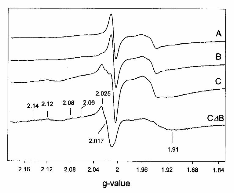
11C). A difference EPR spectrum between acetylene-treated BCR in the presence and absence of
MgATP is presented in Fig.11C∆B. This spectrum was dominated by an axial signal with all g-
values higher than 2 and with features at g=2.14, 2.12, 2.08, 2.06 and 2.017. Additionally, a
broad feature appeared in the g=1.92 region. These signals were not induced by MgATP in the
absence of acetylene. Optimal EPR conditions for the novel signals were 20 mW and 30 K, where
cluster I and II were slightly saturated. The spin concentration amounted to 0.15 mol spin/mol
enzyme. Neither the S=1/2 EPR signals of cluster I and II, nor the rise of the S=7/2 high spin
signals observed in the steady-state of benzoyl-CoA independent ATP-hydrolysis, were affected
by addition of acetylene plus MgATP.
by guest on August 15, 2019
http://ww
w.jbc.org/
Dow
nloaded from
22
DISCUSSION
1. Nature of the metal centers in BCR
The enzymatic reduction of the aromatic ring requires electron transport at very low potentials.
Therefore, the redox centers of BCR were expected to have a very special nature to catalyze such
a demanding process at pH 7 and room temperature. The most prominent example of a similar
ATP-driven redox reaction is nitrogenase, which contains unique redox centers (P-cluster, Mo-
cofactor) [21]. Surprisingly EPR and Moessbauer spectroscopy revealed that BCR contains three
‘normal’ [4Fe-4S] centers as its sole metal redox modules. The molecular composition and the
amino acid sequence is clearly different to nitrogenase, but is very similar to another enzyme
system as discussed below.
The identification of the three [4Fe-4S] clusters in BCR was based on data from Moessbauer
spectroscopy, EPR spectroscopy and metal analysis. Although the data obtained with both
spectroscopic techniques were consistent with our major conclusions, some minor differences
need to be discussed. In all Moessbauer samples the degree of overall reduction was 33 % which
would be consistent with one reduced and two oxidized [4Fe-4S] clusters. However, the results
obtained by EPR spectroscopy pointed to a different interpretation: two partially reduced isolated
clusters were identified with different EPR spectroscopic properties, and EPR signals of
magnetically interacting clusters were detected.
Since the Moessbauer spectroscopic properties were quite similar for all clusters, it was difficult
to distinguish between the presence of one reduced cluster and two half-reduced clusters.
Different conditions used for the both techniques could have influenced the spectroscopic
properties of BCR. Because of the different requirements of each technique, the EPR samples
were ten times less concentrated than the Moessbauer samples (200 mg/ml), in which the
concentration of the [4Fe-4S] clusters reached 3 mM which may have resulted in a strong redox
buffering effect that diminished the extent of their reduction. The degree of reduction was usually
lower in the Moessbauer samples (33 %) than in the EPR samples (up to 50 %). At the high
by guest on August 15, 2019
http://ww
w.jbc.org/
Dow
nloaded from
23
concentrations used for Moessbauer spectroscopy the viscosity was increased which also could
have influenced the protein conformation. The presence of both interacting and non-interacting
clusters observed by EPR indicated the presence of a microheterogeneity in the BCR samples.
The ‘interacting’ and ‘non-interacting’ forms of BCR are considered as two different
conformational states; note that binding of ATP eliminates the interaction [8]. In our enzyme
samples we usually found varying amounts of FAD (0.1-0.3 mol/mol enzyme) [4]. No correlation
between FAD content and enzyme activity has been observed, indicating that it binds
nonspecifically to the enzyme, most probably to the ATP binding sites; substoichiometric binding
of FAD may result in the observed microheterogeneity. In addition minor variations of external
factors, e.g., protein and salt concentration or viscosity, especially in combination with different
freezing procedures (EPR samples were frozen in liquid nitrogen, Moessbauer samples on dry ice)
could have resulted in microheterogeneity as has been reported for other proteins [22]. Inter-
protein magnetic interactions may also play a role at very high concentrations. Major enzyme
concentration effects have been demonstrated, e.g., for glutamine phosphoriboslypyrophosphate
amidotransferase of E. coli where the spin state of a [4Fe-4S] cluster switched from 3/2 to 5/2
when the concentration was increased to 2 mM [23].
The EPR signals observed under various conditions are summarized in Table 2. Clusters I and II
exhibit usual EPR spectroscopic properties for a [4Fe-4S]+2/+1 cluster, with the expected g-values,
although cluster I shows a rather atypical slow spin relaxation. These clusters were reducible to
some extent by both dithionite or deazaflavin/light in the absence of the substrate. However, the
spectroscopic and electrochemical properties of cluster III were more unusual. First, its reduction
was only observed under steady state conditions of substrate reduction, possibly due to a very
negative reduction potential. Second, cluster III displayed an unusual narrow spectrum expanding
over only approximately 18 mT (Fig. 6). In contrast to cluster III, the EPR spectra of clusters I
and II covered a range of more than 30 mT each (Fig. 5). Third, and most remarkably, the
spectrum displayed a split line-shape that was most likely due to the interaction with a second
paramagnetic site. Since a radical mechanism is proposed for BCR [5,6] a free radical would be
an attractive candidate as coupling partner of cluster III. Although radical signals have been
by guest on August 15, 2019
http://ww
w.jbc.org/
Dow
nloaded from
24
detected in different BCR preparations (see. e.g., spectrum A in Fig.6) these signals could not be
assigned to a protein or substrate radical, and artifacts cannot be ruled out. If a radical anion or a
free radical were formed on the substrate, its lifetime is expected to be very short since the
subsequent protonation is considered to be a faster process [24]. Therefore, pre-steady-state EPR
and ENDOR spectroscopy experiments may allow the detection of such a radical using
13C-labeled substrates.
The oxygen-sensitivity of BCR fits with the observed irreversible degradation of [4Fe-4S] to
[3Fe-4S] clusters upon exposure to air; such cluster conversions are well described and are
reversible in some cases [25]. Two observations need to be reconciled: first the high sensitivity of
BCR towards oxygen is consistent with the low reduction potentials of the [4Fe-4S] clusters
(E’° < -550 mV for cluster I and II as estimated from redox titration experiments); secondly
benzoyl-CoA reductase is able to accept electrons from reduced methyl viologen (MV) with
E’° = -448 mV [26]. The reaction rate is linear up to an MVox:MVred ratio of at least 4:1,
indicating that potentials of -400 mV are sufficiently low for an optimal electron transfer rate to
the primary electron accepting site of the enzyme. Furthermore, at –400 mV the spin
concentration of paramagnetic [4Fe-4S] clusters was less than 1 %. This may indicate that
electron transfer to the enzyme is not the rate-limiting step. Cluster I or II are suitable candidates
for the primary electron accepting site, whereas cluster III is probably involved in electron transfer
from cluster I or II to the substrate.
Acetylene stimulated benzoyl-CoA-independent ATPase activity and induced novel axial EPR
signal with all g-values higher than 2.00 (Fig. 11, Table 2). Such signals are typical for [4Fe-4S]3+
clusters of several high-potential iron-sulfur (HiPIP) proteins [27]. It is rather unlikely that
acetylene induced the oxidation of a [4Fe-4S]+1/+2 cluster to the +3 state, although the presence of
all three redox states (+3,+2,+1) has been demonstrated for the Rhodphila globiformis HiPIP
under non-denaturing conditions [27]. Although electron transfer from reduced methyl viologen
to acetylene was not observed, we cannot rule out the possibility that the enzyme reduced
acetylene to ethylene at concentrations similar to that of BCR. Both acetylene and ethylene are
by guest on August 15, 2019
http://ww
w.jbc.org/
Dow
nloaded from
25
potential ligands for metal complexes. Note, however, that addition of ethylene to BCR did not
cause such effects (not shown) but rather partially inactivated the enzyme [3]. Surprisingly, EPR
signals similar to acetylene plus MgATP-treated BCR have also been found in transient complexes
between ethylene and the nitrogenase-Mo-Fe-protein from Klebsiella pneumoniae [28]. Although
the metal clusters of the Mo-Fe protein of nitrogenase are structurally unrelated to the [4Fe-4S]
clusters of BCR it is likely that acetylene (or the putative reduced product ethylene) forms
complexes with a [4Fe-4S] cluster of BCR, altering its EPR spectroscopic properties. Since the
EPR signals of clusters I and II are not affected by acetylene treatment, cluster III is the most
probable candidate for such a complex formation.
2. Comparison with similar enzymes
Amino acid sequence comparisons of the four subunits of BCR from T. aromatica revealed that
they show highest similarities the four putative subunits of benzoyl-CoA reductase from
Rhodopseudomonas palustris [29,30] and to the 2-hydroxy-glutaryl-CoA dehydratase/activase
systems from amino acid fermenting bacteria [29,31-33]. The biochemistry of the former enzyme
has not been studied yet, whereas 2-hydroxyglutaryl-CoA dehydratases from several bacteria have
been purified and characterized [31,32]. These enzymes catalyze the mechanistically difficult
eliminiation of water from the α-position of (R)-2-hydroxyglutaryl-CoA affording (E)-glutaconyl-
CoA. A similar catalytic mechanism, involving a ketyl radical, has been proposed for both BCR
and 2-hydroxyglutaryl-CoA dehydratase [5,34]. In order to initiate enzymatic dehydration these
enzymes require a second protein component, termed activase [35]. Activase catalyzes the ATP
hydrolysis-dependent activation of electrons accepted from an external source, such as the
artificial electron donor Ti(III) citrate.
The 2-hydroxyglutaryl-CoA dehydratases from Acidaminococcus fermentans and Clostridium
symbiosum have been studied in detail [33,36]. Both comprise two different subunits (Hgd A and
Hgd B) with 38-41 % amino acid similarities to the β- and γ-subunits of BCR (Bcr B and Bcr C)
[29]. They contain two (C. symbiosus) or one (A. fermentans) [4Fe-4S] clusters and various
amounts of FMN and/or riboflavin. The biochemistry of the corresponding activase from A.
by guest on August 15, 2019
http://ww
w.jbc.org/
Dow
nloaded from
26
fermentans has been studied. It is a homodimeric protein (Hgd C) containing a single [4Fe-4S]
cluster [35]. The amino acid sequences of the α- and δ-subunit of BCR (Bcr A and Bcr D) show
high similarities to each other (45 % if the extra N-terminal loop of BCR A is disregarded) and to
Hgd C (52 %) [29]. Bcr A, Bcr D and Hgd C all contain identical phosphate and adenosine-
binding motifs similar to many sugar kinases [29,35]. Note that enzymatic ring reduction requires
stoichiometrical ATP-dependent electron activation whereas ATP hydrolysis is only required in
catalytic amounts in the dehydration process: once an activated electron is transferred from
activase to dehydratase it will stay in the activated form for several catalytic cycles [35]. BCR can
be considered as a fusion product of ATP hydrolyzing/electron activating (Bcr A and D) and ketyl
radical forming (Bcr B and C) moieties which are located on two different proteins in the
dehydratase/activase enzyme system. The total amount of three [4Fe-4S] clusters is the same in
BcrBCAD from T. aromatica and in the 2-OH-glutaryl-CoA dehydratase/activase system,
HgdABC2 from C. symbiosum [33].
Due to the similarities of the amino acid sequences of the BCR and the 2-hydroxyglutaryl-CoA
dehydratase/activase subunits, similar spectroscopic and electrochemical properties of the
[4Fe-4S] clusters were expected. Indeed, neither of the one or two [4Fe-4S] clusters of the
2-hydroxyglutaryl-CoA dehydratases from A. fermentans or C. symbiosum was reducible with
commonly used reducing agents [33,37] . The single cluster of activase, however, was reducible
to a paramagnetic S=3/2 high spin state [37]. This incomplete reduction of the whole
dehydratase/activase systems resembles the situation with BCR where only 33-50 % of the three
[4Fe-4S] clusters were reducible.
Only two cysteines are conserved in Hgd C, Bcr A and Bcr D; these are separated by a 38 amino
acid spacer. Therefore, one of the [4Fe-4S] clusters of BCR is probably ligated by two cysteines
from Bcr A and Bcr D, respectively, as has been suggested for the homodimeric activase (HgdC2)
[33]. This would be similar to the [4Fe-4S] cluster ligation of nitrogenase Fe-protein which is
buried at the interface of two identical subunits [21]. The S=3/2 high spin state of the [4Fe-4S]
cluster in both nitrogenase Fe-protein and activase, HgdC, support this assumption, although no
by guest on August 15, 2019
http://ww
w.jbc.org/
Dow
nloaded from
27
amino acid similarities between the two proteins have been found. No S=3/2 EPR signals were
detected in BCR samples, which may be a result of the differences between the composition of the
one-protein component BCR and the two-protein component systems of nitrogenase and
dehydratase/activase. However one important common feature is shared by all three enzymes: all
contain a moiety with low potential redox modules of which the electron transferring operation is
strictly coupled to ATP-hydrolysis. Since in BCR, reduction of cluster III is only observed in the
presence of MgATP and benzoyl-CoA, it is likely that ATP hydrolysis is required for electron
transport from cluster I or II to cluster III.
3. Effect of adenosine nucleotides
The ability to study electron transfer and ATPase activity separately is important because it allows
us to study the effect of ATP hydrolysis alone. Two spectroscopic effects have been reported, the
rise of S=7/2 EPR signals and the elimination of the magnetic interaction of two clusters [7]. The
Moessbauer spectroscopy described in this work has confirmed the presence of the S=7/2 species
assigned it to a [4Fe-4S] cluster which changed from the S=1/2 to the S=7/2 state. This latter
state has only been observed previously for biological iron-chalconide clusters where a bridging
sulfur of a [4Fe-4S] has artificially been replaced by selenium [38,39] and for P-clusters in the
thionine oxidized MoFe protein of nitrogenase [40]. In BCR the switch from the S=1/2 to the
S=7/2 state parallels enzymatic activity and is probably the result of structural changes in the
vicinity of a [4Fe-4S] cluster induced by ATP hydrolysis. In a [4Fe-4S]+1 cluster, all individual Fe-
atoms are in the high-spin state and are coupled antiferromagnetically to give an overall S=1/2
spin due to double- and super-exchange. Structural changes in the protein are likely to induce
changes in the double- and super-exchange interactions, and in vibronic coupling, which could
generate a S=7/2 ground state in [4Fe-4S] clusters [41]. Clearly such changes could have large
effects on the redox properties of a [4Fe-4S] cluster, although it is not possible to define a simple
relationship between spin state and reduction potential.
It is possible to distinguish between the effects of binding and hydrolysis of MgATP using the
non-hydrolyzable ATP analogue AdoPP[NH]P. Such experiments have shown that binding of the
by guest on August 15, 2019
http://ww
w.jbc.org/
Dow
nloaded from
28
nucleotide eliminates the interaction of two [4Fe-4S] clusters, whereas hydrolysis is responsible
for the switch from the S=1/2 to the S=7/2 state. We propose that the competitive inhibitor ADP
accumulates as a result of ATPase activity and binds to the enzyme which therefore remains in the
‘non-interacting’ state. This would indicate that the hydrolysis of ATP, but not its binding, is
responsible for the re-coupling of non-interacting [4Fe-4S] clusters.
ACKNOWLEDGEMENTS: We thank Dr S.J. George, and Dr V. Schünemann for useful
discussions and C. Ebenau-Jehle for the help with the cultivation of T. aromatica.
by guest on August 15, 2019
http://ww
w.jbc.org/
Dow
nloaded from
29
REFERENCES
1. Heider, J. and Fuchs, G. (1997) Eur. J. Biochem. 243, 577-596.
2. Harwood, C.S., Burchhardt, G., Herrmann, H., and Fuchs, G. (1999) FEMS Microbiol. Rev.
22: 439-458.
3. Boll, M. and Fuchs, G. (1995) Eur. J. Biochem. 234, 921-933.
4. Boll, M. and Fuchs, G. (1998) Eur. J. Biochem. 251, 946-954.
5. Buckel, W. and Keese, (1995) Angew. Chem. 107, 1595-1598.
6. Koch, J., Eisenreich, W., Bacher, A., and Fuchs, G. (1993) Eur. J. Biochem. 211, 649-661.
7. Mittelberger, T. (1996) Diploma thesis, University of Freiburg, Germany.
8. Boll, M., Albracht, S. J. P., and Fuchs, G. (1997) Eur. J. Biochem. 244, 840-851.
9. Anders, H.-J., Kaetzke, A., Kämpfer, P., Ludwig, W., and Fuchs, G. (1995) Int. J. Syst.
Bacteriol. 45, 327-333.
10. Brackmann, R. and Fuchs, G. (1993) Eur. J. Biochem. 213, 563-571.
11. Schachter, D. and Taggart, J.V. (1976) J. Biol. Chem. 203, 925-933.
12. Bradford, M. M. (1976) Anal. Biochem. 72, 248-254.
13. Laemmli, U.K. (1970) Nature 227, 680-685.
14. Zehr, B.D., Savin, T.J., and Hall, R.E. (1989) Anal. Biochem, 182, 157-159.
15. Middleton, P.; Dickson D.P.E.; Johnson, C.E., Rush, J.D. (1978) Eur.J.Biochem. 88, 135-14.
16. Abragam, A.; Pryce, M.H.L. (1951) Proc. Roy. Soc. (London) 205A, 135.
17. Thomson, C.L.; Johnson, C.E.; Dickson, D.P.E.; Cammack, R.; Hall, D.O.; Weser, U.; and
Rao, K.K. (1974), Biochem. J. 139, 97-103.
18. Rupp, H., Rao, K.K., and Cammack, R. (1978) Biochim. Biophys. Acta 537, 255-269.
19. Boll, M., Fuchs, G., Tilley, G., Armstrong, F., and Lowe, D.J. (2000) Biochemistry
20. Auric, P.; Gaillard, J.; Meyer, J.; Moulis, J.M. (1987) Biochem. J. 242, 525-530.
21. Kim J. and Rees, D.C. (1994) Biochemistry 33, 389-97.
22. Dunham, W.R., Hagen, W.R., Fee, J.A., Sands, R.H., Dunbar, J.B., and Humblet, C. (1991)
Biochim. Biophys. Acta 1079, 253-262.
23. Onate, Y.A., Vollmer, S.J., Switzer, R.L., and Johnson, M.K. (1989) J. Biol. Chem. 264,
18386-18391.
by guest on August 15, 2019
http://ww
w.jbc.org/
Dow
nloaded from
30
24. Krapcho, A.P. and Bothnerby, A.A. (1959) J. Amer. Chem. Soc. 81, 3658-3666.
25. Beinert, H., Holm, R.H., and Münck, E. (1997) Science 277, 653-659.
26. Fraisse, L. and Simon, H. (1988) Arch. Microbiol. 150, 381-386.
27. Heering, H.A., Bulsink, Y.B., Hagen, W.R., and Meyer, T.E. (1995) Eur. J. Biochem. 232,
811-817.
28. Lowe, D.J., Eady, R.R., and Thorneley, R.N.F. (1978) Biochem. J. 173, 277-290.
29. Breese, K., Boll, M., Alt-Mörbe, J., Schägger, H., and Fuchs, G. (1998) Eur. J. Biochem.
256, 148-154.
30. Egland, P.G., Pelletier, D.A., Dispensa, M., Gibson, J., Harwood, C.S. (1997) Proc. Natl.
Acad. Sci. 94, 6484-6489.
31. Dutscho, R., Wohlfarth, G., Buckel, P., and Buckel, W. (1989) Eur. J. Biochem. 181, 741-
746.
32. Hans, M., Sievers, J., Müller, U., Bill, E., Vorholt, J.A., Linder, D., and Buckel, W. (1999)
Eur. J. Biochem. 265, 404-414.
33. Bendrat, K. Muller, U., Klees, A.H., and Buckel, W. (1993) FEBS Lett. 329, 329-331.
34. Buckel, W. (1996) FEBS Lett. 389, 20-24.
35. Müller, U. and Buckel, W. (1996) Eur. J. Biochem. 230, 698-704.
36. Schweiger, G., Dutscho, R. and Buckel, W. (1987) Eur. J. Biochem. 169, 441-448.
37. Buckel, W., University of Marburg, Germany; personal communication.
38. Moulis, J.-M., Auric, P., Gaillard, J., and Meyer, J. (1984) J. Biol. Chem. 259, 11396-11402.
39. Jung, Y.-S., Vassiliew, I.R., and Golbeck, J.H. (1997) J. Biol. Inorg. Chem. 2, 209-217.
40. Hagen, W.R., Wassink, H., Eady, R.R., Smith, B.E., and Haaker, H. (1987) Eur. J. Biochem.
169, 457-465.
41. Noodleman, L. (1991) Inorg. Chem. 30, 246-256.
by guest on August 15, 2019
http://ww
w.jbc.org/
Dow
nloaded from
31
LEGENDS TO FIGURES
Fig. 1. Reaction catalyzed by benzoyl-CoA reductase.
Fig. 2. Mössbauer spectra of 57Fe-enriched benzoyl-CoA reductase oxidized by thionine. A.
Spectrum taken at 4.2 K in an applied field of 20 mT ⊥ γ, B. spectrum taken at 4.2 K in an
applied field of 7 T || γ. The solid lines are the sum of subspectra 1,2,3 (see Table 1).
Fig. 3. Mössbauer spectra of 57Fe-enriched benzoyl-CoA reductase photoreduced by
deazaflavin. A. Spectrum taken at 120 K in an applied field of 20 mT ⊥ γ, B. spectrum taken at
4.2 K in an applied field of 20 mT ⊥ γ, C. spectrum taken at 4.2 K in an applied field of 7T || γ.
The solid lines are the sum of subspectra 1,2,3,4 (see Table 1).
Fig. 4. EPR spectra of deazaflavin/light reduced benzoyl-CoA reductase. () unlabeled
enzyme, (……) 57Fe-labeled enzyme. EPR parameters: microwave frequency: 9.414 GHz,
modulation amplitude, 1.17 mT, microwave power 2 mW; temperature, 25 K.
Fig. 5. Temperature-dependent EPR spectra of deazaflavin/light-reduced benzoyl-CoA
reductase. The enzyme (120 µM) was reduced anaerobically with 20 µM deazaflavin in 100 mM
Tris/HCl, pH 7.5 for 10 min in white light. A. Representative spectra recorded from 60 K to 15
K. EPR parameters: microwave frequency, 9.415 GHz; modulation amplitude, 1.17 mT;
microwave power, 2 mW for 60 K to 25 K spectra and 0.2 mW for 20 K and 15 K spectra. Note
that the dominant features of the EPR spectrum recorded at 60 K are assigned to cluster I in this
work. 20 K-60 K is the difference spectrum of the 20 K spectrum minus the 60 K spectrum and is
assigned to cluster II. The spectra were normalized for temperature and microwave power. The
numbers in the figure refer to g-values. B. Total S=1/2 spin concentration of the spectra recorded
at 15 K to 77 K. All spin concentrations were corrected for temperature-dependence according to
Curie’s Law .
by guest on August 15, 2019
http://ww
w.jbc.org/
Dow
nloaded from
32
Fig. 6. EPR spectra of dithionite-reduced benzoyl-CoA reductase in the presence of
MgATP (20 mM) and benzoyl-CoA (3 mM). A. Spectrum recorded at 77 K, B. Spectrum
recorded at 40 K, C. 40 K spectrum of dithionite-reduced enzyme in the absence of MgATP and
benzoyl-CoA. B∆∆C. Difference spectrum B minus 0.2 times spectrum C. EPR parameters:
microwave frequency, 9.415 GHz; microwave power, 2 mW; modulation amplitude: 1.27 mT.
Fig. 7. EPR spectra of benzoyl-CoA reductase at different redox potentials. The potentials
were poised by adding ferricyanide to a dithionite-reduced benzoyl-CoA reductase preparation in
the presence of a redox mediator mixture. A. Spectra recorded at 4 K, B. Spectra recorded at 25
K. All spectra were recorded under non-saturating conditions. The numbers in B are g-values.
EPR-parameters: microwave frequency, 9.415-9.416 GHz; microwave power: 0.2 mW in A. and
2 mW in B; modulation amplitude 1.17 mT.
Fig. 8. Mössbauer spectra of 57Fe-enriched BCR reduced by dithionite in the presence of
MgATP. a: spectrum taken at 120 K in an applied field of 20 mT ⊥ γ. b: spectrum taken at 4.2 K
in an applied field of 20 mT ⊥ γ. c: spectrum taken at 4.2 K in an applied field of 7T || γ. The
solid lines are the sum of subspectra 1,2,3,4,5,6 (see Table 1).
Fig. 9. EPR spectra of dithionite-reduced benzoyl-CoA reductase ( 180 µM) in the presence
of MgATP and MgAdoPP[NH]P. 4 K EPR spectra of A. reduced enzyme as isolated, B.
reduced enzyme after 1 min incubation with 20 mM MgATP, C. reduced enzyme after 1 min
incubation with 0.5 mM MgAdoPP[NH]P. Arrow 1 points to the ATP-induced isotropic EPR-
signal at g=5.1; arrow 2 to the broad shoulder in the spectrum of dithionite reduced enzyme in the
absence of ATP or AdoPP[NH]P. EPR parameters: microwave frequency, 9.414 GHz;
microwave power 0.2 mW; modulation amplitude, 1.17 mT.
Fig. 10. EPR spectra of air-inactivated benzoyl-CoA reductase (250 µM). A. Full scan
spectrum recorded at 4 K after 5 min incubation of BCR at air, B. Re-reduction of the enzyme in
A. with excess of dithionite (10 mM), C. EPR spectrum of B. using a parallel mode cavity. D.
by guest on August 15, 2019
http://ww
w.jbc.org/
Dow
nloaded from
33
g=2 region of spectrum A. E. g=2 region of spectrum B. EPR parameters: microwave frequency,
9.409 GHz for spectrum A,B,D,E and 9.250 GHz for spectrum C.; microwave power and
temperature, 2 mW and 4 K in A,C, and D, 0.2 mW and 4 K in B, 2 mW and 26 K in E;
modulation amplitude, 1.17 mT.
Fig. 11. Effect of acetylene on the EPR spectra of dithionite-reduced benzoyl-CoA
reductase. Dithionite reduced benzoyl-CoA reductase was anaerobically incubated with acetylene
(~4 mM) in the presence and absence of MgATP (20 mM) for 10 min at 20°C. Note that the EPR
conditions are optimized for the acetylene-induced signal, under these conditions the usual S=1/2
EPR signals of the [4Fe-4S] clusters are saturated. A. Dithionite reduced enzyme as isolated. B.
Reduced enzyme in the presence of acetylene. C. Reduced enzyme in the presence of acetylene
and MgATP. EPR parameters: microwave frequency, 9.354 GHz; microwave power, 20 mW;
modulation amplitude, 1.17 mT; temperature, 30 K.
by guest on August 15, 2019
http://ww
w.jbc.org/
Dow
nloaded from
34
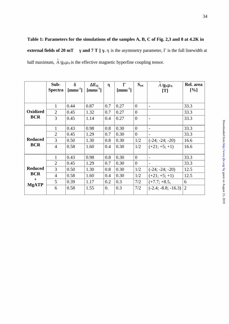
Table 1: Parameters for the simulations of the samples A, B, C of Fig. 2,3 and 8 at 4.2K in
external fields of 20 mT ⊥⊥ γγ and 7 T || γγ. η is the asymmetry parameter, Γ is the full linewidth at
half maximum, τA /gNµN is the effective magnetic hyperfine coupling tensor.
Sub-Spectra
δδ[mms-1]
∆∆EQ
[mms-1]ηη ΓΓ
[mms-1]Stot
τA /gNµµN
[T]Rel. area
[%]
1 0.44 0.87 0.7 0.27 0 - 33.32 0.45 1.32 0.7 0.27 0 33.3Oxidized
BCR 3 0.45 1.14 0.4 0.27 0 - 33.3
1 0.43 0.98 0.8 0.30 0 - 33.32 0.45 1.29 0.7 0.30 0 - 33.33 0.50 1.30 0.8 0.30 1/2 (-24; -24; -20) 16.6Reduced
BCR 4 0.58 1.60 0.4 0.30 1/2 (+21; +5; +1) 16.6
1 0.43 0.98 0.8 0.30 0 - 33.32 0.45 1.29 0.7 0.30 0 - 33.33 0.50 1.30 0.8 0.30 1/2 (-24; -24; -20) 12.54 0.58 1.60 0.4 0.30 1/2 (+21; +5; +1) 12.55 0.39 1.17 0.2 0.3 7/2 (+7.7; +8.5, 6
ReducedBCR
+MgATP
6 0.58 1.55 0. 0.3 7/2 (-2.4; -8.8; -16.3) 2
by guest on August 15, 2019
http://ww
w.jbc.org/
Dow
nloaded from
35
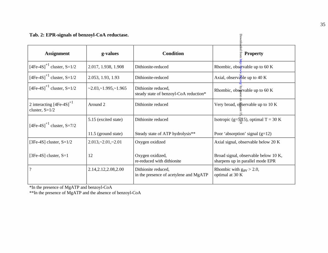
Tab. 2: EPR-signals of benzoyl-CoA reductase.
Assignment g-values Condition Property
[4Fe-4S]+1 cluster, S=1/2 2.017, 1.938, 1.908 Dithionite-reduced Rhombic, observable up to 60 K
[4Fe-4S]+1 cluster, S=1/2 2.053, 1.93, 1.93 Dithionite-reduced Axial, observable up to 40 K
[4Fe-4S]+1 cluster, S=1/2 ~2.03,~1.995,~1.965 Dithionite reduced,steady state of benzoyl-CoA reduction*
Rhombic, observable up to 60 K
2 interacting [4Fe-4S]+1
cluster, S=1/2Around 2 Dithionite reduced Very broad, observable up to 10 K
[4Fe-4S]+1 cluster, S=7/25.15 (excited state) Dithionite reduced Isotropic (g=5.15), optimal T = 30 K
11.5 (ground state) Steady state of ATP hydrolysis** Poor ‘absorption’ signal (g=12)
[3Fe-4S] cluster, S=1/2 2.013,~2.01,~2.01 Oxygen oxidized Axial signal, observable below 20 K
[3Fe-4S] cluster, S=1 12 Oxygen oxidized,re-reduced with dithionite
Broad signal, observable below 10 K,sharpens up in parallel mode EPR
? 2.14,2.12,2.08,2.00 Dithionite reduced,in the presence of acetylene and MgATP
Rhombic with gav > 2.0,optimal at 30 K
*In the presence of MgATP and benzoyl-CoA**In the presence of MgATP and the absence of benzoyl-CoA
by guest on August 15, 2019
http://ww
w.jbc.org/
Dow
nloaded from
Matthias Boll, Georg Fuchs, Christian Meier, Alfred Trautwein and David J. LoweEPR- and Moessbauer Studies of Benzoyl-CoA Reductase
published online July 19, 2000J. Biol. Chem.
10.1074/jbc.M001508200Access the most updated version of this article at doi:
Alerts:
When a correction for this article is posted•
When this article is cited•
to choose from all of JBC's e-mail alertsClick here
by guest on August 15, 2019
http://ww
w.jbc.org/
Dow
nloaded from