Embed Size (px)
Citation preview
C O N T I N U I N G E D U C A T I O N
Hybrid PET/MR Imaging of the Heart: Potential, InitialExperiences, and Future Prospects
Christoph Rischpler, Stephan G. Nekolla, Isabel Dregely, and Markus Schwaiger
Nuklearmedizinische Klinik und Poliklinik, Technische Universität München, Munich, Germany
Learning Objectives: On successful completion of this activity, participants should be able to describe (1) advantages and shortcomings of hybrid PET/MRscanners with respect to cardiovascular applications (e.g. myocardial perfusion imaging and viability imaging), (2) additional value of the MR component incardiac imaging, and (3) technical challenges and workflow considerations regarding hybrid PET/MR scanners in the field of cardiology (e.g. attenuationcorrection and cardiac/respiratory/patient motion).
Financial Disclosure: Dr. Schwaiger is an investigator, meeting participant, and lecturer for Siemens Medical. The authors of this article have indicated noother relevant relationships that could be perceived as a real or apparent conflict of interest.
CME Credit: SNMMI is accredited by the Accreditation Council for Continuing Medical Education (ACCME) to sponsor continuing education for physicians.SNMMI designates each JNM continuing education article for a maximum of 2.0 AMA PRA Category 1 Credits. Physicians should claim only creditcommensurate with the extent of their participation in the activity. For CE credit, participants can access this activity through the SNMMI Web site (http://www.snmmi.org/ce_online) through March 2016.
PET/CT and other combined scanners have in the past decaderapidly emerged as important research tools and are proving tobe invaluable for improved diagnostics in routine nuclearmedicine. The design of hybrid PET/MR scanners presenteda formidable technical challenge, and only recently were theseinstruments introduced to the market. Initial expectations of theperformance of these scanners have been high, notablybecause of the potential for superior tissue contrast inherentin the MR modality, as well as the potential for multiparametricfunctional imaging in conjunction with PET. However, theadditional value and potential clinical role that these newsystems might bring to the cardiac field have yet to bedocumented. This review presents a comparative summary ofthe existing applications for PET and MR in the field ofcardiology and suggests potential cardiac applications exploit-ing unique properties of the newly introduced combined in-strumentation.
Key Words: PET; MR; hybrid imaging; cardiology
J Nucl Med 2013; 54:402–415DOI: 10.2967/jnumed.112.105353
The diverse range of imaging modalities for cardiologyincludes echocardiography, CT, MR imaging, SPECT, andPET, each of which offers distinct properties and advantages.The demand for combined PET/CT instrumentation in clin-ical nuclear medicine has grown remarkably, because of thenotable advantages presented by hybrid imaging with respect
to anatomic localization of lesions. Although these advan-tages were initially driven by the demands of oncology im-aging, the resultant broader availability of PET/CT hasprovided the opportunity to use these scanners for indica-tions beyond oncology, such as in the field of cardiac imag-ing (1). Furthermore, CT images are necessary for rapidattenuation correction, which is of particular importancefor quantification in myocardial perfusion imaging. Throughthe use of suitable CT components and modification of im-aging protocols, we can now visualize coronary anatomy byCT angiography, in conjunction with functional and meta-bolic imaging of the heart by PET, which together provideenhanced diagnostic information for the clinician (2). Draw-ing on this experience with PET/CT, we present in this re-view technical aspects of the design and performance of thenewly available PET/MR scanners and summarize the exist-ing comparative studies of PET and MR imaging in cardiol-ogy, as the basis for predicting the impact of hybrid PET/MRscanners in diagnostics and therapeutics for cardiology.
STRENGTHS ANDWEAKNESSES OF MR VERSUS CT ASAN ADJUNCT TO PET
The major advantages of MR over CT are the higher soft-tissue contrast and the lack of radiation exposure to thepatient. Morphologic information; attenuation correctionmaps—essential for regional quantification of PET-basedphysiologic parameters; and all other information is acquiredwithout ionizing radiation by MR. Different approaches toreduce the radiation dose from CTwere undertaken; nonethe-less, CT-based attenuation correction maps still add, for ex-ample, about 0.8 mSv to a cardiac stress–rest scan (3).
At the same time, the electromagnetic fields necessaryfor MR imaging represent a major disadvantage, especiallyin cardiac imaging. A substantial number of patients cannotbe imaged using MR because of the presence of pace-makers, implantable cardioverter defibrillators, or mechanical
Received Nov. 26, 2012; revision accepted Feb. 1, 2013.For correspondence or reprints contact: Markus Schwaiger, Department of
Nuclear Medicine, Technische Universität München, Klinikum rechts der Isar,Ismaninger Strasse 22, 81675 Munich, Germany.E-mail: [email protected] online Feb. 12, 2013.COPYRIGHT ª 2013 by the Society of Nuclear Medicine and Molecular
Imaging, Inc.
402 THE JOURNAL OF NUCLEAR MEDICINE • Vol. 54 • No. 3 • March 2013
heart valves. In these patients, PET/CT proves to be aninvaluable alternative for noninvasive cardiac imaging.Despite the disadvantage of ionizing radiation, however,
CT has become the most commonly used technique fornoninvasive coronary angiography. It is fast and easy toperform. Also, the use of high-pitched prospectivelyelectrocardiogram-triggered coronary CT angiography withlow voltage has reduced the radiation dose to about 1.3mSv (4). Although there has been considerable progress inMR angiography since the initial published reports, MRacquisition remains for the present a technical challengedue to artifacts arising from cardiac, respiratory, and patientmotion and the need for high spatial resolution while retain-ing high-vessel-to-tissue contrast, and high signal-to-noise-ratio, all within a reasonable scanning time.For the assessment of left ventricular (LV) function, MR
imaging represents the gold standard and has been exten-sively validated. MR shows great accuracy in the de-termination of functional parameters such as LV volumes,mass, ejection fraction, regional wall motion, and myocar-dial thickening (5). Although CT-based assessment of LVfunction correlates well with MR, radiation exposure to thepatient still represents a major issue (6).Another important point that needs to be addressed is the
application of contrast medium. Most patients tolerate MRcontrast agents well, and acute adverse reactions seldomoccur. Furthermore, a rare condition called nephrogenicsystemic fibrosis occurs almost exclusively in patients withacute renal failure or severe chronic kidney disease.Iodinated CT contrast agents may lead to a condition calledcontrast-induced nephropathy, which is a leading cause ofiatrogenic acute kidney failure. Furthermore, severe ad-verse reactions occur more frequently after the applicationof CT contrast medium. Notably, MR perfusion imagingcan also be accomplished without the use of contrastagents, by determining the T1 for both myocardium andblood, a technique called arterial spin-labeled MR imaging(7). Even though this technique proved to have the potentialto assess myocardial perfusion without any contrast me-dium, an advantage that might be especially valuable inpatients with end-stage renal disease or when serial meas-urements are needed, it is far from routine clinical practice(8). Although initial studies for CT myocardial perfusionshowed promising results, 64-slice multidetector CT is—according to a recent study—not feasible for detectingischemia (9).Although molecular information is hard to assess by CT,
as images are based simply on differences in x-rayattenuation, MR offers promising features of molecularimaging, such as spectroscopy. Spectroscopy allows thenoninvasive investigation of myocardial metabolism andcomposition. Using 1H-MR spectroscopy for the regionalquantification of creatine content, viable myocardium canbe distinguished from nonviable (scarred) tissue (10). Thistechnique also allows measurement of the triglyceride con-tent of myocardium, and interestingly, high myocardial
lipid content was linked to low septal wall thickness andhigh LV mass (11). Furthermore, 23Na MR spectroscopy isa promising application for myocardial viability assessmentand the detection of myocardial ischemia (12). Anotherinteresting feature of MR is diffusion spectrum MR imag-ing tractography. This imaging modality was first imple-mented for brain research but also yields great potentialas a research tool for cardiology. This method allows im-aging of the architecture of the organization of myocardialfibers with exceptional resolution (13). Using this tech-nique, Sosnovik et al. revealed the presence of meshlikenetworks of orthogonal myofibers within the infarcted ratmyocardium (14).
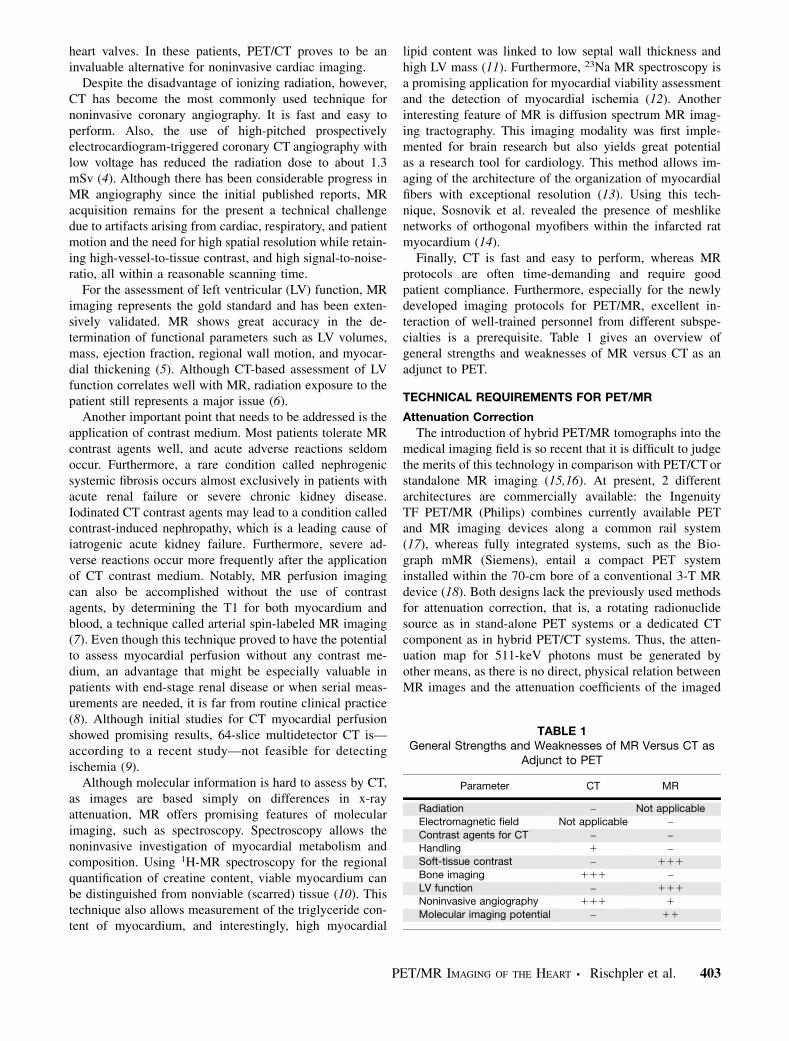
Finally, CT is fast and easy to perform, whereas MRprotocols are often time-demanding and require goodpatient compliance. Furthermore, especially for the newlydeveloped imaging protocols for PET/MR, excellent in-teraction of well-trained personnel from different subspe-cialties is a prerequisite. Table 1 gives an overview ofgeneral strengths and weaknesses of MR versus CT as anadjunct to PET.
TECHNICAL REQUIREMENTS FOR PET/MR
Attenuation Correction
The introduction of hybrid PET/MR tomographs into themedical imaging field is so recent that it is difficult to judgethe merits of this technology in comparison with PET/CT orstandalone MR imaging (15,16). At present, 2 differentarchitectures are commercially available: the IngenuityTF PET/MR (Philips) combines currently available PETand MR imaging devices along a common rail system(17), whereas fully integrated systems, such as the Bio-graph mMR (Siemens), entail a compact PET systeminstalled within the 70-cm bore of a conventional 3-T MRdevice (18). Both designs lack the previously used methodsfor attenuation correction, that is, a rotating radionuclidesource as in stand-alone PET systems or a dedicated CTcomponent as in hybrid PET/CT systems. Thus, the atten-uation map for 511-keV photons must be generated byother means, as there is no direct, physical relation betweenMR images and the attenuation coefficients of the imaged
TABLE 1General Strengths and Weaknesses of MR Versus CT as
Adjunct to PET
Parameter CT MR
Radiation – Not applicable
Electromagnetic field Not applicable –
Contrast agents for CT – –
Handling 1 –
Soft-tissue contrast – 111Bone imaging 111 –
LV function – 111Noninvasive angiography 111 1Molecular imaging potential – 11
PET/MR IMAGING OF THE HEART • Rischpler et al. 403
tissue. However, PET quantification is impossible withouta m-map. In particular, a map containing the attenuationcoefficients for 511-keV photons at each voxel within theentire imaged volume is required. The calculation of thisattenuation map presents a major challenge for PET/MR(19) in general, and imperfections in the m-map can sub-stantially degrade cardiac PET results, as is known fromcardiac PET/CT (20).Several MR approaches for attenuation correction are
currently used, based on segmentation, templates or atlases,or the PET emission data (Table 2). For segmentation-basedapproaches, the attenuation map is segmented into differenttissue classes with a fixed attenuation coefficient per tissueclass, as first proposed by Huang et al. in 1981 (21). Build-ing on this well-established concept, our group proposed touse segmented MR-based attenuation correction for whole-body PET/MR imaging (22). Using water-weighted and fat-weighted images computed from a Dixon MR sequence(23), we classify each voxel as air, lungs, fat, or soft tissue.This scan requires typically 18 s per bed position, measuredduring a single breath-hold. Cortical bone is almost impos-sible to segment from these data and thus, in analogy tosegmented whole-body data from PET-only devices, is ig-nored. In an alternate approach, T1-weighted turbo spin-echo sequences are used for attenuation correction. Boneis also ignored by this approach, but this sequence allowsa shorter imaging time at the expense of not differentiatingbetween fat (m 5 0.086 cm21) and soft tissue (m 5 0.096cm21) (24). The limitations of segmentation-based tech-niques arise from the fixed number of tissue classes thatcan be discriminated and, in the context of heart imaging,from the use of a fixed attenuation coefficient for lung tissue,which is known to vary between patients and locations in the
lung. In addition, ignoring bone obviously results in an un-derestimation of tissue attenuation, especially near the skel-eton. Tumor uptake in PET/MR has differed from that inPET/CT in the range of 5%–15% (22,24) or up to 23%(25). Furthermore, metal implants result in a void of MRsignal and do not contribute to the attenuation map. In ad-dition, the effect of MR contrast agents can be problematicbecause of effects on segmentation of fat, lung, and softtissue arising from reduced T1 values (26). Despite theseconsiderations, the segmentation approach is rather stableand computationally fast and has been implemented in com-mercially available PET/MR systems. The Biograph mMRuses a modification of this approach (22), and our group hasshown a good correlation between PET/CT and PET/MRin oncologic studies (27). The sequential Ingenuity TFPET/MR likewise uses a version of this approach (24).
Template-based approaches match a model to thepatient’s anatomy such that the known attenuation map ofthe model is modified to match the target. In the particularcase of head imaging, a rigid model will suffice, whereasfor whole-body applications, the need for an elastic regis-tration presents an obstacle to implementation for routineuse. In addition, atlas-based methods might fail if anatomicabnormalities are present. A recent implementation of acombined template and segmentation approach has beenshown to be feasible in whole-body imaging (28).
In emission-based approaches to attenuation correction,the recorded PET data themselves might provide the infor-mation needed to calculate an attenuation map. However,the effect of photon scatter limits the applicability of thisapproach (29). Still, it bears consideration that informationderived from emission data was well suited for motion cor-rection in cardiac imaging (20), and Nuyts et al. suggested
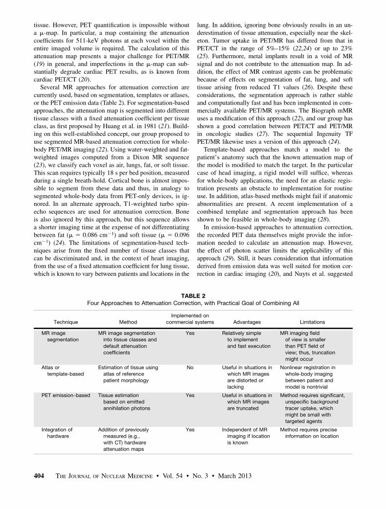
TABLE 2Four Approaches to Attenuation Correction, with Practical Goal of Combining All
Technique Method
Implemented on
commercial systems Advantages Limitations
MR imagesegmentation
MR image segmentationinto tissue classes and
default attenuation
coefficients
Yes Relatively simpleto implement
and fast execution
MR imaging fieldof view is smaller
than PET field of
view; thus, truncation
might occur
Atlas or
template-based
Estimation of tissue using
atlas of referencepatient morphology
No Useful in situations in
which MR imagesare distorted or
lacking
Nonlinear registration in
whole-body imagingbetween patient and
model is nontrivial
PET emission–based Tissue estimationbased on emitted
annihilation photons
Yes Useful in situations inwhich MR images
are truncated
Method requires significant,unspecific background
tracer uptake, which
might be small with
targeted agents
Integration of
hardware
Addition of previously
measured (e.g.,with CT) hardware
attenuation maps
Yes Independent of MR
imaging if locationis known
Method requires precise
information on location
404 THE JOURNAL OF NUCLEAR MEDICINE • Vol. 54 • No. 3 • March 2013
using this approach to recover truncated parts of an attenu-ation map (30).All techniques present their own advantages and dis-
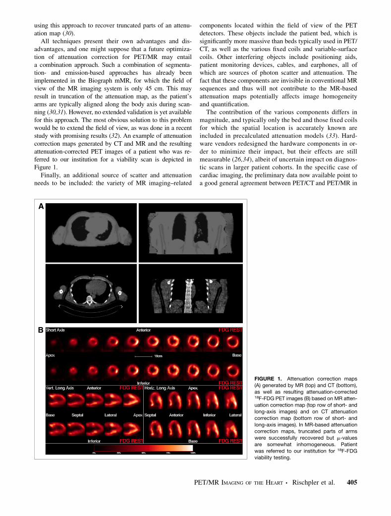
advantages, and one might suppose that a future optimiza-tion of attenuation correction for PET/MR may entaila combination approach. Such a combination of segmenta-tion- and emission-based approaches has already beenimplemented in the Biograph mMR, for which the field ofview of the MR imaging system is only 45 cm. This mayresult in truncation of the attenuation map, as the patient’sarms are typically aligned along the body axis during scan-ning (30,31). However, no extended validation is yet availablefor this approach. The most obvious solution to this problemwould be to extend the field of view, as was done in a recentstudy with promising results (32). An example of attenuationcorrection maps generated by CT and MR and the resultingattenuation-corrected PET images of a patient who was re-ferred to our institution for a viability scan is depicted inFigure 1.Finally, an additional source of scatter and attenuation
needs to be included: the variety of MR imaging–related
components located within the field of view of the PETdetectors. These objects include the patient bed, which issignificantly more massive than beds typically used in PET/CT, as well as the various fixed coils and variable-surfacecoils. Other interfering objects include positioning aids,patient monitoring devices, cables, and earphones, all ofwhich are sources of photon scatter and attenuation. Thefact that these components are invisible in conventional MRsequences and thus will not contribute to the MR-basedattenuation maps potentially affects image homogeneityand quantification.
The contribution of the various components differs inmagnitude, and typically only the bed and those fixed coilsfor which the spatial location is accurately known areincluded in precalculated attenuation models (33). Hard-ware vendors redesigned the hardware components in or-der to minimize their impact, but their effects are stillmeasurable (26,34), albeit of uncertain impact on diagnos-tic scans in larger patient cohorts. In the specific case ofcardiac imaging, the preliminary data now available point toa good general agreement between PET/CT and PET/MR in
FIGURE 1. Attenuation correction maps(A) generated by MR (top) and CT (bottom),
as well as resulting attenuation-corrected18F-FDG PET images (B) based on MR atten-
uation correction map (top row of short- andlong-axis images) and on CT attenuation
correction map (bottom row of short- and
long-axis images). In MR-based attenuationcorrection maps, truncated parts of arms
were successfully recovered but m-values
are somewhat inhomogeneous. Patient
was referred to our institution for 18F-FDGviability testing.
PET/MR IMAGING OF THE HEART • Rischpler et al. 405
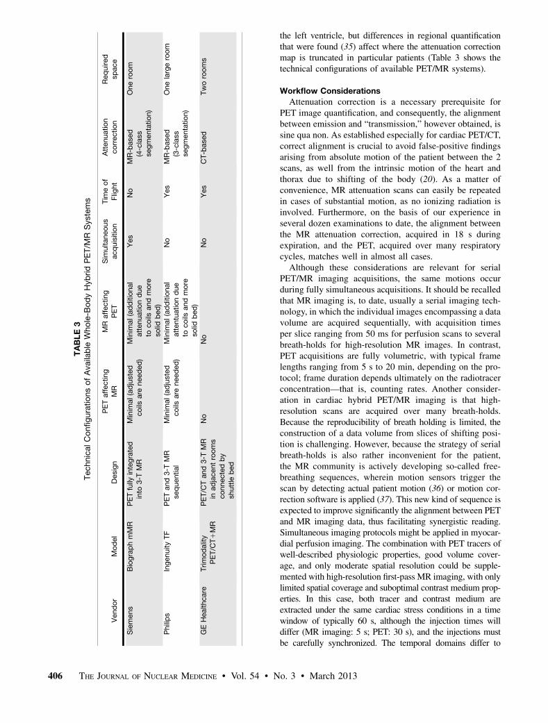
the left ventricle, but differences in regional quantificationthat were found (35) affect where the attenuation correctionmap is truncated in particular patients (Table 3 shows thetechnical configurations of available PET/MR systems).
Workflow Considerations
Attenuation correction is a necessary prerequisite forPET image quantification, and consequently, the alignmentbetween emission and “transmission,” however obtained, issine qua non. As established especially for cardiac PET/CT,correct alignment is crucial to avoid false-positive findingsarising from absolute motion of the patient between the 2scans, as well from the intrinsic motion of the heart andthorax due to shifting of the body (20). As a matter ofconvenience, MR attenuation scans can easily be repeatedin cases of substantial motion, as no ionizing radiation isinvolved. Furthermore, on the basis of our experience inseveral dozen examinations to date, the alignment betweenthe MR attenuation correction, acquired in 18 s duringexpiration, and the PET, acquired over many respiratorycycles, matches well in almost all cases.
Although these considerations are relevant for serialPET/MR imaging acquisitions, the same motions occurduring fully simultaneous acquisitions. It should be recalledthat MR imaging is, to date, usually a serial imaging tech-nology, in which the individual images encompassing a datavolume are acquired sequentially, with acquisition timesper slice ranging from 50 ms for perfusion scans to severalbreath-holds for high-resolution MR images. In contrast,PET acquisitions are fully volumetric, with typical framelengths ranging from 5 s to 20 min, depending on the pro-tocol; frame duration depends ultimately on the radiotracerconcentration—that is, counting rates. Another consider-ation in cardiac hybrid PET/MR imaging is that high-resolution scans are acquired over many breath-holds.Because the reproducibility of breath holding is limited, theconstruction of a data volume from slices of shifting posi-tion is challenging. However, because the strategy of serialbreath-holds is also rather inconvenient for the patient,the MR community is actively developing so-called free-breathing sequences, wherein motion sensors trigger thescan by detecting actual patient motion (36) or motion cor-rection software is applied (37). This new kind of sequence isexpected to improve significantly the alignment between PETand MR imaging data, thus facilitating synergistic reading.Simultaneous imaging protocols might be applied in myocar-dial perfusion imaging. The combination with PET tracers ofwell-described physiologic properties, good volume cover-age, and only moderate spatial resolution could be supple-mented with high-resolution first-pass MR imaging, with onlylimited spatial coverage and suboptimal contrast medium prop-erties. In this case, both tracer and contrast medium areextracted under the same cardiac stress conditions in a timewindow of typically 60 s, although the injection times willdiffer (MR imaging: 5 s; PET: 30 s), and the injections mustbe carefully synchronized. The temporal domains differ to
TABLE3
TechnicalConfigurationsofAvailable
Whole-B
odyHybridPET/M
RSystems
Vendor
Model
Design
PETaffecting
MR
MR
affecting
PET
Sim
ultaneous
acquisition
Tim
eof
Flig
ht
Attenuation
correction
Required
space
Siemens
BiographmMR
PETfully
integrated
into
3-T
MR
Minim
al(adjusted
coils
are
needed)
Minim
al(additional
attenuationdue
tocoils
andmore
solid
bed)
Yes
No
MR-based
(4-class
segmentation)
Oneroom
Philips
IngenuityTF
PETand3-T
MR
sequential
Minim
al(adjusted
coils
are
needed)
Minim
al(additional
attentuationdue
tocoils
andmore
solid
bed)
No
Yes
MR-based
(3-class
segmentation)
Onelargeroom
GEHealthcare
Trimodality
PET/C
T1MR
PET/C
Tand3-T
MR
inadjacentrooms
connectedby
shuttle
bed
No
No
No
Yes
CT-based
Tworooms
406 THE JOURNAL OF NUCLEAR MEDICINE • Vol. 54 • No. 3 • March 2013
a greater extent, and the gating logic is inherently different(prospective vs. retrospective). Whereas high-resolution scanssuch as cine MR imaging and late gadolinium enhancement(LGE) are acquired in less than 5 min, PET recordings usedfor an electrocardiogram-gated reconstruction will take up to20 min. Thus, in patients with greater heart rate variability, thecontractile information potentially differs between modalities.A technical solution synchronizing the contraction for an op-timized display is therefore desirable.In summary, truly simultaneous PET/MR protocols pres-
ent formidable technical difficulties, and perfectly matchingframes are not yet automatically achievable. Consequently,misalignment arising from patient motion is an issue requir-ing close attention. However, in hybrid PET/MR scanners,real-time MR-based motion correction and partial-volumecorrection may eventually become available.From the perspective of workflow, parallel PET/MR im-
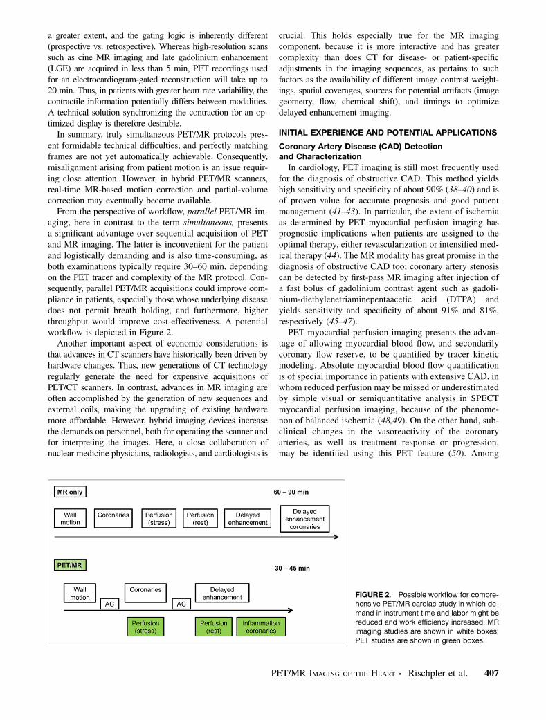
aging, here in contrast to the term simultaneous, presentsa significant advantage over sequential acquisition of PETand MR imaging. The latter is inconvenient for the patientand logistically demanding and is also time-consuming, asboth examinations typically require 30–60 min, dependingon the PET tracer and complexity of the MR protocol. Con-sequently, parallel PET/MR acquisitions could improve com-pliance in patients, especially those whose underlying diseasedoes not permit breath holding, and furthermore, higherthroughput would improve cost-effectiveness. A potentialworkflow is depicted in Figure 2.Another important aspect of economic considerations is
that advances in CT scanners have historically been driven byhardware changes. Thus, new generations of CT technologyregularly generate the need for expensive acquisitions ofPET/CT scanners. In contrast, advances in MR imaging areoften accomplished by the generation of new sequences andexternal coils, making the upgrading of existing hardwaremore affordable. However, hybrid imaging devices increasethe demands on personnel, both for operating the scanner andfor interpreting the images. Here, a close collaboration ofnuclear medicine physicians, radiologists, and cardiologists is
crucial. This holds especially true for the MR imagingcomponent, because it is more interactive and has greatercomplexity than does CT for disease- or patient-specificadjustments in the imaging sequences, as pertains to suchfactors as the availability of different image contrast weight-ings, spatial coverages, sources for potential artifacts (imagegeometry, flow, chemical shift), and timings to optimizedelayed-enhancement imaging.
INITIAL EXPERIENCE AND POTENTIAL APPLICATIONS
Coronary Artery Disease (CAD) Detectionand Characterization
In cardiology, PET imaging is still most frequently usedfor the diagnosis of obstructive CAD. This method yieldshigh sensitivity and specificity of about 90% (38–40) and isof proven value for accurate prognosis and good patientmanagement (41–43). In particular, the extent of ischemiaas determined by PET myocardial perfusion imaging hasprognostic implications when patients are assigned to theoptimal therapy, either revascularization or intensified med-ical therapy (44). The MR modality has great promise in thediagnosis of obstructive CAD too; coronary artery stenosiscan be detected by first-pass MR imaging after injection ofa fast bolus of gadolinium contrast agent such as gadoli-nium-diethylenetriaminepentaacetic acid (DTPA) andyields sensitivity and specificity of about 91% and 81%,respectively (45–47).
PET myocardial perfusion imaging presents the advan-tage of allowing myocardial blood flow, and secondarilycoronary flow reserve, to be quantified by tracer kineticmodeling. Absolute myocardial blood flow quantificationis of special importance in patients with extensive CAD, inwhom reduced perfusion may be missed or underestimatedby simple visual or semiquantitative analysis in SPECTmyocardial perfusion imaging, because of the phenome-non of balanced ischemia (48,49). On the other hand, sub-clinical changes in the vasoreactivity of the coronaryarteries, as well as treatment response or progression,may be identified using this PET feature (50). Among
FIGURE 2. Possible workflow for compre-hensive PET/MR cardiac study in which de-
mand in instrument time and labor might be
reduced and work efficiency increased. MRimaging studies are shown in white boxes;
PET studies are shown in green boxes.
PET/MR IMAGING OF THE HEART • Rischpler et al. 407
the available PET tracers for myocardial perfusion are13N-ammonia (13NH3), 15O-water, 82Rb, and 18F-flurpiridaz,with the last of these currently being evaluated in clinicalphase 3 trials (51).Although the quantification of myocardial perfusion by
PET is well validated and is in routine use at many centers,the usefulness of cardiac MR imaging for flow quantifica-tion is still under investigation. In one of the few publishedreports, Schwitter et al. demonstrated that MR imaging canquantify myocardial perfusion deficits in patients with CAD(52). A flow index using a pixelwise upslope parameter ofthe signal intensity curve during gadolinium-DTPA transitwas calculated. Using this index, sectors defined as pa-thologic by MR showed a linear correlation with 13NH3
PET-defined pathologic sectors (slope 5 0.94, r 5 0.76,P , 0.0001). Subsequently, Ibrahim et al. determined forMR imaging a pathologic stress–rest index threshold, whichwas low compared with validated PET thresholds (1.3 vs.2.5) (53).However, both modalities hold several shortcomings: the
underestimation of the MR-based stress–rest index whencompared with the 13NH3 PET results is one of severalhurdles to be overcome before cardiac MR can find routineuse. Other relevant issues of cardiac MR include the pos-sibility of image-derived assessment of the arterial inputfunction, the rather low distribution volume of gadoli-nium-DTPA, the rapid diffusion of gadolinium-DTPA intothe extracellular space (which poses difficulties for kineticmodeling), and the limited spatial coverage of the left ven-tricle, which is imaged in 3–5 slices. One of the shortcom-ings of PET is that no morphologic information but onlyinformation on perfusion abnormalities is gained. Conse-quently, it is not possible to differentiate between a reducedperfusion that is due to epicardial coronary stenosis and onethat is due to microvascular dysfunction. Furthermore, a re-duced perfusion—being relative or quantitative—is not al-ways indicative of myocardial scarring, especially inpatients with dilated cardiomyopathy (54).
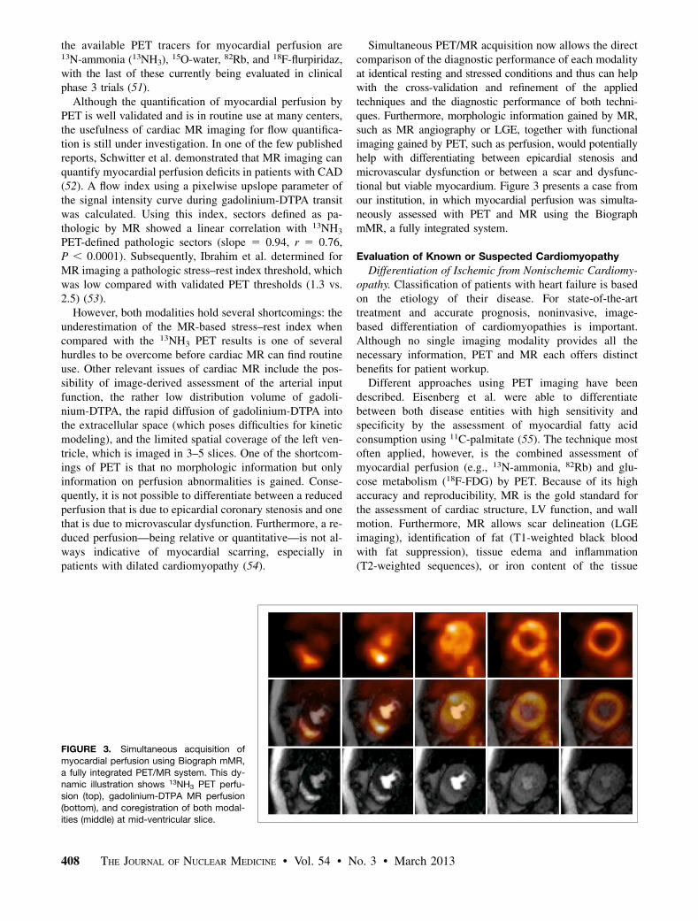
Simultaneous PET/MR acquisition now allows the directcomparison of the diagnostic performance of each modalityat identical resting and stressed conditions and thus can helpwith the cross-validation and refinement of the appliedtechniques and the diagnostic performance of both techni-ques. Furthermore, morphologic information gained by MR,such as MR angiography or LGE, together with functionalimaging gained by PET, such as perfusion, would potentiallyhelp with differentiating between epicardial stenosis andmicrovascular dysfunction or between a scar and dysfunc-tional but viable myocardium. Figure 3 presents a case fromour institution, in which myocardial perfusion was simulta-neously assessed with PET and MR using the BiographmMR, a fully integrated system.
Evaluation of Known or Suspected Cardiomyopathy
Differentiation of Ischemic from Nonischemic Cardiomy-opathy. Classification of patients with heart failure is basedon the etiology of their disease. For state-of-the-arttreatment and accurate prognosis, noninvasive, image-based differentiation of cardiomyopathies is important.Although no single imaging modality provides all thenecessary information, PET and MR each offers distinctbenefits for patient workup.
Different approaches using PET imaging have beendescribed. Eisenberg et al. were able to differentiatebetween both disease entities with high sensitivity andspecificity by the assessment of myocardial fatty acidconsumption using 11C-palmitate (55). The technique mostoften applied, however, is the combined assessment ofmyocardial perfusion (e.g., 13N-ammonia, 82Rb) and glu-cose metabolism (18F-FDG) by PET. Because of its highaccuracy and reproducibility, MR is the gold standard forthe assessment of cardiac structure, LV function, and wallmotion. Furthermore, MR allows scar delineation (LGEimaging), identification of fat (T1-weighted black bloodwith fat suppression), tissue edema and inflammation(T2-weighted sequences), or iron content of the tissue
FIGURE 3. Simultaneous acquisition of
myocardial perfusion using Biograph mMR,
a fully integrated PET/MR system. This dy-namic illustration shows 13NH3 PET perfu-
sion (top), gadolinium-DTPA MR perfusion
(bottom), and coregistration of both modal-
ities (middle) at mid-ventricular slice.
408 THE JOURNAL OF NUCLEAR MEDICINE • Vol. 54 • No. 3 • March 2013
(T2* sequence), all of which are crucial for appropriate di-agnosis. Although in ischemic cardiomyopathy fibrotic tissueis located in the subendocardium and follows the typical vas-cular distribution, in nonischemic cardiomyopathy no scartissue, or primarily subepicardial fibrotic tissue, which doesnot follow the coronary artery distribution, is observed. Also,stress cardiac MR can be performed to assess for CAD.Consequently, simultaneous assessment of the extent and
exact localization of scar tissue by MR, as well as tissueperfusion and glucose metabolism by PET, improves theidentification of the underlying disease.Assessment of Myocardial Viability. The basic principle
underlying myocardial viability imaging by PET is thathypoxia and ischemia lead to a shift in myocardial metab-olism from oxidation of free fatty acids toward glucoseutilization. Myocardium that is chronically hypoperfused orexposed to repetitive stunning, which brings about a stateknown as hibernation, shows chronic dysfunction and isknown to shift toward glucose metabolism. The upregulatedglucose utilization in ischemically compromised tissue hasbeen linked to poor outcome (56), and the extent of the mis-matched area has therapeutic significance (57–59). Viable butdysfunctional myocardium has the potential to recover afterrevascularization (60). Among the different imaging modali-ties available to assess myocardial viability, 18F-FDG PET isconsidered to be the gold standard (61,62). The results ofseveral studies have proven the value of 18F-FDG PET imag-ing in identifying those patients who would benefit from re-vascularization, based on a prediction of improved LVfunction (63–65).An important feature of PET lies in its potential to
quantify tracer uptake, thus allowing differentiation be-tween fully viable, partially viable, and nonviable myocar-dium, within the constraints imposed by spatial resolution.However, MR using the late gadolinium enhancement tech-nique (LGE) is a valuable alternative to 18F-FDG PET formyocardial viability imaging. The application of inversion-re-covery prepared T1-weighted gradient-echo pulse sequences5–20 min after administration of gadolinium-DTPA suppressessignal from remote myocardium and depicts scarred myocar-dium as enhanced areas in comparison to the surroundingnormal and viable myocardium (66,67). The inherently highin-plane resolution of MR allows the observer to distinguishbetween transmural and nontransmural enhancement, and evenvery small areas of subendocardial infarction can be detected.The presence of even a small amount of enhanced myocardiumcarries prognostic significance in patients with suspected CADbut without known prior myocardial infarction (68). A meta-analysis investigated the capabilities of imaging to predictfunctional recovery after revascularization; specificity for18F-FDG PET and delayed-enhancement MR imaging wascomparable (63%), whereas the nuclear medicine techniquehad higher sensitivity (92% vs. 84%) (61).However, it is important to consider the fundamental
differences between these imaging modalities; the in-creased extracellular space in scar tissue determines the
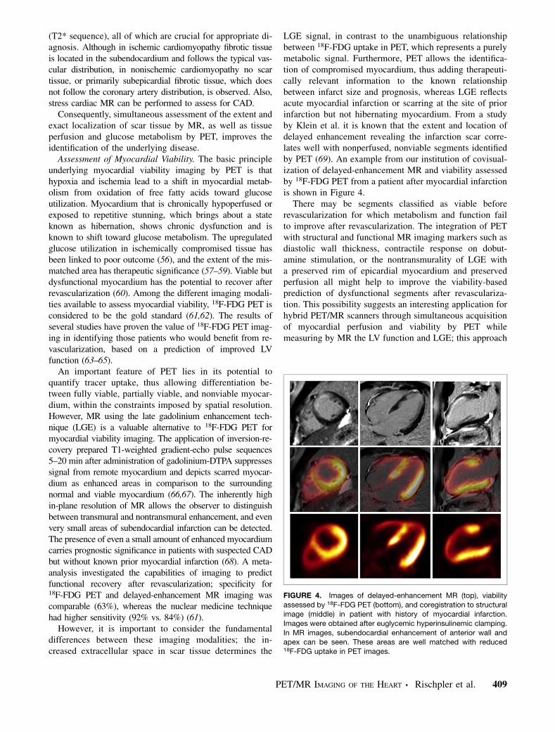
LGE signal, in contrast to the unambiguous relationshipbetween 18F-FDG uptake in PET, which represents a purelymetabolic signal. Furthermore, PET allows the identifica-tion of compromised myocardium, thus adding therapeuti-cally relevant information to the known relationshipbetween infarct size and prognosis, whereas LGE reflectsacute myocardial infarction or scarring at the site of priorinfarction but not hibernating myocardium. From a studyby Klein et al. it is known that the extent and location ofdelayed enhancement revealing the infarction scar corre-lates well with nonperfused, nonviable segments identifiedby PET (69). An example from our institution of covisual-ization of delayed-enhancement MR and viability assessedby 18F-FDG PET from a patient after myocardial infarctionis shown in Figure 4.
There may be segments classified as viable beforerevascularization for which metabolism and function failto improve after revascularization. The integration of PETwith structural and functional MR imaging markers such asdiastolic wall thickness, contractile response on dobut-amine stimulation, or the nontransmurality of LGE witha preserved rim of epicardial myocardium and preservedperfusion all might help to improve the viability-basedprediction of dysfunctional segments after revasculariza-tion. This possibility suggests an interesting application forhybrid PET/MR scanners through simultaneous acquisitionof myocardial perfusion and viability by PET whilemeasuring by MR the LV function and LGE; this approach
FIGURE 4. Images of delayed-enhancement MR (top), viability
assessed by 18F-FDG PET (bottom), and coregistration to structuralimage (middle) in patient with history of myocardial infarction.
Images were obtained after euglycemic hyperinsulinemic clamping.
In MR images, subendocardial enhancement of anterior wall andapex can be seen. These areas are well matched with reduced18F-FDG uptake in PET images.
PET/MR IMAGING OF THE HEART • Rischpler et al. 409
should yield new insights and may allow more reliableprediction of functional recovery after intervention (70,71).Inflammatory and Infiltrative Cardiomyopathies (Myo-
carditis and Sarcoidosis). Myocarditis is an inflammatorydisease arising from various infections (primarily viral butalso from bacteria, fungi, protozoa, or helminths), autoim-mune processes, hypersensitivity reactions, and treatmentwith cardiotoxic drugs. Although invasive endomyocardialbiopsy is the gold standard for the diagnosis of myocarditis,MR imaging is increasingly used for this purpose. The firstcase using this technique was published in 1991 by Gagliardiet al. (72). Since then, different protocols have been estab-lished, based on MR measures such as wall motion abnor-malities, pericardial effusion, increased wall thickness, andchanges in LV mass, which together represent the range offunctional and morphologic abnormalities of the heart thatcan be depicted by MR imaging. However, MR imaging isalso suited to image other pathologic abnormalities of thetissue in myocarditis, such as edema (T2-weighted imaging),hyperemia or capillary leakage (early gadolinium-enhancedT1-weighted imaging), and necrosis or fibrosis (late gadoli-nium-enhanced T1-weighted imaging). In a meta analysisincluding 194 patients, Friedrich et al. reported a diagnosticaccuracy of 78% for MR-based imaging if at least 2 of theaforementioned tissue-based criteria were positive (73).Even though the literature on myocarditis in PET
imaging is scarce, a few potential applications should meritconsideration. An increased number of apoptotic cells havebeen described in the heart of patients with myocarditis(74). Consequently, molecular imaging of apoptosis shouldbe valuable for the diagnosis of myocarditis and potentiallyfor risk stratification and prognosis. In a rat model of lipo-polysaccharide-induced myocardial apoptosis, an observedincrease in heart uptake of 123I-annexin V proved to bea caspase-dependent phenomenon (75). Annexin V repre-sents a specific ligand for externalization of phosphatidyl-serine cells undergoing apoptotic changes (76). Caspasesare also implicated in apoptosis, and caspase-3 has recentlybeen labeled with 18F (77). In a murine model of hepaticapoptosis, highly specific liver uptake of this tracer wasshown (78). Further experiments determining the useful-ness in cardiovascular applications of 18F-labeled caspase-3tracers seem justified.By the hybrid PET/MR approach, imaging not only
might confirm the diagnosis of myocarditis but mightalso—especially the PET part—be useful for assessing dis-ease activity and thus guiding therapy.Cardiac sarcoidosis represents another inflammatory
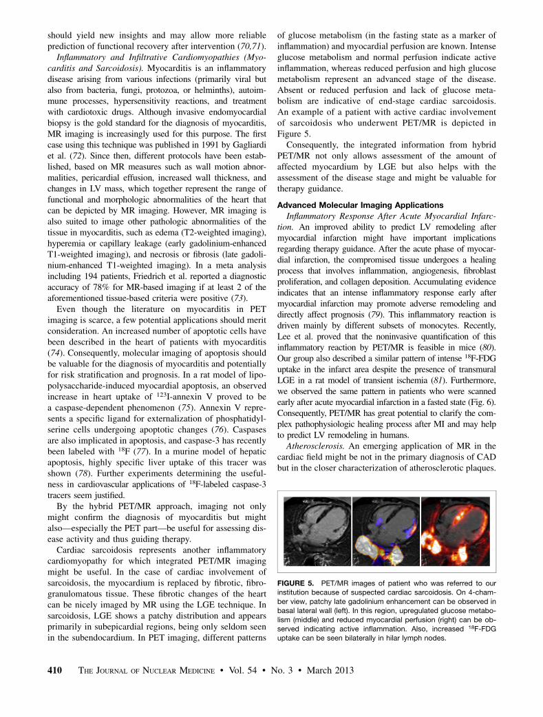
cardiomyopathy for which integrated PET/MR imagingmight be useful. In the case of cardiac involvement ofsarcoidosis, the myocardium is replaced by fibrotic, fibro-granulomatous tissue. These fibrotic changes of the heartcan be nicely imaged by MR using the LGE technique. Insarcoidosis, LGE shows a patchy distribution and appearsprimarily in subepicardial regions, being only seldom seenin the subendocardium. In PET imaging, different patterns
of glucose metabolism (in the fasting state as a marker ofinflammation) and myocardial perfusion are known. Intenseglucose metabolism and normal perfusion indicate activeinflammation, whereas reduced perfusion and high glucosemetabolism represent an advanced stage of the disease.Absent or reduced perfusion and lack of glucose meta-bolism are indicative of end-stage cardiac sarcoidosis.An example of a patient with active cardiac involvementof sarcoidosis who underwent PET/MR is depicted inFigure 5.
Consequently, the integrated information from hybridPET/MR not only allows assessment of the amount ofaffected myocardium by LGE but also helps with theassessment of the disease stage and might be valuable fortherapy guidance.
Advanced Molecular Imaging Applications
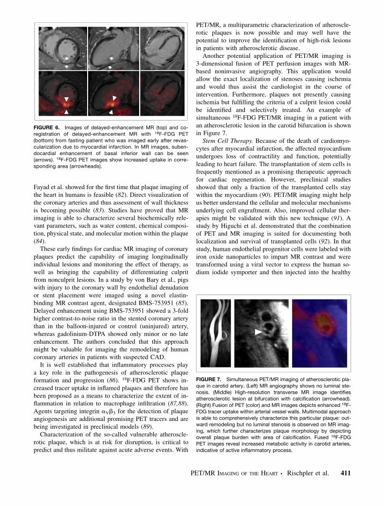
Inflammatory Response After Acute Myocardial Infarc-tion. An improved ability to predict LV remodeling aftermyocardial infarction might have important implicationsregarding therapy guidance. After the acute phase of myocar-dial infarction, the compromised tissue undergoes a healingprocess that involves inflammation, angiogenesis, fibroblastproliferation, and collagen deposition. Accumulating evidenceindicates that an intense inflammatory response early aftermyocardial infarction may promote adverse remodeling anddirectly affect prognosis (79). This inflammatory reaction isdriven mainly by different subsets of monocytes. Recently,Lee et al. proved that the noninvasive quantification of thisinflammatory reaction by PET/MR is feasible in mice (80).Our group also described a similar pattern of intense 18F-FDGuptake in the infarct area despite the presence of transmuralLGE in a rat model of transient ischemia (81). Furthermore,we observed the same pattern in patients who were scannedearly after acute myocardial infarction in a fasted state (Fig. 6).Consequently, PET/MR has great potential to clarify the com-plex pathophysiologic healing process after MI and may helpto predict LV remodeling in humans.
Atherosclerosis. An emerging application of MR in thecardiac field might be not in the primary diagnosis of CADbut in the closer characterization of atherosclerotic plaques.
FIGURE 5. PET/MR images of patient who was referred to ourinstitution because of suspected cardiac sarcoidosis. On 4-cham-
ber view, patchy late gadolinium enhancement can be observed in
basal lateral wall (left). In this region, upregulated glucose metabo-
lism (middle) and reduced myocardial perfusion (right) can be ob-served indicating active inflammation. Also, increased 18F-FDG
uptake can be seen bilaterally in hilar lymph nodes.
410 THE JOURNAL OF NUCLEAR MEDICINE • Vol. 54 • No. 3 • March 2013
Fayad et al. showed for the first time that plaque imaging ofthe heart in humans is feasible (82). Direct visualization ofthe coronary arteries and thus assessment of wall thicknessis becoming possible (83). Studies have proved that MRimaging is able to characterize several biochemically rele-vant parameters, such as water content, chemical composi-tion, physical state, and molecular motion within the plaque(84).These early findings for cardiac MR imaging of coronary
plaques predict the capability of imaging longitudinallyindividual lesions and monitoring the effect of therapy, aswell as bringing the capability of differentiating culpritfrom nonculprit lesions. In a study by von Bary et al., pigswith injury to the coronary wall by endothelial denudationor stent placement were imaged using a novel elastin-binding MR contrast agent, designated BMS-753951 (85).Delayed enhancement using BMS-753951 showed a 3-foldhigher contrast-to-noise ratio in the stented coronary arterythan in the balloon-injured or control (uninjured) artery,whereas gadolinium-DTPA showed only minor or no lateenhancement. The authors concluded that this approachmight be valuable for imaging the remodeling of humancoronary arteries in patients with suspected CAD.It is well established that inflammatory processes play
a key role in the pathogenesis of atherosclerotic plaqueformation and progression (86). 18F-FDG PET shows in-creased tracer uptake in inflamed plaques and therefore hasbeen proposed as a means to characterize the extent of in-flammation in relation to macrophage infiltration (87,88).Agents targeting integrin aVb3 for the detection of plaqueangiogenesis are additional promising PET tracers and arebeing investigated in preclinical models (89).Characterization of the so-called vulnerable atheroscle-
rotic plaque, which is at risk for disruption, is critical topredict and thus militate against acute adverse events. With
PET/MR, a multiparametric characterization of atheroscle-rotic plaques is now possible and may well have thepotential to improve the identification of high-risk lesionsin patients with atherosclerotic disease.
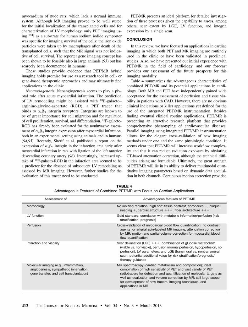
Another potential application of PET/MR imaging is3-dimensional fusion of PET perfusion images with MR-based noninvasive angiography. This application wouldallow the exact localization of stenoses causing ischemiaand would thus assist the cardiologist in the course ofintervention. Furthermore, plaques not presently causingischemia but fulfilling the criteria of a culprit lesion couldbe identified and selectively treated. An example ofsimultaneous 18F-FDG PET/MR imaging in a patient withan atherosclerotic lesion in the carotid bifurcation is shownin Figure 7.
Stem Cell Therapy. Because of the death of cardiomyo-cytes after myocardial infarction, the affected myocardiumundergoes loss of contractility and function, potentiallyleading to heart failure. The transplantation of stem cells isfrequently mentioned as a promising therapeutic approachfor cardiac regeneration. However, preclinical studiesshowed that only a fraction of the transplanted cells staywithin the myocardium (90). PET/MR imaging might helpus better understand the cellular and molecular mechanismsunderlying cell engraftment. Also, improved cellular ther-apies might be validated with this new technique (91). Astudy by Higuchi et al. demonstrated that the combinationof PET and MR imaging is suited for documenting bothlocalization and survival of transplanted cells (92). In thatstudy, human endothelial progenitor cells were labeled withiron oxide nanoparticles to impart MR contrast and weretransformed using a viral vector to express the human so-dium iodide symporter and then injected into the healthy
FIGURE 6. Images of delayed-enhancement MR (top) and co-
registration of delayed-enhancement MR with 18F-FDG PET
(bottom) from fasting patient who was imaged early after revas-cularization due to myocardial infarction. In MR images, suben-
docardial enhancement of basal inferior wall can be seen
(arrows). 18F-FDG PET images show increased uptake in corre-
sponding area (arrowheads).
FIGURE 7. Simultaneous PET/MR imaging of atherosclerotic pla-
que in carotid artery. (Left) MR angiography shows no luminal ste-
nosis. (Middle) High-resolution transverse MR image identifiesatherosclerotic lesion at bifurcation with calcification (arrowhead).
(Right) Fusion of PET (color) and MR images depicts enhanced 18F-
FDG tracer uptake within arterial vessel walls. Multimodal approach
is able to comprehensively characterize this particular plaque: out-ward remodeling but no luminal stenosis is observed on MR imag-
ing, which further characterizes plaque morphology by depicting
overall plaque burden with area of calcification. Fused 18F-FDGPET images reveal increased metabolic activity in carotid arteries,
indicative of active inflammatory process.
PET/MR IMAGING OF THE HEART • Rischpler et al. 411
myocardium of nude rats, which lack a normal immunesystem. Although MR imaging proved to be well suitedfor the initial localization of the transplanted cells and forcharacterization of LV morphology, only PET imaging us-ing 124I as a substrate for human sodium iodide symporterwas specific for imaging survival of the cells; the iron oxideparticles were taken up by macrophages after death of thetransplanted cells, such that the MR signal was not indica-tive of cell survival. The reporter gene imaging concept hasbeen shown to be feasible also in large animals (93) but hasscarcely been documented in humans.These studies provide evidence that PET/MR hybrid
imaging holds promise for use as a research tool in cell- orgene-based therapeutic approaches and may ultimately findapplications in the clinic.Neoangiogenesis. Neoangiogenesis seems to play a piv-
otal role after acute myocardial infarction. The predictionof LV remodeling might be assisted with 18F-galacto–arginine-glycine-aspartate (RGD), a PET tracer thatbinds to anb3 integrin receptors. Integrins are known tobe of great importance for cell migration and for regulationof cell proliferation, survival, and differentiation. 18F-galacto-RGD has already been evaluated for the noninvasive assess-ment of anb3 integrin expression after myocardial infarction,both in an experimental setting using animals and in humans(94,95). Recently, Sherif et al. published a report on theexpression of anb3 integrin in the infarction area early aftermyocardial infarction in rats with ligation of the left anteriordescending coronary artery (96). Interestingly, increased up-take of 18F-galacto-RGD in the infarction area seemed to bea predictor for the absence of subsequent LV remodeling asassessed by MR imaging. However, further studies for theevaluation of this tracer need to be conducted.
PET/MR presents an ideal platform for detailed investiga-tion of these processes given the capability to assess, amongothers, scar extent by LGE, LV function, and integrinexpression by a single scan.
CONCLUSION
In this review, we have focused on applications in cardiacimaging in which both PET and MR imaging are routinelyused in the clinic or have been validated in preclinicalstudies. Also, we have presented our initial experience withPET/MR in the field of cardiology, and our forecastprovides our assessment of the future prospects for thisimaging modality.
Table 4 summarizes the advantageous characteristics ofcombined PET/MR and its potential applications in cardi-ology. Both MR and PET have independently gained wideacceptance for the assessment of perfusion and tissue via-bility in patients with CAD. However, there are no obviousclinical indications or killer applications yet defined for theuse of the integrated PET/MR instrumentation. Besidesfinding eventual clinical routine applications, PET/MR ispresenting an attractive research platform that providescomprehensive phenotyping of cardiovascular diseases.Parallel imaging using integrated PET/MR instrumentationallows for the elegant cross-validation of new imagingmethods under one and the same physiologic condition. Itseems clear that PET/MR will increase workflow complex-ity and that it can reduce radiation exposure by obviatingCT-based attenuation correction, although the technical diffi-culties arising are formidable. Ultimately, the great strengthof PET/MR will lie in its ability to deliver multimodal quan-titative imaging parameters based on dynamic data acquisi-tion in both channels. Continuous motion correction provided
TABLE 4Advantageous Features of Combined PET/MR with Focus on Cardiac Applications
Assessment of. . . Advantageous features of PET/MR
Morphology No ionizing radiation, high soft-tissue contrast, coronaries 1, plaque
imaging 1, cardiac structure 111, fiber architecture 111
LV function Gold standard; correlation with metabolic information/perfusion (riskstratification, prognosis)
Perfusion Cross-validation of myocardial blood flow quantification; no contrast
agents for arterial spin-labeled MR imaging; attenuation correctionby MR; motion and partial-volume correction for myocardial blood
flow quantification
Infarction and viability Scar delineation (LGE) 111; combination of glucose metabolism
(viable vs. nonviable), perfusion (normal perfusion, hypoperfusion, no
perfusion), LV parameters, and LGE (transmural vs. nontransmural
scar); potential additional value for risk stratification/prognosis/therapy guidance
Molecular imaging (e.g., inflammation,
angiogenesis, sympathetic innervation,gene transfer, and cell transplantation)
MR spectroscopy (cardiac metabolism and composition); ideal
combination of high sensitivity of PET and vast variety of PETradiotracers for detection and quantification of molecular targets as
well as localization and volume correction by MR; still large scope
for development of new tracers, imaging techniques, andapplications in MR
412 THE JOURNAL OF NUCLEAR MEDICINE • Vol. 54 • No. 3 • March 2013
by MR imaging may improve substantially the quantificationof regional PET tracer uptake. Access to coregistered, almostsimultaneous physiologic and biologic measurements havealready made PET/MR the most sophisticated quantitativeimaging modality in cardiology, although these applicationsare in their infancy.The financial aspects of MR versus CT as an adjunct to
PET need to be considered. PET/MR is entering routineclinical practice at present, but it is still not clear whetherPET/MR will be able to offer added value or generate anadditional demand for imaging studies that PET/CT cannotsatisfy. Thus, whether the higher acquisition costs for PET/MR will pay off in the long-term still needs to be proven.However, we expect that the combination of molecularimaging with superb functional characterization of cardiacperformance will help to stage heart failure and to developpredictive parameters for tissue recovery and response totherapy. The results of such sophisticated research appli-cations will define new clinical indications, which need tobe validated as superior and cost-effective replacements forexisting imaging approaches in cardiology.
ACKNOWLEDGMENTS
This review has been prepared with the help of manystaff members, but we would like to thank especiallyShelley Zhang and Sebastian Fürst for valuable input, Syl-via Schachoff and Brigitte Dzewas for their technical assis-tance during PET/MR acquisitions, and Dr. Paul Cummingfor professional textual revisions.
REFERENCES
1. Schwaiger M, Ziegler S, Nekolla SG. PET/CT: challenge for nuclear cardiology.
J Nucl Med. 2005;46:1664–1678.
2. Kajander S, Ukkonen H, Sipila H, Teras M, Knuuti J. Low radiation dose im-
aging of myocardial perfusion and coronary angiography with a hybrid PET/CT
scanner. Clin Physiol Funct Imaging. 2009;29:81–88.
3. Koepfli P, Hany TF, Wyss CA, et al. CT attenuation correction for myocardial
perfusion quantification using a PET/CT hybrid scanner. J Nucl Med.
2004;45:537–542.
4. Fink C, Krissak R, Henzler T, et al. Radiation dose at coronary CT angiography:
second-generation dual-source CT versus single-source 64-MDCT and first-gen-
eration dual-source CT. AJR. 2011;196:W550–W557.
5. Bellenger NG, Davies LC, Francis JM, Coats AJ, Pennell DJ. Reduction in
sample size for studies of remodeling in heart failure by the use of cardiovascular
magnetic resonance. J Cardiovasc Magn Reson. 2000;2:271–278.
6. Greupner J, Zimmermann E, Grohmann A, et al. Head-to-head comparison of
left ventricular function assessment with 64-row computed tomography, biplane
left cineventriculography, and both 2- and 3-dimensional transthoracic echocar-
diography: comparison with magnetic resonance imaging as the reference stan-
dard. J Am Coll Cardiol. 2012;59:1897–1907.
7. Zhang H, Shea SM, Park V, et al. Accurate myocardial T1 measurements: toward
quantification of myocardial blood flow with arterial spin labeling. Magn Reson
Med. 2005;53:1135–1142.
8. Zun Z, Varadarajan P, Pai RG, Wong EC, Nayak KS. Arterial spin labeled CMR
detects clinically relevant increase in myocardial blood flow with vasodilation.
JACC Cardiovasc Imaging. 2011;4:1253–1261.
9. Spiro AJ, Haramati LB, Jain VR, Godelman A, Travin MI, Levsky JM. Resting
cardiac 64-MDCT does not reliably detect myocardial ischemia identified by
radionuclide imaging. AJR. 2013;200:337–342.
10. Bottomley PA, Weiss RG. Non-invasive magnetic-resonance detection of crea-
tine depletion in non-viable infarcted myocardium. Lancet. 1998;351:714–718.
11. Szczepaniak LS, Dobbins RL, Metzger GJ, et al. Myocardial triglycerides and
systolic function in humans: in vivo evaluation by localized proton spectroscopy
and cardiac imaging. Magn Reson Med. 2003;49:417–423.
12. Jansen MA, Van Emous JG, Nederhoff MG, Van Echteld CJ. Assessment of
myocardial viability by intracellular 23Na magnetic resonance imaging. Circu-
lation. 2004;110:3457–3464.
13. Wedeen VJ, Hagmann P, Tseng WY, Reese TG, Weisskoff RM. Mapping com-
plex tissue architecture with diffusion spectrum magnetic resonance imaging.
Magn Reson Med. 2005;54:1377–1386.
14. Sosnovik DE, Wang R, Dai G, et al. Diffusion spectrum MR imaging tractog-
raphy reveals the presence of a complex network of residual myofibers in in-
farcted myocardium. Circ Cardiovasc Imaging. 2009;2:206–212.
15. Antoch G, Bockisch A. Combined PET/MR imaging: a new dimension in whole-
body oncology imaging? Eur J Nucl Med Mol Imaging. 2009;36(suppl 1):S113–
S120.
16. Nekolla SG, Martinez-Moeller A, Saraste A. PET and MR imaging in cardiac
imaging: from validation studies to integrated applications. Eur J Nucl Med Mol
Imaging. 2009;36(suppl 1):S121–S130.
17. Zaidi H, Ojha N, Morich M, et al. Design and performance evaluation of
a whole-body Ingenuity TF PET-MR imaging system. Phys Med Biol.
2011;56:3091–3106.
18. Delso G, Furst S, Jakoby B, et al. Performance measurements of the Siemens
mMR integrated whole-body PET/MR scanner. J Nucl Med. 2011;52:1914–
1922.
19. Hofmann M, Pichler B, Scholkopf B, Beyer T. Towards quantitative PET/MR
imaging: a review of MR-based attenuation correction techniques. Eur J Nucl
Med Mol Imaging. 2009;36(suppl 1):S93–S104.
20. Martinez-Möller A, Souvatzoglou M, Navab N, Schwaiger M, Nekolla SG.
Artifacts from misaligned CT in cardiac perfusion PET/CT studies: frequency,
effects, and potential solutions. J Nucl Med. 2007;48:188–193.
21. Huang SC, Carson RE, Phelps ME, Hoffman EJ, Schelbert HR, Kuhl DE. A
boundary method for attenuation correction in positron computed tomography. J
Nucl Med. 1981;22:627–637.
22. Martinez-Möller A, Souvatzoglou M, Delso G, et al. Tissue classification as
a potential approach for attenuation correction in whole-body PET/MR imaging:
evaluation with PET/CT data. J Nucl Med. 2009;50:520–526.
23. Coombs BD, Szumowski J, Coshow W. Two-point Dixon technique for water-fat
signal decomposition with B0 inhomogeneity correction. Magn Reson Med.
1997;38:884–889.
24. Schulz V, Torres-Espallardo I, Renisch S, et al. Automatic, three-segment, MR-
based attenuation correction for whole-body PET/MR data. Eur J Nucl Med Mol
Imaging. 2011;38:138–152.
25. Samarin A, Burger C, Wollenweber SD, et al. PET/MR imaging of bone lesions:
implications for PET quantification from imperfect attenuation correction. Eur J
Nucl Med Mol Imaging. 2012;39:1154–1160.
26. Fürst S, Souvatzoglu M, Rischpler C, Ziegler S, Schwaiger M, Nekolla S. Effects
of MR contrast agents on attenuation map generation and cardiac PET quanti-
fication in PET/MR [abstract]. J Nucl Med. 2012;53(suppl 1):43P.
27. Drzezga A, Souvatzoglou M, Eiber M, et al. First clinical experience with in-
tegrated whole-body PET/MR: comparison to PET/CT in patients with oncologic
diagnoses. J Nucl Med. 2012;53:845–855.
28. Hofmann M, Bezrukov I, Mantlik F, et al. MR imaging-based attenuation cor-
rection for whole-body PET/MR imaging: quantitative evaluation of segmenta-
tion- and atlas-based methods. J Nucl Med. 2011;52:1392–1399.
29. Nuyts J, Dupont P, Stroobants S, Bennick R, Mortelmans L, Suetens P. Simultaneous
maximum a posteriori reconstruction of attenuation and activity distributions from
emission sinograms. IEEE Trans Med Imaging. 1999;18(5):393–403.
30. Nuyts J, Bal G, Kehren F, Fenchel M, Michel C, Watson C. Completion of
a truncated attenuation image from the attenuated PET emission data. IEEE
Trans Med Imaging. September 21, 2012 [Epub ahead of print].
31. Delso G, Martinez-Moller A, Bundschuh RA, Nekolla SG, Ziegler SI. The effect
of limited MR field of view in MR/PET attenuation correction. Med Phys.
2010;37:2804–2812.
32. Blumhagen JO, Ladebeck R, Fenchel M, Scheffler K. MR-based field-of-view
extension in MR/PET: B(0) homogenization using gradient enhancement
(HUGE). Magn Reson Med. November 30, 2012 [Epub ahead of print].
33. Delso G, Martinez-Moller A, Bundschuh RA, et al. Evaluation of the attenuation
properties of MR equipment for its use in a whole-body PET/MR scanner. Phys
Med Biol. 2010;55:4361–4374.
34. MacDonald LR, Kohlmyer S, Liu C, Lewellen TK, Kinahan PE. Effects of MR
surface coils on PET quantification. Med Phys. 2011;38:2948–2956.
35. Nekolla S, Souvatzoglou M, Schachoff S, et al. MR/PET attenuation correction
using a multi tissue model for quantification of cardiac uptake: initial comparison
to PET/CT [abstract]. J Nucl Med. 2011;52(suppl 1):130P.
36. Adluru G, Chen L, Kim SE, et al. Three-dimensional late gadolinium enhance-
ment imaging of the left atrium with a hybrid radial acquisition and compressed
sensing. J Magn Reson Imaging. 2011;34:1465–1471.
PET/MR IMAGING OF THE HEART • Rischpler et al. 413
37. Xue H, Zuehlsdorff S, Kellman P, et al. Unsupervised inline analysis of cardiac
perfusion MR imaging. Med Image Comput Comput Assist Interv. 2009;12:741–
749.
38. Klocke FJ, Baird MG, Lorell BH, et al. ACC/AHA/ASNC guidelines for the
clinical use of cardiac radionuclide imaging: executive summary—a report of the
American College of Cardiology/American Heart Association Task Force on
Practice Guidelines (ACC/AHA/ASNC Committee to Revise the 1995 Guide-
lines for the Clinical Use of Cardiac Radionuclide Imaging). J Am Coll Cardiol.
2003;42:1318–1333.
39. Parker MW, Iskandar A, Limone B, et al. Diagnostic accuracy of cardiac positron
emission tomography versus single photon emission computed tomography for
coronary artery disease: a bivariate meta-analysis. Circ Cardiovasc Imaging.
2012;5:700–707.
40. McArdle BA, Dowsley TF, deKemp RA, Wells GA, Beanlands RS. Does rubidium-
82 PET have superior accuracy to SPECT perfusion imaging for the diagnosis of
obstructive coronary disease? A systematic review and meta-analysis. J Am Coll
Cardiol. 2012;60:1828–1837.
41. Fukushima K, Javadi MS, Higuchi T, et al. Prediction of short-term cardio-
vascular events using quantification of global myocardial flow reserve in
patients referred for clinical 82Rb PET perfusion imaging. J Nucl Med.
2011;52:726–732.
42. Yoshinaga K, Chow BJ, Williams K, et al. What is the prognostic value of
myocardial perfusion imaging using rubidium-82 positron emission tomogra-
phy? J Am Coll Cardiol. 2006;48:1029–1039.
43. Merhige ME, Breen WJ, Shelton V, Houston T, D’Arcy BJ, Perna AF. Impact of
myocardial perfusion imaging with PET and 82Rb on downstream invasive pro-
cedure utilization, costs, and outcomes in coronary disease management. J Nucl
Med. 2007;48:1069–1076.
44. Hachamovitch R, Hayes SW, Friedman JD, Cohen I, Berman DS. Comparison of
the short-term survival benefit associated with revascularization compared with
medical therapy in patients with no prior coronary artery disease undergoing
stress myocardial perfusion single photon emission computed tomography. Cir-
culation. 2003;107:2900–2907.
45. Manning WJ, Atkinson DJ, Grossman W, Paulin S, Edelman RR. First-pass
nuclear magnetic resonance imaging studies using gadolinium-DTPA in patients
with coronary artery disease. J Am Coll Cardiol. 1991;18:959–965.
46. Nandalur KR, Dwamena BA, Choudhri AF, Nandalur MR, Carlos RC. Diagnos-
tic performance of stress cardiac magnetic resonance imaging in the detection of
coronary artery disease: a meta-analysis. J Am Coll Cardiol. 2007;50:1343–
1353.
47. de Jong MC, Genders TS, van Geuns RJ, Moelker A, Hunink MG. Diagnostic
performance of stress myocardial perfusion imaging for coronary artery disease:
a systematic review and meta-analysis. Eur Radiol. 2012;22:1881–1895.
48. Parkash R, deKemp RA, Ruddy TD, et al. Potential utility of rubidium 82 PET
quantification in patients with 3-vessel coronary artery disease. J Nucl Cardiol.
2004;11:440–449.
49. Kajander SA, Joutsiniemi E, Saraste M, et al. Clinical value of absolute quan-
tification of myocardial perfusion with 15O-water in coronary artery disease. Circ
Cardiovasc Imaging. 2011;4:678–684.
50. Schwaiger M, Melin J. Cardiological applications of nuclear medicine. Lancet.
1999;354:661–666.
51. Rischpler C, Park MJ, Fung GS, Javadi M, Tsui BM, Higuchi T. Advances in
PET myocardial perfusion imaging: F-18 labeled tracers. Ann Nucl Med.
2012;26:1–6.
52. Schwitter J, Nanz D, Kneifel S, et al. Assessment of myocardial perfusion in
coronary artery disease by magnetic resonance: a comparison with positron
emission tomography and coronary angiography. Circulation. 2001;103:2230–
2235.
53. Ibrahim T, Nekolla SG, Schreiber K, et al. Assessment of coronary flow reserve:
comparison between contrast-enhanced magnetic resonance imaging and posi-
tron emission tomography. J Am Coll Cardiol. 2002;39:864–870.
54. O’Neill JO, McCarthy PM, Brunken RC, et al. PET abnormalities in patients
with nonischemic cardiomyopathy. J Card Fail. 2004;10:244–249.
55. Eisenberg JD, Sobel BE, Geltman EM. Differentiation of ischemic from non-
ischemic cardiomyopathy with positron emission tomography. Am J Cardiol.
1987;59:1410–1414.
56. Beanlands RS, Hendry PJ, Masters RG, deKemp RA, Woodend K, Ruddy TD.
Delay in revascularization is associated with increased mortality rate in patients
with severe left ventricular dysfunction and viable myocardium on fluorine 18-
fluorodeoxyglucose positron emission tomography imaging. Circulation. 1998;
98:II51–II56.
57. Di Carli MF, Davidson M, Little R, et al. Value of metabolic imaging with
positron emission tomography for evaluating prognosis in patients with coronary
artery disease and left ventricular dysfunction. Am J Cardiol. 1994;73:527–533.
58. D’Egidio G, Nichol G, Williams KA, et al. Increasing benefit from revascular-
ization is associated with increasing amounts of myocardial hibernation: a sub-
study of the PARR-2 trial. JACC Cardiovasc Imaging. 2009;2:1060–1068.
59. Allman KC, Shaw LJ, Hachamovitch R, Udelson JE. Myocardial viability testing
and impact of revascularization on prognosis in patients with coronary artery
disease and left ventricular dysfunction: a meta-analysis. J Am Coll Cardiol.
2002;39:1151–1158.
60. Heyndrickx GR, Millard RW, McRitchie RJ, Maroko PR, Vatner SF. Regional
myocardial functional and electrophysiological alterations after brief coronary
artery occlusion in conscious dogs. J Clin Invest. 1975;56:978–985.
61. Schinkel AF, Poldermans D, Elhendy A, Bax JJ. Assessment of myocardial
viability in patients with heart failure. J Nucl Med. 2007;48:1135–1146.
62. Ghosh N, Rimoldi OE, Beanlands RS, Camici PG. Assessment of myocardial
ischaemia and viability: role of positron emission tomography. Eur Heart J.
2010;31:2984–2995.
63. Schinkel AF, Bax JJ, Poldermans D, Elhendy A, Ferrari R, Rahimtoola SH.
Hibernating myocardium: diagnosis and patient outcomes. Curr Probl Cardiol.
2007;32:375–410.
64. vom Dahl J, Eitzman DT, al-Aouar ZR, et al. Relation of regional function,
perfusion, and metabolism in patients with advanced coronary artery disease
undergoing surgical revascularization. Circulation. 1994;90:2356–2366.
65. Tillisch J, Brunken R, Marshall R, et al. Reversibility of cardiac wall-motion
abnormalities predicted by positron tomography. N Engl J Med. 1986;314:884–
888.
66. Klein C, Nekolla SG, Balbach T, et al. The influence of myocardial blood flow
and volume of distribution on late Gd-DTPA kinetics in ischemic heart failure. J
Magn Reson Imaging. 2004;20:588–593.
67. Klein C, Schmal TR, Nekolla SG, Schnackenburg B, Fleck E, Nagel E. Mech-
anism of late gadolinium enhancement in patients with acute myocardial infarc-
tion. J Cardiovasc Magn Reson. 2007;9:653–658.
68. Kwong RY, Chan AK, Brown KA, et al. Impact of unrecognized myocardial scar
detected by cardiac magnetic resonance imaging on event-free survival in pa-
tients presenting with signs or symptoms of coronary artery disease. Circulation.
2006;113:2733–2743.
69. Klein C, Nekolla SG, Bengel FM, et al. Assessment of myocardial viability with
contrast-enhanced magnetic resonance imaging: comparison with positron emis-
sion tomography. Circulation. 2002;105:162–167.
70. Schmidt M, Voth E, Schneider CA, et al. F-18-FDG uptake is a reliable predic-
tory of functional recovery of akinetic but viable infarct regions as defined by
magnetic resonance imaging before and after revascularization. Magn Reson
Imaging. 2004;22:229–236.
71. Gerber BL, Rochitte CE, Bluemke DA, et al. Relation between Gd-DTPA con-
trast enhancement and regional inotropic response in the periphery and center of
myocardial infarction. Circulation. 2001;104:998–1004.
72. Gagliardi MG, Bevilacqua M, Di Renzi P, Picardo S, Passariello R,
Marcelletti C. Usefulness of magnetic resonance imaging for diagnosis of acute
myocarditis in infants and children, and comparison with endomyocardial bi-
opsy. Am J Cardiol. 1991;68:1089–1091.
73. Friedrich MG, Sechtem U, Schulz-Menger J, et al. Cardiovascular magnetic
resonance in myocarditis: a JACC white paper. J Am Coll Cardiol. 2009;
53:1475–1487.
74. Alter P, Jobmann M, Meyer E, Pankuweit S, Maisch B. Apoptosis in myocarditis
and dilated cardiomyopathy: does enterovirus genome persistence protect from
apoptosis? An endomyocardial biopsy study. Cardiovasc Pathol. 2001;10:229–
234.
75. Pétillot P, Lahorte C, Bonanno E, et al. Annexin V detection of lipopolysaccha-
ride-induced cardiac apoptosis. Shock. 2007;27:69–74.
76. Gerke V, Moss SE. Annexins: from structure to function. Physiol Rev. 2002;
82:331–371.
77. Faust A, Wagner S, Law MP, et al. The nonpeptidyl caspase binding radioligand
(S)-1-(4-(2-[18F]Fluoroethoxy)-benzyl)-5-[1-(2-methoxymethylpyrrolidinyl)sul-
fonyl]isatin ([18F]CbR) as potential positron emission tomography-compatible
apoptosis imaging agent. Q J Nucl Med Mol Imaging. 2007;51:67–73.
78. Zhou D, Chu W, Rothfuss J, et al. Synthesis, radiolabeling, and in vivo evaluation
of an 18F-labeled isatin analog for imaging caspase-3 activation in apoptosis.
Bioorg Med Chem Lett. 2006;16:5041–5046.
79. van der Laan AM, Nahrendorf M, Piek JJ. Healing and adverse remodelling after
acute myocardial infarction: role of the cellular immune response. Heart.
2012;98:1384–1390.
80. Lee WW, Marinelli B, van der Laan AM, et al. PET/MR imaging of inflamma-
tion in myocardial infarction. J Am Coll Cardiol. 2012;59:153–163.
81. Higuchi T, Nekolla SG, Jankaukas A, et al. Characterization of normal and
infarcted rat myocardium using a combination of small-animal PET and clinical
MR imaging. J Nucl Med. 2007;48:288–294.
414 THE JOURNAL OF NUCLEAR MEDICINE • Vol. 54 • No. 3 • March 2013
82. Fayad ZA, Fuster V, Fallon JT, et al. Noninvasive in vivo human coronary artery
lumen and wall imaging using black-blood magnetic resonance imaging. Circu-
lation. 2000;102:506–510.
83. Kim WY, Stuber M, Bornert P, Kissinger KV, Manning WJ, Botnar RM. Three-
dimensional black-blood cardiac magnetic resonance coronary vessel wall imaging
detects positive arterial remodeling in patients with nonsignificant coronary artery
disease. Circulation. 2002;106:296–299.
84. Fuster V, Kim RJ. Frontiers in cardiovascular magnetic resonance. Circulation.
2005;112:135–144.
85. von Bary C, Makowski M, Preissel A, et al. MR imaging of coronary wall
remodeling in a swine model of coronary injury using an elastin-binding contrast
agent. Circ Cardiovasc Imaging. 2011;4:147–155.
86. Ross R. Atherosclerosis: an inflammatory disease. N Engl J Med. 1999;340:115–
126.
87. Rudd JH, Warburton EA, Fryer TD, et al. Imaging atherosclerotic plaque in-
flammation with [18F]-fluorodeoxyglucose positron emission tomography. Cir-
culation. 2002;105:2708–2711.
88. Davies JR, Rudd JH, Weissberg PL, Narula J. Radionuclide imaging for the
detection of inflammation in vulnerable plaques. J Am Coll Cardiol. 2006;47:
C57–C68.
89. Laitinen I, Saraste A, Weidl E, et al. Evaluation of alphavbeta3 integrin-targeted
positron emission tomography tracer 18F-galacto-RGD for imaging of vascular
inflammation in atherosclerotic mice. Circ Cardiovasc Imaging. 2009;2:331–
338.
90. Terrovitis J, Lautamaki R, Bonios M, et al. Noninvasive quantification and optimiza-
tion of acute cell retention by in vivo positron emission tomography after intramyo-
cardial cardiac-derived stem cell delivery. J Am Coll Cardiol. 2009;54:1619–1626.
91. Zhang SJ, Wu JC. Comparison of imaging techniques for tracking cardiac stem
cell therapy. J Nucl Med. 2007;48:1916–1919.
92. Higuchi T, Anton M, Dumler K, et al. Combined reporter gene PET and iron
oxide MR imaging for monitoring survival and localization of transplanted cells
in the rat heart. J Nucl Med. 2009;50:1088–1094.
93. Bengel FM, Anton M, Richter T, et al. Noninvasive imaging of transgene ex-
pression by use of positron emission tomography in a pig model of myocardial
gene transfer. Circulation. 2003;108:2127–2133.
94. Higuchi T, Bengel FM, Seidl S, et al. Assessment of alphavbeta3 integrin ex-
pression after myocardial infarction by positron emission tomography. Cardio-
vasc Res. 2008;78:395–403.
95. Makowski MR, Ebersberger U, Nekolla S, Schwaiger M. In vivo molecular
imaging of angiogenesis, targeting alphavbeta3 integrin expression, in a patient
after acute myocardial infarction. Eur Heart J. 2008;29:2201.
96. Sherif HM, Saraste A, Nekolla SG, et al. Molecular imaging of early alphavbeta3
integrin expression predicts long-term left-ventricle remodeling after myocardial
infarction in rats. J Nucl Med. 2012;53:318–323.
PET/MR IMAGING OF THE HEART • Rischpler et al. 415