Embed Size (px)
Citation preview
Aus dem Institut/der Klinik für Innere Medizin mit Schwerpunkt Nephrologie
der Medizinischen Fakultät Charité – Universitätsmedizin Berlin
DISSERTATION
Incidence and outcome of citrate accumulation in critically ill patients undergoing continuous renal replacement therapy with
regional citrate anticoagulation
zur Erlangung des akademischen Grades
Doctor medicinae (Dr. med.)
vorgelegt der Medizinischen Fakultät
Charité – Universitätsmedizin Berlin
von
Dmytro Khadzhynov
aus Donezk, Ukraine
Datum der Promotion: 27.02.2015

I

II
In memory of my beloved Father. ό α α ά, rest in peace Dad...
III
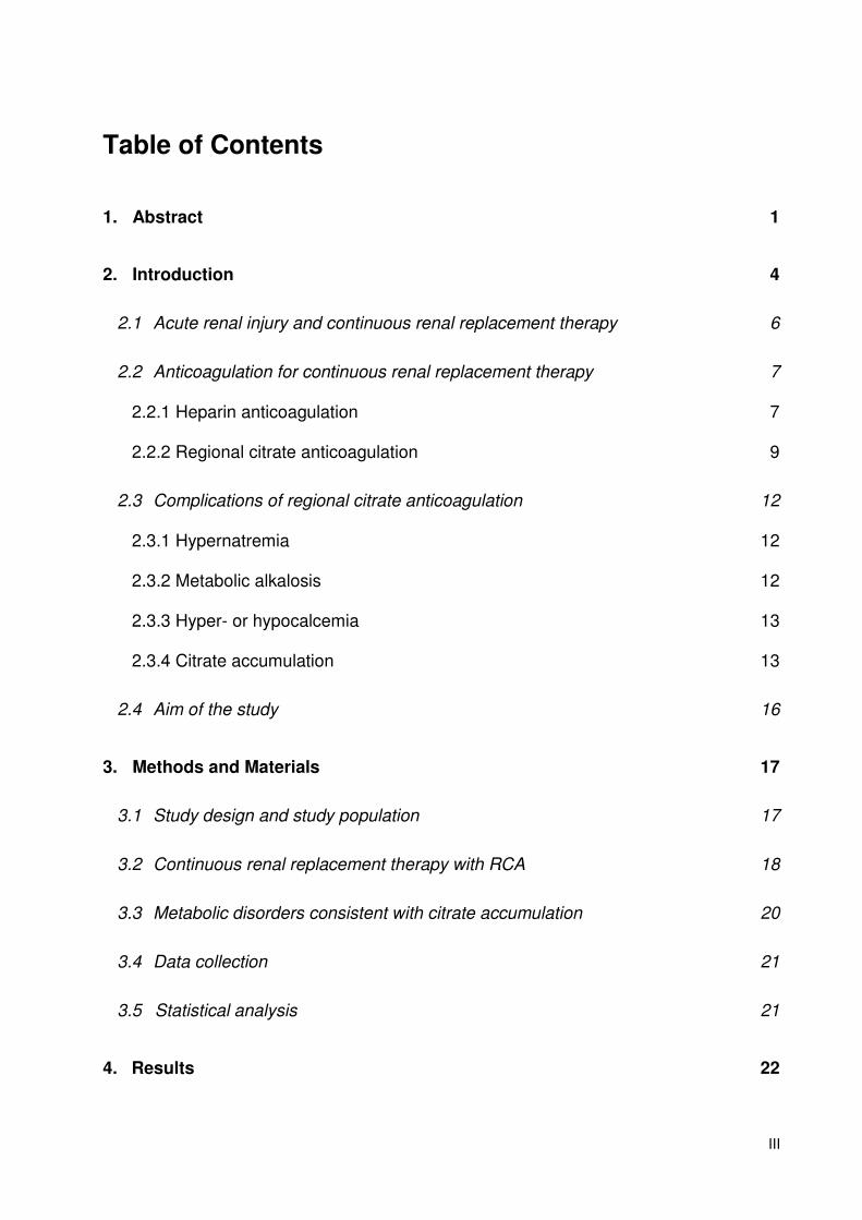
Table of Contents
1. Abstract 1
2. Introduction 4
2.1 Acute renal injury and continuous renal replacement therapy 6
2.2 Anticoagulation for continuous renal replacement therapy 7
2.2.1 Heparin anticoagulation 7
2.2.2 Regional citrate anticoagulation 9
2.3 Complications of regional citrate anticoagulation 12
2.3.1 Hypernatremia 12
2.3.2 Metabolic alkalosis 12
2.3.3 Hyper- or hypocalcemia 13
2.3.4 Citrate accumulation 13
2.4 Aim of the study 16
3. Methods and Materials 17
3.1 Study design and study population 17
3.2 Continuous renal replacement therapy with RCA 18
3.3 Metabolic disorders consistent with citrate accumulation 20
3.4 Data collection 21
3.5 Statistical analysis 21
4. Results 22
IV
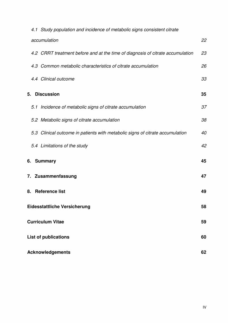
4.1 Study population and incidence of metabolic signs consistent citrate
accumulation 22
4.2 CRRT treatment before and at the time of diagnosis of citrate accumulation 23
4.3 Common metabolic characteristics of citrate accumulation 26
4.4 Clinical outcome 33
5. Discussion 35
5.1 Incidence of metabolic signs of citrate accumulation 37
5.2 Metabolic signs of citrate accumulation 38
5.3 Clinical outcome in patients with metabolic signs of citrate accumulation 40
5.4 Limitations of the study 42
6. Summary 45
7. Zusammenfassung 47
8. Reference list 49
Eidesstattliche Versicherung 58
Curriculum Vitae 59
List of publications 60
Acknowledgements 62
Abstract
1
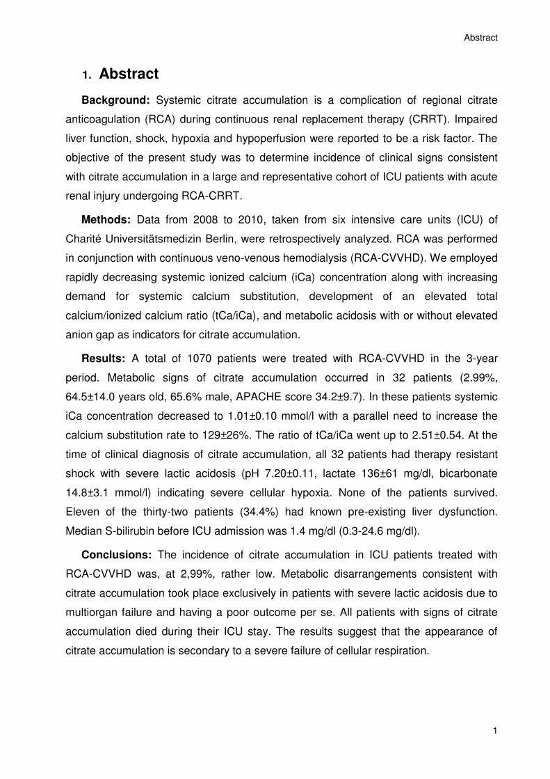
1. Abstract
Background: Systemic citrate accumulation is a complication of regional citrate
anticoagulation (RCA) during continuous renal replacement therapy (CRRT). Impaired
liver function, shock, hypoxia and hypoperfusion were reported to be a risk factor. The
objective of the present study was to determine incidence of clinical signs consistent
with citrate accumulation in a large and representative cohort of ICU patients with acute
renal injury undergoing RCA-CRRT.
Methods: Data from 2008 to 2010, taken from six intensive care units (ICU) of
Charité Universitätsmedizin Berlin, were retrospectively analyzed. RCA was performed
in conjunction with continuous veno-venous hemodialysis (RCA-CVVHD). We employed
rapidly decreasing systemic ionized calcium (iCa) concentration along with increasing
demand for systemic calcium substitution, development of an elevated total
calcium/ionized calcium ratio (tCa/iCa), and metabolic acidosis with or without elevated
anion gap as indicators for citrate accumulation.
Results: A total of 1070 patients were treated with RCA-CVVHD in the 3-year
period. Metabolic signs of citrate accumulation occurred in 32 patients (2.99%,
64.5±14.0 years old, 65.6% male, APACHE score 34.2±9.7). In these patients systemic
iCa concentration decreased to 1.01±0.10 mmol/l with a parallel need to increase the
calcium substitution rate to 129±26%. The ratio of tCa/iCa went up to 2.51±0.54. At the
time of clinical diagnosis of citrate accumulation, all 32 patients had therapy resistant
shock with severe lactic acidosis (pH 7.20±0.11, lactate 136±61 mg/dl, bicarbonate
14.8±3.1 mmol/l) indicating severe cellular hypoxia. None of the patients survived.
Eleven of the thirty-two patients (34.4%) had known pre-existing liver dysfunction.
Median S-bilirubin before ICU admission was 1.4 mg/dl (0.3-24.6 mg/dl).
Conclusions: The incidence of citrate accumulation in ICU patients treated with
RCA-CVVHD was, at 2,99%, rather low. Metabolic disarrangements consistent with
citrate accumulation took place exclusively in patients with severe lactic acidosis due to
multiorgan failure and having a poor outcome per se. All patients with signs of citrate
accumulation died during their ICU stay. The results suggest that the appearance of
citrate accumulation is secondary to a severe failure of cellular respiration.
Abstract
2
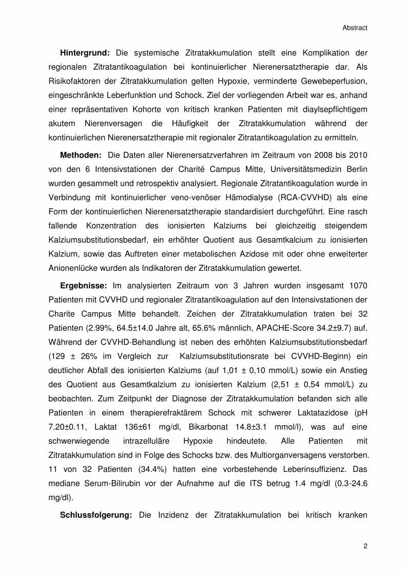
Hintergrund: Die systemische Zitratakkumulation stellt eine Komplikation der
regionalen Zitratantikoagulation bei kontinuierlicher Nierenersatztherapie dar. Als
Risikofaktoren der Zitratakkumulation gelten Hypoxie, verminderte Gewebeperfusion,
eingeschränkte Leberfunktion und Schock. Ziel der vorliegenden Arbeit war es, anhand
einer repräsentativen Kohorte von kritisch kranken Patienten mit diaylsepflichtigem
akutem Nierenversagen die Häufigkeit der Zitratakkumulation während der
kontinuierlichen Nierenersatztherapie mit regionaler Zitratantikoagulation zu ermitteln.
Methoden: Die Daten aller Nierenersatzverfahren im Zeitraum von 2008 bis 2010
von den 6 Intensivstationen der Charité Campus Mitte, Universitätsmedizin Berlin
wurden gesammelt und retrospektiv analysiert. Regionale Zitratantikoagulation wurde in
Verbindung mit kontinuierlicher veno-venöser Hämodialyse (RCA-CVVHD) als eine
Form der kontinuierlichen Nierenersatztherapie standardisiert durchgeführt. Eine rasch
fallende Konzentration des ionisierten Kalziums bei gleichzeitig steigendem
Kalziumsubstitutionsbedarf, ein erhöhter Quotient aus Gesamtkalcium zu ionisierten
Kalzium, sowie das Auftreten einer metabolischen Azidose mit oder ohne erweiterter
Anionenlücke wurden als Indikatoren der Zitratakkumulation gewertet.
Ergebnisse: Im analysierten Zeitraum von 3 Jahren wurden insgesamt 1070
Patienten mit CVVHD und regionaler Zitratantikoagulation auf den Intensivstationen der
Charite Campus Mitte behandelt. Zeichen der Zitratakkumulation traten bei 32
Patienten (2.99%, 64.5±14.0 Jahre alt, 65.6% männlich, APACHE-Score 34.2±9.7) auf.
Während der CVVHD-Behandlung ist neben des erhöhten Kalziumsubstitutionsbedarf
(129 ± 26% im Vergleich zur Kalziumsubstitutionsrate bei CVVHD-Beginn) ein
deutlicher Abfall des ionisierten Kalziums (auf 1,01 ± 0,10 mmol/L) sowie ein Anstieg
des Quotient aus Gesamtkalzium zu ionisierten Kalzium (2,51 ± 0,54 mmol/L) zu
beobachten. Zum Zeitpunkt der Diagnose der Zitratakkumulation befanden sich alle
Patienten in einem therapierefraktärem Schock mit schwerer Laktatazidose (pH
7.20±0.11, Laktat 136±61 mg/dl, Bikarbonat 14.8±3.1 mmol/l), was auf eine
schwerwiegende intrazelluläre Hypoxie hindeutete. Alle Patienten mit
Zitratakkumulation sind in Folge des Schocks bzw. des Multiorganversagens verstorben.
11 von 32 Patienten (34.4%) hatten eine vorbestehende Leberinsuffizienz. Das
mediane Serum-Bilirubin vor der Aufnahme auf die ITS betrug 1.4 mg/dl (0.3-24.6
mg/dl).
Schlussfolgerung: Die Inzidenz der Zitratakkumulation bei kritisch kranken
Abstract
3
dialysepflichtigen Patienten während kontinuierlicher Nierenersatztherapie mit
regionaler Zitratantikoagulation betrug 2.99%. Die metabolischen Veränderungen, die
auf eine Zitratakkumulation hingewiesen haben, traten ausschließlich bei Patienten mit
schwerer Laktatazidose aufgrund eines Multiorganversagens und folglich
eingeschränkter Prognose auf. Alle Patienten mit Zeichen der Zitratakkumulation
verstarben während des ITS-Aufenthalts. Die Ergebnisse sprechen für das Auftreten
der Zitratakkumulation als Folge eines schwerwiegenden Versagens der Zellatmung.
Introduction
4
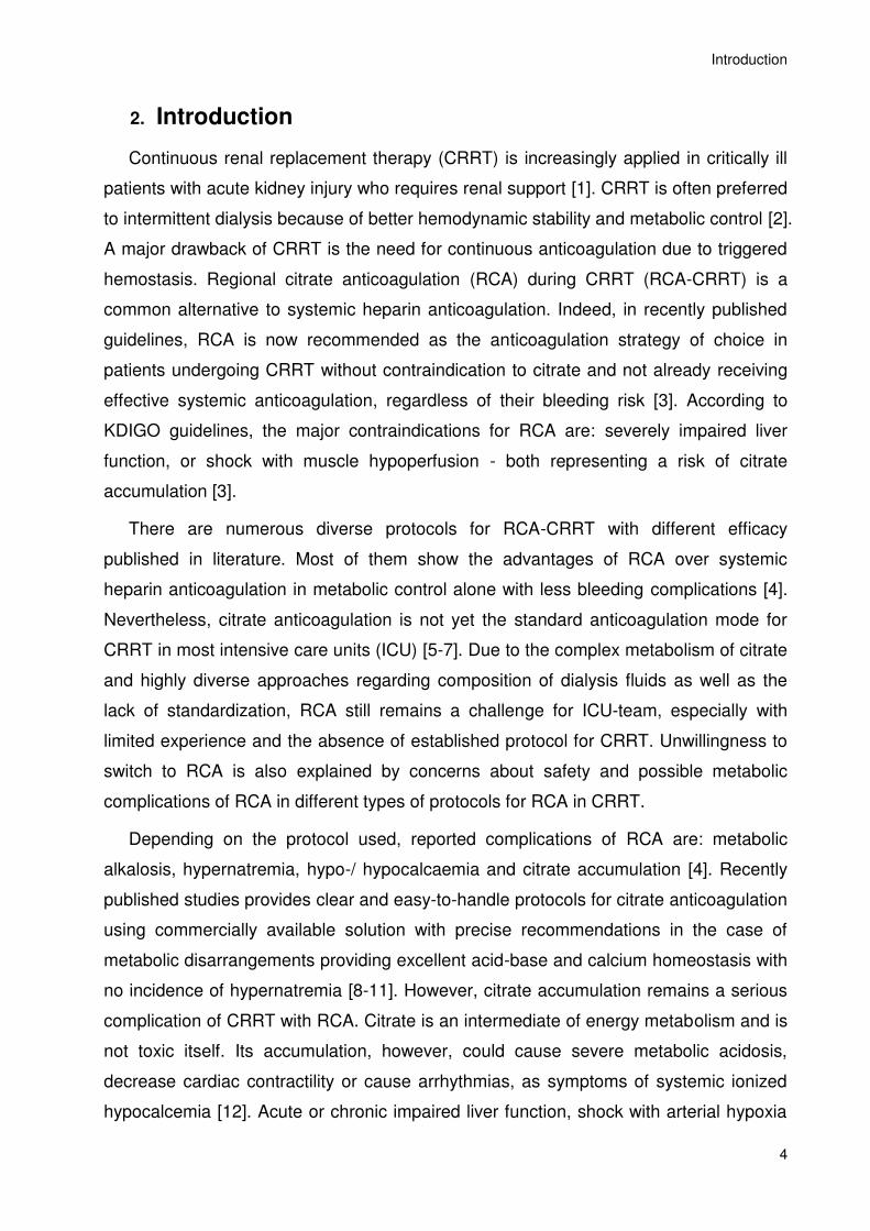
2. Introduction
Continuous renal replacement therapy (CRRT) is increasingly applied in critically ill
patients with acute kidney injury who requires renal support [1]. CRRT is often preferred
to intermittent dialysis because of better hemodynamic stability and metabolic control [2].
A major drawback of CRRT is the need for continuous anticoagulation due to triggered
hemostasis. Regional citrate anticoagulation (RCA) during CRRT (RCA-CRRT) is a
common alternative to systemic heparin anticoagulation. Indeed, in recently published
guidelines, RCA is now recommended as the anticoagulation strategy of choice in
patients undergoing CRRT without contraindication to citrate and not already receiving
effective systemic anticoagulation, regardless of their bleeding risk [3]. According to
KDIGO guidelines, the major contraindications for RCA are: severely impaired liver
function, or shock with muscle hypoperfusion - both representing a risk of citrate
accumulation [3].
There are numerous diverse protocols for RCA-CRRT with different efficacy
published in literature. Most of them show the advantages of RCA over systemic
heparin anticoagulation in metabolic control alone with less bleeding complications [4].
Nevertheless, citrate anticoagulation is not yet the standard anticoagulation mode for
CRRT in most intensive care units (ICU) [5-7]. Due to the complex metabolism of citrate
and highly diverse approaches regarding composition of dialysis fluids as well as the
lack of standardization, RCA still remains a challenge for ICU-team, especially with
limited experience and the absence of established protocol for CRRT. Unwillingness to
switch to RCA is also explained by concerns about safety and possible metabolic
complications of RCA in different types of protocols for RCA in CRRT.
Depending on the protocol used, reported complications of RCA are: metabolic
alkalosis, hypernatremia, hypo-/ hypocalcaemia and citrate accumulation [4]. Recently
published studies provides clear and easy-to-handle protocols for citrate anticoagulation
using commercially available solution with precise recommendations in the case of
metabolic disarrangements providing excellent acid-base and calcium homeostasis with
no incidence of hypernatremia [8-11]. However, citrate accumulation remains a serious
complication of CRRT with RCA. Citrate is an intermediate of energy metabolism and is
not toxic itself. Its accumulation, however, could cause severe metabolic acidosis,
decrease cardiac contractility or cause arrhythmias, as symptoms of systemic ionized
hypocalcemia [12]. Acute or chronic impaired liver function, shock with arterial hypoxia
Introduction
5
and reduced tissue perfusion are the major risk factors for citrate accumulation [3, 13,
14]. Unfortunately, measurement of citrate concentration in blood is not available on a
daily routine basis, and, at least in Germany, available test kits are not approved for
clinical use. However, there are commonly accepted markers for citrate accumulation
such as: metabolic acidosis with or without increased anion gap, ionized hypocalcemia
with simultaneous increased levels of total calcium and an increased total calcium to
ionized calcium ratio (tCa/iCa) [13-15]. Those laboratory parameters do not confirm
citrate accumulation in all cases, and often lead to false positive results, which makes
the diagnosis of citrate accumulation a complex clinical issue. Moreover, information
about the incidence of citrate accumulation in a general cohort of ICU patients
undergoing RCA-CRRT is relatively limited due to the fact that patients with risk factors
for citrate accumulation were either excluded from the prospective trials [8, 11, 16-19],
or the observation of those patients was limited to the study period [20].
In this monocentric retrospective study we collected and analyzed all patients on
RCA-CRRT over a three-year period (from 2008 to 2010) in order to identify risk factors
for citrate accumulation. The primary objective was to reveal the incidence rate of citrate
accumulation in a cohort of non-selected critically ill patients receiving RCA-CRRT. The
secondary objective was to assess the clinical characteristics and outcome related to
citrate accumulation based on a representative population of patients.
Introduction
6
2.1 Acute renal injury and continuous renal replacement therapy
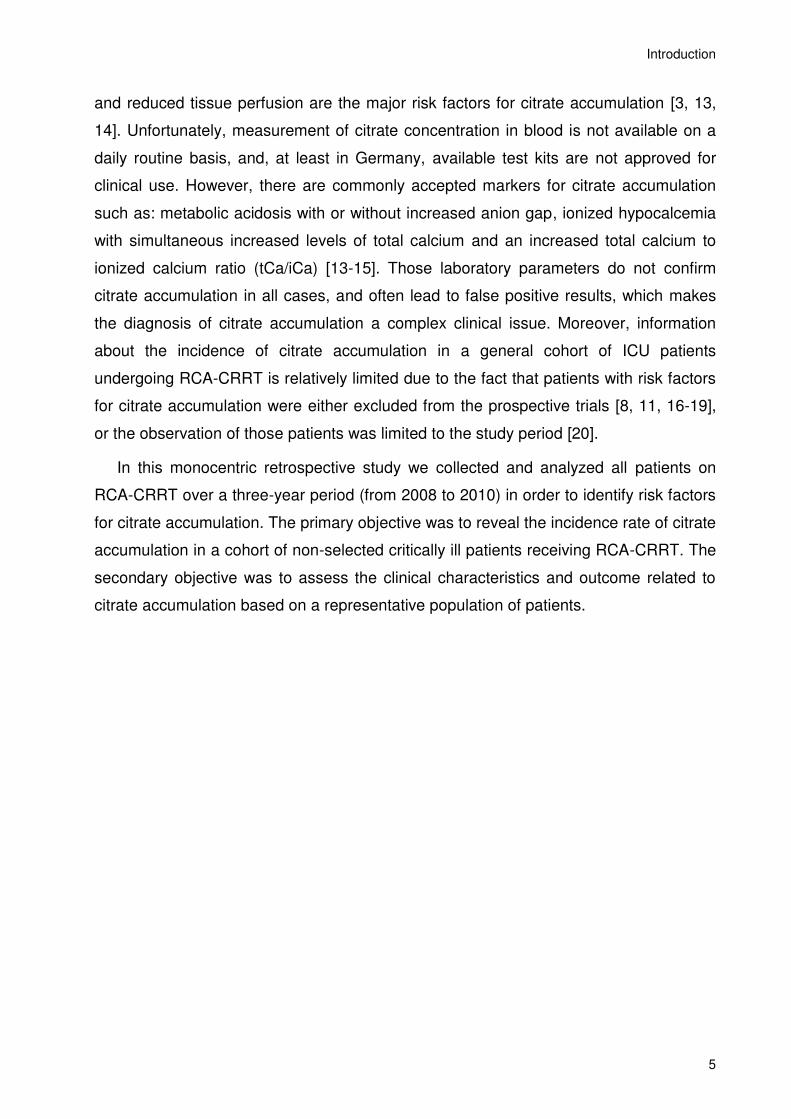
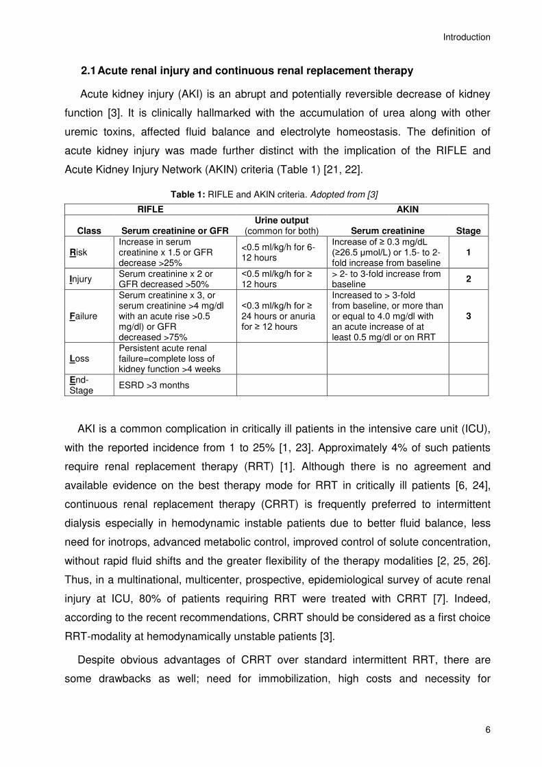
Acute kidney injury (AKI) is an abrupt and potentially reversible decrease of kidney
function [3]. It is clinically hallmarked with the accumulation of urea along with other
uremic toxins, affected fluid balance and electrolyte homeostasis. The definition of
acute kidney injury was made further distinct with the implication of the RIFLE and
Acute Kidney Injury Network (AKIN) criteria (Table 1) [21, 22].
Table 1: RIFLE and AKIN criteria. Adopted from [3]
RIFLE AKIN
Class Serum creatinine or GFR Urine output
(common for both) Serum creatinine Stage
Risk Increase in serum creatinine x 1.5 or GFR decrease >25%
<0.5 ml/kg/h for 6-12 hours
Increase of ≥ 0.3 mg/dL (≥26.5 µmol/L) or 1.5- to 2-fold increase from baseline
1
Injury Serum creatinine x 2 or GFR decreased >50%
<0.5 ml/kg/h for ≥ 12 hours
> 2- to 3-fold increase from baseline
2
Failure
Serum creatinine x 3, or serum creatinine >4 mg/dl with an acute rise >0.5 mg/dl) or GFR decreased >75%
<0.3 ml/kg/h for ≥ 24 hours or anuria for ≥ 12 hours
Increased to > 3-fold from baseline, or more than or equal to 4.0 mg/dl with an acute increase of at least 0.5 mg/dl or on RRT
3
Loss Persistent acute renal failure=complete loss of kidney function >4 weeks
End-Stage
ESRD >3 months
AKI is a common complication in critically ill patients in the intensive care unit (ICU),
with the reported incidence from 1 to 25% [1, 23]. Approximately 4% of such patients
require renal replacement therapy (RRT) [1]. Although there is no agreement and
available evidence on the best therapy mode for RRT in critically ill patients [6, 24],
continuous renal replacement therapy (CRRT) is frequently preferred to intermittent
dialysis especially in hemodynamic instable patients due to better fluid balance, less
need for inotrops, advanced metabolic control, improved control of solute concentration,
without rapid fluid shifts and the greater flexibility of the therapy modalities [2, 25, 26].
Thus, in a multinational, multicenter, prospective, epidemiological survey of acute renal
injury at ICU, 80% of patients requiring RRT were treated with CRRT [7]. Indeed,
according to the recent recommendations, CRRT should be considered as a first choice
RRT-modality at hemodynamically unstable patients [3].
Despite obvious advantages of CRRT over standard intermittent RRT, there are
some drawbacks as well; need for immobilization, high costs and necessity for
Introduction
7
continuous anticoagulation are all major drawbacks of CRRT.
2.2 Anticoagulation for continuous renal replacement therapy
In patients with AKI undergoing RRT, the blood is conducted through the
extracorporeal circuit and dialysis filter. The contact of blood with the alien surface and
air, as well as non-laminar flow in the extracorporeal tubing system, results in activation
of plasmatic coagulation, tissue factors, leucocytes and platelets, which initiate a
clotting. Premature filter clotting reduces circuit lifetime and treatment efficacy, and
increases blood loss, workload and costs. Therefore, improving circuit life is clinically
relevant, especially in terms of CRRT. Protocols involving CRRT with no anticoagulation
were studied in small trials where adequate CRRT-circuits survival was described only
in patients with some degree of coagulopathy [27-29]. Thus anticoagulation is generally
required, representing a great challenge for critically ill patients. Until recently, systemic
anticoagulation with heparins was a common choice.
2.2.1 Heparin anticoagulation
Since its first implementation at the beginning of the 20th century, unfractionated
heparin (UFH), and, much later, low-molecular-weight heparin (LMWH) were
considered standard anticoagulation modalities and, thus, were the most commonly
prescribed anticoagulant agents used during continuous renal replacement therapy
(CRRT) [7]. The overview of advantages and disadvantages of each type of heparin is
presented in Table 2.
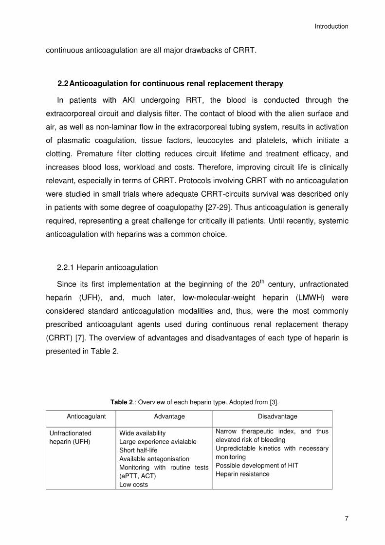
Table 2.: Overview of each heparin type. Adopted from [3].
Anticoagulant Advantage Disadvantage
Unfractionated
heparin (UFH)
Wide availability
Large experience avialable
Short half-life
Available antagonisation
Monitoring with routine tests
(aPTT, ACT)
Low costs
Narrow therapeutic index, and thus
elevated risk of bleeding
Unpredictable kinetics with necessary
monitoring
Possible development of HIT
Heparin resistance
Introduction
8
Low-molecular-weight
heparin (LMWH) *
More predictable kinetics with
possible weight-based dosing
More reliable anticoagulant
response with no required
monitoring
Reduced risk of HIT
Risk of accumulation in kidney failure
Monitoring requires non-routine test
(anti–Factor Xa)
Different drugs not interchangeable
Incomplete reversal by protamine In
most countries more expensive than
unfractionated heparin
* mostly applicable for the intermittent RRT
Nevertheless, anticoagulation with any kind of heparins has a systemic impact, and
thus, their major drawback is bleeding. This is especially the case in CRRT, where
continuous anticoagulation is administered. Besides critically ill patients are, per se,
predisposed to bleeding complications due to endothelial disruption, coagulopathy,
recent surgery, trauma or mucosal lesions. Taking into account each of the application
methods and heparin agents, the incidence of bleeding varies from 10% to 50%, with a
bleeding mortality of 15% [30-32]. Thus, heparin in high doses is at least an important
challenge in critically ill patients with active bleeding or at high risk of bleeding [3].
Anticoagulation with heparins can also cause bleeding-independent complications,
such as: heparin-induced thrombocytopenia (HIT), with incidence between 1% and 5%
[33]; heparin resistance due to reduced antithrombin concentration [34, 35] and heparin-
binding proteins [36]; activation of pro-inflamative processes [35, 37, 38]; and impaired
microcirculation in sepsis [39, 40].
Hence, there is increasing evidence questioning the safety of heparin
anticoagulation during CRRT, particularly in critically ill patients. Several methods of
regional anticoagulation, as an alternative to systemic heparin use, have been proposed
over the past fifty years. The regional citrate anticoagulation demonstrated most
promising results over time. Several randomized clinical trials showed the superiority of
RCA over heparin anticoagulation regarding bleeding incidence and need of blood
transfusion (Table 3).

Introduction
9
Table 3. Comparison of citrate and heparin anticoagulation for CRRT in RCT in respect of
bleeding and transfusion. Adopted from [4].
Bleeding Transfusion (RBC)
Reference Design Citrate Heparin Citrate Heparin
Monchi et al [19] RCOT, n = 20
n = 0 n = 1 0.2 (RBC/day*) P < 0.001
1.0 (RBC/day*)
Kutsogiannis et al [16]
RCT, n = 30
n = 1 (RR 0.17) P = 0.06
n = 8 n = 15 (RR 0.53) P = 0.13
n = 20
Betjes et al [41] RCT, n = 48
0% P < 0.01
33% 0.43 (RBC/day*)
P = 0.01
0.88 (RBC/day*)
Oudemans-Van Straaten et al [11]
RCT, n = 200
6% P = 0.08
16% 0.27 (RBC/day*)
P = 0.31
0.36 (RBC/day*)
Hetzel et al [8] RCT, n = 170
14.5% P = 0.06
5.7%
RCOT, randomized cross-over trial; RCT, randomized controlled trial; RR, relative risk. *Number of red cell units per
day of continuous venovenous hemofiltration.
Hence, according to the latest guidelines, regional citrate anticoagulation during
CRRT should be used independently of the bleeding risk [3].
2.2.2 Regional citrate anticoagulation
Citrate was first applied as a regional anticoagulant for intermittent hemodialysis in
the 1960s-1980s [42, 43]. Since the development of the renal replacement therapy, this
method has received a lot of implementation. Especially encouraging results were
achieved in CRRT, where citrate anticoagulation was first applied in 1990 by Mehta et
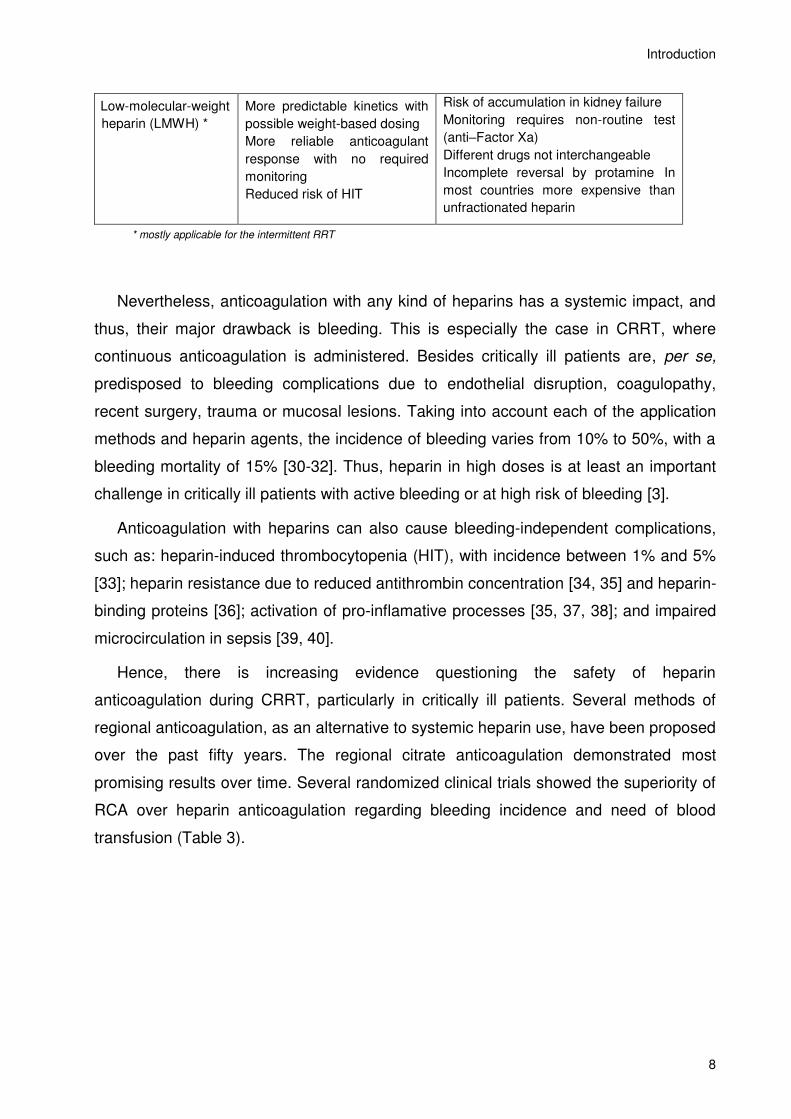
al [44]. Calcium is an important cofactor required at different steps of the coagulation
cascade for proper function. Being infused pre-filter in appropriate doses, citrate acts by
chelating the ionized calcium in the extracorporeal circuit, consequently blocking the
coagulation cascade (Figure 1).
Introduction
10
Figure 1. Calcium as a cofactor in the coagulation cascade. Adopted from [45].
A large amount of calcium-citrate complexes are removed by a dialyser, which
generally requires use of a calcium-free dialysate, and partially enters the systemic
circulation. Having a sieving coefficient of about one, citrate is partially removed by
dialysis as iCa-citrate complexes. Depending on the CRRT modality and blood-to-
effluent ratio, the removed fraction of citrate varies between 20 and 50%[46]. By
entering a patient’s blood circulation, unfiltered calcium-citrate complexes are diluted by
the total blood volume and are rapidly metabolized in the citric acid cycle, as it is
generally supposed, mainly in the liver, kidney and skeletal muscle tissue. Moreover,
the metabolism of citrate is necessary to avoid relevant systemic accumulation of citrate
and calcium-citrate complexes. The concentration of systemic iCa is partially restored
Introduction
11
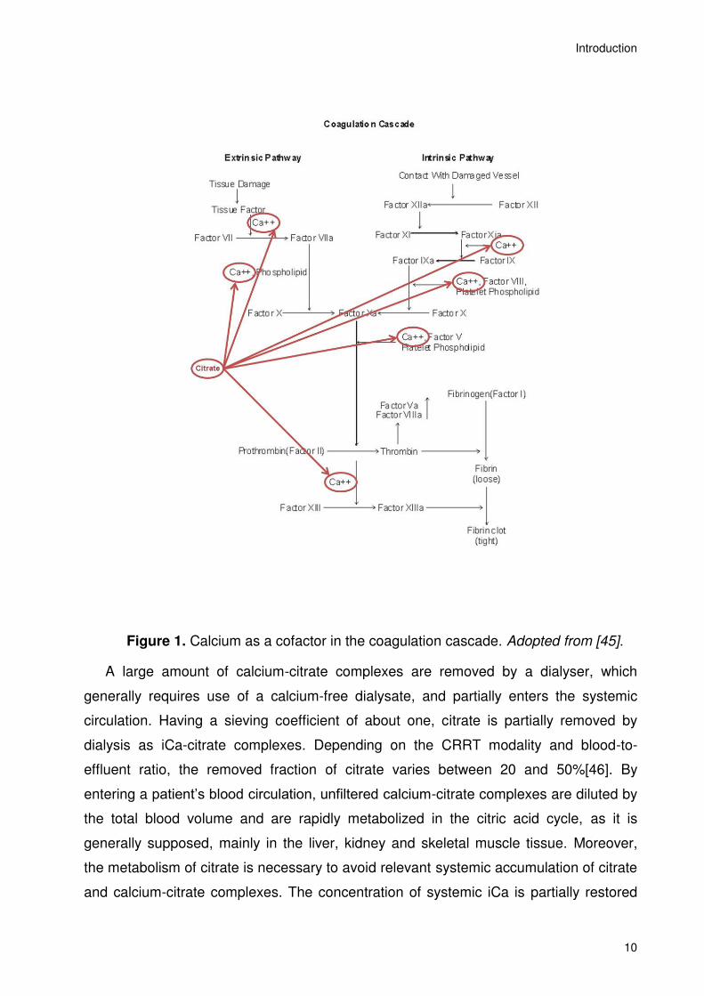
due to liberation of chelated calcium when citrate is metabolized. In RCA protocol using
calcium-free dialysates an additional calcium substitution is needed to compensate for
the loss of calcium into the dialysate. The calcium solution is generally infused into the
venous line (post-filter) of the extracorporeal circuit. As a result, calcium balance
remains in equilibrium. The schematic representation of general principles of regional
citrate anticoagulation is demonstrated in Figure 2.
Figure 2. Principles of regional citrate accumulation
Citrate infusion decreases the concentration of iCa in a dose-dependent manner,
however the relationship between iCa - concentration and the anticoagulation effect is
not in a linear proportion [47].
Anticoagulation is unaffected when iCa concentration is more then 0,50 mmol/L,
whereas concentrations of iCa less then 0,25 – 0,35 mmol/L allow near total inhibition of
anticoagulation [48, 49]. Thus, the anticoagulation effect can and should be monitored
by measuring the concentration of iCa in the extracorporeal circuit (post-filter) with
subsequent adjustment of delivered citrate dose according to the post-filter iCa target
(0,25 – 0,35 mmol/L) [8, 10]. In a situation with normal concentration of iCa in the
Introduction
12
patient’s peripheral blood, one would require a citrate dose of about 4 mmol per liter
blood in the extracorporeal circuit to reach the mentioned post-filter iCa concentration [8,
10]. Some other protocols propose a less complex approach using a fixed citrate dose
proportional to blood flow, with a citrate concentration about 3 mmol per one liter blood.
Those protocols, however, did not show the superiority over heparin anticoagulation in
terms of circuit lifetime [11].
2.3 Complications of regional citrate anticoagulation
Being consequent with the basic principles of this anticoagulation modality, the most
common complications of RCA are:
2.3.1 Hypernatremia
Due to the fact that citrate solution is generally applied as a trisodium citrate, there is
a certain risk of accompanied hypernatremia. However, this complication was often a
problem at the beginning of clinical implementation of RCA, and with the application of
dialysis solutions with reduced sodium concentration for RCA, has become negligible
[11, 41, 50].
2.3.2 Metabolic alkalosis
Additionally to its anticoagulation effects, citrate possesses profound effects on acid-
base homeostasis as well. Being infused in systemic circulation, in a normal
physiological state, citrate is rapidly metabolized to bicarbonate via the Krebs cycle in
liver, kidney and skeletal muscle. Trisodium citrate is converted into citric acid, and after
metabolism 1 mmol of citrate produces 3 mmol of bicarbonate. Thus, citrate also
contributes to the bicarbonate buffer system, and an excessive citrate load leads to
metabolic alkalosis. This complication was relatively common in the beginning of clinical
implementations of RCA [44]. Nevertheless, the metabolic alkalosis could be easily
avoided by using a dialysate solution with reduced bicarbonate concentration [50].
Using such protocols, in case of metabolic alkalosis, bicarbonate overload could be
compensated by elevated dialysate flow rate or reduced citrate load [8, 10, 50].
Moreover, in recently published meta-analysis, it has been shown that RCA has no
Introduction
13
significant increase in the incidence of metabolic alkalosis in comparison with heparin
anticoagulation [51] and, if the RCA protocol is strictly followed, metabolic
disarrangements are easy to identify and control [52].
2.3.3 Hyper- or hypocalcemia
The induction of severe systemic hypocalcemia was the most threatening
complication of RCA in the early years of implementation [12]. Severe hypocalcemia
leads to weakness, muscle cramps, myocardial dysfunction, and, in the most severe
cases, death [53-58]. Systemic hypocalcemia during RCA can occur due to an
inbalance between the elimination of the calcium through the dialysis filter as part of
calcium-citrate complexes and the systemic calcium substitution rate, or technical
failure of the calcium replacement system. Currently, the bedside monitoring of the
ionized calcium has become a standard method available in almost every intensive care
unit and calcium replacement systems have been integrated into the CRRT device.
Since then, disarrangements of calcium homeostasis can be avoided and easily
detected and corrected. Application of the commercially available dialysis solutions
together with CRRT devices designed for RCA-based CRRT minimized the incidence of
hypocalcemia to negligible levels [8, 10, 50].
Hypercalcemia is a well-known problem during citrate anticoagulation [59, 60].
However, it could be easily corrected by adjusting the calcium substitution rate, as
described elsewhere [10].
In some patients undergoing RCA-CRRT, mild to severe ionized hypocalcemia is
accompanied by elevated concentration of total calcium. It is particularly distinct in
patients with slowed down or disturbed citrate metabolism for whatever reasons [60].
This condition is often a hallmark of a possible citrate accumulation and would be
discussed further.
2.3.4 Citrate accumulation
In contrast to other metabolic disarrangements during RCA-CRRT, citrate
accumulation remains a serious complication with often severe consequences. In case
of slowed down or disturbed citrate metabolism, concentration of citrate continuously
rises during the ongoing RCA-CRRT. Inability to metabolize citrate, and subsequent
Introduction
14
citrate accumulation during RCA-CRRT seems to worsen the prognosis of the patients
and indicate a high risk of death [61].
Citrate is not toxic itself, but in the case of impaired citrate metabolism, the essential
part of the physiologically active ionized calcium remains chelated in the calcium-citrate
complexes. Hence, severe ionized hypocalcemia may appear and lead to life-
threatening complications (decreased cardiac contractility, arrhythmias, etc.). Thus,
ionized hypocalcemia is one of the most sensitive indicator and/or most severe
complications of citrate accumulation [62]. In contrast to decreased concentration of
active ionized calcium, the concentrations of physiologically inactive total calcium are
continuously increasing during the citrate accumulation, owing to a growing fraction of
calcium-citrate complexes. Thus the total to ionized calcium ratio is a useful marker to
detect citrate accumulation [60]. The ionized hypocalcaemia is often masked due to
continuously elevated calcium substitution. That is why continuously increasing calcium
demand itself is a possible clinical marker for citrate accumulation. That explains why
total-to-ionized calcium ratio is probably the most specific marker for citrate
accumulation [60, 62, 63].
Besides decreased levels of ionized calcium and elevated concentration of total
calcium, citrate accumulation is accompanied by metabolic acidosis [46]. This occurs
because of a negative bicarbonate balance: on the one hand, missing metabolism of
citrate to bicarbonate; on the other hand, continuous application of the dialysis solution
with reduced bicarbonate concentration applied during RCA-CRRT. Additionally, citrate
accumulation goes together with elevation of anion gap, due to an increased
concentration of citrate in the blood.
Methods for measurement of citrate concentration in blood are available, but
unfortunately, at least in Germany, still not accessible on a daily routine basis.
Consequently, one can only speculate the citrate accumulation according to metabolic
disarrangements. Taken together, commonly accepted clinical markers for citrate
accumulation are:
1) increased ratio of total calcium – to – ionized calcium (tCa/iCa) due to
2) ionized hypocalcemia with simultaneous increased levels of total calcium,
3) metabolic acidosis with or without increased anion gap and
3) elevated demand in calcium substitution.
Introduction
15
However, those laboratory parameters do not confirm citrate accumulation in all
cases, and often lead to false positive results [14, 15, 60, 64]. That makes the diagnosis
of citrate accumulation a complex clinical issue.
Impaired liver function, arterial hypoxia and reduced tissue perfusion are described
in literature as risk factors for citrate accumulation. Recently published clinical practice
guidelines for acute kidney injury postulates a severely impaired liver function or shock
with muscle hypoperfusion as a major contra-indication for the use of RCA [3]. Some
other authors report that high concentration of lactate and severe heart failure should
also be suggested as risk factors for citrate accumulation [65, 66], as Krebs cycle only
works under aerobic conditions.
Nevertheless, there is enough evidence that at least impaired liver function doesn't
have to be seen as an absolute contraindication for RCA [20, 64, 67]. Taking into
consideration that CRRT is often preferred in hemodynamically unstable patients,
recommendation of KDIGO 2012 not to apply RCA-CRRT in patients with septic shock
automatically excludes a significant portion of those ICU-patients, who would
supposedly benefit from CRRT [11]. As a result, some authors are concerned that a
considerable proportion of patients with shock do tolerate citrate anticoagulation,
especially those with septic shock and high lactate levels if circulation improves and
lactate concentration decreases [46].
Taken together, citrate accumulation, a potentially life-threatening complication of
RCA-CRRT, is probably the most important issue that holds back wide implementation
of RCA during CRRT. The frequency of this complication in clinical daily routine is of
great importance and at the same time remains controversial. For instance, patients
with acute liver failure or severe liver cirrhosis were excluded from all until now
published randomized trials [3]. Moreover, until recently there were few reports on the
incidence and outcome of citrate accumulation in critically ill patients treated with RCA-
CRRT. They were based on occasional clinical cases or on the prospective trials what
representing a selected cohort of patients observed over a limited timeframe, and which
may bring a certain bias leading to underestimation of the incidence rate.
Introduction
16
2.4 Aim of the study
In the present single-center retrospective study, all cases with clinical diagnosis of
citrate accumulation in critically ill patients undergoing RCA-CRRT over a three-year
period were collected and analyzed. Due to the fact that RCA-CVVHD as a modality of
CRRT is the modality of choice in all ICUs of our university hospital, the weight-adapted
protocol for RCA-CVVHD according to Morgera et al [10], was conducted in all patients
requiring CRRT, independent of the patients’ liver function status, shock status or risk of
bleeding, without any contraindication. This scenario provides a unique opportunity to
deal with an unselected representative cohort of patients with diverse morbidity having
AKI and undergoing RCA-CRRT.
Thus, the primary objective of our study was:
- to reveal the incidence of metabolic disarrangements consistent with citrate
accumulation in a cohort of unselected critically ill patients receiving RCA-CRRT.
The secondary objectives of our study were:
- (1) to assess the common clinical characteristics of patients with citrate
accumulation, and
- (2) to evaluate the outcome of citrate accumulation based on a representative
patient population.
Methods and Materials
17
3. Methods and Materials
3.1 Study design and study population
The retrospective single-center study, evaluating the incidence of metabolic
disorders consistent with citrate accumulation during RCA-CRRT, was performed in six
intensive care units (one general, two surgical, two medical, and one neurological ICU)
all having a total of 72 beds at the University Hospital Charité Campus Mitte, Berlin,
Germany.
The study was approved by the local ethical review committee (Ethikkommission der
Charité - Universitätsmedizin Berlin; EA1/035/12). Upon approval by the local ethical
review committee, the need for patients’ informed consent was waived due to the
observational character of study combined with the absence of any intervention and the
anonymisation of all data sets used for analysis.
All patients between January 1, 2008 and December 31, 2010 with kidney injury and
treated with the RCA-CRRT, were included in this retrospective analysis. The
interdisciplinary team of nephrologists and the ICU staff determined the diagnosis of
acute kidney injury and indication for CRRT-treatment.
Patients were identified and data were collected from three different sources:
- a computerized billing database (SAP, Germany)
- the patient data management system used in the ICUs (Computer Organized
Patient Report Assistant (COPRA), COPRA System GmbH, Sasbachwalden,
Germany), and
- records of the daily prescriptions of renal replacement procedures used by the
nursing staff at the nephrology department.
Methods and Materials
18
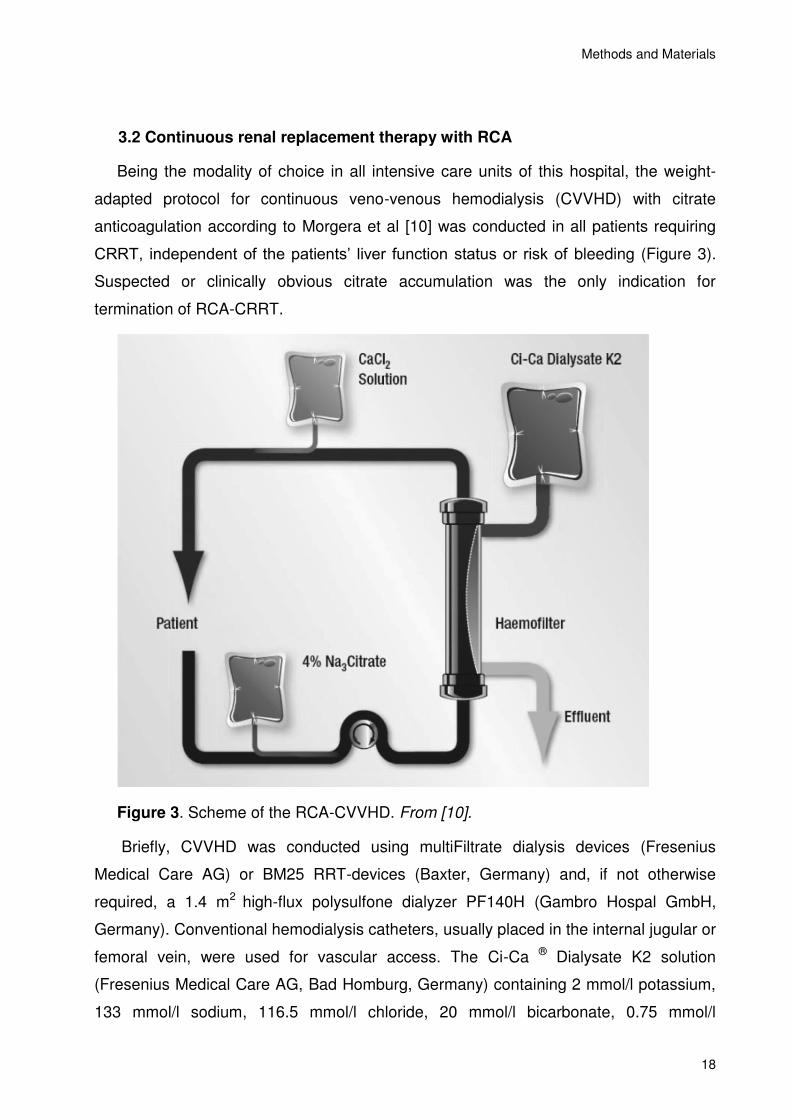
3.2 Continuous renal replacement therapy with RCA
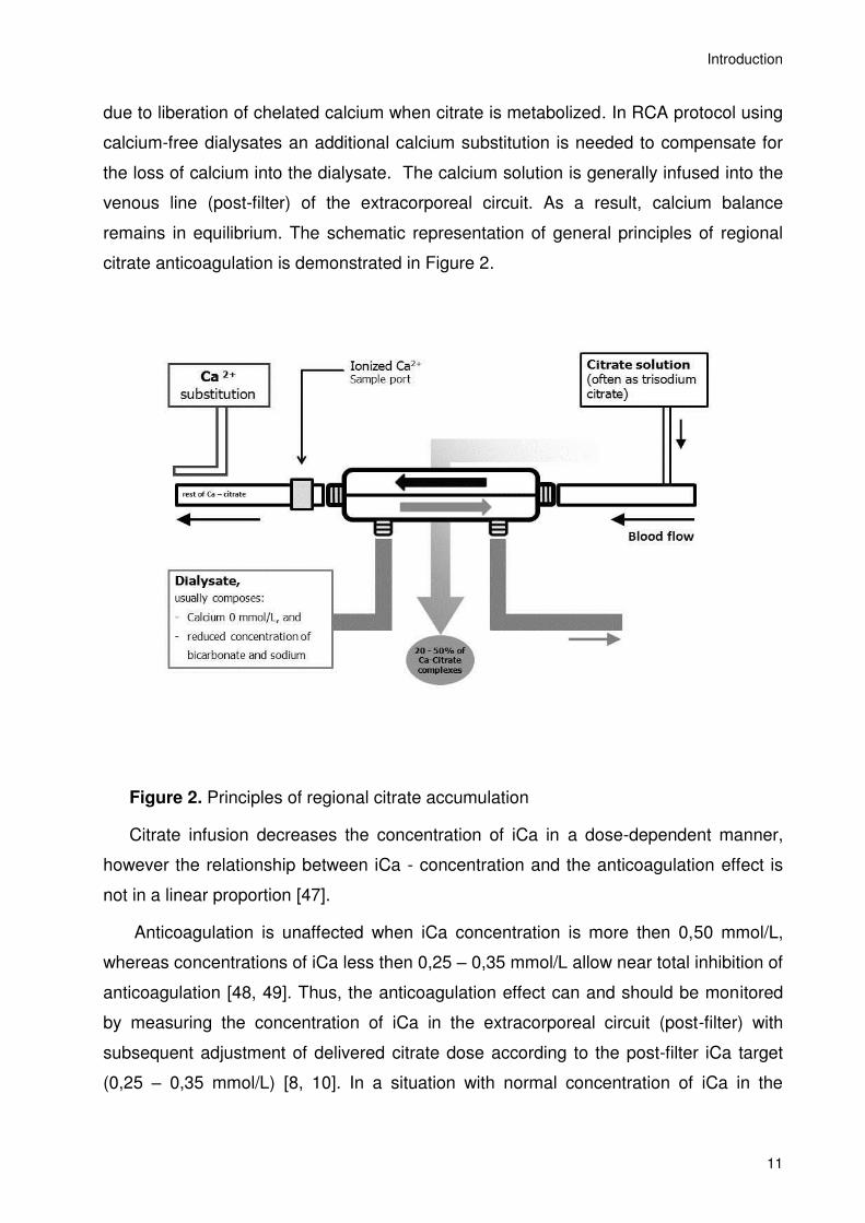
Being the modality of choice in all intensive care units of this hospital, the weight-
adapted protocol for continuous veno-venous hemodialysis (CVVHD) with citrate
anticoagulation according to Morgera et al [10] was conducted in all patients requiring
CRRT, independent of the patients’ liver function status or risk of bleeding (Figure 3).
Suspected or clinically obvious citrate accumulation was the only indication for
termination of RCA-CRRT.
Figure 3. Scheme of the RCA-CVVHD. From [10].
Briefly, CVVHD was conducted using multiFiltrate dialysis devices (Fresenius
Medical Care AG) or BM25 RRT-devices (Baxter, Germany) and, if not otherwise
required, a 1.4 m2 high-flux polysulfone dialyzer PF140H (Gambro Hospal GmbH,
Germany). Conventional hemodialysis catheters, usually placed in the internal jugular or
femoral vein, were used for vascular access. The Ci-Ca ® Dialysate K2 solution
(Fresenius Medical Care AG, Bad Homburg, Germany) containing 2 mmol/l potassium,
133 mmol/l sodium, 116.5 mmol/l chloride, 20 mmol/l bicarbonate, 0.75 mmol/l
Methods and Materials
19
magnesium and zero calcium was used. Blood flow was set accordingly to dialysate
flow to maintain a ratio of 3 to 1. Regional citrate anticoagulation was performed by
infusion of 4% trisodium citrate solution (136 mmol/l; Fresenius Kabi, Bad Homburg,
Germany) in the “arterial” line of the extracorporeal circuit with a starting dose of 4
mmol/l blood. Further, the citrate infusion rate was adjusted to reach post-filter ionized
calcium (iCa) levels of 0.25-0.35 mmol/l. Calcium substitution flow (CaCl2 solution, 91
mmol/l) was initiated with 1.7 mmol calcium per liter total effluent flow and adjusted
accordingly to maintain patients' ionized calcium in the physiological range of 1.1-1.2
mmol/l. The correction of acid-base status was performed, changing the ratio of
blood/dialysate flow: increasing the dialysate flow was used to correct the metabolic
alkalosis, whereas decreasing the dialysate flow led to correction of metabolic acidosis.
Alternatively, correction of acidosis was managed by increasing the citrate infusion with
a parallel increase of blood flow while keeping the dialysate flow constant. Citrate and
calcium infusions were performed through integrated citrate and calcium pumps while
using multiFiltrate Ci-Ca. The dialysis filters were changed routinely after 72 hours with
an allowed tolerance of ±12 hours.
The patients were initially categorized by body weight into three groups (<60kg, 60-
90kg and >90kg) and each group was treated at a matching efficacy level by adaptation
of dialysate flow to maintain an effective dose of 25-30 ml/kg/h.
Methods and Materials
20
3.3 Metabolic disorders consistent with citrate accumulation
As the measurement of citrate blood concentration, at least in Germany, is not
clinically approved for clinical use and, so far, not available for routine bedside testing,
the diagnosis of citrate accumulation could be suspected according to clinical criteria
which have been previously described and are generally accepted, as follows:
1) Decrease of systemic ionized calcium (iCa) (<1.1 mmol/l), despite increasing
calcium replacement;
2) Concomitant increase of total calcium concentration and, thus, increase of total to
ionized calcium ratio (tCa/iCa > 2.1);
3) Relevant metabolic acidosis (pH < 7.2 and/or BE < -5 mmol/l) without, or
4) with an increase of anion gap (> 11 mmol/l).
The ionized hypocalcemia, a sensitive indicator of citrate accumulation, is not
adequately specific for diagnosis of citrate accumulation, and could be related to other
causes. Increase of total-to-ionized calcium ratio, correlating best with citrate plasma
levels, may still not predict citrate accumulation in all cases. Thus, the citrate
accumulation was clinically suspected when the systemic ionized hypocalcemia was
accompanied by at least two additional factors taken from the clinical criteria described.
Moreover, citrate accumulation was also suspected when an unusually high calcium
substitution rate, judged as >3 mmol of calcium substitution per liter of effluent, was
needed [10]. With sufficient evidence of citrate accumulation, the regional citrate
anticoagulation was immediately stopped and, if the clinical situation required further
RRT, CVVHD was performed without anticoagulation, or it was altered to conventional
systemic anticoagulation. CVVHD without RCA was performed using the same CRRT
devices, dialyzers and multiBic® dialysate (Fresenius Medical Care AG, Bad Homburg,
Germany) containing 2 or 4 mmol/l potassium, 140 mmol/l sodium, 111 mmol/l chloride,
35 mmol/l bicarbonate, 0.5 mmol/l magnesium, 1.5 mmol/l calcium, 5.55 mmol/l glucose.
Standard flows with heparin anticoagulated CVVHD were: blood flow of 150 ml/hour,
dialysate flow of 2 l/hour, adjusted according to clinical needs.
Methods and Materials
21
3.4 Data collection
Patient demographics: age, gender, reason for admission, APACHE II score, SOFA
score, length of ICU stay, and ICU mortality. CRRT parameters: duration of RCA-CRRT,
reason for circuit discontinuation, filter life-time and post-filter iCa; blood, citrate,
dialysate and calcium flows; net ultrafiltration; and, if applicable, duration of further
CRRT with other anticoagulation mode when the clinical diagnosis of citrate
accumulation was made.
Biochemical data (creatinine, urea, phosphorus, magnesium and total calcium), total
blood cell count, international normalized ratio (INR) and activated partial
thromboplastin time (aPTT) were measured once daily in the central laboratory,
whereas blood gas analyses (electrolytes, pH, base excess, standard bicarbonate,
ionized calcium and lactate) were measured bed-side (ABL, Radiometer Medical ApS,
Denmark) on a 6-hour basis or more often if there was clinical need. Post filter ionized
calcium for anticoagulation monitoring was measured immediately after initiation of
CRRT and then every 12 hours. Prior to the time of citrate-accumulation diagnosis,
metabolic data were collected during the last hours of treatment with RCA-CVVHD (up
to 48 hours). After the CVVHD anticoagulation mode was switched to heparin,
metabolic data were recorded also for up to 48 hours.
3.5 Statistical analysis
All data are expressed as mean ± standard deviation (SD). According to the
characteristics of the data, group comparisons were carried out by ANOVA and Chi-
Square test. A p value below 0.05 was considered significant. For analysis IBM SPSS
Statistis Version 19 (IBM Corporation, USA) was used.
Results
22
4. Results
4.1 Study population and incidence of metabolic signs consistent citrate
accumulation
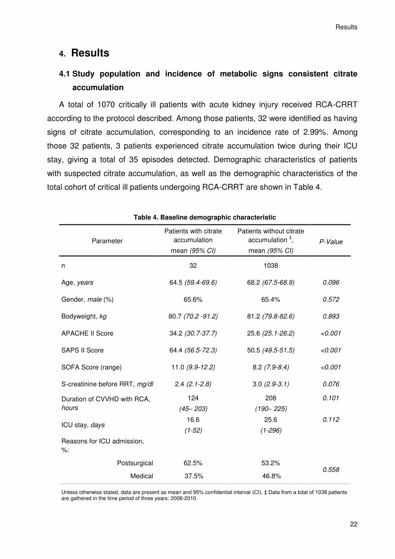
A total of 1070 critically ill patients with acute kidney injury received RCA-CRRT
according to the protocol described. Among those patients, 32 were identified as having
signs of citrate accumulation, corresponding to an incidence rate of 2.99%. Among
those 32 patients, 3 patients experienced citrate accumulation twice during their ICU
stay, giving a total of 35 episodes detected. Demographic characteristics of patients
with suspected citrate accumulation, as well as the demographic characteristics of the
total cohort of critical ill patients undergoing RCA-CRRT are shown in Table 4.
Table 4. Baseline demographic characteristic
Parameter
Patients with citrate
accumulation
mean (95% CI)
Patients without citrate
accumulation ‡,
mean (95% CI)
P-Value
n 32 1038
Age, years 64.5 (59.4-69.6) 68.2 (67.5-68.9) 0.096
Gender, male (%) 65.6% 65.4% 0.572
Bodyweight, kg 80.7 (70.2 -91.2) 81.2 (79.8-82.6) 0.893
APACHE II Score 34.2 (30.7-37.7) 25.6 (25.1-26.2) <0.001
SAPS II Score 64.4 (56.5-72.3) 50.5 (49.5-51.5) <0.001
SOFA Score (range) 11.0 (9.9-12.2) 8.2 (7.9-8.4) <0.001
S-creatinine before RRT, mg/dl 2.4 (2.1-2.8) 3.0 (2.9-3.1) 0.076
Duration of CVVHD with RCA,
hours
124
(45– 203)
208
(190– 225)
0.101
ICU stay, days 16.6
(1-52)
25.6
(1-296)
0.112
Reasons for ICU admission,
%:
Postsurgical 62.5% 53.2% 0.558
Medical 37.5% 46.8%
Unless otherwise stated, data are present as mean and 95% confidential interval (CI). ‡ Data from a total of 1038 patients are gathered in the time period of three years: 2008-2010.
Results
23
As shown in Table 4, there was no significant difference in age, gender, bodyweight,
S-creatinin at initiation of RRT, duration of CRRT, ICU stay, or reasons for ICU-
admission between the patients with citrate accumulation, and other patients treated
with RCA-CVVHD. Remarkably, patients with citrate accumulation had significantly
higher severity of disease, presented in APACHE, SAPS II and SOFA scores, as the
group of patients without signs of citrate accumulation.
4.2 CRRT treatment before and at the time of diagnosis of citrate accumulation
When citrate accumulation was clinically suspected, patients were immediately
switched to heparin anticoagulation with standard dialysis solution, or they received no
anticoagulation at all. Due to a very limited prognosis, treatment of four patients with
suspected citrate accumulation with any further RRT was not applied.
There was no significant difference in the CVVHD flows at the beginning of RCA-
CRRT compared with those immediately before RCA was stopped - blood flow was
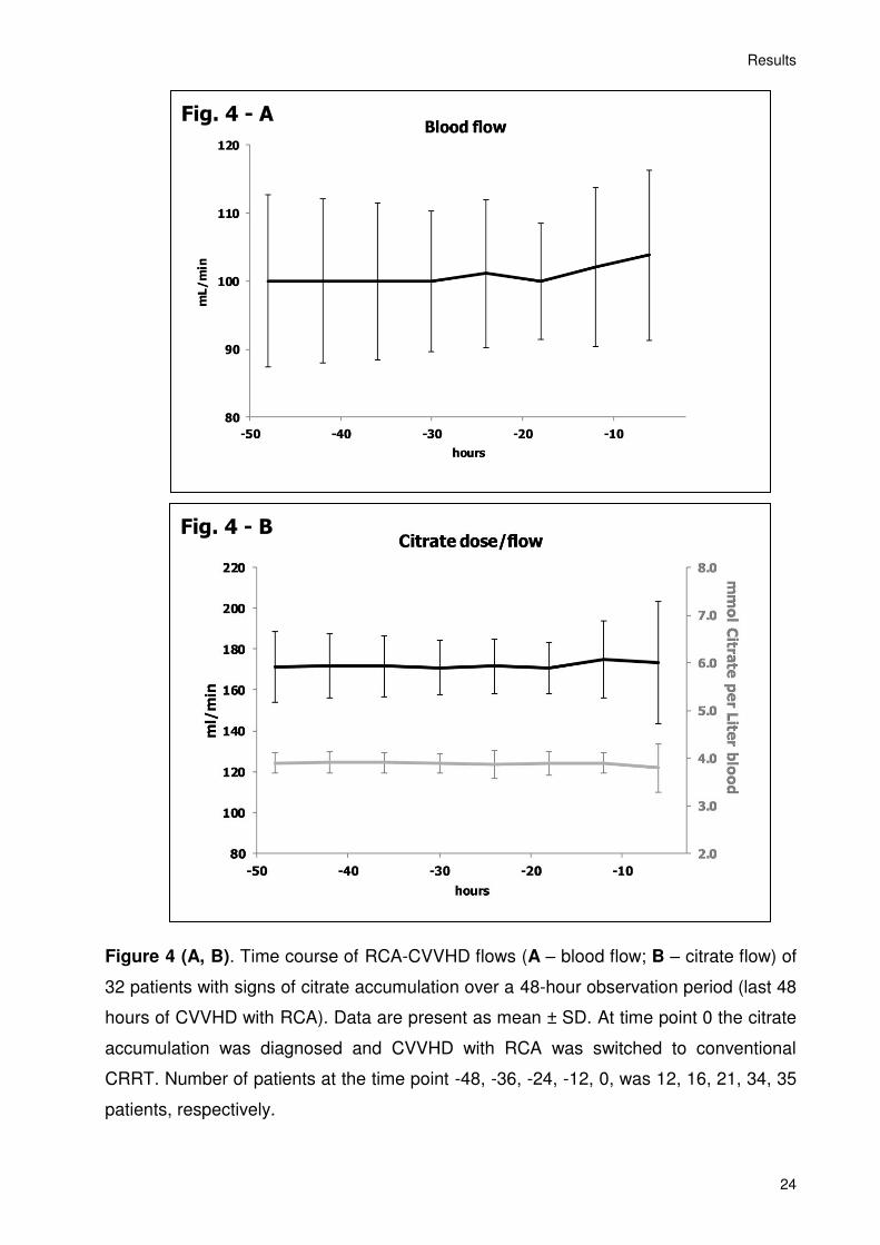
101 ± 8 versus 104 ± 13 mL/hour (p = 0.206) (Figure 4 - A); citrate infusion per liter of
blood flow was 3.9 ± 0.15 versus 3.8 ± 0.40 mmol (p = 0.786) (Figure 4 - B) and
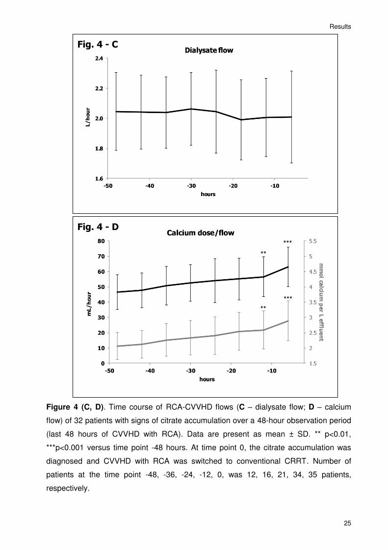
dialysate flow was 2.0 ± 0.2 versus 2.0 ± 0.3 L/hour (p= 0.929) (Figure 4 - C).
Remarkably, calcium substitution per liter effluent flow was significantly elevated at the
time when anticoagulation was switched to heparin compared with that at the beginning
of CRRT with RCA (Figure 4 - D): 2.87 ± 0.66 versus 2.23 ± 0.35 mmol/L effluent (p
<0.001), respectively.
Results
24
Figure 4 (A, B). Time course of RCA-CVVHD flows (A – blood flow; B – citrate flow) of
32 patients with signs of citrate accumulation over a 48-hour observation period (last 48
hours of CVVHD with RCA). Data are present as mean ± SD. At time point 0 the citrate
accumulation was diagnosed and CVVHD with RCA was switched to conventional
CRRT. Number of patients at the time point -48, -36, -24, -12, 0, was 12, 16, 21, 34, 35
patients, respectively.
Fig. 4 - A
Fig. 4 - B
Results
25
Figure 4 (C, D). Time course of RCA-CVVHD flows (C – dialysate flow; D – calcium
flow) of 32 patients with signs of citrate accumulation over a 48-hour observation period
(last 48 hours of CVVHD with RCA). Data are present as mean ± SD. ** p<0.01,
***p<0.001 versus time point -48 hours. At time point 0, the citrate accumulation was
diagnosed and CVVHD with RCA was switched to conventional CRRT. Number of
patients at the time point -48, -36, -24, -12, 0, was 12, 16, 21, 34, 35 patients,
respectively.
Fig. 4 - C
Fig. 4 - D
**
***
**
***
Results
26
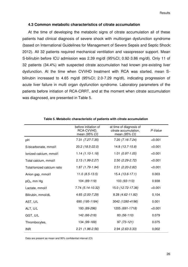
4.3 Common metabolic characteristics of citrate accumulation
At the time of developing the metabolic signs of citrate accumulation all of these
patients had clinical diagnosis of severe shock with multiorgan dysfunction syndrome
(based on International Guidelines for Management of Severe Sepsis and Septic Shock:
2012). All 32 patients required mechanical ventilation and vasopressor support. Mean
S-bilirubin before ICU admission was 2.39 mg/dl (95%CI; 0.92-3.86 mg/dl). Only 11 of
32 patients (34.4%) with suspected citrate accumulation had known pre-existing liver
dysfunction. At the time when CVVHD treatment with RCA was started, mean S-
bilirubin increased to 4.65 mg/dl (95%CI; 2.0-7.29 mg/dl), indicating progression of
acute liver failure in multi organ dysfunction syndrome. Laboratory parameters of the
patients before initiation of RCA-CRRT, and at the moment when citrate accumulation
was diagnosed, are presented in Table 5.
Table 5. Metabolic characteristic of patients with citrate accumulation
before initiation of
RCA-CVVHD, mean (95% CI)
at time of diagnosis of citrate accumulation,
mean (95% CI) P-Value
pH 7.31 (7.27-7.35) 7.20 (7.16-7.24) <0.001
S-bicarbonate, mmol/l 20.2 (18.5-22.0) 14.8 (13.7-15.8) <0.001
Ionized calcium, mmol/l 1.14 (1.10-1.18) 1.01 (0.97-1.05) <0.001
Total calcium, mmol/l 2.13 (1.99-2.27) 2.50 (2.29-2.72) <0.001
Total/ionized calcium ratio 1.87 (1.79-1.94) 2.51 (2.20-2.82) <0.001
Anion gap, mmol/l 11.0 (8.5-13.5) 15.4 (13.6-17.1) 0.003
pO2, mm Hg 104 (89-119) 103 (93-113) 0.938
Lactate, mmol/l 7.74 (5.14-10.32) 15.0 (12.72-17.36) <0.001
Bilirubin, mmol/dL 4.65 (2.00-7.29) 8.28 (4.62-11.92) 0,104
AST, U/L 690 (195-1184) 3042 (1280-4196) 0.001
ALT, U/L 193 (89-296) 1205 (691-1718) <0.001
GGT, U/L 142 (66-218) 83 (56-110) 0.079
Thrombocytes, 134 (99-169) 97 (73-121) 0.075
INR 2.21 (1.96-2.56) 2.94 (2.63-3.33) 0,002
Data are present as mean and 95% confidential interval (CI)
Results
27
As shown in the Table 5, at the time when the citrate accumulation was diagnosed,
patients revealed severe metabolic acidosis with significantly reduced pH and S-
bicarbonate concentration, and base excess; significantly elevated anion gap and
lactate concentration; significantly reduced ionized calcium concentration, significantly
elevated concentration of a total calcium and, consequently, significantly increased
tCa/iCa ratio.
Accordingly, signs of citrate accumulation in actual study consisted of:
1) systemic ionized hypocalcemia (presented in all cases, in 82.9% of cases iCa
was < 1.05 mmol/L);
2) elevated total to ionized systemic calcium ratio (tCa/iCa > 2.1 presented in all
patients, > 2.25 presented in 78% of cases) and
3) severe metabolic acidosis (presented in 94.3% of cases) with
4) increased anion gap (presented in 78% of cases).
All four metabolic signs of citrate accumulation were present in 62,5 % of cases.
Besides the pathognomonic metabolic signs for citrate accumulation, a significant
elevation of AST and ALT, as well as INR, was observed. At the time when citrate
accumulation was diagnosed, there was an aggravation of thrombocytopenia and
increased bilirubinemia, however the differences were not statistically significant.
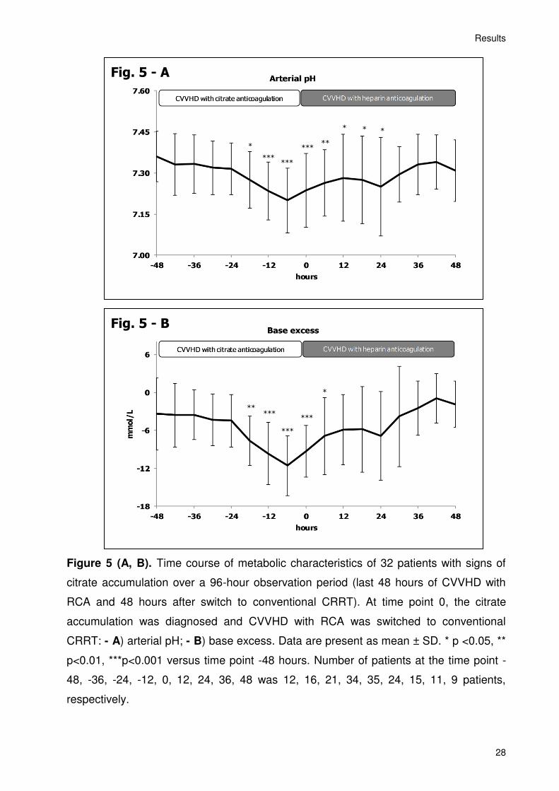
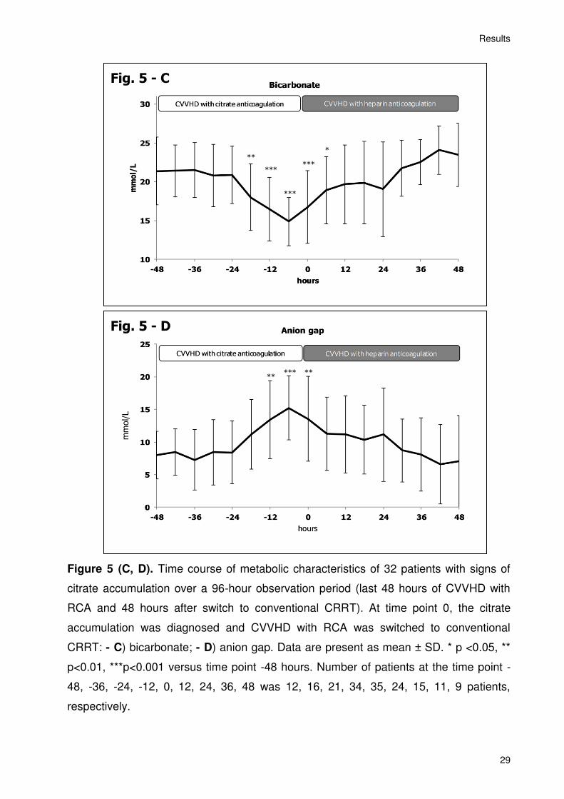
All described metabolic disarrangements progressively increased parallel to each
other, with the most severe changes occurring over the last 24 hours of RCA-CRRT
(Fig. 5 – A, B, C, D). Significant decreases of arterial pH, base excess and bicarbonate
(Fig. 5 – A, B, C, respectively), as well as significant increase of anion gap and lactate
concentration (Fig. 5 – D and Fig. 6, respectively) occurred twelve hours before the
clinical diagnosis of citrate accumulation have been made. Remarkably, all patients had
severe lactic acidosis (pH 7.2 ± 0.11 and lactate 15.0 ± 6.8 mmol/l) at the time when
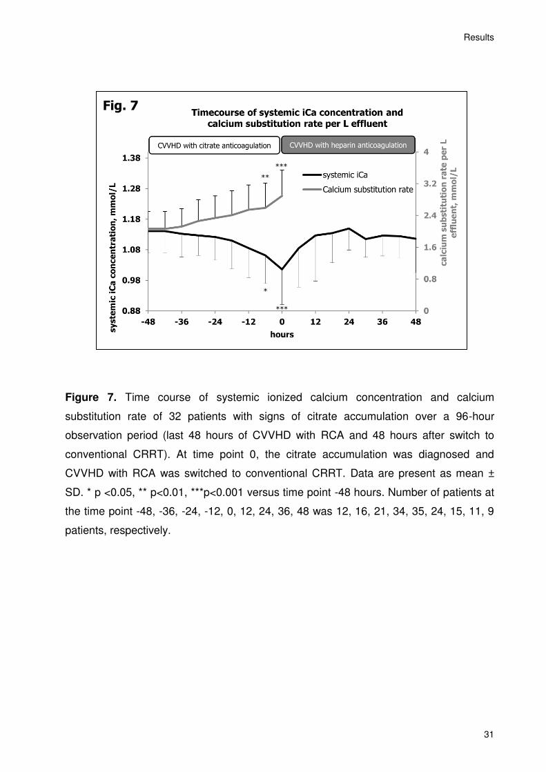
citrate accumulation was diagnosed. It is of interest that both gradually decreasing
ionized calcium and calcium-substitution were 12-24 hours prior to acid-base
disarrangements. However, significant difference was reached only 6 hours prior to
diagnosis of citrate accumulation (Fig. 7).
Results
28
Figure 5 (A, B). Time course of metabolic characteristics of 32 patients with signs of
citrate accumulation over a 96-hour observation period (last 48 hours of CVVHD with
RCA and 48 hours after switch to conventional CRRT). At time point 0, the citrate
accumulation was diagnosed and CVVHD with RCA was switched to conventional
CRRT: - A) arterial pH; - B) base excess. Data are present as mean ± SD. * p <0.05, **
p<0.01, ***p<0.001 versus time point -48 hours. Number of patients at the time point -
48, -36, -24, -12, 0, 12, 24, 36, 48 was 12, 16, 21, 34, 35, 24, 15, 11, 9 patients,
respectively.
Fig. 5 - B
***
*** ***
**
*
Fig. 5 - A
*** ***
*** **
* * *
*
Results
29
Figure 5 (C, D). Time course of metabolic characteristics of 32 patients with signs of
citrate accumulation over a 96-hour observation period (last 48 hours of CVVHD with
RCA and 48 hours after switch to conventional CRRT). At time point 0, the citrate
accumulation was diagnosed and CVVHD with RCA was switched to conventional
CRRT: - C) bicarbonate; - D) anion gap. Data are present as mean ± SD. * p <0.05, **
p<0.01, ***p<0.001 versus time point -48 hours. Number of patients at the time point -
48, -36, -24, -12, 0, 12, 24, 36, 48 was 12, 16, 21, 34, 35, 24, 15, 11, 9 patients,
respectively.
Fig. 5 - C
***
*** ***
* **
Fig. 5 - D
*** **
**
mm
ol/L
Results
30
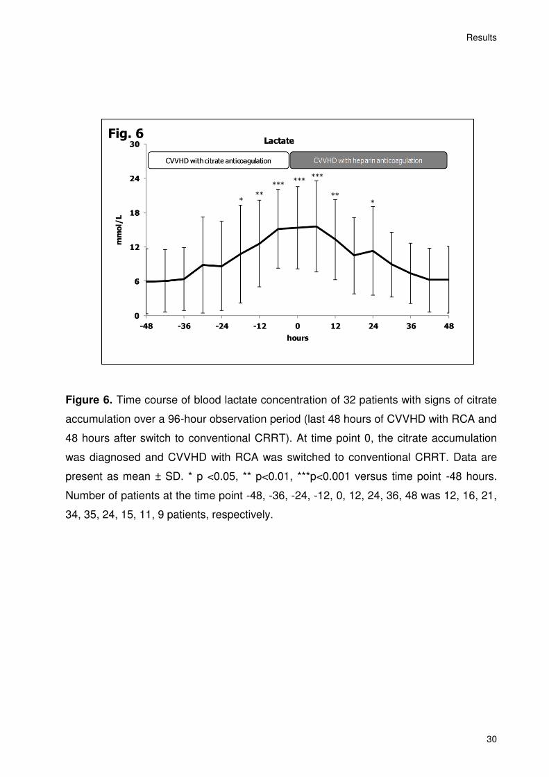
Figure 6. Time course of blood lactate concentration of 32 patients with signs of citrate
accumulation over a 96-hour observation period (last 48 hours of CVVHD with RCA and
48 hours after switch to conventional CRRT). At time point 0, the citrate accumulation
was diagnosed and CVVHD with RCA was switched to conventional CRRT. Data are
present as mean ± SD. * p <0.05, ** p<0.01, ***p<0.001 versus time point -48 hours.
Number of patients at the time point -48, -36, -24, -12, 0, 12, 24, 36, 48 was 12, 16, 21,
34, 35, 24, 15, 11, 9 patients, respectively.
Fig. 6
** *
*** ***
***
** *
Results
31
Figure 7. Time course of systemic ionized calcium concentration and calcium
substitution rate of 32 patients with signs of citrate accumulation over a 96-hour
observation period (last 48 hours of CVVHD with RCA and 48 hours after switch to
conventional CRRT). At time point 0, the citrate accumulation was diagnosed and
CVVHD with RCA was switched to conventional CRRT. Data are present as mean ±
SD. * p <0.05, ** p<0.01, ***p<0.001 versus time point -48 hours. Number of patients at
the time point -48, -36, -24, -12, 0, 12, 24, 36, 48 was 12, 16, 21, 34, 35, 24, 15, 11, 9
patients, respectively.
Fig. 7
***
***
**
*
Results
32
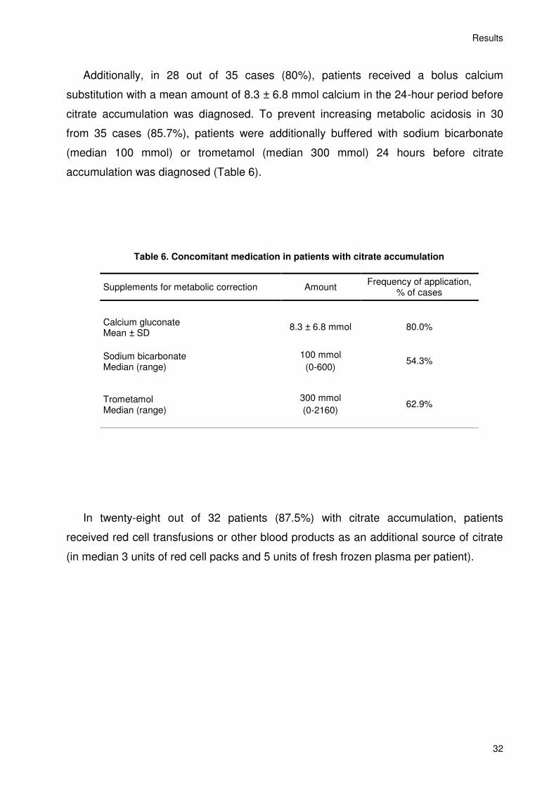
Additionally, in 28 out of 35 cases (80%), patients received a bolus calcium
substitution with a mean amount of 8.3 ± 6.8 mmol calcium in the 24-hour period before
citrate accumulation was diagnosed. To prevent increasing metabolic acidosis in 30
from 35 cases (85.7%), patients were additionally buffered with sodium bicarbonate
(median 100 mmol) or trometamol (median 300 mmol) 24 hours before citrate
accumulation was diagnosed (Table 6).
Table 6. Concomitant medication in patients with citrate accumulation
Supplements for metabolic correction Amount Frequency of application,
% of cases
Calcium gluconate
Mean ± SD 8.3 ± 6.8 mmol 80.0%
Sodium bicarbonate Median (range)
100 mmol
(0-600) 54.3%
Trometamol Median (range)
300 mmol
(0-2160) 62.9%
In twenty-eight out of 32 patients (87.5%) with citrate accumulation, patients
received red cell transfusions or other blood products as an additional source of citrate
(in median 3 units of red cell packs and 5 units of fresh frozen plasma per patient).
Results
33
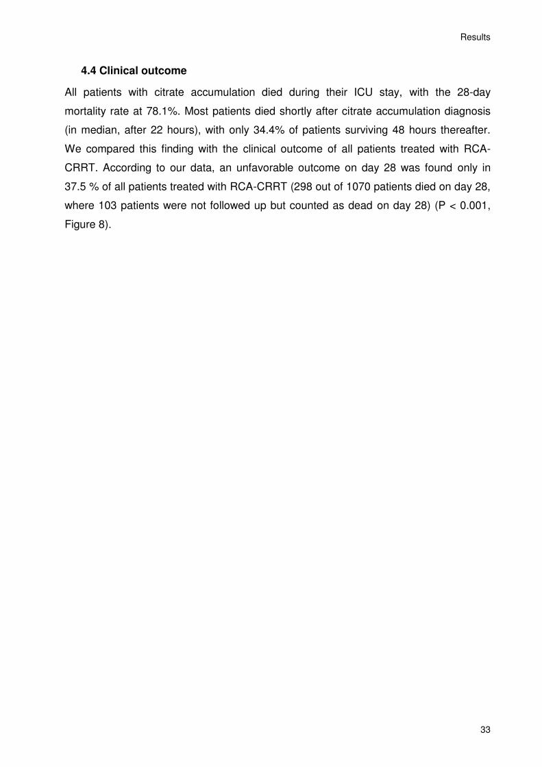
4.4 Clinical outcome
All patients with citrate accumulation died during their ICU stay, with the 28-day
mortality rate at 78.1%. Most patients died shortly after citrate accumulation diagnosis
(in median, after 22 hours), with only 34.4% of patients surviving 48 hours thereafter.
We compared this finding with the clinical outcome of all patients treated with RCA-
CRRT. According to our data, an unfavorable outcome on day 28 was found only in
37.5 % of all patients treated with RCA-CRRT (298 out of 1070 patients died on day 28,
where 103 patients were not followed up but counted as dead on day 28) (P < 0.001,
Figure 8).
Results
34
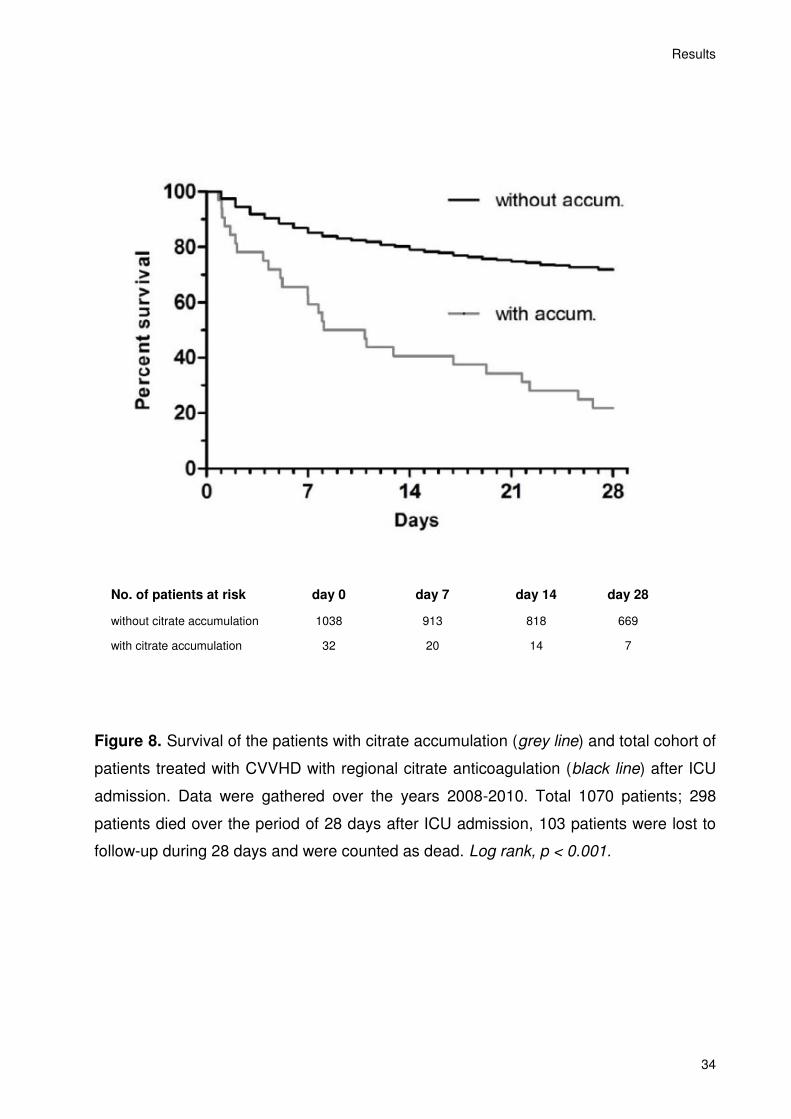
No. of patients at risk day 0 day 7 day 14 day 28
without citrate accumulation 1038 913 818 669
with citrate accumulation 32 20 14 7
Figure 8. Survival of the patients with citrate accumulation (grey line) and total cohort of
patients treated with CVVHD with regional citrate anticoagulation (black line) after ICU
admission. Data were gathered over the years 2008-2010. Total 1070 patients; 298
patients died over the period of 28 days after ICU admission, 103 patients were lost to
follow-up during 28 days and were counted as dead. Log rank, p < 0.001.
Discussion
35
5. Discussion
Taking into consideration the advantages of CRRT, current KDIGO Clinical Practice
Guideline for Acute Kidney Injury (2012) suggests using CRRT, rather than standard
intermittent RRT, for hemodynamically unstable patients, patients with increased
intracranial pressure, or generalized brain edema [3]. The most important disadvantage
of CRRT is the need for continuous anticoagulation. For a long time the anticoagulation
during the CRRT represented a great challenge. Based on recent clinical trials and
meta-analyses, KDIGO Clinical Practice Guideline for Acute Kidney Injury (2012)
suggests using RCA rather than heparin for anticoagulation during CRRT in patients
who do not have contraindications for citrate, independently of their bleeding risk [3],
and who are not already systemically anticoagulated for other reasons. Despite this
recommendation and otherwise obvious advantage of RCA over standard heparin
anticoagulation, the application of RCA in clinical practice is still limited to 0–20% of the
patients/treatments in recently published trials [7]. The main reason for avoiding RCA is
the fear of metabolic disarrangements due to RCA in critically ill patients with AKI
requiring RRT. However, metabolic complications due to RCA are infrequent in patients
without contraindication to RCA. According to the actual KDIGO Clinical Practice
Guideline for AKI, severely impaired liver function or shock with muscle hypoperfusion
represent the major contraindications for the use of RCA [3]. Both shock and decreased
liver function are supposed risk factors for citrate accumulation. In other words, risk for
citrate accumulation represents a major contraindication for RCA-CRRT. Thus, it is
crucial to reveal those patients and their common metabolic characteristics in order to
meet the clinical decision of which anticoagulation to choose during CRRT.
During the last years some clinical evidence has been gathered showing that shock
and liver impairment do not necessary lead to citrate accumulation during RCA-CRRT
[3, 20, 46, 64, 67]. Thus, those factors should be considered as only a relative, and not
absolute, contraindication for RCA. Furthermore, consequently excluding
hemodynamically instable patients in shock for treatment with RCA-CRRT, one actually
excludes a significant part of patients who, according to KDIGO Clinical Practice
Guideline for AKI, would especially profit from CRRT and not from intermittent RRT.
Under these circumstances, it is of particular interest to know what is the actual
incidence of citrate accumulation in critically ill patients undergoing RCA-CVVHD. In our
opinion, that question cannot be addressed at this time, as there is a lack of scientific
Discussion
36
evidence. All available studies describing citrate accumulation during CRRT might have,
at least to some extent, a selection bias (by including only the patients at risk for
bleeding or excluding certain patients because of the liver dysfunction, risk for citrate
accumulation, etc.) and, thus, might cause an underestimation of the incidence of this
complication. Moreover, some trials have a limited observational period, which could
lead to an underestimation of the incidence rate of citrate accumulation [20]. In Charité
university hospital, RCA-CVVHD is the modality of choice for hemodynamically instable
patients with AKI since 2006. Until the actual recommendations of KDIGO Clinical
Practice Guideline for AKI published in 2012, each patient requiring CRRT at our center
was initially treated with RCA-CRRT independently of bleeding risk, liver function,
muscle hypoperfusion, or shock. We performed a retrospective study analyzing the
incidence of citrate accumulation in a three year period (2008-2010).
The aim of the present study was to determine the incidence of metabolic
disarrangements consistent with citrate accumulation and to assess the common
metabolic findings of this complication. The study was performed in a large,
representative group of critically ill patients requiring RRT and treated with standardized
RCA-CRRT.
Discussion
37
5.1 Incidence of metabolic signs of citrate accumulation
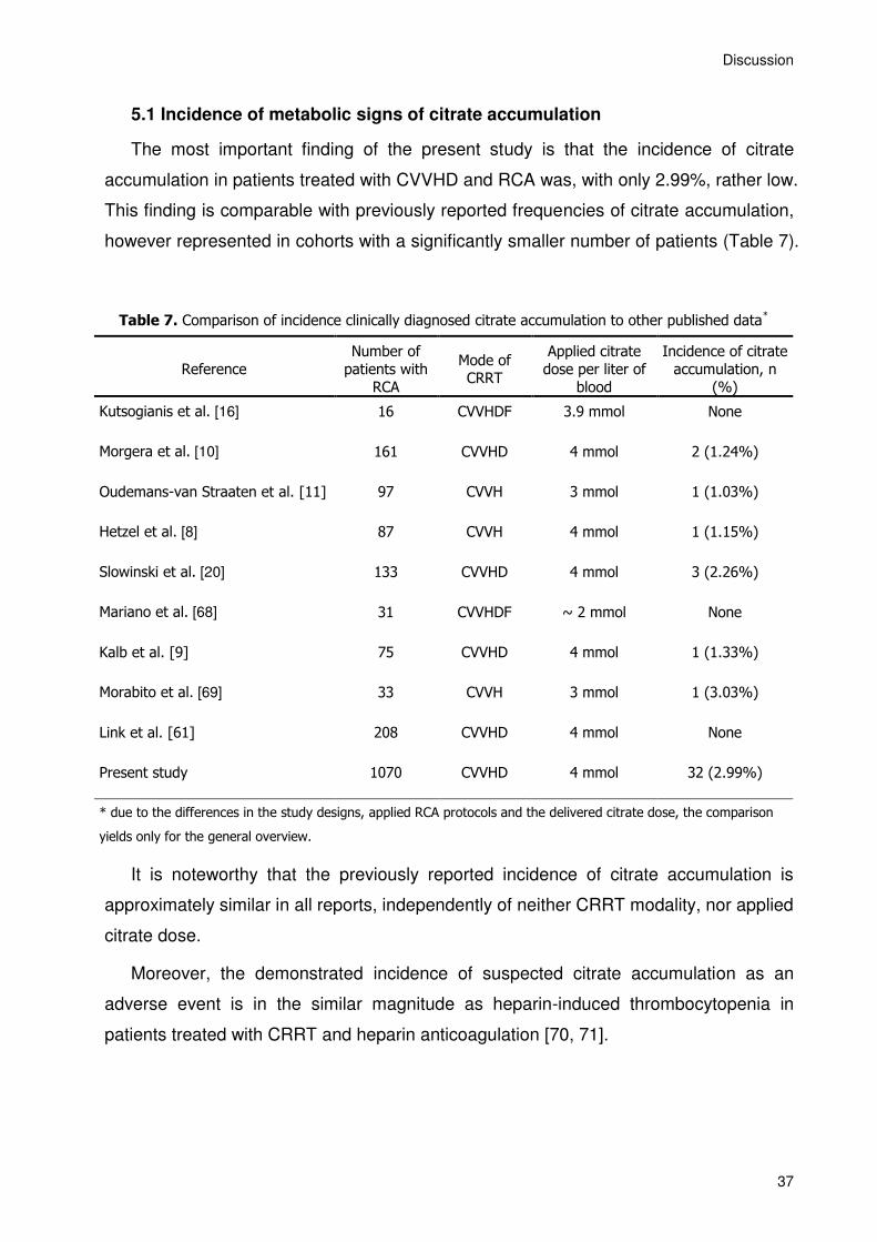
The most important finding of the present study is that the incidence of citrate
accumulation in patients treated with CVVHD and RCA was, with only 2.99%, rather low.
This finding is comparable with previously reported frequencies of citrate accumulation,
however represented in cohorts with a significantly smaller number of patients (Table 7).
Table 7. Comparison of incidence clinically diagnosed citrate accumulation to other published data*
Reference
Number of
patients with
RCA
Mode of CRRT
Applied citrate
dose per liter of
blood
Incidence of citrate
accumulation, n
(%)
Kutsogianis et al. [16] 16 CVVHDF 3.9 mmol None
Morgera et al. [10] 161 CVVHD 4 mmol 2 (1.24%)
Oudemans-van Straaten et al. [11] 97 CVVH 3 mmol 1 (1.03%)
Hetzel et al. [8] 87 CVVH 4 mmol 1 (1.15%)
Slowinski et al. [20] 133 CVVHD 4 mmol 3 (2.26%)
Mariano et al. [68] 31 CVVHDF ~ 2 mmol None
Kalb et al. [9] 75 CVVHD 4 mmol 1 (1.33%)
Morabito et al. [69] 33 CVVH 3 mmol 1 (3.03%)
Link et al. [61] 208 CVVHD 4 mmol None
Present study 1070 CVVHD 4 mmol 32 (2.99%)
* due to the differences in the study designs, applied RCA protocols and the delivered citrate dose, the comparison
yields only for the general overview.
It is noteworthy that the previously reported incidence of citrate accumulation is
approximately similar in all reports, independently of neither CRRT modality, nor applied
citrate dose.
Moreover, the demonstrated incidence of suspected citrate accumulation as an
adverse event is in the similar magnitude as heparin-induced thrombocytopenia in
patients treated with CRRT and heparin anticoagulation [70, 71].
Discussion
38
5.2 Metabolic signs of citrate accumulation
Only 34% of patients with citrate accumulation had a pre-existing liver impairment.
This finding conflicts to some extent with the common concern that patients with liver
failure are the most relevant patient group at risk to develop citrate accumulation. On
the other hand, our finding is in line with a recent prospective observational multicenter
study conducted at our department, where the safety and efficacy of regional citrate
anticoagulation in ICU patients having normal and impaired liver function was evaluated
[20]. Together with other European centers, we have demonstrated that CVVHD with
RCA can be safely used in patients with different grades of liver dysfunction, defined, as
well as in our study, according to the total serum bilirubin. Interestingly, the 2.26%
incidence of clinical diagnosis of citrate accumulation was even slightly lower than that
reported in the current study. Although the attempt to include patients with elevated S-
bilirubin in that study could have been expected to result in a higher incidence rate, the
observation period was limited to first 72 hours of CVVHD treatment with a possible
underestimation of the citrate accumulation frequency in that patient population.
In the present study, citrate accumulation was diagnosed based on ionized
hypocalcemia in combination with elevated total/ionized calcium ratio, metabolic
acidosis with elevated anion gap, and severe lactic acidosis. Despite the calcium
substitution via the dialysis device that increased by a mean of 28.7% compared to
baseline, the majority of patients (80%) required additional bolus calcium substitution.
85.7% of the patients required additional buffering with sodium bicarbonate, trometamol,
or both. Hypocalcemia with sustained increase of calcium substitution by the dialysis
device as well as a parallel continuous increase of lactate concentration, were the first
clinically recognizable signs of imminent citrate accumulation. The increased calcium
substitution requirement as a sign of possible citrate accumulation was also described
in 1999 by Meier-Kriesche et al. [12].
Although most experts and guidelines on citrate anticoagulation report on an
association between citrate accumulation and lactic acidosis, to the best of our
knowledge the incidence, clinical outcome and laboratory findings of citrate
accumulation have been poorly described in the literature so far. There is a very
plausible biochemical rationale for a causal relationship. Both anions require oxygen
during metabolism, which is obvious in the case of complete oxidation but also evident
for other metabolic pathways. For instance, gluconeogenesis from lactate requires more
Discussion
39
ATP-equivalents than those generated in anaerobic glycolysis. This clearly suggests
that ATP from oxidative metabolism is required to convert larger quantities of lactate
into glucose. After entering the Krebs cycle, citrate is metabolized via the isocitrate-
dehydrogenase with hydrogen being transferred to NADH. As the coenzyme NADH
needs to be regenerated to NAD+ in the respiratory chain, the metabolism of citrate is
therefore oxygen-dependent. Thus, the parallel accumulation of citrate and lactate likely
indicates an insufficient oxidative metabolism, e.g. a blockade in the regeneration of
NAD+ from NADH via the respiratory chain. Remarkably, after a switch to conventional
CRRT, the resolution of metabolic disorders was observed. That could be explained by
1) application of bicarbonate- and calcium-rich dialysate, 2) decreasing number of
patients due to death, with only 11 out of 32 patients surviving > 48 hours, and 3) partial
recovery of citrate metabolism in short-term survivors. One of the further assumptive
explanations for lactate levels reduction could be an improvement of cardiac contractility
after normalization of acid-base status and/or resolution of hypocalcaemia.
From our study, it is not possible to determine a threshold for lactate, which predicts
impaired citrate metabolism, although the mean lactate concentration of 14.99 mmol/L
at the time of diagnosis of citrate accumulation was very high. While a high lactate
concentration at the start of CRRT should be an alert of the risk of citrate accumulation,
certainly not all patients with lactic acidosis will obligatorily accumulate citrate. On the
contrary, considerable proportion of shock patients with a high lactate level do
metabolize citrate remarkably well if circulation and oxygen delivery improves during
treatment of shock. Thus we suggest that additional attention and close monitoring of
calcium homeostasis should be assured in patients suffering from a severe lactic
acidosis and treated with any RCA-CRRT set-up.
Finally, it is worth mentioning that the majority of patients with citrate accumulation
received a considerable amount of blood products as an additional source of citrate
over the last 24 hours before this complication occurred. One unit of packed red blood
cells (RBC) contains approximately 16 mmol of citric acid. Thus, patients with massive
blood transfusions are at an additional risk of developing citrate accumulation,
regardless of RCA required for CRRT. This finding correlates with previously published
studies [56-58, 72].
Discussion
40
5.3 Clinical outcome in patients with metabolic signs of citrate accumulation
Another important finding of the actual study is that all patients with citrate
accumulation had, in comparison to the whole study population, significantly higher
APACHE II and SAPS II scores that are well-established as being linked to higher
mortality rates. All of these patients had severe therapy resistant shock with multiple
organ dysfunctions and died during their ICU stay. These findings indicate that those
patients did have a very limited prognosis, per se, and citrate accumulation might simply
reflect the severity of the underlying disorders and the breakdown of cellular respiration.
Our results are in line with the recently published work of Link et al [61]. The authors
proved that in patients on CRRT with RCA total-to-ionized calcium ratio correlates with
the clinical outcome and is an independent predictor of 28-days mortality.
One would argue the 28-days mortality rate (37.5%) of the patients in actual study to
be low in comparison with international prospective multicenter studies on mortality of
critically ill patients with need for RRT [1, 73]. The BEST-Kidney Study included 1753
critically ill patients with AKI, a study population that was comparable with the study
population in actual survey (mean age 63.2 years, 64% males, mean SAPS II Score
50.3, mean SOFA Score 10.5). Indeed, in the BEST-Kidney Study with mainly
conventional anicoagulated RRT, the total 28-days mortality rate was more than 60%
[73]. There are several explanations for the considerably different 28-days mortality rate
between both studies. First of all, the BEST-Kidney Study included patients from
different European centers, thus providing very heterogenic outcomes from center to
center, with hospital mortality rate from 22.2 to 100% [1]. Furthermore, in the BEST-
Kidney Study the proportion of the surgical reasons for ICU admission was considerably
lower than in the actual investigation (41.1 % vs 53.2%). Besides, in the BEST-Kidney
Study 80% of patients received the CRRT with very heterogeneous anticoagulation
modalities. Regional citrate anticoagulation could have a positive effect on the survival
rate due to reduced bleeding risk, especially in patients after surgical intervention [8, 16,
51]. Furthermore, due to the fact that in our study all patients were initially treated
exclusively with RCA, the other possible explanation of a better outcome data from our
study is pro-inflammatory effects of heparin and/or anti-inflammatory properties of
citrate itself [4]. Some data shows that heparin possess potentially pro-inflammatory
effects by, for example, releasing the mediators of inflammation from leucocytes and
thrombocytes [37, 38, 74-76]. On the other hand it has been assumed that a local
Discussion
41
hypocalcemia in the area of dialysis membrane could reduce the release of
inflammatory cytokines from the cells adhered to the membrane [77, 78].
Noteworthy, survival rate in patients treated with RCA-CRRT have been reported
recently in the prospective randomized trial [11]. Authors have shown a significantly
better outcome in the group of patients treated with RCA-CRRT compared to those
treated with CRRT with LMWH-anticoagulation. Remarkably, those effects were
independent from the differences in bleeding incidence. A better outcome of the
patients treated with RCA-CRRT as those, anticoagulated with heparin during CRRT,
described by Oudemans-van Straaten et al, was questioned by Hetzel et al. In the
multicentre, controlled, randomized, open, prospective clinical trial comparing RCA and
systemic anticoagulation with heparin during CRRT. The authors didn’t confirm the
survival benefit of patients treated with RCA-CRRT.
Discussion
42
5.4 Limitations of the study
There are several limitations of the present study to be stressed and addressed.
Firstly, it has the usual limitations of single-center retrospective studies. However, the
number of patients we retrospectively analyzed is considerably large - inclusion of 6
ICUs with different specializations allowed the involvement of a multitude of morbidities
covering the range of what could possibly be present in an ICU, and our data are
comparable with citrate accumulation incidences reported in other prospective trials
using smaller patients numbers (Table 7). Additionally, using citrate as the standard
anticoagulation method for CVVHD in every patient treated with CRRT and without any
exclusion criteria except clinically diagnosed citrate accumulation, allowed us to
eliminate any selection bias, which may be present even in some prospective trials.
Secondly, we used the surrogate criteria to define citrate accumulation without
actually measuring the citrate level. Indeed, the measurement of blood citrate
concentration is, at least in Germany, not clinically approved, and, thus, not available in
laboratory routine, making such investigation infeasible for a large cohort of patients. On
the other hand, the parameters we used are widely accepted. The cut-off levels of total-
to-ionized calcium ratio, the most specific criteria for diagnosis of citrate accumulation,
vary. To increase sensitivity of our monitoring we accepted a cut-off level ratio of >2.1,
as previously described [13, 62].
Thirdly, a possible limitation of the actual study is the use of total serum bilirubin as
a main parameter for liver function. However, there is no physiologic parameter
applicable for clinical use that allows for early detection of hepatic dysfunction; indeed
all current diagnostic criteria are based on laboratory evaluation. Serum bilirubin, as a
steady marker of hepatic impairment [79], is a key component of prognostic scores of
chronic liver disease and cirrhosis as well as prognostic models in patients with acute
liver failure [80]. It could be assumed that international normalized ratio (INR), as a
parameter of liver synthesis, may be a better predictor of the liver ability to metabolise
citrate. However, INR is elevated in the advanced stages of liver failure and only
represents very severe forms of liver dysfunction. Furthermore, the substitution of
plasma products as well as disseminated intravascular coagulopathy, often observed
during severe sepsis, can influence INR. Moreover, serum bilirubin was used for
classification of liver dysfunction in others study as well [20, 60].
Fourthly, in our study all patients were treated with CVVHD as a basis CRRT
Discussion
43
modality, which might have had an impact on citrate clearance. Yet, citrate has a low
molecular weight and its sieving coefficient is close to 1, and thus, its clearance remains
the same during diffusion or convection [81, 82]. In fact, Morgera et al. [10] had based
our RCA-CVVHD protocol on moderately high blood flows as a lower blood flow
reduces citrate requirements for anticoagulation and with a blood-to-dialysate flow ratio
of 3:1, as this allows the assumption of near complete equilibration of low-molecular
weight solutes. Therefore, the citrate exposure during CVVHD should be mainly dose-
dependent and almost irrespective from the renal replacement technique used (CVVH,
CVVHDF), and thus, similar to such described in present study.
And finally, in our center we used only one protocol according to Morgera et al with a
fixed starting dose of citrate of approximately 4 mmol per liter of blood [10]. Indeed, in
patients with ionized calcium concentrations in the normal range, 4 mmol citrate per liter
blood is sufficient to completely inhibit the coagulation cascade in the extracorporeal
circuit. Ionized calcium and coagulation are not linked in a linear proportion. On the
contrary, ionized calcium has to be lowered to <0.40 mmol/l to provide any
anticoagulation, and coagulation ability is completely unaffected with ionized calcium
concentrations above 0.40 mmol/l [49]. To reach a reasonable range (0.25-0.35 mmol/l)
in the extracorporeal circuit from normal blood calcium concentrations (1.1-1.3 mmol/l),
approximately 4 mmol of citrate per liter blood is needed [48]. Therefore, citrate-sparing
protocols are not feasible or at least not ideal for anticoagulation. In patients with clinical
signs or a known risk for citrate accumulation, a strategy to reduce a citrate load should
be applied: reduction of blood flow with subsequent decrease in citrate dose, or/and
increase of extracorporeal citrate clearance by increasing the dialysate flow [61].
Noteworthy, increasing the dialysate flow, at least in our protocol, will subsequently lead
to worsening of the metabolic acidosis, due to the reduced bicarbonate concentration in
the dialysate. However, this could be, at least partially, compensated by additional
bicarbonate infusion. Our retrospective analysis shows that reduction of citrate load was
not consequently applied in each patient with suspected citrate accumulation. Due to
retrospective character of the study, we can only speculate about the reasons: lack of
experience in handling the RCA in early years; rapid onset of metabolic
disarrangements with consequent need of prompt change of CRRT modality; very
limited prognosis of four patients where the CRRT was discontinued; worsening of the
metabolic acidosis when elevating the dialysate flow with reduced bicarbonate
Discussion
44
concentration; and reduced dialysis efficacy when decreasing the blood flow in terms of
hyperkalemia. According to the actual knowledge and our present results, we would
strongly recommend applying strategies to reduce the citrate load in all patients where
citrate accumulation would even be suspected.
Summary
45
6. Summary
Continuous renal replacement therapy (CRRT) is increasingly applied in critically ill
patients with acute kidney injury who requires renal support. CRRT is often preferred to
intermittent dialysis because of better hemodynamic stability and metabolic control.
Regional citrate anticoagulation (RCA) during CRRT (RCA-CRRT) is a common
alternative to systemic heparin anticoagulation, especially by patients with increased
bleeding risk. Indeed, in recently published guidelines RCA is now recommended as the
anticoagulation strategy of choice in patients undergoing CRRT without contraindication
to citrate. According to KDIGO guidelines, the major contraindications for RCA are:
severely impaired liver function or shock with muscle hypoperfusion, both representing
a risk of citrate accumulation.
Citrate accumulation is a serious complication of CRRT with RCA. It could cause
severe metabolic acidosis, decrease cardiac contractility or cause arrhythmias, as
symptoms of systemic ionized hypocalcemia. Acute or chronic impaired liver function,
shock with arterial hypoxia and reduced tissue perfusion are the major risk factors for
citrate accumulation. Unfortunately measurement of citrate concentration in blood is not
available on a daily routine basis, and, at least in Germany, available test kits are not
approved for clinical use. There are however commonly accepted markers for citrate
accumulation, such as: metabolic acidosis with or without increased anion gap, ionized
hypocalcemia with simultaneous increased levels of total calcium and, an increased
total calcium to ionized calcium ratio (tCa/iCa). Those laboratory parameters do not
confirm citrate accumulation in all cases, and often lead to false positive results, what
makes the diagnosis of citrate accumulation a complex clinical issue. More over,
information about the incidence of citrate accumulation in general cohort of ICU patients
undergoing RCA-CRRT is relatively limited, due to the fact that patients with risk factors
for citrate accumulation were usually whether excluded from the prospective trials, or
the observation of those patients were limited to the study period.
In this monocentric retrospective study we collected and analyzed all patients on
RCA-CRRT over a three-year period (from 2008 to 2010) to identify risk factors for
citrate accumulation. The primary objective was to reveal the incidence rate of citrate
accumulation in cohort of non-selected critically ill patients receiving RCA-CRRT. The
secondary objective was to assess the clinical characteristics and outcome related to
citrate accumulation based on a representative population of patients.
Summary
46
The results of the present research show that the incidence of metabolic
disarrangements consistent with citrate accumulation was rather low since it affected
only 2.99% of all RCA-CRRT patients. Our findings show that RCA during CRRT is
feasible and complications are uncommon independently of a liver function, shock or
presented muscle hypoperfusion. Diagnosis of citrate accumulation was found
exclusively in severely ill patients with multiorgan failure and severe lactic acidosis, thus
representing a group of patients with a very poor prognosis per se. All patients with
suspected citrate accumulation died during their ICU stay. Our findings suggest that
citrate accumulation occurs secondary to severe disability of cellular respiration and that
citrate accumulation could be interpreted as a strong indicator for patient's poor
outcome. We suggest that close attention should be paid to patients treated with RCA-
CRRT who have a severe lactic acidosis and an elevated demand for calcium
substitution.
Zusammenfassung
47
7. Zusammenfassung
Die kontinuierliche Nierenersatztherapie wird beim Großteil der kritisch kranken
Patienten mit dialysepflichtigen akutem Nierenversagen auf Intensivstationen
verwendet. Die kontinuierliche Nierenersatztherapie wird insbesondere bei
hämodynamisch instabilen patienten gegenüber der intermittierenden Dialyse wegen
der schonenderen Ultrafiltration und der besseren Stoffwechselkontrolle bevorzugt. Die
regionale Zitratantikoagulation während der kontinuierlichen Nierenersatztherapie ist
eine zunehmend häufiger eingesetzte Alternative zur systemischen Antikoagulation,
insbesondere bei Patienten mit erhöhtem Blutungsrisiko. In den kürzlich veröffentlichten
Leitlinien zur Antikoagulation bei kontinuierlicher Nierensesartztherapie (KDIGO 2012)
wird die regionale Zitratantikoagulation empfohlen als die Antikoagulation der Wahl bei
Patienten ohne Kontraindikation für Zitratantikoagulation und ohne systemische
Antikoagulation aus anderen patientenbezogenen Gründen. Die wichtigsten
Kontraindikationen für RCA laut den KDIGO Leitlinien sind folgende: stark
eingeschränkte Leberfunktion oder Schock mit Muskelminderdurchblutung. Beide
Risikofaktoren entsprechen den Risikofaktoren der Zitratakkumulation.
Die systemische Zitratakkumulation stellt eine Komplikation kontinuierlichen
Nierenersatzverfahrens mit regionaler Zitratantikoagulation dar. Die möglichen
klinischen Folgen der Zitratakkumulation sind schwere metabolische Azidose,
verminderte Herzkontraktilität und Herzrhythmusstörungen. Leider ist die Messung der
Zitratkonzentration im Blut im klinischen Alltag nicht verfügbar. Allgemein anerkannte
Marker der Zitratakkumulation sind rasch fallende Konzentration des ionisierten
Kalziums bei gleichzeitig steigendem Kalziumsubstitutionsbedarf, erhöhter Quotient aus
Gesamtkalzium zu ionisierten Kalzium, sowie metabolische Azidose mit oder ohne
erweiterter Anionenlücke. Diese Laborparameter bestätigen die Zitratakkumulation nicht
in allen Fällen und führen häufig zu falschen positiven Ergebnissen. Somit ist die
Diagnose der Zitratakkumulation ein komplexes klinisches Problem. Weiterhin ist die
Information über die Inzidenz der Zitratakkumulation in allgemeiner Kohorte von
dialysepflichtigen kritisch kranken Patienten auf der Intensivstation gering, weil die
Patienten mit Risikofaktoren für Zitratakkumulation in der Regel aus den bisherigen
prospektiven Studien ausgeschlossen wurden oder der Beobachtungszeitraum
vergleichsweise kurz war.
In dieser monozentrischen retrospektiven Studie wurden alle Patienten die mit
Zusammenfassung
48
kontinuierlicher Nierenersatztherapie mit regionaler Zitratantikoagulation auf der
Intensivstationen behandelt worden über einen Zeitraum von drei Jahren (von 2008 bis
2010) identifiziert und analysiert. Das primäre Ziel war es, die Häufigkeit der
Zitratakkumulation in einer Kohorte von nicht selektierten kritisch kranken Patienten, die
mit RCA-CRRT behandelt wurden, zu ermitteln. Das weitere Ziel war es, die klinischen
Merkmale sowie das Outcome der Zitratakkumulation aufgrund einer repräsentativen
Patientenpopulation zu bewerten.
Die Ergebnisse der vorliegenden Arbeit zeigen, dass die Inzidenz der
Zitratakkumulation mit 2.99 % gering war. Die Ergebnisse zeigen weiterhin, dass die
Zitratantikoagulation während der kontinuierlichen Nierenersatztherapie, unabhängig
von einer Leberfunktion, Schock oder Muskelminderdurchblutung, möglich sowie
komplikationsarm ist. Die Zitratakkumulation ist auschließlich bei Patienten mit
schwerster Laktatazidose bei Multiorganversagens und starkeingeschränkter Prognose
aufgetreten. Die Ergebnisse deuten auf das Auftreten der Zitratakkumulation als Folge
des schwerwiegenden Versagens der Zellatmung hin. Die Zitratakkumulation könnte
damit als ein starker Prädiktor für eine schlechte Prognose des Patienten interpretiert
werden. Die Patienten, die mit RCA-CRRT behandelt werden und gleichzeitig eine
schwere Laktatazidose mit erhöhtem Kalziumsubstitutionsbedarf haben, sind
besonderes zu beachten.
Reference List
49
8. Reference list
1. Uchino S, Kellum JA, Bellomo R, Doig GS, Morimatsu H, Morgera S, Schetz M,
Tan I, Bouman C, Macedo E, Gibney N, Tolwani A, Ronco C: Acute renal failure
in critically ill patients: a multinational, multicenter study. JAMA 2005, 294(7):813-
818.
2. Rabindranath K, Adams J, Macleod AM, Muirhead N: Intermittent versus
continuous renal replacement therapy for acute renal failure in adults. Cochrane
Database Syst Rev 2007(3):CD003773.
3. KDIGO AKI Work Group: KDIGO Clinical Practice Guideline for Acute Kidney
Injury. Kidney Int Suppl 2012, 2(1):1-138.
4. Oudemans-van Straaten HM, Kellum JA, Bellomo R: Clinical review:
anticoagulation for continuous renal replacement therapy--heparin or citrate? Crit
Care 2011, 15(1):202.
5. Bellomo R, Cass A, Cole L, Finfer S, Gallagher M, Lo S, McArthur C,
McGuinness S, Myburgh J, Norton R, Scheinkestel C, Su S: Intensity of
continuous renal-replacement therapy in critically ill patients. N Engl J Med 2009,
361(17):1627-1638.
6. Palevsky PM, Zhang JH, O'Connor TZ, Chertow GM, Crowley ST, Choudhury D,
Finkel K, Kellum JA, Paganini E, Schein RM, Smith MW, Swanson KM,
Thompson BT, Vijayan A, Watnick S, Star RA, Peduzzi P: Intensity of renal
support in critically ill patients with acute kidney injury. N Engl J Med 2008,
359(1):7-20.
7. Uchino S, Bellomo R, Morimatsu H, Morgera S, Schetz M, Tan I, Bouman C,
Macedo E, Gibney N, Tolwani A, Oudemans-van Straaten H, Ronco C, Kellum
JA: Continuous renal replacement therapy: a worldwide practice survey. The
beginning and ending supportive therapy for the kidney (B.E.S.T. kidney)
investigators. Intensive Care Med 2007, 33(9):1563-1570.
8. Hetzel GR, Schmitz M, Wissing H, Ries W, Schott G, Heering PJ, Isgro F,
Kribben A, Himmele R, Grabensee B, Rump LC: Regional citrate versus
systemic heparin for anticoagulation in critically ill patients on continuous
venovenous haemofiltration: a prospective randomized multicentre trial. Nephrol
Reference List
50
Dial Transplant 2011, 26(1):232-239.
9. Kalb R, Kram R, Morgera S, Slowinski T, Kindgen-Milles D: Regional citrate
anticoagulation for high volume continuous venovenous hemodialysis in surgical
patients with high bleeding risk. Ther Apher Dial 2013, 17(2):202-212.
10. Morgera S, Schneider M, Slowinski T, Vargas-Hein O, Zuckermann-Becker H,
Peters H, Kindgen-Milles D, Neumayer HH: A safe citrate anticoagulation
protocol with variable treatment efficacy and excellent control of the acid-base
status. Crit Care Med 2009, 37(6):2018-2024.
11. Oudemans-van Straaten HM, Bosman RJ, Koopmans M, van der Voort PH,
Wester JP, van der Spoel JI, Dijksman LM, Zandstra DF: Citrate anticoagulation
for continuous venovenous hemofiltration. Crit Care Med 2009, 37(2):545-552.
12. Meier-Kriesche HU, Finkel KW, Gitomer JJ, DuBose TD, Jr.: Unexpected severe
hypocalcemia during continuous venovenous hemodialysis with regional citrate
anticoagulation. Am J Kidney Dis 1999, 33(4):e8.
13. Joannidis M, Oudemans-van Straaten HM: Clinical review: Patency of the circuit
in continuous renal replacement therapy. Crit Care 2007, 11(4):218.
14. Oudemans-van Straaten HM: Citrate anticoagulation for continuous renal
replacement therapy in the critically ill. Blood Purif 2010, 29(2):191-196.
15. Mariano F, Bergamo D, Gangemi EN, Hollo Z, Stella M, Triolo G: Citrate
anticoagulation for continuous renal replacement therapy in critically ill patients:
success and limits. Int J Nephrol 2011, 2011:748320.
16. Kutsogiannis DJ, Gibney RT, Stollery D, Gao J: Regional citrate versus systemic
heparin anticoagulation for continuous renal replacement in critically ill patients.
Kidney Int 2005, 67(6):2361-2367.
17. Park JS, Kim GH, Kang CM, Lee CH: Regional anticoagulation with citrate is
superior to systemic anticoagulation with heparin in critically ill patients
undergoing continuous venovenous hemodiafiltration. Korean J Intern Med 2011,
26(1):68-75.
18. Fealy N, Baldwin I, Johnstone M, Egi M, Bellomo R: A pilot randomized
controlled crossover study comparing regional heparinization to regional citrate
anticoagulation for continuous venovenous hemofiltration. Int J Artif Organs 2007,

Reference List
51
30(4):301-307.
19. Monchi M, Berghmans D, Ledoux D, Canivet JL, Dubois B, Damas P: Citrate vs.
heparin for anticoagulation in continuous venovenous hemofiltration: a
prospective randomized study. Intensive Care Med 2004, 30(2):260-265.
20. Slowinski T, Morgera, S., Joannidis, M., Kindgen-Milles, D.: Multicenter
prospective observational study on safety and efficacy of regional citrate
anticoagulation in CVVHD in the presence of liver failure: the Liver Citrate
Anticoagulation Threshold Study (L-CAT). Critical Care 2011, 15(Suppl 1 ):127.
21. Bellomo R, Ronco C, Kellum JA, Mehta RL, Palevsky P: Acute renal failure -
definition, outcome measures, animal models, fluid therapy and information
technology needs: the Second International Consensus Conference of the Acute
Dialysis Quality Initiative (ADQI) Group. Crit Care 2004, 8(4):R204-212.
22. Mehta RL, Kellum JA, Shah SV, Molitoris BA, Ronco C, Warnock DG, Levin A:
Acute Kidney Injury Network: report of an initiative to improve outcomes in acute
kidney injury. Crit Care 2007, 11(2):R31.
23. de Mendonca A, Vincent JL, Suter PM, Moreno R, Dearden NM, Antonelli M,
Takala J, Sprung C, Cantraine F: Acute renal failure in the ICU: risk factors and
outcome evaluated by the SOFA score. Intensive Care Med 2000, 26(7):915-921.
24. Lins RL, Elseviers MM, Van der Niepen P, Hoste E, Malbrain ML, Damas P,
Devriendt J: Intermittent versus continuous renal replacement therapy for acute
kidney injury patients admitted to the intensive care unit: results of a randomized
clinical trial. Nephrol Dial Transplant 2009, 24(2):512-518.
25. Bagshaw SM, Berthiaume LR, Delaney A, Bellomo R: Continuous versus
intermittent renal replacement therapy for critically ill patients with acute kidney
injury: a meta-analysis. Crit Care Med 2008, 36(2):610-617.
26. Pannu N, Klarenbach S, Wiebe N, Manns B, Tonelli M: Renal replacement
therapy in patients with acute renal failure: a systematic review. JAMA 2008,
299(7):793-805.
27. Uchino S, Fealy N, Baldwin I, Morimatsu H, Bellomo R: Continuous venovenous
hemofiltration without anticoagulation. ASAIO J 2004, 50(1):76-80.
28. Morabito S, Guzzo I, Solazzo A, Muzi L, Luciani R, Pierucci A: Continuous renal
Reference List
52
replacement therapies: anticoagulation in the critically ill at high risk of bleeding.
J Nephrol 2003, 16(4):566-571.
29. Tan HK, Baldwin I, Bellomo R: Continuous veno-venous hemofiltration without
anticoagulation in high-risk patients. Intensive Care Med 2000, 26(11):1652-1657.
30. Martin PY, Chevrolet JC, Suter P, Favre H: Anticoagulation in patients treated by
continuous venovenous hemofiltration: a retrospective study. Am J Kidney Dis
1994, 24(5):806-812.
31. van de Wetering J, Westendorp RG, van der Hoeven JG, Stolk B, Feuth JD,
Chang PC: Heparin use in continuous renal replacement procedures: the
struggle between filter coagulation and patient hemorrhage. J Am Soc Nephrol
1996, 7(1):145-150.
32. Oudemans-van Straaten HM, Wester JP, de Pont AC, Schetz MR:
Anticoagulation strategies in continuous renal replacement therapy: can the
choice be evidence based? Intensive Care Med 2006, 32(2):188-202.
33. Warkentin TE, Greinacher A: Heparin-induced thrombocytopenia: recognition,
treatment, and prevention: the Seventh ACCP Conference on Antithrombotic and
Thrombolytic Therapy. Chest 2004, 126(3 Suppl):311S-337S.
34. Cohen JR, Sarfati I, Birnbaum E, Benacquista T, Wise L: The inactivation of
antithrombin III by serum elastase in patients with surgical infections. Am Surg
1990, 56(11):665-667.
35. Jordan RE, Nelson RM, Kilpatrick J, Newgren JO, Esmon PC, Fournel MA:
Antithrombin inactivation by neutrophil elastase requires heparin. Am J Med
1989, 87(3B):19S-22S.
36. Manson L, Weitz JI, Podor TJ, Hirsh J, Young E: The variable anticoagulant
response to unfractionated heparin in vivo reflects binding to plasma proteins
rather than clearance. J Lab Clin Med 1997, 130(6):649-655.
37. Warren BL, Eid A, Singer P, Pillay SS, Carl P, Novak I, Chalupa P, Atherstone A,
Penzes I, Kubler A, Knaub S, Keinecke HO, Heinrichs H, Schindel F, Juers M,
Bone RC, Opal SM: Caring for the critically ill patient. High-dose antithrombin III
in severe sepsis: a randomized controlled trial. JAMA 2001, 286(15):1869-1878.
38. Leithauser B, Schumacher J, Lendemans S, Tillmanns H, Matthias FR:
Reference List
53
Antithrombin attenuates microvascular leakage and leukocyte-endothelial
interaction in response to endotoxin. Semin Thromb Hemost 2002, 28 Suppl
1:87-94.
39. Hoffmann JN, Vollmar B, Laschke MW, Inthorn D, Kaneider NC, Dunzendorfer S,
Wiedermann CJ, Romisch J, Schildberg FW, Menger MD: Adverse effect of
heparin on antithrombin action during endotoxemia: microhemodynamic and
cellular mechanisms. Thromb Haemost 2002, 88(2):242-252.
40. Heinzelmann M, Bosshart H: Heparin binds to lipopolysaccharide (LPS)-binding
protein, facilitates the transfer of LPS to CD14, and enhances LPS-induced
activation of peripheral blood monocytes. J Immunol 2005, 174(4):2280-2287.
41. Betjes MG, van Oosterom D, van Agteren M, van de Wetering J: Regional citrate
versus heparin anticoagulation during venovenous hemofiltration in patients at
low risk for bleeding: similar hemofilter survival but significantly less bleeding. J
Nephrol 2007, 20(5):602-608.
42. Pinnick RV, Wiegmann TB, Diederich DA: Regional citrate anticoagulation for
hemodialysis in the patient at high risk for bleeding. N Engl J Med 1983,
308(5):258-261.
43. Morita Y, Johnson RW, Dorn RE, Hall DS: Regional anticoagulation during
hemodialysis using citrate. Am J Med Sci 1961, 242:32-43.
44. Mehta RL, McDonald BR, Aguilar MM, Ward DM: Regional citrate
anticoagulation for continuous arteriovenous hemodialysis in critically ill patients.
Kidney Int 1990, 38(5):976-981.
45. Robert S, Ghiotto J, Pirotte B, David JL, Masereel B, Pochet L, Dogne JM: Is
thrombin generation the new rapid, reliable and relevant pharmacological tool for
the development of anticoagulant drugs? Pharmacol Res 2009, 59(3):160-166.
46. Oudemans-van Straaten HM, Ostermann M: Bench-to-bedside review: Citrate for
continuous renal replacement therapy, from science to practice. Crit Care 2012,
16(6):249.
47. Toyoshima S, Fukuda T, Masumi S, Nakashima Y, Kawaguchi Y, Nakayama M:
Maximum acceptable infusion rate of citrate: relationship between blood ionized
calcium levels and cardiovascular effects in anesthetized rats. Clin Nutr 2006,
Reference List
54
25(4):653-660.
48. Calatzis A, Toepfer M, Schramm W, Spannagl M, Schiffl H: Citrate
anticoagulation for extracorporeal circuits: effects on whole blood coagulation
activation and clot formation. Nephron 2001, 89(2):233-236.
49. James MF, Roche AM: Dose-response relationship between plasma ionized
calcium concentration and thrombelastography. J Cardiothorac Vasc Anesth
2004, 18(5):581-586.
50. Morgera S, Scholle C, Voss G, Haase M, Vargas-Hein O, Krausch D, Melzer C,
Rosseau S, Zuckermann-Becker H, Neumayer HH: Metabolic complications
during regional citrate anticoagulation in continuous venovenous hemodialysis:
single-center experience. Nephron Clin Pract 2004, 97(4):c131-136.
51. Wu MY, Hsu YH, Bai CH, Lin YF, Wu CH, Tam KW: Regional Citrate Versus
Heparin Anticoagulation for Continuous Renal Replacement Therapy: A Meta-
Analysis of Randomized Controlled Trials. Am J Kidney Dis 2012.
52. Zhang Z, Hongying N: Efficacy and safety of regional citrate anticoagulation in
critically ill patients undergoing continuous renal replacement therapy. Intensive
Care Med 2012, 38(1):20-28.
53. Llach F, Weidmann P, Reinhart R, Maxwell MH, Coburn JW, Massry SG: Effect
of acute and long-standing hypocalcemia on blood pressure and plasma renin
activity in man. J Clin Endocrinol Metab 1974, 38(5):841-847.
54. Zaloga GP: Hypocalcemia in critically ill patients. Crit Care Med 1992, 20(2):251-
262.
55. Denlinger JK, Nahrwold ML: Cardiac failure associated with hypocalcemia.
Anesth Analg 1976, 55(1):34-36.
56. Denlinger JK, Nahrwold ML, Gibbs PS, Lecky JH: Hypocalcaemia during rapid
blood transfusion in anaesthetized man. Br J Anaesth 1976, 48(10):995-1000.
57. Balatresi P, Ristori M: [Changes in peroperative calcemia and massive blood
transfusions with anticoagulant A.C.D. solution]. Acta Anaesthesiol 1965,
16:Suppl 4:213-221.
58. Olinger GN, Hottenrott C, Mulder DG, Maloney JV, Jr., Miller J, Patterson RW,
Reference List
55
Sullivan SF, Buckberg GD: Acute clinical hypocalcemic myocardial depression
during rapid blood transfusion and postoperative hemodialysis: a preventable
complication. J Thorac Cardiovasc Surg 1976, 72(4):503-511.
59. Diaz J, Acosta F, Parrilla P, Sansano T, Contreras RF, Bueno FS, Martinez P:
Correlation among ionized calcium, citrate, and total calcium levels during
hepatic transplantation. Clin Biochem 1995, 28(3):315-317.
60. Meier-Kriesche HU, Gitomer J, Finkel K, DuBose T: Increased total to ionized
calcium ratio during continuous venovenous hemodialysis with regional citrate
anticoagulation. Crit Care Med 2001, 29(4):748-752.
61. Link A, Klingele M, Speer T, Rbah R, Poss J, Lerner-Graber A, Fliser D, Bohm M:
Total-to-ionized calcium ratio predicts mortality in continuous renal replacement
therapy with citrate anticoagulation in critically ill patients. Crit Care 2012,
16(3):R97.
62. Bakker AJ, Boerma EC, Keidel H, Kingma P, van der Voort PH: Detection of
citrate overdose in critically ill patients on citrate-anticoagulated venovenous
haemofiltration: use of ionised and total/ionised calcium. Clin Chem Lab Med
2006, 44(8):962-966.
63. Hetzel GR, Taskaya G, Sucker C, Hennersdorf M, Grabensee B, Schmitz M:
Citrate plasma levels in patients under regional anticoagulation in continuous
venovenous hemofiltration. Am J Kidney Dis 2006, 48(5):806-811.
64. Schultheiss C, Saugel B, Phillip V, Thies P, Noe S, Mayr U, Haller B, Einwachter
H, Schmid RM, Huber W: Continuous venovenous hemodialysis with regional
citrate anticoagulation in patients with liver failure: a prospective observational
study. Crit Care 2012, 16(4):R162.
65. Davenport A, Will EJ, Davison AM: Paradoxical increase in arterial hydrogen ion
concentration in patients with hepatorenal failure given lactate-based fluids.
Nephrol Dial Transplant 1990, 5(5):342-346.
66. Kramer L, Bauer E, Joukhadar C, Strobl W, Gendo A, Madl C, Gangl A: Citrate
pharmacokinetics and metabolism in cirrhotic and noncirrhotic critically ill patients.
Crit Care Med 2003, 31(10):2450-2455.
67. Patel S, Wendon J: Regional citrate anticoagulation in patients with liver failure -
Reference List
56
time for a rethink? Crit Care 2012, 16(5):153.
68. Mariano F, Tedeschi L, Morselli M, Stella M, Triolo G: Normal citratemia and
metabolic tolerance of citrate anticoagulation for hemodiafiltration in severe
septic shock burn patients. Intensive Care Med 2010, 36(10):1735-1743.
69. Morabito S, Pistolesi V, Tritapepe L, Zeppilli L, Polistena F, Strampelli E, Pierucci
A: Regional citrate anticoagulation in cardiac surgery patients at high risk of
bleeding: a continuous veno-venous hemofiltration protocol with a low
concentration citrate solution. Crit Care 2012, 16(3):R111.
70. Spencer FA: Heparin-induced thrombocytopenia: patient profiles and clinical
manifestations. J Thromb Thrombolysis 2000, 10 Suppl 1:21-25.
71. Piednoir P, Allou N, Provenchere S, Berroeta C, Huisse MG, Philip I, Ajzenberg
N: Heparin-induced thrombocytopenia after cardiac surgery: an observational
study of 1,722 patients. J Cardiothorac Vasc Anesth 2012, 26(4):585-590.
72. Schmitt HJ, Gotz E: [Metabolic disorders caused by blood transfusions].
Infusionstherapie 1988, 15(6):254-260.
73. Bagshaw SM, Uchino S, Bellomo R, Morimatsu H, Morgera S, Schetz M, Tan I,
Bouman C, Macedo E, Gibney N, Tolwani A, Oudemans-van Straaten HM,
Ronco C, Kellum JA: Septic acute kidney injury in critically ill patients: clinical
characteristics and outcomes. Clin J Am Soc Nephrol 2007, 2(3):431-439.
74. Borawski J: Myeloperoxidase as a marker of hemodialysis biocompatibility and
oxidative stress: the underestimated modifying effects of heparin. Am J Kidney
Dis 2006, 47(1):37-41.
75. Gritters M, Borgdorff P, Grooteman MP, Schoorl M, Schoorl M, Bartels PC,
Tangelder GJ, Nube MJ: Platelet activation in clinical haemodialysis: LMWH as a
major contributor to bio-incompatibility? Nephrol Dial Transplant 2008,
23(9):2911-2917.
76. Myrup B, Yokoyama H, Kristiansen OP, Ostergaard PB, Olivecrona T: Release
of endothelium-associated proteins into blood by injection of heparin in normal
subjects and in patients with Type 1 diabetes. Diabet Med 2004, 21(10):1135-
1140.
77. Bohler J, Schollmeyer P, Dressel B, Dobos G, Horl WH: Reduction of
Reference List
57
granulocyte activation during hemodialysis with regional citrate anticoagulation:
dissociation of complement activation and neutropenia from neutrophil
degranulation. J Am Soc Nephrol 1996, 7(2):234-241.
78. Bohler J, Donauer J, Birmelin M, Schollmeyer PJ, Horl WH: Mediators of
complement-independent granulocyte activation during haemodialysis: role of
calcium, prostaglandins and leukotrienes. Nephrol Dial Transplant 1993,
8(12):1359-1365.
79. Moseley RH: Sepsis and cholestasis. Clin Liver Dis 2004, 8(1):83-94.
80. O'Grady JG, Alexander GJ, Hayllar KM, Williams R: Early indicators of prognosis
in fulminant hepatic failure. Gastroenterology 1989, 97(2):439-445.
81. Chadha V, Garg U, Warady BA, Alon US: Citrate clearance in children receiving
continuous venovenous renal replacement therapy. Pediatr Nephrol 2002,
17(10):819-824.
82. Swartz R, Pasko D, O'Toole J, Starmann B: Improving the delivery of continuous
renal replacement therapy using regional citrate anticoagulation. Clin Nephrol
2004, 61(2):134-143.
Erklärung an Eides Statt
58
Eidesstattliche Versicherung
„Ich, Dmytro Khadzhynov, versichere an Eides statt durch meine eigenhändige Unterschrift, dass ich die vorgelegte Dissertation mit dem Thema: „Incidence and outcome of citrate accumulation in critically ill patients undergoing continuous renal replacement therapy with regional citrate anticoagulation“ selbstständig und ohne nicht offengelegte Hilfe Dritter verfasst und keine anderen als die angegebenen Quellen und Hilfsmittel genutzt habe.
Alle Stellen, die wörtlich oder dem Sinne nach auf Publikationen oder Vorträgen anderer Autoren beruhen, sind als solche in korrekter Zitierung (siehe „Uniform Requirements for Manuscripts (URM)“ des ICMJE -www.icmje.org) kenntlich gemacht. Die Abschnitte zu Methodik (insbesondere praktische Arbeiten, Laborbestimmungen, statistische Aufarbeitung) und Resultaten (insbesondere Abbildungen, Graphiken und Tabellen) entsprechen den URM (s.o) und werden von mir verantwortet.
Meine Anteile an etwaigen Publikationen zu dieser Dissertation entsprechen denen, die in der untenstehenden gemeinsamen Erklärung mit dem/der Betreuer/in, angegeben sind. Sämtliche Publikationen, die aus dieser Dissertation hervorgegangen sind und bei denen ich Autor bin, entsprechen den URM (s.o) und werden von mir verantwortet.
Die Bedeutung dieser eidesstattlichen Versicherung und die strafrechtlichen Folgen einer unwahren eidesstattlichen Versicherung (§156,161 des Strafgesetzbuches) sind mir bekannt und bewusst.“
Datum Unterschrift
Anteilserklärung an etwaigen erfolgten Publikationen
Dmytro Khadzhynov hatte folgenden Anteil an den folgenden Publikationen:
Publikation: Khadzhynov D, Schelter C, Lieker I, Mika A, Staeck O, Neumayer HH, Peters H, Slowinski T: Incidence and outcome of metabolic disarrangements consistent with citrate accumulation in critically ill patients undergoing continuous venovenous hemodialysis with regional citrate anticoagulation. J Crit Care 2014, 29(2):265-271.
Beitrag im Einzelnen (bitte kurz ausführen): Studiendesign, Datenerhebung, Datenanalyse, Publikationsentwurf, Veröffentlichung.
Unterschrift, Datum und Stempel des betreuenden Hochschullehrers
Unterschrift des Doktoranden/der Doktorandin
Curriculum Vitae
59
Mein Lebenslauf wird aus datenschutzrechtlichen Gründen in der elektronischen Version meiner Arbeit nicht veröffentlicht .
List of publications
60
List of publications
1. Khadzhynov D, Slowinski T, Lieker I, Neumayer HH, Peters H: Evaluation of
acid-base control, electrolyte balance, and filter patency of a prismaflexbased
regional citrate anticoagulation protocol for pre-dilution continuous veno-
venous hemodiafiltration. Clin Nephrol 2014, In Press
2. Khadzhynov D, Schelter C, Lieker I, Mika A, Staeck O, Neumayer HH, Peters
H, Slowinski T: Incidence and outcome of metabolic disarrangements
consistent with citrate accumulation in critically ill patients undergoing
continuous venovenous hemodialysis with regional citrate anticoagulation. J
Crit Care 2014, 29(2):265-271.
3. Wang-Rosenke Y, Khadzhynov D, Loof T, Mika A, Kawachi H, Neumayer HH,
Peters H: Tyrosine kinases inhibition by Imatinib slows progression in chronic
anti-thy1 glomerulosclerosis of the rat. BMC Nephrol 2013, 14:223.
4. Khadzhynov D, Wagner F, Formella S, Wiegert E, Moschetti V, Slowinski T,
Neumayer HH, Liesenfeld KH, Lehr T, Hartter S, Friedman J, Peters H,
Clemens A: Effective elimination of dabigatran by haemodialysis. A phase I
single-centre study in patients with end-stage renal disease. Thromb Haemost
2013, 109(4):596-605.
5. Khadzhynov D, Slowinski T, Lieker I, Neumayer HH, Albrecht D, Streefkerk
HJ, Rebello S, Peters H: Pharmacokinetics of aliskiren in patients with end-
stage renal disease undergoing haemodialysis. Clin Pharmacokinet 2012,
51(10):661-669.
6. Hocher B, Armbruster FP, Stoeva S, Reichetzeder C, Gron HJ, Lieker I,
Khadzhynov D, Slowinski T, Roth HJ: Measuring parathyroid hormone (PTH)
in patients with oxidative stress--do we need a fourth generation parathyroid
hormone assay? PLoS One 2012, 7(7):e40242.
7. Wang-Rosenke Y, Mika A, Khadzhynov D, Loof T, Neumayer HH, Peters H:
Impact of biological gender and soluble guanylate cyclase stimulation on renal
recovery after relief of unilateral ureteral obstruction. J Urol 2012, 188(1):316-
323.
List of publications
61
8. Khadzhynov D, Slowinski T, Lieker I, Spies C, Puhlmann B, Konig T, Uhrig A,
Eggers K, Neumayer HH, Traunmuller F, Joukhadar C, Peters H: Plasma
pharmacokinetics of daptomycin in critically ill patients with renal failure and
undergoing CVVHD. Int J Clin Pharmacol Ther 2011, 49(11):656-665.
9. Wang-Rosenke Y, Mika A, Khadzhynov D, Loof T, Neumayer HH, Peters H:
Stimulation of soluble guanylate cyclase improves renal recovery after relief of
unilateral ureteral obstruction. J Urol 2011, 186(3):1142-1149.
10. Kramer S, Binder E, Loof T, Wang-Rosenke Y, Martini S, Khadzhynov D,
Budde K, Neumayer HH, Peters H: The lymphocyte migration inhibitor FTY720
attenuates experimental hypertensive nephropathy. Am J Physiol Renal
Physiol 2009, 297(1):F218-227.
11. Khadzhynov D, Joukhadar C, Peters H: Plasma and peritoneal dialysate
levels during daptomycin therapy for peritonitis. Am J Kidney Dis 2009,
53(5):911-912.
12. Gadau J, Peters H, Kastner C, Kuhn H, Nieminen-Kelha M, Khadzhynov D,
Kramer S, Castrop H, Bachmann S, Theilig F: Mechanisms of tubular volume
retention in immune-mediated glomerulonephritis. Kidney Int 2009, 75(7):699-
710.
13. Kramer S, Kron S, Wang-Rosenke Y, Loof T, Khadzhynov D, Morgera S,
Kawachi H, Shimizu F, Martini S, Neumayer HH, Peters H: Rosuvastatin is
additive to high-dose candesartan in slowing progression of experimental
mesangioproliferative glomerulosclerosis. Am J Physiol Renal Physiol 2008,
294(4):F801-811.
14. Kramer S, Wang-Rosenke Y, Scholl V, Binder E, Loof T, Khadzhynov D,
Kawachi H, Shimizu F, Diekmann F, Budde K, Neumayer HH, Peters H: Low-
dose mTOR inhibition by rapamycin attenuates progression in anti-thy1-
induced chronic glomerulosclerosis of the rat. Am J Physiol Renal Physiol 2008,
294(2):F440-449.

Acknowledgements
62
Acknowledgements
In the first place, I would like to thank my beloved mother and father for giving me the
chance and possibility to go my own way and for supporting me along this trip. I would
also like to thank them for annoying me by reminding to accomplish my thesis. I am
sorry, dad, that it took me so long and you did not get the chance to enjoy this moment
with me.
I thank my beloved aunt and uncle for their support, encouragement and endless
love. Dear aunt, thank you for encouraging me to study medicine and supporting me
along my studies.
Of course, I thank my supervisor, Prof. Dr. Harm Peters, who initially offered me the
chance to work in the research laboratory at nephrology department and later on
offered me the opportunity to work at the nephrology ward. He helped and supported
me a lot as well as showed a lot of patience along my roundabout way.
I would especially like to thank Dr. Torsten Slowinski for introducing me to the topic of
my project and guiding me throughout the difficult process of publication of our results.
His help as mentor, scientist and a friend cannot be overestimated.
There are many things and skills I learned at the beginning of my laboratory
experience at the working group of Prof. Peters. For technical, scientific and personal
help and support I thank all my colleagues from the lab, Stephanie Krämer, Tanja Loof,
Alice Mike, Sebastian Martini and, of course, Bogdan Iliev. Three years of a great
experience and friendly working atmosphere will stay in my mind forever. The part of
this project would be impossible without perfect help and assistance from Christin
Schlelter.
Working at other clinical projects I received a lot of support from my colleagues from
the department of nephrology. I am much obliged especially to Ina Lieker, Daniel
Wolbergs, Dr. Frederike Bachman, Dr. Bianca Zukunft, Dr. Phillip Bartling, Dr. Phillip
Rösch, Dr. Michael Dürr, Dr. Susanne Kron, Dr. Bjorn Otto, Heiko Valtin and many
others. Especially I would like to thank Dr. Oliver Staeck und Dr. Fabian Halleck for their
input in our common accomplished and future research projects, and, much more, for
their friendship.
I am grateful for the support from the DAAD (Deutscher Akademischer
Austauschdienst), Sonnenfield Stiftung and Chrarité Promotionsstidendium for providing

Acknowledgements
63
me with a scholarship and supporting my research during the first years of my stay in
Germany.
Last but not least, I would like to thank my girlfriend, Anna. Without her help,
everyday support and motivation, I wouldn’t be able to bring this work to the end. Thank
you for enriching my life and making it so much better.

![Citratantikoagulation bei kontinuierlicher ... · 6 Anforderung an das Dialysat [mmol/l] Standard Citrate CiCa K2 Potassium 2 2 Sodium 140 133 Chloride 112 116.5 Magnesium 0.5 0.75](https://img.pdfslide.org/doc/110x75/5c99251309d3f2ea2e8c7670/citratantikoagulation-bei-kontinuierlicher-6-anforderung-an-das-dialysat.jpg)