Embed Size (px)
Citation preview

Patellofemoral Pain, Instability, and Arthritis
Clinical Presentation, Imaging, and Treatment
Bearbeitet vonStefano Zaffagnini, David Dejour, Elizabeth A. Arendt
1st Edition. 2010. Buch. XII, 331 S. HardcoverISBN 978 3 642 05423 5
Format (B x L): 19,3 x 26 cmGewicht: 941 g
Weitere Fachgebiete > Medizin > Sonstige Medizinische Fachgebiete > Radiologie,Bildgebende Verfahren
Zu Inhaltsverzeichnis
schnell und portofrei erhältlich bei
Die Online-Fachbuchhandlung beck-shop.de ist spezialisiert auf Fachbücher, insbesondere Recht, Steuern und Wirtschaft.Im Sortiment finden Sie alle Medien (Bücher, Zeitschriften, CDs, eBooks, etc.) aller Verlage. Ergänzt wird das Programmdurch Services wie Neuerscheinungsdienst oder Zusammenstellungen von Büchern zu Sonderpreisen. Der Shop führt mehr
als 8 Millionen Produkte.
17S. Zaffagnini et al. (eds.), Patellofemoral Pain, Instabilty, and Arthritis, DOI: 10.1007/978-3-642-05424-2_2, © Springer-Verlag Berlin Heidelberg 2010
2.1 Introduction
Patellofemoral disorders represent 20–40% of all knee problems and can be one of the most common com-plaints in sports related injuries. These disorders are a major cause of disability, particularly in females, and in extreme cases may contribute to termination of ath-lete’s career and could lead to degenerative arthritic changes of the knee joint. For these reasons, disorders and in particular patellar instability often pose a diag-nostic and therapeutic dilemma for the orthopedic sur-geon. This dilemma implies that usually no single pathophysiology or therapeutic approach can fully explain and solve patellofemoral instability. In fact the patellofemoral joint is biomechanically one of the most complex human articulations with different ana-tomical components like bone shape, capsuloligament structures, and muscle that could alone or in combina-tion be responsible for patellar instability. These fac-tors are often present in combination in one patient,
but the severity of each pathology can be different resulting in variable patterns of instability and pain that determine that each patient is almost unique; thus the characterization in a classification is a simplifica-tion of a very complex issue. Moreover the multifacto-riality and variability of pathogenesis has determined in the past numerous misunderstanding. These mis-conceptions have been responsible for the high variety of surgical procedures proposed to treat patellofemoral instability, leading to less than completely satisfactory clinical results also related to iatrogenic cause.
Central to the development of a rational therapy for these patients is a complete and deep knowledge of the various anatomical abnormalities that can be respon-sible for patellofemoral instability. For a true compre-hension of the influence on patellar instability by each risk factor it is fundamental to clearly understand the biomechanical rule on which the normal physiology of the patellofemoral joint is based.
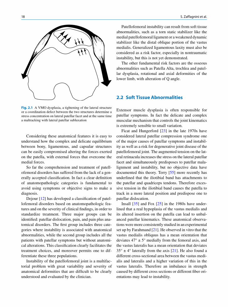
The “valgus law” underlines the prevalence of the lateral structures with respect to the medial ones [23]. The lateral knee compartment of the patellofemoral joint is normally wider than the medial one. In fact the lateral condyle is larger than the medial one with an external part of the patellar groove higher, wider, and forward with respect to the medial compartment. The external patellar facet is larger in respect to the medial facet. At the capsular level is present a prevalence of the lateral retinaculum that is stronger and wider with respect to the medial one (Fig. 2.1).
The patella is the largest sesamoid bone in the body, and resides within biarticular muscles (the quadriceps and patellar tendons). The patella functions both as a lever and a pulley. As a lever, the patella magnifies the forces exerted by the quadriceps on knee extension. As a pulley, the patella redirects the quadriceps force as it undergoes normal lateral tracking during flexion.
Pathophysiology of Lateral Patellar Dislocation
Stefano Zaffagnini, Giovanni Giordano, Danilo Bruni, Giulio Maria Marcheggiani Muccioli, and Maurilio Marcacci
S. Zaffagnini, MD (*) Laboratorio di Biomeccanica, Istituti Ortopedici Rizzoli, via di Barbiano, 1/10, 40100 Bologna, Italy e-mail: [email protected]
G. Giordano, MD Biomechanics Lab., Rizzoli Orthopaedics Institute, via di Barbiano, 1/10, 40100 Bologna, Italy
D. Bruni, MD Biomechanics Lab., Rizzoli Orthopaedics Institute, via di Barbiano, 1/10, 40100 Bologna, Italy
G. Maria Marcheggiani Muccioli, MD Biomechanics Lab., Rizzoli Orthopaedics Institute, via di Barbiano, 1/10, 40100 Bologna, Italy
M. Marcacci, MD Biomechanics Lab., Rizzoli Orthopaedics Institute, via di Barbiano, 1/10, 40100 Bologna, Italy
2
18 S. Zaffagnini et al.
Considering these anatomical features it is easy to understand how the complex and delicate equilibrium between bony, ligamentous, and capsular structures can be easily compromised altering the forces exerted on the patella, with external forces that overcome the medial forces.
So far the comprehension and treatment of patell-ofemoral disorders has suffered from the lack of a gen-erally accepted classification. In fact a clear definition of anatomopathologic categories is fundamental to avoid using symptoms or objective signs to make a diagnosis.
Dejour [12] has developed a classification of patel-lofemoral disorders based on anatomopathologic fea-tures and on the severity of clinical findings, in order to standardize treatment. Three major groups can be identified: patellar dislocation, pain, and pain plus ana-tomical disorders. The first group includes three cate-gories where instability is associated with anatomical abnormalities, while the second group includes all the patients with patellar symptoms but without anatomi-cal alterations. This classification clearly facilitates the treatment choices, and moreover permits one to dif-ferentiate these three populations.
Instability of the patellofemoral joint is a multifac-torial problem with great variability and severity of anatomical deformities that are difficult to be clearly understood and evaluated by the clinician.
Patellofemoral instability can result from soft tissue abnormalities, such as a torn static stabilizer like the medial patellofemoral ligament or a weakened dynamic stabilizer like the distal oblique portion of the vastus medialis. Generalized ligamentous laxity must also be considered as a risk factor, especially in nontraumatic instability, but this is not yet demonstrated.
The other fundamental risk factors are the osseous abnormalities such as Patella Alta, trochlea and patel-lar dysplasia, rotational and axial deformities of the lower limb, with alteration of Q angle.
2.2 Soft Tissue Abnormalities
Extensor muscle dysplasia is often responsible for patellar symptoms. In fact the delicate and complex muscular mechanism that controls the joint kinematics is extremely sensible to small variation.
Ficat and Hungerford [23] in the late 1970s have considered lateral patellar compression syndrome one of the major causes of patellar symptoms and instabil-ity as well as a risk for degenerative joint disease of the patellofemoral joint. The augmented tension on the lat-eral retinacula increases the stress on the lateral patellar facet and simultaneously predisposes to patellar mala-lignment and instability, but no objective data have documented this theory. Terry [55] more recently has underlined that the iliotibial band has attachments to the patellar and quadriceps tendons. Therefore exces-sive tension in the iliotibial band causes the patella to track in a more lateral position and predispose one to patellar dislocation.
Insall [35] and Fox [25] in the 1980s have under-lined that a real hypoplasia of the vastus medialis and its altered insertion on the patella can lead to unbal-anced patellar kinematics. These anatomical observa-tions were more consistently studied in an experimental set up by Farahmand [21]. He observed in vitro that the vastus medialis obliquus has a mean orientation that deviates 47° ± 5° medially from the femoral axis, and the vastus lateralis has a mean orientation that deviates 35° ± 4° laterally from the axis [21]. He also found a different cross-sectional area between the vastus medi-alis and lateralis and a higher variation of this in the vastus lateralis. Therefore an imbalance in strength caused by different cross sections or different fiber ori-entations may lead to instability.
Fig. 2.1 A VMO dysplasia, a tightening of the lateral structure or a coordination defect between the two structures determine a stress concentration on lateral patellar facet and at the same time a maltracking with lateral patellar subluxation
192 Pathophysiology of Lateral Patellar Dislocation
Vastus medialis relaxation reduces lateral patellar stability at all flexion angles. Goh [29] found lateral stability to be reduced by 30% when the vastus media-lis obliquus was relaxed at 20° of knee flexion with a lateral patellar displacement of 4 mm.
A VMO dysplasia does not guarantee the force nec-essary to compensate the force exerted from the lateral structure to stabilize the patella in the trochlea groove. In this type of dysplasia the absence of the oblique muscle fibers causes a worse lever arm. The conse-quences are usually an increased patellar tilt or a ten-dency to patellar subluxation.
Voight [60] also has demonstrated that although the medial and lateral muscle structures are normal, a defect in the muscular coordination, can determine an opposite recruitment order between vastus medialis and lateralis originating in patellar instability.
Passive stabilizers in the patellofemoral joint include patellofemoral and patellotibial ligaments and the reti-nacula. Warren and Marshall describe the MPFL as an extracapsular structure [64]. The size and thickness of the ligament varies considerably among individuals, but it is relatively constant within a given person [65]. The MPFL acts as a static check rein to resist lateral translation of the patella.
Desio [16] reported that the MPFL contributes 60% of the total restraining force against lateral patellar dis-placement with the patellomeniscal ligament the second most important medial stabilizer contributing an aver-age of 22% of the total restraining force. Senavongse [49] found that 20° of knee flexion was the position when 10 mm displacement occurred at the lowest restraining force. However the patella was more resis-tant to medial than lateral 10 mm displacement. Again Senavongse and Amis [48] tried to demonstrate the relative effects of various abnormalities on patellar sta-bility. They found that a relaxed VMO reduced by 30% the force to displace the patella laterally in 20°–90° flexion range, while only by 14% in extension. If the MPFL was ruptured the force required to displace the patella laterally was reduced by 50% in the extended knee, decreasing while the knee flexed. Interestingly abnormal trochlear geometry reduced the lateral stabil-ity by 70% at 30° of flexion.
General hyperlaxity can also be a cause of patell-ofemoral instability related to the insufficiency in con-trolling lateral patellar displacement.
Carson and James [8], evaluating lateral patellar dis-placement in response to applied load at full extension,
found a significantly greater lateral patellar mobility in symptomatic and hyperlaxity patient. The same observation was performed by Fithian [24] at 30° of flexion.
Nomura in 2006 [44], in a case series, showed that a hypermobile patella and generalized joint laxity were significantly important in the recurrent patellar dislo-cation group compared to the control group, with hypermobile patella as a predisposing factor for dislo-cation. Christoforakis [9] still in 2006 has shown that release of the lateral retinaculum reduces at 10° and 20° of flexion the force required to displace the patella by 20%. These findings underline the importance of medial structures like the VMO and MPFL.
2.3 Bone Abnormalities
One of the most important anatomical abnormalities originating in patellar symptoms is the trochlear dys-plasia. This pathology has often been underestimated and initially considered secondary to patellar disloca-tion [13,15,16]. Instead intraoperative observations have confirmed that the intercondylar groove can be found completely flat or even convex [17, 18].
The normal trochlea is concave and strictly corre-lated to the bony contour and depth of the overlying cartilage [49, 50]. Trochlear dysplasia is defined as a groove with a proximal flat articular zone and a distal shallow zone. [15] Trochlear dysplasia was first described many years ago by Richerand [38]. This author, in 1802, described an abnormal lateral condyle in patients with recurrent patellar dislocations.
In the presence of dysplasia, the intercondylar groove may be flattened or even convex. [17, 45] This convexity presents the articular cartilage being thicker centrally than laterally and medially. [49, 50]. These find-ings have been confirmed from other authors [56, 61] utilizing standard x-ray and CT images. In patients with recurrent patellar dislocation Yamada et al. [68] found the convex groove to extend twice as far during flexion as in controls. In the presence of trochlear emi-nence, the patella has to surmount the bump during the early flexion of the knee. [13, 15] The inadequate depth of the intercondylar groove can be total or focal, when affecting only the upper part [14].
Flattening of the groove does not allow the patella to fit into the trochlea during range of motion. Imbalance
20 S. Zaffagnini et al.
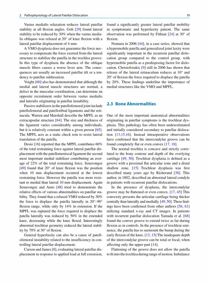
of the patellofemoral joint with risk of patellar disloca-tion is created by this lack of centration, especially in the first degrees of flexion that allows the lateral struc-ture to overtake easily the medial ones. In the presence of this deformity, the stresses are prevalently distrib-uted on the lateral facet instead of the entire groove, originating as long term arthritic degenerative changes of the joint [13, 15]. Quantitatively the convexity (bump or boss) is pathological above 3 mm or more,
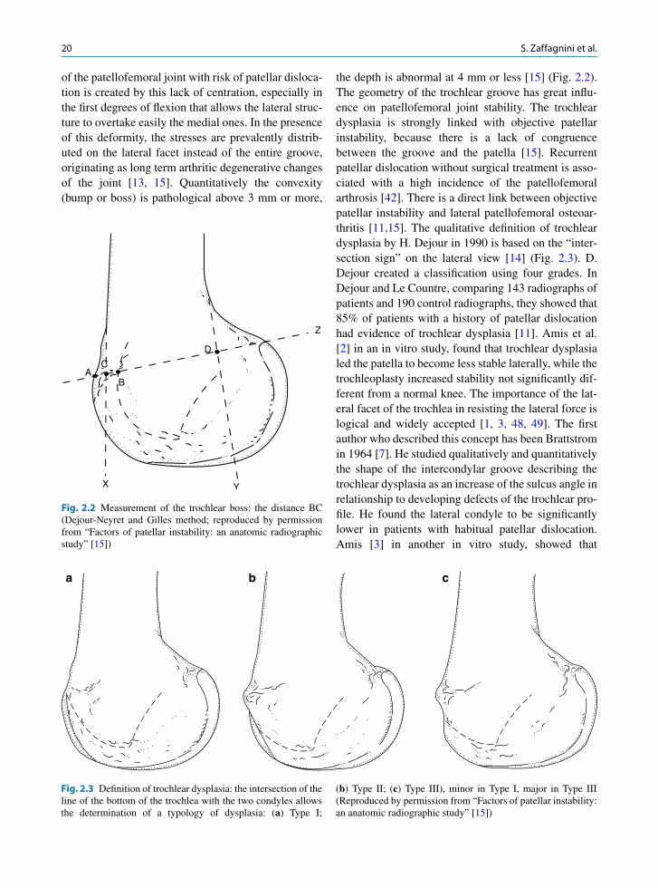
the depth is abnormal at 4 mm or less [15] (Fig. 2.2). The geometry of the trochlear groove has great influ-ence on patellofemoral joint stability. The trochlear dysplasia is strongly linked with objective patellar instability, because there is a lack of congruence between the groove and the patella [15]. Recurrent patellar dislocation without surgical treatment is asso-ciated with a high incidence of the patellofemoral arthrosis [42]. There is a direct link between objective patellar instability and lateral patellofemoral osteoar-thritis [11,15]. The qualitative definition of trochlear dysplasia by H. Dejour in 1990 is based on the “inter-section sign” on the lateral view [14] (Fig. 2.3). D. Dejour created a classification using four grades. In Dejour and Le Countre, comparing 143 radiographs of patients and 190 control radiographs, they showed that 85% of patients with a history of patellar dislocation had evidence of trochlear dysplasia [11]. Amis et al. [2] in an in vitro study, found that trochlear dysplasia led the patella to become less stable laterally, while the trochleoplasty increased stability not significantly dif-ferent from a normal knee. The importance of the lat-eral facet of the trochlea in resisting the lateral force is logical and widely accepted [1, 3, 48, 49]. The first author who described this concept has been Brattstrom in 1964 [7]. He studied qualitatively and quantitatively the shape of the intercondylar groove describing the trochlear dysplasia as an increase of the sulcus angle in relationship to developing defects of the trochlear pro-file. He found the lateral condyle to be significantly lower in patients with habitual patellar dislocation. Amis [3] in another in vitro study, showed that
A
X Y
C
B
D
Z
Fig. 2.2 Measurement of the trochlear boss: the distance BC (Dejour-Neyret and Gilles method; reproduced by permission from “Factors of patellar instability: an anatomic radiographic study” [15])
a b c
Fig. 2.3 Definition of trochlear dysplasia: the intersection of the line of the bottom of the trochlea with the two condyles allows the determination of a typology of dysplasia: (a) Type I;
(b) Type II; (c) Type III), minor in Type I, major in Type III (Reproduced by permission from “Factors of patellar instability: an anatomic radiographic study” [15])
212 Pathophysiology of Lateral Patellar Dislocation
flattening the lateral groove had more influence on patellar laxity than dysfunction of VMO and MPFL. It has been found that the patellar shape could change in trochlear dysplasia. The distal medial facet in dysplas-tic knee does not articulate well with the trochlea, becoming smaller than normal. [4, 26] Fucentese et al., in a comparative MRI study, proposed that the patellar morphology may be not only a result of missing medial patellofemoral pressure in trochlear dysplastic knees, but a decreased medial patellofemoral traction. They found hypotrophic medial patellofemoral restraints and increased lateral patellar tilt in the dysplastic knees. Wiberg [67] has classified radiographically the shape of the patella determining three types of patellar hypopla-sia that can originate from patellar symptoms. Ficat [22, 23] has underlined that the severe dysplasia of the inter-nal facet implies a reduction of the weight-bearing internal area with a surface incongruence and an auto-matic stress concentration on lateral side that can start the degenerative phenomena.
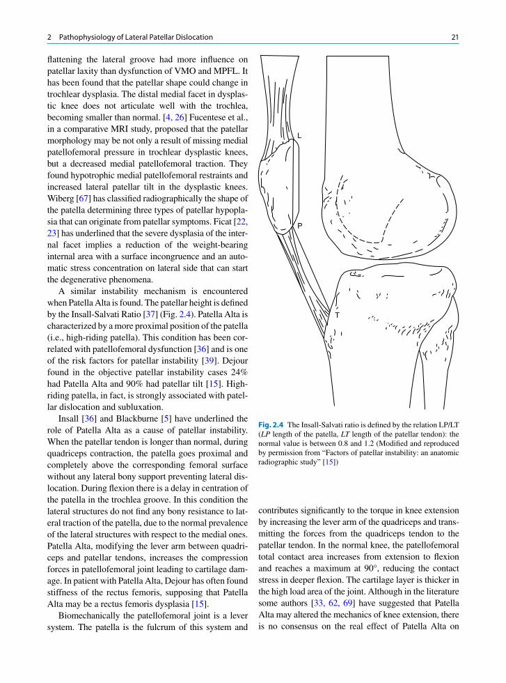
A similar instability mechanism is encountered when Patella Alta is found. The patellar height is defined by the Insall-Salvati Ratio [37] (Fig. 2.4). Patella Alta is characterized by a more proximal position of the patella (i.e., high-riding patella). This condition has been cor-related with patellofemoral dysfunction [36] and is one of the risk factors for patellar instability [39]. Dejour found in the objective patellar instability cases 24% had Patella Alta and 90% had patellar tilt [15]. High-riding patella, in fact, is strongly associated with patel-lar dislocation and subluxation.
Insall [36] and Blackburne [5] have underlined the role of Patella Alta as a cause of patellar instability. When the patellar tendon is longer than normal, during quadriceps contraction, the patella goes proximal and completely above the corresponding femoral surface without any lateral bony support preventing lateral dis-location. During flexion there is a delay in centration of the patella in the trochlea groove. In this condition the lateral structures do not find any bony resistance to lat-eral traction of the patella, due to the normal prevalence of the lateral structures with respect to the medial ones. Patella Alta, modifying the lever arm between quadri-ceps and patellar tendons, increases the compression forces in patellofemoral joint leading to cartilage dam-age. In patient with Patella Alta, Dejour has often found stiffness of the rectus femoris, supposing that Patella Alta may be a rectus femoris dysplasia [15].
Biomechanically the patellofemoral joint is a lever system. The patella is the fulcrum of this system and
contributes significantly to the torque in knee extension by increasing the lever arm of the quadriceps and trans-mitting the forces from the quadriceps tendon to the patellar tendon. In the normal knee, the patellofemoral total contact area increases from extension to flexion and reaches a maximum at 90°, reducing the contact stress in deeper flexion. The cartilage layer is thicker in the high load area of the joint. Although in the literature some authors [33, 62, 69] have suggested that Patella Alta may altered the mechanics of knee extension, there is no consensus on the real effect of Patella Alta on
T
P
L
Fig. 2.4 The Insall-Salvati ratio is defined by the relation LP/LT (LP length of the patella, LT length of the patellar tendon): the normal value is between 0.8 and 1.2 (Modified and reproduced by permission from “Factors of patellar instability: an anatomic radiographic study” [15])
22 S. Zaffagnini et al.
patellofemoral force, contact area, and contact pressure. Singerman, Davy, and Goldberg [51] reported, in an in vitro study, that the patellofemoral contact force, and its point of application on the patella, depended on patellar height. In a high-riding patella the magnitude of the PF contact increases with increasing flexion angle. They report also no increases from 0° to 60° of knee flexion and a significant rising at 90° in PFJ reaction force with Patella Alta. Luyckx [41], using a dynamic knee simulator, reported that the patellofemoral contact force is the sum of the patellar tendon force and the quadriceps tendon forces. In Patella Alta he showed the lowest PF contact force in initial flexion (35–70°) and a higher contact force in deeper flexion (70–120°) than in normal conditions. In this way he demonstrated a direct association between patellar height and maximal con-tact force. He also found that Patella Alta caused the greatest maximal contact force and pressure. In normal conditions the effective moment arm of the quadriceps tendon is greater than that of the patellar tendon because of the distal contact point of the patella during initial flexion [30, 58]. Yamaguchi and Zajac [69], moreover, by a mathematical simulation of Patella Alta to calcu-late a quadriceps moment arm, reported that modified lengthening of the patella or patellar tendon caused alteration of force transmission from quadriceps to patellar tendon. They showed a considerable increasing of moment arm and joint reaction force at flexion above 25–30°, with the Patella Alta condition. It seems that Patella Alta creates a more efficient knee extensor mechanism by a more distal contact point in initial flex-ion (0–60°), whereas, in deeper flexion, it is considered a biomechanical disadvantage [41].
Ward et al. [62, 63] demonstrated in two MRI stud-ies that Patella Alta is correlated with a significantly
larger quadriceps and smaller patellar ligament moment arm than in normal conditions, with a greater transmis-sion force from quadriceps to patellar ligament. They showed 19% less contact area than normal between 0° and 60° of flexion, with lateral displacement and lateral tilt of the patella at 0° of flexion. Patients with Patella Alta and pain have elevated PFJ stress because of smaller PFJ contact areas and interrelate with patel-lofemoral cartilaginous breakdown and degeneration, dysfunction, and subsequent pain. [32, 39] No correla-tion could be found between malalignment and the reduced contact areas [41].
Rotational and axial deformity of the entire leg can play a role in patellar instability.
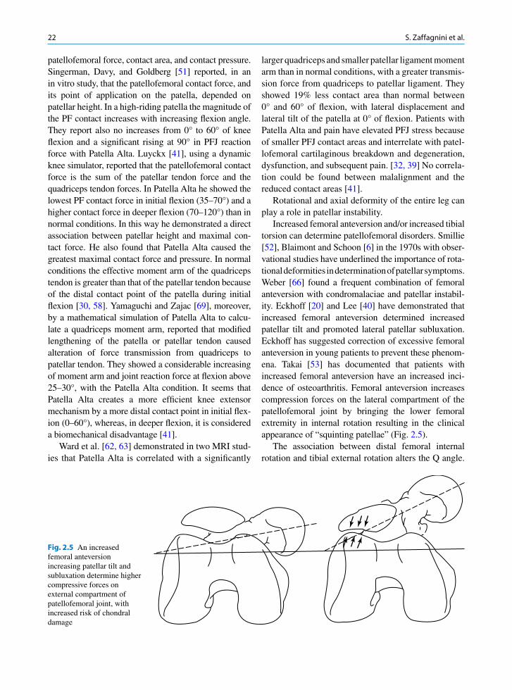
Increased femoral anteversion and/or increased tibial torsion can determine patellofemoral disorders. Smillie [52], Blaimont and Schoon [6] in the 1970s with obser-vational studies have underlined the importance of rota-tional deformities in determination of patellar symptoms. Weber [66] found a frequent combination of femoral anteversion with condromalaciae and patellar instabil-ity. Eckhoff [20] and Lee [40] have demonstrated that increased femoral anteversion determined increased patellar tilt and promoted lateral patellar subluxation. Eckhoff has suggested correction of excessive femoral anteversion in young patients to prevent these phenom-ena. Takai [53] has documented that patients with increased femoral anteversion have an increased inci-dence of osteoarthritis. Femoral anteversion increases compression forces on the lateral compartment of the patellofemoral joint by bringing the lower femoral extremity in internal rotation resulting in the clinical appearance of “squinting patellae” (Fig. 2.5).
The association between distal femoral internal rotation and tibial external rotation alters the Q angle.
Fig. 2.5 An increased femoral anteversion increasing patellar tilt and subluxation determine higher compressive forces on external compartment of patellofemoral joint, with increased risk of chondral damage
232 Pathophysiology of Lateral Patellar Dislocation
Brattstrom [7] described the Q angle as the angle formed by the line of pull of the quadriceps and that of the patellar tendon as they intersect at the center of the patella. The Q angle is largest in extension in relation to the screw home mechanism of the knee. For this measurement to be accurate the patella should be cen-tered on the trochlea. In males the Q angle is normally about 8–10° in females 15 plus or minus 5°. It should be noted that the relationship between the Q angle and clinical signs and symptoms has not always been con-sistent. A possible reason for the lack of association is related to the fact that there has been no consensus with respect to how this measurement should be taken, but more important is the fact that this measurement is taken statically, therefore, the contribution of abnormal segmental motions and muscle activation to the Q angle during dynamic activities may not be appreciated.
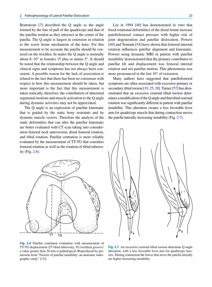
The Q angle is an expression of patellar kinematic that is guided by the static bony restraints and by dynamic muscle vectors. Therefore the analysis of the static deformities that can alter the patellar kinematic are better evaluated with CT scan taking into consider-ation femoral neck anteversion, distal femoral rotation, and tibial rotation. Patellar centration is more reliably evaluated by the measurement of TT-TG that considers femoral rotation as well as the rotation of tibial tuberos-ity (Fig. 2.6).
Lee in 1994 [40] has demonstrated in vitro that fixed rotational deformities of the distal femur increase patellofemoral contact pressure with higher risk of joint degeneration and patellar dislocation. Powers [46] and Tennant [54] have shown that femoral internal rotation influences patellar alignment and kinematic. Powers using dynamic MRI in patient with patellar instability demonstrated that the primary contributor to patellar tilt and displacement was femoral internal rotation and not patellar motion. This phenomena was more pronounced in the last 10° of extension.
Many authors have suggested that patellofemoral symptoms are often associated with excessive primary or secondary tibial torsion [19, 25, 28]. Turner [57] has dem-onstrated that an excessive external tibial torsion deter-mines a modification of the Q angle and that tibial external rotation was significantly different in patient with patellar instability. This alteration creates a less favorable lever arm for quadriceps muscle that during contraction moves the patella laterally increasing instability (Fig. 2.7).
TG TT
Fig. 2.6 Patellar centration evaluation with measurement of TT-TG displacement (TT tibial tuberosity, TG trochlear groove): a value greater than 20 mm is pathological (Reproduced by per-mission from “Factors of patellar instability: an anatomic radio-graphic study” [15])
PaPa
MvMv
Fig. 2.7 An excessive external tibial torsion determine Q angle alteration, with a less favorable lever arm for quadriceps mus-cles. During contraction the forces that move the patella laterally are higher increasing instability
24 S. Zaffagnini et al.
Van Kampen and Huiskes [59], Nagamine [43], and Sakai [47] examined the effect of tibial rotation on patellar three-dimensional movement. Hefzy [31] also studied the change of patellofemoral contact area with tibial rotation.
Apart from abnormal rotations in the transverse plane, excessive frontal plane malalignment can also influence patellofemoral joint.
Fujikawa et al. [27] observed that in varus defor-mity the patella displaces laterally and the lateral facet is hyperstressed with the increased risk of patellar instability. They also observed an association of proxi-mal tibial rotation with varus deformity.
Similar combinations of varus and tibial torsion have been described by Coscia [10] in 1983. In these patients there is an increased risk of patellar instability, moreover the screw home mechanism is reduced or missed and this can originate in degenerative changes of the medial femorotibial compartment and of the lat-eral patellofemoral joint.
Ficat described this phenomena as a cruciate arthritis.A valgus knee alters the Q angle and can be
responsible for dynamic patellar instability. Old observational studies have underlined that an exces-sive valgus knee alignment associated with external tibial rotation determines especially close to exten-sion a lateral patellar displacement especially during quadriceps contraction that increases the risk of patel-lar instability [22, 23].
Coscia [10] has also observed that in a valgus knee it is difficult to achieve knee extension stability due to excessive internal rotation. Therefore these knees remain unstable. During time this pathological situa-tion leads to medial capsular distension further increas-ing knee laxity. In severe valgus articular stability is lacking due to the difficulties in controlling external rotation and the screw home mechanism (Fig. 2.8).
As underlined by Powers [46] a valgus knee is not only determined by static osseous abnormalities but
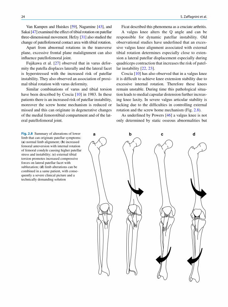
Fig. 2.8 Summary of alterations of lower limb that can originate patellar symptoms: (a) normal limb alignment; (b) increased femoral anteversion with internal rotation of femoral condyle causing higher patellar stress and instability; (c) external tibial torsion promotes increased compressive forces on lateral patellar facet with subluxation; (d) limb alterations can be combined in a same patient, with conse-quently a severe clinical picture and a technically demanding solution
a b c d
252 Pathophysiology of Lateral Patellar Dislocation
also dynamically during certain activities as a result of femoral, tibial, or combined adduction moment. These can results from muscle weakness or imbalance, or abnormalities at the level of the hip and pelvis as well as of the foot.
Torsional defect of the lower extremity can be found often together with different patient penetration origi-nating in a wide variety of clinical aspects that are really difficult to be globally understood.
In all these studies, a pathological value for varus/valgus or rotational deformity that is correlated to clin-ical symptoms has not been detected.
As we have shown the anatomical alterations that can be present with different penetration in each patient are various and complex and create several clinical aspects. Therefore the treatment options should be chosen in relation to the etiologic factors responsible for clinical symptoms in each patient.
A rational treatment of these disorders must foresee the execution of different surgical procedures in the same patient when the symptoms have a multifactorial origin in a manner to completely modify the joint physiology and kinematic.
Even if the surgical procedure acts mostly on pas-sive and static stabilizers of the patella it is fundamen-tal to achieve during surgery a dynamic patellar equilibrium with correct patellar tracking during the whole range of motion. Hughston [34] in 1989 has underlined the importance of dynamic stability of patellofemoral joint.
2.4 Summary
Patellofemoral instability:
Subjective instability with anatomical abnormalities•Traumatic dislocation without anatomical abnor-•malitiesDislocation with anatomical abnormalities•Patellofemoral pain•
Patellofemoral instability:
Soft tissue abnormalities•Osseous abnormalities•Soft tissue and osseous abnormalities•
Soft tissue abnormalities:
Extensor muscle dysplasia•Ipoplasia of the vastus medialis•
Patellofemoral, patellotibial ligaments, and retinac-•ula disordersGeneral hyperlaxity•
Osseous abnormalities:
Trochlear dysplasia•Patella Alta•Rotational and axial deformity of lower limb•Patellar dysplasia•
Acknowledgments We thank Mrs. Silvia Bassini for icono-graphic material, Rizzoli Orthopaedic Institute’s Library Staff, and Tommaso Bonanzinga, MD for helping us in preparing references.
References
1. Amis AA (2007) Current concepts on anatomy and biome-chanics of patellar stability. J Biol Chem 15:48–56
2. Amis AA, Oguz C, Bull AM, Senavongse W, Dejour D (2008) The effect of trochleoplasty on patellar stability and kinematics: a biomechanical study in vitro. J Bone Joint Surg Br 90(7):864–869
3. Amis AA, Senavongse W, Bull AM (2006) Patellofemoral kinematics during knee flexion-extension: an in vitro study. J Orthop Res 24(12):2201–2211
4. Barnett AJ, Gardner RO, Lankester BJ, Wakeley CJ, Eldridge JD (2007) Magnetic resonance imaging of the patella: a comparison of the morphology of the patella in normal and dysplastic knees. J Bone Joint Surg Br 89(6):761–765
5. Blackburne JS, Peel TE (1977) A new method of measuring patellar height. J Bone Joint Surg Br 59(2):241–242
6. Blaimont P, Schoon R (1977) 2 cases of gonarthrosis associ-ated with an internal torsion abnormality of the tibia. Acta Orthop Belg 43(4):476–481
7. Brattstrom H (1964) Shape of the intercondylar groove nor-mally and in recurrent dislocation of the patella. A clinical and x-ray anatomical investigation. Acta Orthop Scand 68(Suppl):1–148
8. Carson WGJ, James SL, Larson RL, Singer KM, Winternitz WW (1984) Patellofemoral disorders: physical and radio-graphic evaluation. Part II: Radiographic examination. Clin Orthop Relat Res 185:178–186
9. Christoforakis J, Bull AM, Strachan RK, Shymkiw R, Senavongse W, Amis AA (2006) Effects of lateral retinacu-lar release on the lateral stability of the patella. Knee Surg Sports Traumatol Arthrosc 14(3):273–277
10. Coscia PL, Fenoglio E, Cerlon C, Mautino F (1983) Fisiopatologia delle lesioni degenerative del ginocchio nei vizi di torsione tibiale. Minerva Orthop 34:497–504
11. Dejour D, Le Coultre B (2007) Osteotomies in patellofemo-ral instabilities. Sports Med Arthrosc 15(1):39–46
12. Dejour D, Nove-Josserand L, Walch G (1998) Patellofemoral disorders-classification and an approach to operative treat-ment for instability. In: Chan KM FF, Maffuli N, et al (ed)
26 S. Zaffagnini et al.
Controversies in orthopedic sports medicine. Williams & Wilkins Asia-Pacific, Ltd., Hong Kong, pp 235–244
13. Dejour H, Walch G, Neyret P, Adeleine P (1990) Dysplasia of the femoral trochlea. Rev Chir Orthop Reparatrice Appar Mot 76(1):45–54
14. Dejour H, Walch G, Neyret P, Adeleine P (1990) Dysplasia of the intercondilar groove. Fr J Orthop Surg 4(1):113–122
15. Dejour H, Walch G, Nove-Josserand L, Guier C (1994) Factors of patellar instability: an anatomic radiographic study. Knee Surg Sports Traumatol Arthrosc 2(1):19–26
16. Desio SM, Burks RT, Bachus KN (1998) Soft tissue restraints to lateral patellar translation in the human knee. Am J Sports Med 26(1):59–65
17. Drew D (1908) Dislocation of patella. Proc R Soc Med 1:1118. Dye SF (1987) An evolutionary perspective of the knee.
J Bone Joint Surg Am 69(7):976–98319. Eckhoff DG, Johnson KK (1994) Three-dimensional com-
puted tomography reconstruction of tibial torsion. Clin Orthop Relat Res 302:42–46
20. Eckhoff DG, Montgomery WK, Kilcoyne RF, Stamm ER (1994) Femoral morphometry and anterior knee pain. Clin Orthop Relat Res 302:64–68
21. Farahmand F, Senavongse W, Amis AA (Jan 1998) Quantitative study of the quadriceps muscles and trochlear groove geometry related to instability of the patellofemoral joint. J Orthop Res 16(1):136–143
22. Ficat P (1973) Les desequilibres rotuliens. De l’hyperpression a l’arthrose. Masson Ed, Paris
23. Ficat P, Hungerford DS (1977) Disorders of the Patellofemoral Joint. Williams & Wilkins, Baltimore, MD
24. Fithian DC, Mishra DK, Balen PF, Stone ML, Daniel DM (1995) Instrumented measurement of patellar mobility. Am J Sports Med 23(5):607–615
25. Fox TA (1975) Dysplasia of the quadriceps mechanism: hyp-oplasia of the vastus medialis muscle as related to the hyper-mobile patella syndrome. Surg Clin North Am 55(1):199–226
26. Fucentese SF, von Roll A, Koch PP, Epari DR, Fuchs B, Schottle PB (2006) The patella morphology in trochlear dys-plasia – a comparative MRI study. Knee 13(2):145–150
27. Fujikawa K, Seedhom BB, Wright V (1983) Biomechanics of the patellofemoral joint. Part II: A study of the effect of simulated femoro-tibial varus deformity on the congruity of the patellofemoral compartment and movement of the patella. Eng Med 12(1):13–21
28. Fulkerson JP, Schutzer SF (1986) After failure of conserva-tive treatment for painful patellofemoral malalignment: lat-eral release or realignment? Orthop Clin North Am 17(2): 283–288
29. Goh JC, Lee PY, Bose K (1995) A cadaver study of the func-tion of the oblique part of vastus medialis. J Bone Joint Surg Br 77(2):225–231
30. Grood ES, Suntay WJ, Noyes FR, Butler DL (1984) Biome-chanics of the knee-extension exercise. Effect of cutting the anterior cruciate ligament. J Bone Joint Surg Am 66(5): 725–734
31. Hefzy MS, Jackson WT, Saddemi SR, Hsieh YF (1992) Effects of tibial rotations on patellar tracking and patell-ofemoral contact areas. J Biomed Eng 14(4):329–343
32. Heino J, Power CM (2002) Patellofemoral stress during walking in persons with and without patellofemoral pain. Med Sci Sports Exerc 34:1582–1593
33. Hirokawa S (1991) Three-dimensional mathematical model analysis of the patellofemoral joint. J Biomech 24(8): 659–671
34. Hughston JC (1989) Patellar subluxation. A recent history. Clin Sports Med 8(2):153–162
35. Insall J (1982) Current Concepts Review: patellar pain. J Bone Joint Surg Am 64(1):147–152
36. Insall J, Goldberg V, Salvati E (1972) Recurrent dislocation and the high-riding patella. Clin Orthop Relat Res 88:67–69
37. Insall J, Salvati E (1971) Patella position in the normal knee joint. Radiology 101(1):101–104
38. Isermeyer H (1967) Über die pathologische Luxation der Patella. Arch Klin Chir 8:1–23
39. Kannus PA (1992) Long patellar tendon: radiographic sign of patellofemoral pain syndrome-a prospective study. Radiology 185:859–863
40. Lee TQ, Anzel SH, Bennett KA, Pang D, Kim WC (1994) The influence of fixed rotational deformities of the femur on the patellofemoral contact pressure in human cadaver knees. Clin Orthop Relat Res 302:69–74
41. Luyckx T, Didden K, Vandenneucker H, Labey L, Innocenti B, Bellemans J (2009) Is there a biomechanical explanation for anterior knee pain in patients with Patella Alta?: influ-ence of patellar height on patellofemoral contact force, con-tact area and contact pressure. J Bone Joint Surg Br 91(3): 344–350
42. Maenpaa H, Lehto MU (1997) Patellofemoral osteoarthritis after patellar dislocation. Clin Orthop Relat Res 339: 156–162
43. Nagamine R, Otani T, White SE, McCarthy DS, Whiteside LA (1995) Patellar tracking measurement in the normal knee. J Orthop Res 13(1):115–122
44. Nomura E, Inoue M, Kobayashi S (2006) Generalized joint laxity and contralateral patellar hypermobility in uni-lateral recurrent patellar dislocators. Arthroscopy 22(8): 861–865
45. Pollard B (1981) Old dislocation of patella reduced by intra-articular operation. Lancet 1:988
46. Powers CM, Ward SR, Fredericson M, Guillet M, Shellock FG (2003) Patellofemoral kinematics during weight-bearing and non-weight-bearing knee extension in persons with lat-eral subluxation of the patella: a preliminary study. J Orthop Sports Phys Ther 33(11):677–685
47. Sakai N, Luo ZP, Rand JA, An KN (1996) In vitro study of patellar position during sitting, standing from squatting, and the stance phase of walking. Am J Knee Surg 9(4): 161–166
48. Senavongse W, Amis AA (2005) The effects of articular, retinacular, or muscular deficiencies on patellofemoral joint stability. J Bone Joint Surg Br 87(4):577–582
49. Senavongse W, Farahmand F, Jones J, Andersen H, Bull AM, Amis AA (2003) Quantitative measurement of patell-ofemoral joint stability: force-displacement behavior of the human patella in vitro. J Orthop Res 21(5):780–786
50. Shih YF, Bull AM, Amis AA (2004) The cartilaginous and osseous geometry of the femoral trochlear groove. Knee Surg Sports Traumatol Arthrosc 12(4):300–306
51. Singerman R, Davy DT, Goldberg VM (1994) Effects of Patella Alta and Patella Infera on patellofemoral contact forces. J Biomech 27(8):1059–1065
272 Pathophysiology of Lateral Patellar Dislocation
52. Smillie IS (1974) The biomechanical basis of osteoarthritis of the knee in total knee replacement. Paper presented at “Total knee replacement” organised by the Institution of Mechanical Engineering, London
53. Takai S, Sakakida K, Yamashita F, Suzu F, Izuta F (1985) Rotational alignment of the lower limb in osteoarthritis of the knee. Int Orthop 9(3):209–215
54. Tennant S, Williams A, Vedi V, Kinmont C, Gedroyc W, Hunt DM (2001) Patellofemoral tracking in the weight-bear-ing knee: a study of asymptomatic volunteers utilising dynamic magnetic resonance imaging: a preliminary report. Knee Surg Sports Traumatol Arthrosc 9(3):155–162
55. Terry GC, Hughston JC, Norwood LA (1986) The anatomy of the iliopatellar band and iliotibial tract. Am J Sports Med 14(1):39–45
56. Trillat A, Dejour H, Couette A (1964) Diagnosis and treat-ment of recurrent dislocations of the patella. Rev Chir Orthop Reparatrice Appar Mot 50:813–824
57. Turner MS (1994) The association between tibial torsion and knee joint pathology. Clin Orthop Relat Res 302: 47–51
58. van Verberg J, Weijus WA (1986) A mathematical model of the patellofemoral joint. J Biomech 19:219–229
59. van Kampen A, Huiskes R (1990) The three-dimensional tracking pattern of the human patella. J Orthop Res 8(3): 372–382
60. Voight ML, Wieder DL (1991) Comparative reflex response times of vastus medialis obliquus and vastus lateralis in normal subjects and subjects with extensor mechanism dys-
function. An electromyographic study. Am J Sports Med 19(2):131–137
61. Wanner JA (1977) Variations in the anterior patellar groove of the human femur. Am J Phys Anthropol 47(1):99–102
62. Ward SR, Terk MR, Powers CM (2005) Influence of Patella Alta on knee extensor mechanics. J Biomech 38(12):2415–2422
63. Ward SR, Terk MR, Powers CM (2007) Patella Alta: asso-ciation with patellofemoral alignment and changes in con-tact area during weight-bearing. J Bone Joint Surg Am 89(8): 1749–1755
64. Warren LA, Marshall JL, Girgis F (1974) The prime static stabilizer of the medical side of the knee. J Bone Joint Surg Am 56(4):665–674
65. Warren LF, Marshall JL (1979) The supporting structures and layers on the medial side of the knee: an anatomical analysis. J Bone Joint Surg Am 61(1):56–62
66. Weber U (1977) Malrotation of distal femur (author’s transl). Z Orthop Ihre Grenzgeb 115(5):707–715
67. Wiberg G (1941) Roentgenographicand anatomic studies on the femoropatellar joint. With special reference to chon-dromalacia patellae. Acta Orthop Scand XII:319
68. Yamada Y, Toritsuka Y, Yoshikawa H, Sugamoto K, Horibe S, Shino K (2007) Morphological analysis of the femoral tro-chlea in patients with recurrent dislocation of the patella using three-dimensional computer models. J Bone Joint Surg Br 89(6):746–751
69. Yamaguchi GT, Zajac FE (1989) A planar model of the knee joint to characterize the knee extensor mechanism. J Biomech 22(1):1–10


![Swiss Payment Standards 2019...techniques – QR Code bar code symbology specification) ISO [2] pain.001.001.03 XML Schema Customer Credit Transfer Initiation V03 ISO [3] pain.001.001.03.ch.02](https://img.pdfslide.org/doc/110x75/5fe7f346efd90445576b4f00/swiss-payment-standards-2019-techniques-a-qr-code-bar-code-symbology-specification.jpg)