Embed Size (px)
Citation preview
This work has been digitalized and published in 2013 by Verlag Zeitschrift für Naturforschung in cooperation with the Max Planck Society for the Advancement of Science under a Creative Commons Attribution4.0 International License.
Dieses Werk wurde im Jahr 2013 vom Verlag Zeitschrift für Naturforschungin Zusammenarbeit mit der Max-Planck-Gesellschaft zur Förderung derWissenschaften e.V. digitalisiert und unter folgender Lizenz veröffentlicht:Creative Commons Namensnennung 4.0 Lizenz.
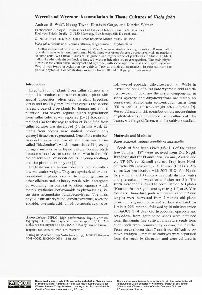
Wyerol and Wyerone Accumulation in Tissue Cultures of Vicia faba Andreas B. Wolff, Maung Thynn, Elisabeth Gorge, and Dietrich Werner
Fachbereich Biologie, Botanisches Institut der Philipps Universität Marburg. Karl von Frisch Straße. D-3550 Marburg, Bundesrepublik Deutschland
Z . Naturforsch. 43c, 636-640 (1988); received March 7/May 30, 1988
Vicia faba, Callus and Liquid Cultures, Regeneration, Phytoalexins
Callus cultures of various cultivars of Vicia faba were studied for regeneration. During callus growth on agar or in liquid medium a black tissue was often observed correlated with an autolysis of some cells. With those tissues callus growth and regeneration of plants was inhibited. In black callus the phytoalexin synthesis is induced without infection by microorganism. The main phyto-alexins in the callus tissue are wyerol and wyerone, with some wyeronic acid and dihydrowyerone. Wyerol was found especially in the cultivar Troy at a high concentration. In four cultivars the pooled phytoalexin concentration varied between 10 and 150 pg-g _ 1 fresh weight.
Introduction
Regeneration of plants from callus cultures is a
method to produce clones from a single plant with
special properties, often used in plant breeding.
Grain and feed legumes are after cereals the second
largest group of crop plants for human and animal
nutrition. For several legume plants regeneration
from callus cultures was reported [1—5]. Recently a
method also for the regeneration of Vicia faba from
callus cultures was developed [6]. In that work ex-
plants from organs were studied, however only
epicotyl tissue was regenerated. One of the main bar-
riers in the in vitro culture of faba bean was the so-
called "blackening", which means that calli growing
on agar surfaces or in liquid culture became black
because of autolysis of some tissue. Also in the field
the "blackening" of shoots occurs in young seedlings
and the plants ultimately die [7].
Phytoalexins are antimicrobial compounds with a
low molecular weight. They are synthesized and ac-
cumulated in plants, exposed to microorganisms or
other elicitors such as heavy metals salts, detergents
or wounding. In contrast to other legumes which
mainly synthesize isoflavonoids as phytoalexins, Vi-cia faba accumulates furanoacetylenes. The main
phytoalexins are wyerone, dihydrowyerone, wyerone
epoxide, wyeronic acid, dihydrowyeronic acid, wye-
Abbreviations: H P L C , high performance liquid chroma-tography; T L C , thin layer chromatography; 2,4D, 2,4-dichlorophenoxyacetic acid; B A P , 6-benzyl-aminopurine.
Reprint requests to Prof. Dr. Werner.
Verlag der Zeitschrift für Naturforschung, D-7400 Tübingen 0341 - 0382/88/0900- 0625 $01.30/0
roi, wyerol epoxide, dihydrowyerol [8]. While in
leaves and pods of Vicia faba wyeronic acid and di-
hydrowyeronic acid are the major components, in
seeds wyerone and dihydrowyerone are mainly ac-
cumulated. Phytoalexin concentration varies from
300 to 1200 pg-g-1 fresh weight after infection [9].
We established in this contribution the accumulation
of phytoalexins in uninfected tissue cultures of faba
beans, with large differences in the cultivars studied.
Materials and Methods
Plant material, culture conditions and media
Seeds of faba bean (Vicia faba L.) of the tannin
free cultivar "TF" were received from Dr. Nagel,
Bundesanstalt für Pflanzenbau, Vienna, Austria and
cv. TP 667, cv. Kristall and cv. Troy from Nord-
deutsche Pflanzenzucht, 2331 Holtsee (F.R.G.). Aft-
er surface sterilization with 30% H 2 0 2 for 20 min
they were rinsed 3 times with sterile distilled water
and presoaked in water on a shaker for 5 h. The
seeds were then allowed to germinate on NB plates
(Nutrient Broth 8 g • T 1 and agar 16 g • 1":') at 28 °C in
the dark. Immature pods with seeds (about 7 mm
length) were harvested from 2 months old plants
grown in a green house and surface sterilized for
1 min in 70% ethanol, followed by 10 min immersion
in NaOCl. 3—4 days old hypocotyls, epicotyls and
cotyledons from germinated seeds were obtained
from the tannin free cultivar. Immature seeds from
open pods were removed by carving the funicle.
From seeds shorter than 7 mm it was difficult to re-
move embryos. Immature embryos were separated
from the seeds by dissection and were cultured in
A. B. Wolff et al. • Wyerol and Wyerone Accumulation in Vicia faba 637
(2x18 cm) test tubes. Pieces of 4—6 mm were excised from immature cotyledons and placed in petridishes containing 20 ml of agar medium.
B5 medium [10] with various phytohormones and 10 amino acids was used for all studies [6]. A l l cul-tures were kept in the dark at 28 °C for 2 weeks and then transferred to 23 °C with a 16 h light and 8 h dark regime. A l l media used were solidified with 0.75% agar and had a pH of 5.5.
For suspension cultures, 6 weeks old calli obtained from epicotyl expiants of the tannin free cultivar were cut into small pieces and placed in 100 ml Er-lenmeyer flasks with 50 ml liquid medium. Incuba-tion was at 23 °C on a shaker (120 rpm).
Extraction, identification and quantification of phytoalexins
Tissues were stored at —20 °C before extraction. Callus tissue was homogenized in ethanol by a mixer and stirred overnight at room temperature. The ex-tract was centrifuged for 5 min at 3000xg. The supernatant being evaporated to dryness was resus-pended in an appropriate volume of ethanol and sub-jected to H P L C or TLC .
With TLC, wyerone derivatives can be detected as blue fluorescent bands under U V at 366 nm [8]. For further identification we used the /^-values pub-lished by Hargeaves [11] followed by a wavelength scan from 200—400 nm to detect the main absorption bands. Wyerone has main U V absorption bands are at 351, 292 and 224 nm in ethanol, dihydrowyerone at 340 and 235 nm in ethanol, wyerol at 312 nm in methanol and wyeronic acid at 356, 284 and 222 nm in methanol [8].
The identification of phytoalexins after separation by H P L C was carried out by comparison with pub-lished /?rvalues [9], [11] and by U V absorption bands as described above. Quantitative assays on H P L C were performed by comparing peak areas to an internal standard and by using the extinction coef-ficient at 351 nm according to Fawcett et al. [12],
High performance liquid chromatography (HPLC)
For H P L C we used a two pump system (LKB 2150) from L K B Instrument GmbH (Gräfling, F.R.G.), a controller (LKB 2152) and an UV/Vis variable wavelength monitor ( LKB 2151). Injection was performed via a Rheodyne injector (Rheodyne Inc., Cotati, C.A., U.S.A.) with a 20 pi loop. Quan-
titative data were obtained by using a Shimadzu Data Processor Chromatopac C - R 3 A (Shimadzu Corpo-ration, Kyota, Japan) with «-butyl salicylate as a standard.
Reversed phase H P L C was carried out as de-scribed by Porter et al. [11], Aliquots of 20 pi of extract with butyl salicylate as internal standard were injected to a Serva Octadecyl = Si 100 Polyol 0.005 mm (4.6 x 200 mm) column with a precolumn (4.6 x 75 mm) filled with the same material. Gra-dient eluation was carried out under room tempera-ture with a flow rate of 1 ml/min and U V detection at 330 nm. Initial solvent conditions were 5% acetic acid — methanol (65:35 vol%). The linear gradient was used for 60 min to a final mixture of 5% acetic acid — methanol — acetonitrile (30:50:20 vol.%). For calibration of the internal standard n-butyle salicylate, the peak area after H P L C separation was compared to a known concentration of wyerone and wyeronic acid.
Thin layer chromatography (TLC)
T L C analysis was done as described by Hargreaves
et al. [13] using Merk silica plates (0.2 mm) with a
solvent system of hexane — acetone 2:1 (v/v).
Results and Discussion
Callus cultures in Vicia faba
In Vicia faba a difficulty in the in vitro culture is the often observed so-called "blackening" of tissue. After placing excised plant tissue for a few hours on different media, some of them became black rapidly. When well formed calli or calli with roots or shoots were transferred to a new medium, some of these also turned black and died afterwards. In cell suspen-sion cultures this problem also occurred when the suspended cell aggregates were transferred to a new medium, especially on agar solidified medium. The phenomenon of "blackening" differs from senes-cence of calli or dried calli. In the latter cases, col-ours of calli changed from cream white or green to pale brown. For this change at least one week is needed. In the case of "blackening", calli or plant material immediately changed to a black colour.
Phytoalexins in calli
When well growing calli were compared with black
calli by T L C some new bands occurred.
10 20 ÎO 10 50
T i m e (min)
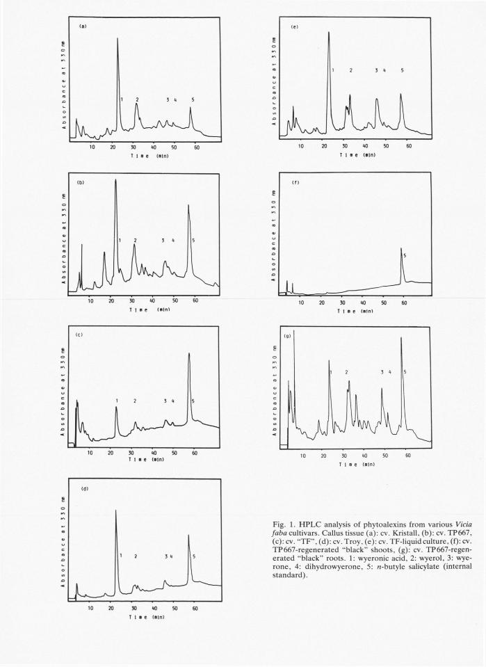
Fig. 1. H P L C analysis of phytoalexins from various Vicia faba cultivars. Callus tissue (a): cv. Kristall, (b): cv. TP667, (c): cv. "TF", (d): cv. Troy, (e): cv. TF-liquid culture, (f): cv. TP667-regenerated "black" shoots, (g): cv. TP667-regen-erated "black" roots. 1: wyeronic acid, 2: wyerol, 3: wye-rone, 4: dihydrowyerone, 5: «-butyle salicylate (internal standard).
A. B. Wolff et al. • Wyerol and Wyerone Accumulation in Vicia faba 639
Phytoalexins in different cultivars of Vicia faba calli
ug Phytoalexin/g fresh weight
fej I L I L SLÜH-Wyeronic acid Wyerol Wyerone
Phytoalexins
DH- wyerone
Vicia faba cultivar
• I Reg.TP-8hoot
Iii Reg.TP-root
• TF (Lc.)
inn Troy
H TF
• TP 667
M Kristall
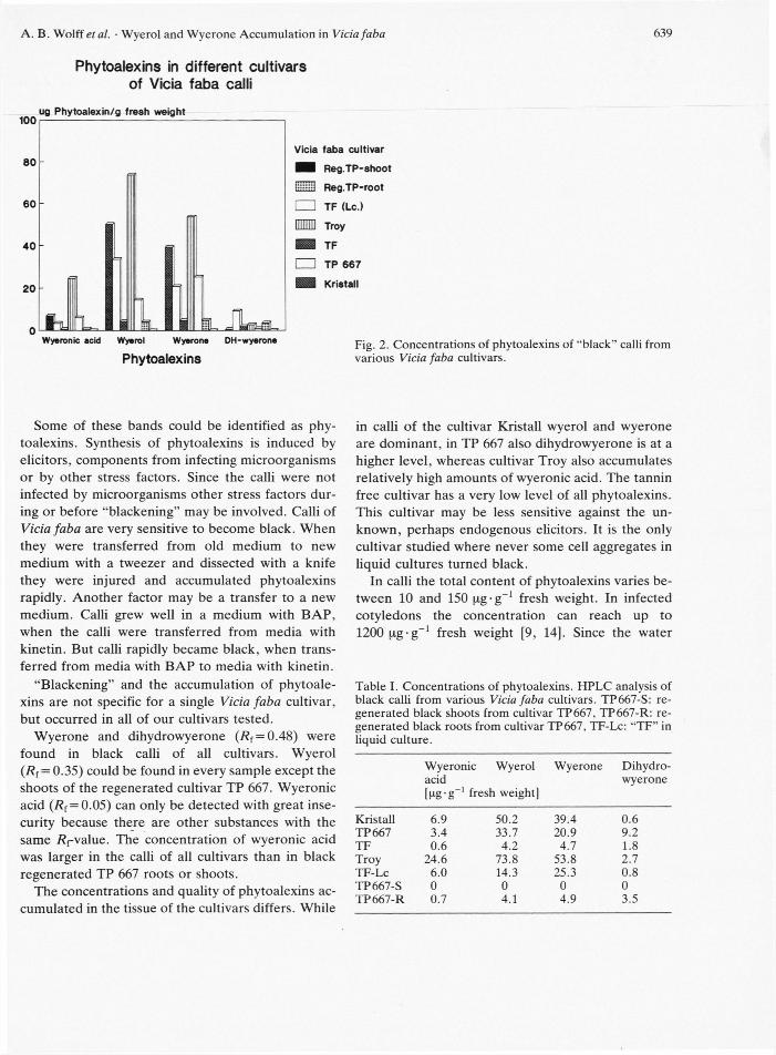
Fig. 2. Concentrations of phytoalexins of "black" calli from various Vicia faba cultivars.
Some of these bands could be identified as phy-toalexins. Synthesis of phytoalexins is induced by elicitors, components from infecting microorganisms or by other stress factors. Since the calli were not infected by microorganisms other stress factors dur-ing or before "blackening" may be involved. Calli of Vicia faba are very sensitive to become black. When they were transferred from old medium to new medium with a tweezer and dissected with a knife they were injured and accumulated phytoalexins rapidly. Another factor may be a transfer to a new medium. Calli grew well in a medium with BAP , when the calli were transferred from media with kinetin. But calli rapidly became black, when trans-ferred from media with B A P to media with kinetin.
"Blackening" and the accumulation of phytoale-xins are not specific for a single Vicia faba cultivar, but occurred in all of our cultivars tested.
Wyerone and dihydrowyerone (/?f = 0.48) were found in black calli of all cultivars. Wyerol (R{ = 0.35) could be found in every sample except the shoots of the regenerated cultivar TP 667. Wyeronic acid (/?f = 0.05) can only be detected with great inse-curity because there are other substances with the same Ärvalue. The concentration of wyeronic acid was larger in the calli of all cultivars than in black regenerated TP 667 roots or shoots.
The concentrations and quality of phytoalexins ac-cumulated in the tissue of the cultivars differs. While
in calli of the cultivar Kristall wyerol and wyerone are dominant, in TP 667 also dihydrowyerone is at a higher level, whereas cultivar Troy also accumulates relatively high amounts of wyeronic acid. The tannin free cultivar has a very low level of all phytoalexins. This cultivar may be less sensitive against the un-known, perhaps endogenous elicitors. It is the only cultivar studied where never some cell aggregates in liquid cultures turned black.
In calli the total content of phytoalexins varies be-tween 10 and 150 pg-g_1 fresh weight. In infected cotyledons the concentration can reach up to 1200 pg-g_1 fresh weight [9, 14]. Since the water
Table I. Concentrations of phytoalexins. H P L C analysis of black calli from various Vicia faba cultivars. TP667-S: re-generated black shoots from cultivar TP667, TP667-R: re-generated black roots from cultivar TP667, TF-Lc: "TF" in liquid culture.
Wyeronic Wyerol acid [pg-g
_1 fresh weight]
Wyerone Dihydro-wyerone
Kristall 6.9 50.2 39.4 0.6 T P 667 3.4 33.7 20.9 9.2 T F 0.6 4.2 4.7 1.8 Troy 24.6 73.8 53.8 2.7 TF-Lc 6.0 14.3 25.3 0.8 TP667-S 0 0 0 0 T P 667-R 0.7 4.1 4.9 3.5
640 A. B. Wolff et al. • Wyerol and Wyerone Accumulation in Vicia faba 640
content in calli is much higher than in cotyledons, the
phytoalexin accumulation per dry matter is more
similar. Beside the handling and the transfer of calli
to new media as elicitoring factor for phytoalexin
synthesis also endogenous elicitors from cell walls of
autolyzing cells may be of importance. Sensitivity of
Vicia faba callus cultures from the cultivars studied
towards different kinds of phytopathogens are un-
known. It is an interesting question, whether the po-
tential to accumulate phytoalexins in calli of different
Vicia faba cultivars can be correlated to resistance
against phytopathogens under field conditions.
Acknowledgements
The work was supported by the B M F T programme
„Wechselwirkungen Pflanzen—Mikroorganismen".
1] S. Arcioni, M . R . Davey, A . V . P. dos Santos, and E . C . Cooking, Z. Pflanzenphysiol. 106, 105 (1982).
2] A . Atanassov and D . C. W . Brown, Plant Cell Tissue Organ Culture 3, 149 (1984).
3] U . B. Barwale, H . R . Kern, and J. M . Widholm, Plan-ta 167, 473 (1986).
4] P. A . Lazzeri, D . F. Hidelbrand, and G . B. Collin, Plant Mol. Bio. Rep. 3, 160 (1985).
5] L. A . Mroginski, K . K. Kartha, and J. P. Schyluk, Can. J. Bot. 59, 826 (1980).
6] M . Thynn and D . Werner, Angew. Bot. 61, 483 (1987).
7] T. B. Jha and S. K . Roy, Cytologia 47, 465 (1982). 8] J. L. Ingham, Phytoalexins from the Leguminosae. In:
Phytoalexins (J. A . Bailey and J. W . Mansfield, eds.), Blackie & Son Ltd, Glasgow and London 1982.
[9] J. W . Mansfield, A . E . A . Porter, and R . V . Small-m a n , Phytochemistry 19, 1057 (1980).
[10] O . L. Gamborg, R . A . Miller, and K . Ojima, Exp. Cell. Res. 50, 151-158 (1980).
[11] A . E . A . Porter, R . V . Smallman, and J. W . Mans-field, J. Chromatogr. 172, 498 (1979).
[12] C . H . Fawcett, D . M . Spencer, R . L. Wain, A . G . Fallis, E. R. H . Jones, M . Le Quan, C. B. Page, V . Thaller, D . C. Shubrook, and P. M . Whitham, J. C h e m . Soc. (C) 1968, 2455.
[13] J. A . Hargreaves, J. W . Mansfield, and S. Rosall, Physiological Plant Pathol. 11, 227 (1977).
[14] J. W . Mansfield, Y . M . Barlow, and A . E. A . Porter (G. P. Chapman & S. A . Tarawali, eds.), Nijhoff/ Tunk 1984.