PTPIP51 – ein multifunktionales Protein im Gehirn
INAUGURAL-DISSERTATION zur Erlangung des Grades eines Doktors der Medizin
des Fachbereichs Medizin der Justus-Liebig-Universität Gießen
vorgelegt von Maxime Jean Viard
aus Köln
Gießen 2011

Aus dem Institut für Anatomie und Zellbiologie
des Fachbereichs Medizin der Justus- Liebig- Universität Gießen
Leitung: Prof. Dr. med. W. Kummer
1. Gutachter: Prof. Dr. Wimmer
2. Gutachter: PD. Dr. Berghoff
Tag der Disputation: 14.03.2012
Eidesstattliche Erklärung
Ich erkläre: Ich habe die vorgelegte Dissertation selbständig und ohne fremde Hilfe
und nur mit den Hilfen angefertigt, die ich in der Dissertation angegeben habe. Alle
Textstellen, die wörtlich oder sinngemäß aus veröffentlichten oder nicht
veröffentlichten Schriften entnommen sind, und alle Angaben, die auf mündlichen
Auskünften beruhen, sind als solche kenntlich gemacht. Bei den von mir
durchgeführten und in der Dissertation erwähnten Untersuchungen habe ich die
Grundsätze guter wissenschaftlicher Praxis, wie sie in der „Satzung der Justus-
Liebig-Universität Gießen zur Sicherung guter wissenschaftlicher Praxis“
niedergelegt sind, eingehalten.
Gießen, den
Maxime Jean Viard

Inhaltsverzeichnis
1. Liste der Publikationen der kumulativen Doktorarbeit .......................... 5
2. Einleitung.............................................................................................. 6
2.1 Protein-Tyrosin- Phosphatase interagierendes Protein 51................................ 6
2.2 Expressionsprofil von PTPIP51 im Mäusegehirn .............................................. 8
2.3 PTPIP51 im Gehirn – ein multifunktionelles Protein?...................................... 14
2.4 PTPIP51 in Glioblastomen .............................................................................. 19
2.5 Zusammenfassung.......................................................................................... 22
2.6 Summary ......................................................................................................... 24
3. Literaturverzeichnis ............................................................................ 26
4. Weitere Publikationen und Poster ...................................................... 41
5. Danksagung ....................................................................................... 42
6. Lebenslauf .......................................................................................... 43
7. Publikationen der kumulativen Doktorarbeit ....................................... 45
7.1. Expression profile of PTPIP51 in mouse brain.
7.2. PTPIP51 – a multifunctional protein in brain tissue
7.3. PTPIP51, a positive modulator of the MAPK/Erk pathway, is upregulated in glioblastoma and interacts with 14-3-3β and PTP1B in situ

Liste der Publikationen der kumulativen Doktorarbeit _____________________________
5
1. Liste der Publikationen der kumulativen Doktorarbeit
(1) Koch P.*, Viard M.*, Stenzinger A., Brobeil A., Tag C., Steger K., Wimmer M.
(2009). Expression profile of PTPIP51 in mouse brain. J Comp Neurol. 517(6):892-
905. (*The first two authors contributed equally to this work, M. Viard= co-first author)
(2) Viard M., Kamm M., Bobrich M., Brobeil A., Petri P. Wimmer M. (2011). PTPIP51
– a multifunctional protein in brain tissue (eingereicht in Journal of Comparative
Neurology)
(3) Petri MK., Koch P., Stenzinger A, Kuchelmeister K., Nestler U., Paradowska A.,
Steger K, .Brobeil A., Viard M., Wimmer M. (2011). PTPIP51, a positive modulator of
the MAPK/Erk pathway, is upregulated in glioblastoma and interacts with 14-3-3β
and PTP1B in situ. Histol Histopathol. (in Druck)
Einleitung _____________________________
6
2. Einleitung
2.1 Protein-Tyrosin- Phosphatase interagierendes Protein 51
Das Protein-Tyrosin-Phosphatase interagierende Protein 51 (PTPIP51) wurde vor ca.
10 Jahren als Interaktionspartner der Protein-Tyrosin-Phosphatase-1B (PTP1B)
identifiziert (Porsche, 2001; Stenziger et al., 2009).
Beim Menschen ist das Gen des PTPIP51 auf Chromosom 15 (15q15.1.) lokalisiert.
Das Gen umfasst 13 Exone, von denen nur die Exone 2-12 kodierend sind. Es hat
inklusive der nicht kodierenden Teile eine Länge von 19.373 Basenpaaren. Das
entsprechende Protein umfasst bei vollständiger Expression 470 Aminosäuren.
Aufgrund seiner Gensequenz ist PTPIP51 auch unter dem Synonym FAM82C (=
family with sequence similarity 82) beziehungsweise FAM82A2 bekannt (Brobeil et
al. 2011a). Die Proteinsequenz ist evolutionär nur gering verändert (evolutionär
konserviertes Protein). Das Protein der Maus weist eine 84% Übereinstimmung zur
Proteinsequenz der humanen Form auf (Stenzinger et al., 2009).
N-terminal verfügt das vollständige Protein über eine „mitochondriale Target-
Sequenz“, die eine Assoziation an Mitochondrien ermöglicht. Neben dieser verfügt
das Protein über weitere spezifische Domänen, wie die „conserved region1 (aa43-
48)“, die „conserved region2 (aa146-154)“, die Tetratricopeptidregion (aa303 -
aa447) und Tyrosine in den Positionen 53, 158, 176 und 300, sowie Serine an den
Stellen 44, 46, 50, 212, 225 (Brobeil et al., 2011a). Bobrich et al. (2011, eingereicht)
zeigten, dass eine Phosphorylierung des Tyrosin 176 die Interaktion von PTPIP51
und 14-3-3beta mit Raf-1 und somit die Aktivität des MAPK-Signalweges erniedrigt,
wogegen eine Interaktion mit PKA zur Phosphorylierung an Serin46 führt und damit
die Aktivität des MAPK-Signalweges erhöht.
Interessanterweise zeigte das PTPIP51 ein sehr spezifisches Expressionsmuster in
zahlreichen Geweben. So wurde nachgewiesen, dass es in der embryonalen
Augenentwicklung (Märker et al., 2008), in den verschiedenen Entwicklungsstadien
der Plazenta, hier in diversen Zelltypen (Stenzinger et al., 2009), im Skelettmuskel,
hier assoziiert mit dem Fasertyp IIa (Barop et al., 2009), in der Epidermis (Pfeiffer,
Einleitung _____________________________
7
2006), in Fettgewebe (Bobrich et al., 2011) und in verschiedenen Zelltypen des
Blutes (Brobeil et al., 2010,2011b) exprimiert wird. Zudem konnten Koch und
Mitarbeiter (2008) PTPIP51-Protein und –mRNA Expression in verschiedenen
Hauttumoren nachweisen, welche das PTPIP51 auch im klinischen Sinne höchst
interessant erscheinen lassen.
PTPIP51 wird in den Perikaryen und Axonen von Neuronen im Nervengewebe
exprimiert (Stenzinger et al., 2005). Das zentrale Nervensystem (ZNS), als zentrales
Integrations-, Koordinations- und Regulationsorgan des Organismus besteht aus
mehreren spezialisierten Regionen. Da das PTPIP51 in verschiedenen Organen sehr
spezifische Expressionsmuster aufweist, stellte sich die Frage, ob die Expression
des Proteins im ZNS möglicherweise lokal auf spezifische Regionen begrenzt
vorkommt. Zur Klärung dieser Frage erschien eine systematische und detaillierte
Analyse der Expression von PTPIP51 im zentralen Nervensystem sinnvoll. Um eine
genauere Vorstellung der PTPIP51 Expression im Gehirn zu gewinnen verwendeten
wir als Modell Gehirne von Mäusen.

Einleitung _____________________________
8
2.2 Expressionsprofil von PTPIP51 im Mäusegehirn
In Wirbeltieren besteht das ZNS aus Gehirn und Rückenmark. Das Gehirn kontrolliert
jedes Organ, entweder über Innervierung via Axonen, Sekretion von
Neurotransmittern oder neuroendokrinen Hormonen. Die Zentralisierung erlaubt eine
schnelle und koordinierte Anpassung an Änderungen in der Umgebung.
Das Gehirn ist anatomisch aufgeteilt in 6 Hauptregionen: das Telencephalon, das
Diencephalon (Thalamus, Hypothalamus, Epiphyse und Hypophyse) das
Mesencephalon (Mittelgehirn), das Kleinhirn (Cerebellum), die Pons
(Metencephalon) und die Medulla oblongata.
Jede Region hat spezifische Funktionen und enthält spezialisierte Zellen wie die
Purkinje Zellen (Cerebellum), Pyramidalzellen (Hippocampus, Kortex, Amygdala),
magno- und parvocelluläre neurosekretorische Zellen (Nucleus paraventrikularis und
supraopticus).
Das Nervengewebe besteht prinzipiell aus zwei Zelltypen: Neuronen und Gliazellen.
Gliazellen sind nicht-neuronale Zellen, die die Homöostase aufrechterhalten, Myelin
bilden, und den Support, sowie den Schutz der Neuronen darstellen. PTPIP51 wird
im gesunden Nervengewebe nur in Neuronen exprimiert, was über die Co-
Expression mit PGP9.5 Protein gezeigt wurde. In den Gliazellen fand sich keine
Expression.
Die genaue Analyse der Expression des PTPIP51 im Gehirn erfolgte an
Serienschnitten von Gehirnen weiblicher Mäuse. Diese Studie zeigte folgende
spezifische Hirnareale mit einer vermehrten PTPIP51 Expression:
• Den piriformen Kortex, ein Hirnareal, das zur sekundären olfaktorischen
Struktur gehört, welche in Zusammenhang mit Emotionen steht und mit
Hyposmie bei Morbus Parkinson in Verbindung gebracht wird (Soudry et
al,. 2011; Baba et al., 2011).
• Den Nucleus accumbens, ein Hirnareal, welches zum mesolimbischen
System gezählt wird. Er spielt eine wesentliche Rolle bei Lernprozessen,
der Impulsivität (Basar K. et al., 2010) und ist klinisch von Bedeutung beim
Krankheitsbild der Schizophrenie und der Sucht (John und Manchanda,
2011).

Einleitung _____________________________
9
• Die Colliculi superiores, bei Tieren auch als Colliculi rostrales bekannt,
einem Kernbereich des Gehirns, welcher für die Verschaltung von
optischen Reflexen und für die Entstehung von Sakkaden zuständig ist (Isa
und Yoshida, 2009).
• Die Pedunculi cerebelli inferiores (untere Kleinhirnstiele). Sie stellen die
Faserverbindung zwischen Kleinhirn und Medulla oblongata dar. Durch sie
laufen als afferente Fasern der Tractus vestibulocerebellaris, der Tractus
spinocerebellaris posterior und der Tractus olivocerebellaris, sowie als
einzige efferente Fasern der Tractus cerebellovestibularis. Sie sind wichtig
für die Erhaltung des Gleichgewichtes und der posturalen Stabilität.
• Das Genu nervi facialis, auch als inneres Fazialisknie bekannt, ein
Hirnareal im Bereich der Medulla oblongata, in dem der Nervus facialis in
seinem Verlauf durch den Abducenskern beeinflusst wird.
• Den spinalen trigeminalen Trakt, ein kompakter Faserverbund zusammen-
gesetzt aus primär sensorischen Fasern der portio major des Nervus
trigeminus.
• Den Nucleus paraventricularis, ein Kerngebiet im Hypothalamus, das
lateral vom dritten Ventrikel liegt. Die magnocellulären und parvocellulären
neurosekretorischen Zellen dieses Kerngebiets produzieren das Hormon
Oxytocin und in geringen Mengen Vasopressin (auch antidiruetisches
Hormon (ADH) genannt) (Russell und Leng, 2000). Oxytocin ist ein
Hormon, welches über die Kontraktion der myoepithelialen Zellen der
Mamma eine Milchausstoßung der Mamillen bewirkt. Vasopressin
wiederum ist bekannt für seine vasokonstriktorische und damit
blutdrucksteigernde Wirkung (Aisenbrey et al., 1981), sowie für die
Reabsorption von Wasser aus den Sammelrohren der Niere. Beim Ausfall
des Hormons kommt es zum Diabetes insipidus. Darüber hinaus enthält
dieses Kerngebiet auch kleine Kerne, die das Corticotropin-releasing
Hormon (CRH) sezernieren. Dieses wird primär über die Eminentia
mediana an den primären hypophysären Pfortaderkreislauf abgegeben
und sorgt auf diese Weise für die Ausschüttung von dem
adrenokorticotropem Hormon und die Stimulierung des Sympathikus. Die
Axone des Nucleus paraventricularis bilden zusammen mit denen des
Nucleus supraopticus den Tractus hypothalamohypophysialis. Auch die
Einleitung _____________________________
10
Nervenzellen des Nucleus supraopticus sind neurosekretorisch tätig und
produzieren wie der Nucleus paraventrikularis Vasopressin und Oxytocin.
Beide Kerngebiete sowie ihre axonalen Verbindungen zur Hypophyse
weisen eine starke PTPIP51 Expression auf. Diese Hormone werden an
ihre Transportproteine Neurophysin I und II gebunden (Fotheringham,
1991; Trembleau, 1994) und als Prohormone in Vesikel eingeschlossen
um dann über die axonalen Nervenzellfortsätze zur Neurohypophyse
(Hypophysenhinterlappen) transportiert zu werden (Brownstein et al.,
1980; Dreifuss, 1975). In unserer Arbeit konnten wir eine Kolokalisation
von PTPIP51 und Neurophysin zeigen und damit eine wahrscheinliche
Beteiligung des PTPIP51 am axonalen Vesikeltransport nachweisen.
Innerhalb der Granula wird das Hormon während des Transports vom
Prohormon-Anteil abgespalten (Brownstein et al. 1980). Außer der
sofortigen neurohypophysären Ausschüttung der Hormone werden auch
einige Granula in den Nervenendigungen gelagert. Ihr Inhalt wird dann bei
Depolarisation der Nervenendigungen durch Exozytose freigesetzt, wo sie
als Neurotransmitter oder Neuromodulatoren fungieren (Buijs et al. 1982).
• Die Neurohypophyse (Hypophysenhinterlappen), sie ist ein Teil der
Hypophyse, welche die von den Hypothalamuskernen produzierten
Hormone Vasopressin und Oxytocin über den Tractus
hypothalamohypophysialis erhält, speichert und bei Bedarf an die Blutbahn
abgibt.
• Den Hippocampus, der im Temporallappen liegt und eine zentrale
Schaltstation des limbischen Systems ist. Zum Hippocampus gehören
mehrere Strukturen, unter anderem der Gyrus dentatus, das Ammonshorn
und das Subiculum. Der eigentliche Hippocampus ist, als archicorticale
Struktur, histologisch in drei Schichten aufgebaut. Die Nervenzellkörper
liegen in der Pyramidenzellschicht (Stratum pyramidale). Die Hauptzellen
sind hier glutamaterge Pyramidenzellen, die Dendriten radial sowohl nach
innen als auch nach außen schicken. Als Eingangsschichten lagern sich
nach außen an die Pyramidenzellschicht das breite Stratum radiatum und
das schmalere Stratum lacunosum-moleculare an, nach innen das Stratum
oriens, welches die Zellkörper der hemmenden Korbzellen enthält. Eine
starke PTPIP51 Expression zeigten die Pyramidalzellen. In seiner
Einleitung _____________________________
11
tangentialen Richtung wird der Hippocampus unterteilt in die CA1 bis CA4-
Regionen. Im Hippocampus fließen Informationen verschiedener
sensorischer Systeme zusammen, die verarbeitet und von dort zum Cortex
zurückgesandt werden. Damit ist er enorm wichtig für die
Gedächtniskonsolidierung, also die Überführung von Gedächtnisinhalten
aus dem Kurzzeit- in das Langzeitgedächtnis. Im adulten Gehirn kann der
Hippocampus neue Verbindungen zwischen bestehenden Nervenzellen
bilden. Diese Neubildungen sind assoziiert an den Erwerb neuer
Gedächtnisinhalte (synaptische Plastizität). Schädigung oder
Abbauprozesse im Bereich des Hippocampus werden mit der Entstehung
der Demenzerkrankung in Zusammenhang gebracht (Varela-Nallar L et al.,
2010; Dhikav V et al., 2011). Darüber hinaus ist der Hippocampus in die
Entstehung von Epilepsieerkrankungen (Oliveira et al., 2011), Stress,
Emotionen (Loureiro M et al., 2011) und Depression (den Heijer T et al.
2011), sowie bei räumlicher Orientierung (Pereira AG et al, 2011)
involviert.
• Das Kleinhirn, es hat zwei Hauptfunktionen für den menschlichen
Organismus: einerseits die Koordination willkürlicher Bewegungen,
andererseits die Kontrolle des Gleichgewichtes (Mauk et al 2000). Eine
Beteiligung bei kognitiven und emotionalen Vorgängen wird ebenfalls
diskutiert (Schmahmann und Sherman, 1998). Der zerebelläre Kortex
besteht aus drei Schichten, der äußeren Molekularschicht, der Purkinjezell-
Schicht und der Granula-Schicht. Die zerebellären Purkinjezellen spielen
eine grundlegende Rolle in der motorischen Koordination und bei
motorischen Lernvorgängen. Um diese Funktionen ausüben zu können
gibt es zwei Arten von axonalen Fasern die zum Kleinhirnkortex
projizieren. Zum einen die Kletterfasern, welche vom unteren Olivenkern
ausstrahlen und Fehlersignale zur Feineinstellung (Präzision) des
Bewegungsprogrammes übermitteln und zum anderen die Moosfasern.
Die T-förmigen Axone der Moosfasern, auch Parallelfasern genannt,
übertragen sensorische und motorische Informationen, die aus dem
„pontocerebellar and spinocerebellar mossy fiber pathway“ (Watanabe,
2008) kommen.
Einleitung _____________________________
12
Die stärkste PTPIP51-Immunoreaktion wurde in Purkinjezellen und ihren Dendriten
beobachtet, wobei die Purkinjezellen durch ihre Calbindin-Expression identifiziert
wurden. Yang und Goldstein (1998) beschrieben, dass Purkinjezellen eine starke
Expression der „motor kinesein family member 3C (KIF3C)“ zeigen, insbesondere in
Zellkörpern und Dendriten. KIF3C ist auch in retinalen Ganglienzellen exprimiert,
welche ebenfalls PTPIP51-positiv sind (Märker et al., 2008).
Betrachtet man die oben genannten PTPIP51-positiven Regionen, so zeigt sich ein
heterogenes Verteilungsmuster im ZNS. Die PTPIP51-positiven Hirnareale weisen
unterschiedliche Funktionen auf, was einer einheitlichen Aufgabe/Wirkung des
Proteins widerspricht.
Weitere funktionelle Studien zeigten eine partielle Kolokalisation von PTP1B und
PTPIP51 in Neuronen. PTP1B wiederum ist an der Regulierung von axonalem
Wachstum beteiligt (Pathre et al., 2001). Dies könnte ebenfalls für das PTPIP51 als
bekanntem Interaktionspartner der PTP1B gelten (Porsche, 2001; Stenzinger et al.,
2005). Ein weiterer Hinweis auf die mögliche Beteiligung von PTPIP51 an der
Regulation des axonalen Wachstums hängt mit der Wirkung des „Ciliary
Neurotrophic Factor” (CNTF) zusammen. Roger et al. (2007) zeigten eine CNTF-
regulierte biphasische Expression von PTPIP51 in der Entwicklungsphase von
Retinazellen. Die Neuronen in den PTPIP51-positiven Arealen wie zum Beispiel der
piriforme Kortex, der Hippocampus, der Nucleus paraventrikularis und supraopticus,
sowie das Kleinhirn exprimieren CNTF-Rezeptoren (Lee et al., 1997). CNTF spielt
eine wichtige Rolle in der Proliferation, der Differenzierung und dem Überleben von
Neuronen, sowie für das axonale Wachstum (Fuhrmann et al., 2003).
Das Immunoblotting von Kortex, Hippocampus und Kleinhirn mit der PTPIP51 zeigte
drei verschiedene Isoformen mit Molekulargewichten von 30kDa, 34kDa und 52kDa.

Einleitung _____________________________
13
Die vorhergehende Arbeit „PTPIP51 in brain tissue“ zeigte ein spezifisches
Verteilungsmuster der PTPIP51-Expression im Mausgehirn. Basierend auf den
oben genannten Hirnregionen und den möglichen bekannten Funktionen des
PTPIP51 wie Proliferation, Differenzierung, Migration und Vesikeltransport (Koch et.
al., 2009) stellt sich die Frage: Wie kann ein Protein an so vielen grundlegend
unterschiedlichen Prozessen teilhaben? Eine mögliche Antwort auf diese Frage ist
die Expression von Isoformen.

Einleitung _____________________________
14
2.3 PTPIP51 im Gehirn – ein multifunktionelles Protein?
Im Immunoblot von Hirngewebe der Maus wurden im Cerebellum, Kortex und
Hippocampus PTPIP51-Formen mit Molekulargewichten von 30, 34 und 52 kDa
identifiziert. Dies impliziert die Existenz von Isoformen.
Die Initialisierung der Translation von mRNA benötigt ein AUG-Triplet mit
spezifischer Umgebungssequenz (GCCRCCCaugG-R steht für Purinbasen) (Kozak
2005). Untersucht man nun den „coding open reading frame (ORF)“ von PTPIP51
nach intern liegenden Startcodons, so zeigen sich 6 AUG-Triplets (Brobeil et al.,
2011a).
Weiterhin lassen sich mögliche Isoformen durch alternatives Spleißen erklären.
Hierbei werden die Introns von den Spleißosomen aus der Prä-mRNA
herausgeschnitten und die Exons zur reifen mRNA zusammengefügt. Diese dient
dann als Matrize der Synthese des Proteins. Beim Zusammenfügen der Exone kann
es zu Umorientierungen, Umlagerungen oder auch zum Auslassen von Exonen
kommen, so dass unterschiedliche mRNAs entstehen und sich die Zahl der
möglichen Proteine erhöht. Alternatives Spleißen führt dazu, dass viele Proteine in
zahllosen Varianten vorkommen (Nilsen und Graveley, 2010). Ein Beispiel für die
Entstehung von Isoformen durch alternatives Spleißen ist die für die Kontakte von
Nervenzellen wichtige Proteinfamilie der Neurexine (Wei et al., 2010).
Die multiplen Isoformen in den verschiedenen Hirnregionen können mögliche
unterschiedliche Funktionen in diesen Bereichen ausüben.
Zur Erfassung dieser Problematik erfolgten detaillierte Untersuchungen an
Hirnregionen mit hoher PTPIP51-Expression, dem Kleinhirn und dem Hippocampus.
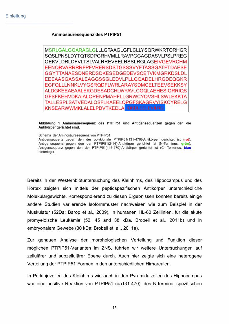
Hierzu wurden peptidspezifische Antikörper gegen die N-terminale Region und
gegen die C-terminale Region des PTPIP51 eingesetzt (Brobeil et al., 2011a).
Einleitung _____________________________
15
Bereits in der Westernblotuntersuchung des Kleinhirns, des Hippocampus und des
Kortex zeigten sich mittels der peptidspezifischen Antikörper unterschiedliche
Molekulargewichte. Korrespondierend zu diesen Ergebnissen konnten bereits einige
andere Studien variierende Isoformmuster nachweisen wie zum Beispiel in der
Muskulatur (52Da; Barop et al., 2009), in humanen HL-60 Zelllinien, für die akute
promyeloische Leukämie (52, 45 and 38 kDa, Brobeil et al., 2011b) und in
embryonalem Gewebe (30 kDa; Brobeil et. al., 2011a).
Zur genauen Analyse der morphologischen Verteilung und Funktion dieser
möglichen PTPIP51-Varianten im ZNS, führten wir weitere Untersuchungen auf
zellulärer und subzellulärer Ebene durch. Auch hier zeigte sich eine heterogene
Verteilung der PTPIP51-Formen in den unterschiedlichen Hirnarealen.
In Purkinjezellen des Kleinhirns wie auch in den Pyramidalzellen des Hippocampus
war eine positive Reaktion von PTPIP51 (aa131-470), des N-terminal spezifischen
Einleitung _____________________________
16
Antikörpers im Soma wie auch im Dendrit zu sehen. Der C-terminal spezifische
Antikörper hingegen wies nur eine Lokalisation im Soma nach.
Subzellulär waren beide PTPIP51-Formen mit dem Golgiapparat assoziiert. Der
Golgiapparat ist an der Morphogenese der Dendriten und Axone beteiligt (Rosso et
al., 2004; Tanabe et al., 2010). Zusätzlich zeigte die C-terminale Form eine
Assoziation mit dem endoplasmatischen Retikulum. Die PTP1B, ein
Interaktionspartner des PTPIP51, ist ebenfalls an das endoplasmtische Retikulum
(ER) gebunden (Pathre et al., 2001). Mittels „Duolink proximity ligation assay“
konnten wir die direkte Interaktion beider Proteine (PTPIP51 und PTP1B) in
Purkinjezellen nachweisen. Bereits Pathre et al. (2001) sowie Fuentes und Arregui
(2009) wiesen eine Beteiligung von PTP1B an Wachstum und Elongation der Axone
nach. Außerdem spielt PTP1B eine wichtige Rolle im „Nerve Growth factor (NGF)“
Signalweg (Shibata et al., 2008). Dieser Signalweg reguliert wiederum den
Neuritenauswuchs, bewahrt sie vor Apoptose und nimmt somit eine funktionelle Rolle
bei Gedächtnisprozessen ein (Chao et al., 2006; Shimoke et al., 2011). Analog zur
intrazellulären Lokalisation der C- terminalen Form des PTPIP51 findet sich NGF
ebenfalls am ER und Golgiapparat (Blöchl et al., 1996). Die Bindung von NGF an
den Neutrophin-Rezeptor p75 (NTR) erhöht die enzymatische Aktivität von PTP1B.
Neben den bereits erwähnten Funktionen ist PTP1B wichtig für das Überleben der
Neurone. Es schützt Neurone vor dem Angriff von Amyloid (Chacon et al., 2011).
Dieser Mechanismus ist noch nicht aufgeklärt (Chacon et al., 2011), könnte aber
durch PTPIP51 vermittelt werden.
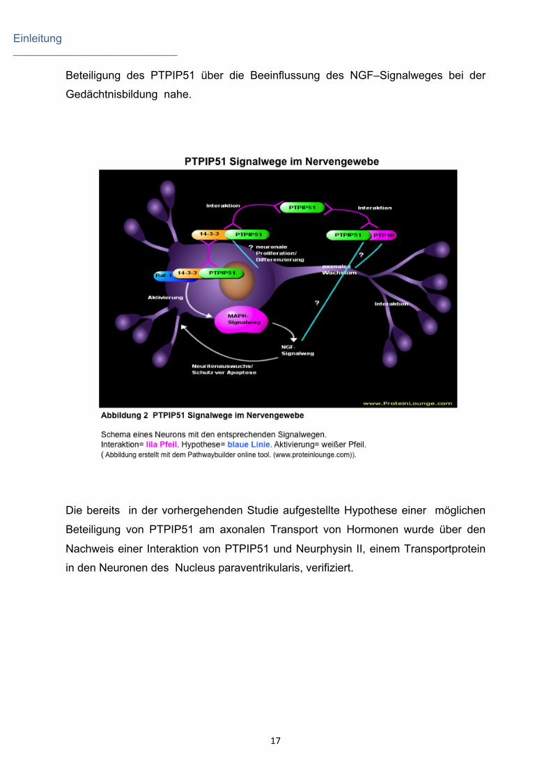
PTPIP51 interagiert mit 14-3-3beta in den Purkinjezellen des Kleinhirns. Die 14-3-3-
Protein-Superfamilie umfasst 7 Isoformen, welche vermutlich im Kleinhirn an der
neuronalen Proliferation, Migration und Differenzierung beteiligt sind (Umahara et al.,
2009). Wie bereits von Yu et al. (2008) berichtet, interagiert PTPIP51 über 14-3-
3beta mit Raf-1 und moduliert darüber die MAPK-Kaskade. Bemerkenswerterweise
ist der NGF- Signalweg mit dem MAPK-Signalweg eng verlinkt (Xing et al., 1998).
NGFs sind wichtig für Gedächtnisprozesse, welche insbesondere bei der
Alzheimererkrankung negativ beeinflusst sind (Chao et al., 2006; Aggleton et al.,
2010). PTPIP51 wird besonderes stark in der Hippocampusregion exprimiert. Diese
Region ist ebenfalls an der Gedächtnisbildung beteiligt. Dies legt eine mögliche
Einleitung _____________________________
17
Beteiligung des PTPIP51 über die Beeinflussung des NGF–Signalweges bei der
Gedächtnisbildung nahe.
Die bereits in der vorhergehenden Studie aufgestellte Hypothese einer möglichen
Beteiligung von PTPIP51 am axonalen Transport von Hormonen wurde über den
Nachweis einer Interaktion von PTPIP51 und Neurphysin II, einem Transportprotein
in den Neuronen des Nucleus paraventrikularis, verifiziert.
Einleitung _____________________________
18
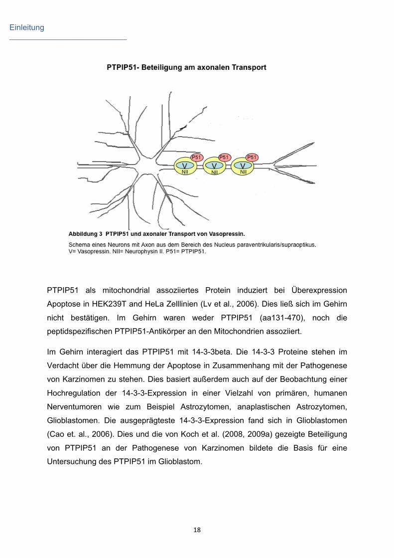
PTPIP51 als mitochondrial assoziiertes Protein induziert bei Überexpression
Apoptose in HEK239T and HeLa Zelllinien (Lv et al., 2006). Dies ließ sich im Gehirn
nicht bestätigen. Im Gehirn waren weder PTPIP51 (aa131-470), noch die
peptidspezifischen PTPIP51-Antikörper an den Mitochondrien assoziiert.
Im Gehirn interagiert das PTPIP51 mit 14-3-3beta. Die 14-3-3 Proteine stehen im
Verdacht über die Hemmung der Apoptose in Zusammenhang mit der Pathogenese
von Karzinomen zu stehen. Dies basiert außerdem auch auf der Beobachtung einer
Hochregulation der 14-3-3-Expression in einer Vielzahl von primären, humanen
Nerventumoren wie zum Beispiel Astrozytomen, anaplastischen Astrozytomen,
Glioblastomen. Die ausgeprägteste 14-3-3-Expression fand sich in Glioblastomen
(Cao et. al., 2006). Dies und die von Koch et al. (2008, 2009a) gezeigte Beteiligung
von PTPIP51 an der Pathogenese von Karzinomen bildete die Basis für eine
Untersuchung des PTPIP51 im Glioblastom.
Einleitung _____________________________
19
2.4 PTPIP51 in Glioblastomen
Das Glioblastoma multiforme (GBM) ist der häufigste und bösartigste primäre
Gehirntumor in der westlichen Welt (Wang et al., 2010; Parsons et al., 2008). Im
Verlauf der letzten Jahrzehnte wurde ein starker Anstieg in der Inzidenz verzeichnet.
18.820 neu aufgetretene Fälle von primären Gehirntumoren werden jährlich in den
USA diagnostiziert, davon entfallen 60% auf Gliome, wobei es sich bei 30-40% um
GBMs handelt (Khan et al., 2009). Bei GBM-Patienten beträgt die durchschnittliche
Überlebenszeit trotz signifikanter technischer und medikamentöser Fortschritte im
therapeutischen Bereich nach wie vor etwa nur ein Jahr (McLendon et al., 2007).
In humanen GBMs korrelieren die beiden Signalmoleküle 14-3-3beta und gamma mit
dem Grad der Malignität (Yang et al., 2009). Aufgrund ihrer spezifischen
Phosphoserin/Phosphothreonin Bindungsstellen besitzen die 14-3-3-Proteine die
Fähigkeit mit vielen verschiedenen Proteinen zu interagieren. Hierzu gehört auch das
PTPIP51. Im Mäusegehirn wurde diese Interaktion in situ nachgewiesen. Zwei
weitere unabhängige Studien (Jin et al., 2004; Ewing et al.. 2007) beschrieben
PTPIP51 als Partner des 14-3-3beta, wobei dadurch die Interaktion mit dem Raf-1
vermittelt wird (Yu et al., 2008).
Die Expression von 14-3-3beta wird als Gradmesser für die Malignität von
Glioblastomen gewertet. Deshalb ist es von höchstem Interesse zu überprüfen, ob
eine erhöhte 14-3-3-Expression gleichzeitig mit einer erhöhten PTPIP51 Expression
in Glioblastomen einhergeht. Zur Klärung dieser Frage wurden Proben von 20
Glioblastomen sowohl auf transkriptioneller, als auch auf translationeller Ebene mit
Hilfe von Immunhistochemie, in situ Hybridisierung und RT-PCR untersucht.
Dabei wurden Grad 2-Astrozytome und GBM mittels RT-PCR analysiert, um eine
potentielle Korrelation zwischen PTPIP51-Expression und Malignitätsgrad zu
überprüfen.
Humane Glioblastomzellen weisen eine Interaktion zwischen PTPIP51 und 14-3-3
beta, sowie PTP1B auf. Beim Vergleich von GBM (WHO Grad lV-glioma) und
niedriggradigen Astrozytomen (Grad ll Glioma) zeigte sich eine zum Tumorgrad
korrelierte Zunahme der Expression von PTPIP51 und 14-3-3beta in den
höhergradigen Hirntumoren. 14-3-3beta vermittelt die Interaktion von PTPIP51 und

Einleitung _____________________________
20
Raf-1, wodurch die Aktivität des MAPK-Signalweges moduliert wird (Lv et al., 2006;
Stenzinger et al., 2009; Yu et al., 2008). Diese Hochregulation von 14-3-3-Proteinen
ist mit einer reduzierten Apoptose-Kapazität assoziiert, da die Antagonisierung oder
Ausschaltung der 14-3-3-Expression verstärkte Apoptose in kultivierten Gliomzellen
bewirkt (Cao et al., 2010). Da der MAPK-Signalweg unter anderem Zellmigration
kontrolliert, könnte eine Aktivitätserhöhung für Rezidive und die schlechte Prognose
von GBM mitverantwortlich sein. Diffus auswandernde Tumorzellen sind in der Lage
gesundes Hirngewebe zu infiltrieren, entgehen so der chirurgischen Extirpation und
bilden neue Tumorherde. In Anbetracht dieser Fakten könnte die Interaktion von
PTPIP51 und 14-3-3beta eine Rolle in der Migration und Proliferation von GBM-
Tumorzellen spielen.
In den Endothelzellen von GBM typischen „glomerulumartigen Gefäßen“ ist eine
deutliche Kolokalisation und Interaktion von PTPIP51 und 14-3-3beta zu beobachten.
Diese abnorme Vaskularisierung und endotheliale Hyperplasie ist eines der
Charakteristika und einer der Mechanismen für die Tumor-Angiogenese der GBMs
(Wang et al., 2010). Die Endothelzellen von Tumoren exprimieren „Epidermal Growth
Factor Receptoren (EGFR)“ (Dhara et al., 2006). Die untersuchten Proben wiesen
eine partielle Kolokalisation von PTPIP51 und dem EGFR auf. EGFR kommt
vermutlich eine zentrale Rolle in der Migration und der lokalen Infiltration von „brain
tumor-initiating cells (BTICs)“ und somit in der Entstehung, der Therapieresistenz
und Entstehung von Krankheitsrezidiven bei Hirntumoren zu (Mimeault M und Batra
SK, 2011). Ausgehend von unseren Ergebnissen könnte PTPIP51 mittels 14-3-3beta
vermittelter Interaktion mit Raf-1 und dem EGFR die Ras/Raf/MEK/ERK-
Signalkaskade aktivieren. Diese Signalkaskade führt schlussendlich auf zellulärer
Ebene zur einer Dysfunktion des Zellzyklus und einer erhöhten proliferativen Aktivität
in GBM (Halatsch et al., 2004). Im Vergleich zum sekundären GBM, welches sich
durch Tumorprogression aus niedriggradigeren Gliomen entwickelt, ist die EGFR-
Genexpression in primären Gliomen etwa fünffach höher, was zu einer
Überexpression in 40% der GBMs führt (Karpel-Massler et al., 2009). Neben dieser
EGFR-Überexpression exprimieren 20% der Gliompatienten die Mutante EGFRvlll
(Jutten et al., 2009). Diese Mutante, die auch in einigen anderen Epitheltumoren
nachgewiesen wurde, ist permanent aktiviert (Yoshimoto et al., 2008; Hama et al.,
2009). Epitheliale Tumoren exprimieren ebenfalls hohe Konzentration von PTPIP51
(Koch et al., 2008; 2009a).
Einleitung _____________________________
21
In Gliomzellen konnte eine direkte Interaktion von PTPIP51 und PTP1B gezeigt
werden. Die PTP1B ist ebenfalls in der Lage die MAPK-Kaskade über c-Src und Ras
zu aktivieren (Stenzinger et al., 2009; Dubé et al., 2004; Dubé und Tremblay, 2004;
Tonks und Muthuswamy, 2007; Zhao et al., 2008). Reichardt et al. (2003) konnten
keine DNA-Amplifikation von PTP1B in humanem GBM nachweisen. Im Gegensatz
dazu zeigt unsere Studie eine Erhöhung der PTP1B Expression. Dies wird auch
bestätigt von Akasaki et al. (2006), die eine Überexpression von PTP1B in Gliomen
zeigten. PTP1B partizipiert in der generellen Onkogenese durch Tyrosin-
Dephosphorylierung von zentralen Signalmolekülen oder durch Hochregulation von
zwei wachstumsfördernden Signalwegen (Arias-Romero et al., 2009). Im
menschlichen Brustdrüsengewebe verknüpft PTP1B die insbesondere für die
Onkogenese wichtige Rezeptor-Tyrosin-Kinase ErbB2 mit zellulären Signalwegen,
welche wiederum atypische Zellteilung und Zellüberleben über Aktivierung von c-Src
und den Übergang in einen Src-abhängigen Phänotyp fördern. Zusätzlich deaktiviert
PTP1B den Ras/MAPK Signalweg Inhibitor (Tonks und Muthuswamy, 2007).
Die hier untersuchten Proben wiesen eine Kolokalisation von PTPIP51 und c-Src im
Glioblastom auf. c-Src wiederum vermittelt die Phosphorylierung von EGFR und
fördert dadurch ein fortschreitendes Tumorwachstum (Tice et al., 1999).
Einleitung _____________________________
22
2.5 Zusammenfassung
Zusammenfassend lässt sich festhalten, dass PTPIP51 in mehreren spezifischen
Gehirnarealen wie dem Kleinhirn, dem Hippocampus, dem Nucleus paraventrikularis,
dem Nucleus supraopticus, dem Nucleus accumbens, dem piriformen Kortex, den
Colliculi superiores, den Pedunculli cerebelli inferiores, dem Genu nervi facialis, dem
spinalen Trigeminaltrakt und in der Neurohypophyse exprimiert wird. Diese lokal
differente Expression ermöglicht die Beteiligung an einer Vielzahl von funktionellen
Prozessen wie zum Beispiel optischen Reflexen, Lernprozessen, Gedächtnisbildung,
Emotionen, Gleichgewicht, Orientierung, Hyposmie bei Morbus Parkinson.
Schizophrenie und Depression.
Auf zellulärer und subzellulärer Ebene zeigt sich ebenfalls ein sehr heterogenes
Expressionsmuster des PTPIP51. Das PTPIP51 interagiert mit der PTP1B, dem 14-
3-3beta und dem Neurophysin II. Durch diese Interaktionen greift PTPIP51 in
zahlreiche essentielle Signalwege wie den MAPK-, den CNTF- und den NGF-
Signalweg ein. Deshalb ist eine der Hauptfunktionen des PTPIP51 im Nervengewebe
wohl die Regulation von Proliferation, Differenzierung, Migration und des
Vesikeltransports. Neben diesen Funktionen kann dem PTPIP51 bedingt durch seine
Involvierung in diesen Signalwegen eine wichtige Rolle in der Pathogenese von
Erkrankungen wie zum Beispiel dem Glioblastom und anderen Hirntumoren, der
Alzheimer-Demenz und dem Morbus Parkinson zukommen. Dies macht das
PTPIP51 nicht nur für die Grundlagenforschung, sondern auch für die Klinik
interessant.
Die heterogene PTPIP51-Expression und dessen Funktionen lassen sich mit der
Existenz möglicher Isoformen des PTPIP51 erklären und wird durch die
unterschiedliche Verteilung mit peptidspezifischen Antikörpern nachgewiesener
Formen gezeigt.
Auf Grund seiner weit gefächerten Funktionen im ZNS kann man das PTPIP51 auch
als ein „multifunktionales Protein“ des Nervengewebes bezeichnen.
Diese Arbeiten stellen die Basis für weitere interessante Studien des PTPIP51 im
ZNS dar.
Einleitung _____________________________
23
So laufen bereits weitere Versuche mit kultivierten Glioblastomzellen, die die
Wirkung von Chemotherapeutika (PD98059, Gefitinib, Cetuximab) auf die PTPIP51-
Expression analysieren. Außerdem wird untersucht, ob die Expressionsrate des
PTPIP51 in Ependymomen als möglicher Marker für verschiedene Stadien etabliert
werden kann. Die Rolle von PTPIP51 bei der erhöhten Ausschüttung von Oxytocin in
weiblichen Ammenratten soll weiteren Aufschluss über die Funktion im axonalen
Transport geben.
Interessanterweise wurde PTP1B auch als Regulator des VCAM-1-Signalweges
identifiziert. Es ist bekannt, dass die Interaktion von α-4 Integrin und VCAM-1 zur
Ansammlung von T-Zellen an der Bluthirnschranke und damit zur perivaskulären
Infiltration von Lymphozyten führt. Dadurch bedingt kommt es zum Beginn von
Erkrankungen des ZNS (Vajkoczy und Menger, 2004). Antikörper gegen VCAM-1
und α-4 Integrin werden bereits in der medikamentösen Therapie von multipler
Sklerose und anderen inflammatorischen Erkrankungen genutzt (Deem et al., 2007).
Colucci et al. (2004) wiesen eine Korrelation zwischen dem klinischen Outcome von
Patienten mit mulitpler Sklerose und der Konzentration von 14-3-3 im Liquor nach, so
dass aktuell eine Studie begonnen wurde, in der die Expression von PTPIP51 und
dessen Interaktionspartner 14-3-3 im Maus EAE-Modell in Abhängigkeit des MS-
Stadiums untersucht wird.
Zusammenfassend kann man sagen, dass diese Arbeit eine wichtige Grundlage für
viele weitere sehr interessante Projekte bezüglich des PTPIP51 im ZNS darstellt. Die
kommenden Projekte werden nun das PTPIP51 in Relation zu Erkrankungen und
mögliche Nutzung für Diagnose und Therapie stellen.

Einleitung _____________________________
24
2.6 Summary
To sum up PTPIP51 is expressed in several specific brain areas: the cerebellum, the
hippocampus, the paraventricular nucleus, the supraoptic nucleus, the nucleus
accumbens, the piriformal cortex, the superior colliculus, the inferior cerebellar
peduncle, the genu of the facial nerve, the spinal trigeminal tract, the
neurohypophysis. This locally restricted different expression allows PTPIP51 to take
part in a multitude of functions e.g. optical reflexes, learning, forming of memories,
emotions, equilibrium, orientation, hyposmy in morbus Parkinson, schizophrenia and
depression.
On cellular and sub- cellular level PTPIP51 displayed a heterogeneous expression.
In brain PTPIP51 interacts with PTP1B,14-3-3 beta and neurophysin II. By these
interactions PTPIP51 is involved in many essential pathways such as MAPK, CNTF
and NGF. Therefore one of the main functions of PTPIP51 in nervous tissue seems
to be the regulation of proliferation, differentiation, migration and vesicle transport.
Besides these functions PTPIP51 plays a central role in the genesis of glioblastoma,
Alzheimer disease and morbus Parkinson. This stresses the importance of basic and
clinical research on PTPIP51.
The heterogeneous expression and function of PTPIP51 can be explained by
possible isoforms. This subject was investigated by peptide specific antibodies.
Based on the broad spectrum of functions in the CNS PTPIP51 is a real
“multifunctional protein”.
The presented manuscript is the basis for many interesting studies of PTPIP51 in
CNS.
A current study with cultured glioblastoma cells investigates the effects of
chemotherapeutics (PD98059, Gefitinib, Cetuximab) on the expression of PTPIP51.
Another study tests whether the grade of PTPIP51- expression in ependymoma is a
possible marker for its staging. The role of PTPIP51 in axonal transport is further
investigated in nurse rats using their increased productions of oxytocin as a trigger.
Interestingly, PTP1B is known as a regulator of the VCAM-1-pathway. The interaction
of α-4 integrin and VCAM-1 results in the accumulation of T-cells at the blood-brain-
Einleitung _____________________________
25
barrier and in a perivascular infiltration of lymphocytes. This induces diseases of the
CNS (Vjakoczy and Menger, 2004). Therefore, in medical therapy of multiple
sclerosis and other inflammatory diseases antibodies against α-4 integrin and VCAM-
1 are applied (Deem et al., 2007). According to Colucci and coworkers (2004) the
clinical outcome of these patients is related to the concentration of 14-3-3 in the
spinal fluid. Just now a study is done investigating the expression of PTPIP51 and its
interaction partner 14-3-3 in relation to the stage of MS in the EAE mouse model.
In summary this PHD thesis is an important basis for many consecutive studies of
PTPIP51 in CNS in relation to diseases and possibly for the use in diagnosis and
therapy.
Literaturverzeichnis _____________________________
26
3. Literaturverzeichnis
Aggleton JP., O'Mara SM., Vann SD., Wright NF., Tsanov M., Erichsen JT. (2010).
Hippocampal-anterior thalamic pathways for memory: uncovering a network of direct
and indirect actions. Eur J Neurosci. 31(12): 2292-307.
Aisenbrey GA., Handelman WA., Arnold P., Manning M., Schrier RW. Vascular
effects of arginine vasopressin during fluid deprivation in the rat. (1981). J Clin Invest.
67(4):961-8.
Akasaki Y., Liu G., Matundan HH., Ng H., Yuan X., Zeng Z., Black KL., Yu JS.
(2006). A peroxisome proliferator-activated receptor-gamma agonist, troglitazone,
facilitates caspase-8 and -9 activities by increasing the enzymatic activity of protein-
tyrosine phosphatase-1B on human glioma cells. J Biol Chem. 281(10):6165-74.
Arias-Romero L.E., Saha S., Villamar-Cruz O., Yip S.C., Ethier S.P., Zhang Z.Y.,
Chernoff J. (2009). Activation of Src by protein tyrosine phosphatase 1B is required
for ErbB2 transformation of human breast epithelial cells. Cancer Res. 69:4582-4588.
Baba T., Takeda A., Kikuchi A., Nishio Y., Hosokai Y., Hirayama K., Hasegawa T.,
Sugeno N., Suzuki K., Mori E., Takahashi S., Fukuda H., Itoyama Y. (2011).
Association of olfactory dysfunction and brain. Metabolism in Parkinson's disease.
Mov Disord.26(4):621-8.
Basar K., Sesia T., Groenewegen H., Steinbusch HW., Visser-Vandewalle V., Temel
Y. (2010). Nucleus accumbens and impulsivity. Prog Neurobiol.92(4):533-57.

Literaturverzeichnis _____________________________
27
Barop J., Sauer H., Steger K., Wimmer M. (2009). Differentiation-dependent
PTPIP51 expression in human skeletal muscle cell culture. J Histochem Cytochem.
57(5):425-35.
Blöchl A., Thoenen H. (1996) Localization of cellular storage compartments and sites
of constitutive and activity-dependent release of nerve growth factor (NGF) in primary
cultures of hippocampal neurons. Mol Cell Neurosci. 7(3):173-90.
Bobrich M., Brobeil A., Mooren FC., Krüger K., Steger K., Tag C., Wimmer M. (2011).
PTPIP51 interaction with PTP1B and 14-3-3ß in adipose tissue of insulin resistant
mice. Int J Obes (Lond). [Epub ahead of print].
Bobrich M., Schwabe S., Viard M., Kamm M., Brobeil A., Mooren FC., Krüger K., Tag
C., Wimmer M. (2011). PTPIP51 – Connecting lipolysis and lipogenesis in adipose
tissue. (eingereicht).
Brobeil A., Graf M., Oeschger S., Steger K., Wimmer M.(2010). PTPIP51-a myeloid
lineage specific protein interacts with PTP1B in neutrophil granulocytes. Blood Cells
Mol Dis. 45(2): 159-68
Brobeil A., Bobrich M., Wimmer M. (2011a). Protein tyrosine phosphatase interacting
protein 51- a jack-of-all-trades protein. Cell Tissue Res. 344(2):189-205.
Brobeil A., Bobrich M., Graf M., Kruchten A., Blau W., Rummel M., Oeschger S.,
Steger K., Wimmer M. (2011b). PTPIP51 is phosphorylated by Lyn and c-Src kinases
lacking dephosphorylation by PTP1B in acute myeloid leukemia. Leuk Res. [Epub
ahead of print].

Literaturverzeichnis _____________________________
28
Brownstein MJ., Russell JT., Gainer H. (1980). Synthesis, transport, and release of
posterior pituitary hormones. Science. 207:373-378.
Buijs RM., Van Heerikhuize JJ. (1982). Vasopressin and oxytocin release in the
brain—a synaptic event. Brain Res. 252(1):71-6.
Cao L., Cao W., Zhang W., Lin H., Yang X., Zhen H., Cheng J., Dong W., Huo J.,
Zhang X. (2008). Identification of 14-3-3 protein isoforms in human astrocytoma by
immunohistochemistry. Neurosci. Lett. 432:94-99.
Cao W., Yang X., Zhou J., Teng Z., Cao L., Zhang X., Fei Z. (2010). Targeting 14-3-3
protein, difopein induces apoptosis of human glioma cells and suppresses tumor
growth in mice. Apoptosis. 15:230-241.
Chacón PJ., Arévalo MA., Tébar AR. (2010) NGF-activated protein tyrosine
phosphatase 1B mediates the phosphorylation and degradation of I-kappa-Balpha
coupled to NF-kappa-B activation, thereby controlling dendrite morphology. Mol Cell
Neurosci. 43(4):384-93.
Chacón PJ., Garcia-Mejias R., Rodriguez-Tebar A. (2011). Inhibition of RhoA
GTPase and the subsequent activation of PTP1B protects cultured hippocampal
neurons against amyloid β toxicity. Mol Neurodegener. 6 (1):14.
Chao MV., Rajagopal R., Lee FS. (2006). Neurotrophin signalling in health and
disease. Clin. Sci. (Lond). 110(2):167-73.

Literaturverzeichnis _____________________________
29
Colucci M., Roccatagliata L., Capello E., Narciso E., Latronico N., Tabaton M.,
Mancardi GL. (2004). The 14-3-3 protein in multiple sclerosis: a marker of disease
severity. Mult Scler. 10(5):477-81.
Deem TL., Abdala-Valencia H., Cook-Mills JM. (2007). VCAM-1 activation of
endothelial cell protein tyrosine phosphatase 1B. J Immunol. 178(6):3865-73.
den Heijer T., Tiemeier H., Luijendijk HJ., van der Lijn F., Koudstaal PJ., Hofman A,,
Breteler MM. (2011). A Study of the Bidirectional Association Between Hippocampal
Volume on Magnetic Resonance Imaging and Depression in the Elderly. Biol
Psychiatry.[Epub ahead of print].
Dhikav V., Anand K. (2011 Potential predictors of hippocampal atrophy in Alzheimer's
disease. Drugs Aging. 28(1):1-11.
Dreifuss JJ. (1975). A review on neurosecretory granules: their contents and
mechanisms of release. Ann N Y Acad Sci. 248:184-201.
Dubé N. and Tremblay M.L. (2004). Beyond the metabolic function of PTP1B. Cell.
Cycle. 3:550-553.
Dubé N., Cheng A. and Tremblay M.L. (2004). The role of protein tyrosine
phosphatase 1B in Ras signalling. Proc. Nat. Acad. Sci. U S A. 101:1834-1839.
Ewing R.M., Chu P., Elisma F., Li H., Taylor P., Climie S., McBroom-Cerajewski L.,
Robinson M.D., O'Connor L., Li M., Taylor R., Dharsee M., Ho Y., Heilbut A., Moore
L., Zhang S., Ornatsky O., Bukhman Y.V., Ethier M., Sheng Y., Vasilescu J., Abu-
Literaturverzeichnis _____________________________
30
Farha M., Lambert J.P., Duewel H.S., Stewart I.I., Kuehl B., Hogue K., Colwill K.,
Gladwish K., Muskat B., Kinach R., Adams S.L., Moran M.F., Morin G.B., Topaloglou
T., Figeys D. (2007). Large-scale mapping of human protein-protein interactions by
mass spectrometry. Mol. Syst. Biol. 3:89.
Fotheringham AP., Davidson YS., Davies I., Morris JA. (1991). Age-associated
changes in neuroaxonal transport in the hypothalamo-neurohypophysial system of
the mouse. Mech Ageing Dev. 60:113-121.
Fuentes F., Arregui CO. (2009). Microtubule and cell contact dependency of ER-
bound PTP1B localization in growth cones. Mol Biol Cell. 20(6):1878-89.
Fuhrmann S., Grabosch K., Kirsch M., Hofmann HD. (2003). Distribution of CNTF
receptor alpha protein in the central nervous system of the chick embryo. J Comp
Neurol. 461(1):111-22.
Fredriksson S. (2009). Visualizing signal transduction pathways by quantifying
protein-protein interactions in native cells and tissue, Nat. Methods 6.
Gerdes JM, Katsanis N. (2005). Microtubule transport defects in neurological and
ciliary disease. Cell Mol Life Sci. 62:1556-1570.
Gibbens GL., Chard T. (1976).Observations on maternal oxytocin release during
human labor and the effect of intravenous alcohol administration. Am J Obstet
Gynecol. 126(2):243-6.
Literaturverzeichnis _____________________________
31
Gillingham AK., Munro S. (2003). Long coiled coil proteins and membrane traffic.
Biochim Biophys Acta. 1641(2-3):71-85.
Goldstein LS. (2001). Kinesin molecular motors: transport pathways, receptors, and
human disease. Proc Natl Acad Sci U S A 98:6999-7003.
Halatsch M.E., Gehrke E.E., Vougioukas V.I., Bötefür I.C., A-Borhani F., Efferth T.,
Gebhart E., Domhof S., Schmidt U., Buchfelder M. (2004). Inverse correlation of
epidermal growth factor receptor messenger RNA induction and suppression of
anchorage-independent growth by OSI-774, an epidermal growth factor receptor
tyrosine kinase inhibitor, in glioblastoma multiforme cell lines. J. Neurosurg. 100:523-
533.
Hama T., Yuza Y., Saito Y., Ouchi J., Kondo S., Okabe M., Yamada H., Kato T.,
Moriyama H., Kurihara S., Urashima M. (2009). Prognostic significance of epidermal
growth factor receptor phosphorylation and mutation in head and neck squamous cell
carcinoma. Oncologist. 14:900-908.
Isa T., Yoshida M. (2009). Saccade control after V1 lesion revisited. Curr Opin
Neurobiol. 19(6):608-14.
Jin J., Smith F.D., Stark C., Wells C.D., Fawcett J.P., Kulkarni S., Metalnikov P.,
O'Donnell P., Taylor P., Taylor L., Zougman A., Woodgett J.R., Langeberg L.K., Scott
J.D., Pawson T. (2004). Proteomic, functional, and domain-based analysis of in vivo
14-3-3 binding proteins involved in cytoskeletal regulation and cellular organization.
Curr. Biol. 14:1436-1450.
Literaturverzeichnis _____________________________
32
John J., Manchanda R. (2011). Modulation of synaptic potentials and cell excitability
by dendritic KIR and KAs channels in nucleus accumbens medium spiny neurons:
Acomputational study. J Biosci. 36(2):309-28.
Jutten B., Dubois L., Li Y., Aerts H., Wouters B.G., Lambin P., Theys J., Lammering
G. (2009). Binding of cetuximab to the EGFRvIII deletion mutant and its biological
consequences in malignant glioma cells. Radiother. Oncol. 92:393-398.
Katz Y., Wang ET., Airoldi EM., Burge CB. (2010). Analysis and design of RNA
sequencing experiments for identifying isoform regulation. Nat. Methods 7(12):1009-
15.
Karpel-Massler G., Schmidt U., Unterberg A., Halatsch M.E. (2009). Therapeutic
inhibition of the epidermal growth factor receptor in high-grade gliomas: where do we
stand? Mol. Cancer. Res. 7:1000-1012.
Khan M.K., Hunter G.K., Vogelbaum M., Suh J.H., Chao S.T. (2009). Evidence-
based adjuvant therapy for gliomas: current concepts and newer developments.
Indian J. Cancer. 46, 96-107.
Koch P., Stenzinger A., Viard M., Märker D., Mayser P., Nilles M., Schreiner D.,
Steger K., Wimmer M. (2008). The novel protein PTPIP51 is expressed in human
keratinocyte carcinomas and their surrounding stroma. J Cell Mol Med. 12(5B):2083-
95.
Literaturverzeichnis _____________________________
33
Koch P., Petri M., Paradowska A., Stenzinger A., Sturm K., Steger K., Wimmer M.
(2009a). PTPIP51 mRNA and protein expression in tissue microarrays and promoter
methylation of benign prostate hyperplasia and prostate carcinoma. Prostate.
69(16):1751-62.
Koch P., Viard M., Stenzinger A., Brobeil A., Tag C., Steger K., Wimmer M. (2009b).
Expression profile of PTPIP51 in mouse brain. J. Comp. Neurol. 517:892-905.
Kozak M. (2005). Regulation of translation via mRNA structure in prokaryotes and
eukaryotes. Gene. 361:13–37.
Leake RD., Weitzman RE., Glatz TH., Fisher DA. (1981). Plasma oxytocin
concentrations in men, nonpregnant women, and pregnant women before and during
spontaneous labor. J Clin Endocrinol Metab. 53(4):730-3.
Lee MY., Hofmann HD., Kirsch M. (1997). Expression of ciliary neurotrophic factor
receptor-alpha messenger RNA in neonatal and adult rat brain: an in situ
hybridization study. Neuroscience. 77(1):233-46.
Loureiro M., Lecourtier L., Engeln M., Lopez J., Cosquer B., Geiger K., Kelche C.,
Cassel JC., Pereira de Vasconcelos A. (2011).The ventral hippocampus is necessary
for expressing a spatial memory. Brain Struct Funct. [Epub ahead of print].
Lv B.E., Yu C.E., Chen Y.Y., Lu Y., Guo J.H., Song Q.S., Ma D.L., Shi T.P. and
Wang L. (2006). Protein tyrosine phophatase interacting interacting protein
51(PTPIP51) is a novel mitochondria protein with an N-terminal mitochondrial
targeting sequence and induces apoptosis. Apoptosis. 11:1489-1501.
Literaturverzeichnis _____________________________
34
Maerker D., Stenzinger A., Schreiner D., Tag C., Wimmer M. (2008). Expression of
PTPIP51 during mouse eye development. Histochem Cell Biol. 129(3):345-56.
Mauk MD., Medina JF., Nores WL., Ohyama T. (2000). Cerebellar function:
coordination, learning or timing? Curr Biol. 10(14):R522-5.
McLendon R.E., Turner K., Perkinson K., Rich J. (2007). Second messenger systems
in human gliomas. Arch. Pathol. Lab. Med. 131:1585-1590.
Mimeault M, Batra SK. (2011). Complex oncogenic signaling networks regulate brain
tumor-initiating cells and their progenies: Pivotal roles of wild-type EGFR, EGFRvIII
mutant and hedgehog cascades and novel multitargeted therapies. Brain Pathol.
[Epub ahead of print]
Nilsen TW., Graveley BR. (2010). Expansion of the eukaryotic proteome by
alternative splicing. Nature 463:457–463.
Oishi K., Okano H., Sawa H. (2007). RMD-1, a novel microtubuleassociated protein,
functions in chromosome segregation in Caenorhabditis elegans. (2009) J Cell Biol.
179:1149–1162.
Oliveira MS., Pacheco LF., Mello CF., Cavalheiro EA., Garrido-Sanabria ER. (2011).
Epileptiform activity in the limbic system. Front Biosci (Schol Ed). 3:565-93.

Literaturverzeichnis _____________________________
35
Parsons DW., Jones S., Zhang X., Lin JC., Leary RJ., Angenendt P., Mankoo P.,
Carter H., Siu IM., Gallia GL., Olivi A., McLendon R., Rasheed BA., Keir S.,
Nikolskaya T., Nikolsky Y., Busam DA., Tekleab H., Diaz LA Jr., Hartigan J., Smith
DR., Strausberg RL., Marie SK., Shinjo SM., Yan H., Riggins GJ., Bigner DD.,
Karchin R., Papadopoulos N., Parmigiani G., Vogelstein B., Velculescu VE., Kinzler
KW. (2008). An integrated genomic analysis of human glioblastoma multiforme.
Science. 321(5897):1807-12.
Pathre P., Arregui C., Wampler T., Kue I., Leung TC., Lilien J., Balsamo J. (2001).
PTP1B regulates neurite extension mediated by cell–cell and cell–matrix adhesion
molecules. J Neurosci Res. 63:143–150.
Pereira AG., Portuguez MW., da Costa DI., Azambuja LS., Marroni SP., da Costa
JC., Pereira-Filho AA. (2011). Route learning performance: is it a hippocampus
function? Cogn Behav Neurol. 24(1):4-10.
Porsche A. (2001). Identifikation von Interaktionspartnern der T-Zell Protein-Tyrosin-
Phosphatase durch das Lex-A Two Hybrid System. Thesis (Ph.D.), University of
Konstanz, (UFO Publishers, Allensbach, Vol. 414).
Reichardt W., Jung V., Brunner C., Klein A., Wemmert S., Romeike B.F., Zang K.D.,
Urbschat S. (2003). The putative serine/threonine kinase gene STK15 on
chromosome 20q13.2 is amplified in human gliomas. Oncol. Rep. 10:1275-1279.
Roger J., Goureau O., Sahel JA., Guillonneau X. (2007). Use of suppression
subtractive hybridization to identify genes regulated by ciliary neurotrophic factor in
postnatal retinal explants. Mol Vis. 13:206-19.
Literaturverzeichnis _____________________________
36
Rosso S., Bollati F., Bisbal M., Peretti D., Sumi T., Nakamura T., Quiroga S., Ferreira
A., Cáceres A. (2004). LIMK1 regulates Golgi dynamics, traffic of Golgi-derived
vesicles, and process extension in primary cultured neurons. Mol Biol Cell.
15(7):3433-49
Russell JA., Leng G. (2000).Veni, vidi, vici: the neurohypophysis in the twentieth
century. Exp Physiol. 85 Spec No:1S-6S. Review.
Schmahmann JD., Sherman JC. (1998). The cerebellar cognitive affective syndrome.
Brain. 121 (Pt 4):561-79.
Shibata T., Nakahara H., Kita N., Matsubara Y., Han C., Morimitsu Y., Iwamoto N.,
Kumagai Y., Nishida M., Kurose H., Aoki N., Ojika M., Uchida K. (2008). A food-
derived synergist of NGF signaling: identification of protein tyrosine phosphatase 1B
as a key regulator of NGF receptor-initiated signal transduction. J Neurochem.
107(5):1248-60.
Shimoke K., Sasaya H., Ikeuchi T.(2011). Analysis of the role of nerve growth factor
in promoting cell survival during endoplasmic reticulum stress in PC12 cells. Methods
Enzymol. 490:53-70.
Soudry Y., Lemogne C., Malinvaud D., Consoli SM., Bonfils P.(2011). Olfactory
system and emotion: common substrates. Eur Ann Otorhinolaryngol Head Neck Dis.
128(1):18-23.
Stenzinger A., Kajosch T., Tag C., Porsche A., Welte I., Hofer HW., Steger K.,
Wimmer M. (2005). The novel protein PTPIP51 exhibits tissue- and cell-specific
expression. Histochem Cell Biol. 123(1):19-28.
Literaturverzeichnis _____________________________
37
Stenzinger A., Schreiner D., Koch P., Hofer H.W. and Wimmer M. (2009). Cell- and
molecular biology of the novel protein tyrosine phosphatase interacting protein 51.
Int. Rev. Cell. Mol. Biol. 275:183-246.
Tanabe K., Kani S., Shimizu T., Bae YK., Abe T., Hibi M. (2010). Atypical protein
kinase C regulates primary dendrite specification of cerebellar Purkinje cells by
localizing Golgi apparatus. J Neurosci. 30(50):16983-92.
Tice D.A., Biscardi J.S., Nickles A.L., Parsons S.J. (1999). Mechanism of biological
synergy between cellular Src and epidermal growth factor receptor. Proc. Nat. Acad.
Sci. USA. 96:1415-1420.
Tonks N.K. and Muthuswamy S.K. (2007). A brake becomes an accelerator: PTP1B -
a new therapeutic target for breast cancer. Cancer Cell. 11:214-216.
Towbin H., Staehelin T., Gordon J. (1979). Electrophoretic transfer of proteins from
polyacrylamide gels to nitrocellulose sheets: procedure and some applications. Proc
Natl Acad Sci USA 76: 4350–4354.
Trembleau A., Morales M., Bloom FE. (1994). Aggregation of vasopressin mRNA in a
subset of axonal swellings of the median eminence and posterior pituitary: light and
electron microscopic evidence. J Neurosci. 14:39-53
Umahara T., Uchihara T., Nakamura A, Iwamoto T. (2009). Isoform- dependent
immunolocalization of 14-3-3 proteins in developing rat cerebellum. Brain Res.
1253:15-26.
Literaturverzeichnis _____________________________
38
Vajkoczy P., Menger MD. (2004). Vascular microenvironment in gliomas. Cancer
Treat Res.117:249-62.
Varela-Nallar L., Aranguiz FC., Abbott AC., Slater PG., Inestrosa NC.(2010). Adult
hippocampal neurogenesis in aging and Alzheimer's disease. Birth Defects Res C
Embryo Today. 90(4):284-96.
Wang R., Chadalavada K., Wilshire J., Kowalik U., Hovinga KE., Geber A., Fligelman
B., Leversha M., Brennan C., Tabar V. (2010). Glioblastoma stem-like cells give rise
to tumour endothelium. Nature. 468(7325):829-33.
Watanabe M., Isobe T., Okuyama T., Ichimura T., Kuwano R., Takahashi Y., Kondo
H. (1991). Molecular cloning of cDNA to rat 14-3-3 eta chain polypeptide and the
neuronal expression of the mRNA in the central nervous system. Brain Res Mol Brain
Res. 10(2):151-8.
Watanabe M., Isobe T., Ichimura T., Kuwano R., Takahashi Y., Kondo H. (1993a).
Molecular cloning of rat cDNAs for beta and gamma subtypes of 14-3-3 protein and
developmental changes in expression of their mRNAs in the nervous system. Brain
Res Dev Brain Res. 17(1-2):135-46.
Watanabe M., Isobe T., Ichimura T., Kuwano R., Takahashi Y., Kondo H. (1993b).
Developmental regulation of neuronal expression for the eta subtype of the 14-3-3
protein, a putative regulatory protein for protein kinase C. Brain Res Dev Brain Res.
73(2):225-35.

Literaturverzeichnis _____________________________
39
Watanabe M., Isobe T., Ichimura T., Kuwano R., Takahashi Y., Kondo H., Inoue Y.
(1994). Molecular cloning of rat cDNAs for the zeta and theta subtypes of 14-3-3
protein and differential distributions of their mRNAs in the brain. Brain Res Mol Brain
Res.(1-2):113-21.
Watanabe M. (2008). Molecular mechanisms governing competitive synaptic wiring
in cerebellar Purkinje cells. Tohoku J Exp Med. 214(3):175-90.
Wei Z., Zhang M. (2010). A structural approach to decipher the neurexin and
neuroligin splice isoform code. Neuron. J. 67(1):1-2.
Xing J., Kornhauser JM., Xia Z., Thiele EA., Greenberg ME. (1998). Nerve growth
factor activates extracellular signal-regulated kinase and p38 mitogen-activated
protein kinase pathways to stimulate CREB serine 133 phosphorylation. Mol Cell
Biol. 18(4):1946-55.
Yang Z., Goldstein LS. (1998). Characterization of the KIF3C neural kinesin-like
motor from mouse. Mol Biol Cell 9:249-261.
Yang X., Cao W., Lin H., Zhang W., Lin W., Cao L., Zhen H., Huo J., Zhang X.
(2009). Isoform-specific expression of 14-3-3 proteins in human astrocytoma. J.
Neurol. Sci. 276:54-59.
Yoshimoto K., Dang J., Zhu S., Nathanson D., Huang T., Dumont R., Seligson D.B.,
Yong W.H., Xiong Z., Rao N., Winther H., Chakravarti A., Bigner D.D., Mellinghoff
I.K., Horvath S., Cavenee W.K., Cloughesy T.F., Mischel P.S. (2008). Development
of a real-time RT-PCR assay for detecting EGFRvIII in glioblastoma samples. Clin.
Cancer. Res. 14:488-493.
Literaturverzeichnis _____________________________
40
Yu C., Han W., Shi T., Lv B., He Q., Zhang Y., Li T., Zhang Y., Song Q., Wang L., Ma
D. (2008). PTPIP51, a novel 14-3-3 binding protein, regulates cell morphology and
motility via Raf-ERK pathway. Cell. Signal. 20:2208-2220.
Zhao Y., Xiao A., Dipierro C.G., Abdel-Fattah R., Amos S., Redpath G.T., Carpenter
J.E., Pieper R.O. and Hussaini, I.M. (2008). H-Ras increases urokinase expression
and cell invasion in genetically modified human astrocytes through Ras/Raf/MEK
signalling pathway. Glia. 56:917-924.
Weitere Publikationen und Poster _____________________________
41
4. Weitere Publikationen und Poster
A) weitere Publikationen
Koch P., Stenzinger A., Viard M., Märker D., Mayser P., Nilles M., Schreiner D.,
Steger K., Wimmer M. (2008). The novel protein PTPIP51 is expressed in human
keratinocyte carcinomas and their surrounding stroma. J Cell Mol Med.12 (5B):2083-
95.
Bobrich M., Schwabe S., Viard M., Kamm M., Brobeil A., Mooren FC., Krüger K., Tag
C., Wimmer M. (2011). PTPIP51 – Connecting lipolysis and lipogenesis in adipose
tissue (eingereicht).
B) Poster
Viard M. Localization of protein tyrosine phosphatase interacting protein 51
(PTPIP51) in mouse brain.(49th Symposium of the Society for Histochemistry
Freiburg im Breisgau, Germany, 2007)
Koch P., Stenzinger A., Viard M., Mayser P., Wimmer M. Expression of the novel
protein PTPIP51 in human keratinocyte carcinomas and their surrounding stroma.
(102nd Annual Meeting of the Anatomische Gesellschaft, Giessen 2007)
Koch P., Stenzinger A., Viard M., Mayser P., Wimmer M. Expression of the novel
protein PTPIP51 in human keratinocyte carcinomas and their surrounding stroma.
(32nd FEBS Congress, Molecular Machines, Vienna 2007)
Danksagung _____________________________
42
5. Danksagung
An erster Stelle möchte ich mich bei Frau Prof. Dr. Wimmer für die Überlassung des
Themas, die Möglichkeit zu Publizieren und die herzliche Aufnahme in ihr Team
sowie für die immerwährende fachliche und freundliche Betreuung meiner Arbeit
bedanken.
Bei Frau Claudia Tag möchte ich mich für die Einarbeitung in die verschiedenen
Methoden und ihre Hilfe in allen Belangen bedanken.
Auch den anderen Mitarbeitern des Instituts für Anatomie und Zellbiologie danke ich
für ihre vielen guten Ratschläge und die sehr nette, freundschaftliche
Arbeitsatmosphäre.
Zudem möchte ich mich bei Herrn Prof. Dr. Kaps und den Kollegen der Neurologie
der Universitätsklinik Gießen bedanken, die mich in den letzten Monaten bei der
Fertigstellung meiner Doktorarbeit unterstützt haben.
Mein weiterer Dank gilt Manuel Bobrich, Alexander Brobeil und Max Kamm für ihre
fachliche Unterstützung, die mir in häufigen Diskussionen und Gesprächen zu Gute
gekommen ist.
Julian Fugmann danke ich für seine Unterstützung bei computerfachlichen Fragen.
Meinen Geschwistern möchte ich für die aufbauenden Gespräche nach so manchen
ernüchternden Tagen im Labor danken.
Ganz besonders bedanken möchte ich mich bei meinen Eltern, die mir mein Studium
erst ermöglicht haben und ohne deren Unterstützung diese Arbeit nie zustande
gekommen wäre.

Der Lebenslauf wurde aus der elektronischen Version der Arbeit entfernt.
The curriculum vitae was removed from the electronic version of the paper.

Publikationen der kumulativen Doktorarbeit _____________________________
45
7. Publikationen der kumulativen Doktorarbeit
7.1. Expression profile of PTPIP51 in mouse brain.
7.2. PTPIP51 – a multifunctional protein in brain tissue
7.3. PTPIP51, a positive modulator of the MAPK/Erk pathway, is upregulated in glioblastoma and interacts with 14-3-3β and PTP1B in situ

Expression Profile of PTPIP51 in Mouse Brain
P. KOCH,1* M. VIARD,1 A. STENZINGER,1 A. BROBEIL,1 C. TAG,1 K. STEGER,2 AND M. WIMMER2
1Institute of Anatomy and Cell Biology, Justus-Liebig-University, 35385 Giessen, Germany2Department of Urology and Pediatric Urology, Justus-Liebig-University, 35385 Giessen, Germany
ABSTRACTThis study demonstrates the expression of the novel proteinprotein tyrosine phophatase-interacting protein 51 (PT-PIP51) in mammalian brain tissue. Serial sections of thewhole adult mouse brain were analyzed for PTPIP51 proteinand mRNA by immunohistochemistry, immunoblotting, RT-PCR, and in situ hybridization. Recent investigations by Yuet al. (2008) describe PTPIP51 as being capable of activatingRaf-1, thereby modulating the MAPK pathway. The role ofRaf-1, as well as of 14-3-3, in neurological disorders is wellestablished. PTPIP51 expression was confined to neurons inthe following structures: the piriform cortex and their con-
nections to the anterior commissure, nucleus accumbens,paraventricular and supraoptical nuclei, neurohypophysis,superior colliculus, genu of facialis nerve, spinal trigeminaltract, inferior cerebellar peduncle, and cerebellum. In thecerebellum, a subpopulation of Purkinje cells and their den-drites was strongly PTPIP51 positive. Moreover, PTPIP51was found to be colocalized with vasopressin and its trans-port protein neurophysin II in the neuroendocrine nuclei andtheir connections to the neurohypophysis. The data pre-sented here suggest a role of PTPIP51 in neuronal ho-meostasis, axonal growth, and transport. J. Comp. Neurol.517:892–905, 2009. © 2009 Wiley-Liss, Inc.
Indexing terms: PTPIP51; PTP1B; CNTF; 14-3-3; Raf-1; neurophysin II; vasopressin; mousebrain; hypothalamus; hypophysis; nucleus accumbens; cerebellum
PTPIP51 is an evolutionarily conserved protein, which wasshown to interact in vitro with two nontransmembrane proteintyrosine phosphatases, protein tyrosine phosphatase 1B(PTP1B) and T-cell protein tyrosine phosphatase (TcPTP;Porsche, 2001; Stenzinger et al., 2005). The interaction takesplace in the region between amino acids 78 and 214. Theprotein is phosphorylated in vitro and in situ at Tyr176 by Srckinase and dephosphorylated by PTP1B and TcPTP (Stenz-inger et al., 2009). In mammals, its expression is associatedwith specific tissues such as epithelia, testis, skeletal muscle,and nervous tissue (Stenzinger et al., 2005). PTPIP51 proteinalso plays a role during mammalian development (Marker etal., 2008), and both mRNA and protein could be traced duringplacental villi formation (Stenzinger et al., 2008) and in variouscarcinomas (Lv et al., 2006; Koch et al., 2008). Further exper-iments demonstrated a vitamin- and cytokine-mediatedPTPIP51 expression in cultured keratinocytes (Stenzinger etal., 2006). Given these findings, we hypothesized PTPIP51 tobe involved in cellular differentiation, motility, cytoskeletonformation, and possibly apoptosis.
Experiments by Lv and colleagues (2006) added evidence tothis assumption by demonstrating that overexpressedPTPIP51 enhances apoptosis in HEK293 cells. Moreover, twoindependent studies by Jin et al. (2004) and Ewing et al. (2007)demonstrated an interaction between the two isoforms 14-3-3-� and 14-3-3-y and PTPIP51. Recent experiments by Yu etal. (2008) confirmed these findings by pull-down experimentsand describe PTPIP51 as interacting with Raf-1 through 14-
3-3, thereby modulating cellular motility and morphology viathe mitogen-activated protein kinase (MAPK) cascade. Boththe Ras/Raf/MEK/ERK pathway and the mammalian 14-3-3superfamily play pivotal roles in neuronal development andmaintenance as well as in many neurological disorders, in-cluding Alzheimer’s and Parkinson’s disease (Dougherty andMorrison, 2004; Mei et al., 2006; McCubrey et al., 2007; Sam-uels et al., 2008). As reported by several research groups, Rassignaling in particular influences neuronal plasticity, synaptictransmission, and short- and long-term memory of adult mice(Brambilla et al., 1997; Atkins et al., 1998; Giese et al., 2001;Dhaka et al., 2003).
In rat retina, transcription of Ptpip51 is governed by ciliaryneurotrophic factor (CNTF; Roger et al., 2007). CNTF, a neu-ropoietic cytokine of the interleukin-6 family, is widely ex-pressed throughout the entire central nervous system (CNS;Sleeman et al., 2000). The cell type, however, that increases
Additional Supporting Information may be found in the online version ofthis article.
The first two authors contributed equally to this work.*Correspondence to: Philipp-Sebastian Koch, Institute of Anatomy and
Cell Biology, Justus-Liebig-University, 35385 Giessen, Germany.E-mail: [email protected]
Received 23 October 2008; Revised 2 February 2009; Accepted 10 Au-gust 2009
DOI 10.1002/cne.22201Published online August 13, 2009 in Wiley InterScience (www.interscience.
wiley.com).
The Journal of Comparative Neurology 517:892–905 (2009)
Research in Systems Neuroscience
© 2009 Wiley-Liss, Inc.

PTPIP51 expression in response to CNTF has not been deter-mined yet. Interestingly, it mediates its action by the differen-tial activation of the JAK-STAT and MAPK signaling pathway(Boulton et al., 1994; Bhattacharya et al., 2008).
Although the neuronal and ganglionic expression ofPTPIP51 in rat peripheral nervous system as well as its local-ization in the hippocampal region of the CNS was alreadydescribed in an organ distribution screening of PTPIP51(Stenzinger et al., 2005), a detailed analysis of PTPIP51 inmammalian CNS is lacking. Therefore, we studied the cell-and tissue-specific expression of PTPIP51 mRNA and proteinin adult mouse brain. Coimmunostainings with neurophysin IIand vasopressin were performed to elucidate functional prop-erties of PTPIP51 in specific regions of mouse brain.
MATERIALS AND METHODSTissue and section preparations
The study was performed on paraffin-embedded and cryo-samples of mouse brain (n � 6; sex: female, age: 14 weeks),fixed in either Bouin fixative or paraformaldehyde. For bothimmunohistochemistry and in situ hybridization, the wholeparaffin-embedded brain of each mouse was serially cut into6-�m thin sections. Every tenth section was dried, deparaf-finized in xylene, and rehydrated in graded alcohol prior toimmunostaining and in situ hybridization, respectively. H&E-stained sections were used for orientation. PTPIP51-positiveregions were identified by comparison with mouse brainmaps: www.mbl.org/mbl_main/atlas.html; www.hms.harvard.edu/research/brain/atlas.html; www.brain-map.org/mouse/atlas.html.
PTPIP51 antibody productionThe cDNA sequence encoding aa 131–470 was inserted
into the BamHI and HindIII sites of the plasmid pQE30 andexpressed as His6-tagged protein in the protease-deficient
Escherichia coli expression strain AD202 [araD139DE(argF-lac)169 ompT1000:kan flhD5301 fruA25 relA1 rps150(strR)rbsR22 deoC1]. The protein was purified to electrophoretichomogeneity by chromatography on an Ni-agarose column(Porsche, 2001). Immunization of rabbits was performed with0.5 mg of the purified protein in 0.5 ml RIBI adjuvant, followedby booster injections with 0.5 and 0.3 mg on days 14 and 21,respectively. The antiserum was collected on day 28. Mono-specific antibodies were prepared following the method de-scribed by Olmsted (1981). Briefly, 2 mg of purified antigenwas blotted on nitrocellulose after SDS electrophoresis. Theprotein band was marked with Ponceau solution and cut out.After blocking of the membrane strip with 1% low-fat milkpowder in phosphate-buffered saline, the membrane was in-cubated with the antiserum for 1 hour, followed by extensivewashing with Tris-EDTA-buffered saline. The antibodies wereeluted with 0.2 M glycine (pH 2.0) for 2 minutes, followed byimmediate neutralization with 1 M triethanolamine.
ImmunohistochemistryPrior to immunostaining, nonspecific binding sites were
blocked with 0.1 M phosphate-buffered saline (PBS; pH 7.4)containing 5% bovine serum albumin and 5% normal goatserum. Indirect immunofluorescence was performed by over-night incubation with primary antibodies (see Table 1) dilutedin PBS at room temperature, followed by washing in PBS andsubsequent incubation for 1 hour at room temperature withthe appropriate secondary antibodies (see Table 1). Then, theslides were washed in PBS, coverslipped in carbonate buff-ered glycerol at pH 8.6, and evaluated either by epifluores-cence microscopy or by sequential confocal laser scanningmicroscopy.
The primary polyclonal antibody to PTPIP51 was visualizedeither by Alexa Fluor 555 secondary antibody or FITC anti-rabbit. Anti-mouse antibodies used for double staining werevisualized by using Alexa Fluor 488 secondary antibody. The
TABLE 1. List of the Antibodies Used in This Study
ImmunogenAntibodysource Clone Dilution Manufacturer
PTPIP51 Human recombinant PTPIP51protein encoding aminoacids (aa) 131-470
Rabbitpolyclonal
1:400 Prof. H.W. Hofer,BiochemicalDepartment, Universityof Konstanz
Vasopressin Synthetic arginine-vasopressin
Rabbitpolyclonal
1:1,000 Prof. Nurnberger,University of Frankfurt
PTP1B Human recombinant proteintyrosine phosphatase 1B(aa 1-321)
Mousemonoclonal
107AT531 1:100 Abgent catalog No.AM8411
Neurophysin II Raised against a peptidemapping near the C-terminus of neurophysin IIof mouse origin (aa 78-128)
Goatpolyclonal
1:1,000 Santa Cruz Biotechnologycatalog No. sc-27093
Glial fibrillary acidic protein Purified porcine glial filamentfrom spinal cord
Mousemonoclonal
GA5 1:200 Chemicon catalog No.MAB3402
PGP9.5 Human recombinant protein,full-length PGP9.5
Mousemonoclonal
10A1 1:100 Neuromics catalog No.MO20002
Calbindin D Bovine kidney calbindin-D Mousemonoclonal
CB-955 1:2,000 Sigma catalog No.015K4826
Antidigoxigenin-fluoresceinFab fragments
Immunization withdigoxigenin
Sheep 1:200 Roche catalog No.1207741
Alexa fluor 555 coupled toanti-rabbit antibody
IgG heavy chains from rabbit Goat 1:800 Invitrogen catalog No.A21428
Alexa fluor 488 coupled toanti-mouse antibody
IgG heavy chains frommouse
Goat 1:800 Invitrogen catalog No.A11029
FITC anti-rabbit antibody IgG from rabbit Goat 1:400 Cappel catalog No. 55651Cy3 donkey anti-goat
antibodyIgG from goat Donkey 1:400 Chemicon catalog No.
AP180C
Research in Systems NeuroscienceThe Journal of Comparative Neurology
893PTPIP51 IN MOUSE BRAIN

primary monoclonal anti-goat antibody neurophysin II, usedfor identification of axonal transport, Cy3 donkey anti-goatwas used as secondary antibody in combination with FITCanti-rabbit as secondary antibody for PTPIP51 visualization.Hypothalamic nuclei were identified by polyclonal anti-rabbitantibody to vasopressin. Nuclei were displayed through DAPI.
Antibody characterizationSee Table 1 for a list of all antibodies used.1) The specificity of the PTPIP51 antibody was tested by
ELISA and by immunoblotting of the isolated purified recom-binant protein staining bands with 52 kDa, 34 kDa, and 30kDa. Immunoblotting of homogenates from porcine spleentissue revealed bands of 48 kDa, 40 kDa, and 29 kDa (Hofer,Buerklen, and Welte, unpublished observations). The antibodybinds to the EGFP fusion PTPIP51 protein expressed inHEK293 (Hofer and Schreiner, unpublished observations).Preabsorbing the PTPIP51 antibody against its antigen com-pletely abolished the immune reaction in all tested samples(Stenzinger et al., 2005; Barop et al., 2009).
2) The calbindin antibody was derived from CB-955 hybrid-oma produced by the fusion of mouse myeloma cells andsplenocytes from BALB/c mice immunized with purified bo-vine kidney calbindin-D-28K. The calbindin D antibody recog-nized on Western blot of rat brain extract a 28-kDa band at theexpected molecular weight for calbindin-D. Recent publica-tions by Kuwajima and coworkers (2006), Levin and coworkers(2006), and Soderling and colleagues (2003) demonstrated astaining pattern of cerebellar Purkinje cells in immunohistro-chemistry of mouse brain and mouse embryo brain sections,comparable to our results. The antibody does not react withother members of the EF-hand family, such as calbindin-D9K,calretinin, parvalbumin, S-100a, S-100b, S100A2, and S100A6.Preabsorbation of this antibody with calbinidin-D28 kDa puri-fied from chick and rat brains or from rat kidney completelyabolished calbindin immunostaining in rat brain (manufactur-er’s data sheet; Pasteels et al., 1987).
3) The glial fibrillary acidic protein (GFAP) antibody recog-nized GFAP on Western blot of mouse brain tissue (50 kDa)and of astrocyte cultures (manufacturer’s data sheet). As re-ported by Debus et al. (1983), the antibody was tested inWestern blot against neurofilament polypeptides purified fromporcine spinal cord. The antibody recognized a single band ofporcine GFAP with an apparent molecular weight of 51 kDa inWestern blot. In tissue sections of human brain and opticnerve, rat brain and spinal cord, and chicken brain, the anti-body stained GFAP as well as tumor cells in human astrocy-toma.
4) The neurophysin II antibody was raised in goat againstthe following peptide: RCQEENYLPSPCQSGQKPCGSG-GRCAAVGICCSDESCVAEPECHDGFFRLT. It recognized onWestern blot of mouse brain and rat pituitary tissue a singleband of 23 kDa at the expected molecular weight (manufac-turer’s data sheet). Indirect ELISA testing was done with theimmunogen neurophysin peptide (aa 78–128; manufacturer’sinformation). This antibody was used for immunohistochemi-cal detection of vassopressinergic neurons in the hypothala-mus of mice (Russell et al., 2003) and for labelling arginine-vasopressin in isolated neurohypophyseal nerve terminals ofthe rat (Custer et al., 2007).
As described in Results, we performed preabsorption ex-periments with 2 �g of neurophysin II antigen (Sc-27093P) per
100 �l incubation mixture with diluted neurophysin II antibody(1:1,000). In the hypothalamic nuclei of mouse brain, neuro-physin II antibody staining was completely knocked out bypreabsorption to neurophysin II antigen.
5) The PGP9.5 antibody (clone 10A1) recognized singlebands of 27 kDa on Western blot of four different humanneural cell lines corresponding to the molecular weight ofprotein gene product 9.5 (Satoh and Kuroda, 2001). In immu-nohistochemistry, the antibody recognizes neurons in mouseembryos (Engleka et al., 2005).
6) The PTP1B antibody recognized a 50-kDa band on West-ern blot of mouse lung tissue corresponding to the expectedmolecular weight of PTP1B. Furthermore, the antibody recog-nized a single band on Western blot of isolated recombinantPTP1B protein (aa 1–321; manufacturer’s data sheet). Thespecificity was tested by ELISA against the recombinant pro-tein (manufacturer’s information) and by preabsorbing theantibody against the blocking peptide (synthetic protein fromthe C-term region of human PTP1B). Preabsorbing the anti-body resulted in a complete lack of staining of mouse brainsections as well as of the positive control tissue.
7) The vasopressin antibody was tested by RIA and immu-nocytochemistry. The antibody expressed only very weakcross-reactivities for oxytocin and vasotocin. For specificitytests, preabsorption of the antisera with its antigen was done(Schindler and Nurnberger, 1990). In addition, the specificitywas confirmed by immunoblotting and by cross-absorptiontests against oxytocin and mesotocin (Sephadex beads;Nurnberger, unpublished observations).
Preabsorption experiments for immunostainingSpecificity of the PTPIP51 immunoreactivity was controlled
by preabsorbing the PTPIP51 antibody with the correspond-ing purified antigen at a concentration of 20 �g/ml for 18hours at 4°C. To exclude cross-reactivity of the PTPIP51 an-tibody with the antigen neurophysin, the PTPIP51 antibody(1:400) was preabsorbed with a mixture of neurophysin I andneurophysin II blocking peptide (Santa Cruz Biotechnology,Santa Cruz, CA; catalog Nos. sc-7810 and sc-27093) in aconcentration of each 2 �g/100 �l incubation mixture. Aspositive control, a normal incubation mixture including thesame concentration of PTPIP51 antibody was used. To con-trol the preabsorption process, the same procedure was per-formed with the antibody to neurophysin II.
Epifluorescence microscopyA Axioplan 2 fluorescence microscope equipped with Plan-
Apochromat objectives (Carl Zeiss, Jena, Germany) was usedfor photodocumentation. For visualization of the secondaryantibody Alexa Fluor 555, an excitation filter with a spectrumof 530–560 nm and an emission filter with a spectrum 572.5–647 nm were used. Alexa Fluor 488 was visualized by anexcitation filter with a range of 460–500 nm and an emissionfilter with a range of 512–542 nm.
ImmunoblottingSamples of brain tissue derived from medial neocortex,
hippocampus, and cerebellum were separated on a 10% SDS-PAGE gel. Transfer on an Immobilon P membrane (Millipore,Bedford, CA) was performed according to Towbin et al. (1979).The membrane was blocked with 10% fat-free milk powder inPBS and subsequently incubated for 1 hour with antibodies
Research in Systems Neuroscience The Journal of Comparative Neurology
894 P. KOCH ET AL.

diluted in 0.5% fat-free milk powder in PBS. Incubation withpolyclonal anti-PTPIP51 was done overnight at 4°C. Alkalinephosphatase-conjugated anti-rabbit immunoglobulins wereapplied for 1 hour at room temperature, and the reaction wasvisualized with the SigmaFast BCIP/NBT substrate. Aprestained molecular weight marker (Fermentas, San Fran-sisco, CA; catalog No. SM0431) was used for calibration.
In situ hybridizationIn situ hybridization was performed as described previously
(Steger et al., 1998, 2000) using a stringency to achieve hy-bridization only with a homology greater than 80%. Briefly,5-�m sections were partially digested with proteinase K, post-fixed in 4% paraformaldehyde, and exposed to 20% aceticacid. After prehybridization in 20% glycerol, sections werecovered with the DIG-labeled sense or antisense cRNAprobes. Production of DIG-labelled cRNA probes was per-formed as described previously (Steger et al., 1998). PCRconditions were 1� 94°C for 3 minutes; 35� 94°C for 30seconds, 66°C for 30 seconds, 72°C for 60 seconds; and 1�72°C for 8 minutes with 5�-GTCTCTCACCTGAGCTG-GCTAGA-3� as forward primer and 5�-GGATAGCCAAA-TCCTCCTTCGTGA-3� as reverse primer. The 247-nucleotideER-product (nt positions 1126–1372, corresponding to aa376–457) of the human PTPIP51 gene was subcloned inpGEM-T (Promega, Heidelberg, Germany). Plasmids weretransformed in the XL1-Blue E. coli strain (Stratagene, Heidel-berg, Germany) and extracted by column purification (Qiagen,Hilden, Germany). In vitro transcription of DIG-labelledPTPIP51-cRNA was performed with a 10� RNA-DIG LabellingMix (Boehringer Mannheim, Mannheim, Germany) and RNApolymerases T3 and SP6. Vectors containing the PTPIP51inserts were digested with NcoI and NotI (New England Bio-labs, Frankfurt, Germany) for the production of sense cRNAand antisense cRNA, respectively. Both cRNAs were used ata dilution of 1:100 (the optimal concentration was determinedby a dot-blot test) in hybridization buffer containing 50%deionized formamide, 10% dextran sulfate, 2� saline sodiumcitrate (SSC), 1� Denhardt’s solution, 10 �g/ml salmon spermDNA, and 10 �g/ml yeast t-RNA. Hybridization was performedovernight at 37°C in a humidified chamber containing 50%formamide in 2� SSC. Posthybridization washes were per-formed according to Lewis and Wells (1992). After blockingwith 3% bovine serum albumin, sections were incubated(overnight at 4°C) with the anti-DIG Fab antibody conjugatedto alkaline phosphate (Boehringer Mannheim). Staining wasvisualized by developing sections with nitroblue tetrazolium/5-bromo-4-chloro-3-indolylphosphate in a humidified cham-ber protected from light. Alternatively, the sections were in-cubated with anti-DIG-fluorescein, Fab fragments (catalogNo. 11 207 741 910; Roche, Indianapolis, IN). For each test,negative controls were performed with DIG-labeled cRNAsense probes.
RNA extractionRNA extraction from cryomaterial was performed with the
RNA extraction kit RNeasy MINI (Qiagen).
First-strand synthesisFirst-strand synthesis was performed by using Omniscript
according to the manufacturer’s protocol (Qiagen).
RT-PCRRT-PCR was performed on an iCycler with Sybr green Su-
permix (Bio-Rad, Munich, Germany) to visualize the amplicons(Brehm et al., 2006). Per sample, 2 �l cDNA was used foramplification of PTPIP51. Cycling conditions were 94°C for 2minutes, followed by 40 cycles of 94°C for 30 seconds, 55°Cfor 30 seconds, and 72°C for 2 minutes. The following primerswere employed: forward primer 5�-AGGGCATCTCGAAA-CGCATCCA-3� and reverse primer 5�-CTTCTAGTTCTTCCA-GGTCTTTTTG-3�, resulting in a 389-bp amplification product.PCR products were visualized by agarose gel electrophoresis.Amplification of a 90-bp �-actin product served as positivecontrol, and negative controls included samples lacking re-verse transcriptase.
Production of DIG-labelled cRNA probesProduction of DIG-labelled cRNA probes was performed as
described previously (Steger et al., 1998). PCR conditionswere 1� 94°C for 3 minutes; 35� 94°C for 30 seconds, 66°Cfor 30 seconds, 72°C for 60 seconds; and 1� 72°C for 8minutes with 5�-GTCTCTCACCTGAGCTGGCTAGA-3� as for-ward primer and 5�-GGATAGCCAAATCCTCCTTCGTGA-3� asreverse primer. The 247-nucleotide ER product (nt position1126–1372, corresponding to aa 376–457) of the humanPTPIP51 gene was subcloned in pGEM-T (Promega). Plas-mids were transformed in the XL1-blue E. coli strain (Strat-agene) and extracted by column purification (Qiagen). In vitrotranscription of DIG-labelled PTPIP51-cRNA was performedwith the 10� RNA-DIG Labelling Mix (Boehringer Mannheim)and RNA polymerases T3 and SP6. Vectors containing thePTPIP51 inserts were digested with NcoI and NotI (New En-gland Biolabs) for the production of sense cRNA and anti-sense cRNA, respectively.
RESULTSThis study demonstrates the expression pattern of PTPIP51
mRNA and protein in neurons making up specific regions ofthe adult mouse brain.
PTPIP51 mRNA expression pattern inmouse brain
In situ hybridization displayed the expression of PTPIP51mRNA in distinct areas of mouse brain (Figs. 1, 5). A stronghybridization signal was detected in the paraventricular nu-cleus (Fig. 1A), piriform area (Fig. 1C), nucleus accumbens(Fig. 1E), cerebellum (Fig. 1G), and hippocampus (Fig. 5B).RT-PCR experiments (Fig. 2) corroborated the in situ hybrid-ization results by demonstrating the transcription of PTPIP51mRNA in the cerebellum and the pituitary gland. Brain tissuederived from the frontal lobe did not show a positive immu-nostaining for the PTPIP51 antigen and was devoid of theencoding mRNA (Fig. 2, lane Fb).
Immunohistochemical detection of PTPIP51 inserial coronal sections of mouse brain
The expression profile of PTPIP51 protein was analyzed bythe use of serial sections of whole mouse brains. Every tenthsection of the series was immunostained for the PTPIP51antigen. Preabsorption experiments corroborated the speci-ficity of the polyclonal PTPIP51 antibody. Preabsorbing the
Research in Systems NeuroscienceThe Journal of Comparative Neurology
895PTPIP51 IN MOUSE BRAIN
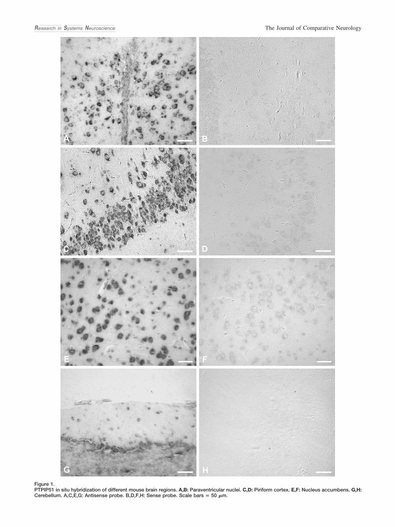
Figure 1.PTPIP51 in situ hybridization of different mouse brain regions. A,B: Paraventricular nuclei. C,D: Piriform cortex. E,F: Nucleus accumbens. G,H:Cerebellum. A,C,E,G: Antisense probe. B,D,F,H: Sense probe. Scale bars � 50 �m.
Research in Systems Neuroscience The Journal of Comparative Neurology
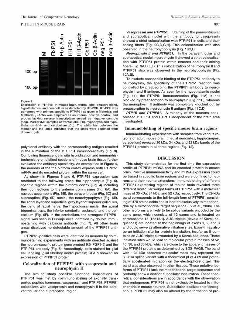
polyclonal antibody with the corresponding antigen resultedin the elimination of the PTPIP51 immunoreactivity (Fig. 3).Combining fluorescence in situ hybridization and immunohis-tochemistry on distinct sections of mouse brain tissue furtherevaluated the antibody specificity. As exemplified in Figure 4,the neurons of the the piriform cortex express both PTPIP51mRNA and its encoded protein within the same cell.
As shown in Figures 5 and 6, PTPIP51 expression wasrestricted to the following areas: the hippocampus (Fig. 5),specific regions within the piriform cortex (Fig. 4) includingtheir connections to the anterior commissure (Fig. 6A), thenucleus accumbens (Fig. 6B), the paraventricular (Fig. 6C) andsupraoptical (Fig. 6D) nuclei, the neurohypophysis (Fig. 6E),the zonal layer and superficial gray layer of superior colliculus,the genu of facial nerve, the hypoglossal nuclei, the spinaltrigeminal tract, the inferior cerebellar peduncle, and the cer-ebellum (Fig. 6F). In the cerebellum, the strongest PTPIP51signal was seen in Purkinje cells identified by double immu-nostaining with calbindin antibody (Fig. 7). All other brainareas displayed no detectable amount of the PTPIP51 anti-gen.
PTPIP51-positive cells were identified as neurons by coim-munostaining experiments with an antibody directed againstthe neuron-specific protein gene product 9.5 (PGP9.5) and thePTPIP51 antibody (Fig. 8). Accordingly, cells stained for glialcell labeling (glial fibrillary acidic protein; GFAP) showed noexpression of PTPIP51 protein.
Colocalization of PTPIP51 with vasopressin andneurophysin II
The aim to study possible functional implications ofPTPIP51 was met by coimmunolabeling of axonally trans-ported peptide hormones, vasopressin and PTPIP51. PTPIP51colocalizes with vasopressin and neurophysin II in the para-ventricular and supraoptical region.
Vasopressin and PTPIP51. Staining of the paraventricularand supraoptical nuclei with the antibody to vasopressinshowed a strict colocalization with PTPIP51 in cells and theirarising fibers (Fig. 9C,D,G,H). This colocalization was alsoobserved in the neurohypophysis (Fig. 10C,D).
Neurophysin II and PTPIP51. In the paraventricular andsupraoptical nuclei, neurophysin II showed a strict colocaliza-tion with PTPIP51 protein within neurons and their arisingfibers (Fig. 9A,B,E,F). This colocalization of neurophysin II andPTPIP51 also was observed in the neurohypophysis (Fig.10A,B).
To exclude nonspecific binding of the PTPIP51 antibody toneurophysins, the specificity of the PTPIP51 reaction wascontrolled by preabsorbing the PTPIP51 antibody to neuro-physin I and II antigen. As seen for the hypothalamic nuclei(Fig. 11), the PTPIP51 immunoreaction (Fig. 11A) is notblocked by preabsorption to neurophysin (Fig. 11B), whereasthe neurophysin II antibody was completely knocked out bypreabsorption to neurophysin II antigen (Fig. 11C,D).
PTP1B and PTPIP51. A minority of the neurons coex-pressed PTPIP51 and PTP1B independent of the brain areainvestigated.
Immunoblotting of specific mouse brain regionsImmunoblotting experiments with samples from various re-
gions of adult mouse brain (medial neocortex, hippocampus,cerebellum) revealed 30 kDa, 34 kDa, and 52 kDa bands of thePTPIP51 protein in all three regions (Fig. 12).
DISCUSSIONThis study demonstrates for the first time the expression
profile of PTPIP51 mRNA and its encoded protein in mousebrain. Positive immunoreactivity and mRNA expression couldbe traced in specific brain regions and were confined to neu-rons and their neurite extensions. Immunoblotting of differentPTPIP51-expressing regions of mouse brain revealed threedifferent molecular weight forms of PTPIP51 with a molecularmass of 30 kDa, 34 kDa, and 52 kDa, respectively. The 52-kDaband corresponds to the full-length form of PTPIP51 consist-ing of 470 amino acids and is located exclusively to mitochon-dria by a mitochondrial target sequence (Lv et al., 2006). Theother isoforms are likely to be splice variants encoded by thesame gene, which consists of 12 exons and is located onchromosome 15 (15q15.1). AUG triplets (devoid of Kozak se-quences) are located at the beginnings of exons 1, 2, and 3and could serve as alternative initiation sites. Exon 4 may alsobe an initiation site for protein translation, insofar as it con-tains an AUG triplet surrounded by a Kozak sequence. Theseinitiation sites would lead to molecular protein masses of 52,45, 38, and 30 kDa, which are close to the apparent masses ofthe PTPIP51 proteins as determined by SDS-PAGE. The bandwith �34-kDa apparent molecular mass may represent the38-kDa splice variant with a theoretical pI of 4.69 and poten-tially accelerated migration on the electrophoretic gel. Thisband was also observed in other tissues. These putative iso-forms of PTPIP51 lack the mitochondrial target sequence andprobably show a distinct subcellular localization. These theo-retical considerations are in accordance with the observationthat endogenous PTPIP51 is not exclusively located to mito-chondria in mouse neurons. Subcellular localization of endog-enous PTPIP51 may also be altered by the formation of pro-
Figure 2.Expression of PTPIP51 in mouse brain, frontal lobe, pituitary gland,hypothalamus, and cerebellum as detected by RT-PCR. RT-PCR wasperformed with primers specific to PTPIP51 as given in Materials andMethods. �-Actin was amplified as an internal positive control, andprobes lacking reverse transcriptase served as negative controls(neg). Marker (M), samples of frontal lobe (Fb), hypophysis and hypo-thalamus (HH), and cerebellum (Cb). The white bar between themarker and the lanes indicates that the lanes were depicted fromdifferent gels.
Research in Systems NeuroscienceThe Journal of Comparative Neurology
897PTPIP51 IN MOUSE BRAIN

Figure 4.PTPIP51 mRNA and protein in the piriform cortex of mouse brain. A: PTPIP51 immunostaining of neurons. B: Fluorescence in situ hybridizationantisense probe of the same section. C: Merge of A and B. A magenta-green version of this figure is provided as Supporting Information for theassistance of color-blind readers. Scale bar � 20 �m.
Figure 5.PTPIP51mRNA expression and immunostaining in the hippocampus. A: Immunostaining. B: In situ hybridization. Scale bars � 20 �m in A; 50�m in B.
Figure 3.Preabsorption experiments. A: Normal PTPIP51 immunoreactivity of neurons in mouse cerebellum. B: Preabsorption of the PTPIP51 antibodywith the purified antigen. Scale bars � 20 �m.
Research in Systems Neuroscience The Journal of Comparative Neurology
898 P. KOCH ET AL.

Figure 6.PTPIP51 immunostaining of brain tissues. A: PTPIP51-positive fibers in anterior commissure. B: PTPIP51-positive cells in the area of nucleusaccumbens. C: PTPIP51-positive cells and fibers arising from the paraventricular nucleus. D: PTPIP51-positive cells and fibers arising from thesupraoptical nucleus. E: PTPIP51-positive fibers in neurohypophysis. F: PTPIP51-positive Purkinje cells and their fibers of cerebellum. Sectionwas taken from nodulus. Insets: Modified diagrams of coronal sections from the Coronal Allen Brain Atlas, displaying the region shown in thefigure. Asterisk, third ventricle; arrow, Purkinje cell. Scale bars � 50 �m in A,C–F; 20 �m in B.
Research in Systems NeuroscienceThe Journal of Comparative Neurology
899PTPIP51 IN MOUSE BRAIN
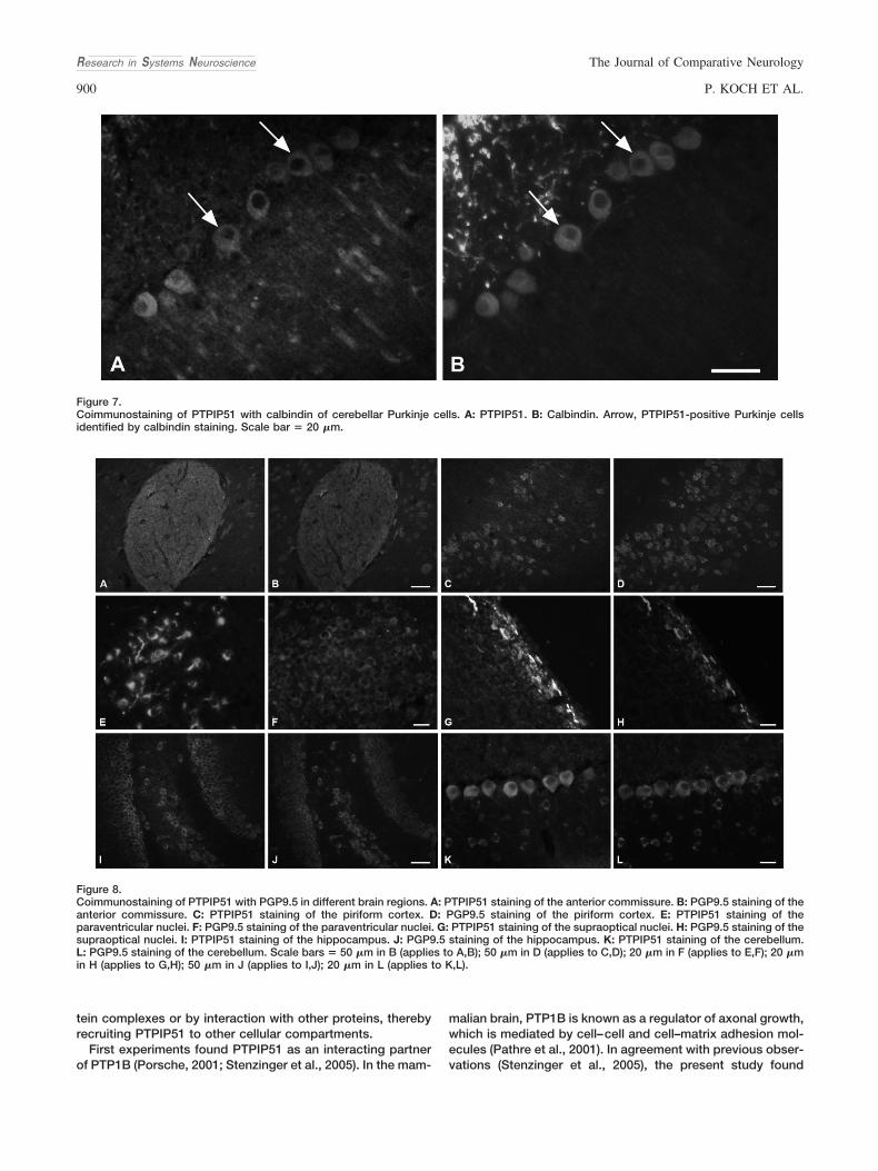
tein complexes or by interaction with other proteins, therebyrecruiting PTPIP51 to other cellular compartments.
First experiments found PTPIP51 as an interacting partnerof PTP1B (Porsche, 2001; Stenzinger et al., 2005). In the mam-
malian brain, PTP1B is known as a regulator of axonal growth,which is mediated by cell–cell and cell–matrix adhesion mol-ecules (Pathre et al., 2001). In agreement with previous obser-vations (Stenzinger et al., 2005), the present study found
Figure 7.Coimmunostaining of PTPIP51 with calbindin of cerebellar Purkinje cells. A: PTPIP51. B: Calbindin. Arrow, PTPIP51-positive Purkinje cellsidentified by calbindin staining. Scale bar � 20 �m.
Figure 8.Coimmunostaining of PTPIP51 with PGP9.5 in different brain regions. A: PTPIP51 staining of the anterior commissure. B: PGP9.5 staining of theanterior commissure. C: PTPIP51 staining of the piriform cortex. D: PGP9.5 staining of the piriform cortex. E: PTPIP51 staining of theparaventricular nuclei. F: PGP9.5 staining of the paraventricular nuclei. G: PTPIP51 staining of the supraoptical nuclei. H: PGP9.5 staining of thesupraoptical nuclei. I: PTPIP51 staining of the hippocampus. J: PGP9.5 staining of the hippocampus. K: PTPIP51 staining of the cerebellum.L: PGP9.5 staining of the cerebellum. Scale bars � 50 �m in B (applies to A,B); 50 �m in D (applies to C,D); 20 �m in F (applies to E,F); 20 �min H (applies to G,H); 50 �m in J (applies to I,J); 20 �m in L (applies to K,L).
Research in Systems Neuroscience The Journal of Comparative Neurology
900 P. KOCH ET AL.

Figure 9.Communostaining of PTPIP51 with neurophysin II and vasopressin of paraventricular (A–D) and supraoptical (E–H) nuclei. A: PTPIP51.B: Neurophysin II. C: PTPIP51. D: Vasopressin. E: PTPIP51. F: Neurophysin II. G: PTPIP51. H: Vasopressin. Arrow indicates double-labeled cells.Scale bars � 50 �m in A,B,E,F; 20 �m in C,D,G,H.
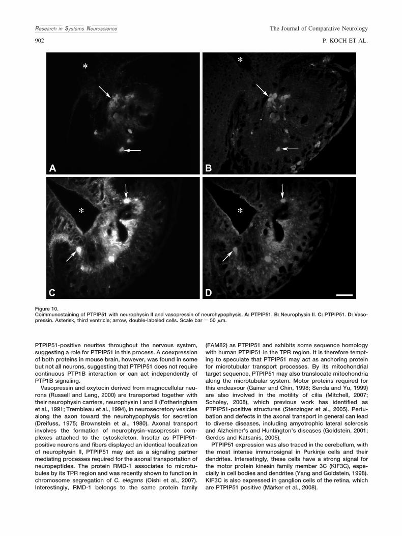
PTPIP51-positive neurites throughout the nervous system,suggesting a role for PTPIP51 in this process. A coexpressionof both proteins in mouse brain, however, was found in somebut not all neurons, suggesting that PTPIP51 does not requirecontinuous PTP1B interaction or can act independently ofPTP1B signaling.
Vasopressin and oxytocin derived from magnocellular neu-rons (Russell and Leng, 2000) are transported together withtheir neurophysin carriers, neurophysin I and II (Fotheringhamet al., 1991; Trembleau et al., 1994), in neurosecretory vesiclesalong the axon toward the neurohypophysis for secretion(Dreifuss, 1975; Brownstein et al., 1980). Axonal transportinvolves the formation of neurophysin–vasopressin com-plexes attached to the cytoskeleton. Insofar as PTPIP51-positive neurons and fibers displayed an identical localizationof neurophysin II, PTPIP51 may act as a signaling partnermediating processes required for the axonal transportation ofneuropeptides. The protein RMD-1 associates to microtu-bules by its TPR region and was recently shown to function inchromosome segregation of C. elegans (Oishi et al., 2007).Interestingly, RMD-1 belongs to the same protein family
(FAM82) as PTPIP51 and exhibits some sequence homologywith human PTPIP51 in the TPR region. It is therefore tempt-ing to speculate that PTPIP51 may act as anchoring proteinfor microtubular transport processes. By its mitochondrialtarget sequence, PTPIP51 may also translocate mitochondriaalong the microtubular system. Motor proteins required forthis endeavour (Gainer and Chin, 1998; Senda and Yu, 1999)are also involved in the motility of cilia (Mitchell, 2007;Scholey, 2008), which previous work has identified asPTPIP51-positive structures (Stenzinger et al., 2005). Pertu-bation and defects in the axonal transport in general can leadto diverse diseases, including amyotrophic lateral sclerosisand Alzheimer’s and Huntington’s diseases (Goldstein, 2001;Gerdes and Katsanis, 2005).
PTPIP51 expression was also traced in the cerebellum, withthe most intense immunosignal in Purkinje cells and theirdendrites. Interestingly, these cells have a strong signal forthe motor protein kinesin family member 3C (KIF3C), espe-cially in cell bodies and dendrites (Yang and Goldstein, 1998).KIF3C is also expressed in ganglion cells of the retina, whichare PTPIP51 positive (Marker et al., 2008).
Figure 10.Coimmunostaining of PTPIP51 with neurophysin II and vasopressin of neurohypophysis. A: PTPIP51. B: Neurophysin II. C: PTPIP51. D: Vaso-pressin. Asterisk, third ventricle; arrow, double-labeled cells. Scale bar � 50 �m.
Research in Systems Neuroscience The Journal of Comparative Neurology
902 P. KOCH ET AL.
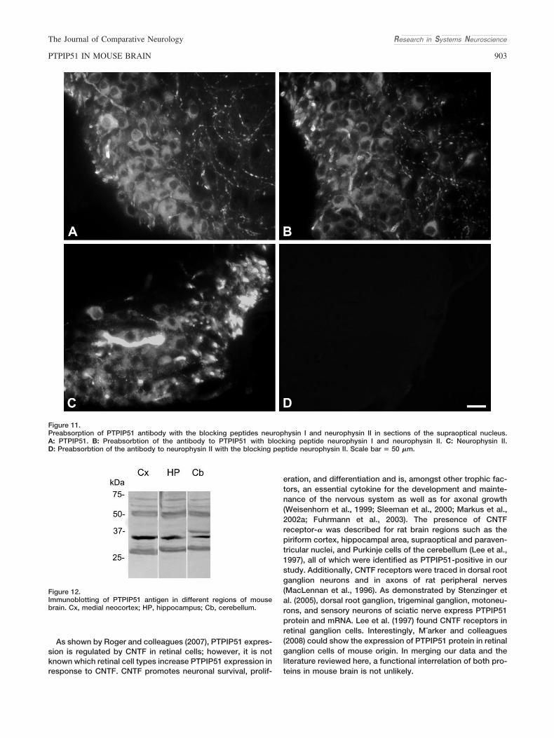
As shown by Roger and colleagues (2007), PTPIP51 expres-sion is regulated by CNTF in retinal cells; however, it is notknown which retinal cell types increase PTPIP51 expression inresponse to CNTF. CNTF promotes neuronal survival, prolif-
eration, and differentiation and is, amongst other trophic fac-tors, an essential cytokine for the development and mainte-nance of the nervous system as well as for axonal growth(Weisenhorn et al., 1999; Sleeman et al., 2000; Markus et al.,2002a; Fuhrmann et al., 2003). The presence of CNTFreceptor-� was described for rat brain regions such as thepiriform cortex, hippocampal area, supraoptical and paraven-tricular nuclei, and Purkinje cells of the cerebellum (Lee et al.,1997), all of which were identified as PTPIP51-positive in ourstudy. Additionally, CNTF receptors were traced in dorsal rootganglion neurons and in axons of rat peripheral nerves(MacLennan et al., 1996). As demonstrated by Stenzinger etal. (2005), dorsal root ganglion, trigeminal ganglion, motoneu-rons, and sensory neurons of sciatic nerve express PTPIP51protein and mRNA. Lee et al. (1997) found CNTF receptors inretinal ganglion cells. Interestingly, M¨arker and colleagues(2008) could show the expression of PTPIP51 protein in retinalganglion cells of mouse origin. In merging our data and theliterature reviewed here, a functional interrelation of both pro-teins in mouse brain is not unlikely.
Figure 11.Preabsorption of PTPIP51 antibody with the blocking peptides neurophysin I and neurophysin II in sections of the supraoptical nucleus.A: PTPIP51. B: Preabsorbtion of the antibody to PTPIP51 with blocking peptide neurophysin I and neurophysin II. C: Neurophysin II.D: Preabsorbtion of the antibody to neurophysin II with the blocking peptide neurophysin II. Scale bar � 50 �m.
Figure 12.Immunoblotting of PTPIP51 antigen in different regions of mousebrain. Cx, medial neocortex; HP, hippocampus; Cb, cerebellum.
Research in Systems NeuroscienceThe Journal of Comparative Neurology
903PTPIP51 IN MOUSE BRAIN
The data presented here suggest a function of PTPIP51 inmouse brain tissue distinct from the observations made by Lvand colleagues (2006), who reported that overexpression ofPTPIP51 enhances apoptosis in HEK 293 cells. The findingsrather point to a role for PTPIP51 as a modulator of theRaf-ERK pathway and interacting partner of PTP1B as well as14-3-3-y and 14-3-3-� (Jin et al., 2004; Ewing et al., 2007; Yuet al., 2008), thereby influencing signal transduction, tran-scription, and protein trafficking for the processes discussedabove, i.e., neuronal maintenance, neurite outgrowth, and ax-onal transport (Markus et al., 2002b; Dougherty and Morrison,2004; Planchamp et al., 2008). Alterations in these pathwayslead to multiple neurological disorders, including Alzheimer’sand Parkinson’s diseases, as well as malignant transformation(Seger and Krebs, 1995; McCubrey et al., 2007; Lyustikman etal., 2008). Further studies are aimed toward investigating thephysiological function of PTPIP51 in neurons as well as itsputative contribution to the neurological diseases mentionedabove.
ACKNOWLEDGMENTSWe thank Hans-Werner Hofer (Department of Biology, Kon-
stanz) for providing us with the antibody PTPIP51, Frank Nurn-berger (Institute of Anatomy, Frankfurt) for the kindly gift of thevasopressin antibody and for providing the necessary infor-mation for this antibody, Phillip Grant (Institute of Anatomyand Cell Biology, Giessen) for critical reading of the manu-script, and Karola Michael (Institute of Anatomy and Cell Bi-ology, Giessen) for help with the design of the figures.
LITERATURE CITEDAtkins CM, Selcher JC, Petraitis JJ, Trzaskos JM, Sweatt JD. 1998. The
MAPK cascade is required for mammalian associative learning. NatNeurosci 1:602– 629.
Barop J, Sauer H, Steger K, Wimmer M. 2009. Differentiation-dependentPTPIP51 expression in human skeletal muscle cell culture. J Histo-chem Cytochem 57:425– 435.
Bhattacharya S, Das AV, Mallya KB, Ahmad I. 2008. CNTF-mediatedsignaling regulates neuronal versus glial differentiation of retinal stemcells/progenitors by concentration-dependent recruitment of MAPKand Jak-STAT pathways in conjunction with Notch signaling. StemCells 26:2611–2624.
Brambilla R, Gnesutta N, Minichiello L, White G, Roylance AJ, Herron CE,Ramsey M, Wolfer DP, Cestari V, Rossi-Arnaud C, Grant SG, ChapmanPF, Lipp HP, Sturani E, Klein R. 1997. A role for the Ras signallingpathway in synaptic transmission and long-term memory. Nature 390:281–286.
Brehm R, Ruttinger C, Fischer P, Gashaw I, Winterhager E, Kliesch S,Bohle RM, Steger K, Bergmann M. 2006. Transition from preinvasivecarcinoma in situ to seminoma is accompanied by a reduction ofconnexin 43 expression in Sertoli cells and germ cells. Neoplasia8:499 –509.
Brownstein MJ, Russell JT, Gainer H. 1980. Synthesis, transport, andrelease of posterior pituitary hormones. Science 207:373–378.
Boulton TG, Stahl N, Yancopoulos GD. 1994. Ciliary neurotrophic factor/leukemia inhibitory factor/interleukin 6/oncostatin M family of cyto-kines induces tyrosine phosphorylation of a common set of proteinsoverlapping those induced by other cytokines and growth factors.J Biol Chem 269:11648 –11655.
Custer EE, Ortiz-Miranda S, Knott TK, Rawson R, Elvey C, Lee RH, LemosJR. 2007. Identification of the neuropeptide content of individual ratneurohypophysial terminals. J Neurosci Methods 163:226 –234.
Debus E, Weber K, Osborn M. 1983. Monoclonal antibodies specific forglial fibrillary acidic (GFA) protein and for each of the neurofilamenttriplet polypeptides. Differentiation 25:193–203.
Dhaka A, Costa RM, Hu H, Irvin DK, Patel A, Kornblum HI, Silva AJ, O’Dell
TJ, Colicelli J. 2003. The RAS effector RIN1 modulates the formation ofaversive memories. J Neurosci 23:748 –757.
Dougherty MK, Morrison DK. 2004. Unlocking the code of 14-3-3. J CellSci 117:1875–1884.
Dreifuss JJ. 1975. A review on neurosecretory granules: their contents andmechanisms of release. Ann N Y Acad Sci 248:184 –201.
Engleka KA, Gitler AD, Zhang M, Zhou DD, High FA, Epstein JA. 2005.Insertion of Cre into the Pax3 locus creates a new allele of Splotch andidentifies unexpected Pax3 derivatives. Dev Biol 280:396 – 406.
Ewing RM, Chu P, Elisma F, Li H, Taylor P, Climie S, McBroom-CerajewskiL, Robinson MD, O’Connor L, Li M, Taylor R, Dharsee M, Ho Y, HeilbutA, Moore L, Zhang S, Ornatsky O, Bukhman YV, Ethier M, Sheng Y,Vasilescu J, Abu-Farha M, Lambert JP, Duewel HS, Stewart II, Kuehl B,Hogue K, Colwill K, Gladwish K, Muskat B, Kinach R, Adams SL, MoranMF, Morin GB, Topaloglou T, Figeys D. 2007. Large-scale mapping ofhuman protein–protein interactions by mass spectrometry. Mol SystBiol 3:89.
Fotheringham AP, Davidson YS, Davies I, Morris JA. 1991. Age-associatedchanges in neuroaxonal transport in the hypothalamo-neurohypophysial sys-tem of the mouse. Mech Ageing Dev 60:113–121.
Fuhrmann S, Grabosch K, Kirsch M, Hofmann HD. 2003. Distribution ofCNTF receptor alpha protein in the central nervous system of the chickembryo. J Comp Neurol 461:111–122.
Gainer H, Chin H. 1998. Molecular diversity in neurosecretion: reflectionson the hypothalamo-neurohypophysial system. Cell Mol Neurobiol 18:211–230.
Gerdes JM, Katsanis N. 2005. Microtubule transport defects in neurolog-ical and ciliary disease. Cell Mol Life Sci 62:1556 –1570.
Giese KP, Friedman E, Telliez JB, Fedorov NB, Wines M, Feig LA, Silva AJ.2001. Hippocampus-dependent learning and memory is impaired inmice lacking the Ras-guanine-nucleotide releasing factor 1 (Ras-GRF1). Neuropharmacology 41:791– 800.
Goldstein LS. 2001. Kinesin molecular motors: transport pathways, recep-tors, and human disease. Proc Natl Acad Sci U S A 98:6999 –7003.
Jin J, Smith FD, Stark C, Wells CD, Fawcett JP, Kulkarni S, Metalnikov P,O’Donnell P, Taylor P, Taylor L, Zougman A, Woodgett JR, LangebergLK, Scott JD, Pawson T. 2004. Proteomic, functional, and domain-based analysis of in vivo 14-3-3 binding proteins involved in cytoskel-etal regulation and cellular organization. Curr Biol 14:1436 –1450.
Koch P, Stenzinger A, Viard M, Marker D, Mayser P, Nilles M, Schreiner D,Steger K, Wimmer M. 2008. The novel protein PTPIP51 is expressed inhuman keratinocyte carcinomas and their surrounding stroma. J CellMol Med 12:2083–2095.
Kuwajima T, Nishimura I, Yoshikawa K. 2006. Necdin promotes GABAergicneuron differentiation in cooperation with Dlx homeodomain proteins.J Neurosci 26:5383–5392.
Lee MY, Hofmann HD, Kirsch M. 1997. Expression of ciliary neurotrophicfactor receptor-alpha messenger RNA in neonatal and adult rat brain:an in situ hybridization study. Neuroscience 77:233–246.
Levin SI, Khaliq ZM, Aman TK, Grieco TM, Kearney JA, Raman IM, MeislerMH. 2006. Impaired motor function in mice with cell-specific knockoutof sodium channel Scn8a (NaV1.6) in cerebellar purkinje neurons andgranule cells. J Neurophysiol 96:785–793.
Lewis FA, Wells M. 1992. Detection of virus in infected human tissue by insitu hybridisation. In: Wilkinson DG, editor. In situ hybridisation, apractical approach. Oxford: Oxford University Press. p 121–135.
Lv BE, Yu CE, Chen YY, Lu Y, Guo JH, Song QS, Ma DL, Shi TP, Wang L.2006. Protein tyrosine phophatase interacting interacting protein 51(PTPIP51) is a novel mitochondria protein with an N-terminal mitochon-drial targeting sequence and induces apoptosis. Apoptosis 11:1489 –1501.
Lyustikman Y, Momota H, Pao W, Holland EC. 2008. Constitutive activa-tion of Raf-1 induces glioma formation in mice. Neoplasia 10:501–510.
MacLennan AJ, Vinson EN, Marks L, McLaurin DL, Pfeifer M, Lee N. 1996.Immunohistochemical localization of ciliary neurotrophic factor recep-tor alpha expression in the rat nervous system. J Neurosci 16:621– 630.
Markus A, Patel TD, Snider WD. 2002a. Neurotrophic factors and axonalgrowth. Curr Opin Neurobiol 12:523–531.
Markus A, Zhong J, Snider WD. 2002b. Raf and akt mediate distinctaspects of sensory axon growth. Neuron 35:65–76.
Marker D, Stenzinger A, Schreiner D, Tag C, Wimmer M. 2008. Expressionof PTPIP51 during mouse eye development. Histochem Cell Biol 129:345–356.
McCubrey JA, Steelman LS, Chappell WH, Abrams SL, Wong EW, ChangF, Lehmann B, Terrian DM, Milella M, Tafuri A, Stivala F, Libra M,
Research in Systems Neuroscience The Journal of Comparative Neurology
904 P. KOCH ET AL.
Basecke J, Evangelisti C, Martelli AM, Franklin RA. 2007. Roles of theRaf/MEK/ERK pathway in cell growth, malignant transformation anddrug resistance. Biochim Biophys Acta 1773:1263–1284.
Mei M, Su B, Harrison K, Chao M, Siedlak SL, Previll LA, Jackson L, CaiDX, Zhu X. 2006. Distribution, levels and phosphorylation of Raf-1 inAlzheimer’s disease. J Neurochem 99:1377–1388.
Mitchell DR. 2007. The evolution of eukaryotic cilia and flagella as motileand sensory organelles. Adv Exp Med Biol 607:130 –140.
Oishi K, Okano H, Sawa H. 2007. RMD-1, a novel microtubule-associatedprotein, functions in chromosome segregation in Caenorhabditis el-egans. J Cell Biol 179:1149 –1162.
Olmsted JB. 1981. Affinity purification of antibodies from diazotized paperblots of heterogeneous protein samples. J Biol Chem 256:11955–11957.
Pasteels B, Miki N, Hatakenaka S, Pochet R. 1987. Immunohistochemicalcross-reactivity and electrophoretic comigration between calbindinD-27 kDa and visinin. Brain Res 412:107–113.
Pathre P, Arregui C, Wampler T, Kue I, Leung TC, Lilien J, Balsamo J.2001. PTP1B regulates neurite extension mediated by cell– cell andcell–matrix adhesion molecules. J Neurosci Res 63:143–150.
Planchamp V, Bermel C, Tonges L, Ostendorf T, Kugler S, Reed JC,Kermer P, Bahr M, Lingor P. 2008. BAG1 promotes axonal outgrowthand regeneration in vivo via Raf-1 and reduction of ROCK activity.Brain 131:2606 –2619.
Porsche A. 2001. Identifikation von Interaktionspartnern der T-zell protein-tyrosin-phosphatase durch das Lex-A two hybrid system. PhD Thesis,University of Konstanz. Allensbach: UFO Publishers, vol 414.
Roger J, Goureau O, Sahel JA, Guillonneau X. 2007. Use of suppressionsubtractive hybridization to identify genes regulated by ciliary neuro-trophic factor in postnatal retinal explants. Mol Vis 13:206 –219.
Russell JA, Leng G. 2000. Veni, vidi, vici: the neurohypophysis in thetwentieth century. Exp Physiol 85(Spec No.):1S– 6S.
Russell TA, Ito M, Yu RN, Martinson FA, Weiss J, Jameson JL. 2003. Amurine model of autosomal dominant neurohypophyseal diabetes in-sipidus reveals progressive loss of vasopressin-producing neurons.J Clin Invest 112:1697–1706.
Samuels IS, Karlo JC, Faruzzi AN, Pickering K, Herrup K, Sweatt JD, SaittaSC, Landreth GE. 2008. Deletion of ERK2 mitogen-activated proteinkinase identifies its key roles in cortical neurogenesis and cognitivefunction. J Neurosci 28:6983– 6995.
Satoh JI, Kuroda Y. 2001. Ubiquitin C-terminal hydrolase-L1 (PGP9.5)expression in human neural cell lines following induction of neuronaldifferentiation and exposure to cytokines, neurotrophic factors or heatstress. Neuropathol Appl Neurobiol 27:95–104.
Schindler CU, Nurnberger F. 1990. Hibernation-related changes in theimmunoreactivity of neuropeptide system in the suprachiasmatic nu-cleus of the ground squirrwl, Spermophilus richardsonii. CTR 262:293–300.
Scholey JM. 2008. Intraflagellar transport motors in cilia: moving along thecell’s antenna. J Cell Biol 180:23–29.
Seger R, Krebs EG. 1995. The MAPK signaling cascade. FASEB J 9:726 –735.
Senda T, Yu W. 1999. Kinesin cross-bridges between neurosecretorygranules and microtubules in the mouse neurohypophysis. NeurosciLett 262:69 –71.
Sleeman MW, Anderson KD, Lambert PD, Yancopoulos GD, Wiegand SJ.2000. The ciliary neurotrophic factor and its receptor, CNTFR alpha.Pharm Acta Helv 74:265–272.
Soderling SH, Langeberg LK, Soderling JA, Davee SM, Simerly R, Raber J,Scott JD. 2003. Loss of WAVE-1 causes sensorimotor retardation andreduced learning and memory in mice. Proc Natl Acad Sci U S A100:1723–1728.
Steger K, Klonisch T, Gavenis K, Drabent B, Doenecke D, Bergmann M.1998. Expression of mRNA and protein of nucleoproteins during hu-man spermiogenesis. Mol Hum Reprod 4:939 –945.
Steger K, Pauls K, Klonisch T, Franke FE, Bergmann M. 2000. Expressionof protamine 1 and 2 mRNA during human spermiogenesis. Mol HumReprod 6:219 –225.
Stenzinger A, Kajosch T, Tag C, Porsche A, Welte I, Hofer HW, Steger K,Wimmer M. 2005. The novel protein PTPIP51 exhibits tissue- andcell-specific expression. Histochem Cell Biol 123:19 –28.
Stenzinger A, Schreiner D, Pfeiffer T, Tag C, Hofer HW, Wimmer M. 2006.EGF, TGF, retinoic acid and 1,25(OH)2D3 regulated expression of thenovel protein PTPIP51 in keratinocytes. Cells Tissues Organs 184:76 –87.
Stenzinger A, Marker D, Koch P, Hoffmann J, Baal N, Steger K, Wimmer M.2008. Protein tyrosine phosphatase interacting protein 51 (PTPIP51),mRNA expression and localization of PTPIP51 and its in vitro interact-ing partner protein tyrosine phosphatase 1B (PTP1B) in human pla-centa of first, second, and third trimester. J Histochem Cytochem57:143–153.
Stenzinger A, Schreiner D, Koch P, Hofer HW, Wimmer M. 2009. Cell andmolecular biology of the novel protein tyrosine phosphatase interactingprotein 51. Int Rev Cell Mol Biol (in press).
Towbin H, Staehelin T, Gordon J. 1979. Electrophoretic transfer of proteinsfrom polyacrylamide gels to nitrocellulose sheets: procedure and someapplications. Proc Natl Acad Sci U S A 76:350 – 4354.
Trembleau A, Morales M, Bloom FE. 1994. Aggregation of vasopressinmRNA in a subset of axonal swellings of the median eminence andposterior pituitary: light and electron microscopic evidence. J Neurosci14:39 –53.
Weisenhorn DM, Roback J, Young AN, Wainer BH. 1999. Cellular aspectsof trophic actions in the nervous system. Int Rev Cytol 189:177–265.
Yang Z, Goldstein LS. 1998. Characterization of the KIF3C neural kinesin-like motor from mouse. Mol Biol Cell 9:249 –261.
Yu C, Han W, Shi T, Lv B, He Q, Zhang Y, Li T, Zhang Y, Song Q, Wang L,Ma D. 2008. PTPIP51, a novel 14-3-3 binding protein, regulates cellmorphology and motility via Raf-ERK pathway. Cell Signal 20:2208 –2220.
Research in Systems NeuroscienceThe Journal of Comparative Neurology
905PTPIP51 IN MOUSE BRAIN

1
PTPIP51 – a multifunctional protein in brain tissue
M. Viard*, M. Kamm*, M. Bobrich*, A. Brobeil*, M. Petri*, M. Wimmer*
* Institute of Anatomy and Cell Biology, Justus-Liebig-University, 35385 Giessen, Germany
Corresponding author:
Maxime Viard
Institute of Anatomy and Cell Biology
Justus-Liebig-University
35385 Giessen
Germany
Phone: +49.641.9947012
Fax: +49.641.99.47009
Keywords: Hippocampus, Cerebellum, 14-3-3, PTP1B, Neurophysin
2
Abstract:
Converging lines of evidence indicate Protein tyrosine phosphatase interacting
protein 51 (PTPIP51) to be a multifunctional protein. We investigated the interaction
of PTPIP51 with Protein tyrosine phosphatase 1B (PTP1B) and 14-3-3 beta in
defined brain areas of adult female mice. In Purkinje-cells of the cerebellum PTPIP51
interacts with both proteins, indicating an involvement in nerve growth factor (NGF)
signaling which plays an important role in memory processes. PTP1B activity is also
known for its influence on neurons to resist the attack of amyloid.
Furthermore we can report an interaction of PTPIP51 with Neurophysin-2 in the
paraventricular nucleus, implying its role in vesicle trafficking.
By the additional use of peptide specific antibodies, targeting C-terminus and N-
terminus of PTPIP51, we were able to demonstrate different expression profiles,
indicating the existence of PTPIP51 isoforms in mouse brain. The expression of
those isoforms varied, depending on the region of the brain and the localization
within the neuron, supporting the theory of an isoform based broad range of function.
Our results underline the hypothesis of PTPIP51 being a multifunctional protein with
special emphasis on nervous tissue, therefore, being potentially involved in different
essential cell functions such as axonal growth and axonal transport.
Introduction
Protein tyrosine phosphatase interacting protein 51 (PTPIP51), originally detected by
a yeast two hybrid screen in order to identify potential substrates of Protein Tyrosine
Phosphatase 1B (PTP1B) and T- cell Protein Tyrosine Phosphatase (TcPTP), has
been proven to be expressed in various tissues (Stenzinger et al., 2009). However,
up to now little is known about the definite function of PTPIP51 in brain tissue.
Previous studies by Koch et al. 2009 revealed a distinct expression of PTPIP51 in
defined brain areas such as cerebellum, hippocampus and the nuclei of
hypothalamus. On cellular level strongest PTPIP51 signals were seen in Purkinje
cells of the cerebellum, neurons of the hippocampus, paraventricular and supraoptic

3
nuclei and their axons. Basic studies in brain tissue concerning the functions of
PTPIP51 hinted at an involvement in essential cellular processes such as
proliferation, differentiation, motility and axonal transport. Immunoblotting of medial
neocortex, hippocampus and cerebellum of adult mouse brain revealed putative
PTPIP51 isoforms with 30, 34 and 52 kDa (Koch et al., 2009). The existence of
PTPIP51 isoforms may provide an explanation for the hypothesized broad spectrum
of functions within these different neurons.
Previous in situ studies revealed an interaction of PTPIP51 with PTP1B in different
tissues (Brobeil et al., 2010; Bobrich et al., 2011). In gliomas, PTP1B is expressed
and interacts with PTPIP51 (Petri et al., in press) and it regulates axonal growth
mediated by cell–cell and cell–matrix adhesion molecules (Pathre et al., 2001).
Besides PTP1B, PTPIP51 is also known to interact with 14-3-3 beta and 14-3-3
gamma as demonstrated by two independent studies (Jin et al., 2004; Ewing et al.,
2007). Furthermore, the work by Petri et al. (in press) could relate the extent of
PTPIP51-14-3-3 beta interaction to the grade of glioma malignancy. It is well known
that Purkinje cells express several 14-3-3 isoform transcripts during development
primarily 14-3-3 beta (Watanabe et al., 1991, 1993a,b, 1994; Umahara et al., 2009).
The suggested role within axonal transport by the observed association of
neurophysin and PTPIP51 is supported by the fact that PTPIP51 is alternatively
known as regulator of microtubule dynamics protein 3 (RMD-3). RMD-3 belongs to
the same protein family as regulator of microtubule dynamics protein 1 (RMD-1).
RMD-1 was identified as a member of a new microtubule associated protein (MAP)
family. Vesicle trafficking is functionally associated with coiled-coil proteins and
PTPIP51 exhibits the same coiled-coil domains as RMD-1 (Gillingham and Munro
2003; Oishi et al., 2007). These facts provide evidence that PTPIP51 is involved in
regulatory processes of vesicle trafficking through interactions with the microtubule
cytoskeleton, as described before by Brobeil et al. (2010) and Koch et al. (2009).
Based on the above named findings, this study aimed to investigate the expression
pattern of potential PTPIP51 isoforms in the brain tissues and the direct interaction of
PTPIP51 with 14-3-3 beta, PTP1B and neurophysin II to clarify the function of
PTPIP51 in brain tissue.
4
Materials and Methods
Animals were raised and housed under standard laboratory conditions. All animal
experiments were performed in compliance with German law on the use of laboratory
animals.
Tissue and section preparations.
Tissue and section preparation was performed as described by Koch et al. (2009).
The study was performed with paraffin-embedded samples of mouse brain (n = 9;
sex: female, genus C57Bl6J, age: 8-12 weeks), either fixed in Bouin fixative or 4%
paraformaldehyde (PFA). For immunohistochemistry the paraffin-embedded brain of
each mouse was cut into 6 µm thin sections, dried, deparaffinized in xylene and
rehydrated in graded alcohol prior to immunostaining. PTPIP51 positive regions were
identified by comparison with mouse brain maps: www.mbl.org/mblmain/atlas.html;
www.hms.harvard.edu/research/brain/atlas.html;www.brainmap.org/mouse/atlas.html
. Frontal and sagittal vibratome sections of cerebellum and hippocampus (thickness
50 µm) of adult female mice (6-8 weeks) were also performed. For this purpose
animals were sacrificed after anaesthesia with pentobarbital and subsequently
transcardial perfused and fixed overnight in 4% PFA.
PTPIP51 (aa 131-470) and peptide specific PTPIP51 antibodies
PTPIP51 (aa 131-470) antibody was produced as described previously (Koch et al.,
2009). To identify different isoforms of the protein, antibodies against defined peptide
sequences of PTPIP51 were used (BioLux, Stuttgart, Germany). The following
sequence was used for the identification of the N-terminus: SRLGALGGARAGLGC
(exon 2) and the following sequence was used for the identification of the C-terminus:
CIQKDLEELEVILRD (exon 13). Identity and purity of each synthetized peptide were
approved by ESI-MS and UV-analysis. Rabbits were immunized with the KLH-
conjugated peptides. The specificity of each antibody was tested by ELISA and
5
Western blotTo verify the use of these peptide specific antibodies for immunostaining
preabsorption experiments were performed (Brobeil et al., 2010). see Figure 1.
Immunoblotting
Brain tissue samples, derived from medial neocortex, hippocampus, and cerebellum
were lysed and separated on 10% SDS-PAGE. According to Towbin et al. (1979) the
transfer on an Immobilon P membrane (Millipore, Bedford, CA) was performed. For
membrane blocking 10% fat-free milk powder in PBS was used and subsequently
incubated for 1 hour. Incubation with either the PTPIP51 (aa 131-470) or the peptide
specific PTPIP51 antibodies was done overnight at 4°C and washed three times with
TBST for 5 min. The secondary antibody alkaline phosphatase-conjugated anti-rabbit
were applied for 1 hour at room temperature, and the reaction was visualized with the
SigmaFast BCIP/NBT substrate. For calibration, a prestained molecular weight
marker (Biorad, Cat# 161-0374) was used (Stenzinger et al., 2005; Brobeil et al.,
2010). Immunoblotting was performed four times (n=4).
Immunohistochemistry
Immunohistochemistry was performed as previously described by Koch et al. (2009).
Prior to immunostaining nonspecific binding sites were blocked with 0.1 M phosphate
buffered saline (PBS, pH 7.4) containing 5% bovine serum albumin and 5% normal
goat serum. Samples were incubated overnight at room temperature with primary
antibodies (see table 1) diluted in PBS, followed by washing in PBS (three time for 10
min) and subsequent incubation for 1 h at room temperature with the respective
secondary antibodies (see table 1). The slides were washed in PBS, coverslipped in
carbonate buffered glycerol at pH 8.6 and evaluated either by epifluorescence
microscopy or by sequential confocal laser scanning microscopy.
PTPIP51 (aa131-470) and peptide specific PTPIP51 antibodies were visualized
either by Alexa Fluor 555 secondary antibody or FITC anti-rabbit. Anti-mouse
antibodies used for double staining were visualized by using Alexa Fluor 488
secondary antibody. The primary monoclonal anti-goat antibody neurophysin II used
6
for identification of axonal transport, was visualized using Cy3 donkey anti-goat as
secondary antibody in combination with FITC anti-rabbit as secondary antibody for
PTPIP51 visualization. Nuclei were displayed through Dapi. Every immunostaining
was performed at least 15 (n=15) times to verify the results.
Duolink proximity ligation assay (DPLA)
Interaction of PTPIP51 with PTP1B, 14-3-3 beta and Neurophysin II was verified by
proximity ligation assay kit Duolink II (PLA probe anti-rabbit minus, Cat.# 90602; PLA
probe anti-mouse plus, Cat.# 90701; Dection Kit 563, Cat.# 90134). The DuoLink
proximity ligation assay is based on the principle that proteins, localized within a
certain on-site proximity to each other, precisely 40nm or less (Fredriksson S., 2009),
perform protein-protein-interactions. To start the procedure, specimens are incubated
with primary, antibodies from different species against the proteins that are
investigated based on previously assumed interaction. Primary antibodies are
recognized by species-specific PLA probes. PLA probes consist of antibodies,
coupled with a DNA strand in either plus or minus orientation. Given that the distance
between the antibodies is equal to or less than 40nm, the DNA strands can be
hybridized using connector oligonucleotides. The connector oligonucleotides are
subsequently connected by enzymatic ligation to form a circular template, which,
after a multitude of amplifications, can be detected by a fluorophore labelled
complementary oligonucleotide.
In this study we used PLA probe anti-rabbit minus, binding the primary
PTPIP51(aa131-470) antibody, PLA probe anti-mouse plus, binding the primary
PTP1B and 14-3-3 antibody and PLA probe anti-goat minus, binding the primary
neurophysin II antibody. Methanol-fixed air-dried samples were pre-incubated with
blocking agent for 1h. After washing in PBS for 10 min, primary antibodies to
PTPIP51 (1:1000) and PTP1B (1:500) or 14-3-3 beta (1:100) and Neurophysin II
(1:1000) were applied to the samples. Incubation was done overnight in a pre-heated
humidity chamber at 37°C. Slides were washed three times in PBS for 10 min.
Duolink PLA probes detecting rabbit or mouse antibodies were diluted in the blocking
agent in a concentration of 1:5 and applied to the slides followed by incubation for 2h
in a pre-heated humidity chamber at 37°C. Unbound PLA probes were removed by
7
washing three times in PBS for 10 min. For hybridization of the two Duolink PLA
probes Duolink Hybridization stock was diluted 1:5 in high purity water and slides
were incubated in a pre-heated humidity chamber for 15 min at 37°C. The slides
were washed in TBS-T for 1 min under gentle agitation. The samples were then
incubated in the ligation solution consisting of Duolink Ligation stock (1:5) and
Duolink Ligase (1:40), diluted in high purity water, for 90 min at 37°C. Detection of
the amplified probe was done with the Duolink Detection kit. Duolink Detection stock
was diluted 1:5 in high purity water and applied for 1 h at 37°C. Final washing steps
were done by SCC buffer and 70% ethanol. Every duolink proximity ligation assay
was performed 5 times (n=5) to verify an interaction.
Epifluorescence microscopy
The Axioplan 2 fluorescence microscope equipped with Plan-Apochromat objectives
(Carl Zeiss Jena, Germany) was used for photo documentation. For visualization of
the secondary antibody Alexa Fluor 555 an excitation filter with a spectrum of 530-
560nm and an emission filter with a spectrum 572-647nm were used. Alexa Fluor
488 was visualized by an excitation filter with a range of 460-500nm and an emission
filter with a range of 512-542 nm.
Confocal laser scanning microscopy and 3D reconstruction.
Confocal microscopy was performed with a Leica TCS SP2 AOBS laser scanning
microscope equipped with a Leica HCX PL APO CD 63x oil immersion lens (NA 1.4).
Detector gain and amplifier offset were initially set to obtain pixel densities within a
linear range. The confocal orthographic projections were done using Imaris software
(Zürich, Switzerland). Confocal images were exported from the Leica software and
stored as TIFF files. Figures were prepared in Photoshop CS2 (Adobe, San Jose,
CA). Image brightness and contrast were adjusted

8
Results
This study demonstrates the expression pattern of PTPIP51 and supposed PTPIP51-
isoforms, identified by peptide specific antibodies, in neurons from the hippocampus,
cerebellum and paraventricular nucleus as well as the subcellular localization in the
adult mouse brain. Furthermore, the direct in situ interaction of PTPIP51 with 14-3-3
beta, PTP1B and neurophysin II in brain was substantiated.
Immunoblotting of specific mouse brain regions
Immunoblotting experiments with samples from various regions of adult mouse brain
(hippocampus, medial neocortex, cerebellum) revealed different molecular weights of
30, 34 and 52 kDa in all three regions, identified by the PTPIP51 (aa 131-470)
antibody (Figure 2A). Using a C-terminus specific antibody, immunoblotting revealed
different expression patterns in the three brain regions, with molecular weight of 70
kDa in hippocampus (Figure 2B1), 38 and 70 kDa in medial cortex (Figure 2B2) and
38, 65 and 70 kDa in cerebellum (Figure 2B3). As shown in Figure 2C, no region
specific differences were recognized by the N-terminus specific antibody. All three
investigated regions showed 3 bands with molecular weights of 25, 30 and 34 kDa,
except the hippocampal tissue, which in addition displayed a 45 kDa band (Figure
2C1).
Immunostaining of PTPIP51 by peptide specific antibodies in mouse brain areas
To detect the region specific expression profile of PTPIP51, immunostainings, using
the PTPIP51 (aa 131-470) and the peptide specific antibodies, were employed.
Hippocampus
The soma and the dendrites of pyramidal cells in hippocampus displayed positive
reaction to the PTPIP51 (aa 131 – 470) antibody (Figure 3A-B), whereas staining
against the C-terminus was restricted to soma (Figure 3C-D) and staining against the
N-terminus was intense in the soma and dendrites (Figure 3E-F).
9
Cerebellum.
The soma and the dendrites of Purkinje cells in cerebellum displayed positive
reaction to the PTPIP51 (aa 131 – 470) antibody (Figure 4A-B), whereas staining
against the C-terminus was restricted to the perinuclear area (Figure 4C-D) and
staining against the N-terminus was intense in the soma and dendrites (Figure 4E-F).
Subcellular localization of PTPIP51 (aa 131-470) in neurons of the cerebellum by confocal 3D reconstruction
Co-immunostaining of PTPIP51 (aa131-470) and antibodies identifying either Golgi
apparatus, mitochondria or endoplasmic reticulum (ER) showed that PTPIP51 was
neither associated with Golgi apparatus nor with mitochondria (Figure 5A,B). In
addition, a partial colocalization of PTPIP51 (aa131-470) with the endoplasmic
reticulum was observed (Figure 5C).
Subcellular localization of specific PTPIP51 peptides in neurons of hippocampus by confocal 3D reconstruction
Co-immunostaining of peptide specific PTPIP51 and antibodies identifying either
Golgi apparatus, mitochondria or ER showed that the N-terminus specific peptide
was not associated to the ER (Figure 6F), whereas a clear, yet only punctual,
colocalization of the C- terminus specific peptide and the endoplasmic reticulum was
found (Fig 5 C). In addition, a partial colocalization of PTPIP51 C- and N-terminus
specific peptides with the Golgi apparatus was found (Figure 6A,D), interestingly not
in mitochondria (Figure 6B,E).
Co- localization of PTPIP51 (aa 131- 470) and interacting partners in Purkinje cells of the cerebellum by confocal 3D reconstruction
Co-immunostudies of PTPIP51 (aa131-470) and either PTP1B and 14-3-3 beta
showed a co- localization of PTPIP51 (aa131-470) with PTP1B and 14-3-3 beta
(Figure 7) in Purkinje cells of cerebellum.
10
Interaction of PTPIP51 (aa 131- 470) with PTP1B and 14-3-3 beta in Purkinje cells of the cerebellum and neurophysin II in neurons of the paraventricular nucleus by Duolink proximity ligation assay (DPLA)
To confirm an interaction of PTPIP51 (aa131-470) with PTP1B and 14-3-3 beta a
Duolink proximity ligation assay was performed with cerebellum of adult mouse. In
Purkinje cells of all investigated samples PTPIP51 (aa131-470) was interacting with
PTP1B as well as with 14-3-3 beta, as evidenced by a positive DPLA signal
(fluorescence dots). Every dot corresponds to an interaction between PTPIP51 and
PTP1B (Figure 8A), or as seen in Figure 8B an interaction between PTPIP51 and 14-
3-3 beta.
To confirm an interaction of PTPIP51 and neurophysin II a Duolink proximity ligation
assay was performed with the paraventricular nucleus of adult mouse. Every dot
corresponds to an interaction between PTPIP51 and neurophysin II (Figure 8C)
Discussion:
PTPIP51 was described as a multifunctional protein (Brobeil et al., 2011a). Preceding
studies of PTPIP51 expression in various tissues hinted to an involvement in
essential cellular processes such as proliferation, differentiation, migration and
apoptosis (Koch et al., 2009).
Here we present data that strongly suggests the expression of different PTPIP51
isoforms on cellular and subcellular levels in the central nervous system. In addition
the interaction of PTPIP51 with 14-3-3 beta and PTP1B were analyzed to clarify the
involvement of PTPIP51 in signaling processes within the nervous system.
Preliminary studies (Koch et al., 2009) reported the expression of PTPIP51 proteins
with molecular weights of 30, 34, and 52 kDa in the medial neocortex, hippocampus
and cerebellum indicating the prevalence of various PTPIP51 isoforms. Initiation of
mRNA translation requires an AUG triplet with a specific surrounding sequence
(GCCRCCCaugG - R stands for purine bases) as already described by Kozak
(2005). Scanning the coding open reading frame (ORF) of PTPIP51 for internal start
11
codons displayed six additional AUG triplets (Brobeil et al., 2011a). Using alternative
initiation codons calculated molecular weights of 45, 38 and 30 kDa were found
(ProteinCaclculator v3, 3 http:// www.scripps.edu/-edputnam/protcalc.html;
Stenzinger et al.2005).
Another mechanism that is suitable to explain the existence of additional PTIPIP51
isoforms is alternative splicing, a process, which is well known to create protein
diversity (Nilsen and Gravely, 2010).
Katz et al. (2010) claim that multiple isoforms can often differ in their function. The
differential expression of PTPIP51 isoforms could be the basis for the proposed
multiple functions associated with their localization in the brain.
Immunoblotting of the cerebellum, hippocampus and cortex with the peptide specific
antibodies confirmed these results.
The identified different molecular forms corresponded to calculated isoforms based
on the leaky scanning mechanism, representing molecular weights of 52, 45, 38 and
30 kDa. Corresponding weights were seen in muscle tissue (52 kDa; Barop et al.,
2009), in human HL-60 cell line representing acute promyelocytic leukemia (52, 45
and 38 kDa; Brobeil et al., 2011b) and in embryonic tissue (30 kDa; Brobeil et al.,
2011a).
On subcellular level PTPIP51 antibodies revealed a heterogenous expression
pattern.
The soma of the Purkinje cells and Pyramidal cells reacted positive for PTPIP51
(aa131-470) as well as for the C- and N- terminal specific antibodies. No C-terminal
comprising isoform was detected in the dendrites. This suggests to a different
function of PTPIP51 within the soma compared to the dendrite.
The C- and N- terminal specific peptides were associated with the Golgi- apparatus,
which is involved in dendritic and axonal morphogenesis (Rosso et al., 2004; Tanabe
et al., 2010). Additionally, the C- terminal specific peptide showed a colocalization
with the ER.
PTP1B, an ER-bound protein tyrosine phosphatase, is known as a regulator of
axonal growth, which is mediated by cell– cell and cell–matrix adhesion molecules

12
(Pathre et al., 2001). In Purkinje cells PTP1B was co-localized with PTPIP51,
indicating an interaction of both proteins and it was corroborated by the duolink
proximity ligation assay. Fuentes and Arregui (2009) showed an involvement of
PTP1B in controlling growth cone dynamics and axon elongation in neurons.
Furthermore, PTP1B plays an important role in nerve growth factor (NGF) signaling
pathway, which has been reported to have the potential to elongate neurites, to
prevent apoptosis and on functional level to influence memory processes (Chao et
al., 2006; Shimoke et al., 2011). Intracellular, NGF is associated to ER and Golgi-
apparatus (Blöchl et al., 1996). This resembles the expression pattern of PTPIP51 C-
terminus. The constitutive secretion of NGF is confined to the neural soma and very
proximal parts of the dendrites (Blöchl et al., 1996), underlying the colocalization with
the C- terminus of PTPIP51. Dendrite patterning in hippocampal neurons is controlled
by NGF, which activates the Src kinase, needed for NF-k-B activation and Hes1
expression (Chacón et al., 2010). In the context of Src-kinase-signaling, PTPIP51
has been shown to be an in vitro substrate of c-Src (Stenzinger et al., 2009).
Additional to its role in neurite outgrowth, PTP1B activity is also needed for neuron
survival, assisting hippocampal neurons resist the attack of amyloid (Chacón et al.,
2011). Binding of NGF to neutrophin receptor p75 (NTR) increases the enzymatic
activity of PTP1B. Yet, the mechanism of this activation is still unknown (Chacón et
al., 2011) and may involve PTPIP51.
The 14-3-3-protein superfamily comprises seven isoforms, which have been
suggested to play a role in neuronal proliferation, migration and differentiation of the
rat cerebellum (Umahara et al., 2009).
PTPIP51 interacted with 14-3-3 beta in the soma of Purkinje cells. As reported by Yu
et al. (2008), PTPIP51 also interacts with Raf-1, an interaction that is mediated by 14-
3-3 beta, thereby modulating the activity of the MAPK- cascade. Interestingly, NGF
signaling is as well linked to the MAPK pathway (Xing et al., 1998). NGFs are
important for memory processes, which are specifically affected in Alzheimer’s
disease (Chao et al., 2006; Aggleton et al., 2010). Since PTPIP51 is strongly
expressed in the hippocampus area (Koch et al., 2009) and thus could be involved in
memory process by affecting NGF signaling.

13
Previous studies revealed a colocalization of PTPIP51 with vasopressin and its
carrier neurophysin II, suggesting a participation in axonal transport processes (Koch
et al., 2009). By proving a direct interaction of PTPIP51 with the carrier protein
neurophysin II in neurons of the paraventricular nucleus, we were able to verify this
theory.
Lv et al. (2006) described PTPIP51 as a mitochondrial associated protein and
inducer of apoptosis in HEK293T and HeLa cell lines. Since no colocalization of
either PTPIP51 peptides with mitochondria was observed, a role in apoptosis does
not appear to be the standout-function of PTPIP51 in brain tissue.
In summary, PTPIP51 is implied in the progression of dendritic and axonal growth, as
well as axonal transport. PTPIP51 exerts these functions through interaction with key
molecules PTP1B, 14-3-3 beta and Neurophysin ll. An additional function of PTPIP51
could be the modulation of NGF-signaling, therefore participating in memory
processes and possibly being involved in the genesis of Alzheimer’s disease. This
functional diversity is probably based on the expression of multiple PTPIP51 isoforms
and is strongly supported by the data presented here. In addition the multitude of
such different functions can be explained by the expression of various isoforms and
their different subcellular localization.
Following this, further experiments have to be performed to investigate the function of
PTPIP51 in brain and thereby its potential involvement in Morbus Alzheimer or other
pathological entities of the brain.
Acknowledgements
We are grateful to Mrs. C.Tag (Institute of Anatomy and Cell Biology, Giessen), for
her excellent technical assistance.
Dedication
This publication is dedicated to Mr. Hans Werner Hofer who promoted the research
on PTPIP51 with all his energy. He died much too early († 18.05.2011).

14
References
Aggleton JP, O'Mara SM, Vann SD, Wright NF, Tsanov M, Erichsen JT. 2010.
Hippocampal-anterior thalamic pathways for memory: uncovering a network of direct
and indirect actions. Eur J Neurosci 31(12): 2292-307.
Barop J, Sauer H, Steger K, Wimmer M. 2009. Differentiation-dependent PTPIP51
expression in human skeletal muscle cell culture. J Histochem Cytochem 57(5):425-
35.
Blöchl A, Thoenen H. 1996. Localization of cellular storage compartments and sites
of constitutive and activity-dependent release of nerve growth factor (NGF) in primary
cultures of hippocampal neurons. Mol Cell Neurosci 7(3):173-90.
Bobrich M, Brobeil A, Mooren FC, Krüger K, Steger K, Tag C, Wimmer M. 2011.
PTPIP51 interaction with PTP1B and 14-3-3ß in adipose tissue of insulin resistant
mice. Int J Obes (Lond) [Epub ahead of print].
Brobeil A, Graf M, Oeschger S, Steger K, Wimmer M. 2010. PTPIP51-a myeloid
lineage specific protein interacts with PTP1B in neutrophil granulocytes. Blood Cells
Mol Dis 45(2): 159-68
Brobeil A, Bobrich M, Wimmer M. 2011a. Protein tyrosine phosphatase interacting
protein 51 a jack of all trades protein. Cell Tissue Res 344(2):189-205.
15
Brobeil A, Bobrich M, Graf M, Kruchten A, Blau W, Rummel M, Oeschger S, Steger
K, Wimmer M. 2011b. PTPIP51 is phosphorylated by Lyn and c-Src kinases lacking
dephosphorylation by PTP1B in acute myeloid leukemia. Leuk Res [Epub ahead of
print].
Chacón PJ, Arévalo MA, Tébar AR. 2010. NGF-activated protein tyrosine
phosphatase 1B mediates the phosphorylation and degradation of I-kappa-Balpha
coupled to NF-kappa-B activation, thereby controlling dendrite morphology. Mol Cell
Neurosci 43(4): 384-93.
Chacón PJ, Garcia-Mejias R, Rodriguez-Tebar A. 2011. Inhibition of RhoA GTPase
and the subsequent activation of PTP1B protects cultured hippocampal neurons
against amyloid β toxicity. Mol Neurodegener 4;6 (1):14.
Chao MV, Rajagopal R, Lee FS. .2006. Neurotrophin signalling in health and
disease. Clin. Sci (Lond) 110(2): 167-73.
Gillingham AK., Munro S. .2003. Long coiled coil proteins and membrane traffic.
Biochim Biophys Acta 1641(2-3): 71-85.
Ewing RM, Chu P, Elisma F, Li H, Taylor P, Climie S, McBroom-Cerajewski L,
Robinson MD, O'Connor L, Li M, Taylor R, Dharsee M, Ho Y, Heilbut A, Moore L,
Zhang S, Ornatsky O, Bukhman YV, Ethier M, Sheng Y, Vasilescu J, Abu-Farha M,
Lambert JP, Duewel HS, Stewart II, Kuehl B, Hogue K, Colwill K, Gladwish K, Muskat
B, Kinach R, Adams SL, Moran MF, Morin GB, Topaloglou T, Figeys D. 2007. Large-
scale mapping of human protein-protein interactions by mass spectrometry. Mol.
Syst. Biol 3: 89.
16
Fuentes F, Arregui C. 2009. Microtubule and cell contact dependency of ER-bound
PTP1B localization in growth cones. Mol Biol Cell 20(6):1878-89.
Fredriksson S. 2009. Visualizing signal transduction pathways by quantifying
proteinprotein interactions in native cells and tissue, Nat. Methods 6
Katz Y, Wang ET, Airoldi EM, Burge CB.2010.. Analysis and design of RNA
sequencing experiments for identifying isoform regulation. Nat. Methods 7(12): 1009-
15.
Koch P, Viard M, Stenzinger A, Brobeil A, Tag C, Steger K, Wimmer M. 2009.
Expression profile of PTPIP51 in mouse brain. J. Comp. Neurol 517: 892-905.
Jin J, Smith F.D, Stark C, Wells C.D, Fawcett J.P, Kulkarni S, Metalnikov P,
O'Donnell P, Taylor P, Taylor L, Zougman A, Woodgett J.R, Langeberg L.K, Scott
J.D, Pawson T. 2004. Proteomic, functional, and domain-based analysis of in vivo
14-3-3 binding proteins involved in cytoskeletal regulation and cellular organization.
Curr. Biol 14: 1436-1450.
Lv B.E, Yu C.E, Chen Y.Y, Lu Y., Guo J.H, Song Q.S, Ma D.L, Shi T.P, Wang L.
2006. Protein tyrosine phophatase interacting interacting protein 51 (PTPIP51) is a
novel mitochondria protein with an N-terminal mitochondrial targeting sequence and
induces apoptosis. Apoptosis 11: 1489-1501.
Nilsen TW, Graveley BR. 2010. Expansion of the eukaryotic proteome by alternative
splicing. Nature 463: 457–463.

17
Oishi K, Okano H, Sawa H. 2007. RMD-1, a novel microtubuleassociated protein,
functions in chromosome segregation in Caenorhabditis elegans. J Cell Biol 179:
1149–1162.
Pathre P, Arregui C, Wampler T, Kue I, Leung TC, Lilien J, Balsamo J. 2001. PTP1B
regulates neurite extension mediated by cell– cell and cell–matrix adhesion
molecules. J Neurosci Res 63: 143–150.
Petri M, Koch P, Stenzinger A, Kuchelmeister A,. Nestler U, Paradowska A,. Steger
K, Brobeil A, Viard M, Wimmer M. 2011. PTPIP51, a positive modulator of the
MAPK/Raf/Erk pathway, is upregulated in glioblastoma and interacts with 14-3-3β
and PTP1B in situ. Journal of Histology and Histopathology “in press”.
Rosso S, Bollati F, Bisbal M, Peretti D, Sumi T, Nakamura T, Quiroga S, Ferreira A,
Cáceres A. 2004. LIMK1 regulates Golgi dynamics, traffic of Golgi-derived vesicles,
and process extension in primary cultured neurons. Mol Biol Cell 15(7):3433-49.
Shimoke K, Sasaya H, Ikeuchi T.2011. Analysis of the role of nerve growth factor in
promoting cell survival during endoplasmic reticulum stress in PC12 cells. Methods
Enzymol 490:53-70.
Tanabe K, Kani S, Shimizu T, Bae YK, Abe T, Hibi M. 2010. Atypical protein kinase C
regulates primary dendrite specification of cerebellar Purkinje cells by localizing Golgi
apparatus. J Neurosci 30(50):16983-92.
Towbin H, Staehelin T, Gordon J.1979. Electrophoretic transfer of proteins from
polyacrylamide gels to nitrocellulose sheets: procedure and some applications, Proc
Natl Acad Sci 76: 4350–4354.

18
Stenzinger A, Kajosch T, Tag C, Porsche A, Welte I, Hofer HW, Steger K, Wimmer
M. 2005. The novel protein PTPIP51 exhibits tissue- and cell-specific expression.
Histochem Cell Biol 123(1):19-28.
Stenzinger A, Schreiner D, Koch P, Hofer H.W, Wimmer M. 2009. Cell- and
molecular biology of the novel protein tyrosine phosphatase interacting protein 51.
Int. Rev. Cell. Mol. Biol 275: 183-246.
Umahara T, Uchihara T, Nakamura A, Iwamoto T. 2009. Isoform- dependent
immunolocalization of 14-3-3 proteins in developing rat cerebellum. Brain Res 1253:
15-26.
Watanabe M, Isobe T, Okuyama T, Ichimura T, Kuwano R, Takahashi Y, Kondo H.
1991. Molecular cloning of cDNA to rat 14-3-3 beta chain polypeptide and the
neuronal expression of the mRNA in the central nervous system. Brain Res Mol Brain
Res 10(2):151-8.
Watanabe M, Isobe T, Ichimura T, Kuwano R, Takahashi Y, Kondo H. 1993a.
Molecular cloning of rat cDNAs for beta and gamma subtypes of 14-3-3 protein and
developmental changes in expression of their mRNAs in the nervous system. Brain
Res Dev Brain Res 17(1-2):135-46.
Watanabe M, Isobe T, Ichimura T, Kuwano R, Takahashi Y, Kondo H. 1993b.
Developmental regulation of neuronal expression for the eta subtype of the 14-3-3
protein, a putative regulatory protein for protein kinase C. Brain Res Dev Brain Res
73(2): 225-35.
19
Watanabe M, Isobe T, Ichimura T, Kuwano R, Takahashi Y, Kondo H, Inoue Y. 1994.
Molecular cloning of rat cDNAs for the zeta and theta subtypes of 14-3-3 protein and
differential distributions of their mRNAs in the brain. Brain Res Mol Brain Res (1-2):
113-21.
Xing J, Kornhauser JM, Xia Z, Thiele EA, Greenberg ME. 1998. Nerve growth factor
activates extracellular signal-regulated kinase and p38 mitogen-activated protein
kinase pathways to stimulate CREB serine 133 phosphorylation. Mol Cell Biol
18(4):1946-55.
Yu C, Han W, Shi T, Lv B, He Q, Zhang Y, Li T, Zhang Y, Song Q, Wang L., Ma D.
2008. PTPIP51, a novel 14-3-3 binding protein, regulates cell morphology and
motility via Raf-ERK pathway. Cell. Signal 20: 2208-2220.
20
Legends
Figure 1 Aminoacid sequence and antibody description
PTPIP51(aa1-470): Aminoacid sequence based on the coding exon sequence 2- 13.
PTPIP51(131-470): antibody target sequence highlighted in black. PTPIP51(2-14): antibody
target sequence highlighted in black. PTPIP51(446-470): antibody target sequence
highlighted in black.
Figure 2 Immunoblotting of PTPIP51 (aa 131-470) and peptide specific antibodies in different regions of mouse brain.
1: hippocampus, 2: medial neocortex, 3: cerebellum.
A: Immunoblot probed with the PTPIP51 (aa 131-470) antibody. B: Immunoblot probed with
the C-terminus specific antibody. C: Immunoblot probed with the N-terminus specific
antibody.
Figure 3 Immunostaining of PTPIP51 (aa 131-470) and peptide specific antibodies in hippocampus
A,B) PTPIP51 (aa 131-470) staining in hippocampus. C,D) C-terminus specific antibody
staining in hippocampus. E,F) N-terminus specific antibody staining in hippocampus. Bar:
20µm. arrow: dendrites.
Figure 4 Immunostaining of PTPIP51 (aa 131-470) and peptide specific antibodies in cerebellum
A,B) PTPIP51 (aa 131-470) staining in cerebellum. C,D) C-terminus specific antibody
staining in cerebellum. E,F) N-terminus specific antibody staining in cerebellum. Bar: 20µm.
arrow: dendrites.
21
Figure 5 Subcellular localization of PTPIP51 (aa 131-470) in neurons of the cerebellum analyzed by CLSM
A) Overlay of golgin, identifying Golgi apparatus and PTPIP51 (aa131-470). B) Overlay of
SDH identifying mitochondria and PTPIP51 (aa131-470). C) Overlay PDI identifying
endoplasmic reticulum and PTPIP51 (aa131-470). C1) Overlay, plane YZ. C2) Overlay,
plane XZ. Bar: 3 µm.
Figure 6 Subcellular localization of PTPIP51 with peptide specific antibodies in neurons of the hippocampus analyzed by CLSM
A) Overlay of Golgin identifying Golgi apparatus and C-terminus specific antibody. A1)
Overlay, plane YZ. A2) Overlay, plane XY. B) Overlay of SDH identifying mitochondria and
C-terminus specific antibody. C) Overlay of PDI identifying endoplasmic reticulum and C-
terminus specific antibody. C1) Overlay, plane YZ. C2) Overlay, plane XY. D) Overlay of
Golgin and N-terminus specific antibody. D1) Overlay, plane YZ. D2) Overlay, plane XY. E)
Overlay of SDH and PTPIP51 N-terminus specific antibody. F) Overlay of PDI and N-
terminus specific antibody. Bar: 5 µm. arrows: co- localization points
Figure 7 Co-immunostaining of PTPIP51 (aa131-470) with PTP1B and 14-3-3 beta, in Purkinje cells of the cerebellum
A) PTP1B immunostaining. B) PTPIP51 (aa131-470) immunostaining. C) Overlay of
PTPIP51 (aa131-470) and PTP1B. D) 14-3-3 beta immunostaining E) PTPIP51 (aa131-470)
immunstaining. F) Overlay of PTPIP51 (aa131-470) and 14-3-3 beta. A-C Bar: 5 µm. D-F
Bar: 10µm.
Figure 8 Duolink proximity ligation assay for PTPIP51 (aa 131-470) with PTP1B and 14-3-3 beta in Purkinje cells of the cerebellum and with Neurophysin II in neurons of the paraventricular nucleus (Interaction of both proteins is seen as dots).
A) PTPIP51 (aa 131-470) and 14-3-3 beta. B) PTPIP51 (aa 131-470) and PTP1B C)
PTPIP51 (aa 131-470) and Neurophysin II.
22
Table 1: List of antibodies used in this study.
Immunogen Antibody
Source Clone Dilution Manufacturer
PTPIP51 Human recombinant PTPIP51 encoding amino acids (aa) 131-470
Rabbit polyclonal 1:400
Prof. HW Hofer, Biochemical Department, University Konstanz, Germany
PTPIP51 C-Terminus
Purified total IgG fraction CIQKDLEELEVILRD
Rabbit polyclonal 1:1000
PTPIP51 N-Terminus
Purified total IgG fraction SRLGALGGARAGLGC
Rabbit polyclonal 1:1000
BioLux, Stuttgart, Germany
Anti PTP1B
Human recombinant protein tyrosine phosphatise 1B (PTP1B)
Mouse monoclonal AE4-2J 1:40
CALBIOCHEM Germany Cat# PHO2
14-3-3 beta Epitope mapping the C-terminus of human origin
Mouse monoclonal A-6 1:100
Santa Cruz Cat# sc-25276
Neurophysin II
raised against a peptide mapping near the C-terminus of Neurophysin II of mous origin
Goat polyclonal V-15 1:1000
SantaCruz USA Cat# sc-27093
anti-PDI Recombinant full length protein (Rat)
Mouse monoclonal RL77 1:50 Abcam
Cat# ab5484 anti-Golgin-97
recognize the 97 kDa form of the golgin protein
Mouse monoclonal CDF4 1:250
MoBiTeC Cat# A-21270
SDHB 30 kDa subunit Mouse monoclonal 21A11 1:100 Invitrogen
Cat# 518471 Secondary antibodies Alexa fluor 555
IgG heavy chains from rabbit Goat 1:800 Invitrogen
Cat# A21428 Alexa fluor 488
IgG heavy chains from mouse Goat 1:800 Invitrogen
Cat# A11029 FITC anti rabbit Goat 1:400 Cappel
Cat# 55651 Cy3 donkey anti-goat IgG from donkey Donkey 1:400 Chemicon
Cat# AP180C

Figure 1
Figure 1 Aminoacid sequence and antibody description
PTPIP51(aa1-470): Aminoacid sequence based on the coding exon sequence 2- 13.
PTPIP51(131-470): antibody target sequence highlighted in black. PTPIP51(2-14):
antibody target sequence highlighted in black. PTPIP51(446-470): antibody target
sequence highlighted in black.
23

Figure 2
Figure 2 Immunoblotting of PTPIP51 (aa 131-470) and peptide specific antibodies in different regions of mouse brain.
1: hippocampus, 2: medial neocortex, 3: cerebellum.
A: Immunoblot probed with the PTPIP51 (aa 131-470) antibody. B: Immunoblot
probed with the C-terminus specific antibody. C: Immunoblot probed with the N-
terminus specific antibody.
24
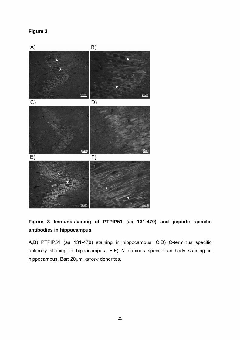
Figure 3
Figure 3 Immunostaining of PTPIP51 (aa 131-470) and peptide specific antibodies in hippocampus
A,B) PTPIP51 (aa 131-470) staining in hippocampus. C,D) C-terminus specific
antibody staining in hippocampus. E,F) N-terminus specific antibody staining in
hippocampus. Bar: 20µm. arrow: dendrites.
25
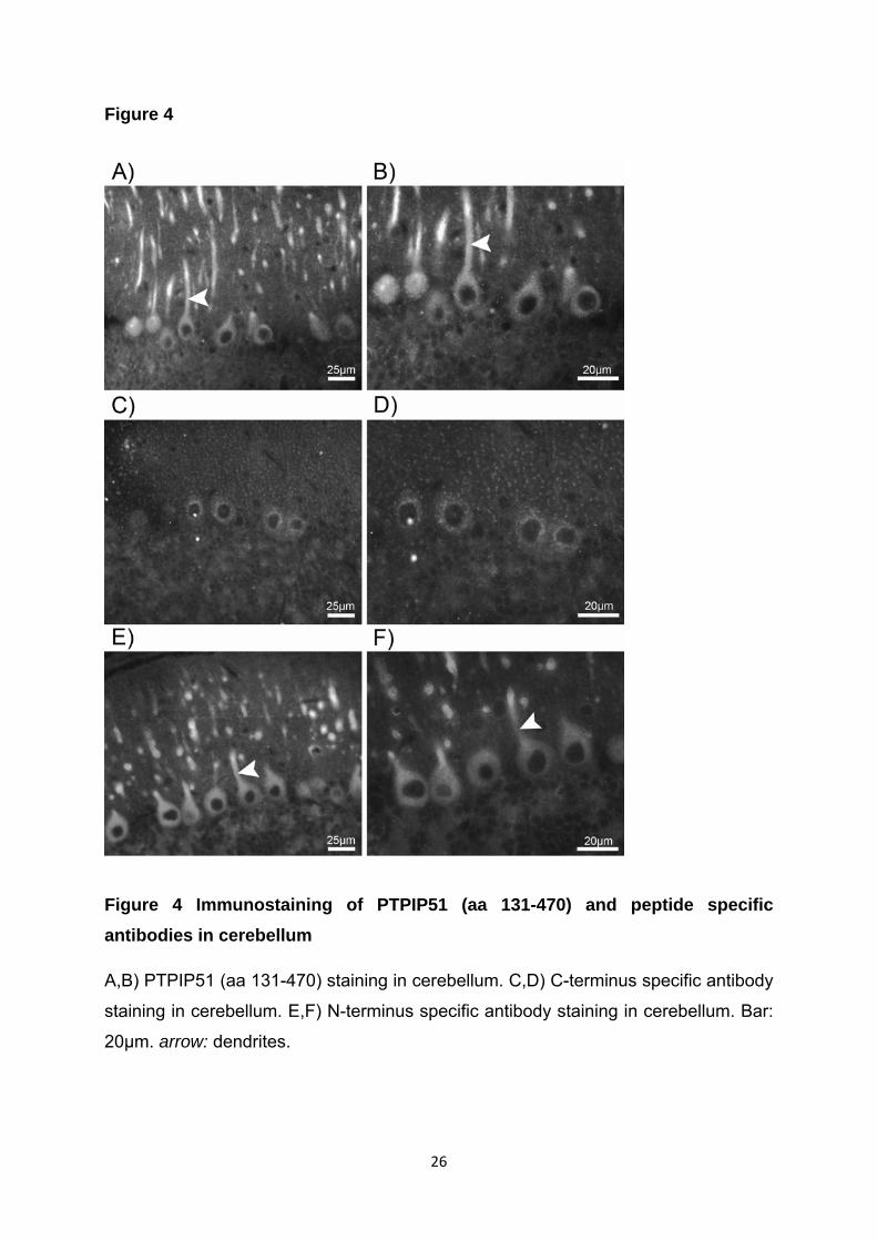
Figure 4
Figure 4 Immunostaining of PTPIP51 (aa 131-470) and peptide specific antibodies in cerebellum
A,B) PTPIP51 (aa 131-470) staining in cerebellum. C,D) C-terminus specific antibody
staining in cerebellum. E,F) N-terminus specific antibody staining in cerebellum. Bar:
20µm. arrow: dendrites.
26
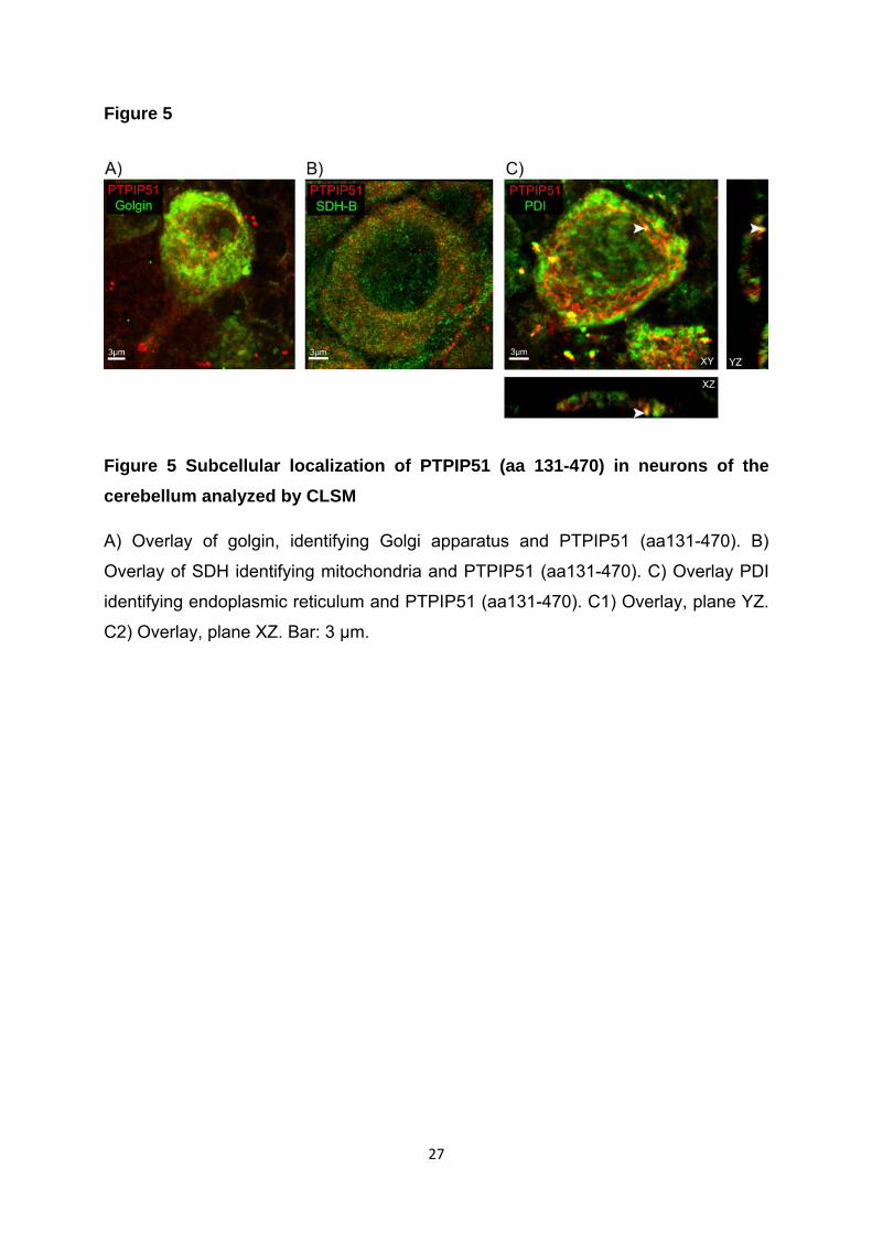
Figure 5
Figure 5 Subcellular localization of PTPIP51 (aa 131-470) in neurons of the cerebellum analyzed by CLSM
A) Overlay of golgin, identifying Golgi apparatus and PTPIP51 (aa131-470). B)
Overlay of SDH identifying mitochondria and PTPIP51 (aa131-470). C) Overlay PDI
identifying endoplasmic reticulum and PTPIP51 (aa131-470). C1) Overlay, plane YZ.
C2) Overlay, plane XZ. Bar: 3 µm.
27

Figure 6
Figure 6 Subcellular localization of PTPIP51 with peptide specific antibodies in
neurons of the hippocampus analyzed by CLSM
A) Overlay of Golgin identifying Golgi apparatus and C-terminus specific antibody.
A1) Overlay, plane YZ. A2) Overlay, plane XY. B) Overlay of SDH identifying
mitochondria and C-terminus specific antibody. C) Overlay of PDI identifying
endoplasmic reticulum and C-terminus specific antibody. C1) Overlay, plane YZ. C2)
Overlay, plane XY. D) Overlay of Golgin and N-terminus specific antibody. D1)
Overlay, plane YZ. D2) Overlay, plane XY. E) Overlay of SDH and PTPIP51 N-
terminus specific antibody. F) Overlay of PDI and N-terminus specific antibody. Bar: 5
µm. arrows: co- localization points
28

Figure 7
Figure 7 Co-immunostaining of PTPIP51 (aa131-470) with PTP1B and 14-3-3 beta, in Purkinje cells of the cerebellum
A) PTP1B immunostaining. B) PTPIP51 (aa131-470) immunostaining. C) Overlay of
PTPIP51 (aa131-470) and PTP1B. D) 14-3-3 beta immunostaining E) PTPIP51
(aa131-470) immunstaining. F) Overlay of PTPIP51 (aa131-470) and 14-3-3 beta. A-
C Bar: 5 µm. D-F Bar: 10µm.
29
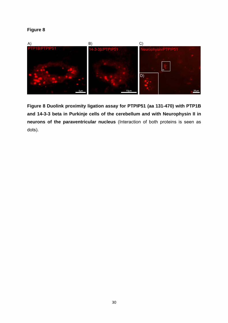
Figure 8
Figure 8 Duolink proximity ligation assay for PTPIP51 (aa 131-470) with PTP1B and 14-3-3 beta in Purkinje cells of the cerebellum and with Neurophysin II in neurons of the paraventricular nucleus (Interaction of both proteins is seen as
dots).
30
PTPIP51, a positive modulator of the MAPK/Erk pathway, is upregulated in glioblastoma and interacts with 14-3-3β and PTP1B in situ.
M. K. Petri*, P. Koch*, A. Stenzinger$, K. Kuchelmeister#, U. Nestler+, A.
Paradowska°, K. Steger°, A. Brobeil*, M. Viard*, and M. Wimmer*
*Institute of Anatomy and Cell Biology, Justus-Liebig-University, 35385 Giessen,
Germany
$Institute of Pathology, University Hospital Heidelberg, 69120 Heidelberg, Germany
#Institute of Neuropathology, University Hospital Bonn, 53105 Bonn, Germany
+Department of Neurosurgery, Justus-Liebig-University, 35385 Giessen, Germany
°Department of Urology and Pediatric Urology, Justus-Liebig-University, 35385
Giessen, Germany
Corresponding author:
Meike Katinka Petri
Institute of Anatomy and Cell Biology, Justus-Liebig-University Giessen
35385 Giessen, Germany
Phone: +49.641.9947012; Fax: +49.641.9947009
Running title: PTPIP51, 14-3-3β and PTP1B in glioblastoma
Keywords: glioblastoma, PTPIP51, 14-3-3β, EGFR, MAPK, Raf-1, glioma
1
Abstract
Glioblastoma multiforme (GBM) is the most common and most malignant primary
brain tumour. Protein tyrosine phosphatase interacting protein 51 (PTPIP51) is an
interaction partner of 14-3-3β, which correlates with the grade of malignancy in
gliomas. In this study PTPIP51 and its interacting partners 14-3-3β, PTP1B, c-Src,
Raf-1 as well as EGFR were investigated in human glioblastoma.
Twenty glioblastoma samples were analyzed on transcriptional and translational level
by immunohistochemistry, in situ hybridization and RT-PCR. To compare PTPIP51
expression in gliomas of different malignancies, quantitative RT-PCR for grade II
astrocytoma and GBM samples was employed. Additionally, we analyzed the
correlation between PTPIP51 and 14-3-3β transcription, and checked for in situ
interaction between PTPIP51 and 14-3-3β and PTP1B, respectively.
PTPIP51 and 14-3-3β mRNA showed a tumour grade dependent upregulation in
gliomas. Glioblastoma cells displayed a strong immunoreaction of PTPIP51, which
co-localized with 14-3-3β and PTP1B. The duolink proximity ligation assay
corroborated a direct in situ interaction of PTPIP51 with both proteins, known to
interact with PTPIP51 in vitro. The in vitro interacting partners Raf-1 and c-Src
showed a partial co-localization. Besides, immune cells located in capillaries or
infiltrating the tumour tissue and endothelial cells of pseudoglomerular vessels
revealed a high PTPIP51 expression.
The upregulation of PTPIP51 and its connection with the EGFR/MAPK pathway by
14-3-3β via Raf-1 and by PTP1B via c-Src, argue for a functional role of PTPIP51 in
the pathogenesis of human glioblastoma.
Introduction
The incidence of gliomas is increasing worldwide. 18,820 new cases of human
primary central nervous system tumours are diagnosed annually in the United States
of America, about 60% are gliomas and 30-40% of these account for glioblastoma
(GBM) (Khan et al., 2009). GBM is the most frequent and most malignant form of
2
neuroepithelial tumour. The mean survival time of GBM patients is still around one
year, despite significant advances in therapeutic options (McLendon et al., 2007).
PTPIP51 is a novel protein that has been shown to be expressed in many human
cancers (Lv et al., 2006; Stenzinger et al., 2009). As demonstrated by Koch et al.
(2008; 2009b) PTPIP51 is expressed in human keratinocyte carcinomas and prostate
cancer. Comparing benign prostate hyperplasia with prostate carcinoma provided
evidence that PTPIP51 expression is partially controlled by promoter methylation. Lv
et al. (2006) demonstrated PTPIP51 mRNA-expression in various carcinomas.
PTPIP51 is evolutionary conserved and was shown to interact in vitro with the non-
transmembrane protein-tyrosine phosphatase, Protein Tyrosine Phosphatase 1B
(PTP1B) (Stenzinger et al., 2005; Stenzinger et al., 2009). The protein is
phosphorylated in vitro and in situ at Tyr176 by Src kinase and dephosphorylated by
PTP1B (Stenzinger et al., 2009). In HEK 293 and HeLa cells, PTPIP51
overexpression was shown to enhance apoptosis (Lv et al., 2006).
Recently, two independent studies by Jin et al. (2004) and Ewing et al.. (2007)
demonstrated the interaction between the two isoforms 14-3-3β, 14-3-3γ and
PTPIP51. The study of Yang et al. (2009) demonstrated a correlation between the
grade of malignancy and the expression of 14-3-3β and 14-3-3η in gliomas. In
contrast, normal brain tissue was not found to express these two 14-3-3 isoforms.
The isoforms β and η belong to a superfamily of 14-3-3 proteins, which are
differentially expressed in many human tissues. 14-3-3 proteins have been implicated
in numerous cellular processes, such as tumourigenesis, cell cycle control and
apoptosis (Cao et al., 2008).
Yu and co-workers (2008) reported PTPIP51 to interact with Raf-1 through 14-3-3
protein, thereby modulating cellular motility and morphology by the mitogen activated
protein kinase (MAPK) cascade. The MAPK/Erk pathway is involved in a variety of
cellular functions such as growth, proliferation, differentiation, migration and
apoptosis. This pathway has been extensively studied in glioblastoma cells (Lopez-
Gines et al., 2008). The data available so far consider PTPIP51 to play a role in
cellular differentiation, motility, cytoskeleton formation and apoptosis.
This study primarily aimed to investigate the PTPIP51-expression profile in GBM,
applying RT-PCR, quantitative real time PCR, in situ hybridization and
3
immunohistochemistry. Moreover, cell specific co-expression of the proteins 14-3-3β,
Raf-1, PTP1B, c-Src and EGFR were assessed at the mRNA and protein level.
Furthermore, the direct interaction of PTPIP51 with 14-3-3β and PTP1B in situ was
substantiated.
An earlier study by Koch et al. (2009a) displayed the expression of PTPIP51 to be
restricted to neurons in specific areas of normal mouse brain and glial cells did not
show an expression of PTPIP51.
Data obtained in the present study will set the base for further studies that aim to
investigate the putative role of PTPIP51 in glioblastoma formation.
Material and Methods
Samples of twenty glioblastoma cases and samples of four astrocytomas were
included in this study (Table 1).
Glioblastoma specimens were stained immunohistochemically for PTPIP51, 14-3-3β,
c-Src, Raf-1, PTP1B, EGFR, apoptosis (TUNEL) and proliferation (Ki67). The cell
specific expression of PTPIP51 was corroborated by means of in situ hybridization.
Four intraoperatively obtained tissue samples of primary glioblastoma and one of
recurrent glioblastoma were examined for mRNA expression of PTPIP51, 14-3-3β, c-
Src, Raf-1 and PTP1B. Moreover, to obtain quantitative data on the transcriptional
activity of the 14-3-3 β and PTPIP51 gene in grade II astrocytoma (n=4 paraffin
embedded samples) and glioblastoma (n= 5 paraffin embedded samples) kinetic
PCR was employed.
Immunohistochemistry: The tumour tissue was fixed in neutral-buffered formalin for
48h, embedded in paraffin, sectioned and stained with H&E. The samples were
categorized according to the WHO classification and diagnosed as GBM.
Paraffin samples were obtained from the Institute of Neuropathology, University
Hospital Bonn. The patients had given informed consent that parts of the histological
specimens can be used for research purposes. The sections were deparaffinized in
4
xylene and rehydrated in graded ethanol. Prior to the staining procedure, antigen
retrieval using microwave-oven heating (2x5min, 800W) in 10mM standard sodium
citrate buffer (pH 6.0) was carried out for all antibodies used in this study.
Nonspecific binding sites were blocked with phosphate-buffered saline (PBS)
containing 5% bovine serum albumin and 5% normal goat serum. Immunoreaction
with the primary antibodies (see Table 2) was performed overnight at room
temperature, followed by incubation with the appropriate secondary antibodies (see
Table 2) for 1 h at room temperature. Subsequently, the slides were coverslipped in
carbonate buffered glycerol at pH 8.6.
The polyclonal antibody against PTPIP51 was raised as described and characterized
in previous studies (Koch et al., 2009a).
Primary antibodies were visualized by Alexa fluor 488 and Alexa fluor 555 secondary
antibodies. For each series of antibody staining sections were incubated with
medium lacking PTPIP51 antibody, serving as an internal negative control. Nuclei
were displayed by DAPI.
Apoptosis was detected using the in situ cell death detection kit ApopTag (Chemicon
International, USA # S7110) which employs an indirect TUNEL method, utilizing an
anti-digoxigenin antibody that is conjugated to a fluorescein reporter molecule. It
provides indirect immunofluorescence staining. Results were analyzed by using
fluorescence microscopy. The kit was used according to the instructions by the
manufacturer.
Immunofluorescence analysis and photodocumentation were performed on an
Axioplan 2 fluorescence microscope equipped with Plan-Apochromat objectives (Carl
Zeiss Jena, Germany). Visualization of the secondary antibody Alexa fluor 555 was
achieved with an excitation filter of 530-560nm and an emission filter with a range
572.5-647nm. Alexa Fluor 488 was visualized by an excitation filter with 460-500nm
and an emission filter 512-542 nm.
Duolink proximity ligation assay (DPLA): Interaction of PTPIP51 with either PTP1B or
14-3-3β was detected by the proximity ligation assay kit DuoLink (Olink biosciences,
Uppsala, Sweden, PLA probe anti-rabbit minus for the detection of the rabbit
PTPIP51 antibody, Cat.# 90602; PLA probe anti-mouse plus for the detection of the
mouse anti PTP1B or 14-3-3β antibody, Cat.# 90701; Detection Kit 563, Cat.#
90134). The DuoLink proximity ligation assay secondary antibodies only hybridise
5

when the two different PLA probes (probe anti-rabbit minus and probe anti-mouse
plus) have bound to proteins in proximity closer than 40 nm. This proximity results in
ligation forming a circular template and amplification step the fluorophore coupled
testing probe binds the amplified oligonucleotide strands. Addition of the fluorescent
labelled oligonucleotides that hybridize to the rolling circle amplification (RCA)
product leads to a point-shaped signal that is visible in fluorescence microscopy.
Methanol-fixed air-dried samples were per-incubated with blocking agent for 1h. After
washing in PBS for 10 min, primary antibodies for PTPIP51 and PTP1B, or PTPIP51
and 14-3-3β were applied to the samples. The antibodies were diluted in the blocking
agent at a concentration of 1:500 and 1:100, respectively. Incubation was done
overnight in a pre-heated humidity chamber. Slides were washed three times in PBS
for 10 min. Duolink PLA probes detecting rabbit or mouse antibodies were diluted in
the blocking agent at a concentration of 1:5 and applied to the slides following
incubation for 2h in a pre-heated humidity chamber at 37°C. Washing three times in
PBS for 10 min removed unbound PLA probes. For hybridization of the two Duolink
PLA probes Duolink Hybridization stock was diluted 1:5 in high purity water and
slides were incubated in a pre-heated humidity chamber for 15 min at 37°C. The
slides were washed in TBS-T for 1 min under gentle agitation. The samples were
incubated in the ligation solution consisting of Duolink Ligation stock (1:5) and
Duolink Ligase (1:40) diluted in high purity water for 90 min at 37°C. Detection of the
amplified probe was done with the Duolink Detection kit. Duolink Detection stock was
diluted 1:5 in high purity water and applied for 1 h at 37°C. Final washing steps were
done by SCC buffer and 70% ethanol.
H&E: The histomorphological characteristics were evaluated by hematoxylin and
eosin staining.
In situ hybridization: In situ hybridization was performed as described previously
(Koch et al., 2009a).
Reverse transcriptase-polymerase chain reaction (RT-PCR): For assessment of RT-
PCR five glioblastoma tissue specimens were obtained during neurosurgical
resection. The tissue samples were immediately transferred into RNA-later (Qiagen,
Hilden, Germany) and stored deep frozen at – 20°C according to the manufacturer’s
instructions. All tissue samples used for RT-PCR were obtained from the Department
of Neurosurgery, Justus-Liebig-University, Giessen. Prior to resection, patients had
6
given informed consent to using parts of the histological specimen for research
purposes.
The RNA extraction was performed using the RNA extraction kit RNeasy MINI
(Qiagen, Hilden, Germany) according to the manufacturer’s instructions.
Qualitative reverse transcriptase PCR was performed on an iCycler using SYBR
Green Supermix (BioRad, Munich, Germany) to visualize the respective amplicons.
2µl cDNA were used per sample. Cycling conditions were 94 °C for 2 min, followed
by 40 cycles of 94 °C (PTPIP51); 58°C (PTP1B); 63°C (c-Src); 63,5°C (Raf-1) and
60°C (14-3-3β) for 30 sec, 55 °C for 30 sec and 72 °C for 2 min. The primers were
employed for PTPIP51, PTP1B, c-Src, Raf-1 and 14-3-3β (see Table 3).
PCR products were visualized by agarose gel electrophoresis. While amplification of
a 90 bp β-actin product served as positive control, negative controls included
samples lacking reverse transcriptase.
Quantitative real time PCR: Five paraffin-embedded GBM tissues and four samples
from patients with grade II astrocytoma were used for quantification of mRNA. The
purification of RNA was done using the QIAamp DNA FFPE tissue kit (Qiagen,
Hilden, Germany) according to the manufacturer’s instruction.
The amplification of cDNA was carried out in 25 µl reaction volume on the iCycler iQ
Real-Time PCR Detection System (Bio Rad, Munich, Germany). The final reaction
tubes contained 100nM as PTPIP51, 14-3-3β and reference genes β-actin and
GAPDH (see Table 3), 12.5µl iQ SYBR Green Supermix (Bio Rad) and 2µl of DNA
template. The PCR conditions were 94°C for 3 min followed by 40 cycles for 30sec,
60°C for 30sec and 72°C for 1min. Melting curves were generated for both genes
after amplification. Negative controls were included in each run. The selection of
appropriate combination of reference genes for expression analysis of PTPIP51 in
GBM and astrocytoma II tissue was carried out using NormFinder Program. PCR-
products were additionally electrophoresed on a 3 % agarose gel and visualized by
GelRed reagent.
7

Results
This study demonstrates for the first time the cell-specific expression of PTPIP51
mRNA and protein in human glioblastoma. Immunohistochemical experiments and in
situ hybridization revealed a strong expression of PTPIP51 in GBM tumour cells and
endothelial cells and immune cells. Additionally, the PTPIP51 expression profile was
assayed in apoptosis by TUNEL and in proliferation with Ki67 by double
immunostaining. Furthermore, we explored the interacting partners of PTPIP51, 14-
3-3β, Raf-1, PTP1B, c-Src and EGFR. The transcription of the respective signalling
partners was shown by qualitative reverse transcriptase PCR. The correlation of the
tumour grade of gliomas with the quantitative expression of both, PTPIP51 and 14-3-
3β, was examined by quantitative real time PCR comparing GBM and grade II
astrocytoma.
In situ expression profile of PTPIP51 mRNA and protein:
Immunohistochemical staining of 15 GBM samples revealed PTPIP51 protein
expression in tumour cells as identified by subsequent H&E staining of the sections
(Fig.1 A-C). Probing glioblastoma samples with both PTPIP51 antibody and GFAP
antibody confirmed the expression of PTPIP51 in malignant glial cells (Fig.1 D-F).
Endothelial cells (Fig.2 J–L), smooth muscle cells (Fig.2 G–I) and immune cells also
showed PTPIP51 expression. In endothelial cells of normal and of pseudoglomerular
vessels PTPIP51 protein was restricted to the plasmamembrane and to the nucleus.
Granulocytes located in capillary lumina or infiltrating the tumour, as well as the
necrotic tissue, displayed a strong PTPIP51 immunoreaction (Fig.2 A-C). In contrast
to granulocytes, B lymphocytes did not show any PTPIP51 expression (Fig.2 D-F).
Matching the protein expression profile, PTPIP51 mRNA was found in the cytoplasm
of tumour cells and in the cytoplasm of endothelial cells as detected by in situ
hybridization (Fig. 3).
In situ expression profile of PTPIP51 and its interacting partners:
PTPIP51 and 14-3-3β showed a strict co-localization. Both proteins were found in
glioblastoma cells, endothelial cells and in immune cells. The glioblastoma cells
displayed a strong cytoplasmic reaction (Fig. 4 A-C). In astrocytoma, a co-localization
for PTPIP51 and 14-3-3β was seen, but to a much lower extent (Fig. 4 D-F).
8

To corroborate the interaction of PTPIP51 and 14-3-3β, a duolink proximity ligation
assay was applied to GBM and astrocytoma sections. In all investigated samples,
hybridized and amplified antibody-linked nucleotide strands were detected. Every dot
corresponds to an interaction between PTPIP51 and 14-3-3β.
As seen in Figure 4 G and H, glioblastoma and astrocytoma cells displayed
hybridized and amplified antibody-linked nucleotide strands, revealing the in situ
interaction between PTPIP51 and 14-3-3β.
GBM tumour cells situated around pseudoglomerular vessels, as well as endothelial
cells and immune cells, displayed a co-localization of PTPIP51 with PTP1B (Fig.5 A-
C). To confirm an in situ interaction of PTPIP51 and PTP1B, a duolink proximity
ligation assay was performed. As seen in Figure 5 D glioblastoma cells displayed
hybridized and amplified antibody-linked nucleotid strands, revealing the interaction
between PTPIP51 and PTP1B.
Raf-1 and PTPIP51 displayed a strong co-localization in the vessels surrounding
tumour cells (Fig.6 A-C).
There was only a partial co-localization of PTPIP51 and c-Src (Fig.6 D-F). In contrast
to high PTPIP51 expression in the cytoplasm of tumour and immune cells c-Src is
only present in some of these cells.
A majority of tumour cells displayed a co-localization of PTPIP51 and EGFR (data
not shown).
Stained by Ki-67, proliferating cells showed a strong expression of PTPIP51 in the
cytoplasm with elevated concentration near the plasmamembrane (Fig.7 A-C).
TUNEL assay analysis of GBM sections did not show PTPIP51 positive cells
executing apoptosis (Fig.7 D-F).
mRNA expression of PTPIP51 and its interaction partners:
Qualitative reverse transcriptase PCR analysis: The mRNA expression of PTPIP51
and its in vitro interaction partners was tested by reverse transcriptase PCR. As
demonstrated in Figure 8, the samples 1, 2, 3 and 5 expressed a considerable
amount of PTPIP51, PTP1B, 14-3-3β, Raf-1 and c-Src. A different expression pattern
was observed in sample number 4, which had been obtained from a patient with a
9
recurrent GBM. The histopathological findings of this specimen showed healthy
cerebral tissue with only singular tumour cells. In this case attenuated expression of
PTPIP51 and PTP1B was found, c-Src was barely detectable, and Raf-1 was lacking (Fig.8).
Comparison of PTPIP51 expression levels in grade II astrocytoma and glioblastoma
by quantitative PCR:
PTPIP51 expression levels of 5 GBM and 4 grade II astrocytoma samples were
assessed by quantitative PCR. Candidate reference genes for normalization and the
best combination of two genes were calculated according to their expression stability
by the NormFinder program. The best fitting combination proved to be β-actin with
GAPDH.
The results of expression analysis showed slightly elevated levels of PTPIP51 (mean
value 5,23 +/- 1,01) in the group of GBM when compared to low grade astrocytoma
(mean value 4,62 +/- 1,49). 14-3-3β expression in glioblastoma (mean value 3,42 +/-
1.10) was significantly higher than in grade II astrocytoma samples (mean value 0,88
+/- 2,74) (Fig. 9).
Discussion
Our results revealed elevated levels of PTPIP51 expression in GBM (grade IV
glioma) samples when compared to low grade astrocytomas (grade II glioma), which
correlated with the expression levels of 14-3-3β. This is consistent with recent studies
displaying a tumour grade dependent expression of two isoforms of 14-3-3β and 14-
3-3η in gliomas. Healthy cerebral tissue is completely lacking in both isoforms (Yang
et al., 2009). The upregulation of 14-3-3 proteins seems to be associated with the
reduced capacity of apoptosis, as antagonizing 14-3-3 or silencing its expression
induces apoptosis in cultured glioma cells (Cao et al., 2010).
14-3-3β and 14-3-3γ mediate the interaction of PTPIP51 and Raf-1, thereby
modulating the activity of the MAPK-cascade (Lv et al., 2006; Yu et al., 2008,
Stenzinger et al., 2009;). The MAPK pathway plays an important role in cell migration
10
and seems to be one of the main reasons for recurrences and poor prognosis of
glioblastoma. It is considered that migrating tumour cells infiltrate the healthy tissue
surrounding the glioblastoma and in this way can escape surgical extirpation and
give rise to regrowth. With regard to these findings the interaction of PTPIP51 with
14-3-3β, which was confirmed by the duolink proximity ligation assay, may mirror the
role of PTPIP51 protein in migration and proliferation of GBM tumour cells.
A strong co-localization and interaction of PTPIP51 and 14-3-3β was also found in all
endothelial cells of GBM-typical glomerulum-like vessels of glioblastoma.
This interaction may contribute to the well known high activation of the MAPK/Erk
pathway via EGFR, resulting in dysfunction of cell cycle control and upregulation of
proliferation in GBM.
The non-transmembrane Protein Tyrosine Phosphatase 1B (PTP1B), a known
interacting partner of PTPIP51, is able to activate the MAPK cascade on c-Src and
Ras level (Dubé et al., 2004; Dubé and Tremblay, 2004; Tonks and Muthuswamy,
2007; Zhao et al., 2008, Stenzinger et al., 2009). Reichardt and coworkers (2003)
were unable to detect DNA amplification of PTP1B in human gliomas. However, in
our study PTP1B expression was upregulated and highly co-localized with the
PTPIP51 protein. The observed upregulation is consistent with the data reported by
Akasaki and co-workers (2006), who also reported PTP1B to be overexpressed in
gliomas. Furthermore, a direct in situ interaction of PTPIP51 and PTP1B in
glioblastoma cells was corroborated by the duolink proximity ligation assay. These
results underline the significance of our observations of upregulated PTPIP51
expression levels in glioblastomas.
PTP1B contributes to oncogenesis by the loss of tyrosine phosphorylation of key
signalling proteins or by up-regulation of two growth-promoting pathways (Arias-
Romero et al., 2009). In human mammary cells PTP1B links an important oncogenic
receptor tyrosine kinase, namely ErbB2, to signalling pathways that promote aberrant
cell division and survival by activation of Src and inducing a Src-dependent
transformed phenotype. It deactivates the Ras/MAPK pathway inhibitor (Tonks and
Muthuswamy, 2007).
C-Src mediates the phosphorylation of EGFR, thereby promoting tumour progression
(Tice et al., 1999). The ultimate cellular response to the activation of EGFR signalling
cascade via MAPK pathway is DNA synthesis and cell division (Halatsch et al.,
11

2004). In our samples PTPIP51 and EGFR were partly co-localized, suggesting a
synergistic effect on cell proliferation, migration and oncogenic transformation. This
may be exerted by sharing the same final signalling pathway, PTPIP51 via 14-3-3β
interaction with Raf-1 and EGFR activating the Ras/Raf/MAPK/ERK pathway.
Compared to secondary GBM, developed by progression from lower grade gliomas,
EGFR gene amplification has been shown to be five times higher in primary
glioblastoma, which leads to overexpression in 40 % of GBM (Karpel-Massler et al.,
2009). Besides this EGFR overexpression, EGFR is expressed as the mutated
EGFRvIII (epidermal growth factor receptor variant III) form of the receptor in 20 % of
GBM cases (Jutten et al., 2009). This constitutively active mutant form of the EGFR,
which is commonly expressed in glioblastoma, is also detected in a number of
epithelial cancers (Yoshimoto et al., 2008; Hama et al., 2009) also known to express
high concentrations of PTPIP51, e.g. non melanoma skin cancer, prostate cancer
(Koch et al., 2008; 2009b) and breast cancer. EGFR signalling cascade via MAPK
pathway is modulated by PTP1B through c-Src and by 14-3-3β through Raf-1 (Yu et
al., 2008), both interaction partners of PTPIP51. This in situ interaction in GBM was
substantiated in our study by duolink proximity ligation assay.
Glial malignant transformation might be correlated to the status of PTPIP51 gene
promotor methylation, since high grade gliomas (GBM) also presented a higher
mRNA expression of PTPIP51 in comparison to low grade gliomas (grade II
astrocytoma).
To sum up, in neuroepithelial tumours, PTPIP51 expression increases with the grade
of malignancy and PTPIP51 interacts in situ with 14-3-3β and PTP1B. The data
presented in this study suggest an important role of PTPIP51 in glioblastoma
formation.
Acknowledgments
We are grateful to Mrs. K. Michael (Institute of Anatomy and Cell Biology, Giessen)
for help with the design of the figures and to Mrs. A. Peters (Department of
Neurosurgery, Giessen) for help in providing the glioblastoma samples. The excellent
12
techncial assistance of Mrs. A. Erkel, Mrs. B. Fröhlich (Department of Urology and
Pediatric Urology, Giessen), Mrs. A. zur Mühlen (Institute of Neuropathology, Bonn)
and Mrs. C. Tag (Institute of Anatomy and Cell Biology, Giessen) is gratefully
acknowledged.
References
Akasaki Y., Liu G., Matundan H.H., Ng H., Yuan X., Zeng Z., Black K.L. and Yu J.S.
(2006). A peroxisome proliferator-activated receptor-gamma agonist, troglitazone,
facilitates caspase-8 and -9 activities by increasing the enzymatic activity of protein-
tyrosine phosphatase-1B on human glioma cells. J. Biol. Chem. 281, 6165- 6174.
Arias-Romero L.E., Saha S., Villamar-Cruz O., Yip S.C., Ethier S.P., Zhang Z.Y. and
Chernoff J. (2009). Activation of Src by protein tyrosine phosphatase 1B is required
for ErbB2 transformation of human breast epithelial cells. Cancer. Res. 69, 4582-
4588.
Cao L., Cao W., Zhang W., Lin H., Yang X., Zhen H., Cheng J., Dong W., Huo J. and
Zhang X. (2008). Identification of 14-3-3 protein isoforms in human astrocytoma by
immunohistochemistry. Neurosci. Lett. 432, 94-99.
Cao W., Yang X., Zhou J., Teng Z., Cao L., Zhang X. and Fei Z. (2010). Targeting
14-3-3 protein, difopein induces apoptosis of human glioma cells and suppresses
tumor growth in mice. Apoptosis. 15, 230-241.
Dubé N. and Tremblay M.L. (2004). Beyond the metabolic function of PTP1B. Cell.
Cycle. 3, 550-553.
Dubé N., Cheng A. and Tremblay M.L. (2004). The role of protein tyrosine
phosphatase 1B in Ras signalling. Proc. Nat. Acad. Sci. U S A. 101, 1834-1839.
Ewing R.M., Chu P., Elisma F., Li H., Taylor P., Climie S., McBroom-Cerajewski L.,
Robinson M.D., O'Connor L., Li M., Taylor R., Dharsee M., Ho Y., Heilbut A., Moore
13

L., Zhang S., Ornatsky O., Bukhman Y.V., Ethier M., Sheng Y., Vasilescu J., Abu-
Farha M., Lambert J.P., Duewel H.S., Stewart I.I., Kuehl B., Hogue K., Colwill K.,
Gladwish K., Muskat B., Kinach R., Adams S.L., Moran M.F., Morin G.B., Topaloglou
T. and Figeys D. (2007). Large-scale mapping of human protein-protein interactions
by mass spectrometry. Mol. Syst. Biol. 3, 89.
Gajadhar A. and Guha A. (2010). A proximity ligation assay using transiently
transfected, epitope-tagged proteins: application for in situ detection of dimerized
receptor tyrosine kinases. Biotechniques. 48, 145-152
Halatsch M.E., Gehrke E.E., Vougioukas V.I., Bötefür I.C., A-Borhani F., Efferth T.,
Gebhart E., Domhof S., Schmidt U. and Buchfelder M. (2004). Inverse correlation of
epidermal growth factor receptor messenger RNA induction and suppression of
anchorage-independent growth by OSI-774, an epidermal growth factor receptor
tyrosine kinase inhibitor, in glioblastoma multiforme cell lines. J. Neurosurg. 100,
523-533.
Hama T., Yuza Y., Saito Y., Ouchi J., Kondo S., Okabe M., Yamada H., Kato T.,
Moriyama H., Kurihara S. and Urashima M. (2009). Prognostic significance of
epidermal growth factor receptor phosphorylation and mutation in head and neck
squamous cell carcinoma. Oncologist. 14, 900-908.
Jin J., Smith F.D., Stark C., Wells C.D., Fawcett J.P., Kulkarni S., Metalnikov P.,
O'Donnell P., Taylor P., Taylor L., Zougman A., Woodgett J.R., Langeberg L.K., Scott
J.D. and Pawson T. (2004). Proteomic, functional, and domain-based analysis of in
vivo 14-3-3 binding proteins involved in cytoskeletal regulation and cellular
organization. Curr. Biol. 14, 1436-1450.
Jutten B., Dubois L., Li Y., Aerts H., Wouters B.G., Lambin P., Theys J. and
Lammering G. (2009) Binding of cetuximab to the EGFRvIII deletion mutant and its
biological consequences in malignant glioma cells. Radiother. Oncol. 92, 393-398.
14

Karpel-Massler G., Schmidt U., Unterberg A. and Halatsch M.E. (2009). Therapeutic
inhibition of the epidermal growth factor receptor in high-grade gliomas: where do we
stand? Mol. Cancer. Res. 7, 1000-1012.
Khan M.K., Hunter G.K., Vogelbaum M., Suh J.H. and Chao S.T. (2009). Evidence-
based adjuvant therapy for gliomas: current concepts and newer developments.
Indian J. Cancer. 46, 96-107.
Koch P., Viard M., Stenzinger A., Brobeil A., Tag C., Steger K. and Wimmer M.
(2009a). Expression profile of PTPIP51 in mouse brain. J. Comp. Neurol. 517, 892-
905.
Koch P., Petri M., Paradowska A., Stenzinger A., Sturm K., Steger K. and Wimmer
M. (2009b). PTPIP51 mRNA and protein expression in tissue microarrays and
promoter methylation of benign prostate hyperplasia and prostate carcinoma.
Prostate. 69, 1751-1762.
Koch P., Stenzinger A., Viard M., Märker D., Mayser P., Nilles M., Schreiner D.,
Steger K. and Wimmer M. (2008). The novel protein PTPIP51 is expressed in human
keratinocyte carcinomas and their surrounding stroma. J. Cell. Mol. Med. 12, 2083-
2095.
Lopez-Gines C., Gil-Benso R., Benito R., Mata M., Pereda J., Sastre J., Roldan P.,
Gonzalez-Darder J. and Cerdá-Nicolás M. (2008). The activation of ERK1/2 MAP
kinases in glioblastoma pathobiology and its relationship with EGFR amplification.
Neuropathol. 28, 507-515.
Lv B.E., Yu C.E., Chen Y.Y., Lu Y., Guo J.H., Song Q.S., Ma D.L., Shi T.P. and
Wang L. (2006). Protein tyrosine phophatase interacting interacting protein 51
(PTPIP51) is a novel mitochondria protein with an N-terminal mitochondrial targeting
sequence and induces apoptosis. Apoptosis. 11, 1489-1501.
McLendon R.E., Turner K., Perkinson K. and Rich J. (2007). Second messenger
systems in human gliomas. Arch. Pathol. Lab. Med. 131, 1585-1590.
15
Reichardt W., Jung V., Brunner C., Klein A., Wemmert S., Romeike B.F., Zang K.D.
and Urbschat S. (2003). The putative serine/threonine kinase gene STK15 on
chromosome 20q13.2 is amplified in human gliomas. Oncol. Rep. 10, 1275-1279.
Söderberg O., Gullberg M., Jarvius M., Ridderstråle K., Leuchowius K.J., Jarvius J.,
Wester K., Hydbring P., Bahram F., Larsson L.G. and Landegren U. (2006). Direct
observation of individual endogenous protein complexes in situ by proximity ligation.
Nat. Methods. 3, 995-1000.
Stenzinger A., Kajosch T., Tag C., Porsche A., Welte I., Hofer H.W., Steger K. and
Wimmer M. (2005). The novel protein PTPIP51 exhibits tissue- and cell-specific
expression. Histochem. Cell. Biol. 123, 19-28.
Stenzinger A., Schreiner D., Koch P., Hofer H.W. and Wimmer M. (2009). Cell- and
molecular biology of the novel protein tyrosine phosphatase interacting protein 51.
Int. Rev. Cell. Mol. Biol. 275, 183-246.
Tice D.A., Biscardi J.S., Nickles A.L. and Parsons S.J. (1999). Mechanism of
biological synergy between cellular Src and epidermal growth factor receptor. Proc.
Nat. Acad. Sci. USA. 96, 1415-1420.
Tonks N.K. and Muthuswamy S.K. (2007). A brake becomes an accelerator: PTP1B -
a new therapeutic target for breast cancer. Cancer Cell. 11, 214-216.
Yadavilli S., Mayo L.D., Higgins M., Lain S., Hegde V. and Deutsch W.A. (2009).
Ribosomal protein S3: A multi-functional protein that interacts with both p53 and
MDM2 through its KH domain. DNA Repair. 8, 1215-1224.
Yang X., Cao W., Lin H., Zhang W., Lin W., Cao L., Zhen H., Huo J. and Zhang X.
(2009). Isoform-specific expression of 14-3-3 proteins in human astrocytoma. J.
Neurol. Sci. 276, 54-59.
16

Yoshimoto K., Dang J., Zhu S., Nathanson D., Huang T., Dumont R., Seligson D.B.,
Yong W.H., Xiong Z., Rao N., Winther H., Chakravarti A., Bigner D.D., Mellinghoff
I.K., Horvath S., Cavenee W.K., Cloughesy T.F. and Mischel P.S. (2008).
Development of a real-time RT-PCR assay for detecting EGFRvIII in glioblastoma
samples. Clin. Cancer. Res. 14, 488-493.
Yu C., Han W., Shi T., Lv B., He Q., Zhang Y., Li T., Zhang Y., Song Q., Wang L. and
Ma D. (2008). PTPIP51, a novel 14-3-3 binding protein, regulates cell morphology
and motility via Raf-ERK pathway. Cell. Signal. 20, 2208-2220.
Zhao Y., Xiao A., Dipierro C.G., Abdel-Fattah R., Amos S., Redpath G.T., Carpenter
J.E., Pieper R.O. and Hussaini, I.M. (2008). H-Ras increases urokinase expression
and cell invasion in genetically modified human astrocytes through Ras/Raf/MEK
signalling pathway. Glia. 56, 917-924.
17
Legends
Fig. 1 Immunostaining of PTPIP51 in sections of human glioblastoma
A) PTPIP51 immunostaining B) H&E. staining of section A C) DAPI staining of
section A D) PTPIP51 immunostaining E) GFAP staining of section D F) DAPI
staining of section D Arrows: PTPIP51 positive glioblastoma cells,
arrowheads: PTPIP51 positive endothelial cells, double arrowheads: PTPIP51
and GFAP positive reactive astrocyte, double arrow: PTPIP51 positive
immune cell (neutrophil granulocyte). BAR = 50µm
Fig. 2 PTPIP51 expression in immune cells, smooth muscle cells and endothelial cells
A) PTPIP51 immunostaining B) granulocyte marker of section A C) Merge of A, B
and DAPI D) PTPIP51 immunostaining E) B lymphocyte in section D detected by
CD20 F) Merge of D, E and DAPI. BAR (A-F)= 10μm G) PTPIP51 immunostaining H)
α-sma of section G I) Merge of G,H and DAPI J) PTPIP51 immunostaining K)
endothelial cells detected by CD34 in section J L) Merge of J, K and DAPI.
Arrowhead: neutrophil granulocytes; blank arrowhead: B lymphocytes; double arrow:
smooth muscle cells; arrow: endothelial cells; white circle: lipofuscin granula . BAR
(G-L) = 20µm
Fig. 3 In situ hybridization of PTPIP51 in sections of human glioblastoma
A) anti-sense probe B) sense probe of parallel section to A C) anti-sense probe D)
sense probe in parallel section to C Arrows: PTPIP51 mRNA in glioblastoma cells,
arrowhead: PTPIP51 mRNA in endothelial cells, double arrow: immune cells,
asterisk: necrotic tissue. BAR = 50µm
18

Fig. 4 PTPIP51 and its interacting partner 14-3-3β - in situ co-localization analysis and Duolink proximity ligation assay in sections of human glioblastoma and astrocytoma
A) PTPIP51 immunostaining GBM B) 14-3-3β immunostaining of section A C) Merge
of A and B D) PTPIP51 immunostaining astrocytoma E) 14-3-3β immunostaining of
section D F) Merge of D and E. Arrows: PTPIP51 positive glioblastoma cells,
arrowheads: PTPIP51 positive endothelial cells, double-arrows: immune cells. BAR =
50µm G) GBM duolink proximity ligation assay for PTPIP51 and 14-3-3β H)
astrocytoma duolink proximity ligation assay for PTPIP51 and 14-3-3β. Interaction of
both proteins is seen as orange dots (arrows) I) Negative control. White circles:
lipofuscin granules. BAR = 10µm
Fig. 5 PTPIP51 and its interacting partner PTP1B in sections of human glioblastom
A) PTPIP51 immunostaining B) PTP1B immunostaining of section A C) Merge of
section A and B D) Duolink proximity ligation assay for PTPIP51 and PTP1B.
Interaction of both proteins is seen as orange dots (arrows). BAR (A,B, C) = 20μm,
BAR (D) = 10μm
Fig. 6 PTPIP51 and its interacting partners Raf-1 and c-Src in sections of human glioblastoma
A) PTPIP51 immunostaining B) Raf-1 immunostaining of section A C) Merge of A
and B D) PTPIP51 immunostaining E) c-Src immunostaining of section D F) Merge of
D and E. Arrows: PTPIP51 positive glioblastoma cells, arrowheads: PTPIP51 positive
endothelial cells, double-arrows: immune cells. BAR = 50µm
19
Fig. 7 PTPIP51 protein expression and investigation of proliferation and apoptosis by Ki67 antibody and TUNEL-assay
A) PTPIP51 immunostaining B) Ki67 immunostaining of section A C) Merge of A and
B D) PTPIP51 immunostaining E) TUNEL-assay of section D, insert = H.E. staining
of detail F) Merge of D and E. Arrows: PTPIP51 positive glioblastoma cells,
arrowheads: PTPIP51 positive endothelial cells, blank arrowhead: apoptotic pericyte.
BAR = 50µm
Fig. 8 Expression of PTPIP51 and its interaction partners as detected by reverse transcriptase-PCR in tissues of human glioblastoma
Qualitative reverse transcriptase-PCR was performed using primers specific to
PTPIP51, PTP1B, Raf-1, c-Src and 14-3-3β as given in Materials and Methods
section. β-actin was amplified as an internal positive control and probes lacking
reverse transcriptase served as negative controls (N).
Fig. 9 Quantitative real time PCR analysis of PTPIP51 and 14-3-3β in glioblastoma and grade II astrocytoma
A: Diagram shows the results of quantitative expression analysis. Slightly elevated
levels of PTPIP51 are seen in the group of glioblastoma (GBM) when compared to
low grade astrocytoma. 14-3-3β expression in glioblastoma was significantly higher
than in grade II astrocytoma samples.
Amplification of β-actin and GAPDH was used as a combination of reference genes.
B: Gel: bands exclusively detecting mRNA with the expected amplification size with
the primers used in 7A. Negative control (N).
20
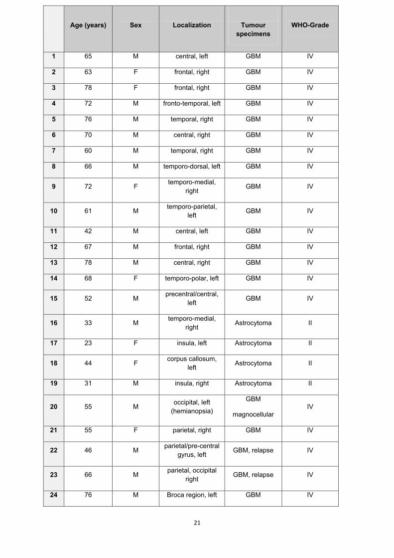
Age (years)
Sex
Localization
Tumour specimens
WHO-Grade
1 65 M central, left GBM IV
2 63 F frontal, right GBM IV
3 78 F frontal, right GBM IV
4 72 M fronto-temporal, left GBM IV
5 76 M temporal, right GBM IV
6 70 M central, right GBM IV
7 60 M temporal, right GBM IV
8 66 M temporo-dorsal, left GBM IV
9 72 F temporo-medial, right GBM IV
10 61 M temporo-parietal, left GBM IV
11 42 M central, left GBM IV
12 67 M frontal, right GBM IV
13 78 M central, right GBM IV
14 68 F temporo-polar, left GBM IV
15 52 M precentral/central, left GBM IV
16 33 M temporo-medial, right Astrocytoma II
17 23 F insula, left Astrocytoma II
18 44 F corpus callosum, left Astrocytoma II
19 31 M insula, right Astrocytoma II
20 55 M occipital, left (hemianopsia)
GBM
magnocellular IV
21 55 F parietal, right GBM IV
22 46 M parietal/pre-central gyrus, left GBM, relapse IV
23 66 M parietal, occipital right GBM, relapse IV
24 76 M Broca region, left GBM IV
21

Table 1: Tumour specimens included in the study Tumours No. 1 – 19 were analyzed by
immunocytochemistry and No. 11 – 19 by quantitative real time PCR. Tumours No. 20 – 24 were
analyzed by reverse transcriptase PCR.
22
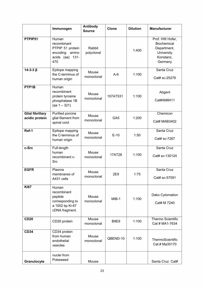
Immunogen Antibody Source Clone Dilution Manufacturer
PTPIP51 Human recombinant PTPIP 51 protein encoding amino acids (aa) 131-470
Rabbit polyclonal 1:400
Prof. HW Hofer, Biochemical Department,
University Konstanz, Germany
14-3-3 β Epitope mapping the C-terminus of human origin
Mouse monoclonal A-6 1:100
Santa Cruz
Cat# sc-25276
PTP1B Human recombinant protein tyrosine phosphatase 1B (aa 1 – 321)
Mouse monoclonal 107AT531 1:100
Abgent
Cat#AM8411
Glial fibrillary acidic protein
Purified porcine glial filament from spinal cord
Mouse monoclonal GA5 1:200
Chemicon
Cat# MAB3402
Raf-1 Epitope mapping the C-terminus of human origin
Mouse monoclonal E-10 1:50
Santa Cruz
Cat# sc-7267
c-Src Full-length human recombinant c-Src
Mouse monoclonal 17AT28 1:100
Santa Cruz
Cat# sc-130124
EGFR Plasma membranes of A431 cells
Mouse monoclonal 2E9 1:75
Santa Cruz
Cat# sc-57091
Ki67 Human recombinant peptide corresponding to a 1002 bp Ki-67 cDNA fragment.
Mouse monoclonal MIB-1 1:100
Dako Cytomation
Cat# M 7240
CD20 CD20 protein Mouse monoclonal B9E9 1:100 Thermo Scientific
Cat # MA1-7634
CD34 CD34 protein from human endothelial vesicles
Mouse monoclonal QBEND-10 1:100
ThermoScientific Cat.# Ma35170
Granulocyte
nuclei from Pokeweed
Mouse
Santa Cruz Cat#
23
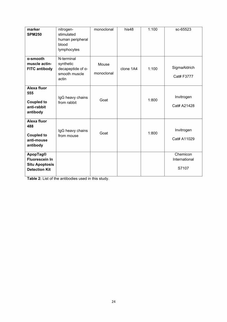
marker SPM250
nitrogen-stimulated human peripheral blood lymphocytes
monoclonal his48 1:100 sc-65523
α-smooth muscle actin- FITC antibody
N-terminal synthetic decapeptide of α-smooth muscle actin
Mouse
monoclonal clone 1A4 1:100
SigmaAldrich
Cat# F3777
Alexa fluor 555
Coupled to anti-rabbit antibody
IgG heavy chains from rabbit Goat 1:800
Invitrogen
Cat# A21428
Alexa fluor 488
Coupled to anti-mouse antibody
IgG heavy chains from mouse Goat 1:800
Invitrogen
Cat# A11029
ApopTag® Fluorescein In Situ Apoptosis Detection Kit
Chemicon International
S7107
Table 2: List of the antibodies used in this study.
24

Primer RT-PCR (fresh tissue specimen) size template
PTPIP51 forward 5´-GCAGGTGGTGCTATCAGGTC-3´
reverse 5’-AGCTCCAGGGCCAACTTCATC-3’
232 BP 1294 –1525
PTP1B forward 5’-GGAGATGGAAAAGGAGTTC-3’
reverse 5’-TGCTTTTCTGCTCCCACAC-3’
311 BP 177 - 487
14-3-3β forward 5’-ATTCGTCTTGGTCTGGCACT-3’
reverse 5’-CAGGCTACAGGCCTTTTCAG-3’.
78 BP 689 – 766,
784 - 861
c-Src forward 5’-ATGGTGAACCGCGAGGTGCT-3’
reverse 5’-GATCCAAGCCGAGAAGCCGGTCTG-3’
244 BP 1753 - 1996
Raf-1 forward 5’-CAGCCCTGTCCAGTAGC-3’
reverse 5’-GCCTGACTTTACTGTTGC-3’
614 BP 1287 - 1900
β-actin forward 5’-TTCCTTCCTGGGCATGGAGT-3’
reverse 5’-TACAGGTCTTTGCGGATGTC-3’
90 BP 2439 - 2528
Primer quantitative RT-PCR (paraffin embedded tissue)
PTPIP51 forward 5’-TCAAGGAGCATGTGGACAAA-3’
reverse 5’-ATAGCACCACCTGCCAAGAA-3’
80 BP 1228 - 1307
14-3-3β forward 5’-ATTCGTCTTGGTCTGGCACT-3’
reverse 5’-CAGGCTACAGGCCTTTTCAG-3’.
78 BP 689 – 766,
784 - 861
β-actin forward 5’-TTCCTTCCTGGGCATGGAGT-3’
reverse 5’-TACAGGTCTTTGCGGATGTC-3’
90 BP 2439 - 2528
GAPDH forward 5’-ATGCCAGTGAGCTTCCCGTTCA-3’
reverse 5’-TGGTATCGTGGAAGGACTCATGA-3’
189 BP
25
628 - 794
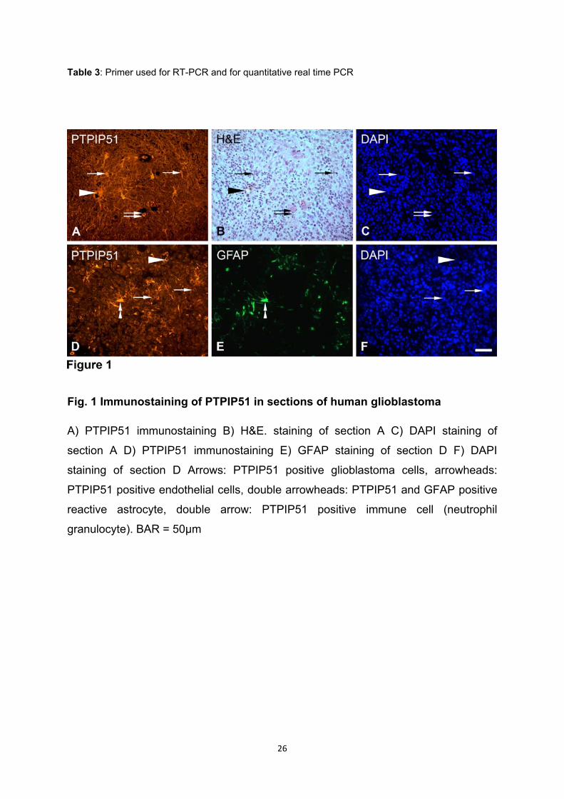
Table 3: Primer used for RT-PCR and for quantitative real time PCR
Fig. 1 Immunostaining of PTPIP51 in sections of human glioblastoma
A) PTPIP51 immunostaining B) H&E. staining of section A C) DAPI staining of
section A D) PTPIP51 immunostaining E) GFAP staining of section D F) DAPI
staining of section D Arrows: PTPIP51 positive glioblastoma cells, arrowheads:
PTPIP51 positive endothelial cells, double arrowheads: PTPIP51 and GFAP positive
reactive astrocyte, double arrow: PTPIP51 positive immune cell (neutrophil
granulocyte). BAR = 50µm
26

Fig. 2 PTPIP51 expression in immune cells, smooth muscle cells and endothelial cells
A) PTPIP51 immunostaining B) granulocyte marker of section A C) Merge of A, B
and DAPI D) PTPIP51 immunostaining E) B lymphocyte in section D detected by
CD20 F) Merge of D, E and DAPI. BAR (A-F)= 10μm G) PTPIP51 immunostaining H)
α-sma of section G I) Merge of G,H and DAPI J) PTPIP51 immunostaining K)
endothelial cells detected by CD34 in section J L) Merge of J, K and DAPI.
Arrowhead: neutrophil granulocytes; blank arrowhead: B lymphocytes; double arrow:
smooth muscle cells; arrow: endothelial cells; white circle: lipofuscin granula . BAR
(G-L) = 20µm
27
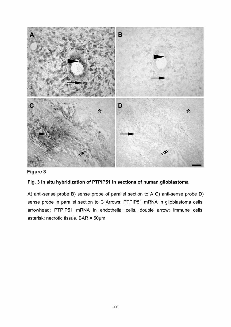
Fig. 3 In situ hybridization of PTPIP51 in sections of human glioblastoma
A) anti-sense probe B) sense probe of parallel section to A C) anti-sense probe D)
sense probe in parallel section to C Arrows: PTPIP51 mRNA in glioblastoma cells,
arrowhead: PTPIP51 mRNA in endothelial cells, double arrow: immune cells,
asterisk: necrotic tissue. BAR = 50µm
28

Fig. 4 PTPIP51 and its interacting partner 14-3-3β - in situ co-localization analysis and Duolink proximity ligation assay in sections of human glioblastoma and astrocytoma
A) PTPIP51 immunostaining GBM B) 14-3-3β immunostaining of section A C) Merge
of A and B D) PTPIP51 immunostaining astrocytoma E) 14-3-3β immunostaining of
section D F) Merge of D and E. Arrows: PTPIP51 positive glioblastoma cells,
arrowheads: PTPIP51 positive endothelial cells, double-arrows: immune cells. BAR =
50µm G) GBM duolink proximity ligation assay for PTPIP51 and 14-3-3β H)
astrocytoma duolink proximity ligation assay for PTPIP51 and 14-3-3β. Interaction of
both proteins is seen as orange dots (arrows) I) Negative control. White circles:
lipofuscin granules. BAR = 10µm
29

Fig. 5 PTPIP51 and its interacting partner PTP1B in sections of human glioblastom
A) PTPIP51 immunostaining B) PTP1B immunostaining of section A C) Merge of
section A and B D) Duolink proximity ligation assay for PTPIP51 and PTP1B.
Interaction of both proteins is seen as orange dots (arrows). BAR (A,B, C) = 20μm,
BAR (D) = 10μm
30

Fig. 6 PTPIP51 and its interacting partners Raf-1 and c-Src in sections of human glioblastoma
A) PTPIP51 immunostaining B) Raf-1 immunostaining of section A C) Merge of A
and B D) PTPIP51 immunostaining E) c-Src immunostaining of section D F) Merge of
D and E. Arrows: PTPIP51 positive glioblastoma cells, arrowheads: PTPIP51 positive
endothelial cells, double-arrows: immune cells. BAR = 50µm
31
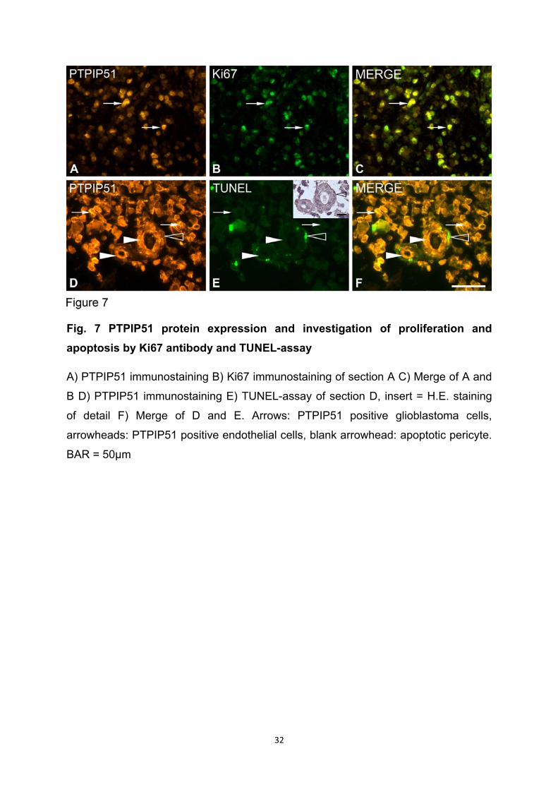
Fig. 7 PTPIP51 protein expression and investigation of proliferation and apoptosis by Ki67 antibody and TUNEL-assay
A) PTPIP51 immunostaining B) Ki67 immunostaining of section A C) Merge of A and
B D) PTPIP51 immunostaining E) TUNEL-assay of section D, insert = H.E. staining
of detail F) Merge of D and E. Arrows: PTPIP51 positive glioblastoma cells,
arrowheads: PTPIP51 positive endothelial cells, blank arrowhead: apoptotic pericyte.
BAR = 50µm
32

Fig. 8 Expression of PTPIP51 and its interaction partners as detected by reverse transcriptase-PCR in tissues of human glioblastoma
Qualitative reverse transcriptase-PCR was performed using primers specific to
PTPIP51, PTP1B, Raf-1, c-Src and 14-3-3β as given in Materials and Methods
section. β-actin was amplified as an internal positive control and probes lacking
reverse transcriptase served as negative controls (N).
33

Fig. 9 Quantitative real time PCR analysis of PTPIP51 and 14-3-3β in glioblastoma and grade II astrocytoma
A: Diagram shows the results of quantitative expression analysis. Slightly elevated
levels of PTPIP51 are seen in the group of glioblastoma (GBM) when compared to
low grade astrocytoma. 14-3-3β expression in glioblastoma was significantly higher
than in grade II astrocytoma samples.
Amplification of β-actin and GAPDH was used as a combination of reference genes.
B: Gel: bands exclusively detecting mRNA with the expected amplification size with
the primers used in 7A. Negative control (N).
34
Recommended