Embed Size (px)
Citation preview

Research Article
Analysis of Spontaneous Abortions Using Genomics, Proteomics and In silico Tools - A. Carvalho1,2, J. Ferreira1,2, R. Pinto Leite3, M. Souto3, P. Botelho3, O. Moutinho3, L. Pinto1,2, H. Santos4,5, J. Capelo4,5 and G. Igrejas1,2,4,6,#*1Department of Genetics and Biotechnology, University of Tras-os-Montes and Alto Douro, Vila Real, Portugal2Functional Genomics and Proteomics Unit, University of Tras -os-Montes and Alto Douro, Vila Real, Portugal3Cytogenetics Laboratory, Genetics Service, Hospitalar Centre of Tras -os-Montes and Alto Douro, Vila Real, Portugal4Bioscope Group, UCIBIO-REQUIMTE. Faculty of Sciences and Technology, New University of Lisbon, Campus de Caparica, Caparica, Portugal5ProteoMass Scientific Society, Faculty of Sciences and Technology, Campus de Caparica, Portugal6UCIBIO-REQUIMTE, Faculty of Science and Technology, University Nova of Lisbon, Caparica, Portugal#These authors contributed equally to this work
*Address for Correspondence: Gilberto Igrejas, Functional Genomics and Proteomics Unit, Department of Genetics and Biotechnology, University of Tras-os-Montes and Alto Douro, Vila Real, Portugal, Quinta dos Prados, 5000-801 Vila Real, Portugal, Tel: +351-259-350-530; Fax: +351-259-350-480; E-mail: [email protected]
Submitted: 09 August 2017; Approved: 02 October 2017; Published: 03 October 2017
Cite this article: Carvalho A, Ferreira J, Pinto Leite R, Souto M, Botelho P, et al. Analysis of Spontaneous Abortions Using Genomics, Proteomics and In silico Tools. Int J Proteom Bioinform. 2017;2(1): 012-026.
Copyright: © 2017 Carvalho A, et al. This is an open access article distributed under the Creative Commons Attribution License, which permits unrestricted use, distribution, and reproduction in any medium, provided the original work is properly cited.
International Journal ofProteomics & Bioinformatics
International Journal of Proteomics & Bioinformatics
SCIRES Literature - Volume 2 Issue 1 - www.scireslit.com Page - 013
INTRODUCTIONIt has been estimated that about 20% of all recognized clinical
pregnancies end in spontaneous abortion, mainly in the first trimester [1]. Risk factors associated with the occurrence of a sporadic miscarriage have been established and are related to different etiologies [2]. Possible causes reported include genetic or structural abnormalities, infection, and endocrine or immune dysfunction. Genetic factors are the most prevalent and may be due to numerical chromosomal abnormalities, aberrant gene expression, mutations or Single Nucleotide Polymorphisms (SNPs) [2,3]. Many miscarriages however remain inexplicable or the causes are the subject of debate. As a problem that affects numerous couples, it is crucial to add new techniques to conventional ones to increase the quality of prognosis and diagnosis for those affected. Genomic, proteomic and bioinformatic techniques can be used as powerful tools to provide an integrated molecular analysis of genotypic and phenotypic factors potentially related to sporadic miscarriage [4]. Some relevant studies of spontaneous miscarriage, summarized in table 1, have started to tackle the problem in this way.
Genomic methods are important in determining whether certain polymorphisms in genes are associated to pathologies that influence normal pregnancy development, leading to a spontaneous abortion [5]. Some genes involved in angiogenesis (VEGF, LEPR, PAI-I and TGFB1) and apoptosis (BID, Caspases 3, 8, 9 and 10) are crucial for correct embryonic development. Many SNPs that could influence the occurrence of a sudden miscarriage or lead to embryo/fetus anomalies during the pregnancy have been reported in the literature and in databases [6-13].
Proteomics is the study of all the proteins expressed in a given biological system including the abundance, activity, structure, properties or modification of those proteins, and the way they interact with each other. Proteomic analysis may be a way of identifying proteins with adverse effects on human embryonic development [14]. Many mathematical approaches and algorithms relevant to biological and medical concepts can be implemented in bioinformatics [15]. A vast number of bioinformatics tools can now be used for in silico analysis, enabling predictions about genes, transcripts and specific proteins implicated in the occurrence of sudden miscarriage and their impact [16,17].
Taking first a genomic approach, we aimed to detect whether any previously reported SNPs were present in the sequences of
genomic amplicons from spontaneous abortion samples. We focused on certain SNPs in genes that encode mediators which play roles in the reproductive process as they might have had an impact on the occurrence of the spontaneous abortion. In order to elucidate the possible causes related to the miscarriages studied, a second objective was to identify anomalies in the expression of proteins that might have had adverse effects on human embryonic development.
MATERIAL AND METHODSCell culture and sampling
Nine samples of spontaneous abortions that occurred at 8-21 weeks of gestation were analyzed. Samples were provided by the Genetic Service of the Hospital Centre of Tras-os-Montes and Alto Douro, following approval from the ethics committee. All samples had a normal karyotype, and no relevant risk factors that may have compromised the normal course of pregnancy, such as tobacco, alcohol or drug abuse, consanguinity or relevant medical history, had been associated to the parents (Table 2).
DNA extraction and PCR amplification
DNA was extracted from the abortion samples using the Citogene Cell and Tissue Kit (Citomed). PCR was used to amplify intronic, exonic and 3’ Untranslated Regions (3’UTR) of selected genes from those samples and amplicons were analysed by gel electrophoresis. DNA from a healthy woman with no history of reproductive difficulties was used as a positive control template. Selected amplicons were purified and sequenced.
Primers for PCR were designed with the Primer3Plus tool to encompass regions of interest in the selected genes [18]. Details of the primers and PCR conditions are described in tables 3 & 4, respectively.
Sequencing and sequence analysis
Sanger sequencing of PCR products was carried out using protocols recommended by the Biometra PCR Thermal Cycler manufacturer. A series of bioinformatics tools was used to perform In silico predictions. Primer3plus [18], Multiple primer analyzer (Life Technologies) and NCBI Primer BLAST [19] were used to evaluate the efficiency and complexity of chosen primers and to verify the number of possible amplification products. Geneious R6 (http://www.geneious.com [20]) was used to store, visualize and edit sequences, chromatograms, alignments and amino acid conversions. Human Splice Finder (HSF) [21] was used to predict possible variations in
ABSTRACTStudies show that about 20% of all recognized clinical pregnancies end in spontaneous abortion, mainly in the first trimester. Risk
factors associated with the occurrence of a sporadic miscarriage have been established, with genetic factors being the most prevalent. As a problem that affects many couples, it is important to increase the quality of prognosis and diagnosis.
In this study, the genomic sequences of spontaneous abortion samples with normal karyotypes were analyzed, and Single Nucleotide Polymorphisms (SNPs) were found to be the most common alterations in a selected set of candidate genes. Using the Human Splice Finder bioinformatics tool, it was estimated that 75% and 23% of these differences were in intronic and exonic regions, respectively. A total of 54% of the amino acid substitutions encoded by SNPs in exonic regions would lead to an alteration in the function of some protein domains and/or be deleterious to some protein structures, according to ProtFun 2.2 and PolyPhen-2 predictions.
In a proteomic analysis comparing the samples, it was possible to identify 23 altered proteins that were related to glycolysis, regulation of the cell cycle, transcription and angiogenic mechanisms, or stress responses. Heat shock proteins 7C and 90 were also identified. In conclusion, genomics, proteomics and bioinformatics techniques can be used to provide an integrated molecular analysis of genotypic and phenotypic factors potentially related to sudden miscarriage.
Keywords: Spontaneous Abortions; Genomics; Bioinformatics; Proteomics; Angiogenesis; Apoptosis
International Journal of Proteomics & Bioinformatics
SCIRES Literature - Volume 2 Issue 1 - www.scireslit.com Page - 014
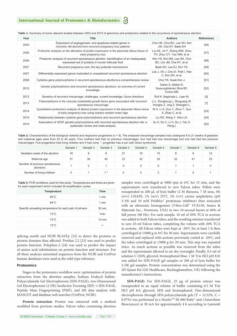
Table 2: Characteristics of the biological material and respective progenitors (n = 9). The analysed miscarriage samples had undergone 8 to 21 weeks of gestation and maternal ages were from 22 to 40 years. Four mothers had had no previous miscarriages, four had had one miscarriage and one had had two previous miscarriages. Five progenitors had living children and 4 had none. *, progenitor has a son with Down syndrome).
Sample 1 Sample 2 Sample 3 Sample 4 Sample 5 Sample 6 Sample 7 Sample 8 Sample 9
Gestation week of the abortion 18 8 8 21 10 15 8 8 12
Maternal age 31 36 40 22 32 33 32 41 25
Number of previous spontaneous abortions 1 2 1 0 0 0 1 1 0
Number of living children 0 1 1 * 0 0 2 1 0 1
splicing motifs and NCBI BLASTp [22] to detect the proteins or protein domains thus affected. Protfun 2.2 [23] was used to predict protein function. Polyphen-2 [24] was used to predict the impact of amino acid substitutions on protein function and structure. For all these analyses annotated sequences from the NCBI and UniProt human databases were used as the wild-type reference.
Proteomics
Stages in the proteomics workflow were: optimization of protein extraction from the abortion samples, Sodium Dodecyl Sulfate-Polyacrylamide Gel Electrophoresis (SDS-PAGE), two-Dimensional Gel Electrophoresis (2-DE) Isoelectric Focusing (IEF) × SDS-PAGE, Peptide Mass Fingerprinting (PMF), and MS data analysis with MASCOT and database web searches (UniProt, NCBI).
Protein extraction: Protein was extracted with a method modified from previous studies. Falcon tubes containing abortion
samples were centrifuged at 5000 rpm at 4ºC for 15 min, and the supernatants were transferred to new Falcon tubes. Pellets were resuspended in 200 µL of lysis buffer (2 M thiourea, 7 M urea, 4% (w/v) CHAPS, 1% (w/v) DTT, 2% (v/v) carrier ampholytes (pH 3-10) and 10 mM Pefabloc® proteinase inhibitor) then sonicated with an ultrasonic homogenizer (Vibra-Cell™ VCX130, Sonics & Materials Inc., Newtown, USA) in two 10-second bursts at 60% of full power (60 Hz). For each sample, 10 ml of 20% TCA in acetone was added to both Falcon tubes, and the resulting mixture transferred to new 25-ml Falcon tubes, completing the volume with 20% TCA in acetone. All Falcon tubes were kept at -20ºC for at least 1 h then centrifuged at 13000 g at 4ºC for 30 min. Supernatants were carefully removed and replaced with acetone previously cooled at -20ºC, and the tubes centrifuged at 13000 g for 20 min. This step was repeated twice. As much acetone as possible was removed from the tubes and the supernatants allowed to air-dry overnight. Finally, 0.5 ml of solution C (SDS, glycerol, bromophenol blue, 1 M Tris HCl pH 8.0) was added for SDS-PAGE gel samples or 200 µl of lysis buffer for 2-DE gel samples. Protein concentration was determined using the 2D Quant Kit (GE Healthcare, Buckinghamshire, UK) following the manufacturer’s instructions.
SDS-PAGE: For SDS-PAGE, 25 µg of protein extract was resuspended in an equal volume of buffer containing 0.5 M Tris HCl pH 8.0, glycerol, SDS and bromophenol. One-dimensional electrophoresis through SDS-polyacrylamide gels (T = 12.52%, C = 0.97%) was performed in a HoeferTM SE 600 Ruby® unit (Amersham Biosciences) at 30 mA for approximately 4 h according to Laemmli
Table 1: Summary of some relevant studies between 2003 and 2015 of genomics and proteomics related to the occurrence of spontaneous abortion.
Year Title Authors References
2003 Expression of angiogenesis- and apoptosis-related genes inchorionic villi derived from recurrent pregnancy loss patients
Choi HK, Choi BC, Lee SH, Kim JW, Cha KY, Baek KH [46]
2006 Proteomic analysis on the alteration of protein expression in the placental villous tissue of early pregnancy loss
Liu AX, Jin F, Zhang WW, Zhou TH, Zhou CY, Yao WM, et al. [47]
2006 Proteomic analysis of recurrent spontaneous abortion: Identification of an inadequately expressed set of proteins in human follicular fluid
Kim YS, Kim MS, Lee SH, Choi BC, Lim JM, Cha KY, et al. [48]
2007 Recurrent pregnancy loss: the key potential mechanisms Baek KH, Lee EJ, Kim YS. [49]
2007 Differentially expressed genes implicated in unexplained recurrent spontaneous abortion Lee J, Oh J, Choi E, Park I, Han C, Kim DH, et al. [50]
2008 Cytokine gene polymorphisms in recurrent spontaneous abortions:a comprehensive review Choi YK, Kwak Kim J. [51]
2012 Genetic polymorphisms and recurrent spontaneous abortions: an overview of current knowledge
Daher S, Mattar R, Gueuvoghlanian Silva BY,
Torloni MR.[52]
2012 Genetics of recurrent miscarriage: challenges, current knowledge, future directions Rull K, Nagirnaja L, Laan M. [4]
2013 Polymorphisms in the vascular endothelial growth factor gene associated with recurrent spontaneous miscarriage
Li L, Donghong L, Shuguang W, Hongbo Z, Jing Z, Shengbin L. [7]
2014 Quantitative proteomics analysis of altered protein expression in the placental villous tissue of early pregnancy loss using isobaric tandem mass tags
Ni X, Li X, Guo Y, Zhou T, Guo X, Zhao C, et al. [53]
2015 Relationship between cytokine gene polymorphisms and recurrent spontaneous abortion Liu RX, Wang Y, Wen LH. [54]
2015 Association of VEGF genetic polymorphisms with recurrent spontaneous abortion risk: a systematic review and meta-analysis
Xu X, Du C, Li H, Du J, Yan X, Peng L [55]
Table 3: PCR conditions used for this study. Temperatures and times are given for each experiment which included 30 amplification cycles.
Temperature Time
95°C 1 min
94°C 1 min
Specific annealing temperature for each pair of primers 30 sec
72°C 1min
72°C 10 min
12°C ∞
International Journal of Proteomics & Bioinformatics
SCIRES Literature - Volume 2 Issue 1 - www.scireslit.com Page - 015
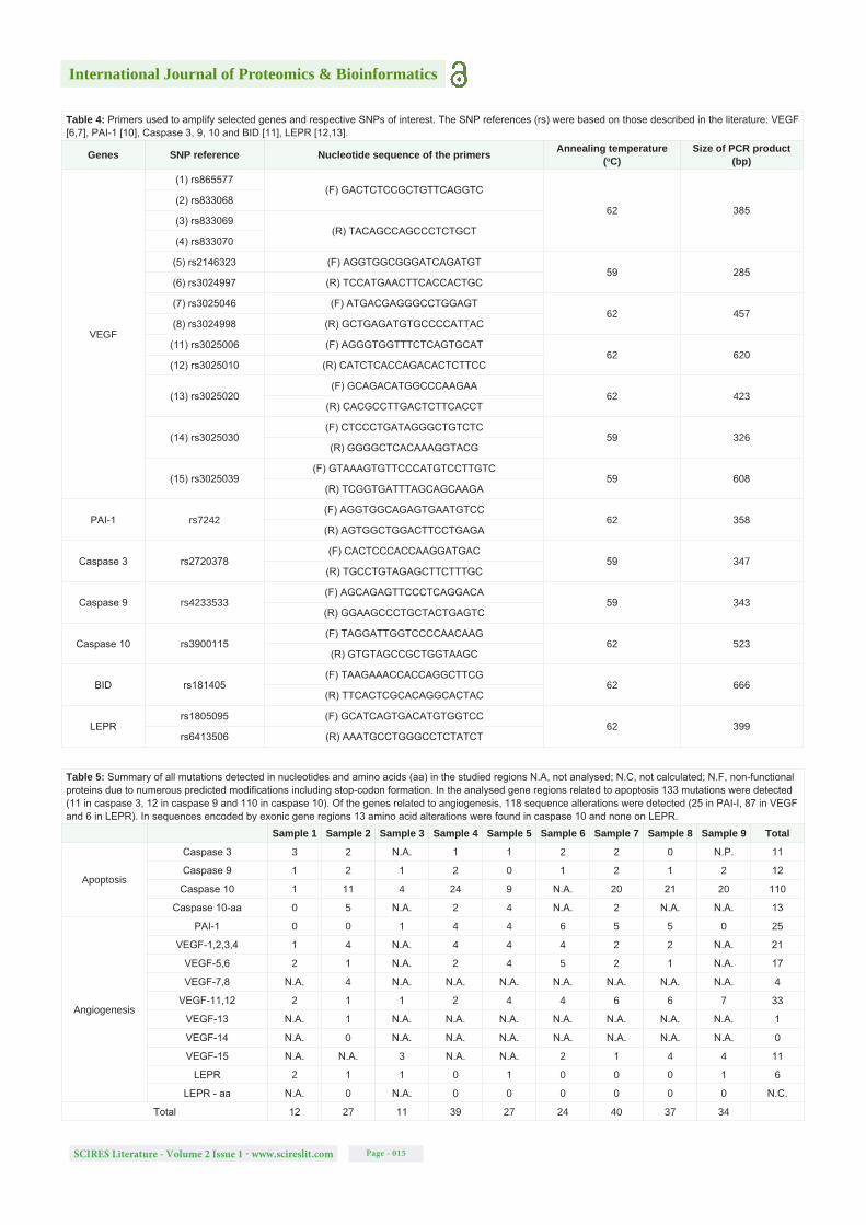
Table 4: Primers used to amplify selected genes and respective SNPs of interest. The SNP references (rs) were based on those described in the literature: VEGF [6,7], PAI-1 [10], Caspase 3, 9, 10 and BID [11], LEPR [12,13].
Genes SNP reference Nucleotide sequence of the primers Annealing temperature (oC)
Size of PCR product (bp)
VEGF
(1) rs865577(F) GACTCTCCGCTGTTCAGGTC
62 385(2) rs833068
(3) rs833069(R) TACAGCCAGCCCTCTGCT
(4) rs833070
(5) rs2146323 (F) AGGTGGCGGGATCAGATGT59 285
(6) rs3024997 (R) TCCATGAACTTCACCACTGC
(7) rs3025046 (F) ATGACGAGGGCCTGGAGT62 457
(8) rs3024998 (R) GCTGAGATGTGCCCCATTAC
(11) rs3025006 (F) AGGGTGGTTTCTCAGTGCAT62 620
(12) rs3025010 (R) CATCTCACCAGACACTCTTCC
(13) rs3025020(F) GCAGACATGGCCCAAGAA
62 423(R) CACGCCTTGACTCTTCACCT
(14) rs3025030(F) CTCCCTGATAGGGCTGTCTC
59 326(R) GGGGCTCACAAAGGTACG
(15) rs3025039(F) GTAAAGTGTTCCCATGTCCTTGTC
59 608(R) TCGGTGATTTAGCAGCAAGA
PAI-1 rs7242(F) AGGTGGCAGAGTGAATGTCC
62 358(R) AGTGGCTGGACTTCCTGAGA
Caspase 3 rs2720378(F) CACTCCCACCAAGGATGAC
59 347(R) TGCCTGTAGAGCTTCTTTGC
Caspase 9 rs4233533(F) AGCAGAGTTCCCTCAGGACA
59 343(R) GGAAGCCCTGCTACTGAGTC
Caspase 10 rs3900115(F) TAGGATTGGTCCCCAACAAG
62 523(R) GTGTAGCCGCTGGTAAGC
BID rs181405(F) TAAGAAACCACCAGGCTTCG
62 666(R) TTCACTCGCACAGGCACTAC
LEPRrs1805095 (F) GCATCAGTGACATGTGGTCC
62 399rs6413506 (R) AAATGCCTGGGCCTCTATCT
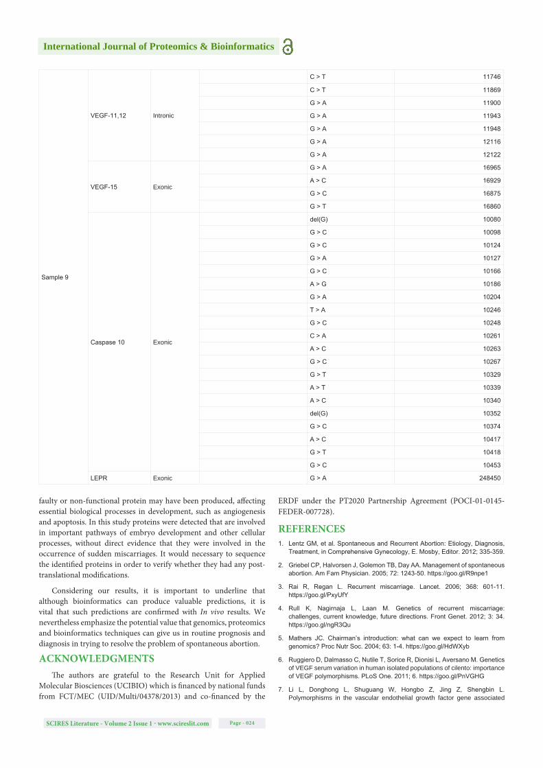
Table 5: Summary of all mutations detected in nucleotides and amino acids (aa) in the studied regions N.A, not analysed; N.C, not calculated; N.F, non-functional proteins due to numerous predicted modifications including stop-codon formation. In the analysed gene regions related to apoptosis 133 mutations were detected (11 in caspase 3, 12 in caspase 9 and 110 in caspase 10). Of the genes related to angiogenesis, 118 sequence alterations were detected (25 in PAI-I, 87 in VEGF and 6 in LEPR). In sequences encoded by exonic gene regions 13 amino acid alterations were found in caspase 10 and none on LEPR.
Sample 1 Sample 2 Sample 3 Sample 4 Sample 5 Sample 6 Sample 7 Sample 8 Sample 9 Total
Apoptosis
Caspase 3 3 2 N.A. 1 1 2 2 0 N.P. 11
Caspase 9 1 2 1 2 0 1 2 1 2 12
Caspase 10 1 11 4 24 9 N.A. 20 21 20 110
Caspase 10-aa 0 5 N.A. 2 4 N.A. 2 N.A. N.A. 13
Angiogenesis
PAI-1 0 0 1 4 4 6 5 5 0 25
VEGF-1,2,3,4 1 4 N.A. 4 4 4 2 2 N.A. 21
VEGF-5,6 2 1 N.A. 2 4 5 2 1 N.A. 17
VEGF-7,8 N.A. 4 N.A. N.A. N.A. N.A. N.A. N.A. N.A. 4
VEGF-11,12 2 1 1 2 4 4 6 6 7 33
VEGF-13 N.A. 1 N.A. N.A. N.A. N.A. N.A. N.A. N.A. 1
VEGF-14 N.A. 0 N.A. N.A. N.A. N.A. N.A. N.A. N.A. 0
VEGF-15 N.A. N.A. 3 N.A. N.A. 2 1 4 4 11
LEPR 2 1 1 0 1 0 0 0 1 6
LEPR - aa N.A. 0 N.A. 0 0 0 0 0 0 N.C.
Total 12 27 11 39 27 24 40 37 34
International Journal of Proteomics & Bioinformatics
SCIRES Literature - Volume 2 Issue 1 - www.scireslit.com Page - 016
[25] with some modifications [26]. Gels were stained for 24 h Coomassie Brilliant Blue R250 then washed overnight in distilled water. Gels were fixed in 6% TCA for 4 h then in 5% glycerol for 2 h.
Two-dimensional gel electrophoresis (IEF× SDS-PAGE): Two-dimensional gel electrophoresis was done according to the principles of O Farrell [27] using the ImmobilineTM pH Gradient (IPG) technology [28]. For Isoelectric Focusing (IEF), precast 13-cm IPG strips with a non-linear gradient from pH 3 to pH 10 (pH 3-10 NL, Amersham Biosciences) were rehydrated for approximately 16 h in a reswelling tray with 250 µL of rehydration buffer (8 M urea, 1% CHAPS, 0.4% DTT, 0.5% carrier ampholyte IPG buffer pH 3-10) and covered in Dry Strip Cover Fluid (Plus One, Amersham Biosciences). Using the cup-loading procedure, 150 µg of protein sample were loaded onto the IPG strip [28] and nine IEF runs were conducted for a total of 18 h 10 min (linear gradient of 500 V for 2 h, linear gradient of 1000 V for 3 h, linear gradient of 3000 V for 3 h, linear gradient of 7000 V for 4 h and a final step of 7000 V for 6 h 10 min). Focused IPG strips were placed in a primary equilibration buffer (6 M urea, 30% (w/v) glycerol, 2% (w/v) SDS in 0.05 M Tris–HCl buffer pH 8.8, bromophenol blue) supplemented with 1% DTT for 15 min, then with 4% iodoacetamide for an additional 15 min. Equilibrated IPG strips were washed in SDS-electrophoresis buffer and submitted to SDS-PAGE in conditions similar to those described above [25,26]. After SDS-PAGE, 2D gels were fixed with a 40% methanol/10% acetic acid solution for 1 h and stained overnight with Coomassie Brilliant Blue G-250. A 25% methanol solution was used to wash the 2D gels which were then preserved in distilled water.
Protein identification: Coomassie-stained protein spots were excised manually from all reference gels and later analyzed using Matrix-Assisted Laser Desorption/Ionization-Time of Flight Mass Spectrometry (MALDI-TOF). Spots were washed twice in 200 µl of 25 mM Ammonium Bicarbonate (Ambic), 50% Acetonitrile (ACN) for 15 min at 37ºC, then in 50 µl ACN and dried for 5 min in a SpeedVac concentrator (Thermo Scientific Savant). Each dried spot was digested with 15 µl of trypsin solution (0.02 µg/µL trypsin, 12.5 mM Ambic, 2% (v/v) can) for 30 min on ice, then incubated with 30 µl of 12.5 mM Ambic at 37ºC for up to 18 h (overnight). Samples were chilled before adding 20 µl of 5% formic acid and incubating them at 37ºC for 15 min. Then 25 µl of 50% ACN, 0.1% Tri Fluoroacetic Acid (TFA) was added and samples were dried in the SpeedVac for up to 10 h. Just before MS analysis, peptides were dissolved in 10 µl of 0.3% formic acid and incubated at 37ºC for 15 min. On a 384-spot ground-steel MALDI target plate 0.5 µl of protein solution was placed and overlaid with 1 µl of matrix solution (5 mg/mL α-cyano-4-hydroxycinnamic acid in 0.1% (v/v) TFA, 50% (v/v) ACN, 8 mM ammonium phosphate). Mass spectra were generated with a MALDI-TOF/TOF Ultraflex mass spectrometer (Bruker Daltonics) operating in positive ion reflectron-mode, and acquired in the m/z range of 600-3500, at laser frequency of 50 Hz (trypsin peak at m/z ~842). External calibration was performed with [M + H]+ monoisotopic peaks of bradykinin 1–7 (m/z 757.3992), angiotensin II (m/z 1046.5418), angiotensin I (m/z 1296.6848), substance P (m/z 1758.9326), ACTH clip 1–17 (m/z 2093.0862), ACTH18–39 (m/z 2465.1983) and somatostatin 28 (m/z 3147.4710). The MASCOT search engine was used to match the peptide masses obtained to customized databases (UniProt and NCBI) according to the following search criteria: proteolytic enzyme, trypsin/P; one missed cleavage allowed; carbamidomethylation as a fixed modification; methionine oxidation as a variable modification; and peptide tolerance error window up to 50 ppm. Finally, a peptide
match was considered significant when the probability of it being a random event was below the default threshold used (p ˂ 0.05), i.e., with a frequency less than 5%.
RESULTS AND DISCUSSION Genomics and In silico analysis
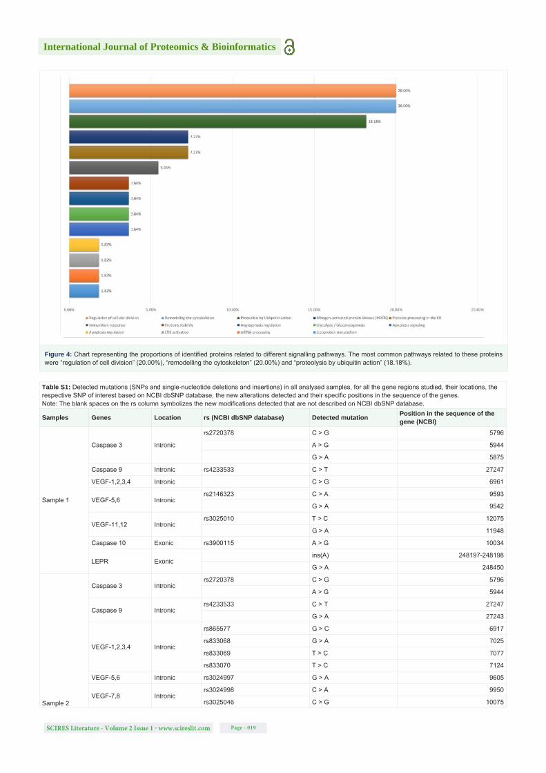
New SNPs that had not been previously associated to any medical condition were identified. To understand the possible impact of these mutations on splicing, predictions were made in both intronic and exonic regions of genes of interest using HSF [21]. Altogether 15 regions were selected for the VEGF gene, 2 regions each for the LEPR and TGFB1 genes, and 1 region each for the PAI-I, BID, and caspase 3, 8, 9 and 10 genes. Of 72 amplicons analyzed 42 were from intronic regions, 17 from exonic regions and 13 from 3’UTR. The results obtained from the TGFB1, BID and caspase 8 sequences were not exploitable. All mutations detected are summarized in table 5 and described in more detail in table S1.
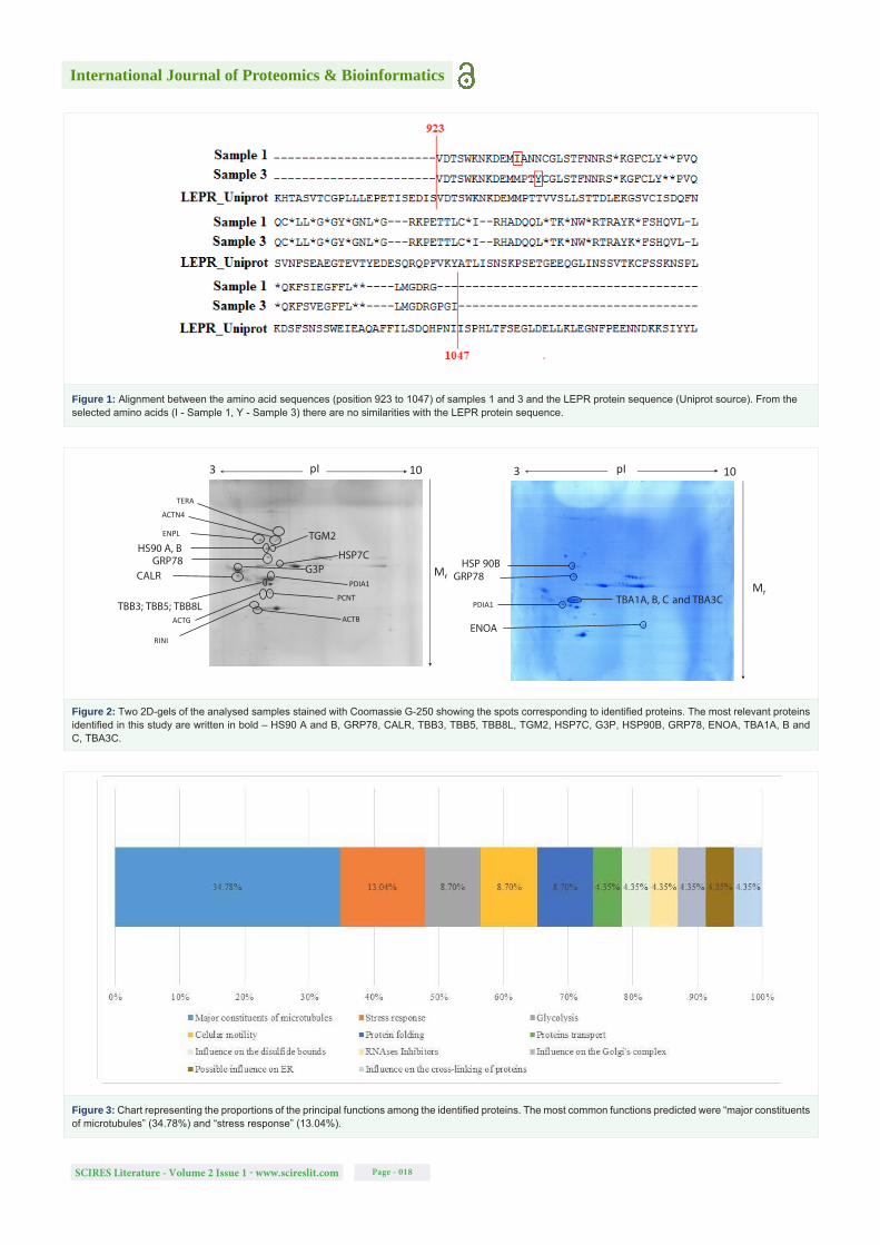
Using the HSF tool it was estimated that 75% and 23% of the modifications detected in intronic and exonic regions (both in deep regions), respectively, could possibly lead to alterations in splicing. The formation of new cryptic splice sites was predicted, as well as the disruption or creation of exonic splicing enhancers or silencers, which may silence the wild-type splice sites or enhance cryptic sites and lead to intron retention, exon skipping or formation of premature stop codons [29]. According to ProtFun 2.2 [23] and PolyPhen-2 predictions [24], 54% of the amino acid substitutions encoded by the SNPs could lead to an alteration in the function of a death effector domain of caspase-10 and/or cause damage to that protein’s structure. In an alignment of amino acid sequences between positions 65 to 116, the residues the most affected are at positions 81 (E > K), 103 (E > D) and 116 (R > K). In some cases, for example in the study of the LEPR protein region, it was not possible to use PolyPhen-2 [24] due to the numerous amino acid substitutions and stop codons formed (Figure 1).
Regarding 3’UTR, there were no bioinformatics tools readily available to predict the impact of nucleotide modifications on 3’UTR and little theoretical basis from which to estimate the impact of the detected mutations, but we can suggest that the detected alterations could lead to the loss of some functions of the gene regions affected. This is because 3’UTR have a crucial role in regulating mRNA expression, functioning as binding sites to regulatory proteins and miRNAs, and are known to influence some aspects of embryo development and spermatogenesis in mammals [30,31].
Proteomics analysis
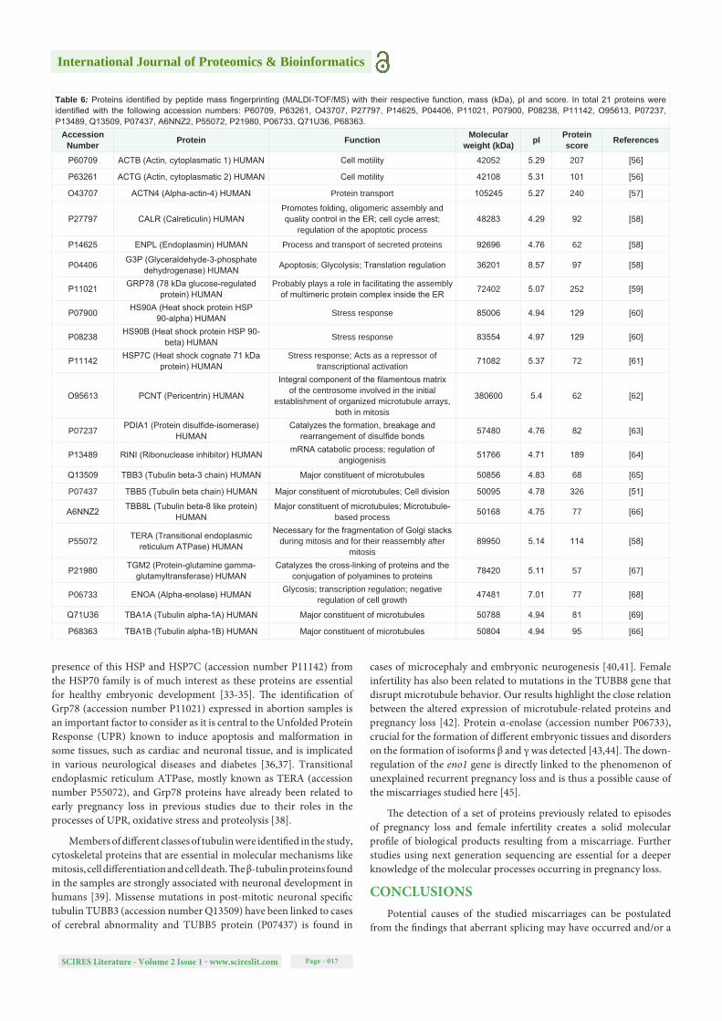
The 2D proteomic profile of the spontaneous abortion samples from SDS-PAGE × IEF and peptide mass fingerprinting (MALDI-TOF/MS) led to the identification of 26 proteins (Figure 2). The identified proteins are described in Table 6 according to their function (Figure 3) and the signaling pathways in which they are involved depending on which analysis was performed (Figure 4).
The presence of HSP90α and β, HSP7C, TERA, Grp78, β-tubulin and α-enolase among the identified proteins indicates functions mainly related to microtubules, stress responses and DNA repair were deregulated in the tissue samples. Although the proteins were not sequenced, it is relevant to consider the pathways in which they may act. Heat Shock Protein 90 (HSP90) is closely related to Akt kinase, which has a fundamental role in the cardiovascular system [32]. The
International Journal of Proteomics & Bioinformatics
SCIRES Literature - Volume 2 Issue 1 - www.scireslit.com Page - 017
presence of this HSP and HSP7C (accession number P11142) from the HSP70 family is of much interest as these proteins are essential for healthy embryonic development [33-35]. The identification of Grp78 (accession number P11021) expressed in abortion samples is an important factor to consider as it is central to the Unfolded Protein Response (UPR) known to induce apoptosis and malformation in some tissues, such as cardiac and neuronal tissue, and is implicated in various neurological diseases and diabetes [36,37]. Transitional endoplasmic reticulum ATPase, mostly known as TERA (accession number P55072), and Grp78 proteins have already been related to early pregnancy loss in previous studies due to their roles in the processes of UPR, oxidative stress and proteolysis [38].
Members of different classes of tubulin were identified in the study, cytoskeletal proteins that are essential in molecular mechanisms like mitosis, cell differentiation and cell death. The β-tubulin proteins found in the samples are strongly associated with neuronal development in humans [39]. Missense mutations in post-mitotic neuronal specific tubulin TUBB3 (accession number Q13509) have been linked to cases of cerebral abnormality and TUBB5 protein (P07437) is found in
cases of microcephaly and embryonic neurogenesis [40,41]. Female infertility has also been related to mutations in the TUBB8 gene that disrupt microtubule behavior. Our results highlight the close relation between the altered expression of microtubule-related proteins and pregnancy loss [42]. Protein α-enolase (accession number P06733), crucial for the formation of different embryonic tissues and disorders on the formation of isoforms β and γ was detected [43,44]. The down-regulation of the eno1 gene is directly linked to the phenomenon of unexplained recurrent pregnancy loss and is thus a possible cause of the miscarriages studied here [45].
The detection of a set of proteins previously related to episodes of pregnancy loss and female infertility creates a solid molecular profile of biological products resulting from a miscarriage. Further studies using next generation sequencing are essential for a deeper knowledge of the molecular processes occurring in pregnancy loss.
CONCLUSIONS Potential causes of the studied miscarriages can be postulated
from the findings that aberrant splicing may have occurred and/or a
Table 6: Proteins identified by peptide mass fingerprinting (MALDI-TOF/MS) with their respective function, mass (kDa), pI and score. In total 21 proteins were identified with the following accession numbers: P60709, P63261, O43707, P27797, P14625, P04406, P11021, P07900, P08238, P11142, O95613, P07237, P13489, Q13509, P07437, A6NNZ2, P55072, P21980, P06733, Q71U36, P68363.
Accession Number Protein Function Molecular
weight (kDa) pI Protein score References
P60709 ACTB (Actin, cytoplasmatic 1) HUMAN Cell motility 42052 5.29 207 [56]
P63261 ACTG (Actin, cytoplasmatic 2) HUMAN Cell motility 42108 5.31 101 [56]
O43707 ACTN4 (Alpha-actin-4) HUMAN Protein transport 105245 5.27 240 [57]
P27797 CALR (Calreticulin) HUMANPromotes folding, oligomeric assembly and quality control in the ER; cell cycle arrest;
regulation of the apoptotic process48283 4.29 92 [58]
P14625 ENPL (Endoplasmin) HUMAN Process and transport of secreted proteins 92696 4.76 62 [58]
P04406 G3P (Glyceraldehyde-3-phosphate dehydrogenase) HUMAN Apoptosis; Glycolysis; Translation regulation 36201 8.57 97 [58]
P11021 GRP78 (78 kDa glucose-regulated protein) HUMAN
Probably plays a role in facilitating the assembly of multimeric protein complex inside the ER 72402 5.07 252 [59]
P07900 HS90A (Heat shock protein HSP 90-alpha) HUMAN Stress response 85006 4.94 129 [60]
P08238 HS90B (Heat shock protein HSP 90-beta) HUMAN Stress response 83554 4.97 129 [60]
P11142 HSP7C (Heat shock cognate 71 kDa protein) HUMAN
Stress response; Acts as a repressor of transcriptional activation 71082 5.37 72 [61]
O95613 PCNT (Pericentrin) HUMAN
Integral component of the filamentous matrix of the centrosome involved in the initial
establishment of organized microtubule arrays, both in mitosis
380600 5.4 62 [62]
P07237 PDIA1 (Protein disulfide-isomerase) HUMAN
Catalyzes the formation, breakage and rearrangement of disulfide bonds 57480 4.76 82 [63]
P13489 RINI (Ribonuclease inhibitor) HUMAN mRNA catabolic process; regulation of angiogenisis 51766 4.71 189 [64]
Q13509 TBB3 (Tubulin beta-3 chain) HUMAN Major constituent of microtubules 50856 4.83 68 [65]
P07437 TBB5 (Tubulin beta chain) HUMAN Major constituent of microtubules; Cell division 50095 4.78 326 [51]
A6NNZ2 TBB8L (Tubulin beta-8 like protein) HUMAN
Major constituent of microtubules; Microtubule-based process 50168 4.75 77 [66]
P55072 TERA (Transitional endoplasmic reticulum ATPase) HUMAN
Necessary for the fragmentation of Golgi stacks during mitosis and for their reassembly after
mitosis89950 5.14 114 [58]
P21980 TGM2 (Protein-glutamine gamma-glutamyltransferase) HUMAN
Catalyzes the cross-linking of proteins and the conjugation of polyamines to proteins 78420 5.11 57 [67]
P06733 ENOA (Alpha-enolase) HUMAN Glycosis; transcription regulation; negative regulation of cell growth 47481 7.01 77 [68]
Q71U36 TBA1A (Tubulin alpha-1A) HUMAN Major constituent of microtubules 50788 4.94 81 [69]
P68363 TBA1B (Tubulin alpha-1B) HUMAN Major constituent of microtubules 50804 4.94 95 [66]
International Journal of Proteomics & Bioinformatics
SCIRES Literature - Volume 2 Issue 1 - www.scireslit.com Page - 018
Figure 1: Alignment between the amino acid sequences (position 923 to 1047) of samples 1 and 3 and the LEPR protein sequence (Uniprot source). From the selected amino acids (I - Sample 1, Y - Sample 3) there are no similarities with the LEPR protein sequence.
3 pI 10
Mr
ACTB
CALR
HS90 A, B
PDIA1
ENPL
TERA
GRP78
TBB3; TBB5; TBB8L
ACTN4
TGM2
ACTG
RINI
G3P
PCNT
HSP7C
3 pI 10
Mr
PDIA1
GRP78HSP 90B
ENOA
TBA1A, B, C and TBA3C
Figure 2: Two 2D-gels of the analysed samples stained with Coomassie G-250 showing the spots corresponding to identified proteins. The most relevant proteins identified in this study are written in bold – HS90 A and B, GRP78, CALR, TBB3, TBB5, TBB8L, TGM2, HSP7C, G3P, HSP90B, GRP78, ENOA, TBA1A, B and C, TBA3C.
Figure 3: Chart representing the proportions of the principal functions among the identified proteins. The most common functions predicted were “major constituents of microtubules” (34.78%) and “stress response” (13.04%).
International Journal of Proteomics & Bioinformatics
SCIRES Literature - Volume 2 Issue 1 - www.scireslit.com Page - 019
Figure 4: Chart representing the proportions of identified proteins related to different signalling pathways. The most common pathways related to these proteins were “regulation of cell division” (20.00%), “remodelling the cytoskeleton” (20.00%) and “proteolysis by ubiquitin action” (18.18%).
Table S1: Detected mutations (SNPs and single-nucleotide deletions and insertions) in all analysed samples, for all the gene regions studied, their locations, the respective SNP of interest based on NCBI dbSNP database, the new alterations detected and their specific positions in the sequence of the genes. Note: The blank spaces on the rs column symbolizes the new modifications detected that are not described on NCBI dbSNP database.
Samples Genes Location rs (NCBI dbSNP database) Detected mutation Position in the sequence of the gene (NCBI)
Sample 1
Caspase 3 Intronic
rs2720378 C > G 5796
A > G 5944
G > A 5875
Caspase 9 Intronic rs4233533 C > T 27247
VEGF-1,2,3,4 Intronic C > G 6961
VEGF-5,6 Intronicrs2146323 C > A 9593
G > A 9542
VEGF-11,12 Intronicrs3025010 T > C 12075
G > A 11948
Caspase 10 Exonic rs3900115 A > G 10034
LEPR Exonic ins(A) 248197-248198
G > A 248450
Sample 2
Caspase 3 Intronicrs2720378 C > G 5796
A > G 5944
Caspase 9 Intronicrs4233533 C > T 27247
G > A 27243
VEGF-1,2,3,4 Intronic
rs865577 G > C 6917
rs833068 G > A 7025
rs833069 T > C 7077
rs833070 T > C 7124
VEGF-5,6 Intronic rs3024997 G > A 9605
VEGF-7,8 Intronicrs3024998 C > A 9950
rs3025046 C > G 10075

International Journal of Proteomics & Bioinformatics
SCIRES Literature - Volume 2 Issue 1 - www.scireslit.com Page - 020
G > C 10015
A > C 10014
VEGF-11,12 Intronic rs3025006 C > T 11746
VEGF-13 Intronic G > A 13622
Caspase 10 Exonic
rs3900115 A > G 10034
A > G 10034
G > C 10098
G > C 10124
G > C 10166
A > G 10186
G > A 10204
C > A 10261
G > C 10374
A > C 10417
G > T 10418
G > C 10453
LEPR Exonic G > A 248450
Sample 3
Caspase 9 Intronic A > C 27208
VEGF-11,12 Intronic G > A 11948
VEGF-15 3'-UTR
G > C 16998
G > C 16993
G > A 16965
PAI-1 3'-UTR A > G 12958
Caspase 10 Exonic
ins(G) 10050-10051
A > G 10247
A > G 10276
G > A 10352
LEPR Exonic ins (T) 248204-248205
Sample 4
Caspase 3 Intronic rs2720378 C > G 5796
Caspase 9 Intronicrs4233533 C > T 27247
G > A 27243
VEGF-1,2,3,4 Intronic
rs865577 G > C 6917
rs833068 G > A 7025
rs833069 T > C 7077
rs833070 T > C 7124
VEGF-5,6 Intronicrs3024997 G > A 9605
del(G) 9708
VEGF-11,12 Intronicrs3025006 C > T 11746
G > A 11900
PAI-1 3'-UTR
T > G 12961
T > A 12968
T > G 12974
C > G 12982
Caspase 10 Exonic
A > T 10097
G > C 10098
G > A 10121
A > T 10123
G > C 10124
A > G 10153

International Journal of Proteomics & Bioinformatics
SCIRES Literature - Volume 2 Issue 1 - www.scireslit.com Page - 021
del(A) 10154
G > T 10158
G > C 10166
G > A 10186
del(A) 10187
G > A 10204
T > A 10246
G > C 10248
C > A 10261
A > C 10263
G > C 10267
G > T 10329
A > T 10339
A > C 10340
G > C 10374
A > C 10417
G > T 10418
G > C 10453
Sample 5
Caspase 3 Intronic G > A 5875
VEGF-1,2,3,4 Intronic
rs865577 G > C 6917
rs833068 G > A 7025
rs833069 T > C 7077
rs833070 T > C 7124
VEGF-5,6 Intronic
rs3024997 G > A 9605
ins(A) 9521-9522
G > A 9542
G > A 9711
VEGF-11,12 Intronic
rs3025006 C > T 11746
G > A 11900
G > A 11943
G > A 12122
PAI-1 3'-UTR
rs7242 T > G 12904
T > A 12961
T > G 12968
T > G 12974
Caspase 10 Exonic
G > C 10098
G > C 10124
A > G 10186
G > A 10204
C > A 10261
G > C 10374
A > C 10417
G > T 10418
G > C 10453
LEPR Exonic G > A 248450
Caspase 3 Intronicdel(G) 5980
G > A 5875
Caspase 9 Intronic G > A 27243

International Journal of Proteomics & Bioinformatics
SCIRES Literature - Volume 2 Issue 1 - www.scireslit.com Page - 022
Sample 6
VEGF-1,2,3,4 Intronic
rs865577 G > C 6917
rs833068 G > A 7025
rs833069 T > C 7077
rs833070 T > C 7124
VEGF-5,6 Intronic
rs3024997 G > A 9605
ins(A) 9521-9522
G > A 9542
VEGF-11,12 Intronic
rs3025006 C > T 11746
G > A 11900
G > A 11943
G > A 12122
PAI-1 3'-UTR
T > G 12851
del(G) 12859
G > A 12897
A > C 12920
del(C) 12929
Sample 7
Caspase 3 Intronicrs2720378 C > G 5796
G > A 5875
Caspase 9 Intronicrs4233533 C > T 27247
A > G 27208
VEGF-1,2,3,4 Intronicrs865577 G > C 6917
rs833069 T > C 7077
VEGF-5,6 Intronicrs3024997 G > A 9605
C > A 9521-9522
VEGF-11,12 Intronic
rs3025006 C > T 11746
G > A 11900
G > A 11943
G > A 11948
G > A 12116
G > A 12122
VEGF-15 3'-UTR G > C 16860
PAI-1 3'-UTR
rs7242 T > G 12904
T > G 12961
T > A 12968
T > G 12974
C > G 12982
Caspase 10 Exonic
G > C 10098
G > A 10121
G > C 10124
G > A 10127
del(G) 10155
G > C 10166
A > G 10186
G > A 10204
G > A 10209
G > C 10248
C > A 10261
A > C 10263

International Journal of Proteomics & Bioinformatics
SCIRES Literature - Volume 2 Issue 1 - www.scireslit.com Page - 023
G > C 10267
G > T 10329
A > T 10339
A > C 10340
G > C 10374
A > C 10417
G > T 10418
G > C 10453
Sample 8
Caspase 9 Intronic G > A 27243
VEGF-1,2,3,4 Intronicrs865577 G > C 6917
rs833069 T > C 7077
VEGF-5,6 Intronic rs3024997 G > A 9605
VEGF-11,12 Intronic
rs3025006 C > T 11746
rs3025010 T > C 12075
C > T 11869
G > A 11948
G > A 12116
G > A 12122
VEGF-15 3'-UTR ins(A) 16534-16535
PAI-1 3'-UTR
T > G 12961
T > A 12968
T > G 12974
C > G 12982
C > A 12985
Caspase 10 Exonic
del(G) 10080
G > C 10098
G > A 10121
G > C 10124
G > A 10127
G > C 10166
A > G 10186
G > A 10204
G > A 10209
G > C 10248
C > A 10261
A > C 10263
G > C 10267
G > T 10329
AZT 10339
A > C 10340
G > A 10352
G > C 10374
A > C 10417
G > T 10418
G > C 10453
Caspase 9 Intronicrs4233533 C > T 27247
A > G 27208
International Journal of Proteomics & Bioinformatics
SCIRES Literature - Volume 2 Issue 1 - www.scireslit.com Page - 024
faulty or non-functional protein may have been produced, affecting essential biological processes in development, such as angiogenesis and apoptosis. In this study proteins were detected that are involved in important pathways of embryo development and other cellular processes, without direct evidence that they were involved in the occurrence of sudden miscarriages. It would necessary to sequence the identified proteins in order to verify whether they had any post-translational modifications.
Considering our results, it is important to underline that although bioinformatics can produce valuable predictions, it is vital that such predictions are confirmed with In vivo results. We nevertheless emphasize the potential value that genomics, proteomics and bioinformatics techniques can give us in routine prognosis and diagnosis in trying to resolve the problem of spontaneous abortion.
ACKNOWLEDGMENTSThe authors are grateful to the Research Unit for Applied
Molecular Biosciences (UCIBIO) which is financed by national funds from FCT/MEC (UID/Multi/04378/2013) and co-financed by the
ERDF under the PT2020 Partnership Agreement (POCI-01-0145-FEDER-007728).
REFERENCES1. Lentz GM, et al. Spontaneous and Recurrent Abortion: Etiology, Diagnosis,
Treatment, in Comprehensive Gynecology, E. Mosby, Editor. 2012; 335-359.
2. Griebel CP, Halvorsen J, Golemon TB, Day AA. Management of spontaneous abortion. Am Fam Physician. 2005; 72: 1243-50. https://goo.gl/R9npe1
3. Rai R, Regan L. Recurrent miscarriage. Lancet. 2006; 368: 601-11. https://goo.gl/PxyUfY
4. Rull K, Nagirnaja L, Laan M. Genetics of recurrent miscarriage: challenges, current knowledge, future directions. Front Genet. 2012; 3: 34. https://goo.gl/ngR3Qu
5. Mathers JC. Chairman’s introduction: what can we expect to learn from genomics? Proc Nutr Soc. 2004; 63: 1-4. https://goo.gl/HdWXyb
6. Ruggiero D, Dalmasso C, Nutile T, Sorice R, Dionisi L, Aversano M. Genetics of VEGF serum variation in human isolated populations of cilento: importance of VEGF polymorphisms. PLoS One. 2011; 6. https://goo.gl/PnVGHG
7. Li L, Donghong L, Shuguang W, Hongbo Z, Jing Z, Shengbin L. Polymorphisms in the vascular endothelial growth factor gene associated
Sample 9
VEGF-11,12 Intronic
C > T 11746
C > T 11869
G > A 11900
G > A 11943
G > A 11948
G > A 12116
G > A 12122
VEGF-15 Exonic
G > A 16965
A > C 16929
G > C 16875
G > T 16860
Caspase 10 Exonic
del(G) 10080
G > C 10098
G > C 10124
G > A 10127
G > C 10166
A > G 10186
G > A 10204
T > A 10246
G > C 10248
C > A 10261
A > C 10263
G > C 10267
G > T 10329
A > T 10339
A > C 10340
del(G) 10352
G > C 10374
A > C 10417
G > T 10418
G > C 10453
LEPR Exonic G > A 248450
International Journal of Proteomics & Bioinformatics
SCIRES Literature - Volume 2 Issue 1 - www.scireslit.com Page - 025
with recurrent spontaneous miscarriage. J Matern Fetal Neonatal Med. 2013; 26: 686-690. https://goo.gl/MdCbX1
8. Andraweera PH, Dekker GA, Thompson SD, North RA, McCowan LM, Roberts CT. The interaction between the maternal BMI and angiogenic gene polymorphisms associates with the risk of spontaneous preterm birth. Mol Hum Reprod. 2012; 18: 459-465. https://goo.gl/Zua9Px
9. Magdoud K, Granados Herbepin V, Messaoudi S, Hizem S, Bouafia N, Almawi WY, et al. Genetic variation in TGFB1 gene and risk of idiopathic recurrent pregnancy loss. Mol Hum Reprod. 2013; 19: 438-443. https://goo.gl/uxW8Qv
10. Daelemans C, Ritchie ME, Smits G, Abu-Amero S, Sudbery IM, Forrest MS, et al. High-throughput analysis of candidate imprinted genes and allele-specific gene expression in the human term placenta. BMC Genet. 2010; 11: 25. https://goo.gl/7st6AA
11. Ester AR, Weymouth KS, Burt A, Wise CA, Scott A, Gurnett CA, et al. Altered transmission of HOX and apoptotic SNPs identify a potential common pathway for clubfoot. Am J Med Genet A. 2009; 149: 2745-2752. https://goo.gl/ZYwLZP
12. Davidson CM, Northrup H, King TM, Fletcher JM, Townsend I, Tyerman GH, et al. Genes in glucose metabolism and association with spina bifida. Reprod Sci. 2008. 15: 51-58. https://goo.gl/tcEJZn
13. Varkonyi T, Lazar L, Molvarec A, Than NG, Rigo J Jr, Nagy B. Leptin receptor (LEPR) SNP polymorphisms in HELLP syndrome patients determined by quantitative real-time PCR and melting curve analysis. BMC Med Genet. 2010; 11: 25. https://goo.gl/hxx2eT
14. Wright PC, Noirel J, Ow SY, Fazeli A. A review of current proteomics technologies with a survey on their widespread use in reproductive biology investigations. Theriogenology. 2012; 77: 738-765. https://goo.gl/2b3VzU
15. Stephane Ballereau, Enrico Glaab, Alexei Kolodkin, Amphun Chaiboonchoe, Maria Biryukov, Nikos Vlassis. Functional Genomics, Proteomics, Metabolomics and Bioinformatics for Systems Biology. 2013; 3-41. https://goo.gl/VvmD9g
16. Hocquette JF. Where are we in genomics? J Physiol Pharmacol. 2005; 56: 37-70. https://goo.gl/iD5H4H
17. Luscombe, NM, Greenbaum D, Gerstein M. What is bioinformatics? A proposed definition and overview of the field. Methods Inf Med. 2001; 40: 346-358. https://goo.gl/2wuq2c
18. Untergasser A, Cutcutache I, Koressaar T, Ye J, Faircloth BC, Remm M. Primer3-new capabilities and interfaces. Nucleic Acids Res. 2012; 40. https://goo.gl/b6iFMH
19. Ye J, Coulouris G, Zaretskaya I, Cutcutache I, Rozen S, Madden TL. Primer-BLAST: a tool to design target-specific primers for polymerase chain reaction. BMC Bioinformatics. 2012; 13: 134. https://goo.gl/55swpE
20. Kearse M, Moir R, Wilson A, Stones-Havas S, Cheung M, Sturrock S. Geneious Basic: an integrated and extendable desktop software platform for the organization and analysis of sequence data. Bioinformatics. 2012; 28: 1647-1649. https://goo.gl/FNKX8N
21. Desmet FO, Hamroun D, Lalande M, Collod-Beroud G, Claustres M, Beroud C. Human Splicing Finder: an online bioinformatics tool to predict splicing signals. Nucleic Acids Res. 2009; 37: 67. https://goo.gl/aKTYck
22. Altschul SF, Gish W, Miller W, Myers EW, Lipman DJ. Basic local alignment search tool. J Mol Biol. 1990; 215: 403-410. https://goo.gl/NNajAa
23. Jensen LJ, Gupta R, Blom N, Devos D, Tamames J, Kesmir C, et al. Prediction of human protein function from post-translational modifications and localization features. J Mol Biol. 2002; 319: 1257-1265. https://goo.gl/qUTxtP
24. Adzhubei IA, Schmidt S, Peshkin L, Ramensky VE, Gerasimova A, Bork P, et al. A method and server for predicting damaging missense mutations. Nat Methods. 2010; 7: 248-249. https://goo.gl/Bx2jCe
25. Laemmli UK. Cleavage of structural proteins during the assembly of the head of bacteriophage T4. Nature. 1970; 227: 680-685. https://goo.gl/9CDc8B
26. Igrejas G. Genetic, biochemical and technological factors associated to the utilization of common wheat (Triticum aestivum L.). [PhD thesis] Vila Real, Portugal: University of Tras-os-Montes e Alto Douro, 2000.
27. O’Farrell PH. High resolution two-dimensional electrophoresis of proteins. J Biol Chem. 1975; 250: 4007-4021. https://goo.gl/1ZaWLV
28. Angelika Gorg, Andreas Klaus, Carsten Luck, Florian Weiland, Walter Weiss. Two-dimensional electrophoresis with immobilized ph gradients for proteome analysis. A laboratory manual. Technische Universitat Munchen. 2007. https://goo.gl/m6hfBG
29. Antonarakis SE, Cooper DN. Human Gene Mutation in Inherited Disease: Molecular Mechanisms and Clinical Consequences, in Emery and Rimoin’s Essential Medical Genetics, Rimoin DL, Pyeritz RE, Korf BR, Editors. Elsevier. 2013; 1-48.
30. Kuersten, S, Goodwin EB. The power of the 3’ UTR: translational control and development. Nat Rev Genet. 2003; 4: 626-37. https://goo.gl/K9HbRf
31. Barrett LW, Fletcher S, Wilton SD. Regulation of eukaryotic gene expression by the untranslated gene regions and other non-coding elements. Cell Mol Life Sci. 2012; 69: 3613-3634. https://goo.gl/UrfYy6
32. Abeyrathna, P, Su Y. The critical role of Akt in cardiovascular function. Vascul Pharmacol, 2015; 74: 38-48. https://goo.gl/3eWQkG
33. Christians ES, Zhou Q, Renard J, Benjamin IJ. Heat shock proteins in mammalian development. Semin Cell Dev Biol. 2003; 14: 283-290. https://goo.gl/S6S3av
34. Neuer A, Spandorfer SD, Giraldo P, Dieterle S, Rosenwaks Z, Witkin SS. The role of heat shock proteins in reproduction. Hum Reprod Update. 2000; 6: 149-59. https://goo.gl/gXEamL
35. Shah M, Stanek J, Handwerger S. Differential localization of heat shock proteins 90, 70, 60 and 27 in human decidua and placenta during pregnancy. Histochem J. 1998; 30: 509-518. https://goo.gl/rXLiMi
36. Rauch F, Prud’homme J, Arabian A, Dedhar S, St-Arnaud R. Heart, brain, and body wall defects in mice lacking calreticulin. Exp Cell Res. 2000; 256: 105-11. https://goo.gl/pChoue
37. Liu M, Dudley SC Jr. Role for the Unfolded Protein Response in Heart Disease and Cardiac Arrhythmias. Int J Mol Sci, 2016; 17. https://goo.gl/A4SYaD
38. Gao HJ, Zhu YM, He WH, Liu AX, Dong MY, Jin M, et al. Endoplasmic reticulum stress induced by oxidative stress in decidual cells: a possible mechanism of early pregnancy loss. Mol Biol Rep. 2012; 39: 9179-9186. https://goo.gl/PQqp2k
39. Katsetos CD, Herman MM, Mork SJ. Class III beta-tubulin in human development and cancer. Cell Motil Cytoskeleton. 2003; 55: 77-96. https://goo.gl/2UZ7LX
40. Saillour Y, Broix L, Bruel-Jungerman E, Lebrun N, Muraca G, Rucci J, et al. Beta tubulin isoforms are not interchangeable for rescuing impaired radial migration due to Tubb3 knockdown. Hum Mol Genet. 2014; 23: 1516-1526. https://goo.gl/tXJ6M4
41. Breuss M, Heng JI, Poirier K, Tian G, Jaglin XH, Qu Z, et al. Mutations in the beta-tubulin gene TUBB5 cause microcephaly with structural brain abnormalities. Cell Rep. 2012; 2: 1554-1562. https://goo.gl/AX4o6B
42. Feng R, Sang Q, Kuang Y, Sun X, Yan Z, Zhang S, et al. Mutations in TUBB8 and Human Oocyte Meiotic Arrest. N Engl J Med. 2016; 374: 223-232. https://goo.gl/vSfzFK
43. Keller A, Rouzeau JD, Farhadian F, Wisnewsky C, Marotte F, Lamande N, et al. Differential expression of alpha- and beta-enolase genes during rat heart development and hypertrophy. Am J Physiol. 1995; 269: 1843-1851. https://goo.gl/SN9Nrj
44. Schmechel DE, Brightman MW, Marangos PJ. Neurons switch from non-neuronal enolase to neuron-specific enolase during differentiation. Brain Res. 1980; 190: 195-214. https://goo.gl/w1DC24
45. Gharesi-Fard B, J Zolghadri, E Kamali Sarvestani. Alteration in the expression of proteins in unexplained recurrent pregnancy loss compared with in the normal placenta. J Reprod Dev, 2014; 60: 261-267. https://goo.gl/koLChe
46. Choi HK, Choi BC, Lee SH, Kim JW, Cha KY, Baek KH. Expression of angiogenesis- and apoptosis-related genes chorionic villi derived from recurrent pregnancy loss patients. Mol Reprod Dev. 2003; 66: 24-31. https://goo.gl/1QJAd6
47. Liu AX, Jin F, Zhang WW, Zhou TH, Zhou CY, Yao WM, et al. Proteomic analysis on the alteration of protein expression in the placental villous tissue of early pregnancy loss. Biol Reprod. 2006; 75: 414-420. https://goo.gl/qRDsJS
International Journal of Proteomics & Bioinformatics
SCIRES Literature - Volume 2 Issue 1 - www.scireslit.com Page - 026
48. Kim YS, Kim MS, Lee SH, Choi BC, Lim JM, Cha KY, et al. Proteomic analysis of recurrent spontaneous abortion: Identification of an inadequately expressed set of proteins in human follicular fluid. Proteomics. 2006; 6: 3445-3454.
49. Baek KH, Lee EJ, Kim YS. Recurrent pregnancy loss: the key potential mechanisms. Trends Mol Med. 2007; 13: 310-317. https://goo.gl/db5LPM
50. Lee J, Oh J, Choi E, Park I, Han C, Kim DH, et al. Differentially expressed genes implicated in unexplained recurrent spontaneous abortion. Int J Biochem Cell Biol. 2007; 39: 2265-2277. https://goo.gl/Vwo8GX
51. Choi YK, Kwak Kim J. Cytokine gene polymorphisms in recurrent spontaneous abortions:a comprehensive review. Am J Reprod Immunol. 2008; 60: 91-110. https://goo.gl/v5UFK9
52. Daher S, Mattar R, Gueuvoghlanian Silva BY, Torloni MR. Genetic polymorphisms and recurrent spontaneous abortions: an overview of current knowledge. Am J Reprod Immunol. 2012; 67: 341-347. https://goo.gl/ULkgfD
53. Ni X, Li X, Guo Y, Zhou T, Guo X, Zhao C, et al. Quantitative proteomics analysis of altered protein expression in the placental villous tissue of early pregnancy loss using isobaric tandem mass tags. Biomed Res Int. 2014; 647143. https://goo.gl/vtbabJ
54. Liu RX, Wang Y, Wen LH. Relationship between cytokine gene polymorphisms and recurrent spontaneous abortion. Int J Clin Exp Med. 2015; 8: 9786-9792. https://goo.gl/BZE5UQ
55. Xu X, Du C, Li H, Du J, Yan X, Peng L. Association of VEGF genetic polymorphisms with recurrent spontaneous abortion risk: a systematic review and meta-analysis. PLoS One. 2015; 10: 0123696. https://goo.gl/K83Xd9
56. Riviere JB, van Bon BW, Hoischen A, Kholmanskikh SS, O’Roak BJ, Gilissen C, et al. De novo mutations in the actin genes ACTB and ACTG1 cause Baraitser-Winter syndrome. Nat Genet. 2012; 44: 440-444. https://goo.gl/adkVWB
57. Safarikova M, Reiterova J, SafrankovaH, Stekrova J, ZidkovaA, Obeidova L, et al., Mutational analysis of ACTN4, encoding alpha-actinin 4, in patients with focal segmental glomerulosclerosis using HRM method. Folia Biol (Praha). 2013; 59: 110-115. https://goo.gl/PSTgVK
58. Vaca Jacome AS, Rabilloud T, Schaeffer-Reiss C, Rompais M, Ayoub D, Lane L, et al. N-terminome analysis of the human mitochondrial proteome. Proteomics. 2015; 15: 2519-2524. https://goo.gl/ozeeT4
59. Macias AT, Williamson DS, Allen N, Borgognoni J, Clay A, Daniels Z, et al.
Adenosine-derived inhibitors of 78 kDa glucose regulated protein (Grp78) ATPase: insights into isoform selectivity. J Med Chem. 2011; 54: 4034-4041. https://goo.gl/nRrYbC
60. Retzlaff M, Stahl M, Eberl HC, Lagleder S, Beck J, Kessler H, et al. Hsp90 is regulated by a switch point in the C-terminal domain. EMBO Rep. 2009; 10: 1147-1153. https://goo.gl/BH5Tm8
61. Matsumura Y, Sakai J, Skach WR. Endoplasmic reticulum protein quality control is determined by cooperative interactions between Hsp/c70 protein and the CHIP E3 ligase. J Biol Chem. 2013; 288: 31069-31079. https://goo.gl/9deHvJ
62. Pagan JK, Marzio A, Jones MJ, Saraf A, Jallepalli PV, Florens L, et al. Degradation of Cep68 and PCNT cleavage mediate Cep215 removal from the PCM to allow centriole separation, disengagement and licensing. Nat Cell Biol. 2015; 17: 31-43. https://goo.gl/NudLfE
63. Gallina A, Hanley TM, Mandel R, Trahey M, Broder CC, Viglianti GA , et al. Inhibitors of protein-disulfide isomerase prevent cleavage of disulfide bonds in receptor-bound glycoprotein 120 and prevent HIV-1 entry. J Biol Chem. 2002; 277: 50579-50588. https://goo.gl/1B9smW
64. Johnson RJ, McCoy JG, Bingman CA, Phillips GN Jr, Raines RT, et al., Inhibition of human pancreatic ribonuclease by the human ribonuclease inhibitor protein. J Mol Biol. 2007; 368: 434-449. https://goo.gl/GxeSqB
65. Tischfield MA, Baris HN, Wu C, Rudolph G, Van Maldergem L, He W, et al. Human TUBB3 mutations perturb microtubule dynamics, kinesin interactions, and axon guidance. Cell. 2010; 140: 74-87. https://goo.gl/zQ6d5Q
66. Rogowski K, Juge F, van Dijk J, Wloga D, Strub JM, Levilliers N, et al. Evolutionary divergence of enzymatic mechanisms for posttranslational polyglycylation. Cell. 2009; 137: 1076-1087. https://goo.gl/2cG2so
67. Porzio O, Massa O, Cunsolo V, Colombo C, Malaponti M, Bertuzzi F, et al. Missense mutations in the TGM2 gene encoding transglutaminase 2 are found in patients with early-onset type 2 diabetes. Mutation in brief no. 982. Online. Hum Mutat. 2007; 28: 1150. https://goo.gl/31jtD6
68. Pancholi V. Multifunctional alpha-enolase: its role in diseases. Cell Mol Life Sci. 2001; 58: 902-920. https://goo.gl/iDe1YR
69. Poirier K, Keays DA, Francis F, Saillour Y, Bahi N, Manouvrier S, et al. Large spectrum of lissencephaly and pachygyria phenotypes resulting from de novo missense mutations in tubulin alpha 1A (TUBA1A). Hum Mutat. 2007; 28: 1055-1064. https://goo.gl/uFfu1r