Embed Size (px)
Citation preview
I
Aus der Abteilung für Präventive Zahnmedizin und Kinderzahnheilkunde (Leiter: Univ.Prof. Dr. Ch. Splieth)
im Zentrum für Zahn-Mund- und Kieferheilkunde (Geschäftsführender Direktor: Univ.- Prof. Dr. Dr. h. c. G. Meyer)
der Universitätsmedizin der Ernst-Moritz-Arndt-Universität Greifswald
Comparison of the Prevalence of Molar Incisor
Hypomineralization in Dubai/United Arab Emirate and
Greifswald/Germany
Inaugural - Dissertation zur Erlangung des akademischen Grades
Doktor der Zahnmedizin (Dr. med. dent.) der Universitätsmedizin der Ernst-Moritz-Arndt-Universität Greifswald
2014
vorgelegt von: Somayeh Haidary
geb. am: 03.12.1986
in: Herat/Afghanistan

II
Dekan: Prof. Dr. med. dent. Reiner Biffar 1. Gutachter: Prof. Dr. med. dent. Christian H. Splieth
2. Gutachter: OA PD Dr. Alexander Welk
Ort, Raum: Greifswald, W.-Rathenau-Str. 42 a, Hörsaal der Zahnklinik
Tag der Disputation: 18. February 2015

III
The Road Not Taken
I shall be telling this with a sigh
Somewhere ages and ages hence
Two roads diverged in a wood, and I
I took the one less traveled by,
And that has made all the difference.
Robert Frost, Mountain Interval. 1916

IV
Table of contents
Page
1. Introduction 1
2. Review of the literature 4
2.1 Molar Incisor Hypomineralization (MIH) 4
2.2 Characteristics of MIH 4
2.3 MIH criteria according to EAPD 6
2.4 Clinical management of MIH 8
2.5 Etiology 9
2.5.1 Amelogenesis 9
2.5.2 Acquired aetiological factors 10
2.6 Differential diagnosis 13
2.6.1 Enamel hypomineralization 14
2.6.2 Fluorosis 14
2.6.3 Enamel hypoplasia 15
2.6.4 Amelogenesis Imperfecta (AI) 15
2.6.5 Trauma as localized factor 16
2.6.6 Dental caries 16
2.7 Epidemiology 17
2.7.1 Molar Incisor Hypomineralization epidemiology (MIH) 17
2.7.2 Dental caries 18
2.7.3 Dental fluorosis 19
3. Aim of the study 20
4. Material and methods 21
4.1 Ethical committee 21
4.2 Material 21
4.3 Sample 22
4.4 Dental examination 23
V
4.4.1 Caries scores 23
4.4.2 Fluorosis 23
4.4.3 MIH 24
4.5 Calibration 26
4.5.1 DMFT/dmft 26
4.5.2 MIH and fluorosis 26
4.6 Statistical analysis 27
4.6.1 Method of analysis 27
4.6.2 Method of comparison 27
5. Results 28
5.1 Sample 28
5.2 dmft/DMFT 29
5.3 MIH prevalence 30
5.3.1 Distribution of MIH criteria 31
5.3.2 Severity of MIH 34
5.4 Fluorosis 36
6. Discussion 37
6.1 Discussion of the aim 37
6.2 Discussion of the method 37
6.2.1 Sample 37
6.2.2 Calibration 38
6.2.3 Data collection form 38
6.2.4 Examination and diagnosis of MIH, caries, and fluorosis 38
6.2.5 Method of comparison 39
6.3 Discussion of the results 40
6.3.1 MIH prevalence 40
6.3.2 Distribution of MIH 40
6.3.3 Distribution of possible etiological factors of MIH 42
6.3.4 DMFT/dmft 43

VI
6.3.5 Fluorosis 44
6.4 Discussion of the challenges 45
7. Conclusion 46
8. Summary 47
9. Reference 49
10. Attachments 63
11. Declaration 78
12. Dedication 79
13. Acknowledgment 80
14. Curriculum vitae 81
VII
List of tables
Page
Table 1: Diagnostic criteria developed to identify MIH 6
Table 2: MIH standard criteria presented in EAPD 2003 7
Table 3: Fluorosis diagnostic criteria, according to the Dean classification index 23
Table 4: Sample age population and distribution of MIH in different age groups 28
Table 5: DMFT value in children with and without MIH 29
Table 6: Sample age population and distribution of fluorosis in different age groups 36
VIII
List of figures
Page
1: Common etiological factors of Developmental Defect of Enamel 1
2: Oral hygiene status in MIH 5
3: Clinical appearance of MIH 7
4: Clinical management and approaches of Molar Incisor Hypomineralization 9
5: Multifactorial etiology of MIH 11
6: Differential diagnosis of MIH 13
7: Material used in the examination 21
8: Graphical representation of sample selection in Dubai and Greifswald 22
9: Distribution of age and gender in Dubai’s sample 29
10: Comparison of DMFT values among children with and without MIH in Dubai 30
11: Prevalence (%) MIH in Dubai and Greifswald 30
12: Distribution (%) of affected permanent molars in MIH cases, in Dubai 31
13: Distribution (%) of MIH alterations in Dubai and Greifswald 32
14: Teeth Distribution (%) with MIH in Dubai and Greifswald 33
15: Distribution (%) of MIH in maxilla and mandible 33
16: Distribution (%) of filling materials used for MIH in Dubai and Greifswald 34
17: Distribution (%) of hypersensitivity in Dubai and Greifswald 35
18: Severity grade distribution (%) “Mild” and “Severe” in MIH cases in Dubai 35
19: Age and severity distribution (%) of fluorosis in Dubai 36
IX
List of abbreviations
Abbreviations Definition
AI Amelogenesis Imperfecta
DDE Developmental Defects of Enamel Index
DMFT Decayed Missing Filled Teeth (permanent dentition)
dmft decayed missing filled teeth (primary dentition)
EAPD European Academy of Paediatric Dentistry
ECC Early Childhood Caries
EDI Enamel Defect Index
FDI Fédération Dentaire Internationale
GIC Glass Ionomer Cement
mDDE Index moderate Developmental Defects of Enamel Index
MIH Molar Incisor Hypomineralization
n Number
P P-Value
PEB Post-eruptive Breakdown
RTA Road and Traffic Authority
SPSS Statistical Package for Social Science
UBA Umweltbundesamt
WHO World Health Organization
yrs Years
1
1. Introduction
Defects in tooth structure results in weakness and increase caries susceptibility.
Developmental Defects of Enamel (DDE) affected a large number of the world
population, which are generally associated with various complications including dental
pain, deformity, and increase caries risk [Hall, 1994; Arrow, 2008]. DDE can have a
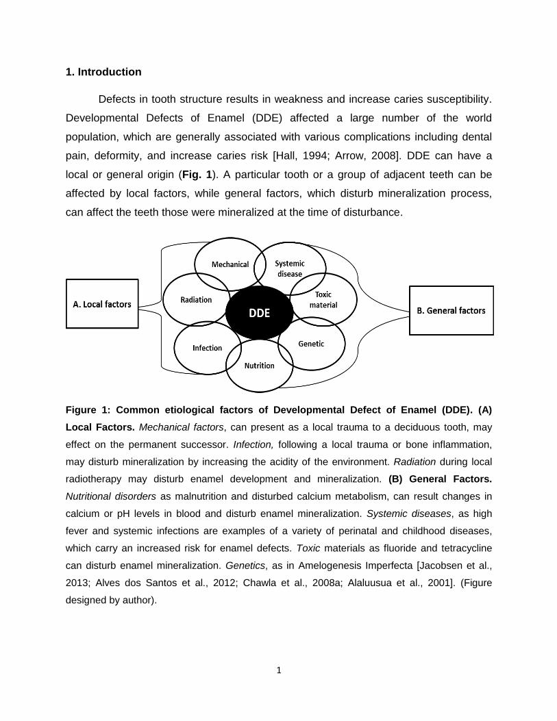
local or general origin (Fig. 1). A particular tooth or a group of adjacent teeth can be
affected by local factors, while general factors, which disturb mineralization process,
can affect the teeth those were mineralized at the time of disturbance.
Figure 1: Common etiological factors of Developmental Defect of Enamel (DDE). (A)
Local Factors. Mechanical factors, can present as a local trauma to a deciduous tooth, may
effect on the permanent successor. Infection, following a local trauma or bone inflammation,
may disturb mineralization by increasing the acidity of the environment. Radiation during local
radiotherapy may disturb enamel development and mineralization. (B) General Factors.
Nutritional disorders as malnutrition and disturbed calcium metabolism, can result changes in
calcium or pH levels in blood and disturb enamel mineralization. Systemic diseases, as high
fever and systemic infections are examples of a variety of perinatal and childhood diseases,
which carry an increased risk for enamel defects. Toxic materials as fluoride and tetracycline
can disturb enamel mineralization. Genetics, as in Amelogenesis Imperfecta [Jacobsen et al.,
2013; Alves dos Santos et al., 2012; Chawla et al., 2008a; Alaluusua et al., 2001]. (Figure
designed by author).
2
Enamel hypoplasia, enamel hypomineralization and fluorosis are the major
developmental defects of enamel [FDI Commission on Oral Health R&E, 1992; Den
Besten, 1994].
Enamel hypoplasia: A quantitative defect of enamel surface, associated with reduced
enamel thickness, smooth borders and without fracture margins which is
macroscopically detectable. The defect may present in different forms, such as pits or
rows, shallow or deep, local or generally dispersed on entire enamel surface [Sabel et
al., 2010].
Hypomineralized enamel: A qualitative defect of enamel surface, associated with normal
enamel thickness at the time of eruption, however a post-eruptive breakdown (PEB)
with fractured edges can be detected short after eruption. The color of the defect may
vary from white, yellow or brown [Fagrell et al., 2011; Weerhejim et al., 2003].
Fluorosis: A qualitative defect of enamel surface, which can also manifest as
quantitative defect in severe cases. It occurs as a result of exposure to high fluoride
concentration during the developmental stage [Den Besten, 1994] with different clinical
presentation (Table 3).
From developmental defect of enamel, Molar Incisor Hypomineralization (MIH)
has become of great concern worldwide in the last few decades. MIH is predominantly a
result of disrupted mineralization process, and more specifically of permanent first
molars and incisors. Despite the fact numerous studies investigated this epidemic
condition, still the etiological factors associated with MIH are unknown [Alaluususa,
2010]. Furthermore there is a significant difference in prevalence at the international
level in young population (2.4%-40.2%) [Weerheijm, 2004; Crombie, 2009; Jälevik,
2010; Alaluusua, 2010]. Nevertheless, this variation in prevalence could be a result of
the difference in socioeconomic status or health care systems, which can be revealed
by conducting comparison studies [Steele et al., 2014; Struzycka, 2014]. Therefore, to
understand the disease process of MIH, there are substantial needs for more
3
comparison studies, which could identify the causative factors associated with this
condition [Abbas et al., 2010; Adriaenssens et al., 2011].
The number of MIH prevalence studies in the Middle East are few and it has
been shown that this region experiencing high level of MIH prevalence [Ghanim et al.,
2011; Zawaideh et al., 2011; Kuscu et al., 2009]. In addition, decayed, missing, filled
tooth (DMFT/dmft) is reported as a parameter with high prevalence in Middle East [Al-
Bluwi, 2014; Hashim et al., 2013; El-Nadeef, 2009]. The City of Dubai in United Arab
Emirates (UAE), is one of the fast growing cities in the Middle East (68% population
increase between 1995 and 2013) [Ministry of Economy in United Arab Emirates, 1995;
Population Bulletin Emirate of Dubai, 2013] and high level of DMFT/dmft, in young age
groups have been reported [El-Nadeef, 2009].
Based on the previous studies, which reported high level in dmft/DMFT indices in
the Middle East, we hypothesize that the prevalence of MIH in Dubai/UAE, which
represents a developed Middle Eastern city is also high and determining it helps in
identifying the etiological factors associated with MIH, if compared with
Greifswald/Germany, a developed European city with considerable difference in
cultural, healthcare system, and socioeconomic status. Thus the aim of this study was
to identify the prevalence of MIH in Dubai/UAE using the European Academy of
Pediatric Dentistry (EAPD) criteria, and to compare it with the Greifswald/Germany MIH
prevalence results, which were obtained by the same research group using similar
study conditions. The study selected Dubai/UAE and Greifswald/Germany due to the
considerable difference in cultural, health care system, and possibly different
socioeconomic status, which could allow for a better understanding of MIH and its
etiology. In addition, this study can help in increasing the awareness of MIH among the
Dubai’s local clinicians and health authorities, which would enhance the preventive
regulation of such a devastating dental condition.
4
2. Review of the literature
2.1 Molar Incisor Hypomineralization
In the late 1970s, demarcated opacities in molars that are occasionally
associated with enamel breakdown, was identified for the first time and was named with
different terms (i.e. idiopathic hypomineralization, Morbus S, Cheese molars) [Van
Amerongen, 1995; Clarkson, 1989]. The causative factor is not identified [Weerheijm et
al., 2003; Weerheijm et al., 2001]. The principal criteria for such hypominaralization
defects were introduced officially in 2003 at the European Academy of Pediatric
Dentistry and since then it has been named as “EAPD criteria” and it is used mainly to
identify MIH. In addition, the defect was given the name “Molar Incisor
Hypominaralisation” (MIH), which is caused by a systemic disturbance during crown
formation and mineralization and it is seen mostly in permanent first molars and
occasionally associated with incisors [Koch et al., 1987; Jälevik et al., 2000; Weerheijm
et al., 2003]. Deciduous second molars, permanent second molars, tips of permanent
canine, and premolars have been also described in some MIH cases [Weerheijm et al.,
2003; Lygidakis et al., 2010; Heitmüller et al., 2012]. Involvement of such teeth could be
due to the overlap of hypomineralization period of the mentioned teeth [Elfrink et al.,
2012]. Among all the associating teeth with MIH, deciduous second molar
hypomineralization is believed to be a significant clinical sign and predictor for MIH in
permanent dentition which can be an aid in early prevention [Elfrink et al., 2012; Petrou
et al., 2013].
2.2 Characteristics of Molar Incisor Hypomineralization
Demarcated opacities are clearly bordered with well-mineralized intact surface
and a glossy appearance. The color of the opacities differs from white, beige to dark
yellow colors. It was reported that demarcated opacities are the most common enamel
defects among MIH teeth [Ghanim et al., 2011; Wogelius et al., 2008]. The severity of
MIH may vary considerably in the same patient; one to all four of the PMF may be
affected. However in some cases only slight demarcated opacities are shown with no

5
further complication. Disturbed enamel mineralization result into an abnormal and weak
enamel integration, which is susceptible to caries and dental wear [Bhaskar et al., 2014;
Lygidakis et al., 2010; William et al., 2006]. In some cases newly erupted MIH teeth can
have severe structural breakdown under normal load of masticatory function. Such
complications occur because histologically there are areas of porosity of varying
degrees [Chawla et al., 2008a; Weerheijm et al., 2003].
In severe cases, MIH affected teeth are very sensitive to stimulations such as air,
cold and hot, as well as mechanical stimuli, which make effective oral hygiene
procedures difficult [Da Costa-Silva et al., 2010; Kilpatrick, 2009]. Insufficient oral
hygiene, due to hypersensitivity is a considerable fact in MIH cases [Leppäniemi et al.,
2001; Weeerheijm, 2003 and 2004] (Fig. 2).
Figure 2: Oral hygiene status in MIH; Plaque detector displays higher accumulation of plaque
on MIH teeth comparing to adjacent teeth without MIH. (A) Tooth 16 with MIH and post-eruptive
breakdown. (B) Teeth 36, 46 affected with MIH and present as post-eruptive breakdown.
(Photos by author).
Several studies showed that MIH has different variables of characteristics and most of
them stated that there is no gender specification in MIH cases [Martínez Gómez et al.,
2012; Jasulaityte et al., 2007; Behrendt et al., 2004; Jälevik et al., 2001] However, few
studies did not support this finding [Chawla et al., 2008a; Cho et al., 2008; Dietrich et
al., 2003; Lygidakis et al., 2008a]. It is common to see MIH in one or more than one
teeth, but the number of affected teeth differs in between studies (mean 2.4 to 5.7) as
well as the number of affected molars (mean 1.5 to 3.16) [Ghanim et al., 2011;
Lygidakis et al., 2008a; Wogelius et al., 2008; Cho et al., 2008; Muratbegovic et al.,
2007; Calderara et al., 2005; Jasulaityte et al., 2003]. In addition, incisors association
6
has been shown in 2.2% of the cases [Jälevik et al., 2001; Jasulaityte et al., 2003;
Calderara et al., 2005; Muratbegovic et al., 2007; Preusser et al., 2007; Willmott et al.,
2008; Cho et al., 2008; Lygidakis et al., 2008a; Wogelius et al., 2008; Chawla et al.,
2008a-b; Ghanim et al., 2011]. While some studies showed that maxillary molar are
more affected than mandibular molars [Gomez et al., 2012; Lygidakis et al., 2008a;
Preusser et al., 2007], other reports showed no statistically significance difference
[Jälevik et al., 2001; Chawla et al., 2008a].
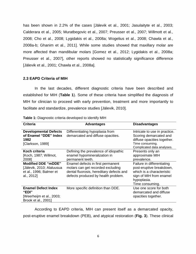
2.3 EAPD Criteria of MIH
In the last decades, different diagnostic criteria have been described and
established for MIH (Table 1). Some of these criteria have simplified the diagnosis of
MIH for clinician to proceed with early prevention, treatment and more importantly to
facilitate and standardize, prevalence studies [Jälevik, 2010].
Table 1: Diagnostic criteria developed to identify MIH
Criteria Advantages Disadvantages
Developmental Defects of Enamel “DDE” Index 1982 [Clarkson, 1989]
Differentiating hypoplasia from demarcated and diffuse opacities.
Intricate to use in practice. Scoring demarcated and diffuse opacities together. Time consuming. Complicated data analyses.
Koch criteria [Koch, 1987; Willmot, 2008]
Defining the prevalence of idiopathic enamel hypomineralization in permanent teeth.
Presents only an approximate MIH prevalence.
Modified DDE “mDDE” [Jälevik, 2010; Alaluusua et al., 1996; Balmer et al., 2012]
Enamel defects in first permanent molars can get recorded excluding dental fluorosis, hereditary defects and defects produced by health problem.
Failure in differentiating post-eruptive breakdown, which is a characteristic sign of MIH from enamel hypoplasia. Time consuming.
Enamel Defect Index “EDI” [Weerheijm et al., 2003; Brook et al., 2001]
More specific definition than DDE. Use one score for both demarcated and diffuse opacities together.
According to EAPD criteria, MIH can present itself as a demarcated opacity,
post-eruptive enamel breakdown (PEB), and atypical restoration (Fig. 3). These clinical

7
presentations can help in the acceleration of clinical evaluation and treatment
[Weerheijm et al., 2003; Lygidakis et al., 2010].
Figure 3: Clinical appearance of MIH. (A) Demarcated opacities on erupting tooth 46. (B)
Demarcated opacities in association with post-eruptive breakdown on tooth 26. (C) Atypical
(occlusal-distal) restorations and demarcated opacities (mesial) on tooth 16. (Photos by author).
Therefore, to avoid the problems of previous indices, the EAPD has developed
the standardized MIH criteria [Weerheijm et al., 2003] (Table 2).
Table 2: MIH standard criteria presented in EAPD 2003
EAPD Criteria 2003 EAPD Criteria
Demarcated opacities
Demarcated opacities of different sizes, located on the crown. Different in color, from white, creamy, and yellow to brownish discoloration.*
Post-eruptive breakdown (PEB)
Different degrees of enamel hypomineralization increase level of porosity, which in the severe cases crown is susceptible to collapse under masticatory forces and leads to unprotected dentin and high sensitivity and increasing the speed of caries process.
Atypical restoration Restoration with similar pattern of defect extension.
Failure in eruption or extracted teeth
Absence of the tooth is possible to be a sign of severely destroyed crown that led to extraction. It could be as well a sign of failure in eruption based on presence of other molars with MIH, otherwise it is not a diagnostic source for MIH.
* According to the EAPD guidance 2010, defects less than 1mm are not recommended to be record.
Tooth sensitivity which is usually reported in MIH cases, is other characteristic of
MIH. The sensitivity differs from mild grade, which is a consequence of external stimuli,
to severe spontaneous hypersensitivity [Lygidakis et al. 2010]. Although some authors
suggested two scores for severity scales in order to limit the diagnostic variations that
A B C
8
exist in the literatures, some other prefer a three score severity classification according
to the clinical features [Lygidakis et al., 2008; da Costa-Silva et al., 2010; Mathu-Muju,
2006]. The two degrees for sensitivity based on clinical features are categorized as a
“mild” or “severe”. In the mild cases, there are occasional sensitivity to external stimuli
such as air and water but no brushing sensitivity and difficulty. It is stated that in severe
cases persistent and spontaneous hypersensitivity affecting oral hygiene procedure,
which lead to higher caries level [Lygidakis et al., 2010]. In case of an extracted or
missing tooth, it should be recorded as MIH, only when the dental record of the patient
shows a diagnosis of MIH prior to extraction or presence of other permanent first molars
with MIH.
2.4 Clinical Consideration of MIH
MIH has some clinical considerations in order to understand, prevent, manage and treat
the etiological factors [Jälevik et al., 2002]. It is useful to increase the frequency of
dental check-ups in children with history of repeated illnesses in the first three years
after birth and children with opacities on deciduous second molars, before and during
the eruption period of the permanent first molars. The signs of hypomineralization, in the
crown of unerupted permanent first molars affected with MIH, can be detected
sometimes on a radiograph even prior to eruption [William et al., 2006a], However it is
questionable if there is an indication to expose children to x-ray prior to eruption of
permanent first molars or incisors in order to detect MIH These check-ups give an
advantage to detect and manage MIH conditions in early stage since the clinical
management of this condition is challenging due to the rapid development of caries,
limited cooperation of a young child, difficulty in achieving anesthesia, repeated
marginal breakdown of restorations, discomfort and sensitivity stimulated by tooth
brushing on affected teeth [William et al., 2006; Fayel, 2003; Jälevik et al., 2002].
According to the characteristics of MIH, the management can be complicated.
Therefore, it is strongly suggested that the patients diagnosed with MIH receive early
intensive prevention therapy based on the severity level of MIH, such as fluoride
varnish, a fissure sealing, glass ionomer cement (GIC) restoration, composite
restoration, and stainless steel crowns [Lygidakis et al., 2010; William et al., 2006b;
9
Fayle, 2003; Koch, 2000] (Fig. 4). In addition, an early diagnosis is of high importance
since an inappropriate orthodontist approach on MIH teeth, can result in negative
consequences, such as extraction of the molars in the age of 8 to 10 years [Kellerhoff et
al., 2004].
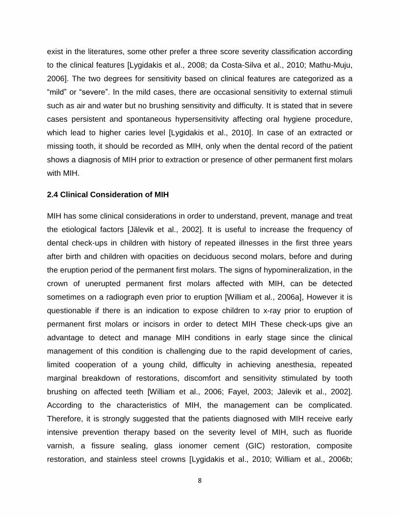
Figure 4: Clinical management and approaches of Molar Incisor Hypomineralization (A)
Tooth affected with mild Molar Incisor Hypomineralization (MIH) is treated with fluoride varnish
to induce remineralization and reduce cariogenicity, and post-eruptive breakdown. (B) Moderate
MIH teeth sealed with Glass Ionomer Cement (GIC). (C) Severe MIH covered with Stainless
Steel Crowns (SSC) to block the hypersensitivity. All the treatments require follow up every 3
months along with orthodontic consultation. (Photos by author).
2.5 Etiology
2.5.1 Amelogenesis
Although the developmental process of amelogenesis is genetically controlled,
environmental disturbances can affect the development especially at the early stage of
maturation [Alaluusua, 2010]. Disturbance during enamel formation affects the quality
and quantity of the enamel, depending on the phase of amelogenesis and the duration
of the disturbance on the ameloblast [Suga, 1989; Alaluusua, 2010]. Teeth development
and mineralization start before birth and continues till late teen ages when the
mineralization of the permanent molars is completed. In the beginning of the second
trimester of pregnancy, the deciduous lower incisors show the first sign of mineralization
which continues till the age of three month. The permanent first molars are the first teeth
from permanent dentition which start mineralization at birth and complete approximately
three years of age [Reid et al., 2006]. Secretory cells produce enamel from specialized
10
enamel forming cells called Ameloblasts, which are highly specialized cells of
ectodermal origin [Simmer, 2010; Mahoney, 2010].
Histologically, developmental disturbances of organic matrix during enamel
formation and maturation stages result in a defect known as enamel hypoplasia.
Transitional ameloblast cell is the most vulnerable cell which needs to undergo the
complete maturation otherwise the outcome is hypomineralization of full thickness
enamel. Disturbed resorptive potential of ameloblast or inhibition of proteolytic enzyme
lead to protein retention and interference with crystal growth and enamel maturation,
which can reduce thickness of enamel layer resulting in white spots, grooves, fissures,
and depressions in the enamel surface. Furthermore the conditions that affect the
matrix pH and impaired calcium metabolism during enamel maturation may result in
MIH. Therefore, enamel defects may contribute as local, systemic or genetic etiological
factors. Neonatal disturbance of enamel formation and maturation can be a result of
premature birth or hypocalcaemia [Alam et al., 2006; Garg et al., 2012].
Fluorosis: As mentioned in MIH process, any impairment during developmental
stage of enamel leads to subsequent changes and defect. During enamel development
an interface among ameloblasts, matrix, and mineral can result changes in enamel
formation. Excessive amount of fluoride in body system during the enamel formation
has a negative effect on ameloblasts, causing a hypomineralization defect in the enamel
layer, known as fluorosis. The excess fluoride ions in the matrix during mineralization
decrease the concentration of free calcium ions and interfere with the proteinases
process leading to protein degradation during the maturation phase of amelogenesis.
Therefore, there is higher fluoride level and protein content in enamel with fluorosis than
normal enamel, which result an increase in enamel porosity and physical changes such
as white spots, grooves, fissures, and different grades of discolorations. Most clinical
feature seen in a tooth affected with fluorosis is striation pattern, parallel lines on the
enamel surface [Den Besten, 2002; Aoba et al., 2002].
11
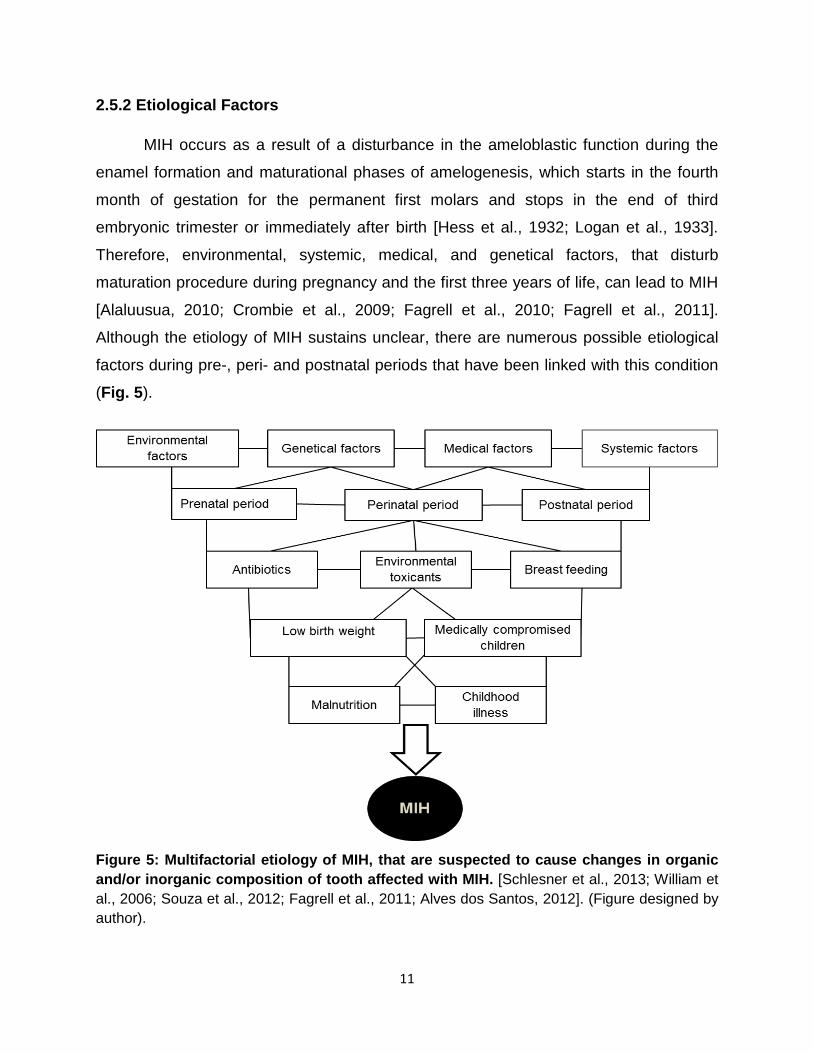
2.5.2 Etiological Factors
MIH occurs as a result of a disturbance in the ameloblastic function during the
enamel formation and maturational phases of amelogenesis, which starts in the fourth
month of gestation for the permanent first molars and stops in the end of third
embryonic trimester or immediately after birth [Hess et al., 1932; Logan et al., 1933].
Therefore, environmental, systemic, medical, and genetical factors, that disturb
maturation procedure during pregnancy and the first three years of life, can lead to MIH
[Alaluusua, 2010; Crombie et al., 2009; Fagrell et al., 2010; Fagrell et al., 2011].
Although the etiology of MIH sustains unclear, there are numerous possible etiological
factors during pre-, peri- and postnatal periods that have been linked with this condition
(Fig. 5).
Figure 5: Multifactorial etiology of MIH, that are suspected to cause changes in organic
and/or inorganic composition of tooth affected with MIH. [Schlesner et al., 2013; William et
al., 2006; Souza et al., 2012; Fagrell et al., 2011; Alves dos Santos, 2012]. (Figure designed by
author).

12
Prenatal period: It was shown that the mothers of the children with MIH had medical
illness during pregnancy more than the mothers of children without MIH. However there
was no specific related illness [Whatling et al., 2008; Lygidakis et al., 2008].
Perinatal period: There is a controversy in results regarding the effect of disturbance in
this period. A study in Greece showed that MIH was more frequent in children born by
mother who had Caesarian section, prolonged delivery, premature birth and twining,
compared to the control group children [Lygidakis et al., 2008]. However in an English
[Whatling et al., 2008] and a German [Diedrich et al., 2003] study, perinatal problems
could not be linked with MIH.
Postnatal period: Most of the researches showed that there is a direct relation between
postnatal medical problem such as hemolytic anemia, which occurs in the first three
years of life and MIH [Jälevik et al., 2001; Beentjes et al., 2002; Lygidakis et al., 2008;
Kusku et al., 2008; Alaluusua, 2010].
Childhood illness: It can be of high concern, since it was found in many studies that
there is an association between MIH and childhood illness such as high fever [Beentjes
et al., 2002; Jälevik et al., 2001; Tapias-Ledesma et al., 2003].
Medically compromised children: These children have shown higher prevalence of
dental defect due to their medical conditions (e.g. coeliac disease) and treatments
[Crombie et al., 2009].
Antibiotics: Studies in concern of antibiotic relation to MIH show, that there is a direct
link between these two issues [Jälevik et al., 2001; Beentjes et al., 2002; Whatling et al.,
2008; Laisi et al., 2009]. Amoxicillin and erythromycin use, in the first year of life showed
increase in the MIH [Laisi et al., 2009].
Environmental toxicants: Exposure to high level of polychlorinated biophenyls (PCBs)
an industrial process waist, and dioxins a by-product of manufacturing process, is
associated with enamel defects as hypoplasia and demarcated opacity hypoplasia
[Alaluusua et al., 2001; Jan et al., 2007].
Breast feeding: In one report it was shown that prolonged breast feeding and the toxin
in the breast milk has been associated with MIH [Alaluusua et al., 1996a; Fagrell et al.,
2011].
13
Low birth weight: Children with the history of low birth weight were shown to be at higher
risk of enamel hypoplasia compare to the control group [Masumo et al., 2013].
Malnutrition: Malnutrition during early childhood is also among possible etiological factor
of MIH [Nelson-Piercy, 1998; Fagrell et al., 2011].
Genetics: It was reported that specific genes are involved in enamel and dentin
structures formation [Bailleul-Forestier et al., 2008].
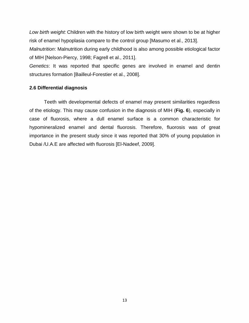
2.6 Differential diagnosis
Teeth with developmental defects of enamel may present similarities regardless
of the etiology. This may cause confusion in the diagnosis of MIH (Fig. 6), especially in
case of fluorosis, where a dull enamel surface is a common characteristic for
hypomineralized enamel and dental fluorosis. Therefore, fluorosis was of great
importance in the present study since it was reported that 30% of young population in
Dubai /U.A.E are affected with fluorosis [El-Nadeef, 2009].
14
Figure 6: Differential diagnosis of MIH. (A) MIH, which presented as demarcated opacities on
tooth 46 and distal cusp post-eruptive breakdown on tooth 36. (Figure by author) (B) Caries on
tooth 46 opacities on occlusal enamel surface due to demineralization in response to metabolic
and chemical activity of oral biofilm, which presents as spots or diffuse opacities. (Figure by
author) (C) Enamel hypoplasia, a quantitative defect of enamel, showing localized decrease of
enamel thickness with no fracture line and sharp edges, affecting all teeth in symmetry (reprint
with copyright permissions by Jacobsen [2013]). (D) Amelogenesis imperfecta, defect of genetic
origin, all teeth and surfaces are affected, which is usually diagnosed base on radiographic form
of taurodont (E) Fluorosis, diffuse opacities affecting the homologous teeth in symmetry, which
depends on the time and duration of exposure to excessive fluoride. (Figure by author) (F)
Localized trauma, a localized defect, which could be a result of a traumatic injury or a prolonged
periapical inflammation of a primary tooth, that affect the delicate developmental procedure of
permanent successor by increasing the acidity of environment, leading to hypominaralization
[Petrou et al., 2013].
2.6.1 Enamel hypomineralization
Hypomineralized enamel has normal thickness at the time of eruption and it is
associated with white, brown or yellow discoloration. Change in the refractive index
(propagation of light, or any other radiation, through a medium) caused by the increased
degree of porosity in the enamel results in a clinical discoloration. To some extend the
color seen is dependent on the degree of hypomineralization and possibly also to the
protein content in the enamel [Farah et al., 2010a; Da Costa-Silva et al., 2011]. A dull
15
enamel surface is characteristic for hypomineralized enamel, which is also seen in case
of dental fluorosis. An association of opaque clinical appearance with distinct borders, a
well mineralized intact and glossy surface, is the characteristic of the hypomineralized
area. In severe cases of enamel hypomineralization, the surface may collapse and a
loss of substance with fractured borders will be seen. Fractured edges are seen with
post-eruptive breakdown (PEB) [Fagrell et al., 2011].
2.6.2 Fluorosis
Fluorosis is an enamel developmental defect as a result of exposure to high
fluoride concentration during the developmental stage [Den Besten, 1994]. The affected
teeth contain low mineral and high level of porosity [Abanto Alvarez, 2009]. The safe
level of daily fluoride intake is 0.05-0.07mg F/Kg/day and a chromic consumption during
teeth development to more than this amount can cause fluorosis [Burt, 1992]. Severity
of fluorosis depends on the age, duration of exposure, weight, degree of physical
activity, nutrition, and bone growth. The main fluoride source is fluoridated water, which
range between 0.7 to 1.0 ppm depending on the season of the year and geographic
area. Fluoridated water is responsible for 40% of dental fluorosis [Richards et al., 1967].
Fluoride supplements, mostly recommended in the fluoride deficient areas, have shown
to be a contributory factor of dental fluorosis in fluoridated and non-fluoridated area.
However it is more risky in fluoridated area [Jackson et al., 1999; Pendrys et al., 1998;
Mascarenhas, 2000]. Fluoride containing toothpastes can also be a contribution factor,
especially in children under age of 6, since they swallow around 30% of the toothpaste
during brushing. Hence if this action is associated with fluoridated water the fluorosis
risk is increased [Villena, 2000].
Diffuse opacities of fluorosis should not be included in the scoring for MIH tooth
since the opacity in MIH is well demarcated [Weerheijm et al., 2003]. Fluorosis has
classical appearances; it is bilateral, substantial symmetry on homologous teeth, and
diffuse opacity in form of striation or banding, which follow the lines of enamel
development. Fluorosis can occur on any tooth, however, MIH is affecting only Molars
and incisors [Levy, 2003]. In severe cases of fluorosis, where the hypomineralization
16
extends to the dentin enamel junction and can cause extensive post-eruptive enamel
breakdown and brown to black staining [Wright et al., 1996]. In addition mild fluorosis is
a caries resistant defect [Waidyasekera et al., 2007], while MIH is caries susceptible in
all phases [William et al., 2006]. Unlike MIH and caries, fluorosis does not result in pain
or abscesses, and no anesthetic (e.g. local or general) is required for the treatment
[Mullen, 2005].
2.6.3 Enamel hypoplasia
A quantitative defect is associated with a reduced localized thickness of enamel,
rough or pitted surface, smooth and rounded borders, and no fractured boundaries.
Such condition occurs due to disruption of the secretory phase of amelogenesis while
MIH is a qualitative defect of enamel which occurs due to disturbed mineralization
phase of amelogenesis [Suckling, 1989]. EHP can occur in a single tooth or multiple
teeth [Seow, 1991]. The defect might have a different depth in the enamel, one surface
or all the enamel surfaces [Sabel et al., 2010].
2.6.4 Amelogenesis imperfecta
Amelogenesis imperfecta (AI) is a genetically originated defect, which affects all
the permanent dentition. In AI, the effect is more generalized and the molars may also
appear taurodont on radiograph. This defect is often associated with family history
[Witkop, 1988; Lygidakis et al., 2010]. MIH is affecting the teeth in asymmetrical pattern
but only in very severe cases, the molars are equally affected and mimic the
appearance of Amelogenesis imperfecta.
2.6.5 Trauma as localized factor
Traumatic injury as well as a prolonged periapical inflammation of a primary tooth
could affect the delicate developmental procedure of permanent successor by
increasing the acidity of environment, leading to hypominaralization [Alaluusua et al.,
2001; Chawla et al., 2008a].
17
2.6.6 Dental caries
A chronic infectious bacterial disease, a demineralization response to metabolic
and chemical activity of oral biofilm content, such as acid by-product of bacterial
metabolism, which dissolves the mineral after disseminating into the enamel and dentin
by initiating an environmental imbalance in the oral microbiome [Featherstone, 2008].
Dental plaque on the tooth surface, which presents itself as a classic biofilm is formed
by oral microorganisms. Acid product of cariogenic microorganisms and large scale
change in protein expression, influence the biofilm formation by decreasing the pH level
of oral environment below 5.5, which induce demineralization of enamel hydroxyapatite
crystals and proteolytic breakdown of the hard tissues structure of the tooth. This may
change the surface texture of enamel and dentin [Takahashi, 2008] and appears as a
spot but not well demarcated opacity, which can be differentiate from MIH [Kidd, 2004;
Chawla et al., 2008a].
Caries is a multifactorial disease, but it mainly occurs due to coexistence of three
principal factors, which are acidogenic and acidophilic microorganisms (e.g.
Streptococcus mutans, non-mutans streptococci group, Actinomyces and
Lactobacillus), carbohydrates containing food, and host factors [Struzycka, 2014],
however additional etiological factors such as educational, behavioral, and
socioeconomic status have strong influence in caries development [Steele et al., 2014;
Struzycka, 2014].
18
2.7 Epidemiology
2.7.1 MIH
High prevalence of this MIH has been shown in great number of studies
[Lygidakis, 2010]. Based on these studies the prevalence of MIH ranges from 2.8% to
40.2% and it differs between regions and studies [Jälevik et al., 2002; Jälevik, 2010]. In
Europe, the prevalence of MIH ranges from 3.6% to 25% [Weerheijm et al., 2003] and
studies have demonstrated that MIH prevalence has increased remarkably in Germany
[Jasulaityte, 2003]. Although limited numbers of studies have taken place in the Middle
East, high prevalence’s of MIH were reported in this region (8.6% to 20%) [Allazzam et
al., 2014; Zawaideh et al., 2011; Ghanim et al., 2012; Ghanim et al., 2011; Ghanim et
al., 2014]. In addition to Middle East, other parts of the world such as Asia and Africa
are also having limited number of MIH studies. This deficiency in data leads to a
weakness in comparable studies, which might cause limitation in the understanding of
MIH and its causative factors, since potential etiological factors in different countries
cannot be compared [Jälevik et al., 2001; Jasulaityte et al., 2007]. More prevalence
studies are needed to investigate the etiological factors of MIH. The comparison of the
MIH prevalence and possible etiological factors among different countries can be
performed only by epidemiological studies, which used the identical criteria and the
same calibration system.
19
2.7.2 Dental caries
Caries is the most common dental disease, which has been found in almost
every population, studied over a million years ago. It has increased considerably after
introduction of sugar to the Western world in the year 1000 A.D [Keene, 1980; Price,
1989]. Epidemiological studies of dental caries, especially in children, show increasing
levels in many developing countries and decrease in many highly developed countries
of the world, which indicate that socioeconomic factors have great impact in determining
the ratio of caries risk in developing countries [Winter, 1990; Steele et al., 2014;
Struzycka, 2014].
The caries prevalence is high in the Middle East and parts of Europe [Struzycka,
2014; Khan, 2014]. Based on the latest studies in UAE, Dubai has the highest
dmft/DMFT prevalence in comparison to the other cities of UAE [El-Nadeef, 2009 and
2010]. Caries values were also high in Germany [DAJ, 2000]. In 1990, the dmft/DMFT
values in Greifswald were higher than most of Mecklenburg-Vorpommern state of the
Germany. However, during the last decades these values dramatically decreased,
through applying a highly structured public dental health program, in which schools are
visited regularly by dental hygienists, dental assistant or even dentists for theoretical
and practical oral prevention [Pieper, 2010; Splieth, 2013]. Therefore, similar to the
study carried out in Greifswald, an estimation of caries prevalence in Dubai and
comparison to the previous studies, can help improve the clinicians` and public’s
awareness and to diminish this problem with improving preventive strategies.

20
2.7.3 Dental fluorosis
In 1925, fluoride found to be caries resistant and in 1931 it was proved that the
fluoride in natural water could cause a specific developmental defect, which was called
as fluorosis. In 1942 Dean has announced the safe fluoride level (1ppm or below)
[Dean, 1942]. This finding was a scientific revolution that helped dentistry to step in the
preventive medicine. Artificial water fluoridation with amount of 1 ppm started in 1945 in
a low fluoride area to reproduce the beneficial dental health effect, which had a positive
outcome and studies reported 50% and more reductions in dental caries experience.
However, the result was obtained at a time when the only source of fluoride was
fluoridated water [Mullen, 2005; Murray et al., 1982]. Nevertheless, it has always been
known that a low level of enamel discoloration would be associating with the water
fluoridation [Murray et al., 1982]. In addition, the prevalence of fluorosis has increased
over the past fifty years [McKnight, 1998].
Germany is from the countries with low fluoride concentration in natural water
and more than 90% of the drinking water contains less than 0.3 mg/L fluoride per liter
[BfR, 2005] and currently there is no artificial water fluoridation in drinking water of
Germany. Low level of fluorosis was reported in Germany (4.9%-11.3%) with mild
severity [Pieper et al., 2008; Momeni et al., 2007]. The Middle East, including UAE,
however, is a region where the drinking water has a naturally high level of fluoride
[WHO, 1994]. However tap water was reported by a local study to be 0.00 ppm [Nimr,
1997]. Nevertheless almost 90% of the population in UAE are using bottled water
[Nsanze, 1999] containing a controlled level of fluoride, which is below the conventional
level and relatively lower than the national standards [Abouleish, 2012; Nimr, 1997].
Although the level of fluoride in water supplements in Dubai does not exceed 0.7 ppm
(Tap water 0.00 ppm, bottle water 0.03-0.68 ppm) [Abouleish, 2012; Nimr, 1997], the
prevalence of fluorosis is significantly high and affected 30% of Dubai’s young
population [El-Nadeef, 2009]. Fluorosis is assumed to have a masking effect on MIH
teeth and its prevalence [Chawla et al., 2008]. This fact gives a great importance in
studies, to include the differential diagnosis of MIH (e.g., fluorosis) in researches to
avoid any confusion between MIH and its differential diagnosis.
21
3. Aim of the Study
The purpose of this study was to find the prevalence of MIH in the city of
Dubai/UAE and to compare it with the literature on the prevalence of MIH, especially
with a detailed study conducted with the same methodology in the city of
Greifswald/Germany. Furthermore, this study explored the prevalence of fluorosis and
caries in Dubai in order to assess the chance and influence of the differential diagnoses
and to analyze the possible relationship between MIH and caries level. This could help
to optimize the recording of enamel defects and especially MIH, to find indications for
etiological factors and also to assess the needs for treatment in Dubai/UAE. In addition,
dental caries and fluorosis prevalence in this study was compared with other studies in
Dubai, which could possibly be a trigger for further studies to develop adequate
prevention and treatment procedures.
22
4. Materials and Methods
For standardization, this study was based on the same methodology as in the
community based study on MIH and caries conducted in Greifswald, Germany [Petrou
et al., 2013]. The epidemiological data of the Greifswald study was also used further in
the comparison process of this research.
4.1 Ethics Committee
This study has received the approval from the Ethical Committee in Greifswald:
[Reg.-Nr.: BB 102/12] (Attachment No. I). In addition, an ethical approval was taken
from Ministry of Health in UAE/Dubai [Reg.-Nr.: 122012-1] (Attachment No. II).
4.2 Materials
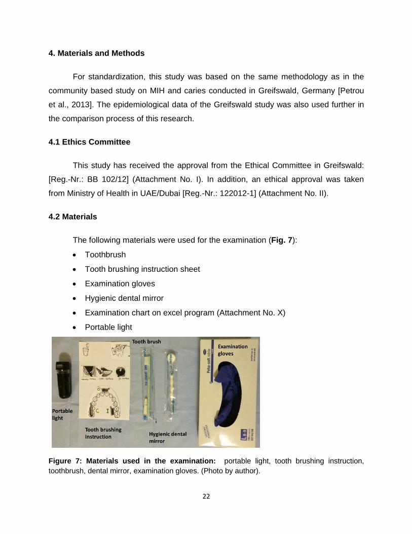
The following materials were used for the examination (Fig. 7):
Toothbrush
Tooth brushing instruction sheet
Examination gloves
Hygienic dental mirror
Examination chart on excel program (Attachment No. X)
Portable light
Figure 7: Materials used in the examination: portable light, tooth brushing instruction,
toothbrush, dental mirror, examination gloves. (Photo by author).
23
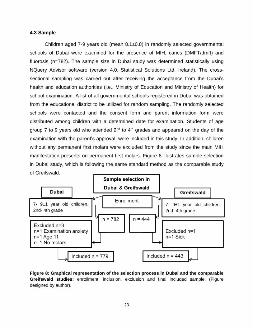
4.3 Sample
Children aged 7-9 years old (mean 8.1±0.8) in randomly selected governmental
schools of Dubai were examined for the presence of MIH, caries (DMFT/dmft) and
fluorosis (n=782). The sample size in Dubai study was determined statistically using
NQuery Advisor software (version 4.0, Statistical Solutions Ltd. Ireland). The cross-
sectional sampling was carried out after receiving the acceptance from the Dubai’s
health and education authorities (i.e., Ministry of Education and Ministry of Health) for
school examination. A list of all governmental schools registered in Dubai was obtained
from the educational district to be utilized for random sampling. The randomly selected
schools were contacted and the consent form and parent information form were
distributed among children with a determined date for examination. Students of age
group 7 to 9 years old who attended 2nd to 4th grades and appeared on the day of the
examination with the parent’s approval, were included in this study. In addition, children
without any permanent first molars were excluded from the study since the main MIH
manifestation presents on permanent first molars. Figure 8 illustrates sample selection
in Dubai study, which is following the same standard method as the comparable study
of Greifswald.
Figure 8: Graphical representation of the selection process in Dubai and the comparable
Greifswald studies: enrollment, inclusion, exclusion and final included sample. (Figure
designed by author).
7- 9±1 year old children,
2nd- 4th grade
n = 782
Included n = 779
Excluded n=3
n=1 Examination anxiety
n=1 Age 11 n=1 No molars
7- 9±1 year old children,
2nd- 4th grade
n = 444
Excluded n=1
n=1 Sick
Included n = 443
Sample selection in
Dubai & Greifswald
Enrollment
Dubai Greifswald
24
4.4 Dental examination
The examination was performed by one calibrated examiner (Somayeh Haidary,
DDS), who was previously trained by the calibrated examiner (M. Petrou) who
performed the equivalent German study and advisor (Prof. Dr. Ch. H. Splieth) in
Greifswald. During the examination, a dentist accompanied the examiner to assist only
in recording the data into computer.
4.4.1 Caries scores
The caries scores in the primary and permanent dentition were calculated
according to the World Health Organization criteria for the dmft/DMFT-index [WHO,
1997], which is the sum of decayed, missing, and filled tooth in 28 permanent and 20
primary teeth per child. The overall mean of the dmft/DMFT is calculated by dividing the
sum of all dmf/DMF-teeth by the number of participating children.
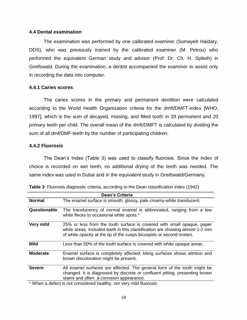
4.4.2 Fluorosis
The Dean’s Index (Table 3) was used to classify fluorosis. Since the index of
choice is recorded on wet teeth, no additional drying of the teeth was needed. The
same index was used in Dubai and in the equivalent study in Greifswald/Germany.
Table 3: Fluorosis diagnostic criteria, according to the Dean classification index (1942)
Dean’s Criteria
Normal The enamel surface is smooth, glossy, pale creamy-white translucent.
Questionable The translucency of normal enamel is abbreviated, ranging from a few white flecks to occasional white spots.*
Very mild 25% or less from the tooth surface is covered with small opaque, paper white areas. Included teeth in this classification are showing almost 1-2 mm of white opacity at the tip of the cusps bicuspids or second molars.
Mild Less than 50% of the tooth surface is covered with white opaque areas.
Moderate Enamel surface is completely affected; biting surfaces shows attrition and brown discoloration might be present.
Severe All enamel surfaces are affected. The general form of the tooth might be changed. It is diagnosed by discrete or confluent pitting. presenting brown stains and often a corrosion appearance.
* When a defect is not considered healthy, nor very mild fluorosis

25
4.4.3 MIH
The following criteria, which were based on the EAPD criteria [Weerheijm et al.,
2001b; Lygidakis et al., 2010] were used to diagnose MIH cases:
1. Examination of permanent first molars and incisors (12 index teeth).
2. Examination should to be performed on wet teeth after cleaning.
3. Each tooth should be recorded for:
Demarcated opacities, but not less than 1 mm
Enamel breakdown following tooth eruption
Atypical restoration
Extraction due to MIH
Molar or incisor tooth failed in erupting
Since the examiner had no access to dental treatment data of the children, therefore,
missing teeth that are extracted or failed to erupt in MIH cases were categorized as
“missing”.
The examination chart includes (Attachment No. X):
Date of the examination
Name of the school
Class number/grade
Birthday
Age
Gender
dmft/DMFT
Fluorosis
MIH index tooth/teeth number with
o MIH defect, not less than 1 mm
o Presence of demarcated opacities
o Post-eruptive breakdown (PEB)
26
o Atypical restoration, type of restoration
o Hypersensitivity
o Missing tooth/teeth, is used and coded since there is no access to
dental record
Co-existence of caries/orthodontic treatment
Children with one of the above clinical MIH criteria with at least one affected
permanent molar were considered as MIH case. To avoid the masking effect in cases
where the DMFT score is more than 5, the MIH criteria were investigated carefully.
Defects less than 1mm, which affected only incisors ware not included as a MIH case.
The affected index-teeth, their clinical view, atypical restorations, and the restoration
material used on MIH affected tooth, were also recorded in an Excel spreadsheet.
Teeth, which had only atypical restoration and did not have another MIH signs, were not
included in MIH cases.
One of the diagnostic features of MIH severity is hypersensitivity. Therefore,
children with suspected MIH were asked, if they had any pain or sensitivity during
air/water stimuli and teeth brushing. The children were also asked if the pain comes
occasionally and by stimuli and if it is persistent and spontaneous. The following
standardized questions were asked from all the children where MIH condition is
suspected to justify the kind of discomfort:
Does it hurt when you brush your teeth? What about your back teeth?
Does it hurt when you drink a hot tea or cacao? Do you enjoy having ice cream
or does it make your teeth hurt?
The severity of MIH cases were then recorded based on the EAPD criteria [Lygidakis et
al., 2010].
The examination procedure was structured in the following way:
Brushing the teeth prior to examination under the instruction and observation of a
calibrated examiner and a school teacher.
Examination by the dentist under supervision of one school member/teacher.
27
4.5 Calibrations
The examination was carried out by a single calibrated examiner (dentist
Somayeh Haidary) who received standardized caries calibrations trainings for the
standardized German dental school examinations (Deutsche Arbeitsgemeinschaft für
Jugendzahnpflege) and was trained in Greifswald university dental clinic (Feb-Jun
2012), by Prof. Dr. Ch. H. Splieth and Dr. M. A. Petrou. Dr. Petrou was previously
calibrated and attained the “gold standard” of the German epidemiological study in
identifying and differentiating MIH defects [Pieper, 2010].
4.5.1 Calibration for dmft/DMFT
The school examination procedures were standardized according to DMFT/dmft
values. The same criteria which was used in the German study were used for the
calibration of Dr. Haidary by Prof. Dr. Ch. H. Splieth (kappa value>0.9) [Pieper, 2010].
4.5.2 Calibration for MIH and Fluorosis
Prior to the calibration, a clinical and theoretical training of the examiner (Dr.
Haidary) on the differential diagnosis of MIH including dental fluorosis was performed.
For the theoretical training, the international literature [Chawla et al., 2008 and Lygidakis
et al., 2010] as well as the previous presentations of German data were used. The
calibration was performed with the use of a slide presentation of 20 clinical pictures of
MIH and other enamel defects. The same pictures were used to calibrate the examiner
of the Greifswald study. The examiner achieved an intra- and inter-kappa values > 0.9
for MIH and fluorosis. Children diagnosed with MIH, were given the diagnostic form to
inform their parents about the findings including DMFT/dmft, fluorosis, and MIH.
28
4.6 Statistical Analysis
4.6.1 Method of Analysis
Following the examination, the data were collected, coded, and statistically
analyzed with the Statistical Package for Social Science (SPSS) system 18.0 [SPSS for
Windows, version 18.0, SPSS Inc., Chicago, IL, USA]. In this system the mean values
and the standard deviation of dmft, DMFT, age, gender distribution, prevalence of MIH,
and distribution of MIH criteria were achieved using descriptive statistics. The difference
and probable association of children’s age, gender, caries experience, and presence of
MIH were statistically analyzed using t-test, Levene-test, Chi-square-test (𝑋2-test), and
Pearson correlation.
4.6.2 Method of Comparison
Prevalence of MIH in the city of Dubai/UAE was compared with the literatures on
the prevalence of MIH, especially a detailed comparative study from
Greifswald/Germany, since the same methodology was used in the both studies.
The comparison of MIH and caries prevalence in Dubai/U.A.E with
Greifswald/Germany was performed using the data obtained from the MIH study in
Greifswald/Germany [Petrou et al., 2013].

29
5. Results
The results of this study are presented in each section accordingly with
immediate comparison to the data of the study with identical methodology from
Greifswald [Petrou et al., 2013].
5.1 Sample
779 school children were examined (Table 4) with an age range of 7 to 9±1
years in Dubai (mean age 8.1±0.8 yrs). The sample in Greifswald, which was directly
compared to the Dubai’s data, consisted of 443 children (mean age 8.45±1.0 yrs)
[Petrou et al., 2013]. In contrast to the Greifswald sample, in Dubai study the sample
was not evenly distributed based on gender (Greifswald: 228 Female, 212 Male; Dubai:
515 Female, 264 Male) (Fig. 9). However, there was no significant gender difference in
MIH prevalence in the Dubai (7.57% Female, 7.59% Male) and Greifswald studies
(4.37% Female, 4.21% Male) (p>0.05).
Table 4: Sample age population and distribution of MIH (n, %) in different age groups in Dubai.
30
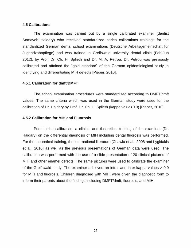
Figure 9: Distribution of age and gender in Dubai sample in comparison to the data with
identical methodology from Greifswald [Petrou et al., 2013]. (A) Age distribution, Dubai
(mean age 8.1±0.8 yrs), Greifswald (mean age 8.45±1.0 yrs). (B) Gender distribution, Dubai
(66% Female, 34% Male), Greifswald (51% Female, 49% Male). However, no significant gender
differences in MIH prevalence in Dubai (7.57% Female, 7.59% Male) and Greifswald studies
(4.37% Female, 4.21% Male) were seen.
5.2 DMFT/dmft
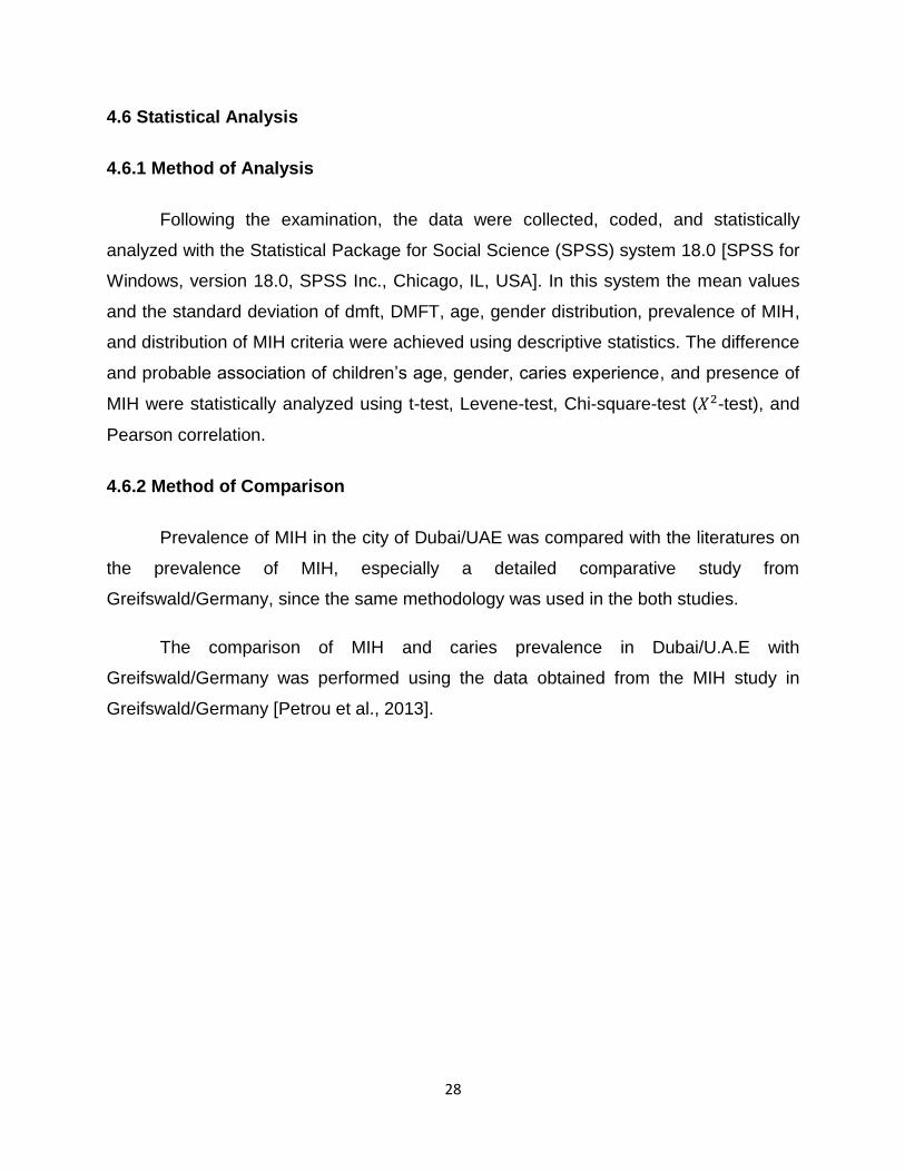
The DMFT/dmft indices in Dubai were significantly higher (DMFT: 2.41±1.7; dmft:
5.46±3.1) than Greifswald (DMFT: 0.1±0.4; dmft: 0.9±1.7). The difference of DMFT
index between children with and without MIH was statistically significant in both cities
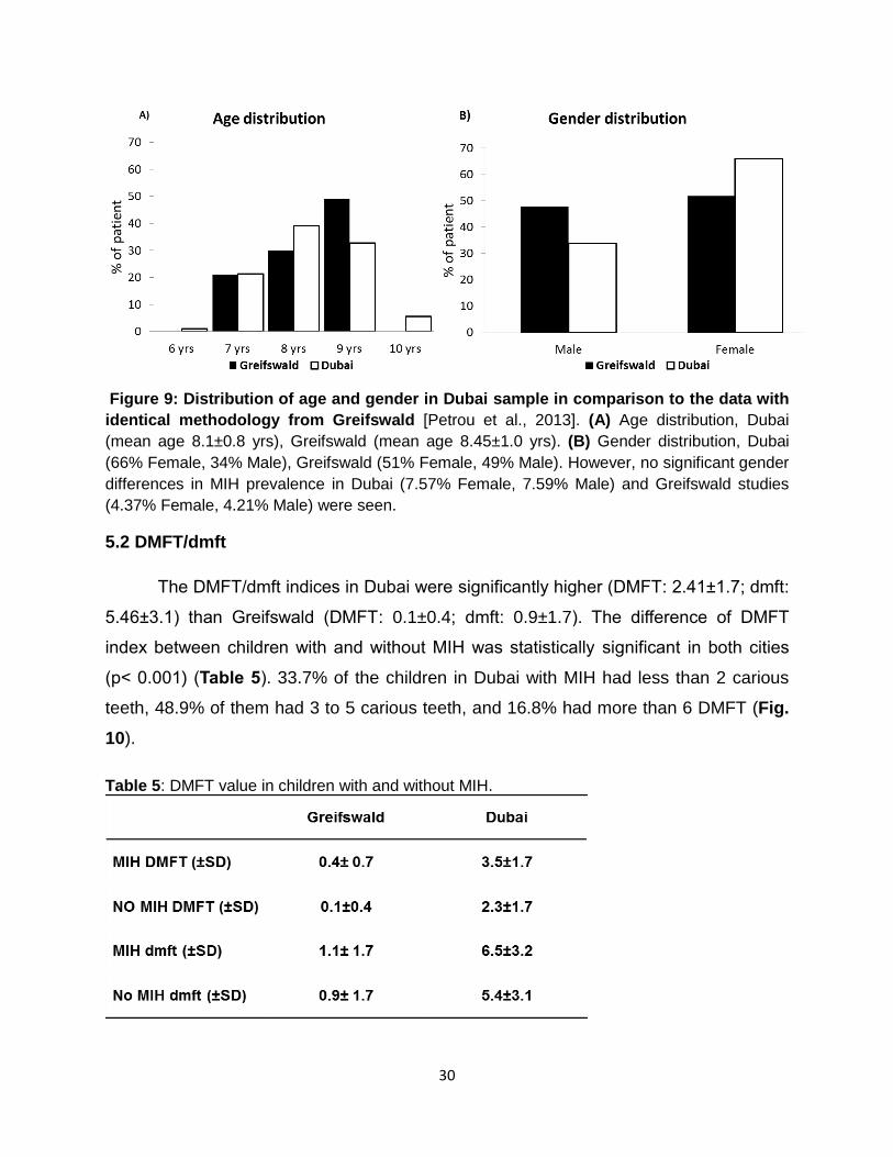
(p< 0.001) (Table 5). 33.7% of the children in Dubai with MIH had less than 2 carious
teeth, 48.9% of them had 3 to 5 carious teeth, and 16.8% had more than 6 DMFT (Fig.
10).
Table 5: DMFT value in children with and without MIH.
31
Figure 10: Comparison of DMFT values among children with and without MIH in Dubai
(p< 0.001). 33.7% of the MIH cases had DMFT<2, almost 50% had DMFT 3_5 and 16.8%
DMFT>6, which is significantly higher than the DMFT values in children without MIH (DMFT<2=
59.8%; DMFT 3_5= 34.5%; DMFT>6= 5.5 (p< 0.001).
5.3 Prevalence of MIH
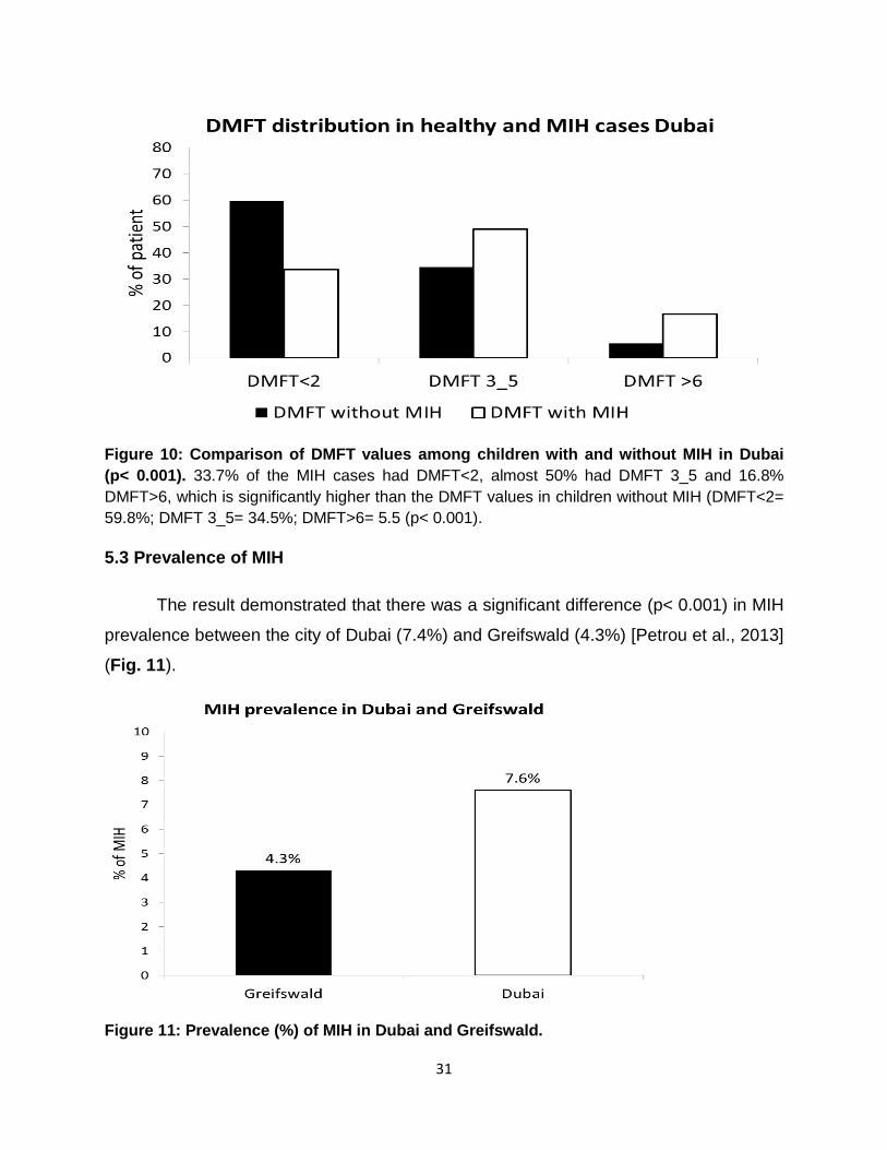
The result demonstrated that there was a significant difference (p< 0.001) in MIH
prevalence between the city of Dubai (7.4%) and Greifswald (4.3%) [Petrou et al., 2013]
(Fig. 11).
Figure 11: Prevalence (%) of MIH in Dubai and Greifswald.
32
5.3.1 Distribution of MIH criteria
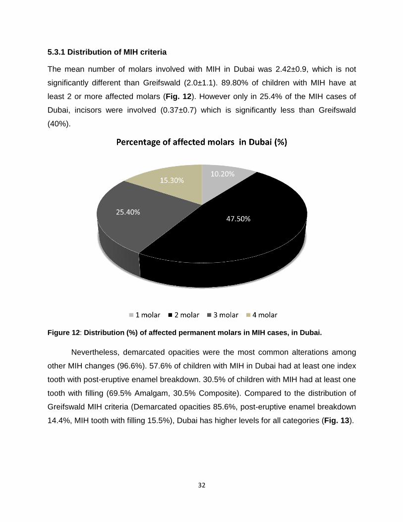
The mean number of molars involved with MIH in Dubai was 2.42±0.9, which is not
significantly different than Greifswald (2.0±1.1). 89.80% of children with MIH have at
least 2 or more affected molars (Fig. 12). However only in 25.4% of the MIH cases of
Dubai, incisors were involved (0.37±0.7) which is significantly less than Greifswald
(40%).
Figure 12: Distribution (%) of affected permanent molars in MIH cases, in Dubai.
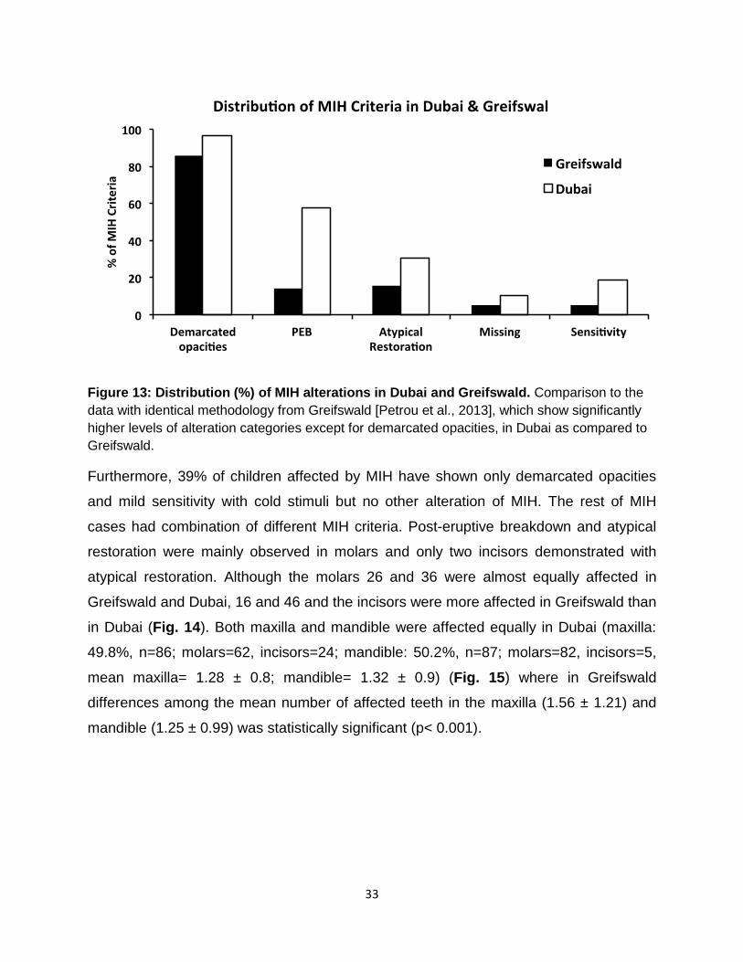
Nevertheless, demarcated opacities were the most common alterations among
other MIH changes (96.6%). 57.6% of children with MIH in Dubai had at least one index
tooth with post-eruptive enamel breakdown. 30.5% of children with MIH had at least one
tooth with filling (69.5% Amalgam, 30.5% Composite). Compared to the distribution of
Greifswald MIH criteria (Demarcated opacities 85.6%, post-eruptive enamel breakdown
14.4%, MIH tooth with filling 15.5%), Dubai has higher levels for all categories (Fig. 13).
33
Figure 13: Distribution (%) of MIH alterations in Dubai and Greifswald. Comparison to the
data with identical methodology from Greifswald [Petrou et al., 2013], which show significantly
higher levels of alteration categories except for demarcated opacities, in Dubai as compared to
Greifswald.
Furthermore, 39% of children affected by MIH have shown only demarcated opacities
and mild sensitivity with cold stimuli but no other alteration of MIH. The rest of MIH
cases had combination of different MIH criteria. Post-eruptive breakdown and atypical
restoration were mainly observed in molars and only two incisors demonstrated with
atypical restoration. Although the molars 26 and 36 were almost equally affected in
Greifswald and Dubai, 16 and 46 and the incisors were more affected in Greifswald than
in Dubai (Fig. 14). Both maxilla and mandible were affected equally in Dubai (maxilla:
49.8%, n=86; molars=62, incisors=24; mandible: 50.2%, n=87; molars=82, incisors=5,
mean maxilla= 1.28 ± 0.8; mandible= 1.32 ± 0.9) (Fig. 15) where in Greifswald
differences among the mean number of affected teeth in the maxilla (1.56 ± 1.21) and
mandible (1.25 ± 0.99) was statistically significant (p< 0.001).

34
Figure 14: Comparison of teeth distribution (%) with MIH in Dubai and Greifswald.
Comparison to the data with identical methodology from Greifswald [Petrou et al., 2013]
Figure 15: Distribution (%) of MIH in maxilla and mandible. Comparison to the data with
identical methodology form Greifswald [Petrou et al., 2013]. Dubai (Maxilla: 49.8%); (Mandible:
50.2%). Greifswald (Maxilla: 61%) (Mandible: 39%). Although there is no significant difference in
the mean number of affected teeth per jaw in Dubai (maxilla= 2.28 ± 0.81; mandible= 2.32 ±
0.93) it is statistically significant (p< 0.001) in Greifswald (maxilla= 1.56 ± 1.21; mandible= 1.25
± 0.99).
35
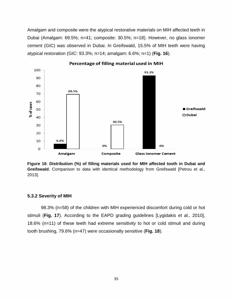
Amalgam and composite were the atypical restorative materials on MIH affected teeth in
Dubai (Amalgam: 69.5%; n=41; composite: 30.5%; n=18). However, no glass ionomer
cement (GIC) was observed in Dubai. In Greifswald, 15.5% of MIH teeth were having
atypical restoration (GIC: 93.3%; n=14; amalgam: 6.6%; n=1) (Fig. 16).
Figure 16: Distribution (%) of filling materials used for MIH affected tooth in Dubai and
Greifswald. Comparison to data with identical methodology from Greifswald [Petrou et al.,
2013].
5.3.2 Severity of MIH
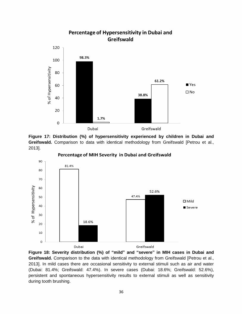
98.3% (n=58) of the children with MIH experienced discomfort during cold or hot
stimuli (Fig. 17). According to the EAPD grading guidelines [Lygidakis et al., 2010],
18.6% (n=11) of these teeth had extreme sensitivity to hot or cold stimuli and during
tooth brushing, 79.6% (n=47) were occasionally sensitive (Fig. 18).
36
Figure 17: Distribution (%) of hypersensitivity experienced by children in Dubai and
Greifswald. Comparison to data with identical methodology from Greifswald [Petrou et al.,
2013].
Figure 18: Severity distribution (%) of “mild” and “severe” in MIH cases in Dubai and
Greifswald. Comparison to the data with identical methodology from Greifswald [Petrou et al.,
2013]. In mild cases there are occasional sensitivity to external stimuli such as air and water
(Dubai: 81.4%; Greifswald: 47.4%). In severe cases (Dubai: 18.6%; Greifswald: 52.6%),
persistent and spontaneous hypersensitivity results to external stimuli as well as sensitivity
during tooth brushing.
37
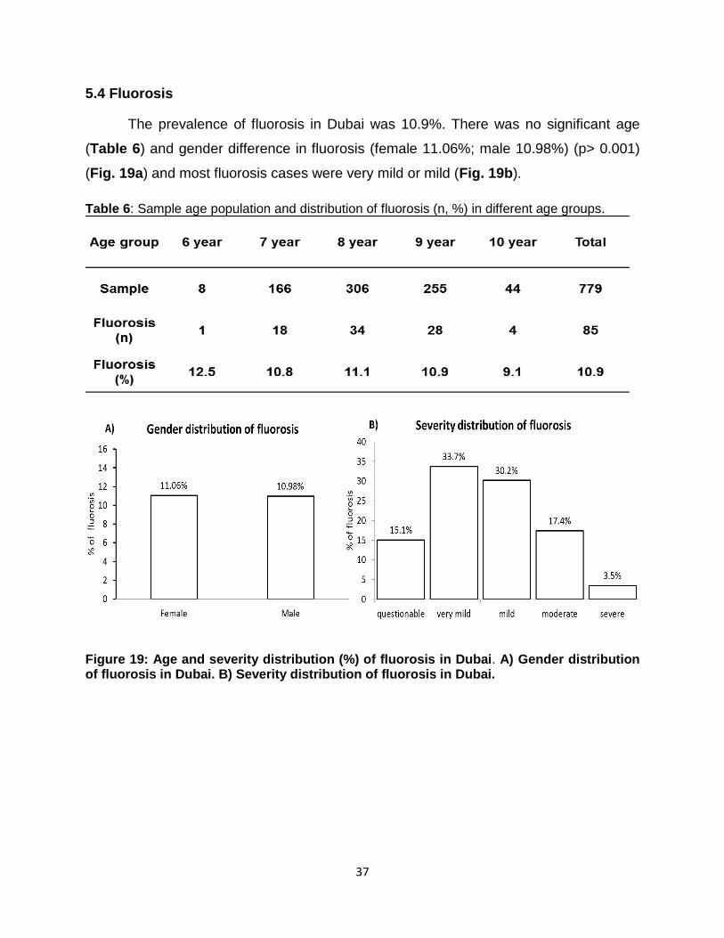
5.4 Fluorosis
The prevalence of fluorosis in Dubai was 10.9%. There was no significant age
(Table 6) and gender difference in fluorosis (female 11.06%; male 10.98%) (p> 0.001)
(Fig. 19a) and most fluorosis cases were very mild or mild (Fig. 19b).
Table 6: Sample age population and distribution of fluorosis (n, %) in different age groups.
Figure 19: Age and severity distribution (%) of fluorosis in Dubai. A) Gender distribution of fluorosis in Dubai. B) Severity distribution of fluorosis in Dubai.
38
6. Discussion
6.1 Discussion of the aim
This is the first study to determine the prevalence of MIH in UAE and to compare
it directly to an equivalent study in Greifswald/Germany with different socioeconomic
status, geographic origin, and healthcare system. Both studies used the same
examination criteria and study design and the examiners were calibrated in identical
locations and with identical concepts. These standards allow a direct comparison
between different geographical and cultural areas. The results obtained from this
comparison study may contribute in understanding MIH and the etiological factors
associated with this condition. Furthermore, to compare the dental caries and fluorosis
with the literatures to evaluate the caries status and the prevention progress in Dubai,
as well as the fluorosis status in this region.
6.2 Discussion of the method
6.2.1 Sample
To have a standard and representative sample size for the Dubai study, the
sample size was calculated statistically using a NQuery Advisor software (version 4.0,
Statistical Solutions Ltd. Ireland). Based on this calculation, representative sample size
was determined to be about 835 children for a prevalence of 10% and a 95%
confidence interval within a range of 8-12%. An identical calculation method was
performed for the sample in the Greifswald study. To avoid the risk of selection bias and
to have the chance of examining all healthy children, the sample in Dubai study was
collected from governmental schools where the obligatory oral examination are
emphasized by the ministry of health. This was considered because children attending
dental clinics or university hospitals were those who are requiring treatment, having
emergency condition (e.g. pain, high caries level, trauma, MIH), or/and a general health
problem [Jälevik, 2010]. The same pattern was performed in sample selection of the
Greifswald study, making both studies representative and comparable. In addition, the
schools in the Greifswald sample were selected randomly by the community services to
avoid any possible selection bias and the schools in Dubai were also selected randomly
39
from a list of governmental school, which was received from the Ministry Of Education
(MOE), therefore, increasing the internal validity of the study. In both studies, the
specific age group (i.e., 7-91 year) was chosen according to the eruption time of first
molars and incisors to avoid the masking effect from advanced carious lesions and
extraction, which can minimize the chance of proper diagnosis [Balmer et al., 2012].
6.2.2 Calibration
The examiner in this study was calibrated with the gold standard as in the
German study and reached high Kappa values (Kappa value>0.9). This makes the
study strongly comparable and the data obtained through this procedure reliable. In
addition, to ensure a high inter-examiner reliability, the calibration was conducted at
different sites (i.e., site A: university library and site B: university dental clinic). During
the calibration sessions, 20 photographs demonstrating various enamel defects with
different level of severity as well as different MIH scores were used. This calibration
method has been proved to be the standard method for MIH diagnosis for many other
studies [Elfrink et al., 2009; Wong et al., 2005].
6.2.3 Data Collection Forms
The evaluation process of MIH in both studies was preformed according to the
EAPD criteria (Table 2). Demographic parameters (i.e., age, gender), DMFT/dmft
values as well as fluorosis and its different degrees were recorded (Attachment No. X).
The data collection sheet was identical for all children. All the healthy, MIH and fluorosis
cases were recorded on one sheet. Different criteria and degrees of MIH and fluorosis
were coded according to the standard criteria to accelerate the recording and to ease
the analysis (Attachment No. XI, XII).
6.2.4 Examination and diagnosis of MIH, caries, and fluorosis
The EAPD criteria were used for the evaluation of MIH in Dubai to avoid
diagnostic errors, which were reported in previous studies using the MIH criteria
established before 2003 (Table 1) [Dietrich et al., 2003; Balmer et al., 2012]. This
standard evaluation process allows clinicians to recognize and differentiate MIH
40
accurately from any other enamel defects [Weerheijm et al., 2003]. The EAPD criteria
allow a standard comparison with the Greifswald and other international studies which
had used the same criteria [Weerheijm et al., 2003; Lygidakis et al., 2010]. A defect size
of less than 1 mm was not recorded as a MIH case, since it can result in misdiagnosis
[Jälevik, 2010]. The recorded affected tooth with MIH, usually presented a single
criterion or combination of criteria on different surfaces of the same tooth, which
confirms the strength of diagnosis in this study. Defects on incisors only, without a molar
involvement, were excluded as has been suggested, since incisor defects alone can be
as a result of local trauma, local inflammation, fluorosis, and hypoplasia [Chawla et al.,
2008a]. To determine the MIH severity in Dubai, the hypersensitive teeth were recorded
based on child responses to standard questions, which were also asked in the
Greifswald study (Section 3.4.3). According to the EAPD guidelines [Lygidakis et al.,
2010], having pain during brushing MIH teeth is a symptom of severe form of MIH, this
standard gave us an assurance not to encounter complication in diagnosis of MIH cases
that are associated with high caries levels, since about 66% of the MIH cases had high
DMFT values (DMFT>3) (Fig. 10; Table 5). This standard gives no value in mild MIH
cases, as the hypersensitivity in mild cases cannot be differentiated when both MIH and
dmft/DMFT are associated, however standard diagnostic criteria of EAPD and strong
calibration, reassure a correct diagnosis of MIH and differentiate it from caries. Standard
criteria were used to investigate caries and fluorosis in order to have standard results to
compare with the available studies in Dubai and to differentiate the findings from MIH
(Sections 3.4.1; 3.4.2).
6.2.5 Method of comparison
The data of the German study [Petrou et al., 2013] was used for comparison.
This study with an identical and a well-designed methodology allows a perceptible
comparison with the data from Dubai. The German study took place in few cities of
Germany including Greifswald, Heidelberg, Hamburg, and Düsseldorf. The city of
Greifswald was chosen due to access to the detailed data as well as the remarkable
socioeconomic and health system differences from Dubai. However, there is no
research in Dubai, which studied caries level in age group of 6 to 10 years old.
41
Therefore, a recent caries literature review [Al-Bluwi, 2014] and a study by El-Nadeef
[2009] were used to compare caries and fluorosis level. The age groups in both studies
are between 4 to 6 (mean dmft 5.1 to 8.4) and 12 (mean DMFT 1.6 to 3.24) years old.
6.3 Discussion of the results
6.3.1 MIH prevalence
Based on the results obtained in this study there is a significant difference in the
prevalence of MIH between Dubai/UAE and Greifswald/Germany [Dubai: 7.4%;
Greifswald: 4.3%;]. However, MIH prevalence in Dubai is quite low in comparison with
other cities in Germany (Düsseldorf=14.6%, Hamburg=14.0%) and Middle East
countries, where MIH prevalence ranges from 8.6% to 20% [Allazzam et al., 2014;
Zawaideh et al., 2011; Ghanim et al., 2013; Ghanim et al., 2011; Ghanim et al., 2014].
Considering the high calibration value (Kappa> 0.9) and the “gold standard”
investigator, who did the examination, this significant difference in MIH prevalence,
could not be caused by an examiner bias. It should be bearing in mind that the
prevalence of MIH in Dubai does not represent the MIH prevalence of the entire UAE
and further studies are advised to provide a representative prevalence for all United
Arab Emirates.
6.3.2 Distribution of MIH
The distribution of MIH obtained from Dubai/UAE was compared with
Greifswald/Germany and other international studies. As a result, no significant
difference (p> 0.001) in the distribution of MIH could be found for the variable gender in
Dubai, which is comparable with the Greifswald study (Dubai: Female 7.57% and Male
7.58%; Greifswald: 4.21% Male and 4.37% Female). This was in agreement with other
international studies [Leppäniemi et al., 2001; Jälevik et al., 2001; Calderara et al.,
2005; Muratbegovic et al., 2007; Chawla et al., 2008a; Jasulaityte et al., 2008; Martínez
Gómez et al., 2012]. Unlike Greifswald, no significant difference (p> 0.001) in MIH
distribution in the upper and lower jaw was found in Dubai (Maxilla 49.8% and Mandible
50.2%) which is comparable to the international studies (Fig. 15) [Weerheijm et al.,
42
2001b; Chawla et al., 2008a; Cho et al., 2008; Ghanim et al., 2011]. Mean value of
affected teeth per child with MIH in Dubai (2.7±1.1) is comparable to Greifswald
(2.8±1.7) and other international studies (2.4 to 5.7) [Calderara et al., 2005; Cho et al.,
2008; Jälevik et al., 2001]. Incisors affected with MIH in Dubai (25.4%) (mean 0.37±0.7),
were significantly less than Greifswald (40%). However, it is comparable to the
international reports (5.26% to 57.89%) [Jasulaityte et al., 2008; Lygidakis et al., 2008].
Mean value of affected molars per child with MIH (2.42±0.9) (Fig. 12), was found in
Dubai to be comparable to Greifswald (2.0±1.1) and to other international studies (1.5 to
3.16) [Jälevik et al., 2001; Dietrich et al., 2003; Petrou et al., 2013]. Demarcated opacity
was the most common criterion of MIH in Dubai and Greifswald, (Dubai: 96.6%;
Greifswald: 85.6%) (Fig. 13) and other international studies [Jasulaityte et al., 2007;
Muratbegovic et al., 2008; Soviero et al., 2009; Ghanim et al., 2011; Petrou et al., 2013;
Heimüller et al., 2012]. Despite the fact of being uncertain about the previous status of
the MIH tooth with restoration, atypical restorations are considered to be a severe form
of MIH due to the invasion [Wogelius et al., 2008]. Nevertheless, 30.5% of children with
MIH had at least one tooth with a filling, this was significantly higher than in Greifswald
(15.5%). Although composite and glass ionomer cement (GIC) are the restorations of
choice in MIH cases [Lygidakis, 2008; Fayle, 2003], no glass ionomer cement (GIC)
was detected in Dubai and mostly amalgam was observed in MIH cases (Amalgam:
69.5%; n=41; composite: 30.5%; n=18) (Fig. 16). Severity and sensitivity due to
structural breakdown were associated with a high number of MIH affected teeth [Petrou
et al., 2013; Jälevik et al., 2001; Leppäniemi et al., 2001; Jasulaityte et al., 2007;
Chawla et al., 2008a-b; Ghanim et al., 2011] as well as caries. 98.3% of the children in
Dubai experienced tooth sensitivity, which could be also due to high caries level (Fig.
17). However, only 18.6% of the MIH cases had severe hypersensitivity, especially
during tooth brushing (Fig.18), which is a specific sign of MIH. Older children with MIH
were more likely to have severe lesions compared to Greifswald, which can be due to
the difference in implementing the prevention and treatment methods between the two
cities (Fig. 16) [Leppäniemi et al., 2001; Lygidakis et al., 2008a; Jasulaityte et al., 2008;
Petrou et al., 2013].
43
6.3.3 Distribution of possible etiological factors of MIH
Since the examiners in Dubai had no access to the information related to
etiological factors (e.g. environmental factors, antibiotics use, breastfeeding, C-section,
etc.) of the selected sample population, no specific and valid results regarding
etiological factors of MIH can be concluded. Although the main purpose of this study
was not to determine the etiological factors related to MIH, some probable assumptions
can be drawn from our results, and available local and international data. However, the
potential differences between Dubai and Greifswald and other Middle East studies can
be as a result of the variance between the cities in multiple acquired factors, including
environmental, healthcare, and socioeconomic factors [Steele, 2014; Struzycka, 2014;
Winter, 1990].
Environmental Factors: Environment factors are among the most important etiological
factors that might cause MIH. These results were obtained from studies that have
compared two environmentally different areas such as urban and rural areas [Souza et
al., 2012]. However, some other studies have shown no significant difference between
the prevalence of MIH in polluted and non-polluted areas [Kusku, 2008, Kusku et al.,
2009; Petrou et al., 2013]. To avoid this possible confusion factor bias in our study, the
schools and their locations were selected randomly and if there are more than one
school in the same area, one was excluded. Nevertheless Dubai air pollution is at a high
level, mainly due to the rapid growth of industry and increased use of automobiles. It
was found by Roads and Transport Authority (RTA), that there is an increase of 12%
per year in using automobiles, which is five times more than other areas of the world
[RTA, 2008]. This can cause an increase of different harmful pollutants, including
nitrogen oxides, hydrocarbons, carbon monoxide, and carbon dioxide, which can
indicate the pollution as one of the risk factors in Dubai [Al-Zubaidi et al., 2002; Corder,
2008; RTA, 2009]. However such pollution is not a concern in Greifswald [UBA, 2003]
since the number of vehicles are low in this area. Comparing the result of our study with
the Germany and other international, and Middle East studies, population tend to be an
environmental risk factor, since cities with higher population showed higher prevalence
(e.g. Dubai population 2.2 million=7.6% MIH; Hamburg population 1.8 million=14% MIH;

44
Düsseldorf population 1.5 million=14.6% MIH [Petrou et al., 2013]; Mosul, Iraq
population 1.8million=21.5% MIH [Ghanim et al., 2011]; Shiraz, Iran population 1.7
million= 20.2% MIH [Ghanim et al., 2014]).
Antibiotics: Studies have shown that antibiotics are being misused in Middle Eastern
countries including UAE [Habibzadeh, 2012; Bin Abdulhak, 2011]. An important
contributing factor for this phenomenon is the cultural habit [Habibzadeh, 2012] where
antibiotics are used without prescription either as a self-treatment action [Abasaeed,
2009] or pharmacists are willing to provide patients with antibiotics without prescription
[Zaghloul, 2013; Habibzadeh, 2012; Bin Abdulhak, 2011]. The excess use of antibiotics
have been linked with MIH due it’s possible negative effect on the amelogenesis phase
of maturation [Alaluusua, 2010; Chawla et al., 2008a-b; Lygidakis et al., 2008b;
Lygidakis et al., 2010]. Nevertheless MIH prevalence is low despite high antibiotic
prescription in Dubai, which was also demonstrated in the German study where higher
prevalence of MIH in cities with low antibiotic prescriptions compared to cities with high
antibiotic prescriptions was observed [Petrou et al., 2013; BARMER, 2010; de With et
al., 2004]. However, neither the study in Dubai nor in Greifswald had direct information
regarding antibiotic use by the selected sample population and conclusions are based
on the reports from health system regulation regarding antibiotics prescription and
available studies regarding antibiotics use in UAE [BARMER, 2010; de With et al., 2004;
Zaghloul, 2013; Habibzadeh, 2012; Bin Abdulhak, 2011; Abasaeed, 2009].
6.3.4 DMFT/dmft
The DMFT/dmft indices in Dubai were recorded to be high (DMFT: 2.41±1.7;
dmft: 5.46±3.1) and comparable to other studies in UAE (DMFT mean: 1.6 to 3.24; dmft
mean: 5.1 to 8.4) [Al-Bluwi, 2014; El-Nadeef, 2009], which supports the strength of the
outcomes in this study. The caries prevalence obtained in this study and the most
recent study [Al-Bluwi, 2014], are relatively similar compared to a previous study [El-
Nadeef, 2009], which indicates no progress in the caries prevention. Therefore, an
appropriate caries prevention program is needed to control the caries progress in Dubai.
In addition, the mean DMFT/dmft indices in Dubai were higher than Greifswald (DMFT:
45
0.1±0.4; dmft: 0.9±1.7). The fact of high caries prevalence has been reported and
discussed in different studies performed in the UAE different regions [Al-Bluwi, 2014;
Hashim et al., 2013; Hashim et al., 2011; El-Nadeef, 2009; Hashim et al., 2009; Ur
Rehman et al., 2008; Hashim et al., 2008; Hashim et al., 2006]. Based on the reported
fact, MIH cases were recorded carefully by a strongly calibrated examiner, using the
EAPD criteria, which allow differentiating MIH from other enamel defects, the possibility
of a masking effect of high caries prevalence in this study has been minimized. Higher
DMFT/dmft values associated with MIH defect was observed in this study (Table 5),
which have been reported in many international studies [Leppäniemi et al., 2001;
Weerheijm et al., 2003] as well as in the German study [Petrou et al., 2013]. This
association indicates that MIH can increase caries susceptibility, specifically due to the
weak structure of the affected tooth and the limitation of the oral hygiene maintenance
as a result of discomfort and pain [Lygidakis et al., 2010]. Therefore, early diagnosis
and treating hypersensitive MIH teeth are the optimal prevention measures to reduce
caries levels in MIH patients.
6.3.5 Fluorosis
The prevalence of fluorosis in the age group of 7-91 year old in Dubai is 10.9%
(Fig. 18). No significant age (Table 6) and gender (female 11.06%; male 10.98%)
differences were observed (Fig. 19a) (p> 0.001). The majority of these cases were
presented in a form of a very mild to mild (63.9%) form according to the Dean’s criteria
(Fig. 19b). Currently, there is no study reporting the prevalence of fluorosis in
Greifswald. However, the prevalence of fluorosis in Germany ranged from 4.9% to
11.3% and the majority of the cases were presented in a mild form of fluorosis [Pieper
et al., 2008; Momeni et al., 2007]. Although, there is no water fluoridation in Germany
and UAE, and the concentration of the fluoride in water supplements in UAE is lower
than the recommended amount (1mg/L), the reason why fluorosis prevalence is still
high, is unclear [Nimr, 1997; Momeni et al., 2007]. Although the prevalence of fluorosis
in Dubai is relatively high, it does not provoke serious health concerns, since it is mostly
“very mild, mild, and moderate” (based on Dean’s criteria), and only affecting the
appearance of the teeth (i.e., esthetic issue) with no detrimental effect on teeth
46
structures [Muñoz, 2013; Abanto Alvarez, 2009]. In fact, teeth affected with moderate
fluorosis have shown to be more resistant to caries lesions [Waidyasekera, 2007].
Therefore, the treatment option of fluorosis cases in Dubai is mostly esthetic to cover
the discoloration. However, this condition can be prevented by controlling the fluoride
intake during the teeth development.
6.4 Discussion of the challenges
In this study, we reported the prevalence of MIH in Dubai/UAE and compared it
with Greifswald/Germany using identical diagnostic criteria and the same calibration
system, which gave us a reliable prevalence. The strong methodology in this study of
one examiner who was previously calibrated according to the standard calibration
method, the examination in identical locations (i.e. schools), using similar clinical
conditions (i.e. Class rooms), and standard instruments, increased the value of these
results. A possible challenge was MIH masked due to the high prevalence of fluorosis
and caries in Dubai. However, the standard EAPD criteria and strong calibration has
reduced this challenge. The use of similar comparison approach between Middle
Eastern and European studies has not been well explored or applied yet due to
absence of access to patients medical histories, which could potentially provide a
powerful mean to determine the etiological factors of MIH. Nevertheless, the lack of
access to the medical record of the sample group in Dubai was also a challenge, which
limited further comparison of etiological factors. Another limitation is that the sample
collected from one city (e.g. Dubai) cannot provide a representative prevalence for the
whole country (UAE). Therefore, further studies of the prevalence of MIH as well as its
local contributing factors in UAE, Middle East, and Europe should be initiated for the
aim of having a more comprehensive picture of the MIH prevalence in the region, as
well as to provide evidence that increase the understanding of the main etiological
factors of this condition. The German study and specifically the Greifswald data were
successfully used as reference for comparison in this study due to the strong and
similar methodology used in Greifswald study and also due to the opportunity and
access to the detailed data, facilities for training and calibration through the same
process.
47
7. Conclusions
Here, we presented the prevalence of MIH in Dubai/UAE for the first time, which
represents a developed Middle Eastern city and compared it to results obtained from
Greifswald/Germany, which represents a developed European city. The results have
shown that the prevalence of MIH in Dubai/UAE is higher than Greifswald/Germany.
However, in comparison to the literature, the prevalence of MIH in Dubai is lower than
other Middle Eastern cities.
Furthermore, we have shown that there is a higher caries level associated with
MIH in Dubai. This is also true in Greifswald, Germany and other international studies
[Jeremias et al., 2013; Grošelj et al., 2013; Lygidakis, 2010]. In addition, we have
reported the prevalence of caries and fluorosis in Dubai and compared them to
Greifswald and the previous studies in Dubai. Nevertheless, caries values presented in
this study and previous studies indicate that strong attention is required from health
authority to this topic.
This research provides a strong and comparable source of information on the
prevalence of MIH in Dubai for other studies, since it followed strictly all methodological
and clinical standards suggested for the assessment and diagnosis of MIH, which are
the EAPD criteria [Weerheijm et al., 2001b; Lygidakis et al., 2010].
The findings presented in this study require particular attention from the local
health authorities and general practitioners for such developmental defect to facilitate
early and adequate diagnosis and treatment. This could be achieved by implementing
continuing education courses on MIH detection, diagnosis, and treatment for general
practitioners. Furthermore, this study has the potential to trigger new studies that would
help in understanding the MIH etiology.
48
8. Summary
Background: Defects in enamel mineralization occur during tooth development
resulting in structural breakdown. Among such defects, Molar Incisor
Hypomineralization (MIH) is one of the most common enamel defects that occur in
children, but the literature is still lacking data on the occurrence and etiology of MIH.
Identifying this defect in population is of particular importance due to the association
with increase of caries susceptibility and interference with treatment planning.
Aim: The purpose of this study was to find the prevalence of MIH in Dubai/United Arab
Emirates (UAE) and to compare it with MIH prevalence in Greifswald/Germany.
Furthermore, to explores prevalence of fluorosis and caries in Dubai in order to assess
the chance and influence of differential diagnoses and to analyze the possible
relationship between MIH and caries level. In addition, dental caries and fluorosis
prevalence in this study will be compared with valid literatures in Dubai, which could
possibly be a trigger for further studies to develop adequate prevention and treatment
procedures.
Design: In this cross-sectional study, 779 children (female: 513; male: 261) age of 7 to
9 (±1) year-old in the city of Dubai/UAE were randomly selected and examined. The
examination was carried out for children in Dubai governmental schools by one
examiner who had previously been trained and calibrated with the MIH diagnostic
criteria of the European Academy of Pediatric Dentistry (EAPD), fluorosis diagnostic
criteria of Dean, and dmft/DMFT values for caries diagnosis. The data from Greifswald
had been collected in year 2011 by the community dental services from 443 children,
with same age group.
Results: The percentage of children with MIH in Dubai/UAE was 7.6% and higher than
in Greifswald with 4.3%. There was no significant gender difference in Dubai (7.57%
female, 7.58% male) or Greifswald (4.34% female, 4.21% male). The DMFT/dmft
indices were generally high in Dubai with a mean DMFT of 2.41±1.7 and dmft 5.46±3.1,
while Greifswald had much lower caries values (DMFT 0.1±0.5 and dmft 0.9±1.7). The
difference of DMFT index between children with and without MIH was statistically

49
significant for Dubai (p< 0.001). However caries prevalence is comparable with the
results from the previous caries studies in Dubai.
Conclusion: The prevalence of MIH and, especially, the caries values are higher
among the children population of Dubai/UAE compared to Greifswald/Germany and no
difference in fluorosis and caries prevalence’s were observed in comparison to previous
studies in Dubai. These findings require particular attention from the health authorities
and the local dentists to such defects that might facilitate adequate prevention and
treatment options.
50
9. Reference
1. Abanto Alvarez J, Rezende KM, Marocho SM, Alves FB, Celiberti P, Ciamponi
AL. Dental fluorosis: exposure, prevention and management. Med Oral Patol
Oral Cir Bucal. 2009; 14:103-107
2. Abasaeed A, Vlcek J, Abuelkhair M, Kubena A. Self-medication with antibiotics
by the community of Abu Dhabi Emirate, United Arab Emirates. J Infect Dev
Ctries. 2009; 3:491-497
3. Abbas S, Ihle P, Heymans L, Küpper-Nybelen J, Schubert I. Differences in
antibiotic prescribing between general practitioners and pediatricians in Hesse,
Germany. Dtsch Med Wochenschr. 2010; 135:1792-1797
4. Abouleish M.Y.Z. Concentration of Selected Anions in Bottled Water in the
United Arab Emirates. Water. 2012; 4:496-509
5. Adriaenssens N, Coenen S, Versporten A, Muller A, Minalu G, Faes C,
Vankerckhoven V, Aerts M, Hens N, Molenberghs G. European Surveillance of
Antimicrobial Consumption (ESAC): outpatient antibiotic use in Europe (1997–
2009), Antimicrobial Chemotherapy. 2011; 66 Suppl 6:57-70
6. Alaluusua S, Bäckman B, Brook AH, Lukinmaa PL. Developmental defects of the
dental hard tissues and their treatment. In: Koch G., Poulsen S. Pediatric
Dentistry. A clinical approach. Blackwell Munksgaard, Copenhagen. 2001: 273-
299
7. Alaluusua S. Aetiology of Molar Incisor Hypomineralisation: A systematic review.
Eur Arch Paediatr Dent. 2010; 11:53-58
8. Alaluusua S, Lukinmaa PL, Koskimies M, Pirinen S, Hölttä P, Kallio M, Holttinen
T, Salmenperä L. Developmental dental defects associated with long breast
feeding. Eur J Oral Sci. 1996; 104:493-497
9. Alam M, Raza SJ, Sherali AR, Akhtar AS. Neonatal complications in infants born
to diabetic mothers. J Coll Physicians Surg Pak. 2006; 16:212-215
10. Al-Bluwi GS. Epidemiology of dental caries in children in the United Arab
Emirates. Int Dent J. 2014; 64:219-228
51
11. Allazzam SM, Alaki SM, El Meligy OA. Molar incisor hypomineralization,
prevalence, and etiology. Int J Dent. 2014; 2014:234508
12. Al-Zubaidi MS, Sabie, K K. Sustainable Transportation in United Arab Emirates. J
Transportation Research. 2002; 2:63-73
13. Arrow P. Prevalence of developmental enamel defects of the first permanent
molars among school children in Western Australia. Aust Dent J. 2008; 53:250-
259
14. Aoba T, Fejerskov O. Dental fluorosis: chemistry and biology. Critical Reviews of
Oral Biology and Medicine. 2002; 13:155-170
15. Bailleul-Forestier I, Berdal A, Vinckier F, de Ravel T, Fryns JP, Verloes A. The
genetic basis of inherited anomalies of the teeth. Part 2: syndromes with
significant dental involvement. Eur J Med Genet. 2008; 51:383-408
16. Balmer R, Toumba J, Godson J, Duggal M.The prevalence of molar incisor
hypomineralisation in Northern England and its relationship to socioeconomic
status and water fluoridation. Int J Paediatr Dent. 2012; 22:250-257
17. BARMER GEK 2010. https://antibiotika.faktencheck-gesundheit.de/antibiotika-
regional/ (page reviewed 28. Nov. 2014)
18. Bhaskar SA, Hegde S. Molar-incisor hypomineralization: Prevalence, severity
and clinical characteristics in 8 to 13 year old children of Udaipur, India. J Indian
Soc Pedod Prev Dent. 2014; 32:322-329
19. Bin Abdulhak AA, Altannir MA, Almansor MA, Almohaya MS, Onazi AS, Marei
MA, Aldossary OF, Obeidat SA, Obeidat MA, Riaz MS, Tleyjeh IM. Non
prescribed sale of antibiotics in Riyadh, Saudi Arabia: a cross sectional study.
BMC Public Health. 2011; 11:538
20. Brook AH, Elcock C, Hallonsten A-L, Poulsen S, Andreasen J, Koch G,Yeung
CA, Dosanjh T. The development of a new index to measure enamel defects. In:
Brook AH (ed). Dental Morphology. Sheffield, United Kingdom: Sheffield
Academic Press. 2001; 59-66
21. BfR (Bundesinstitut für Risikobewertung). Durchschnittlicher Fluoridgehalt in
Trinkwasser ist in Deutschland niedrig. Information Nr. 037/2005 des BfR vom
12.07.2005
52
22. Burt BA. The changing patterns of systemic fluoride intake. J Dent Res. 1992;
71:1228-1237
23. Calderara PC, Gerthoux PM, Mocarelli P, Lukinmaa PL, Tramacere PL,
Alaluusua S. The prevalence of Molar Incisor Hypomineralisation (MIH) in a
group of Italian school children. Eur J Paediatr Dent. 2005; 6:79-83
24. Chawla N, Messer LB, Silva M. Clinical studies on molar incisor
hypomineralisation part 1: distribution and putative associations. Eur Arch
Paediatr Dent. 2008a; 9:180-190
25. Chawla N, Messer LB, Silva M. Clinical studies on molar-incisor-
hypomineralisation part 2: development of a severity index. Eur Arch Paediatr
Dent. 2008b; 9:191-199
26. Cho SY, Ki Y, Chu V. Molar incisor hypomineralization in Hong Kong Chinese
children. Int J Paediatr Dent. 2008; 18:348-352
27. Clarkson J. Review of terminology, classifications, and indices of developmental
defects of enamel. Adv Dent Res. 1989; 3: 104-109
28. Corder A. "Dubai's traffic pollution among world's worst". Arabian Bussines.com.
Dubai. 7 March 2008. http://www.arabianbusiness.com/dubai-s-traffic-pollution-
among-world-s-worst-52827.html#.VH8hnjGG91Y (Page viewed Nov. 2014)
29. Crombie F, Manton D, Kilpatrick N. Aetiology of molar incisor hypomineralisation:
a critical review. Int J Paediatr Dent. 2009; 19: 73-83
30. Da Costa-Silva CM, Jeremias F, de Souza JF, Cordeiro Rde C, Santos-Pinto L,
Zuanon AC. Molar incisor hypomineralization: prevalence, severity and clinical
consequences in Brazilian children. Int J Paediatr Dent. 2010; 20: 426-434
31. Da Costa-Silva CM, Ambrosano GM, Jeremias F, De Souza JF, Mialhe FL.
Increase in severity of molar incisor hypomineralization and its relationship with
the color of enamel opacity: a prospective cohort study. Int J Paediatr Dent.
2011; 21:333-341
32. DAJ. (Deutsche Arbeitsgemeinschaft für Jugendzahnpflege) Grundsätze für
Maßnahmen zur Förderung der Mundgesundheit im Rahmen der
Gruppenprophylaxe nach § 21 SGB V. Geändert am 20.06.2000

53
33. Dean, 1942. As Reproduced in “Health Effects of Ingested Fluoride” National
Academy of Sciences. 1993; pp.169
34. Den Besten PK, et al. Effects of fluoride on rat dental enamel matrix proteinases.
Archives of Oral Biology. 2002; 47: 763-770
35. Den Besten PK. Dental fluorosis: its use as a biomarker. Adv Dent Res. 1994;
8:105-110.
36. de With K, Schröder H, Meyer E, Nink K, Hoffmann S, Steib-Bauert M,
Kämmerer R, Rueß S, Daschner FD, Kern WV. Antibiotikaanwendung in
Deutschland im europäischen Vergleich. Deutsche Medizinische Wochenschrift
2004; 129:1987-1992
37. Dietrich G, Sperling S, Hetzer G. Molar incisor hypomineralisation in a group of
children and adolescents living in Dresden (Germany). Eur J Paediatr Dent.
2003; 4:133-137
38. Elfrink ME, Schuller AA, Weerheijm KL, Veerkamp JS. Hypomineralized second
primary molars: prevalence data in Dutch 5 year olds. Caries Res. 2008; 42:282-
285
39. Elfrink ME, Ten Cate JM, Jaddoe VW, Hofman A, Moll HA, Veerkamp JS.
Deciduous Molar Hypomineralization and Molar Incisor Hypomineralization. J
Dent Res. 2012; 91:551-555
40. Fagrell TG, Dietz W, Jälevik B, Norén JG. Chemical, mechanical and
morphological properties of hypomineralized enamel of permanent first molars.
Acta Odontol Scand. 2010; 68:215-222
41. Fagrell TG, Ludvigsson J, Ullbro C, Lundin SA, Koch G. Aetiology of severe
demarcated enamel opacities, an evaluation based on prospective medical and
social data from 17,000 children. Swed Dent J. 2011; 35:57-67
42. Farah R, Drummond B, Swain M, Williams S. Linking the clinical presentation of
molar incisor hypomineralisation to its mineral density. Int J Paediatr Dent. 2010;
20:353-360
43. Fayle SA. Molar incisor hypomineralization: Restorative management. Eur J
Paediatr Dent. 2003; 4:121-126
54
44. FDI Commission on Oral Health Research & Epidemiology. A review of the
developmental defects of enamel index (DDE Index). Commission on Oral
Health, Research & Epidemiology. Report of an FDI Working Group. Int Dent J.
1992; 42:411-426
45. Featherstone JD. Dental caries: a dynamic disease process. Aust Dent J. 2008;
53:286-291
46. Garg N, Jain AK, Saha S, Singh J. Essentiality of early diagnosis of molar incisor
hypomineralization in children and review of its clinical presentation, etiology and
management. Int J Clin Pediatr Dent. 2012; 5:190-196
47. Ghanim A, Bagheri R, Golkari A, Manton D. Molar-incisor hypomineralisation: a
prevalence study amongst primary schoolchildren of Shiraz, Iran. Eur Arch
Paediatr Dent. 2014; 15:75-82
48. Ghanim A, Morgan M, Mariño R, Bailey D, Manton D. Molar-incisor
hypomineralisation: prevalence and defect characteristics in Iraqi children. Int J
Paediatr Dent. 2011; 21:413-421
49. Ghanim A, Manton D, Mariño R, Morgan M, Bailey D. Prevalence of demarcated
hypomineralisation defects in second primary molars in Iraqi children. Int J
Paediatr Dent. 2012; 13:111-118
50. Gottberg B, Berné J, Quiñónez B, Solórzano E. Prenatal effects by exposing to
amoxicillin on dental enamel in Wistar rats. Med Oral Patol Oral Cir Bucal. 2014;
19:38-43.
51. Grošelj M, Jan J. Molar incisor hypomineralisation and dental caries among
children in Slovenia. Eur J Paediatr Dent. 2013; 14:241-245.
52. Habibzadeh A. Antibiotic misuse and antibiotic-resistant bacteria. The Lancet
Middle East Edition, NIOC Medical Education and Research Center, Shiraz, Iran.
2012;http://download.thelancet.com/flatcontentassets/pdfs/Jan12_MiddleEastEd
_Antibiotics.pdf (Page viewed Nov. 2014)
53. Hall RK. Developmental defects of tooth enamel. In pediatric Orofacial Medicine
and Pathology 1st Ed. London Chapman and Hall, 1994; pp 10-11, pp 184-202, p
391

55
54. Hashim R, Thomson WM, Ayer K, Lewsely J, Awad M. Dental caries experience
and use of dental services among preschoolers in Ajman, UAE. International
Journal of Paediatric Dentistry. 2006; 16: 257-262
55. Hashim R, Thomson WM, Awad M. Prevalence of dental caries among young
children in the Arab countries. Dental News Journal. 2008; 15:17-26
56. Hashim R, Williams S, Thomson WM. Diet and caries experience among
preschool children in Ajman, United Arab Emirates. European Journal of oral
sciences. 2009; 117: 734-740
57. Hashim R, Fitzgerald R, Schafer C, Thomson WM. Mothers’ understanding of
dental- caries related feeding practices and children’s use of dental care in
Ajman. Social Science and Dentistry. 2011; 1: 97-107
58. Hashim R, Williams S, Thomson WM. Oral hygiene and dental caries in 5- to 6-
year-old children in Ajman, United Arab Emirates. Int J Dent Hyg. 2013; 11:208-
215
59. Heitmüller D, Thiering E, Hoffmann U, Heinrich J, Manton D, Kühnisch J,
Neumann C, Bauer CP, Heinrich-Weltzien R, Hickel R. Is there a positive
relationship between molar incisor hypomineralisations and the presence of
dental caries?. Int J Paediatr Dent. 2013; 23:116-124
60. Jackson RD, Kelly SA, Katz B, Brizendine E, Stookey GK. Dental fluorosis in
children residing in communities with different water fluoride levels: 33-month
follow-up. Pediatr Dent. 1999; 21:248-254
61. Jacobsen PE, Henriksen TB, Haubek D, Østergaard JR. Developmental Enamel
Defects in Children Prenatally Exposed to Anti-Epileptic Drugs. PLoS ONE 2013;
8:e58213
62. Jälevik B, Klingberg G, Barregård L, Norén JG. The prevalence of demarcated
opacities in permanent first molars in a group of Swedish children. Acta Odontol
Scand. 2001; 59:255-260
63. Jälevik B, Klingberg GA. Dental treatment, dental fear and behaviour
management problems in children with severe enamel hypomineralization of their
permanent first molars. Int J Paediatr Dent. 2002; 12:24-32
56
64. Jälevik B. Prevalence and Diagnosis of Molar-Incisor- Hypomineralisation (MIH):
A systematic review. Eur Arch Paediatr Dent. 2010; 11:59-64
65. Jälevik B, Noren J.G. "Enamel hypomineralization of permanent first molars: a
morphological study and survey of possible aetiological factors. Int J Paediatr
Dent. 2000; 10:278-289
66. Jan J, Sovcikova E, Kocan A, Wsolova L, Trnovec T. Developmental dental
defects in children exposed to PCBs in eastern Slovakia. Chemosphere. 2007;
67:350-354
67. Jasulaityte L, Veerkamp JS, Weerheijm KL. Molar incisor hypomineralization:
review and prevalence data from the study of primary school children in
Kaunas/Lithuania. Eur Arch Paediatr Dent. 2007; 8:87-94
68. Jasulaityte L, Weerheijm KL, Veerkamp JS. Prevalence of molar-incisor-
hypomineralisation among children participating in the Dutch National
Epidemiological Survey (2003). Eur Arch Paediatr Dent. 2008; 9:218-223
69. Jeremias F, de Souza JF, Silva CM, Cordeiro Rde C, Zuanon AC, Santos-Pinto
L. Dental caries experience and Molar-Incisor Hypomineralization. Acta Odontol
Scand. 2013; 71:870-876
70. Keene, H. "History of Dental Caries in Human Populations: The First Million
Years" Symposium and Workshop on Animal Models in Cariology, Sturbridge,
Massachusets,1980.http://www.uic.edu/classes/osci/osci590/11_1Epidemiology.
htm (Page viewed Nov. 2014)
71. Kellerhoff NM, Lussi A. "Molar-incisor hypomineralization". Schweiz Monatsschr
Zahnmed. 2004; 114:243-253
72. Khan SQ. Dental caries in Arab League countries: a systematic review and meta-
analysis.int Dent J. 2014; 64:173-180
73. Kidd EA. How 'clean' must a cavity be before restoration? Caries Res. 2004;
38:305-313
74. Koch G, Hallonsten AL, Ludvigsson N, Hansson BO, Holst A, Ullbro C.
Epidemiologic study of idiopathic enamel hypomineralization in permanent teeth
of Swedish children. Community Dent Oral Epidemiol. 1987; 15:279-85
57
75. Koch MJ, Garcia-Godoy F. The clinical performance of laboratory-fabricated
crowns placed on first permanent molars with developmental defects. J Am Dent
Assoc. 2000; 131:1285-1290
76. Kusku OO, Caglar E, Sandalli N. The prevalence and aetiology of molar-incisor
hypomineralisation in a group of children in Istanbul. Eur J Paediatr Dent. 2008;
9:139-144
77. Kuscu OO, Caglar E, Aslan S, Durmusoglu E, Karademir A, Sandalli N. The
prevalence of molar incisor hypomineralization (MIH) in a group of children in a
highly polluted urban region and a windfarm-green energy island. Int J Paediatr
Dent. 2009; 19:176-185
78. Laisi S, Ess A, Sahlberg C, Arvio P, Lukinmaa PL, Alaluusua S. Amoxicillin may
cause molar incisor hypomineralization. J Dent Res. 2009; 88:132-136
79. Leppäniemi A, Lukinmaa PL, Alaluusua S. Nonfluoride hypomineralizations in the
permanent first molars and their impact on the treatment need. Caries Res. 2001;
35:36-40
80. Levy SM. An updated on fluorides and fluorosis. J Can Dent Assoc. 2003;
69;286-291
81. Lygidakis NA, Dimou G, Briseniou E. Molar-incisor-hypomineralisation (MIH).
Retrospective clinical study in Greek children. I. Prevalence and defect
characteristics. Eur Arch Paediatr Dent. 2008; 9:200-206
82. Lygidakis NA, Dimou G, Marinou D. Molar-incisor-hypomineralisation (MIH). A
retrospective clinical study in Greek children. II. Possible medical aetiological
factors. Eur Arch Paediatr Dent. 2008; 9:207-217
83. Lygidakis NA, Wong F, Jälevik B, Vierrou AM, Alaluusua S, Espelid I. Best
Clinical Practice Guidance for clinicians dealing with children presenting with
Molar-Incisor-Hypomineralisation (MIH): An EAPD Policy Document. Eur Arch
Paediatr Dent. 2010; 11:75-81
84. Lygidakis NA. Treatment modalities in children with teeth affected by molar-
incisor enamel hypomineralisation (MIH): A systematic review. Eur Arch Paediatr
Dent. 2010; 11:65-74
58
85. Mahoney P. Two dimensional patterns of human enamel thickness on deciduous
(dm1, dm2) and permanent first (M1) mandibular molars. Arch Oral Biol. 2010;
55:115-126
86. Alves dos Santos MP, Maia LC. Molar Incisor Hypomineralization:Morphological,
Aetiological, Epidemiological and Clinical Considerations, Contemporary
Approach to Dental Caries, Dr. Ming-Yu Li (Ed.), 2012; ISBN: 978-953-51-0305-
9. http://cdn.intechopen.com/pdfs-wm/32183.pdf (Page viewed Nov. 2014)
87. Martínez Gómez TP, Guinot Jimeno F, Bellet Dalmau LJ, Giner Tarrida L.
Prevalence of molar incisor hypomineralisation observed using transillumination
in a group of children from Barcelona (Spain).Int J Paediatr Dent. 2012; 22:100-
109
88. Martins VM, Sousa RV, Rocha ES, Leite RB, Paiva SM, Granville-Garcia AF.
Dental trauma among Brazilian schoolchildren: prevalence, treatment and
associated factors. Eur Arch Paediatr Dent. 2012; 13:232-237
89. Mascarenhas AK. Risk factors for dental fluorosis: a review of the recent
literature. Pediatr Dent. 2000; 22:269-277
90. Masumo R, Bårdsen A, Astrøm AN. Developmental defects of enamel in primary
teeth and association with early life course events: a study of 6-36 month old
children in Manyara, Tanzania. BMC Oral Health. 2013; 13:21
91. Mathu-Muju K, Wright JT. Diagnosis and treatment of molar incisor
hypomineralization. Compend Contin Educ Dent. 2006; 27:604-610
92. McKnight. A pilot study of esthetic perceptions of dental fluorosis vs. selected
other dental conditions. ASDC J Dent Child. 1998; 65:233-238
93. Momeni A, Neuhäuser A, Renner N, Heinzel-Gutenbrunner M, Abou-Fidah J,
Rasch K, Kröplin M, Fejerskov O, Pieper K. Prevalence of dental fluorosis in
German schoolchildren in areas with different preventive programmes. Caries
Res. 2007; 41:437-444
94. Muñoz MA, Arana-Gordillo LA, Gomes GM, Gomes OM, Bombarda NH, Reis A,
Loguercio AD. Alternative esthetic management of fluorosis and hypoplasia
stains: blending effect obtained with resin infiltration techniques. J Esthet Restor
Dent. 2013; 25:32-39
59
95. Mullen J. History of Water Fluoridation. British Dental Journal. 2005; 199:1 - 4
96. Muratbegovic A, Markovic N, Ganibegovic Selimovic M. Molar incisor
hypomineralisation in Bosnia and Herzegovina: aetiology and clinical
consequences in medium caries activity population. Eur Arch Paediatr Dent.
2007; 8:189-194
97. Muratbegovic A, Zukanovic A, Markovic N. Molar-incisor-hypomineralisation
impact on developmental defects of enamel prevalence in a low fluoridated area.
Eur Arch Paediatr Dent. 2008; 9:228-231
98. Murray JJ, Rugg-Gunn A. Fluorides and Caries Prevention London, ed 2.
Wright 1982
99. Nelson-Piercy C. Treatment of nausea and vomiting in pregnancy. When should
it be treated and what can be safely taken? 1998; 19:155-164.
100. Nimr HIS. Fluoride supplementation and sources of drinking water in Dubai.
Dental news. 1997; 19: 37-39
101. Nsanze H, Babarinde Z, Al Kohaly H. Microbiological quality of bottled drinking
water in the UAE and the effect of storage at different temperatures. Environ. Int.
1999; 25: 53-57
102. Schlesner OR, Fátima DD, Luciano C, Almeida RJ. Molar incisor
hypomineralization: three case reports and discussion of etiology, diagnosis, and
management strategies. J Stomatos. 2013; 19:4-9
103. Pendrys DG, Katz RV. Risk factors for enamel fluorosis in optimally fluoridated
children born after the US manufacturers’ decision to reduce the fluoride
concentration of infant formula. Am J Epidemiol. 1998; 148:967-974
104. Petrou MA, Giraki M, Bissar AR, Basner R, Wempe C, Altarabulsi MB, Schäfer
M, Schiffner U, Beikler T, Schulte AG, Splieth CH. Prevalence of Molar Incisor
Hypomineralisation among school children in four German cities. Int J Paediatr
Dent. 2014; 24:434-440
105. Pieper K, Neuhäuser A, Renner N, Abou-Fidah J, Rasch K, Kröplin M, Völkner-
Stetefeld P, Heinzel-Gutenbrunner M, Jablonski-Momeni A. Fluoroseprävalenz
bei 15-Jährigen in drei Regionen mit unterschiedlichen Prophylaxe-programmen.
Deutsche Zahnärztliche Zeitschrift. 2008; 63:16-29
60
106. Pieper K. Epidemiologische Begleituntersuchungen zur Gruppenprophylaxe 2009
Gutachten (DAJ). Bonn. 2010: 10-11;122-134
107. Population Bulletin Emirate of Dubai, Population and Vital Statistics, Dubai
statistics center 2013.
http://www.dsc.gov.ae/EN/Themes/Pages/Publications.aspx?TopicId=23 (Page
viewed Nov. 2014)
108. Preusser SE, Ferring V, Wleklinski C, Wetzel WE. Prevalence and severity of
molar incisor hypomineralization in a region of Germany - a brief communication.
J Public Health Dent. 2007; 67:148-150
109. Price, W. Nutrition and Physical Degeneration. New Canaan: Keats Publishing,
Inc. 1989
110. Radwan H. Patterns and determinants of breastfeeding and complementary
feeding practices of Emirati Mothers in the United Arab Emirates. BMC Public
Health. 2013; 13:171
111. Reid DJ, Dean MC. Variation in modern human enamel formation times. J Hum
Evol. 2006; 50:329-346
112. Richards LF, Westmoreland WW, Tashiro M, McKay CH, Morrison JT.
Determining optimum fluoride levels for community water supplies in relation to
temperature. J Am Dent Assoc. 1967; 74:389-397
113. Robke F.J, Buitkamp M. Häufigkeit der Nuckelflaschenkaries bei Vorschulkindern
in einer westdeutschen Großstadt. Oralprophylaxe. 2002; 24:59-65
114. RTA, (road and traffic authority) http://www.arabianbusiness.com/dubai-s-traffic-
pollution-among-world-s-worst-52827.html#.VGuM3_mG91Y (Page viewed Nov.
2014)
115. Sabel N, Klingberg G, Dietz W, Nietzsche S, Norén JG. Polarized light and
scanning electron microscopic investigation of enamel hypoplasia in primary
teeth. Int J Paediatr Dent. 2010; 20:31-36
116. Seow WK. Biological mechanisms of early childhood caries. Community Dent
Oral Epidemiol. 1998; 26:8-27
117. Seow WK Enamel hypoplasia in the primary dentition: a review. ASDC J Dent
Child. 1991; 58:441-451
61
118. Seow WK. Clinical diagnosis of enamel defects: pitfalls and practical guidelines.
Int Dent J. 1997; 47:173-182
119. Shahraban A, Abdulla K, Björksten B, Hofvander Y. Patterns of breast feeding
and weaning in the United Arab Emirate. J Trop Pediatr. 1991; 37:13-26
120. Simmer JP, Papagerakis P, Smith CE, Fisher DC, Rountrey AN, Zheng L, Hu JC.
Regulation of dental enamel shape and hardness. J Dent Res. 2010; 89:1024-
1038
121. Souza JF, Costa-Silva CM, Jeremias F, Santos-Pinto L, Zuanon AC, Cordeiro
RC. Molar Incisor Hypomineralisation: Possible aetiological factors in children
from urban and rural areas. Eur Arch Paediatr Dent. 2012; 13:164-170
122. Soviero V, Haubek D, Trindade C, Da Matta T, Poulsen S. Prevalence and
distribution of demarcated opacities and their sequelae in permanent 1st molars
and incisors in 7 to 13 year old Brazilian children. Acta Odontol Scand. 2009;
67:170-175
123. Steele J, Shen J, Tsakos G, Fuller E, Morris S, Watt R, Guarnizo-Herreño C,
Wildman J. The Interplay between Socioeconomic Inequalities and Clinical Oral
Health. J Dent Res. 2014. pii: 0022034514553978
124. Struzycka I.The oral microbiome in dental caries. Pol J Microbiol. 2014; 63:127-
135
125. Suga S. Enamel hypomineralisation viewed from pattern of progressive
mineralisation of human and monkey developing enamel. Adv Dent Res. 1989;
3:188-198
126. Takahashi N, Nyvad B. Caries ecology revisited: microbial dynamics and the
caries process. Caries Res. 2008; 42:409-418
127. Tapias-Ledesma MA, Jimenes R, Lamas F. Factors associated with first molar
dental enamel defects: a multivariate epidemiological approach. J Dent Child
2003; 70:215-220
128. UBA (UMWELTBUNDESAMT) (Hrsg.) (2003). CO2 – Minderung im Verkehr.
Berlin. http://www.umweltbundesamt.org/fpdf-l (Page viewed Nov. 2014)
62
129. Ur Rehman MM, Mahmood N, Ur Rehman B. The relationship of caries with oral
hygiene status and extra-oral risk factors. J Ayub Med Coll Abbottabad. 2008;
20:103-108
130. Van Amerongen WE, Kreulen CM. Cheese molars: a pilot study of the etiology of
hypocalcifications in first permanent molars. J Dent Child. 1995; 62: 266-269
131. Villena RS. An investigation of the transverse technique of dentifrice application
to reduce the amount of fluoride dentifrice for young children. Pediatr Dent 2000;
22:312-317
132. Waidyasekera PG, Nikaido T, Weerasinghe DD, Wettasinghe KA, Tagami J.
Caries susceptibility of human fluorosed enamel and dentine. J Dent. 2007;
35:343-349
133. Weerheijm KL, Groen HJ, Beentjes VE, Poorterman JH. Prevalence of cheese
molars in eleven-year-old Dutch children. ASDC J Dent Child. 2001; 68:259-262
134. Weerheijm KL, Duggal M, Mejàre I, Papagiannoulis L, Koch G, Martens LC,
Hallonsten AL. Judgement criteria for molar incisor hypomineralisation (MIH) in
epidemiologic studies: a summary of the European meeting on MIH held in
Athens, 2003. Eur J Paediatr Dent. 2003; 4:110-113
135. Weerheijm KL. Molar incisor hypomineralization (MIH): clinical presentation,
aetiology and management. Dent Update. 2004; 31:9-12
136. Whatling R, Fearne JM. Molar incisor hypomineralization: a study of aetiological
factors in a group of UK children Int J Paediatr Dent. 2008;18:155-162
137. WHO (World Health Organization). Fluorides and oral health. Report of a WHO
Expert Committee on Oral Health Status and Fluoride Use. WHO Technical
Report Series, 846. Geneva; 1994
138. WHO (World Health Organization). Oral health surveys: basic methods. 4th ed.
Geneva; 1997, 4th ed
139. William V, Messer LB, Burrow MF. Molar incisor hypomineralization: review and
recommendations for clinical management. Pediatr Dent. 2006; 28:224-32
140. William V, Burrow MF, Palamara JE, Messer LB. Microshear bond strength of
resin composite to teeth affected by molar hypomineralization using 2 adhesive
systems. Pediatr Dent. 2006; 28:233-241

63
141. Willmott NS, Bryan RA, Duggal MS. Molar-incisor-hypomineralisation: a literature
review. Eur Arch Paediatr Dent. 2008; 9:172-179
142. Winter GB. Epidemiology of dental caries. Arch Oral Biol. 1990; 35:1-7
143. Witkop CJ,Jr:Amelogenesis imperfecta, dentinogenesis imperfecta and dentin
dysplasia revisited: problem in classification. J Oral Pathol. 1988; 17:547-553
144. Wogelius P, Haubek D, Poulsen S. Prevalence and distribution of demarcated
opacities in permanent 1st molars and incisors in 6 to 8-year-old Danish children.
Acta Odontol Scand 2008; 66:58-64
145. Wong HM, McGrath C, Lo EC, King NM. Photographs as a means of assessing
developmental defects of enamel. Community Dent Oral Epidemiol. 2005;
33:438-446
146. Wright JT, Chen SC, Hall KI, Yamauchi M, Bawden JW. Protein characterization
of fluorosed human enamel. J Dent Res. 1996; 75:1936-1941
147. Zaghloul AA, Elsergany M, El-Enein NA, Alsuwaidi H, Ayoub M. Over-the-counter
medication patterns in households in Sharjah, United Arab Emirates. Risk Manag
Healthc Policy. 2013; 7:19-24
148. Zawaideh FI, Al-Jundi SH, Al-Jaljoli MH. Molar incisor hypomineralisation:
prevalence in Jordanian children and clinical characteristics. Eur Arch Paediatr
Dent. 2011; 12:31-36

64
10. Attachments
I. Ethical approval from Greifswald University

65

66
II. Ethical approval from UAE Minstory of Health
67

68
III. Approval from Dubai Ministry Of Education
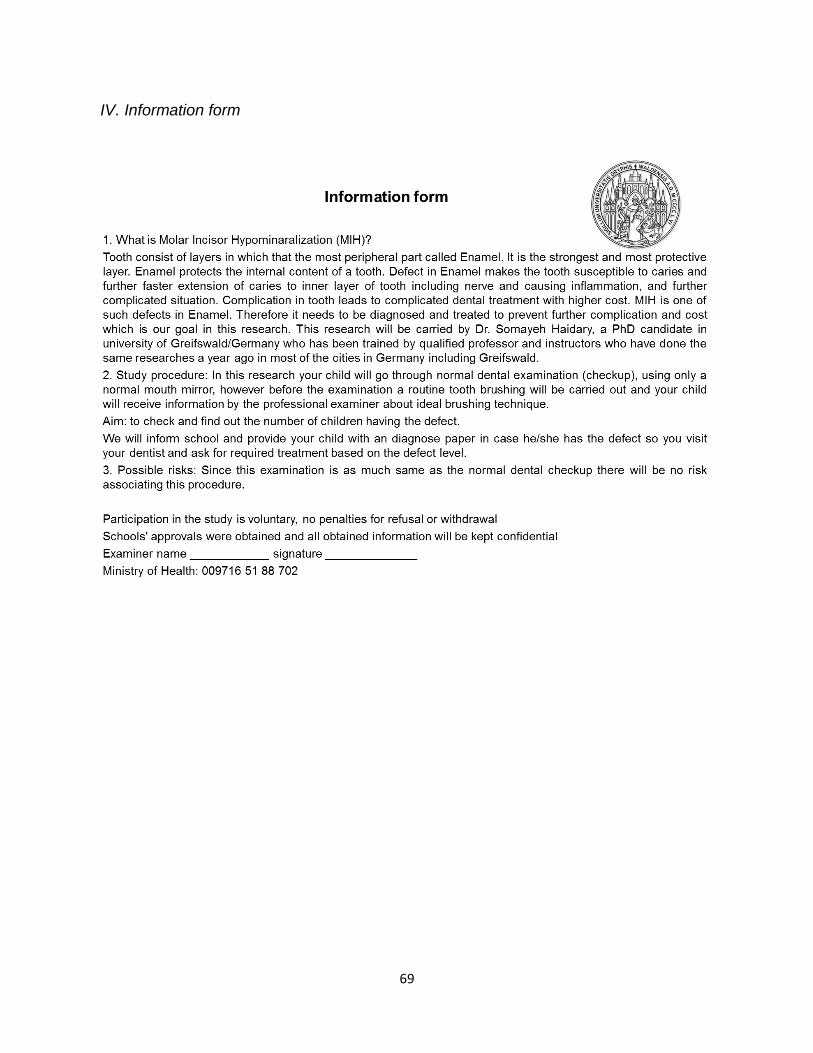
69
IV. Information form
70
V. Information form translated to Arabic Language

71
VI. Consent form

72
VII. Consent form translated to Arabic Language
73
VIII. Diagnosis form

74
IX. Diagnosis form translated to Arabic

75
X. Examination Chart
76
XI. Index table coding
77
XII. Coding table
78
XIII. Presented Conference Abstract
Conference Abstract: European Academy of Paediatric Dentistry 2014, Sopot, Poland
Comparison of Molar-Incisor Hypominaralization Prevalence in Dubai/United Arab
Emirates and Greifswald/Germany
Somayeh Haidary1*, Marina-Agathi Petrou1, Raghad Hashim2, Azza Alhumrani2, Christian Splieth1
1 Preventive and Pediatric Dentistry Department, University of Greifswald, Greifswald, Germany 2 Growth and Development Department, College of Dentistry, Ajman University of Science and Technology, Ajman, UAE *Correspondence to: Somayeh Haidary, 17475 Greifswald, Germany.
E-mail: [email protected]
Background: Defects in enamel mineralization occur during tooth development resulting in structural breakdown. Among such defects, Molar-Incisor Hypomineralization (MIH) is one of the most common enamel defects that occur in children. Identifying this defect in population is of particular importance due to the association with increase caries susceptibility and interference with treatment planning. A recent study in Greifswald/Germany reported a prevalence of MIH 4.29%. Aim: The purpose of this study was to find the prevalence of MIH in Dubai/United Arab
Emirates (UAE) and to compare it with MIH prevalence in Greifswald/Germany.
Design: In this cross-sectional study, 779 children (female: 513; male: 261) age of 7-9 (±1) year-old in the city of Dubai/UAE were randomly selected and examined. The examination was carried out for children in Dubai governmental schools by one examiner who had previously been trained and calibrated with the MIH diagnostic criteria of the European Academy of Pediatric Dentistry (EAPD).
Results: The percentage of children with MIH in Dubai/UAE was 7.6%. There was no
significant gender difference in Dubai (7.57% female, 7.58% male) or Greifswald (4.34%
female, 4.21% male). The DMFT/dmft indices were generally high in Dubai (DMFT=
2.41±1.7; dmft= 5.46±3.1). However, the difference of DMFT index between children
with and without MIH was statistically significant (p < 0.001).
Conclusion: The prevalence of MIH is higher among the children population of
Dubai/UAE compared to Greifswald/Germany. This finding requires a particular
attention from the local dentists to such developmental defect to facilitate adequate
treatment.

79
11. Declaration
I hereby declare that I have written this thesis independently and have used no other
than the means and sources mentioned. The thesis has so far not been submitted to
any other faculty. I declare that I have not completed any doctoral process
unsuccessfully and no withdrawal exists of an already acquired doctoral degree.
Greifswald, 15/11/2014 Somayeh Haidary
Eidesstattliche Erklärung
Hiermit erkläre ich, dass ich die vorliegende Dissertation selbständig verfasst und keine
anderen als die angegebenen Hilfsmittel benutzt habe. Die Dissertation ist bisher keiner
anderen Fakultät, keiner anderen wissenschaftlichen Einrichtung vorgelegt worden. Ich
erkläre, dass ich bisher kein Promotionsverfahren erfolglos beendet habe und dass eine
Aberkennung eines bereits erworbenen Doktorgrades nicht vorliegt.
Greifswald, 15/11/2014 Somayeh Haidary

80
12. Dedication
I am thankful to GOD for being here and reach this point I am, I am thankful to him for
giving me countless blessings in my life.
I dedicate my work to my family who supported me through the difficulties I faced. I am
grateful for having each of them in my life.
I thank my parents for giving me a chance to improve myself and allowing me to go
further in my studies. Not every Afghan girl has such a chance in life. I thank my lovely
beautiful mom for teaching me being kind and good to people. I thank her for her
patience with me. I thank her for her love and guidance towards GOD and his love. I
love you mom, without you and dad I would not be here.
I dedicate this work especially to my loving fiancé with special feeling of gratitude,
whose words of encouragement and support cheered me up, and kept my spirit high,
who have never left my side, who had believe in me even when I didn’t. My dear, here I
thank you from all my heart for your unconditional support. I am blessed and honored to
have you.
I also dedicate this dissertation to my family and friends in Greifswald who have
supported me throughout these three years. Who have always been beside me when I
needed help. I will always appreciate all they have done for me and keep their love in
my heart.
I have learned, there is nothing undoable in life as long as you believe in god by your
heart.
81
13. Acknowledgment
I wish to thank Prof. Dr. Ch. H. Splieth for giving me the opportunity for being here, my
colleagues, better to say my family in the dental clinic for the support and
encouragement, and for the generosity with their expertise and love.
I would like to acknowledge and thank Dr. Marina Petrou and Dr. Ruth Santamaria for
their limitless help. Special thanks to, Dr. Mohammad Alkilzy, and Dr. Julian
Schmoeckel for their kind instruction.
I would like to thank Dr.Raghad Hashim, Head of Growth & Development Department,
College of Dentistry, Ajman University (AUST), without her guidance this research could
not start. Special thanks to Dr. Azza Alhumrani for her kind help and support.
I would like to acknowledge the Ministry Of Health (MOH) especially Dr. Kalthoom
Hassan, director of specialized health care department MOH, for her kind guidance
through my research application. Sincere thanks to ministry of education in UAE for
supporting this research. Many thanks to all the schools for providing the requested
assistance, their excitement and willingness to cooperate made the completion of this
research a pleasant experience despite all the difficulties.