Embed Size (px)
Citation preview
Degradation of vasopressin precursor and
pathogenic mutants in diabetes insipidus
Inauguraldissertation
zur
Erlangung der Würde eines Doktors der Philosophie
vorgelegt der
Philosophisch-Naturwissenschaftlichen Fakultät
der Universität Basel
von
Michael Friberg
aus Zürich, ZH
Basel, 2007
Genehmigt von der Philosophisch-Naturwissenschaftlichen Fakultät
auf Antrag von
Prof. M. Spiess
Prof. H.-P. Hauri
PD Dr. J. Rutishauser
Basel, den 05.07.05
Hans-Jakob Wirz
Dekan
___________________________________________________________________________
Acknowledgements
I’d like to thank the following people for their support during the course of this work:
Prof. Dr. Martin Spiess for providing me with the opportunity to become a member of his
research group, and for his critical discussions
PD Dr. med. Jonas Rutishauser for entrusting the continuation of his project to my care, and
for continuing support and guidance
Nicole Beuret for valuable discussions and expert technical assistance
Cristina Baschong, Pascal Crottet, Tina Junne-Bieri, Marie Higy, Szymon Kobialka, Daniel
Meyer, Adriana Pagano, Hans Stettler, and Gregor Suri, for their input, technical support, and
for creating a superb working environment
Special thanks go to:
Mr. Mark Schiffer and Mr. Clyde Hayes, who encouraged me to go into science
My parents, Alice and Reto, who provided moral and emotional encouragement
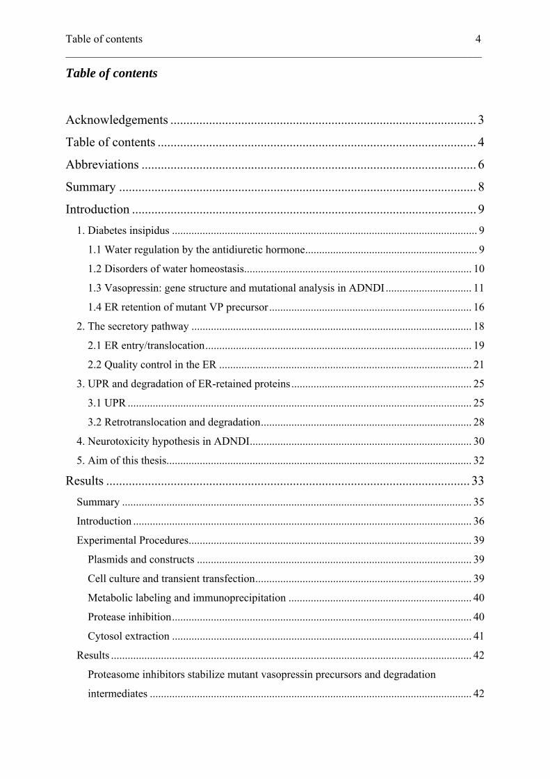
Table of contents 4 ___________________________________________________________________________
Table of contents
Acknowledgements ............................................................................................... 3
Table of contents ................................................................................................... 4
Abbreviations ........................................................................................................ 6
Summary ............................................................................................................... 8
Introduction ........................................................................................................... 9
1. Diabetes insipidus .............................................................................................................. 9
1.1 Water regulation by the antidiuretic hormone.............................................................. 9
1.2 Disorders of water homeostasis.................................................................................. 10
1.3 Vasopressin: gene structure and mutational analysis in ADNDI............................... 11
1.4 ER retention of mutant VP precursor......................................................................... 16
2. The secretory pathway ..................................................................................................... 18
2.1 ER entry/translocation................................................................................................ 19
2.2 Quality control in the ER ........................................................................................... 21
3. UPR and degradation of ER-retained proteins................................................................. 25
3.1 UPR ............................................................................................................................ 25
3.2 Retrotranslocation and degradation............................................................................ 28
4. Neurotoxicity hypothesis in ADNDI................................................................................ 30
5. Aim of this thesis.............................................................................................................. 32
Results ................................................................................................................. 33
Summary .............................................................................................................................. 35
Introduction .......................................................................................................................... 36
Experimental Procedures...................................................................................................... 39
Plasmids and constructs ................................................................................................... 39
Cell culture and transient transfection.............................................................................. 39
Metabolic labeling and immunoprecipitation .................................................................. 40
Protease inhibition............................................................................................................ 40
Cytosol extraction ............................................................................................................ 41
Results .................................................................................................................................. 42
Proteasome inhibitors stabilize mutant vasopressin precursors and degradation
intermediates .................................................................................................................... 42
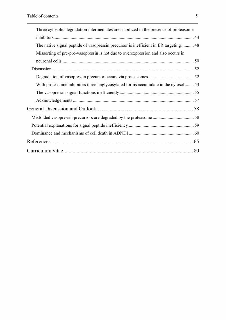
Table of contents 5 ___________________________________________________________________________
Three cytosolic degradation intermediates are stabilized in the presence of proteasome
inhibitors........................................................................................................................... 44
The native signal peptide of vasopressin precursor is inefficient in ER targeting........... 48
Missorting of pre-pro-vasopressin is not due to overexpression and also occurs in
neuronal cells.................................................................................................................... 50
Discussion ............................................................................................................................ 52
Degradation of vasopressin precursor occurs via proteasomes........................................ 52
With proteasome inhibitors three unglycosylated forms accumulate in the cytosol........ 53
The vasopressin signal functions inefficiently ................................................................. 55
Acknowledgements .......................................................................................................... 57
General Discussion and Outlook......................................................................... 58
Misfolded vasopressin precursors are degraded by the proteasome .................................... 58
Potential explanations for signal peptide inefficiency ......................................................... 59
Dominance and mechanisms of cell death in ADNDI ......................................................... 60
References ........................................................................................................... 65
Curriculum vitae.................................................................................................. 80
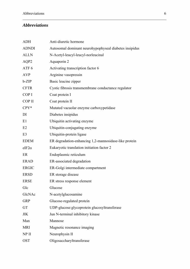
Abbreviations 6 ___________________________________________________________________________
Abbreviations ADH Anti-diuretic hormone
ADNDI Autosomal dominant neurohypophyseal diabetes insipidus
ALLN N-Acetyl-leucyl-leucyl-norleucinal
AQP2 Aquaporin 2
ATF 6 Activating transcription factor 6
AVP Arginine vasopressin
b-ZIP Basic leucine zipper
CFTR Cystic fibrosis transmembrane conductance regulator
COP I Coat protein I
COP II Coat protein II
CPY* Mutated vacuolar enzyme carboxypetidase
DI Diabetes insipidus
E1 Ubiquitin activating enzyme
E2 Ubiquitin-conjugating enzyme
E3 Ubiquitin-protein ligase
EDEM ER degradation-enhancing 1,2-mannosidase-like protein
eIF2α Eukaryotic translation initiation factor 2
ER Endoplasmic reticulum
ERAD ER-associated degradation
ERGIC ER-Golgi intermediate compartment
ERSD ER storage disease
ERSE ER stress response element
Glc Glucose
GlcNAc N-acetylglucosamine
GRP Glucose-regulated protein
GT UDP-glucose:glycoprotein glucosyltransferase
JIK Jun N-terminal inhibitory kinase
Man Mannose
MRI Magnetic resonance imaging
NP II Neurophysin II
OST Oligosaccharyltransferase
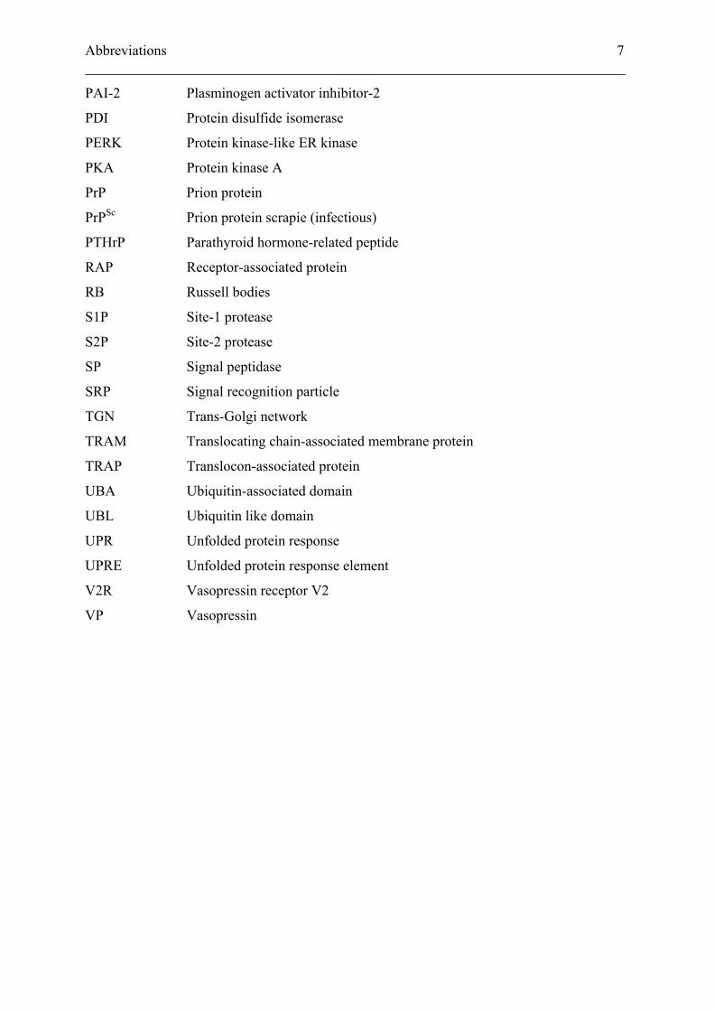
Abbreviations 7 ___________________________________________________________________________
PAI-2 Plasminogen activator inhibitor-2
PDI Protein disulfide isomerase
PERK Protein kinase-like ER kinase
PKA Protein kinase A
PrP Prion protein
PrPSc Prion protein scrapie (infectious)
PTHrP Parathyroid hormone-related peptide
RAP Receptor-associated protein
RB Russell bodies
S1P Site-1 protease
S2P Site-2 protease
SP Signal peptidase
SRP Signal recognition particle
TGN Trans-Golgi network
TRAM Translocating chain-associated membrane protein
TRAP Translocon-associated protein
UBA Ubiquitin-associated domain
UBL Ubiquitin like domain
UPR Unfolded protein response
UPRE Unfolded protein response element
V2R Vasopressin receptor V2
VP Vasopressin
Summary 8 ___________________________________________________________________________
Summary
The nonapeptide hormone, arginine vasopressin, plays a decisive role in the regulation of
fluid balance by reducing free water clearance through reabsorption of water in the renal
collecting ducts. Mutations in the gene encoding arginine vasopressin cause autosomal
dominant neurohypophyseal diabetes insipidus, a disease characterized by excessive urine
production and strong thirst. Post mortem examination of affected individuals suggests a
selective degeneration of vasopressinergic neurons in the hypothalamus. On a molecular level,
the disease is linked to a trafficking defect. Mutant vasopressin precursor is retained in the
endoplasmic reticulum, while the wild-type is transported to mature secretory granules at
synaptic processes. How this trafficking defect of the vasopressin precursor is interrelated
with the degeneration of neurons is unknown. A plausible hypothesis is that mutant proteins,
or degradation products thereof, are toxic to neurons.
Accordingly, we analyzed the fate of mutant vasopressin precursor arrested in the
endoplasmic reticulum of transfected cell lines. Proteasomal, but not lysosomal, inhibitors
induced stabilization of mutant precursors and the accumulation of three distinct non-
glycosylated cytosolic species: pre-pro-vasopressin, pro-vasopressin, and an N-terminally
truncated form. These results provide evidence that mutant precursor, after translocation into
the ER lumen, is retrotranslocated to the cytosol and degraded by the proteasome.
Furthermore, a fraction of the newly synthesized precursor, even of wild-type, was found not
to be translocated, but to be synthesized into the cytosol due to inefficiency of the vasopressin
signal peptide.
In autosomal dominant neurohypophyseal diabetes insipidus, neurotoxicity may thus result
from degradation intermediates and/or by ER retention directly. Both mistargeted and
retrotranslocated proteins add to the cytosolic pool of these degradation products.
Neurodegeneration might occur in heterozygous individuals once a critical concentration of
toxic material is exceeded.
Introduction 9 ___________________________________________________________________________
Introduction
1. Diabetes insipidus
1.1 Water regulation by the antidiuretic hormone
Water homeostasis is critical to survival of cells and organisms. Accordingly, the balance of
water uptake and excretion needs to be carefully controlled. We ingest water as part of our
daily diet. Loss of water occurs during the maintenance of body temperature by sweating, or
when removing waste products by either urination or defecation. Additional moisture is lost
during exhalation, but the largest loss of fluid will ordinarily occur through urination. The
amount of urine an individual produces varies depending on kidney activity, while the
quantity of water filtered from the blood passing through the kidney remains relatively
constant under physiological conditions. An adult human produces about 180 litres of primary
filtrate per day, of which about 90% is held back by the proximal compartments of the
nephron. The remaining 10% reach the distal collecting tubules, where water reabsorption is
controlled by anti-diuretic hormone (ADH), also known as vasopressin (VP). ADH-dependent
water reabsorption reduces the volume we excrete each day to an approximate 1% of the
original primary filtrate.
Vasopressin synthesis occurs in magno- and parvocellular neurons of the hypothalamus.
Hypovolemia and/or hypernatremia stimulate release of vasopressin to the blood stream.
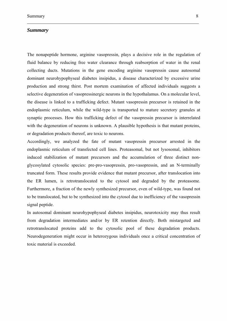
Water reabsorption in the kidney is achieved via a G-protein coupled receptor cascade.
Vasopressin from the bloodstream binds to the vasopressin receptor V2 (V2R) at the
basolateral side of the cell in renal collecting ducts (Figure 1). This leads to an elevation of
intracellular cAMP levels by the activation of adenylate cyclase and subsequent
phosphorylation of the water channel aquaporin 2 (AQP2) at its cytoplasmic C-terminus by
protein kinase A. Consequently, transport vesicles containing AQP2 fuse with the apical
plasma membrane and permit increased reabsorption of water from the collecting ducts. In
parallel, synthesis of AQP2 is induced. Water molecules leave the cell through aquaporin 3
and 4 channels at the basolateral membrane. Removal of AVP from the V2R results in the
internalisation of AQP2, ending the cycle (Rutishauser and Kopp, 1999; Levin et al., 2001).
Introduction 10 ___________________________________________________________________________
Rutishauser and Kopp (1999)
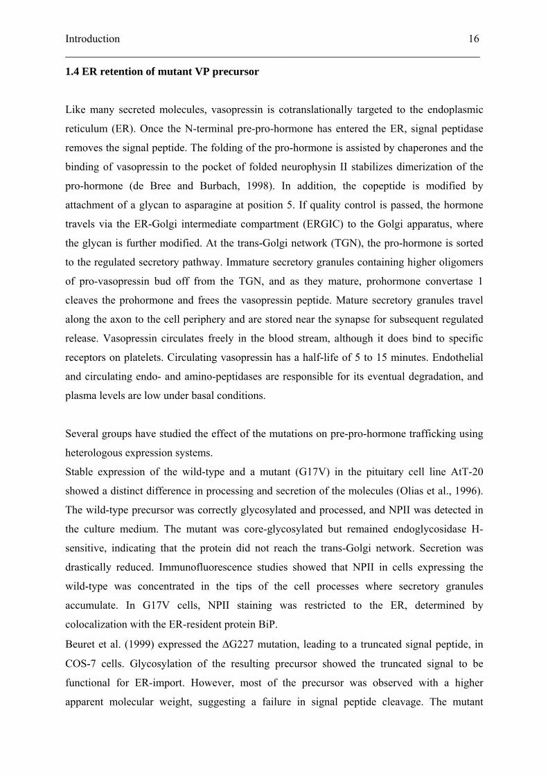
FIG. 1. Arginine vasopressin (AVP) binds to its receptor and activates protein kinase A (PKA), inducing fusion
of aquaporin 2 containing vesicles with the plasma membrane. Water resorption persists as long as AVP remains
bound to its receptor. Subsequently AQP2 is reinternalized.
1.2 Disorders of water homeostasis
Failure to efficiently concentrate urine will lead to polyuria and consequently polydipsia. This
clinical condition, characterized by the production of large quantities of dilute urine, is known
as diabetes insipidus (DI). Three mechanisms are known to cause DI. Deficiency of VP itself
causes neurohypophyseal diabetes insipidus. Renal resistance to the antidiuretic action of VP,
e.g. due to injury of the nephron or to mutations in the VP receptor or the aquaporins, results
in nephrogenic diabetes insipidus. Finally, inappropriate and excessive drinking without a
somatic cause leads to dipsogenic diabetes insipidus.
Introduction 11 ___________________________________________________________________________
DI and its subtypes can be diagnosed by a water deprivation test. It compares plasma
osmolality with urine osmolality during a defined time period of water deprivation, and
assesses the response of these parameters to exogenous VP administration. Neurohypophyseal
diabetes insipidus results from acquired pathologies affecting the vasopressinergic cells, such
as trauma, infiltrating or inflammatory diseases, or tumors. Rarely, the disease is congenital
due to mutations in the gene encoding pre-pro-vasopressin. This disease is called autosomal
dominant neurohypophyseal diabetes insipidus (ADNDI). Although it is rather rare, the
disorder shows a high penetrance with symptoms beginning weeks to months after birth.
Presentation of neurohypophyseal diabetes insipidus implies destruction or loss-of-function of
more than 80% of magnocellular neurons. Further clinical information may be obtained from
magnetic resonance imaging (MRI). In normal subjects the posterior pituitary shows a high
intensity signal on T1-weighted images. Absence of such a signal has been correlated with
neurohypophyseal DI, although studies have failed to display a strict cosegregation of
morphological abnormalities and clinical symptoms (Miyamoto et al., 1991; Rutishauser et
al., 1996; Gagliardi et al., 1997).
1.3 Vasopressin: gene structure and mutational analysis in ADNDI
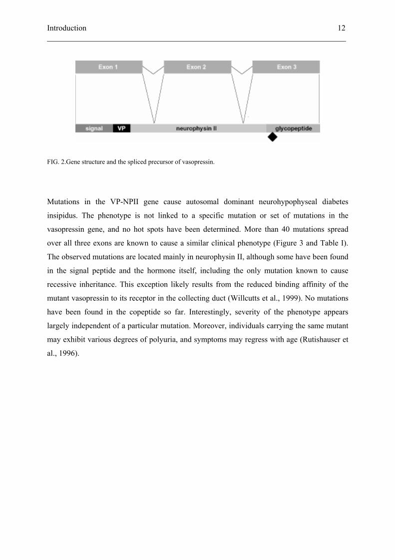
The vasopressin gene, located on chromosome 20p13, consists of three exons (Figure 2). The
first exon encodes a signal peptide of 19 amino acids, followed by the 9 amino acid hormone,
a 3 amino acid linker, and the 9 N-terminal amino acids of neurophysin II (NPII),
vasopressin’s carrier protein. Exon 2 encodes 67 amino acids, which form the central part of
NPII. Exon 3 constitutes the C-terminal 17 amino acids of NPII, a single amino acid linker,
and a C-terminal glycopeptide of 39 amino acids, also known as copeptide, bearing a single
N-linked glycosylation site.
Introduction 12 ___________________________________________________________________________
FIG. 2.Gene structure and the spliced precursor of vasopressin.
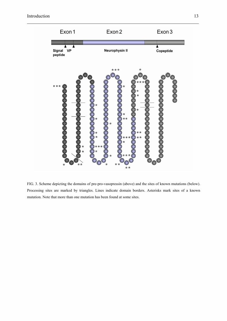
Mutations in the VP-NPII gene cause autosomal dominant neurohypophyseal diabetes
insipidus. The phenotype is not linked to a specific mutation or set of mutations in the
vasopressin gene, and no hot spots have been determined. More than 40 mutations spread
over all three exons are known to cause a similar clinical phenotype (Figure 3 and Table I).
The observed mutations are located mainly in neurophysin II, although some have been found
in the signal peptide and the hormone itself, including the only mutation known to cause
recessive inheritance. This exception likely results from the reduced binding affinity of the
mutant vasopressin to its receptor in the collecting duct (Willcutts et al., 1999). No mutations
have been found in the copeptide so far. Interestingly, severity of the phenotype appears
largely independent of a particular mutation. Moreover, individuals carrying the same mutant
may exhibit various degrees of polyuria, and symptoms may regress with age (Rutishauser et
al., 1996).
Introduction 13 ___________________________________________________________________________
FIG. 3. Scheme depicting the domains of pre-pro-vasopressin (above) and the sites of known mutations (below).
Processing sites are marked by triangles. Lines indicate domain borders. Asterisks mark sites of a known
mutation. Note that more than one mutation has been found at some sites.
Introduction 14 ___________________________________________________________________________
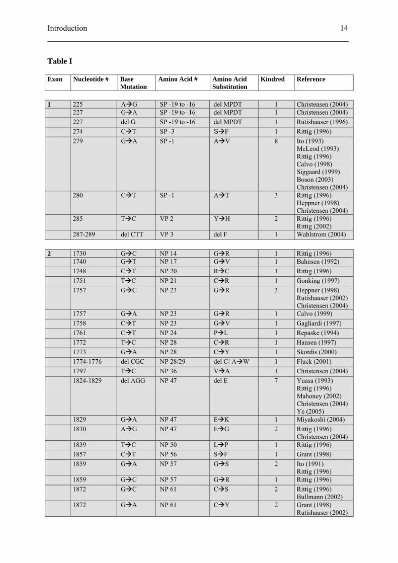
Table I
Exon Nucleotide # Base Mutation
Amino Acid # Amino Acid Substitution
Kindred Reference
1 225 A G SP -19 to -16 del MPDT 1 Christensen (2004) 227 G A SP -19 to -16 del MPDT 1 Christensen (2004) 227 del G SP -19 to -16 del MPDT 1 Rutishauser (1996) 274 C T SP -3 S F 1 Rittig (1996) 279 G A SP -1 A V 8 Ito (1993)
McLeod (1993) Rittig (1996) Calvo (1998) Siggaard (1999) Boson (2003) Christensen (2004)
280 C T SP -1 A T 3 Rittig (1996) Heppner (1998) Christensen (2004)
285 T C VP 2 Y H 2 Rittig (1996) Rittig (2002)
287-289 del CTT VP 3 del F 1 Wahlstrom (2004) 2 1730 G C NP 14 G R 1 Rittig (1996) 1740 G T NP 17 G V 1 Bahnsen (1992) 1748 C T NP 20 R C 1 Rittig (1996) 1751 T C NP 21 C R 1 Gonking (1997) 1757 G C NP 23 G R 3 Heppner (1998)
Rutishauser (2002) Christensen (2004)
1757 G A NP 23 G R 1 Calvo (1999) 1758 C T NP 23 G V 1 Gagliardi (1997) 1761 C T NP 24 P L 1 Repaske (1994) 1772 T C NP 28 C R 1 Hansen (1997) 1773 G A NP 28 C Y 1 Skordis (2000) 1774-1776 del CGC NP 28/29 del C/ A W 1 Fluck (2001) 1797 T C NP 36 V A 1 Christensen (2004) 1824-1829 del AGG NP 47 del E 7 Yuasa (1993)
Rittig (1996) Mahoney (2002) Christensen (2004) Ye (2005)
1829 G A NP 47 E K 1 Miyakoshi (2004) 1830 A G NP 47 E G 2 Rittig (1996)
Christensen (2004) 1839 T C NP 50 L P 1 Rittig (1996) 1857 C T NP 56 S F 1 Grant (1998) 1859 G A NP 57 G S 2 Ito (1991)
Rittig (1996) 1859 G C NP 57 G R 1 Rittig (1996) 1872 G C NP 61 C S 2 Rittig (1996)
Bullmann (2002) 1872 G A NP 61 C Y 2 Grant (1998)
Rutishauser (2002)
Introduction 15 ___________________________________________________________________________
1873 C A NP 61 C X 3 Rittig (1996) Grant (1998) Christensen (2004)
1874 G T NP 62 G W 1 Nagasaki (1995) 1883 G T NP 65 G C 2 Rittig (1996)
Christensen (2004) 1884 G A NP 65 G D 1 Christensen (2004) 1884 G T NP 65 G V 2 Ueta (1996)
Rauch (1996) 1886 C T NP 66 R C 1 Rutishauser (1999) 1887 G C NP 66 R P 1 Mundschenk (2001) 1889 T G NP 67 C G 1 DiMeglio (2001) 1891 C A NP 67 C X 1 Nagasaki (1995) 1907 T G NP 73 C G 1 Christensen (2004) 1908 G T NP 73 C F 1 Santiprabhob
(2002) 1910 T C NP 74 C R 1 Rutishauser (2002) 1911 G A NP 74 C Y 1 Fujii (2000) 3 2094 C A NP 79 C X 1 Rittig (1996) 2101 G T NP 82 E X 2 Calvo (1998) 2106-2107 CG GT NP 83 E X 2 Rittig (1996)
Bullmann (2002) 2110 T G NP 85 C G 2 Abbes (2000)
Nijenhuis (2001) 2110 T C NP 85 C R 1 Abbes (2000) 2112 C G NP 85 C W 1 Christensen (2004) 2116 G T NP 87 E X 1 Rittig (1996) X = stop codon
Introduction 16 ___________________________________________________________________________
1.4 ER retention of mutant VP precursor
Like many secreted molecules, vasopressin is cotranslationally targeted to the endoplasmic
reticulum (ER). Once the N-terminal pre-pro-hormone has entered the ER, signal peptidase
removes the signal peptide. The folding of the pro-hormone is assisted by chaperones and the
binding of vasopressin to the pocket of folded neurophysin II stabilizes dimerization of the
pro-hormone (de Bree and Burbach, 1998). In addition, the copeptide is modified by
attachment of a glycan to asparagine at position 5. If quality control is passed, the hormone
travels via the ER-Golgi intermediate compartment (ERGIC) to the Golgi apparatus, where
the glycan is further modified. At the trans-Golgi network (TGN), the pro-hormone is sorted
to the regulated secretory pathway. Immature secretory granules containing higher oligomers
of pro-vasopressin bud off from the TGN, and as they mature, prohormone convertase 1
cleaves the prohormone and frees the vasopressin peptide. Mature secretory granules travel
along the axon to the cell periphery and are stored near the synapse for subsequent regulated
release. Vasopressin circulates freely in the blood stream, although it does bind to specific
receptors on platelets. Circulating vasopressin has a half-life of 5 to 15 minutes. Endothelial
and circulating endo- and amino-peptidases are responsible for its eventual degradation, and
plasma levels are low under basal conditions.
Several groups have studied the effect of the mutations on pre-pro-hormone trafficking using
heterologous expression systems.
Stable expression of the wild-type and a mutant (G17V) in the pituitary cell line AtT-20
showed a distinct difference in processing and secretion of the molecules (Olias et al., 1996).
The wild-type precursor was correctly glycosylated and processed, and NPII was detected in
the culture medium. The mutant was core-glycosylated but remained endoglycosidase H-
sensitive, indicating that the protein did not reach the trans-Golgi network. Secretion was
drastically reduced. Immunofluorescence studies showed that NPII in cells expressing the
wild-type was concentrated in the tips of the cell processes where secretory granules
accumulate. In G17V cells, NPII staining was restricted to the ER, determined by
colocalization with the ER-resident protein BiP.
Beuret et al. (1999) expressed the ΔG227 mutation, leading to a truncated signal peptide, in
COS-7 cells. Glycosylation of the resulting precursor showed the truncated signal to be
functional for ER-import. However, most of the precursor was observed with a higher
apparent molecular weight, suggesting a failure in signal peptide cleavage. The mutant
Introduction 17 ___________________________________________________________________________
precursor was almost completely retained in the ER, corroborated by costaining for the ER
resident protein p63.
Nijenhuis et al. (1999) studied a variety of mutants by stably expressing them in the
neuroendocrine cell line Neuro2a and the rat pheochromocytoma cell line PC12/PC2. When
comparing G14R, E47R, ΔE47, G57S, and G65V to the wild-type, all mutants were found to
be impaired in processing and secretion, albeit to different extents (in decreasing order of
impairment: G65V≥G14R>ΔE47≥E47G>G57S). Sensitivity to endoglycosidase H indicated
retention of the precursors in the ER. Immunofluorescence studies using transiently
transfected Neuro2a cells demonstrated that the mutant prohormone was found in large
accumulations in the ER, which colocalized with the ER marker protein disulfide isomerase
(PDI).
Analogous studies with the E87X mutant in PC12/PC2 and the mouse pituitary cell line AtT-
20 also showed reduced processing and secretion and colocalization with PDI (Nijenhuis et
al., 2000).
Expression studies in PC12/PC2 cells using wild-type vasopressin and the C85G mutant
confirmed previous observations. Processing and secretion was only observed for the wild-
type (Nijenhuis et al., 2001). The mutant was retained in the ER, as indicated by
endoglycosidase H sensitivity. Transient transfection of Neuro2a cells demonstrated that the
mutant was not only confined to the normal reticular ER. Costaining with PDI showed these
areas to represent enlarged ER subcompartments. Such large areas of altered ER morphology
could be responsible not only for severe dysfunction but also for death of the host cells in vivo
(Aridor and Hannan, 2000; Rutishauser and Spiess, 2002).
Siggaard et al. (1999) examined the signal peptide mutant A(-1)T in Neuro2a cells. They
observed an 8-fold reduction in vasopressin secretion when compared to the wild-type,
accompanied by the accumulation of improper signal cleavage. The precursor was found to
colocalize with glucose-regulated protein 78, indicating an ER localization.
Although the degree of retention varies among different mutants, inefficient ER exit leading
to reduced secretion of mutant hormone precursors apparently is a common denominator in
the pathogenesis of ADNDI. In order to understand the processes leading to this trafficking
defect, we need to briefly look at the mechanisms involved in secretion of proteins from the
cell.
Introduction 18 ___________________________________________________________________________
2. The secretory pathway
Hormones are molecules acting on target tissues which are typically remote from the site of
production. In order for secretion to occur, the proteins must thus enter the secretory pathway.
While all cells possess the means to constitutively release proteins form the cell, endocrine
cells need to regulate hormone release in response to a specific stimulus. Therefore, they are
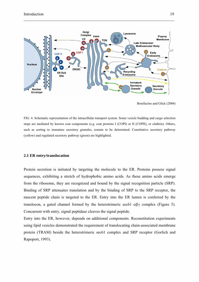
endowed with an additional release mechanism, the regulated secretory pathway (Figure 4).
If a protein is to be released from the cell, it is targeted to the ER. In mammalian cells this is a
cotranslational process. As the newly synthesized polypeptide emerges into the ER lumen, it
may be modified by core glycosylation. In addition, chaperones promote proper folding.
Before the secretory protein can leave the ER it needs to pass quality control, a mechanism
which prevents further transport of incorrectly folded precursors, such as proteins which are
either non-functional or thermo-labile because they have not attained their native
conformation. The protein then travels via the ERGIC to the Golgi apparatus. Here further
modifications such as complex glycosylation or sulfation may occur. The TGN is the
compartment where proteins destined for regulated secretion are segregated from those taking
other pathways. The hormone precursors are sorted to immature secretory granules, which
mature as they travel to the cell periphery. During the maturation process, the pro-hormones
are activated by pro-hormone convertases. The mature secretory granules are then stored near
the cell periphery, from where they can be rapidly released in response to the proper stimuli.
Introduction 19 ___________________________________________________________________________
Bonifacino and Glick (2004)
FIG. 4. Schematic representation of the intracellular transport system. Some vesicle budding and cargo selection
steps are mediated by known coat components (e.g. coat proteins I (COPI) or II (COPII), or clathrin). Others,
such as sorting to immature secretory granules, remain to be determined. Constitutive secretory pathway
(yellow) and regulated secretory pathway (green) are highlighted.
2.1 ER entry/translocation
Protein secretion is initiated by targeting the molecule to the ER. Proteins possess signal
sequences, exhibiting a stretch of hydrophobic amino acids. As these amino acids emerge
from the ribosome, they are recognized and bound by the signal recognition particle (SRP).
Binding of SRP attenuates translation and by the binding of SRP to the SRP receptor, the
nascent peptide chain is targeted to the ER. Entry into the ER lumen is conferred by the
translocon, a gated channel formed by the heterotrimeric sec61 αβγ complex (Figure 5).
Concurrent with entry, signal peptidase cleaves the signal peptide.
Entry into the ER, however, depends on additional components. Reconstitution experiments
using lipid vesicles demonstrated the requirement of translocating chain-associated membrane
protein (TRAM) beside the heterotrimeric sec61 complex and SRP receptor (Gorlich and
Rapoport, 1993).
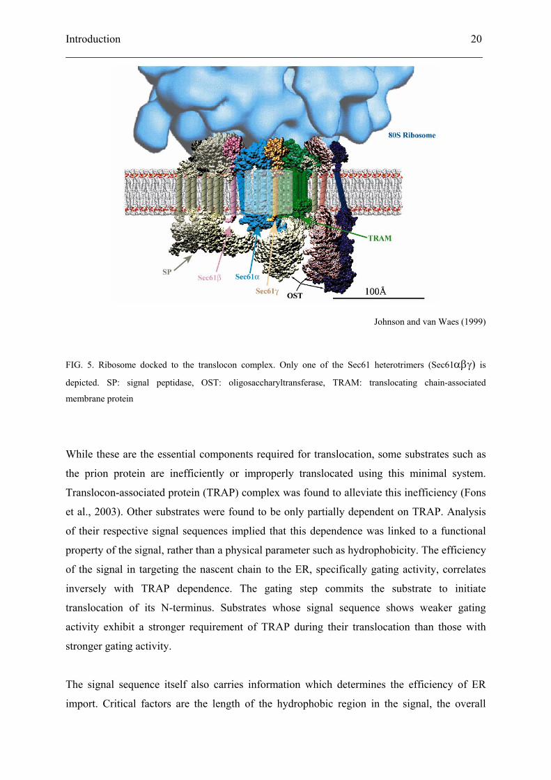
Introduction 20 ___________________________________________________________________________
Johnson and van Waes (1999)
FIG. 5. Ribosome docked to the translocon complex. Only one of the Sec61 heterotrimers (Sec61αβγ) is
depicted. SP: signal peptidase, OST: oligosaccharyltransferase, TRAM: translocating chain-associated
membrane protein
While these are the essential components required for translocation, some substrates such as
the prion protein are inefficiently or improperly translocated using this minimal system.
Translocon-associated protein (TRAP) complex was found to alleviate this inefficiency (Fons
et al., 2003). Other substrates were found to be only partially dependent on TRAP. Analysis
of their respective signal sequences implied that this dependence was linked to a functional
property of the signal, rather than a physical parameter such as hydrophobicity. The efficiency
of the signal in targeting the nascent chain to the ER, specifically gating activity, correlates
inversely with TRAP dependence. The gating step commits the substrate to initiate
translocation of its N-terminus. Substrates whose signal sequence shows weaker gating
activity exhibit a stronger requirement of TRAP during their translocation than those with
stronger gating activity.
The signal sequence itself also carries information which determines the efficiency of ER
import. Critical factors are the length of the hydrophobic region in the signal, the overall
Introduction 21 ___________________________________________________________________________
distribution of charged amino acids with respect to the hydrophobic region, and the position
of the signal in relation to the start codon, i.e. the size of the N-terminal region synthesized
prior to the signal (Wahlberg and Spiess, 1997; Goder and Spiess, 2001).
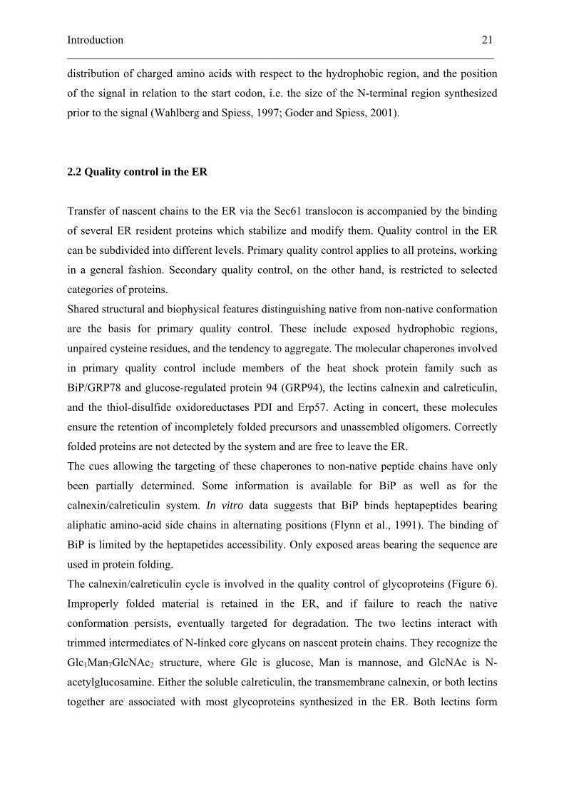
2.2 Quality control in the ER
Transfer of nascent chains to the ER via the Sec61 translocon is accompanied by the binding
of several ER resident proteins which stabilize and modify them. Quality control in the ER
can be subdivided into different levels. Primary quality control applies to all proteins, working
in a general fashion. Secondary quality control, on the other hand, is restricted to selected
categories of proteins.
Shared structural and biophysical features distinguishing native from non-native conformation
are the basis for primary quality control. These include exposed hydrophobic regions,
unpaired cysteine residues, and the tendency to aggregate. The molecular chaperones involved
in primary quality control include members of the heat shock protein family such as
BiP/GRP78 and glucose-regulated protein 94 (GRP94), the lectins calnexin and calreticulin,
and the thiol-disulfide oxidoreductases PDI and Erp57. Acting in concert, these molecules
ensure the retention of incompletely folded precursors and unassembled oligomers. Correctly
folded proteins are not detected by the system and are free to leave the ER.
The cues allowing the targeting of these chaperones to non-native peptide chains have only
been partially determined. Some information is available for BiP as well as for the
calnexin/calreticulin system. In vitro data suggests that BiP binds heptapeptides bearing
aliphatic amino-acid side chains in alternating positions (Flynn et al., 1991). The binding of
BiP is limited by the heptapetides accessibility. Only exposed areas bearing the sequence are
used in protein folding.
The calnexin/calreticulin cycle is involved in the quality control of glycoproteins (Figure 6).
Improperly folded material is retained in the ER, and if failure to reach the native
conformation persists, eventually targeted for degradation. The two lectins interact with
trimmed intermediates of N-linked core glycans on nascent protein chains. They recognize the
Glc1Man7GlcNAc2 structure, where Glc is glucose, Man is mannose, and GlcNAc is N-
acetylglucosamine. Either the soluble calreticulin, the transmembrane calnexin, or both lectins
together are associated with most glycoproteins synthesized in the ER. Both lectins form
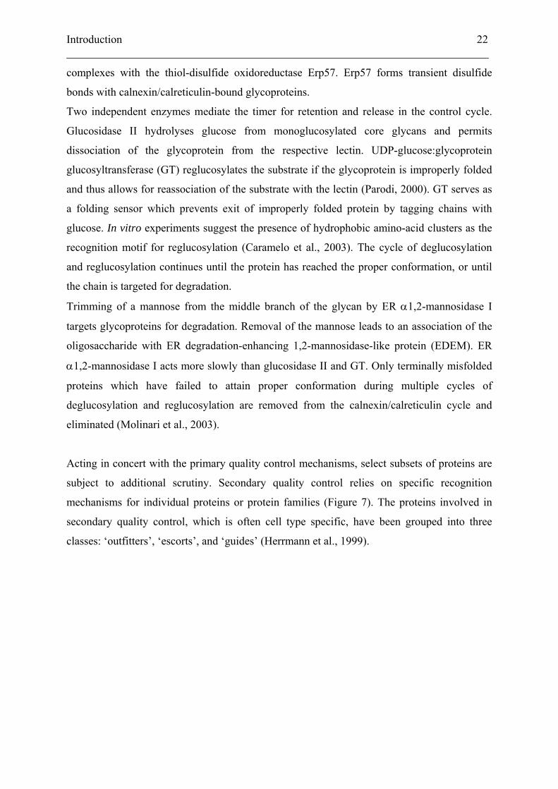
Introduction 22 ___________________________________________________________________________
complexes with the thiol-disulfide oxidoreductase Erp57. Erp57 forms transient disulfide
bonds with calnexin/calreticulin-bound glycoproteins.
Two independent enzymes mediate the timer for retention and release in the control cycle.
Glucosidase II hydrolyses glucose from monoglucosylated core glycans and permits
dissociation of the glycoprotein from the respective lectin. UDP-glucose:glycoprotein
glucosyltransferase (GT) reglucosylates the substrate if the glycoprotein is improperly folded
and thus allows for reassociation of the substrate with the lectin (Parodi, 2000). GT serves as
a folding sensor which prevents exit of improperly folded protein by tagging chains with
glucose. In vitro experiments suggest the presence of hydrophobic amino-acid clusters as the
recognition motif for reglucosylation (Caramelo et al., 2003). The cycle of deglucosylation
and reglucosylation continues until the protein has reached the proper conformation, or until
the chain is targeted for degradation.
Trimming of a mannose from the middle branch of the glycan by ER α1,2-mannosidase I
targets glycoproteins for degradation. Removal of the mannose leads to an association of the
oligosaccharide with ER degradation-enhancing 1,2-mannosidase-like protein (EDEM). ER
α1,2-mannosidase I acts more slowly than glucosidase II and GT. Only terminally misfolded
proteins which have failed to attain proper conformation during multiple cycles of
deglucosylation and reglucosylation are removed from the calnexin/calreticulin cycle and
eliminated (Molinari et al., 2003).
Acting in concert with the primary quality control mechanisms, select subsets of proteins are
subject to additional scrutiny. Secondary quality control relies on specific recognition
mechanisms for individual proteins or protein families (Figure 7). The proteins involved in
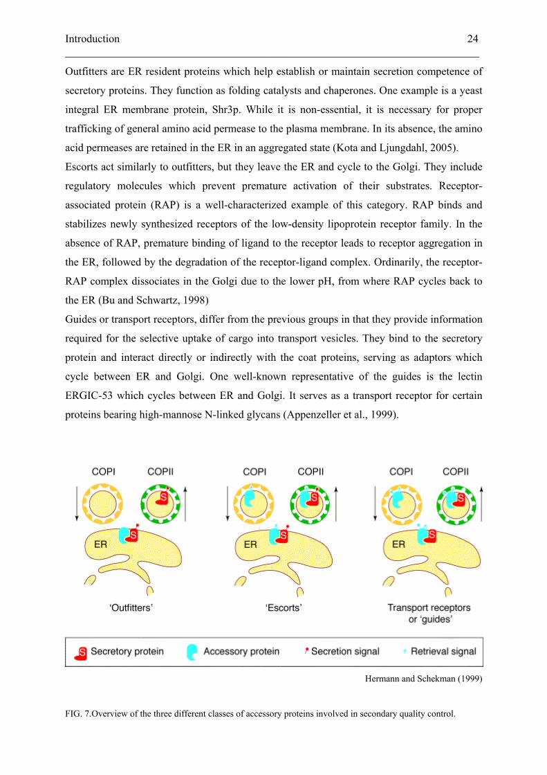
secondary quality control, which is often cell type specific, have been grouped into three
classes: ‘outfitters’, ‘escorts’, and ‘guides’ (Herrmann et al., 1999).
Introduction 23 ___________________________________________________________________________
Ellgaard and Helenius (2003)
FIG. 6. Scheme depicting the steps of quality control undertaken by an N-linked glycoprotein. The processing of
the core glycan tree helps ensure folding fidelity. Properly folded proteins will eventually leave the ER via exit
sites, while terminally misfolded material is targeted for degradation via the cytosolic proteasome pathway. M
(mannose), G (glucose)
Introduction 24 ___________________________________________________________________________
Outfitters are ER resident proteins which help establish or maintain secretion competence of
secretory proteins. They function as folding catalysts and chaperones. One example is a yeast
integral ER membrane protein, Shr3p. While it is non-essential, it is necessary for proper
trafficking of general amino acid permease to the plasma membrane. In its absence, the amino
acid permeases are retained in the ER in an aggregated state (Kota and Ljungdahl, 2005).
Escorts act similarly to outfitters, but they leave the ER and cycle to the Golgi. They include
regulatory molecules which prevent premature activation of their substrates. Receptor-
associated protein (RAP) is a well-characterized example of this category. RAP binds and
stabilizes newly synthesized receptors of the low-density lipoprotein receptor family. In the
absence of RAP, premature binding of ligand to the receptor leads to receptor aggregation in
the ER, followed by the degradation of the receptor-ligand complex. Ordinarily, the receptor-
RAP complex dissociates in the Golgi due to the lower pH, from where RAP cycles back to
the ER (Bu and Schwartz, 1998)
Guides or transport receptors, differ from the previous groups in that they provide information
required for the selective uptake of cargo into transport vesicles. They bind to the secretory
protein and interact directly or indirectly with the coat proteins, serving as adaptors which
cycle between ER and Golgi. One well-known representative of the guides is the lectin
ERGIC-53 which cycles between ER and Golgi. It serves as a transport receptor for certain
proteins bearing high-mannose N-linked glycans (Appenzeller et al., 1999).
Hermann and Schekman (1999)
FIG. 7.Overview of the three different classes of accessory proteins involved in secondary quality control.
Introduction 25 ___________________________________________________________________________
3. UPR and degradation of ER-retained proteins
3.1 UPR
The presence of irrevocably misfolded proteins in the ER is detrimental to the cell over a
prolonged time period. The material interferes with proper trafficking by detaining
components of the secretory pathway. Two processes aid eukaryotic cells to reduce this bulk
load: ER-associated degradation (ERAD) and the unfolded protein response (UPR). The two
responses are interconnected. Induction of UPR leads to an increase in ERAD capacity, while
efficient ERAD is dependent on an intact UPR. Loss of ERAD results in a constitutive UPR
induction. Loss of both ERAD and UPR strongly diminishes cell viability.
One of the key classes of target genes of UPR is ER-resident chaperones. An increase in
chaperone concentration helps to reduce the amount of unfolded proteins and precludes their
aggregation.
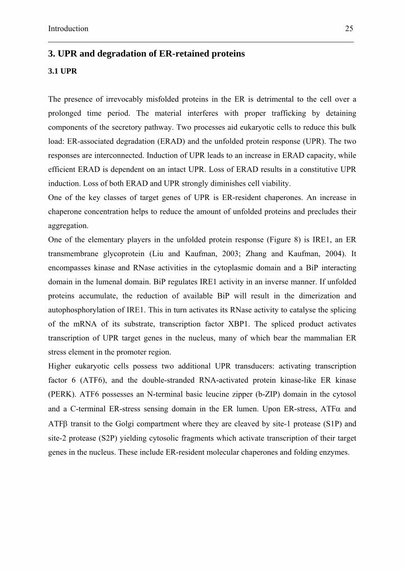
One of the elementary players in the unfolded protein response (Figure 8) is IRE1, an ER
transmembrane glycoprotein (Liu and Kaufman, 2003; Zhang and Kaufman, 2004). It
encompasses kinase and RNase activities in the cytoplasmic domain and a BiP interacting
domain in the lumenal domain. BiP regulates IRE1 activity in an inverse manner. If unfolded
proteins accumulate, the reduction of available BiP will result in the dimerization and
autophosphorylation of IRE1. This in turn activates its RNase activity to catalyse the splicing
of the mRNA of its substrate, transcription factor XBP1. The spliced product activates
transcription of UPR target genes in the nucleus, many of which bear the mammalian ER
stress element in the promoter region.
Higher eukaryotic cells possess two additional UPR transducers: activating transcription
factor 6 (ATF6), and the double-stranded RNA-activated protein kinase-like ER kinase
(PERK). ATF6 possesses an N-terminal basic leucine zipper (b-ZIP) domain in the cytosol
and a C-terminal ER-stress sensing domain in the ER lumen. Upon ER-stress, ATFα and
ATFβ transit to the Golgi compartment where they are cleaved by site-1 protease (S1P) and
site-2 protease (S2P) yielding cytosolic fragments which activate transcription of their target
genes in the nucleus. These include ER-resident molecular chaperones and folding enzymes.
Introduction 26 ___________________________________________________________________________
Zhang and Kaufman (2004)
FIG. 8. Illustration of the interplay of IRE1 and ATF6 in promoting the expression of genes involved in both the
unfolded protein response and the ER stress response via the respective response elements (UPRE and ERSE).
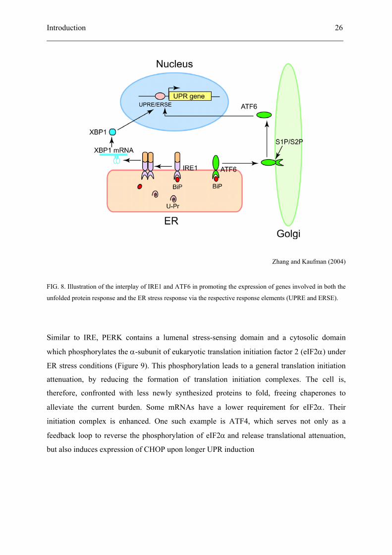
Similar to IRE, PERK contains a lumenal stress-sensing domain and a cytosolic domain
which phosphorylates the α-subunit of eukaryotic translation initiation factor 2 (eIF2α) under
ER stress conditions (Figure 9). This phosphorylation leads to a general translation initiation
attenuation, by reducing the formation of translation initiation complexes. The cell is,
therefore, confronted with less newly synthesized proteins to fold, freeing chaperones to
alleviate the current burden. Some mRNAs have a lower requirement for eIF2α. Their
initiation complex is enhanced. One such example is ATF4, which serves not only as a
feedback loop to reverse the phosphorylation of eIF2α and release translational attenuation,
but also induces expression of CHOP upon longer UPR induction
Introduction 27 ___________________________________________________________________________
Zhang and Kaufman (2004)
FIG. 9. The effect of PERK on general translation, as well as its influence on induction of specific unfolded
protein response genes.
Prolonged activation of UPR leads to apoptotic cell death through two of the key players.
Activated IRE1 recruits Jun N-terminal inhibitory kinase (JIK) and TRAF2. JIK activates
apoptosis-signaling kinase 1, which in turn activates JNK and mitochondria/Apaf1-dependent
caspases. TRAF2 release from the ER-associated apoptosis effector procaspase-12, permits
the clustering and activation of caspase 12. It acts on caspase 9 which in turn activates caspase
3, leading to apoptosis.
The b-ZIP transcription factor CHOP induced by prolonged UPR via the eIF2α and ATF4
pathway activates caspase 3 through unknown intermediates leading to cell death (Kaufman,
2002; Rutkowski and Kaufman, 2004).
In yeast, activation of UPR induces transcription of several genes encoding ERAD proteins.
Among these are ubiquitin-conjugating enzymes and ubiquitin-ligases, as well as a player of
the (retro-)translocon, Hrd3. Studies in mammalian cells have demonstrated the necessity of
the IRE-XBP1 signaling in regulating ERAD. Notably, induction of EDEM depends solely on
this pathway. The efficient targeting of defective glycoproteins for degradation is therefore
preceded by the activation of the unfolded protein response.
Introduction 28 ___________________________________________________________________________
3.2 Retrotranslocation and degradation
It has been estimated that as much as 30% of all newly synthesized proteins fail to pass
quality control and need to be degraded (Schubert et al., 2000). Degradation of such
misfolded proteins occurs outside the ER, thus requiring retrotranslocation. The protein
destined for degradation is unfolded and exits the ER via the Sec61 complex (Wiertz et al.,
1996). Studies in yeast using mutated vacuolar enzyme carboxypetidase Y (CPY*) have
elucidated many of the players involved in ER quality control and degradation (Kostova and
Wolf, 2003). The transport from ER to the cytosol requires directionality. This might be
mediated by the existence of two subsets of translocons with distinct compositions (Plemper
et al., 1999). An important player for the removal of proteins from the ER to the cytosol is the
Cdc48-Ufd1-Npl4 complex. Cdc48, p97 in mammals, belonging to the AAA ATPase family,
is thought to pull the polypeptide chain through the pore in an ATP-dependent manner (Ye et
al., 2001). Cdc48/p97 possesses a homo-hexameric ring structure. ATP hydrolysis promotes a
strong conformational change. The emerging model suggests that the substrate emerges from
the (retro-)translocon and becomes polyubiquitinated which allows binding of the Cdc48-
Ufd1-Npl4 complex. The conformational change of ATP hydrolysis then serves as the
racheting mechanism providing unidirectionality of retrotranslocation.
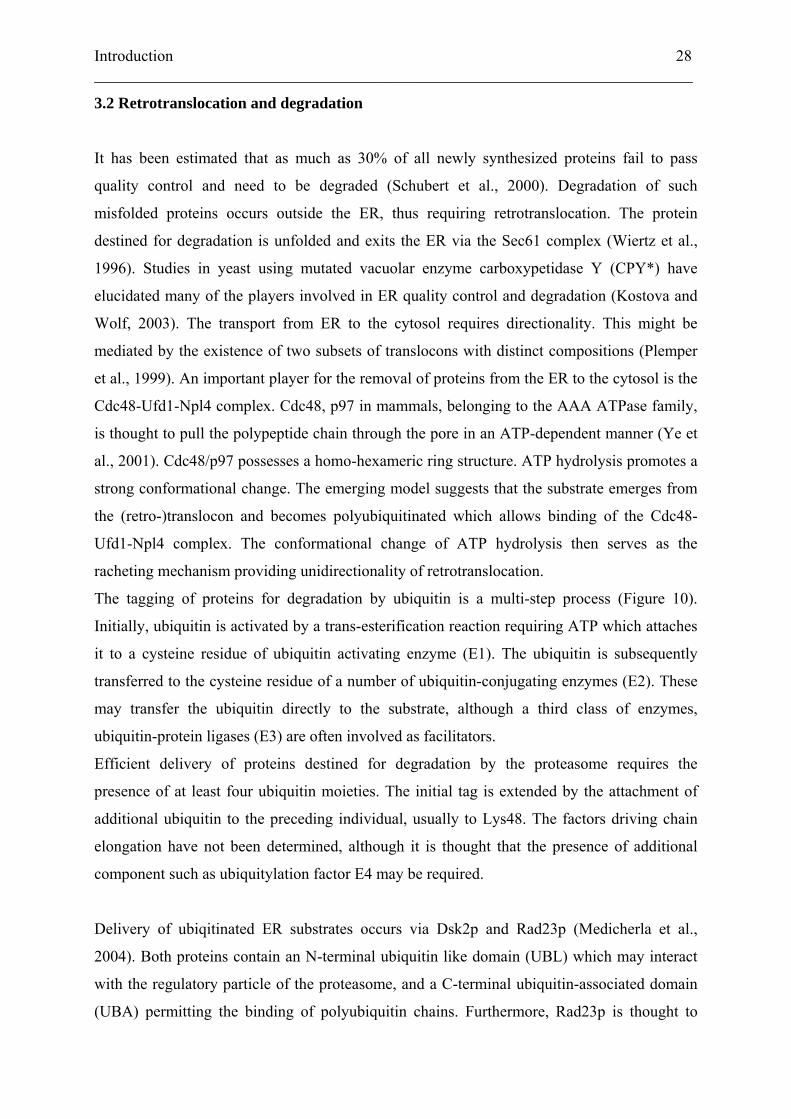
The tagging of proteins for degradation by ubiquitin is a multi-step process (Figure 10).
Initially, ubiquitin is activated by a trans-esterification reaction requiring ATP which attaches
it to a cysteine residue of ubiquitin activating enzyme (E1). The ubiquitin is subsequently
transferred to the cysteine residue of a number of ubiquitin-conjugating enzymes (E2). These
may transfer the ubiquitin directly to the substrate, although a third class of enzymes,
ubiquitin-protein ligases (E3) are often involved as facilitators.
Efficient delivery of proteins destined for degradation by the proteasome requires the
presence of at least four ubiquitin moieties. The initial tag is extended by the attachment of
additional ubiquitin to the preceding individual, usually to Lys48. The factors driving chain
elongation have not been determined, although it is thought that the presence of additional
component such as ubiquitylation factor E4 may be required.
Delivery of ubiqitinated ER substrates occurs via Dsk2p and Rad23p (Medicherla et al.,
2004). Both proteins contain an N-terminal ubiquitin like domain (UBL) which may interact
with the regulatory particle of the proteasome, and a C-terminal ubiquitin-associated domain
(UBA) permitting the binding of polyubiquitin chains. Furthermore, Rad23p is thought to
Introduction 29 ___________________________________________________________________________
play a role in deglycosylation of retrotranslocated glycoproteins by recruiting N-glycanase
(Suzuki et al., 2001).
The tagged substrate can bind to the 26S proteasome, the active degradation machinery,
which deubiquitylates and unfolds it prior to degradation. The proteasome consists of the 20S
core cylinder encompassing three catalytically active sites, and two 19S cap components
responsible for recognition, binding and unfolding of ubiquitnated proteins. The cap consists
of two functionally distinct parts, the base and the lid. The lid is necessary for
deubiquitylation, freeing ubiquitin for another round of protein targeting. The base, consisting
of a ring of six ATPases, both binds the tagged substrate and is thought to control access to
the core in a similar manner to Cdc48 in retrotranslocation.
Kostova and Wolf (2003)
FIG. 10. The cycle undertaken by ubiquitin moieties used in tagging defective protein substrate for degradation
(right). Schematic of the 26S proteasome with a linearized core segment. The active sites are marked in red
(left).
Introduction 30 ___________________________________________________________________________
4. Neurotoxicity hypothesis in ADNDI
Mutations in a secretory protein like pro-vasopressin are likely to disturb correct folding,
leading to retention of the protein by quality control. Such material will typically be degraded
via the ubiquitin-proteasome pathway. In a heterozygous situation, one might expect the
products of the wild-type allele to generate functional hormone, possibly of a reduced amount.
However, the ADNDI mutations listed in Table I are all dominant.
The mechanism explaining dominant inheritance has currently not been determined.
Dimerization of pro-vasopressin, beginning in the ER, might explain retention of the product
of the wild type allele only to some extent. The dimers consisting of one mutant and one wild
type pro-hormone would likely fail to pass quality control, leading to their eventual
degradation. Ito et al (1999) addressed this question when they epitope-tagged the wild-type
and several mutants (A(-1)T, ΔE47, G57S, and C67X) and transfected them into human
embryonic kidney cells, tsa 201. Crosslinking experiments revealed homo- and heterodimer
formation between wild-type and mutant precursors. Furthermore, mutant precursor was
found to inhibit trafficking of wild-type precursor from the ER to the Golgi apparatus.
Ultimately, fewer vasopressin-containing secretory granules would be available for release at
the cell periphery. However, the cell might adapt to the stronger demand for vasopressin by
increasing expression of the gene. This would still result in a recessive phenotype. Notably, in
similar experiments we were not able to confirm interaction of wild-type and mutant
vasopressin precursor.
Clinical data from individuals affected by diabetes insipidus has led to the development of a
hypothesis about the aetiology of the disease. A decrease in circulating vasopressin paralleled
by gliosis and hypocellularity of vasopressinergic neurons in the hypothalamus supported the
notion that vasopressin mutants exert a toxic effect on their host cells, resulting in
neurodegeneration. Support for this has been obtained from a limited number of post mortem
studies (Hanhart, 1940; Gaupp, 1941; Braverman et al., 1965; Nagai et al., 1984; Bergeron et
al., 1991). The selective destruction of the vasopressinergic magnocellular cells expressing
mutant hormone would explain the dominant inheritance. Progressive cell death could
account for the gradual development of clinical symptoms. How the mutants exert this
detrimental effect remains the major open question in understanding the disease.
Introduction 31 ___________________________________________________________________________
Support for the toxic hypothesis came from other heterologous expression studies. Ito et al.
(1997) used stably transfected Neuro2a cells to asses an effect of vasopressin mutants on cell
viability. Expression of mutant precursors (A(-1)T, ΔE47, G57S, and C67X) as such was not
found to considerably affect growth of cells. When the cells were differentiated to a neuronal
phenotype with valproic acid, however, viability was significantly reduced (C67X>A(-
1)T>G57S>ΔE47). Concurrent metabolic labeling and immunofluorescence studies confirmed
the reduced secretion of mutant precursors and their accumulation within the ER observed by
other groups
Beuret et al. (1999) investigated the molecular consequences of the signal peptide mutant
ΔG227. The mutation leads to translation initiation at an alternative ATG site, and displays
reduced cleavage of the signal peptide. Transient transfection of COS-7 cells showed that the
mutant was retained in the ER, and led to the formation of disulfide-linked aggregates. This
was not surprising since the uncleaved signal contains an unpaired cysteine at position -11.
Abolishing this unpaired cysteine did not alter the properties of the precursor protein. The
double mutant ΔG227/ C(-11)S showed a similar phenotype as ΔG227, ER-localized
precursor with an uncleaved signal peptide. To determine if uncleaved signal peptide
interferes with disulfide bond formation, a cysteine residue in the vasopressin region of a
wild-type precursor was altered to serine. The mutant, C6S, mimicked the aggregation
phenotype, while being largely confined to the ER, despite proper cleavage of the signal
peptide. Evidently, two independent processes, unpaired cysteine residues leading to aberrant
disulfide bond formation, and interference of uncleaved signal peptide with correct folding,
contribute to ER retention and formation of disulfide-linked aggregates.
The accumulation of material in the ER is a phenomenon common to a group of disorders
known as ER storage diseases (ERSD)(Rutishauser and Spiess, 2002). The material stems
from nascent proteins failing quality control mechanisms in the ER. The disease phenotypes
may result from the deficiency of a particular protein at its site of action, these are generally
recessive diseases, or the mutant protein may exert a toxic effect. In the latter case, the mutant
protein itself or one of its degradation products is detrimental to cell viability. Mutant proteins
might aggregate and thus prevent further transport, detaining chaperones and clogging the ER
until parts are rendered non-functional and need to be degraded.
The use of animal models to examine ADNDI has met with limited success. A mutation (del
G in NP 65, aka Brattleboro mutation) causes a frameshift leading to a strongly altered C-
Introduction 32 ___________________________________________________________________________
terminus. The precursor, bearing a poly Lys tail extending beyond the translational stop
codon, causes a recessive disease phenotype in the rat (Schmale et al., 1984). Furthermore, the
C67X mutation failed to reproduce a human disease phenotype in rats (Si-Hoe et al., 2000).
Although the mutant was retained in the ER and water homeostasis was affected, no cell death
or atrophy of magnocellular neurons was observed. Nevertheless, a recent paper supported the
neurodegeneration hypothesis in ADNDI using a murine knock-in model (Russell et al.,
2003). Mice expressing the C67X neurophysin II mutation showed polyuria and polydipsia
which worsened with progressing age. This was paralleled by loss of neurons in the
supraoptic and paraventricular nuclei, along with an induction of the chaperone BiP in these
cells. Furthermore, VP gene products could not be found in the neuronal projections,
indicating a trafficking defect. The observed neurodegenereation, however, did not appear in
mice expressing the common A(-1)T mutation.
5. Aim of this thesis
Experiments have shown ADNDI to be linked to a defect in protein trafficking. Protein
precursors of mutant vasopressin were found to accumulate in the ER. In addition,
experiments in our lab have demonstrated the formation of disulfide-linked aggregates formed
by mutant- but not wild-type vasopressin precursor (Beuret et al., 1999). Whether the
appearance of aggregates plays a direct or indirect role in the progression of the disease will
warrant further examination. The accumulation of proteins in the ER has been linked to
various classes of human disorders. Generally, terminally misfolded secretory protein
precursors are removed from the ER by shuttling them into the ERAD pathway. Apparently,
mutant vasopressin leads to toxic proteins or degradation fragments thereof, resulting in cell
death.
To investigate the basis of the neurotoxic effect, we decided to examine the mechanism of
degradation of ADNDI mutant pro-vasopressin.
Results 33 ___________________________________________________________________________
Results
Results 34 ___________________________________________________________________________
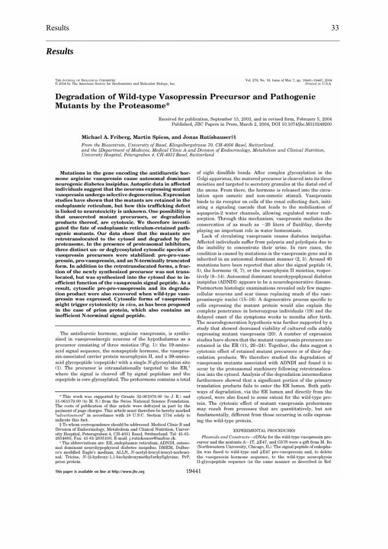
Degradation of wild-type vasopressin precursor and
pathogenic mutants by the proteasome
Michael A. Friberg, Martin Spiess, Jonas Rutishauser1
Biozentrum, University of Basel, Klingelbergstrasse 70, CH-4056 Basel, Switzerland, and 1Department of Medicine, Medical Clinic A and Division of Endocrinology, Metabolism and
Clinical Nutrition, University Hospital, Petersgraben 4, CH-4031 Basel, Switzerland
Results 35 ___________________________________________________________________________
Summary
Mutations in the gene encoding the antidiuretic hormone arginine vasopressin cause
autosomal dominant neurogenic diabetes insipidus. Autoptic data in affected individuals
suggest that the neurons expressing mutant vasopressin undergo selective degeneration.
Expression studies have shown that the mutants are retained in the endoplasmic reticulum, but
how this trafficking defect is linked to neurotoxicity is unknown. One possibility is that
unsecreted mutant precursors, or degradation products thereof, are cytotoxic. We therefore
investigated the fate of endoplasmic reticulum-retained pathogenic mutants. Our data show
that the mutants are retrotranslocated to the cytosol and degraded by the proteasome. In the
presence of proteasomal inhibitors, three distinct un- or deglycosylated cytosolic species of
vasopressin precursors were stabilized: pre-pro-vasopressin, pro-vasopressin, and an N-
terminally truncated form. In addition to the retrotranslocated forms, a fraction of the newly
synthesized precursor was not translocated, but synthesized into the cytosol due to inefficient
function of the vasopressin signal peptide. As a result, cytosolic pre-pro-vasopressin and its
degradation product were also recovered when wild-type vasopressin was expressed.
Cytosolic forms of vasopressin might trigger cytotoxicity in vivo, as has been proposed in the
case of prion protein, which also contains an inefficient N-terminal signal peptide.
Results 36 ___________________________________________________________________________
Introduction
The antidiuretic hormone, arginine vasopressin, is synthesized in vasopressinergic neurons of
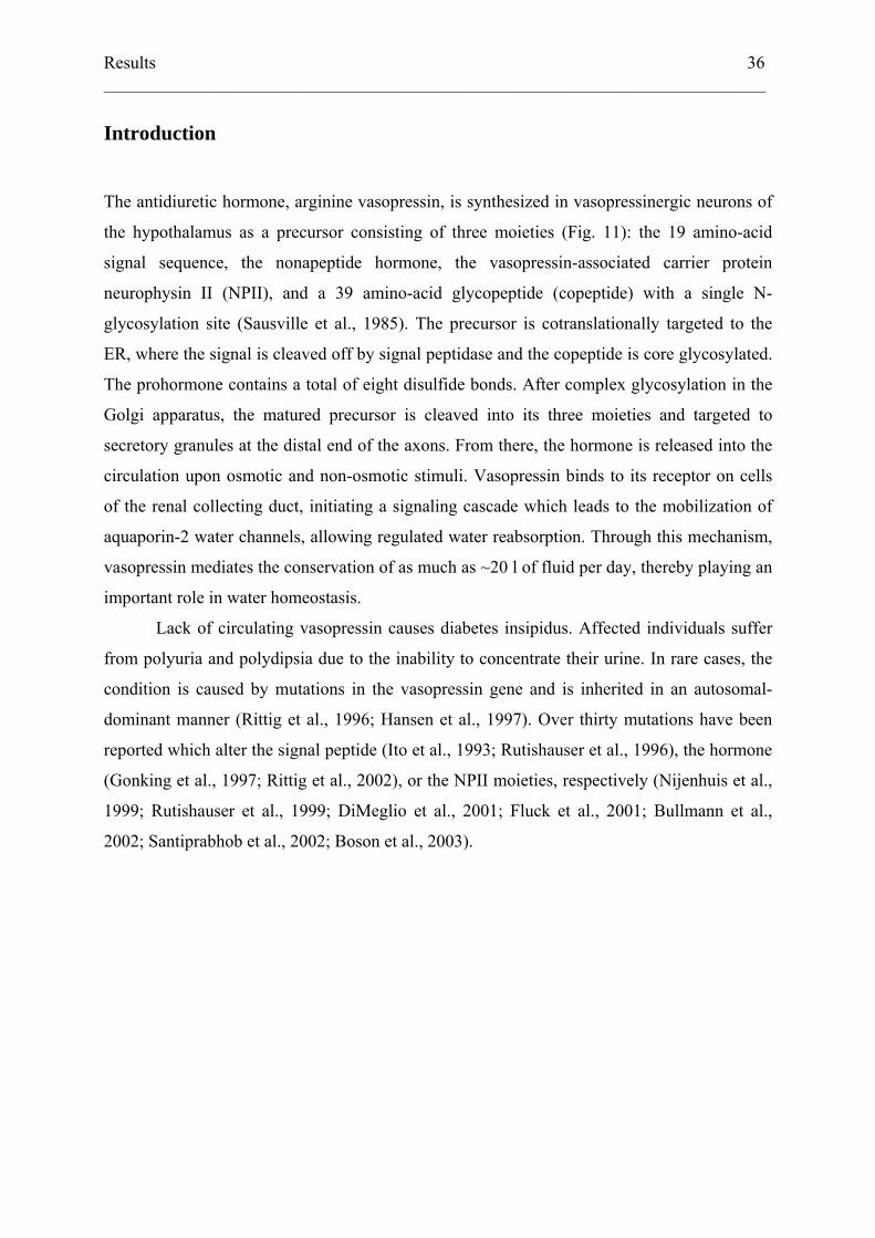
the hypothalamus as a precursor consisting of three moieties (Fig. 11): the 19 amino-acid
signal sequence, the nonapeptide hormone, the vasopressin-associated carrier protein
neurophysin II (NPII), and a 39 amino-acid glycopeptide (copeptide) with a single N-
glycosylation site (Sausville et al., 1985). The precursor is cotranslationally targeted to the
ER, where the signal is cleaved off by signal peptidase and the copeptide is core glycosylated.
The prohormone contains a total of eight disulfide bonds. After complex glycosylation in the
Golgi apparatus, the matured precursor is cleaved into its three moieties and targeted to
secretory granules at the distal end of the axons. From there, the hormone is released into the
circulation upon osmotic and non-osmotic stimuli. Vasopressin binds to its receptor on cells
of the renal collecting duct, initiating a signaling cascade which leads to the mobilization of
aquaporin-2 water channels, allowing regulated water reabsorption. Through this mechanism,
vasopressin mediates the conservation of as much as ~20 l of fluid per day, thereby playing an
important role in water homeostasis.
Lack of circulating vasopressin causes diabetes insipidus. Affected individuals suffer
from polyuria and polydipsia due to the inability to concentrate their urine. In rare cases, the
condition is caused by mutations in the vasopressin gene and is inherited in an autosomal-
dominant manner (Rittig et al., 1996; Hansen et al., 1997). Over thirty mutations have been
reported which alter the signal peptide (Ito et al., 1993; Rutishauser et al., 1996), the hormone
(Gonking et al., 1997; Rittig et al., 2002), or the NPII moieties, respectively (Nijenhuis et al.,
1999; Rutishauser et al., 1999; DiMeglio et al., 2001; Fluck et al., 2001; Bullmann et al.,
2002; Santiprabhob et al., 2002; Boson et al., 2003).
Results 37 ___________________________________________________________________________
FIG. 11. Wild-type and mutant vasopressin precursor and its signal sequence. The domain organization of
the vasopressin precursor is shown with disulfide bridges as line connections and the glycosylation site as a
diamond. The positions of mutations in mutant vasopressin precursors used in this study are indicated by an
arrow. Below, the sequence of the signal peptide and the N-terminal portion of pro-vasopressin is shown. To
prevent ER targeting, L(-9) of the signal peptide was mutated to R as indicated.
Autosomal dominant neurohypophyseal diabetes insipidus (ADNDI) appears to be a
neurodegenerative disease. Post-mortem histologic examinations revealed only few magno-
cellular neurons and scar tissue replacing much of the vasopressinergic nuclei (Hanhart, 1940;
Gaupp, 1941; Braverman et al., 1965; Bergeron et al., 1991). A degenerative process specific
to cells expressing the mutant protein would also explain the complete penetrance in
heterozygous individuals (Mahoney et al., 2002) and the delayed onset of the symptoms
weeks to months after birth. The neurodegeneration hypothesis was further supported by a
study which showed decreased viability of cultured cells stably expressing mutant vasopressin
(Ito and Jameson, 1997). A number of expression studies have shown that the mutant
vasopressin precursors are retained in the ER (Olias et al., 1996; Beuret et al., 1999; Ito et al.,
1999; Nijenhuis et al., 1999; Siggaard et al., 1999; Evans et al., 2000). Together, the data
suggest a cytotoxic effect of retained mutant precursors or of their degradation products.
Results 38 ___________________________________________________________________________
We therefore studied the degradation of vasopressin mutants associated with ADNDI
and found it to occur by the proteasomal machinery following retrotranslocation into the
cytosol. Analysis of the degradation intermediates furthermore showed that a significant
portion of the primary translation products fails to enter the ER lumen. Both pathways of
degradation, via the ER lumen and directly from the cytosol, were also found to some extent
for the wild-type protein. The cytotoxic effect of mutant vasopressin prohormone may result
from processes that are quantitatively, but not fundamentally different from those occurring in
cells expressing the wild-type protein.
Results 39 ___________________________________________________________________________
Experimental Procedures
Plasmids and constructs
cDNAs for the wild-type vasopressin precursor and the mutants A(-1)T, ΔE47, and G57S
were a gift from M. Ito (Northwestern University, Chicago, IL). The signal peptide of
enkephalin was fused to wild-type and ΔE47 pro-vasopressin and, to delete the vasopressin
hormone sequence, to the wild-type neurophysin II-glycopeptide sequence (in the same
manner as described in (Cescato et al., 2000). Point mutations D(-17)R, L(-9)R, and
R(8)E/K(11)E/R(12)E, were generated by polymerase chain reaction. All cDNAs were
subcloned into the pRc/RSV expression plasmid (Invitrogen) and verified by DNA
sequencing.
Cell culture and transient transfection
COS-1 and CV-1 cells were grown in Dulbecco’s modified Eagle’s medium (DMEM; Sigma)
supplemented with 10% fetal calf serum, 100 units/ml penicillin, 100 μg/ml streptomycin, 2
mM L-glutamine at 37°C in 7.5% CO2. Neuro2A cells COS-1 cells were transiently
transfected in 6-well plates using Lipofectine (Life Technologies, Inc.) and used 2–3 days
after transfection. Neuro2a were grown in DMEM containing 4500 mg/l glucose in 5% CO2.
They were transfected using Metafectene (Biontex Laboratories). To produce stably
expressing cell lines, the cDNA of the vasopressin precursor was subcloned into the
expression vector pCB6 and transfected into CV-1 cells using calcium phosphate
precipitation. Clonal cell lines resistant to 0.5 mg/ml G418-sulfate were isolated and screened
for pro-vasopressin expression by immunofluorescence.
Results 40 ___________________________________________________________________________
Metabolic labeling and immunoprecipitation
For labeling experiments, transfected cells were starved for 30 min in DMEM without
cysteine and methionine (Sigma) supplemented with 2 mM L-glutamine. Cells were labeled
for the times indicated with 100 μCi/ml [35S] protein labeling mix (DuPont-NEN) in
starvation medium and chased in starvation medium supplemented with excess cysteine and
methionine. Cells were transferred to 4°C, washed with phosphate-buffered saline (PBS),
lysed in 500 μl of lysis buffer (PBS, 1% Triton X-100, 0.5% deoxycholate, 2 mM
phenylmethylsulfonyl fluoride), and scraped. After 10 min centrifugation in a microfuge, the
lysate was subjected to immunoprecipitation using rabbit polyclonal anti-neurophysin II or
anti-vasopressin antibodies (ICN). The immune complexes were isolated with protein A-
Sepharose (Zymed) and analyzed by electrophoresis on 10% polyacrylamide Tris/trycine
SDS-gels and autoradiography. For deglycosylation, immunoprecipitates were either boiled
for 2 min in 50 µl 50 mM Na-citrate, pH 6, 1% SDS and incubated with 1 mU endo-β-N-
acetylglucosaminidase H (Roche Biochemicals) for 2 h at 37°C, or they were boiled in 100 µl
0.1 M Na-phosphate, pH 6.8, containing 50 mM EDTA, 1% β-mercaptoethanol, 0.1% SDS
and incubated with 0.25 U endoglycosidase F/N-glycosidase F (Roche Biochemicals) for 3 h
at 37°C
Protease inhibition
Stock solutions of 10 mM N-Acetyl-leucyl-leucyl-norleucinal (ALLN), 1 mM lactacystin, 1
mM pepstatin A (all from Sigma) in DMSO, and of 10 mM leupeptin (Roche Biochemicals)
in water were prepared. For application to the cells, they were diluted into DMEM to final
concentrations of 250 μM ALLN, 25 or 40 μM lactacystin, 100 μM leupeptin, and 5 μM
pepstatin A. ALLN was added to the cells 90 min and lactacystin 10 min prior to the
experiment and was freshly added to the starvation, labeling, and chase media. The lysosomal
inhibitors leupeptin and pepstatin A were applied to the cells 16 h before the experiment and
were present during all further incubations.
Results 41 ___________________________________________________________________________
Cytosol extraction
To separate the cytosol from microsomes, labeled cells were incubated at 4°C in swelling
buffer (15 mM HEPES/KOH, pH 7.2, 15 mM KCl) supplemented with 2 mM
phenylmethylsulfonyl fluoride and protease inhibitor cocktail (from a 500-fold concentrated
stock of 1 mg/ml each of pepstatin A, leupeptin, chymostatin, antipain, and 5 mg/ml
benzamidine, dissolved in 40% DMSO and 60% ethanol) for 15 min at 4°C, scraped, and
centrifuged for 30 min at 136000×g. The supernatant containing the cytosol and the
resuspended organelle pellet were subjected to immunoprecpitation and analyzed as above.
Results 42 ___________________________________________________________________________
Results
Proteasome inhibitors stabilize mutant vasopressin precursors and degradation
intermediates
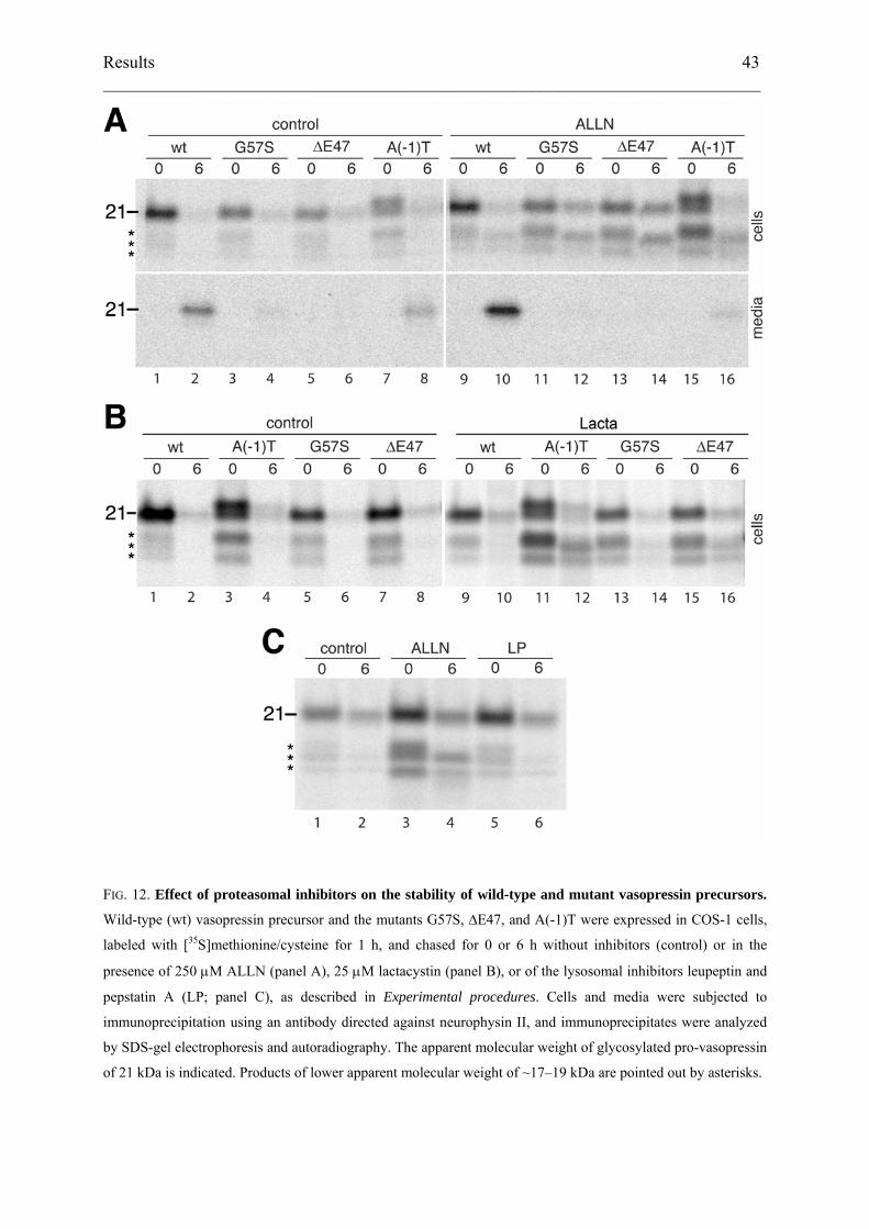
To test the fate of wild-type and mutant pre-pro-vasopressin in COS-1 cells, transiently
transfected cells were radiolabeled with [35S]methionine/cysteine for 1 h and chased with
excess unlabeled methionine/cysteine for 0 or 6 h. Cells and media were subjected to
immunoprecipitation using an antibody directed against neurophysin II followed by SDS-gel
electrophoresis and fluorography (Fig. 2A, lanes 1–8). Upon pulse-labeling, wild-type protein
and the mutants ∆E47 and G57S were found as a major species of ~21 kDa corresponding to
N-glycosylated pro-vasopressin. The mutant A(-1)T, in which mutation of the last residue of
the signal sequence causes inefficient signal cleavage (Ito et al., 1993), appeared as two major
products corresponding to glycosylated pre-pro-vasopressin and pro-vasopressin. In all cases,
additional faint bands in the range of~17–19 kDa were produced. After 6 h of chase, wild-type
pro-vasopressin and the signal-cleaved fraction of the A(-1)T mutant were secreted into the
medium. Since COS cells lack prohormone processing enzymes, intact glycosylated pro-
vasopressin of 21 kDa was recovered. Hardly any protein could be detected in the cells,
indicating that the mutants ∆E47, G57S, and the uncleaved fraction of A(-1)T had been
retained and degraded.
To test for degradation via the ER-associated degradation pathway, the proteasomal peptide
inhibitor N-acetyl-leucyl-leucyl-norleucinal (ALLN) was added to the medium 90 min before
and during the pulse and the chase periods (Fig. 12A, lanes 9–16). ALLN stabilized the
putative degradation intermediates of ~17–19 kDa for wild-type and mutant precursors, and to
variable extent also the full-size, glycosylated band of the mutant precursors, consistent with
proteasomal degradation of retained protein. This was confirmed by experiments using
lactacystin, a more specific proteasomal inhibitor. Addition of 25 μM lactacystin stabilized
low molecular weight forms that were indistinguishable from those seen with ALLN
treatment (Fig. 12B). In contrast, a mixture of leupeptin and pepstatin A, two inhibitors of
lysosomal degradation, had no stabilizing effect on the mutant ∆E47 (Fig. 12C). These results
indicate that mutant pro-vasopressin as well as a fraction of wild-type pro-vasopressin is
degraded by the proteasome in a process that involves intermediates of 17–19 kDa.
Results 43 ___________________________________________________________________________
FIG. 12. Effect of proteasomal inhibitors on the stability of wild-type and mutant vasopressin precursors.
Wild-type (wt) vasopressin precursor and the mutants G57S, ∆E47, and A(-1)T were expressed in COS-1 cells,
labeled with [35S]methionine/cysteine for 1 h, and chased for 0 or 6 h without inhibitors (control) or in the
presence of 250 μM ALLN (panel A), 25 μM lactacystin (panel B), or of the lysosomal inhibitors leupeptin and
pepstatin A (LP; panel C), as described in Experimental procedures. Cells and media were subjected to
immunoprecipitation using an antibody directed against neurophysin II, and immunoprecipitates were analyzed
by SDS-gel electrophoresis and autoradiography. The apparent molecular weight of glycosylated pro-vasopressin
of 21 kDa is indicated. Products of lower apparent molecular weight of ~17–19 kDa are pointed out by asterisks.
Results 44 ___________________________________________________________________________
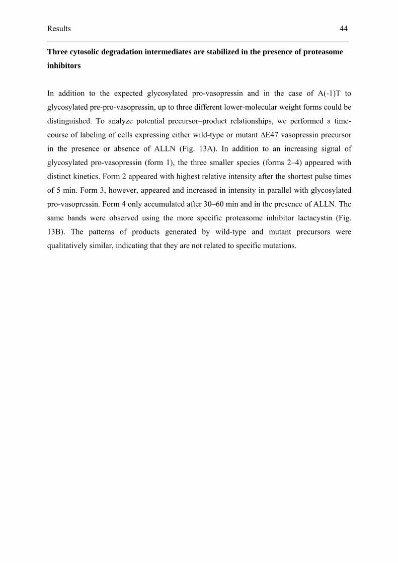
Three cytosolic degradation intermediates are stabilized in the presence of proteasome
inhibitors
In addition to the expected glycosylated pro-vasopressin and in the case of A(-1)T to
glycosylated pre-pro-vasopressin, up to three different lower-molecular weight forms could be
distinguished. To analyze potential precursor–product relationships, we performed a time-
course of labeling of cells expressing either wild-type or mutant ∆E47 vasopressin precursor
in the presence or absence of ALLN (Fig. 13A). In addition to an increasing signal of
glycosylated pro-vasopressin (form 1), the three smaller species (forms 2–4) appeared with
distinct kinetics. Form 2 appeared with highest relative intensity after the shortest pulse times
of 5 min. Form 3, however, appeared and increased in intensity in parallel with glycosylated
pro-vasopressin. Form 4 only accumulated after 30–60 min and in the presence of ALLN. The
same bands were observed using the more specific proteasome inhibitor lactacystin (Fig.
13B). The patterns of products generated by wild-type and mutant precursors were
qualitatively similar, indicating that they are not related to specific mutations.
Results 45 ___________________________________________________________________________
FIG. 13. Time-course of appearance of different vasopressin precursor forms. COS-1 cells expressing wild-
type (wt) or ∆E47 mutant vasopressin precursor were labeled for 5–60 min with [35S]methionine/cysteine. Cells
were incubated with proteasome inhibitor (+) before and during labeling as described in Experimental
procedures or were untreated (–). ALLN (250 µM) was used as inhibitor in panel A, and lactacystin (40 µM) in
panel B. Vasopressin products were immunoprecipitated and analyzed by SDS-gel electrophoresis and
autoradiography.
Results 46 ___________________________________________________________________________
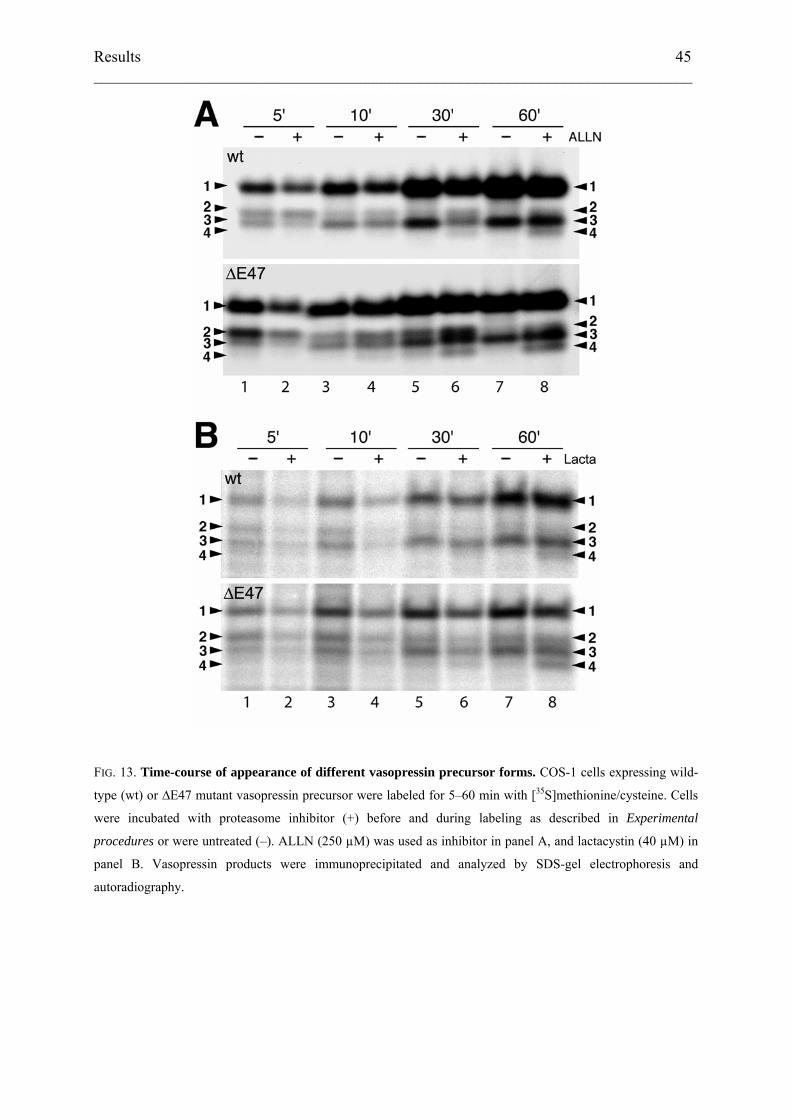
To characterize the different forms, immunoprecipitates of ALLN-treated labeled cells
expressing ∆E47 vasopressin precursor were incubated with endoglycosidase H or F (Fig.
14A, lane 1–3). The 21-kDa form 1 was deglycosylated to an apparent molecular weight of
~18 kDa corresponding to form 3. In contrast, the lower bands were insensitive to
deglycosylation. This suggested that product 3 corresponds to un- or deglycosylated pro-
vasopressin and product 4 to a subsequent degradation intermediate lacking a short segment of
the polypeptide at the N- or C-terminus. Upon immunoprecipitation using an antibody
directed against the vasopressin hormone, form 4 was not recovered (Fig. 14A, lane 4),
indicating that it lacks the hormone sequence at the N-terminus.
Based on its size, product 2 likely represents pre-pro-vasopressin, the primary
translation product that had not been translocated to the ER lumen. For comparison, we
expressed various mutant precursors to serve as size markers (Fig. 14B). A mutant with a
nonfunctional signal sequence (L(-9)R; lane 2), in which an arginine disrupts the hydrophobic
core, comigrated with form 2. Only a very small fraction was glycosylated but not processed
by signal peptidase (arrowhead). A mutant lacking the signal peptide entirely (∆SP), i.e. pro-
vasopressin synthesized into the cytosol, migrated like band 3 (lane 3). In a further construct
the hormone domain was deleted (∆VP) by fusing NPII-glycopeptide to the signal sequence of
pre-pro-enkephalin. In addition to a glycosylated product of ~21 kDa, this construct also
produced a 17-kDa form comigrating with band 4 (lane 4). Interestingly, this product of 17
kDa was generated by all constructs, indicating that N-terminal clipping occurred
independently of whether the protein was initially inserted into the ER or synthesized directly
into the cytosol, and whether a signal sequence was still attached or not.
ER-associated degradation involves the retrotranslocation of unfolded or misfolded
proteins from the ER lumen back to the cytosol where they are exposed to cytosolic N-
glycanase (Hirsch et al., 2003; Kostova and Wolf, 2003). To determine the localization of the
low-molecular weight forms, cells expressing wild-type or ∆E47 vasopressin precursor were
labeled for 1 h in the presence of ALLN, broken by swelling and scraping, and subjected to
ultracentrifugation. We then analyzed the immunoprecipitated products in the membrane
pellet (M) and the cytosol fraction (C) in comparison to the unfractionated total cell lysate (L;
Fig. 14C). The experiment was performed with cells labeled for 5 min (lanes 1–3), producing
predominantly form 2, or for 60 min (lanes 4–9), generating forms 3 and 4 in addition to
Results 47 ___________________________________________________________________________
FIG. 14. Characterization of low molecular weight vasopressin products. Panel A: COS-1 cells expressing the
vasopressin precursor mutant ∆E47 were incubated with ALLN, labeled with [35S]methionine/cysteine for 1 h,
subjected to immunoprecipitation using anti-neurophysin II (αNP) or anti-vasopressin (αVP) antibodies and
analyzed either directly (–) or after deglycosylation using endoglycosidase H (H) or endoglycosidase F (F). Panel
B: COS-1 cells expressing the following mutant precursors were labeled for 30 min in the presence of ALLN and
analyzed as above: ∆E47, the signal peptide mutant L(-9)R, the signal peptide deletion mutant ∆SP, and the
mutant ∆VP which lacks the vasopressin hormone sequence. Panel C: COS-1 cells expressing wild-type or ∆E47
mutant precursor as indicated were incubated with ALLN and labeled for 5 min to generate (besides form 1)
predominantly form 2, or for 60 min to generate predominantly forms 3 and 4. The labeled cells were then
broken by swelling and centrifuged at high speed. The supernatants containing cytosolic proteins (C) and the
membrane pellets (M) were analyzed in parallel to unfractionated aliquots of total cell lysates by
immunoprecipitation, gel electrophoresis and autoradiography.
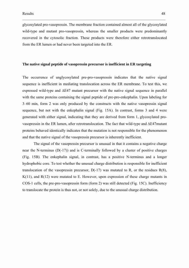
Results 48 ___________________________________________________________________________
glycosylated pro-vasopressin. The membrane fraction contained almost all of the glycosylated
wild-type and mutant pro-vasopressin, whereas the smaller products were predominantly
recovered in the cytosolic fraction. These products were therefore either retrotranslocated
from the ER lumen or had never been targeted into the ER.
The native signal peptide of vasopressin precursor is inefficient in ER targeting
The occurrence of unglycosylated pre-pro-vasopressin indicates that the native signal
sequence is inefficient in mediating translocation across the ER membrane. To test this, we
expressed wild-type and ∆E47 mutant precursor with the native signal sequence in parallel
with the same proteins containing the signal peptide of pre-pro-enkephalin. Upon labeling for
3–60 min, form 2 was only produced by the constructs with the native vasopressin signal
sequence, but not with the enkephalin signal (Fig. 15A). In contrast, forms 3 and 4 were
generated with either signal, indicating that they are derived from form 1, glycosylated pro-
vasopressin in the ER lumen, after retrotranslocation. The fact that wild-type and ΔE47mutant
proteins behaved identically indicates that the mutation is not responsible for the phenomenon
and that the native signal of the vasopressin precursor is inherently inefficient.
The signal of the vasopressin precursor is unusual in that it contains a negative charge
near the N-terminus (D(-17)) and is C-terminally followed by a cluster of positive charges
(Fig. 15B). The enkephalin signal, in contrast, has a positive N-terminus and a longer
hydrophobic core. To test whether the unusual charge distribution is responsible for inefficient
translocation of the vasopressin precursor, D(-17) was mutated to R, or the residues R(8),
K(11), and R(12) were mutated to E. However, upon expression of these charge mutants in
COS-1 cells, the pre-pro-vasopressin form (form 2) was still detected (Fig. 15C). Inefficiency
to translocate the protein is thus not, or not solely, due to the unusual charge distribution.

Results 49 ___________________________________________________________________________
FIG. 15. Signal dependence of form 2 generation. Panel A: COS-1 cells expressing wild-type or mutant ∆E47
precursor with either the native vasopressin signal (V) or the signal sequence of pre-pro-enkephalin (E) were
incubated with ALLN and labeled for 3–60 min with [35S]methionine/cysteine before immunoprecipitation, gel
electrophoresis and autoradiography. Panel B: The sequence of the enkephalin signal (E) fused to pro-
vasopressin (gray) is shown in comparison to the native vasopressin signal (V). Mutation of the charges flanking
the hydrophobic core of the signal in constructs V(D>R) and V(RKR>EEE) are indicated. Panel C: COS-1 cells
expressing the vasopressin precursor with the native signal (V), with the enkephalin signal (E), or with the
charge mutants V(D>R) and V(RKR>EEE) were labeled for 5 min and analyzed as in A.
Results 50 ___________________________________________________________________________
Missorting of pre-pro-vasopressin is not due to overexpression and also occurs in
neuronal cells
To exclude the possibility that mistargeting of pre-pro-vasopressin is simply a consequence of
high-level expression in COS cells, we examined the polypeptides produced in stably
transfected CV-1 cells, the parental cell line of COS-1 cells lacking the large T antigen
driving the SV40 promoter/origin of replication present in our expression plasmids. In
labeling and immunoprecipitation experiments, the stable CV-1 cell line expressing wild-type
vasopressin precursor yielded a somewhat lower signal from the same number of cells than
COS cells of which only 5–10% were transfected. The CV-1 cells are therefore producing at
least 10–20 fold less of the protein per cell. Even in this situation, all three low molecular
weight forms were made (Fig. 16A). In particular, form 2, pre-pro-vasopressin, was made as
well. The inefficiency of the vasopressin signal is thus also apparent at moderate rates of
synthesis.
In vivo, the vasopressin precursor is expressed by hypothalamic secretory neurons. We
tested whether the products observed in fibroblasts were also generated in Neuro2a cells, a
mouse neuroblastoma cell line that endogenously expresses secretogranin II and that had been
shown to sort exogenous pro-opiomelanocortin into dense-core granules (Chevrier et al.,
1991). In transfected Neuro2a cells expressing wild-type or ∆E47 mutant vasopressin
precursor and labeled for 5–60 min, pre-pro-vasopressin (form 2) and unglycosylated pro-
vasopressin (form 3) were detected and stabilized by ALLN as in COS-1 and CV-1 cells (Fig.
16B). However, form 4 corresponding to N-terminally truncated pro-vasopressin could not be
detected. Whereas the inefficiency of the vasopressin signal is observed in all cell types
tested, the cytosolic protease responsible for N-terminal truncation of vasopressin precursors
in COS-1 and CV-1 cells, is missing in Neuro2a cells.
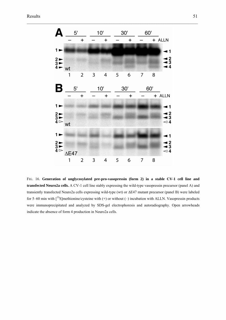
Results 51 ___________________________________________________________________________
FIG. 16. Generation of unglycosylated pre-pro-vasopressin (form 2) in a stable CV-1 cell line and
transfected Neuro2a cells. A CV-1 cell line stably expressing the wild-type vasopressin precursor (panel A) and
transiently transfected Neuro2a cells expressing wild-type (wt) or ∆E47 mutant precursor (panel B) were labeled
for 5–60 min with [35S]methionine/cysteine with (+) or without (–) incubation with ALLN. Vasopressin products
were immunoprecipitated and analyzed by SDS-gel electrophoresis and autoradiography. Open arrowheads
indicate the absence of form 4 production in Neuro2a cells.
Results 52 ___________________________________________________________________________
Discussion
Degradation of vasopressin precursor occurs via proteasomes
In cells expressing mutant vasopressin precursor, products of lower molecular weight than
full-size glycosylated pro-vasopressin were stabilized by proteasomal inhibitors. No
stabilization was observed with inhibitors of lysosomal proteases. This confirmed the
expectation that the mutant proteins retained in the ER by the lumenal quality control system
are subject to ER-associated degradation (ERAD), i.e. proteolysis by the cytosolic
proteasome. Interestingly, significant stabilization of the same types of intermediates was also
observed in cells expressing the wild-type protein. This may be due to a considerable number
of polypeptides that never attained the native structure owing to errors in translation or post-
translational processes necessary for proper protein folding. It has previously been estimated
that about one third of newly synthesized total protein is rapidly degraded (Schubert et al.,
2000).
Proteins targeted for proteasomal degradation are often, but not always, tagged by
ubiquitin (Ciechanover, 1994; Hiller et al., 1996; Fisher et al., 1997; Zhou et al., 1998;
Yewdell, 2001). We were unable to demonstrate ubiquitination of vasopressin polypeptides
using multiple approaches, including immunoblotting of immunoprecipitated vasopressin
precursor with anti-ubiquitin antibodies, increasing the cells' ubiquitin pool by overexpressing
a ubiquitin cDNA, or coexpression of dominant-negative ubiquitin constructs (K48R and
K48RG76A). It is unclear whether we did not reach sufficient amounts of ubiquitinated
material, whether the ubiquitinated form might not be recognized by our antibodies, or
whether the vasopressin mutants are targeted to the proteasome through alternative pathways,
such as neddylation or sumoylation (Gong et al., 2000; Kamitani et al., 2001; Aguilar and
Wendland, 2003). To detect ubiquitinylated proteins is notoriously difficult. In general only a
small amount of the protein is detectable in ubiquitinated forms, which furthermore are
heterogeneous in size. In addition, rapid deubiquitination may occur in cell lysates.
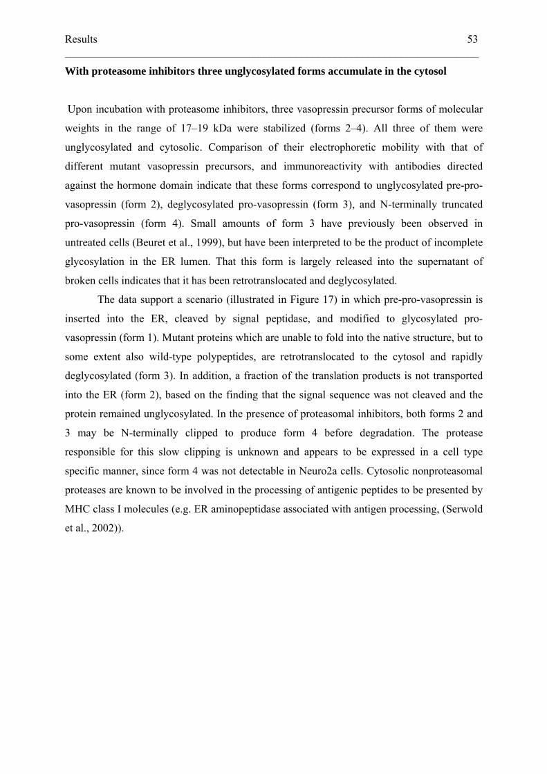
Results 53 ___________________________________________________________________________
With proteasome inhibitors three unglycosylated forms accumulate in the cytosol
Upon incubation with proteasome inhibitors, three vasopressin precursor forms of molecular
weights in the range of 17–19 kDa were stabilized (forms 2–4). All three of them were
unglycosylated and cytosolic. Comparison of their electrophoretic mobility with that of
different mutant vasopressin precursors, and immunoreactivity with antibodies directed
against the hormone domain indicate that these forms correspond to unglycosylated pre-pro-
vasopressin (form 2), deglycosylated pro-vasopressin (form 3), and N-terminally truncated
pro-vasopressin (form 4). Small amounts of form 3 have previously been observed in
untreated cells (Beuret et al., 1999), but have been interpreted to be the product of incomplete
glycosylation in the ER lumen. That this form is largely released into the supernatant of
broken cells indicates that it has been retrotranslocated and deglycosylated.
The data support a scenario (illustrated in Figure 17) in which pre-pro-vasopressin is
inserted into the ER, cleaved by signal peptidase, and modified to glycosylated pro-
vasopressin (form 1). Mutant proteins which are unable to fold into the native structure, but to
some extent also wild-type polypeptides, are retrotranslocated to the cytosol and rapidly
deglycosylated (form 3). In addition, a fraction of the translation products is not transported
into the ER (form 2), based on the finding that the signal sequence was not cleaved and the
protein remained unglycosylated. In the presence of proteasomal inhibitors, both forms 2 and
3 may be N-terminally clipped to produce form 4 before degradation. The protease
responsible for this slow clipping is unknown and appears to be expressed in a cell type
specific manner, since form 4 was not detectable in Neuro2a cells. Cytosolic nonproteasomal
proteases are known to be involved in the processing of antigenic peptides to be presented by
MHC class I molecules (e.g. ER aminopeptidase associated with antigen processing, (Serwold
et al., 2002)).
Results 54 ___________________________________________________________________________
FIG. 17. Schematic summary of the products and degradation intermediates of vasopressin precursor. The
majority of vasopressin precursor is translocated into the ER lumen, cleaved by signal peptidase and glycosylated
to form 1 (glycosylated pro-vasopressin). Molecules unable to fold are retrotranslocated into the cytosol and
deglycosylated to form 3 (deglycosylated pro-vasopressin). A fraction of precursor proteins (even of the wild-
type) is not translocated and is found as cytosolic pre-pro-vasopressin (form 2). Forms 2 and 3 are stabilized by
proteasomal inhibitors. In COS-1 and CV-1 cells, but not in Neuro2a cells, they give rise to an N-terminally
processed form 4. Primarily forms 2, 3, and 4 accumulate upon addition of proteasome inhibitors indicating that
they are substrates of the proteasome (shown on the right).
Results 55 ___________________________________________________________________________
The vasopressin signal functions inefficiently
The production of pre-pro-vasopressin suggests that the native vasopressin signal is
inefficient. This is not due to incomplete signal cleavage, since no glycosylated pre-pro-
vasopressin could be detected. In ADNDI mutants affecting the cleavage efficiency of the
signal, such as A(-1)T (mutation of the cleavage site (Ito et al., 1993)) and ∆G227 (truncation
of the signal; (Beuret et al., 1999)), glycosylated pre-pro-vasopressin is easily detected. A(-
1)T also revealed increased form 2 (Fig. 12A, lane 15, and Fig. 12B, lane 11), since
retrotranslocated polypeptides add to those that were primarily synthesized into the cytosol.
Therefore, the defect in the native vasopressin signal is due to inefficient targeting or
translocation. The phenomenon is not an artefact of overexpression and potential saturation of
the secretory route, since it is also observed in CV-1 cells expressing at least ten times less
protein per cell. Moreover, it is detected in Neuro2a cells which have characteristics of
neuroendocrine cells and is thus likely to occur also in vasopressinergic cells in vivo.
The charges flanking the hydrophobic core of signal sequences largely determine
signal orientation in the ER translocation machinery (Goder and Spiess, 2001). Typically, the
N-terminal portion of signal peptides is positively charged (the positive-inside rule; (von
Heijne, 1990)), or at least more positive than the C-terminal flanking sequence (Hartmann et
al., 1989). This is not the case for the vasopressin signal where the charge difference ∆(C–N)
calculated according to the rules by Hartmann et al. (Hartmann et al., 1989) is +2.
Surprisingly, however, the unusual charge distribution is not responsible for the translocation
inefficiency: mutation of D(-17) to R or of R(8), K(11), and R(12) to E did not reduce the
production of pre-pro-vasopressin despite an improved charge difference of 0 and -4,
respectively. The efficiency of the enkephalin signal fused to pro-vasopressin is thus also not
just due to the positive N-terminus. The hydrophobic core of the enkephalin signal is longer
and more hydrophobic (in total and on average) than that of the vasopressin signal. This might
account for more efficient recognition by signal recognition particle (Hatsuzawa et al., 1997).
Inefficient function of signal sequences is observed rarely. Plasminogen activator
inhibitor-2 (PAI-2) is found as a secreted and a cytosolic form because of an internal,
uncleaved signal that is inefficient both in binding signal recognition particle and in
subsequent interaction with the translocation complex (Belin et al., 1996). Parathyroid
hormone-related peptide (PTHrP) precursor is in part found as pre-pro-PTHrP in the cell and
accumulates in nucleoli due to a nucleolar targeting signal (Amizuka et al., 2000). Although
Results 56 ___________________________________________________________________________
this signal has a positive N-terminus, its C-terminal sequence is even more positively charged.
In both cases, dual localization may reflect separate functions in different compartments.
Of special pathophysiological interest is the case of the prion protein (PrP). A
particular misfolded conformation of PrP (PrPSc), which is favored by certain mutations in the
protein, causes neurodegenerative disorders (prion diseases). The N-terminal signal of PrP is
inefficient and membrane targeting of a fraction of the protein is rescued by a C-terminal
hydrophobic sequence (Hölscher et al., 2001). PrP can adopt multiple membrane topologies,
including a fully translocated form (SecPrP), two transmembrane forms with either the N- or
the C-terminal portion of the polypeptide translocated into the ER lumen (NtmPrP and CtmPrP,
respectively), and a cytosolic form (Hegde et al., 1998; Hegde and Rane, 2003). Again, the
charge difference of the signal is the opposite of that typical for secretory signal peptides. It
has been shown that mutant PrP as well as a significant portion of wild-type PrP is degraded
via the proteasome, since cytosolic, unglycosylated forms accumulate upon treatment with
proteasomal inhibitors (Ma and Lindquist, 2001; Yedidia et al., 2001). It has recently been
proposed that the cytosolic forms of PrP trigger at least some neurodegenerative prion
diseases, because expression of a cytosolic form of PrP lacking the N-terminal signal and the
C-terminal glycosylphosphatidylinositol anchor sequence was toxic in Neuro2a cells and
rapidly caused neurodegeneration in transgenic mice (Ma et al., 2002). In addition, conversion
to a PrPSc-like conformation was found to be increased for a mutant PrP causing heritable
prion disease correlating with its increased transport into the cytosol (Ma and Lindquist,
2002). To which extent the situation of mutant vasopressin precursor parallels that of PrP
remains to be determined.
It is not known at present whether ADNDI results from a toxic product in the cytosol
or in the ER lumen. Several factors that are induced by ER stress have been shown to mediate
cell damage. The transcription factor CHOP has been shown to induce an apoptotic response
in renal tubular cells of mice injected intraperitoneally with tunicamycin (Zinszner et al.,
1998), and downstream effectors of CHOP have been characterized (Wang et al., 1998). In a
mouse model of non-autoimmune diabetes mellitus, hyperglycemia occurs as a consequence
of apoptotic destruction of pancreatic β cells induced by ER stress due to ER retention of a
mutant insulin precursor. Targeted deletion of CHOP delays the onset of diabetes in these
mice (Oyadomari et al., 2002). Tunicamycin-induced apoptotic destruction of renal tubular
cells is significantly alleviated in mice deficient in caspase-12, which is located at the ER and
mediates ER-associated apoptosis in a specific manner (Nakagawa et al., 2000). Caspase-12
also mediates amyloid-β-induced toxicity in primary cortical neurons cultured ex vivo, but is
Results 57 ___________________________________________________________________________
not activated by non-ER-associated apoptotic signals, such as cycloheximide, tumor necrosis
factor, or anti-Fas, again illustrating its specific role in ER-associated cell damage.
In ADNDI, cytotoxic pathways triggered by ER-retention directly and/or by
retrotranslocated degradation intermediates in the cytosol may be involved in the degeneration
of vasopressinergic neurons. Our findings indicate that proteasomal degradation of
mistargeted precursors occurs even when two wild-type alleles are expressed. In heterozygous
individuals, a critical concentration of toxic molecules may be exceeded, leading to neuronal
degeneration.
Acknowledgements
We thank Nicole Beuret for valuable discussions and expert technical assistance. This work
was supported by grants 32-061978.00 (to J.R.) and 31-061579.00 (to M.S.) from the Swiss
National Science Foundation.
General discussion and outlook 58 ___________________________________________________________________________
General Discussion and Outlook
In a subset of patients with DI, dominant-negative mutations in the gene of vasopressin, the
anti-diuretic hormone, are responsible for the disease. Diverse mutations (Table I) lead to a
similar phenotype, clinically characterized by polyuria and polydipsia. Post mortem
examination of affected individuals revealed a hypocellularity and gliosis of vasopressinergic
magnocellular neurons. The processes linking vasopressin deficiency and/or mutant precursor
retention to eventual cell death have not been established and remain to be clarified.
Misfolded vasopressin precursors are degraded by the proteasome
Our metabolic labeling experiments demonstrated that vasopressin mutants (∆E47 and G57S
completely, and A(-1)T partially) failed to exit the cell, as no protein was detected in the
supernatant of transfected cells. Immunofluorescence staining showed the precursors to be
localized to and retained in the ER. The material found in the ER remained there for only a
short period of time, as pulse-chase experiments of mutant vasopressin failed to show a signal
after a chase period of 6 hours. During this time, the bulk of the unreleased material must have
been removed from the cell by a degradative mechanism.
Experiments using different protease inhibitors suggested that the retained material is shuttled
into the ERAD pathway and degraded by the proteasome. Inhibitors of the 26S proteasome,
ALLN and lactacystin, used in different cell types (COS-1 and N2a) provide evidence that the
improperly folded material is cleared from the cell via the proteasome. The use of pepstatin
and leupeptin, inhibitors of the lysosome, did not stabilize mutant vasopressin precursor.
Interestingly, the use of proteasomal inhibitors revealed that a considerable fraction of the
precursor material is unable to enter the ER lumen, indicated by uncleaved signal peptide
remaining attached to the precursor molecule. This was true not only for the diverse mutants
examined, but also for the wild-type. Replacing the native signal peptide of vasopressin with
the signal peptide of enkephalin enhanced ER translocation, inasmuch as no precursor with
signal was detectable. The findings suggest an inefficiency of the vasopressin signal peptide
in conferring translocation into the ER lumen.
General discussion and outlook 59 ___________________________________________________________________________
Potential explanations for signal peptide inefficiency
Poor transport of a nascent peptide chain into the ER can be due to different factors. Entry
into the secretory pathway is initiated by the recognition of a hydrophobic stretch of amino
acids in the signal peptide of a secretory protein by SRP. The hydrophobic stretch in the wild-
type vasopressin signal is shorter than in the more efficient enkephalin signal sequence
(Figure 15 B). Decreased binding of SRP to the hydrophobic stretch in the signal peptide
would reduce ER delivery.
Another possibility has been brought up by Fons et al. (2003). Subsequent to targeting a
nascent chain to the ER via SRP and its receptor, the protein needs to be translocated across
the ER membrane. This gating step was re-enacted using purified components in a
proteoliposome system (Gorlich and Rapoport, 1993). Fons et al. (2003) demonstrated that
efficient targeting of nascent chains to the ER lumen may depend on TRAP. PrP exhibits poor
targeting in proteoliposomes supplemented with the minimal translocation machinery. Import
was improved with increasing amount of TRAP in the proteoliposomes. Different signal
peptides fused to the PrP mature domain demonstrated an inverse relationship between TRAP
dependence for ER import and the signal peptide’s ability to induce translocation, the signal’s
gating strength (Kim et al., 2002). Only if the gating capability of the signal is very strong,
does TRAP become dispensable. TRAP dependence could, however, not be linked to an
obvious property of the signal peptide, such as overall length, length of the hydrophobic
domain, or charge differential across the hydrophobic domain. Since the gating strength does
not depend on a structural but rather on a functional parameter, comparison of signal
sequences cannot provide a reliable prediction in this direction. A testing series analogous to
Fons et al. (2003), using the vasopressin signal fused to PrP might help determine if this
underlies the observed signal inefficiency. A construct bearing an enkephalin signal should
thus exhibit a lower dependence on TRAP than one bearing the native vasopressin signal
peptide. Low TRAP expression in magnocellular neurons could prevent efficient ER targeting
of vasopressin precursor, predisposing the cells to an increase in mistargeted proteins if
additional mutations occur.
Finally, failure of the signal to properly orient the precursor in the translocon could prevent
translocation. The unusual charge distribution of the vasopressin signal is in disagreement
with von Heijne’s positive-inside rule. However, we have shown that this is not the main
reason for inefficient targeting, since charge mutations in the signal did not abrogate the
observed phenotype (Figure 15 C). Even a charge distribution favouring the translocation of
General discussion and outlook 60 ___________________________________________________________________________
the C-terminal part of the precursor was not adequate to prevent the appearance of pre-pro-
vasopressin. The relatively short hydrophobic region would also not be a likely candidate for
hindering reorientation of the signal from a head-on insertion to translocation of its C-
terminus. Extending the hydrophobic stretch by leucines was found to actually impede
reorientation of pre-pro-vasopressin (Eusebio et al., 1998).
Dominance and mechanisms of cell death in ADNDI
Mutations in the vasopressin precursor leading to DI are dominant. The reason for this is most
likely a cytotoxic effect generated by the mutant proteins. Successive loss of vasopressinergic
magnocellular neurons leading to a declining quantity of secreted vasopressin corresponds to
the progressive nature of the disease. The irreversible decline of hypothalamic cells would
further account for the observed high penetrance.
Several mechanisms may be responsible for cytotoxicity. The large variety of mutations
described for vasopressin all produce a similar phenotype. This makes it improbable that each
mutation is directly involved in toxicity. It appears more likely that they all act in a similar
way to prevent normal folding and to generate a common misfolded toxic conformation. A
dramatic example for a toxic protein is the prion protein which can adopt an extremely stable
folding conformation leading to scrapie in sheep or Creutzfeldt-Jacob disease in humans by
promoting the misfolding of normal prion protein. This novel conformation is inherently
toxic, poisoning the host cell by interfering with essential processes.
In general, misfolding of the protein due to mutations may cause toxicity in different ways.
The protein may aggregate, induce autophagy, or overload the quality control and degradative
mechanisms, all possibly cumulating in cell death. The cell uses chaperones to prevent the
accumulation of misfolded proteins, and to keep unfolded proteins in solution. Terminally
misfolded proteins are targeted for degradation to clear the cellular machinery of potentially
toxic aggregates. Our experiments showed that expression of mutant vasopressin pro-hormone
leads to formation of disulfide-linked aggregates (Beuret et al., 1999)(and unpublished).
These aggregates were not observed when expressing wild type. Immunofluorescence studies
showed no obvious ER deformation in transfected cells. Thus, aggregate formation may
represent the beginning of neuronal toxicity. Eukaryotic cells use two major routes to remove
undesired material. The ubiquitin-proteasome system removes short-lived nuclear and
General discussion and outlook 61 ___________________________________________________________________________
cytosolic, as well as ER-synthesized proteins. The proteasome requires at least partial
unfolding of the substrate for degradation to proceed and is, therefore, relatively inefficient at
degrading aggregated proteins. Larger complexes and even whole organelles are degraded by
the autophagy-lysosomal pathway. This involves the formation of double-membrane
structures which eventually fuse with lysosomes.
ER-synthesized proteins which are destined for degradation need to clear two hurdles if they
are to be efficiently eliminated. Initially, they need to be extracted from the ER lumen, and
subsequently shuttled to the proteasome for degradation. Inefficiency of the former process
will lead to the deposition of material in the ER lumen, which is the hallmark of ER storage
diseases (ERSD) (Kim and Arvan, 1998; Aridor and Hannan, 2000; Rutishauser and Spiess,
2002). Russell bodies (RB) are dilated ER cisternae containing condensed immunoglobulins.
Valetti et al (1991) demonstrated that synthesis of a mutated immunoglobulin which is neither
secreted nor degraded is sufficient to induce formation of RB in different tissues among
multiple species. RB membranes are separated from the ER lumen, and while the soluble BiP
and PDI are excluded from these membranes, ribosomes and calnexin, both associated with
membranes, are found on ER as well as on RB. Dilated cisternae are found in a number of
secretory cells of different origin. This suggests that condensation of transport-incompetent
proteins in the ER may be the common cause for these elements. One such example is the
accumulation of mutated α1 anti-trypsin in hepatocytes in patients carrying the Z-variant
allele (PiZ). Deposition of PiZ damages the hepatocytes, leading to liver disease (Carlson et
al., 1989).
How RB are ultimately disposed of is not clearly understood. One model suggests fusion of
cisternae with lysosomes, another speculates on the formation of autophagic vesicles. In
ADNDI, there is no evidence that RB might be formed in neuronal cells transfected with
vasopressin mutants (Beuret et al., 1999; Nijenhuis et al., 1999; Friberg et al., 2004). This is
illustrated by the fact that the addition of lysosomal inhibitors will neither cause accumulation
of vasopressin precursor nor the formation of RB.
Proteins which have been retrotranslocated but not degraded immediately run a high risk of
aggregating in the cytosol. Integral membrane proteins endowed with highly hydrophobic
regions are prime examples of such candidates. One well-studied paradigm is the Δ508 mutant
form of the cystic fibrosis transmembrane conductance regulator (CFTR). Dislocation from
the ER results in indigestible cytoplasmic aggregates which are ultimately sequestered to
inclusion bodies termed aggresomes (Johnston et al., 1998). Aggresomes are found around the
microtubule organizing center. Proteins are collected there by active minus end transport
General discussion and outlook 62 ___________________________________________________________________________
along microtubules. Characteristic proteins found in aggresomes include the intermediate
filament vimentin, molecular chaperones and proteasome subunits. The reason for
sequestering aggregated proteins to a central cell site is not quite clear. It is thought that it
might facilitate disposal of aggregates by an autophagic route, particularly in large
mammalian cells such as neurons (Kopito, 2000). In post-mitotic cells effective clearance is
very important because accumulation of material cannot be diluted by cell proliferation.
Immunofluorescence and electron microscopy studies in stably transfected and differentiated
neuronal cells, challenged by proteasomal inhibitors, might help determine if such a
degradation pathway is followed by mutant vasopressin precursor as an alternate degradation
route. Webb et al. (2003) found that α-synuclein, the major aggregate protein in Parkinson’s
disease, was indeed degraded by both the proteasome and autophagy.
Castino et al. (2005) introduced autophagy to the issue of cell debris degradation in ADNDI.
Expression of the C67X mutant in rat and Neuro2a cells led to autophagic structures in the
cell body, implicating the lysosome in the degradation process. The mutant caused the
appearance of enlarged vesicles in the ER of transfected cells, confirmed by colocalization
with calnexin and PDI. These markers set them apart from RB. A subset of C67X protein
appeared to be found in the TGN, where it colocalized with the lysosomal marker cathepsin
D. Inhibition of the autophagic-lysosomal pathway induced apoptosis of the mutant
expressing cells, confirmed by the release of cytochrome c from mitochondria and the
activation of caspases. Apparently, activation of autophagy serves as a survival mechanism to
prevent cell death caused by the stress of mutant vasopressin precursor. In contrast, our
findings did not demonstrate a role for the lysosome in vasopressin degradation. Their
observation may thus indicate a step occurring at a later time point, perhaps as a result of the
gradual build up of aggregated mutant vasopressin which obstructs the ER.
Overloading the cellular control systems can have other detrimental effects. If traffic to the
proteasome is impeded or if the amount of misfolded protein surpasses the degradative
capacity of the ubiquitin-proteasome pathway, material will agglomerate in the cytosol.
One interesting example is the cellular phenotype of mutations in the proteolipid protein gene
which are responsible for a demyelination disease, Pelizeaeus-Merzbacher disease. These
mutants lead to the accumulation of the gene products in the ER, causing ER stress.
Eventually, the oligodendrocytes undergo apoptosis (Gow et al., 1998). In motor neuron
disease, kinesin accumulates in sphere-like structures among massive accumulations of highly
phosphorylated neurofilaments. These spheroids which impair anterograde transport in the
axons of motor neurons are thought to result from disturbed transport of neurofilaments
General discussion and outlook 63 ___________________________________________________________________________
(Toyoshima et al., 1998). One could speculate that mutation of the vasopressin wild-type
which is already targeted inefficiently leads to an overload of the degradation pathway
causing an accumulation of an ordinarily removed toxic intermediate form. Alternatively,
overloading the degradation system may prevent the efficient removal of other proteins,
allowing them to accumulate to toxic levels in the cell.
Ideally, one would like to observe the fate of vasopressinergic neurons in vivo. Specific loss of
magnocellular neurons in the hypothalamus is difficult to study. Mouse models offer evidence
that the destruction of neurons occurs only among this subspecies of vasopressinergic
neurons. This loss which Russell et al. (2003) were able to mimic in their knock-in mice was
only observable in the C67X mutant mice. Those carrying the A(-1)T mutation, the most
predominant among human patients, did not exhibit death of the corresponding magnocellular
neurons.
The mechanism of cell death itself is unclear in the animals. Accumulation of unfolded
protein in the ER should invoke UPR. Failure to cope with the material present would then
lead to ERAD and possibly to the induction of cell death via the expression of CHOP. Yet,
C67X animals lacked an observable CHOP induction, perhaps due to the relatively small
population of cells dying at any given time.
A suitable system which permits the study of cell death in response to its exposition to mutant
vasopressin precursor has remained intangible. Ito et al. (1997) had most closely achieved this
goal. His Neuro2a neuroblastoma cell line exhibited reduced cell viability in differentiated
cells expressing various mutant vasopressins, but not wild-type over a time period of six
weeks. Our future goal is to build on their results and try to define a minimal toxic element of
vasopressin precursor by stably expressing wild-type, several mutants, and selected tagged
deletion constructs of vasopressin precursor in Neuro2a and rat hypothalamic cells. The toxic
fragment would have to lie N-terminal of the shortest known truncation mutant found to date,
C61X. To ensure comparable expression levels, all constructs will be transfected into the
same mother cell line bearing a Flp recombination site. In parallel, we will examine the
possible contribution of the inefficient signal to toxicity. The mutant coagulation factor X,
“Santo Domingo”, causes a severe bleeding disorder. It is imported into the ER but its signal
remains uncleaved (Racchi et al., 1993). Its recessive inheritance establishes it as a non-
cytotoxic control protein with failed trafficking properties. The enkephalin signal peptide-
bearing vasopressin precursor could serve as a control for efficient ER import. It will be
General discussion and outlook 64 ___________________________________________________________________________
interesting to determine whether the enkephalin signal rescues or ameliorates the survival rate
of otherwise toxic mutants, and whether these fusion constructs still induce aggregate
formation.
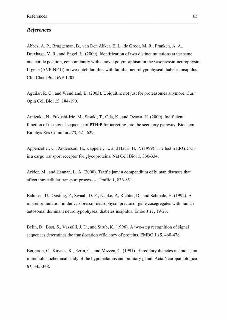
References 65 ___________________________________________________________________________
References
Abbes, A. P., Bruggeman, B., van Den Akker, E. L., de Groot, M. R., Franken, A. A.,
Drexhage, V. R., and Engel, H. (2000). Identification of two distinct mutations at the same
nucleotide position, concomitantly with a novel polymorphism in the vasopressin-neurophysin
II gene (AVP-NP II) in two dutch families with familial neurohypophyseal diabetes insipidus.
Clin Chem 46, 1699-1702.
Aguilar, R. C., and Wendland, B. (2003). Ubiquitin: not just for proteasomes anymore. Curr
Opin Cell Biol 15, 184-190.
Amizuka, N., Fukushi-Irie, M., Sasaki, T., Oda, K., and Ozawa, H. (2000). Inefficient
function of the signal sequence of PTHrP for targeting into the secretory pathway. Biochem
Biophys Res Commun 273, 621-629.
Appenzeller, C., Andersson, H., Kappeler, F., and Hauri, H. P. (1999). The lectin ERGIC-53
is a cargo transport receptor for glycoproteins. Nat Cell Biol 1, 330-334.
Aridor, M., and Hannan, L. A. (2000). Traffic jam: a compendium of human diseases that
affect intracellular transport processes. Traffic 1, 836-851.
Bahnsen, U., Oosting, P., Swaab, D. F., Nahke, P., Richter, D., and Schmale, H. (1992). A
missense mutation in the vasopressin-neurophysin precursor gene cosegregates with human
autosomal dominant neurohypophyseal diabetes insipidus. Embo J 11, 19-23.
Belin, D., Bost, S., Vassalli, J. D., and Strub, K. (1996). A two-step recognition of signal
sequences determines the translocation efficiency of proteins. EMBO J 15, 468-478.
Bergeron, C., Kovacs, K., Ezrin, C., and Mizzen, C. (1991). Hereditary diabetes insipidus: an
immunohistochemical study of the hypothalamus and pituitary gland. Acta Neuropathologica
81, 345-348.
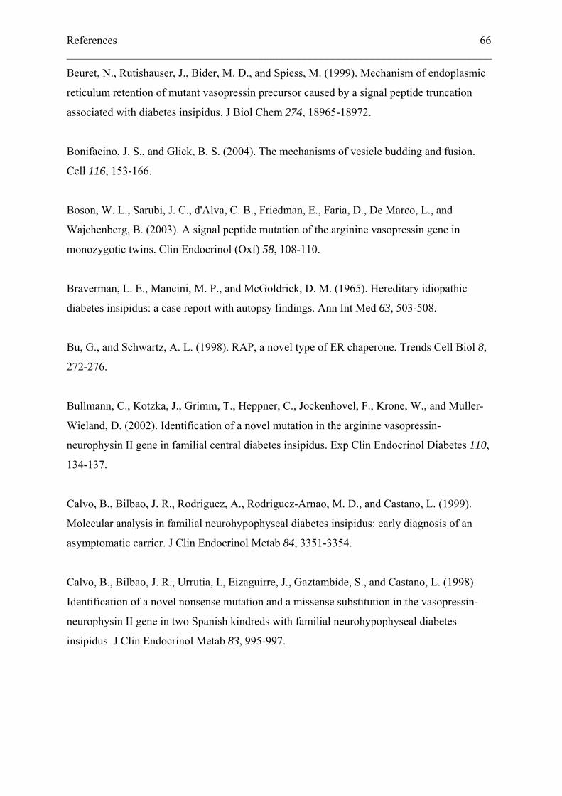
References 66 ___________________________________________________________________________
Beuret, N., Rutishauser, J., Bider, M. D., and Spiess, M. (1999). Mechanism of endoplasmic
reticulum retention of mutant vasopressin precursor caused by a signal peptide truncation
associated with diabetes insipidus. J Biol Chem 274, 18965-18972.
Bonifacino, J. S., and Glick, B. S. (2004). The mechanisms of vesicle budding and fusion.
Cell 116, 153-166.
Boson, W. L., Sarubi, J. C., d'Alva, C. B., Friedman, E., Faria, D., De Marco, L., and
Wajchenberg, B. (2003). A signal peptide mutation of the arginine vasopressin gene in
monozygotic twins. Clin Endocrinol (Oxf) 58, 108-110.
Braverman, L. E., Mancini, M. P., and McGoldrick, D. M. (1965). Hereditary idiopathic
diabetes insipidus: a case report with autopsy findings. Ann Int Med 63, 503-508.
Bu, G., and Schwartz, A. L. (1998). RAP, a novel type of ER chaperone. Trends Cell Biol 8,
272-276.
Bullmann, C., Kotzka, J., Grimm, T., Heppner, C., Jockenhovel, F., Krone, W., and Muller-
Wieland, D. (2002). Identification of a novel mutation in the arginine vasopressin-
neurophysin II gene in familial central diabetes insipidus. Exp Clin Endocrinol Diabetes 110,
134-137.
Calvo, B., Bilbao, J. R., Rodriguez, A., Rodriguez-Arnao, M. D., and Castano, L. (1999).
Molecular analysis in familial neurohypophyseal diabetes insipidus: early diagnosis of an
asymptomatic carrier. J Clin Endocrinol Metab 84, 3351-3354.
Calvo, B., Bilbao, J. R., Urrutia, I., Eizaguirre, J., Gaztambide, S., and Castano, L. (1998).
Identification of a novel nonsense mutation and a missense substitution in the vasopressin-
neurophysin II gene in two Spanish kindreds with familial neurohypophyseal diabetes
insipidus. J Clin Endocrinol Metab 83, 995-997.
References 67 ___________________________________________________________________________
Caramelo, J. J., Castro, O. A., Alonso, L. G., De Prat-Gay, G., and Parodi, A. J. (2003). UDP-
Glc:glycoprotein glucosyltransferase recognizes structured and solvent accessible
hydrophobic patches in molten globule-like folding intermediates. Proc Natl Acad Sci U S A
100, 86-91.
Carlson, J. A., Rogers, B. B., Sifers, R. N., Finegold, M. J., Clift, S. M., DeMayo, F. J.,
Bullock, D. W., and Woo, S. L. (1989). Accumulation of PiZ alpha 1-antitrypsin causes liver
damage in transgenic mice. J Clin Invest 83, 1183-1190.
Castino, R., Davies, J., Beaucourt, S., Isidoro, C., and Murphy, D. (2005). Autophagy is a
prosurvival mechanism in cells expressing an autosomal dominant familial neurohypophyseal
diabetes insipidus mutant vasopressin transgene. Faseb J.
Cescato, R., Dumermuth, E., Spiess, M., and Paganetti, P. A. (2000). Increased generation of
alternatively cleaved beta-amyloid peptides in cells expressing mutants of the amyloid
precursor protein defective in endocytosis. J Neurochem 74, 1131-1139.
Chevrier, D., Fournier, H., Nault, C., Zollinger, M., Crine, P., and Boileau, G. (1991).
Expression of porcine pro-opiomelanocortin in mouse neuroblastoma (Neuro2A) cells:
targeting of the foreign neuropeptide to dense-core vesicles. Mol Cell Endocrinol 79, 109-118.
Christensen, J. H., Siggaard, C., Corydon, T. J., deSanctis, L., Kovacs, L., Robertson, G. L.,
Gregersen, N., and Rittig, S. (2004). Six novel mutations in the arginine vasopressin gene in
15 kindreds with autosomal dominant familial neurohypophyseal diabetes insipidus give
further insight into the pathogenesis. Eur J Hum Genet 12, 44-51.
Ciechanover, A. (1994). The ubiquitin-proteasome proteolytic pathway. Cell 79, 13-21.
de Bree, F. M., and Burbach, J. P. (1998). Structure-function relationships of the vasopressin
prohormone domains. Cell Mol Neurobiol 18, 173-191.
DiMeglio, L. A., Gagliardi, P. C., Browning, J. E., Quigley, C. A., and Repaske, D. R. (2001).
A missense mutation encoding cys(67) --> gly in neurophysin ii is associated with early onset
autosomal dominant neurohypophyseal diabetes insipidus. Mol Genet Metab 72, 39-44.
References 68 ___________________________________________________________________________
Ellgaard, L., and Helenius, A. (2003). Quality control in the endoplasmic reticulum. Nat Rev
Mol Cell Biol 4, 181-191.
Eusebio, A., Friedberg, T., and Spiess, M. (1998). The role of the hydrophobic domain in
orienting natural signal sequences within the ER membrane. Exp Cell Res 241, 181-185.
Evans, D. A., De Bree, F. M., Nijenhuis, M., Van Der Kleij, A. A., Zalm, R., Korteweg, N.,
Van Leeuwen, F. W., and Burbach, J. P. (2000). Processing of frameshifted vasopressin
precursors. J Neuroendocrinol 12, 685-693.
Fisher, E. A., Zhou, M., Mitchell, D. M., Wu, X., Omura, S., Wang, H., Goldberg, A. L., and
Ginsberg, H. N. (1997). The degradation of apolipoprotein B100 is mediated by the ubiquitin-
proteasome pathway and involves heat shock protein 70. J Biol Chem 272, 20427-20434.
Fluck, C. E., Deladoey, J., Nayak, S., Zeller, O., Kopp, P., and Mullis, P. E. (2001).
Autosomal dominant neurohypophyseal diabetes insipidus in a Swiss family, caused by a
novel mutation (C59Delta/A60W) in the neurophysin moiety of prepro-vasopressin-
neurophysin II (AVP-NP II). Eur J Endocrinol 145, 439-444.
Flynn, G. C., Pohl, J., Flocco, M. T., and Rothman, J. E. (1991). Peptide-binding specificity of
the molecular chaperone BiP. Nature 353, 726-730.
Fons, R. D., Bogert, B. A., and Hegde, R. S. (2003). Substrate-specific function of the
translocon-associated protein complex during translocation across the ER membrane. J Cell
Biol 160, 529-539.
Friberg, M. A., Spiess, M., and Rutishauser, J. (2004). Degradation of wild-type vasopressin
precursor and pathogenic mutants by the proteasome. J Biol Chem 279, 19441-19447.
Fujii, H., Iida, S., and Moriwaki, K. (2000). Familial neurohypophyseal diabetes insipidus
associated with a novel mutation in the vasopressin-neurophysin II gene. Int J Mol Med 5,
229-234.
References 69 ___________________________________________________________________________
Gagliardi, P. C., Bernasconi, S., and Repaske, D. R. (1997). Autosomal dominant
neurohypophyseal diabetes insipidus associated with a missense mutation encoding Gly23--
>Val in neurophysin II. J Clin Endocrinol Metab 82, 3643-3646.
Gaupp, R. (1941). Über den Diabetes Insipidus. Z Neurol Psychiatr 171, 514-546.
Goder, V., and Spiess, M. (2001). Topogenesis of membrane proteins: determinants and
dynamics. FEBS Lett 504, 87-93.
Gong, L., Kamitani, T., Millas, S., and Yeh, E. T. (2000). Identification of a novel
isopeptidase with dual specificity for ubiquitin- and NEDD8-conjugated proteins. J Biol
Chem 275, 14212-14216.
Gonking, N. Q., Cherkow, B. S., Robertson, G. L., Rittig, S., Siggaard, C., and Pedersen, E.
B. (1997). Familial Neurohypophyseal Diabetes Insipidus: A Novel Mutation Presenting with
Eneuresis. J Investig Med 45, Abstract 29a.
Gorlich, D., and Rapoport, T. A. (1993). Protein translocation into proteoliposomes
reconstituted from purified components of the endoplasmic reticulum membrane. Cell 75,
615-630.
Gow, A., Southwood, C. M., and Lazzarini, R. A. (1998). Disrupted proteolipid protein
trafficking results in oligodendrocyte apoptosis in an animal model of Pelizaeus-Merzbacher
disease. J Cell Biol 140, 925-934.
Grant, F. D., Ahmadi, A., Hosley, C. M., and Majzoub, J. A. (1998). Two novel mutations of
the vasopressin gene associated with familial diabetes insipidus and identification of an
asymptomatic carrier infant. J Clin Endocrinol Metab 83, 3958-3964.
Hanhart, E. (1940). Die Erbpathologie des Diabetes Insipidus, Vol Vol.4 Part 2 (Berlin,
Verlag Julius Springer).
Hansen, L. K., Rittig, S., and Robertson, G. L. (1997). Genetic basis of familial
neurohypophyseal diabetes insipidus. Trends Endocrinol Metab 8, 363-372.
References 70 ___________________________________________________________________________
Hartmann, E., Rapoport, T. A., and Lodish, H. F. (1989). Predicting the orientation of
eukaryotic membrane-spanning proteins. Proc Natl Acad Sci USA 86, 5786-5790.
Hatsuzawa, K., Tagaya, M., and Mizushima, S. (1997). The hydrophobic region of signal
peptides is a determinant for SRP recognition and protein translocation across the ER
membrane. J Biochem (Tokyo) 121, 270-277.
Hegde, R. S., Mastrianni, J. A., Scott, M. R., DeFea, K. A., Tremblay, P., Torchia, M.,
DeArmond, S. J., Prusiner, S. B., and Lingappa, V. R. (1998). A transmembrane form of the
prion protein in neurodegenerative disease. Science 279, 827-834.
Hegde, R. S., and Rane, N. S. (2003). Prion protein trafficking and the development of
neurodegeneration. Trends Neurosci 26, 337-339.
Heppner, C., Kotzka, J., Bullmann, C., Krone, W., and Muller-Wieland, D. (1998).
Identification of mutations of the arginine vasopressin-neurophysin II gene in two kindreds
with familial central diabetes insipidus. J Clin Endocrinol Metab 83, 693-696.
Herrmann, J. M., Malkus, P., and Schekman, R. (1999). Out of the ER--outfitters, escorts and
guides. Trends Cell Biol 9, 5-7.
Hiller, M. M., Finger, A., Schweiger, M., and Wolf, D. H. (1996). ER degradation of a
misfolded luminal protein by the cytosolic ubiquitin-proteasome pathway. Science 273, 1725-
1728.
Hirsch, C., Blom, D., and Ploegh, H. L. (2003). A role for N-glycanase in the cytosolic
turnover of glycoproteins. EMBO J 22, 1036-1046.
Hölscher, C., Bach, U. C., and Dobberstein, B. (2001). Prion protein contains a second
endoplasmic reticulum targeting signal sequence located at its C terminus. J Biol Chem 276,
13388-13394.
References 71 ___________________________________________________________________________
Ito, M., and Jameson, J. L. (1997). Molecular basis of autosomal dominant neurohypophyseal
diabetes insipidus. Cellular toxicity caused by the accumulation of mutant vasopressin
precursors within the endoplasmic reticulum. J Clin Invest 99, 1897-1905.
Ito, M., Mori, Y., Oiso, Y., and Saito, H. (1991). A single base substitution in the coding
region for neurophysin II associated with familial central diabetes insipidus. Journal of
Clinical Investigation 87, 725-728.
Ito, M., Oiso, Y., Murase, T., Kondo, K., Saito, H., Chinzei, T., Racchi, M., and Lively, M. O.
(1993). Possible involvement of inefficient cleavage of preprovasopressin by signal peptidase
as a cause for familial central diabetes insipidus. J Clin Invest 91, 2565-2571.
Ito, M., Yu, R. N., and Jameson, J. L. (1999). Mutant vasopressin precursors that cause
autosomal dominant neurohypophyseal diabetes insipidus retain dimerization and impair the
secretion of wild-type proteins. J Biol Chem 274, 9029-9037.
Johnson, A. E., and van Waes, M. A. (1999). The translocon: a dynamic gateway at the ER
membrane. Annu Rev Cell Dev Biol 15, 799-842.
Johnston, J. A., Ward, C. L., and Kopito, R. R. (1998). Aggresomes: a cellular response to
misfolded proteins. J Cell Biol 143, 1883-1898.
Kamitani, T., Kito, K., Fukuda-Kamitani, T., and Yeh, E. T. (2001). Targeting of NEDD8 and
its conjugates for proteasomal degradation by NUB1. J Biol Chem 276, 46655-46660.
Kaufman, R. J. (2002). Orchestrating the unfolded protein response in health and disease. J
Clin Invest 110, 1389-1398.
Kim, P. S., and Arvan, P. (1998). Endocrinopathies in the family of endoplasmic reticulum
(ER) storage diseases: disorders of protein trafficking and the role of ER molecular
chaperones. Endocr Rev 19, 173-202.
Kim, S. J., Mitra, D., Salerno, J. R., and Hegde, R. S. (2002). Signal sequences control gating
of the protein translocation channel in a substrate-specific manner. Dev Cell 2, 207-217.
References 72 ___________________________________________________________________________
Kopito, R. R. (2000). Aggresomes, inclusion bodies and protein aggregation. Trends Cell Biol
10, 524-530.
Kostova, Z., and Wolf, D. H. (2003). For whom the bell tolls: protein quality control of the
endoplasmic reticulum and the ubiquitin-proteasome connection. EMBO J 22, 2309-2317.
Kota, J., and Ljungdahl, P. O. (2005). Specialized membrane-localized chaperones prevent
aggregation of polytopic proteins in the ER. J Cell Biol 168, 79-88.
Levin, M. H., Haggie, P. M., Vetrivel, L., and Verkman, A. S. (2001). Diffusion in the
endoplasmic reticulum of an aquaporin-2 mutant causing human nephrogenic diabetes
insipidus. J Biol Chem 276, 21331-21336.
Liu, C. Y., and Kaufman, R. J. (2003). The unfolded protein response. J Cell Sci 116, 1861-
1862.
Ma, J., and Lindquist, S. (2001). Wild-type PrP and a mutant associated with prion disease are
subject to retrograde transport and proteasome degradation. Proc Natl Acad Sci USA 98,
14955-14960.
Ma, J., and Lindquist, S. (2002). Conversion of PrP to a self-perpetuating PrPSc-like
conformation in the cytosol. Science 298, 1785-1788.
Ma, J., Wollmann, R., and Lindquist, S. (2002). Neurotoxicity and neurodegeneration when
PrP accumulates in the cytosol. Science 298, 1781-1785.
Mahoney, C. P., Weinberger, E., Bryant, C., Ito, M., and Jameson, J. L. (2002). Effects of
aging on vasopressin production in a kindred with autosomal dominant neurohypophyseal
diabetes insipidus due to the DeltaE47 neurophysin mutation. J Clin Endocrinol Metab 87,
870-876.
McLeod, J. F., Kovacs, L., Gaskill, M. B., Rittig, S., Bradley, G. S., and Robertson, G. L.
(1993). Familial neurohypophyseal diabetes insipidus associated with a signal peptide
mutation. J Clin Endocrinol Metab 77, 599A-599G.
References 73 ___________________________________________________________________________
Medicherla, B., Kostova, Z., Schaefer, A., and Wolf, D. H. (2004). A genomic screen
identifies Dsk2p and Rad23p as essential components of ER-associated degradation. EMBO
Rep 5, 692-697.
Miyakoshi, M., Kamoi, K., Murase, T., Sugimura, Y., and Oiso, Y. (2004). Novel mutant
vasopressin-neurophysin II gene associated with familial neurohypophyseal diabetes
insipidus. Endocr J 51, 551-556.
Miyamoto, S., Sasaki, N., and Tanabe, Y. (1991). Magnetic resonance imaging in familial
central diabetes insipidus. Neuroradiology 33, 272-273.
Molinari, M., Calanca, V., Galli, C., Lucca, P., and Paganetti, P. (2003). Role of EDEM in the
release of misfolded glycoproteins from the calnexin cycle. Science 299, 1397-1400.
Mundschenk, J., Rittig, S., Siggaard, C., Hensen, J., and Lehnert, H. (2001). A new mutation
of the arginine vasopressin-neurophysin II gene in a family with autosomal dominant
neurohypophyseal diabetes insipidus. Exp Clin Endocrinol Diabetes 109, 406-409.
Nagai, I., Li, C. H., Hsieh, S. M., Kizaki, T., and Urano, Y. (1984). Two cases of hereditary
diabetes insipidus, with an autopsy finding in one. Acta Endocrinol (Copenh) 105, 318-323.
Nagasaki, H., Ito, M., Yuasa, H., Saito, H., Fukase, M., Hamada, K., Ishikawa, E., Katakami,
H., and Oiso, Y. (1995). Two novel mutations in the coding region for neurophysin-II
associated with familial central diabetes insipidus. J Clin Endocrinol Metab 80, 1352-1356.
Nakagawa, T., Zhu, H., Morishima, N., Li, E., Xu, J., Yankner, B. A., and Yuan, J. (2000).
Caspase-12 mediates endoplasmic-reticulum-specific apoptosis and cytotoxicity by amyloid-
beta. Nature 403, 98-103.
Nijenhuis, M., van den Akker, E. L., Zalm, R., Franken, A. A., Abbes, A. P., Engel, H., de
Wied, D., and Burbach, J. P. (2001). Familial neurohypophysial diabetes insipidus in a large
Dutch kindred: effect of the onset of diabetes on growth in children and cell biological defects
of the mutant vasopressin prohormone. J Clin Endocrinol Metab 86, 3410-3420.
References 74 ___________________________________________________________________________
Nijenhuis, M., Zalm, R., and Burbach, J. P. (1999). Mutations in the vasopressin prohormone
involved in diabetes insipidus impair endoplasmic reticulum export but not sorting. J Biol
Chem 274, 21200-21208.
Nijenhuis, M., Zalm, R., and Burbach, J. P. (2000). A diabetes insipidus vasopressin
prohormone altered outside the central core of neurophysin accumulates in the endoplasmic
reticulum. Mol Cell Endocrinol 167, 55-67.
Olias, G., Richter, D., and Schmale, H. (1996). Heterologous expression of human
vasopressin-neurophysin precursors in a pituitary cell line: defective transport of a mutant
protein from patients with familial diabetes insipidus. DNA Cell Biol 15, 929-935.
Oyadomari, S., Koizumi, A., Takeda, K., Gotoh, T., Akira, S., Araki, E., and Mori, M. (2002).
Targeted disruption of the Chop gene delays endoplasmic reticulum stress-mediated diabetes.
J Clin Invest 109, 525-532.
Parodi, A. J. (2000). Protein glucosylation and its role in protein folding. Annu Rev Biochem
69, 69-93.
Plemper, R. K., Bordallo, J., Deak, P. M., Taxis, C., Hitt, R., and Wolf, D. H. (1999). Genetic
interactions of Hrd3p and Der3p/Hrd1p with Sec61p suggest a retro-translocation complex
mediating protein transport for ER degradation. J Cell Sci 112 ( Pt 22), 4123-4134.
Racchi, M., Watzke, H. H., High, K. A., and Lively, M. O. (1993). Human coagulation factor
X deficiency caused by a mutant signal peptide that blocks cleavage by signal peptidase but
not targeting and translocation to the endoplasmic reticulum. J Biol Chem 268, 5735-5740.
Rauch, F., Lenzner, C., Nurnberg, P., Frommel, C., and Vetter, U. (1996). A novel mutation
in the coding region for neurophysin-II is associated with autosomal dominant
neurohypophyseal diabetes insipidus. Clin Endocrinol (Oxf) 44, 45-51.
Repaske, D. R., and Browning, J. E. (1994). A de novo mutation in the coding sequence for
neurophysin-II (Pro24-->Leu) is associated with onset and transmission of autosomal
dominant neurohypophyseal diabetes insipidus. J Clin Endocrinol Metab 79, 421-427.
References 75 ___________________________________________________________________________
Rittig, S., Robertson, G. L., Siggaard, C., Kovacs, L., Gregersen, N., Nyborg, J., and
Pedersen, E. B. (1996). Identification of 13 new mutations in the vasopressin-neurophysin II
gene in 17 kindreds with familial autosomal dominant neurohypophyseal diabetes insipidus.
Am J Hum Genet 58, 107-117.
Rittig, S., Siggaard, C., Ozata, M., Yetkin, I., Gregersen, N., Pedersen, E. B., and Robertson,
G. L. (2002). Autosomal dominant neurohypophyseal diabetes insipidus due to substitution of
histidine for tyrosine(2) in the vasopressin moiety of the hormone precursor. J Clin
Endocrinol Metab 87, 3351-3355.
Russell, T. A., Ito, M., Yu, R. N., Martinson, F. A., Weiss, J., and Jameson, J. L. (2003). A
murine model of autosomal dominant neurohypophyseal diabetes insipidus reveals
progressive loss of vasopressin-producing neurons. J Clin Invest 112, 1697-1706.
Rutishauser, J., Boni-Schnetzler, M., Boni, J., Wichmann, W., Huisman, T., Vallotton, M. B.,
and Froesch, E. R. (1996). A novel point mutation in the translation initiation codon of the
pre-pro-vasopressin-neurophysin II gene: cosegregation with morphological abnormalities and
clinical symptoms in autosomal dominant neurohypophyseal diabetes insipidus. J Clin
Endocrinol Metab 81, 192-198.
Rutishauser, J., and Kopp, P. (1999). Aquaporin-2 water channel mutations and nephrogenic
diabetes insipidus: new variations on a theme. Eur J Endocrinol 140, 137-139.
Rutishauser, J., Kopp, P., Gaskill, M. B., Kotlar, T. J., and Robertson, G. L. (1999). A novel
mutation (R97C) in the neurophysin moiety of prepro-vasopressin-neurophysin II associated
with autosomal-dominant neurohypophyseal diabetes insipidus. Mol Genet Metab 67, 89-92.
Rutishauser, J., Kopp, P., Gaskill, M. B., Kotlar, T. J., and Robertson, G. L. (2002). Clinical
and molecular analysis of three families with autosomal dominant neurohypophyseal diabetes
insipidus associated with a novel and recurrent mutations in the vasopressin-neurophysin II
gene. Eur J Endocrinol 146, 649-656.
Rutishauser, J., and Spiess, M. (2002). Endoplasmic reticulum storage diseases. Swiss Med
Wkly 132, 211-222.
References 76 ___________________________________________________________________________
Rutkowski, D. T., and Kaufman, R. J. (2004). A trip to the ER: coping with stress. Trends
Cell Biol 14, 20-28.
Santiprabhob, J., Browning, J., and Repaske, D. (2002). A missense mutation encoding
Cys73Phe in neurophysin II is associated with autosomal dominant neurohypophyseal
diabetes insipidus. Mol Genet Metab 77, 112.
Sausville, E., Carney, D., and Battey, J. (1985). The human vasopressin gene is linked to the
oxytocin gene and is selectively expressed in a cultured lung cancer cell line. J Biol Chem
260, 10236-10241.
Schmale, H., Ivell, R., Breindl, M., Darmer, D., and Richter, D. (1984). The mutant
vasopressin gene from diabetes insipidus (Brattleboro) rats is transcribed but the message is
not efficiently translated. EMBO J 3, 3289-3293.
Schubert, U., Anton, L. C., Gibbs, J., Norbury, C. C., Yewdell, J. W., and Bennink, J. R.
(2000). Rapid degradation of a large fraction of newly synthesized proteins by proteasomes.
Nature 404, 770-774.
Serwold, T., Gonzalez, F., Kim, J., Jacob, R., and Shastri, N. (2002). ERAAP customizes
peptides for MHC class I molecules in the endoplasmic reticulum. Nature 419, 480-483.
Siggaard, C., Rittig, S., Corydon, T. J., Andreasen, P. H., Jensen, T. G., Andresen, B. S.,
Robertson, G. L., Gregersen, N., Bolund, L., and Pedersen, E. B. (1999). Clinical and
molecular evidence of abnormal processing and trafficking of the vasopressin preprohormone
in a large kindred with familial neurohypophyseal diabetes insipidus due to a signal peptide
mutation. J Clin Endocrinol Metab 84, 2933-2941.
Si-Hoe, S. L., De Bree, F. M., Nijenhuis, M., Davies, J. E., Howell, L. M., Tinley, H., Waller,
S. J., Zeng, Q., Zalm, R., Sonnemans, M., et al. (2000). Endoplasmic reticulum derangement
in hypothalamic neurons of rats expressing a familial neurohypophyseal diabetes insipidus
mutant vasopressin transgene. Faseb J 14, 1680-1684.
References 77 ___________________________________________________________________________
Skordis, N., Patsalis, P. C., Hettinger, J. A., Kontou, M., Herakleous, E., Krishnamani, M. R.,
and Phillips, J. A., 3rd (2000). A novel arginine vasopressin-neurophysin II mutation causes
autosomal dominant neurohypophyseal diabetes insipidus and morphologic pituitary changes.
Horm Res 53, 239-245.
Suzuki, T., Park, H., Kwofie, M. A., and Lennarz, W. J. (2001). Rad23 provides a link
between the Png1 deglycosylating enzyme and the 26 S proteasome in yeast. J Biol Chem
276, 21601-21607.
Toyoshima, I., Sugawara, M., Kato, K., Wada, C., Hirota, K., Hasegawa, K., Kowa, H.,
Sheetz, M. P., and Masamune, O. (1998). Kinesin and cytoplasmic dynein in spinal spheroids
with motor neuron disease. J Neurol Sci 159, 38-44.
Ueta, Y., Taniguchi, S., Yoshida, A., Murakami, I., Mitani, Y., Hisatome, I., Manabe, I., Sato,
R., Tsuboi, M., Ohtahara, A., et al. (1996). A new type of familial central diabetes insipidus
caused by a single base substitution in the neurophysin II coding region of the vasopressin
gene. J Clin Endocrinol Metab 81, 1787-1790.
Valetti, C., Grossi, C. E., Milstein, C., and Sitia, R. (1991). Russell bodies: a general response
of secretory cells to synthesis of a mutant immunoglobulin which can neither exit from, nor be
degraded in, the endoplasmic reticulum. J Cell Biol 115, 983-994.
von Heijne, G. (1990). The signal peptide. J Mem Biol 115, 195-201.
Wahlberg, J. M., and Spiess, M. (1997). Multiple determinants direct the orientation of signal-
anchor proteins: The topogenic role of the hydrophobic signal domain. J Cell Biol 137, 555-
562.
Wahlstrom, J. T., Fowler, M. J., Nicholson, W. E., and Kovacs, W. J. (2004). A novel
mutation in the preprovasopressin gene identified in a kindred with autosomal dominant
neurohypophyseal diabetes insipidus. J Clin Endocrinol Metab 89, 1963-1968.
References 78 ___________________________________________________________________________
Wang, X. Z., Kuroda, M., Sok, J., Batchvarova, N., Kimmel, R., Chung, P., Zinszner, H., and
Ron, D. (1998). Identification of novel stress-induced genes downstream of chop. EMBO J
17, 3619-3630.
Webb, J. L., Ravikumar, B., Atkins, J., Skepper, J. N., and Rubinsztein, D. C. (2003). Alpha-
Synuclein is degraded by both autophagy and the proteasome. J Biol Chem 278, 25009-
25013.
Wiertz, E. J., Tortorella, D., Bogyo, M., Yu, J., Mothes, W., Jones, T. R., Rapoport, T. A., and
Ploegh, H. L. (1996). Sec61-mediated transfer of a membrane protein from the endoplasmic
reticulum to the proteasome for destruction. Nature 384, 432-438.
Willcutts, M. D., Felner, E., and White, P. C. (1999). Autosomal recessive familial
neurohypophyseal diabetes insipidus with continued secretion of mutant weakly active
vasopressin. Hum Mol Genet 8, 1303-1307.
Ye, L., Li, X., Chen, Y., Sun, H., Wang, W., Su, T., Jiang, L., Cui, B., and Ning, G. (2005).
Autosomal Dominant Neurohypophyseal Diabetes Insipidus with Linkage to Chromosome
20p13 but without Mutations in the AVP-NPII Gene. J Clin Endocrinol Metab.
Ye, Y., Meyer, H. H., and Rapoport, T. A. (2001). The AAA ATPase Cdc48/p97 and its
partners transport proteins from the ER into the cytosol. Nature 414, 652-656.
Yedidia, Y., Horonchik, L., Tzaban, S., Yanai, A., and Taraboulos, A. (2001). Proteasomes
and ubiquitin are involved in the turnover of the wild-type prion protein. EMBO J 20, 5383-
5391.
Yewdell, J. W. (2001). Not such a dismal science: the economics of protein synthesis, folding,
degradation and antigen processing. Trends Cell Biol 11, 294-297.
Yuasa, H., Ito, M., Nagasaki, H., Oiso, Y., Miyamoto, S., Sasaki, N., and Saito, H. (1993).
Glu-47, which forms a salt bridge between neurophysin-II and arginine vasopressin, is deleted
in patients with familial central diabetes insipidus. J Clin Endocrinol Metab 77, 600-604.
References 79 ___________________________________________________________________________
Zhang, K., and Kaufman, R. J. (2004). Signaling the unfolded protein response from the
endoplasmic reticulum. J Biol Chem 279, 25935-25938.
Zhou, M., Fisher, E. A., and Ginsberg, H. N. (1998). Regulated Co-translational
ubiquitination of apolipoprotein B100. A new paradigm for proteasomal degradation of a
secretory protein. J Biol Chem 273, 24649-24653.
Zinszner, H., Kuroda, M., Wang, X., Batchvarova, N., Lightfoot, R. T., Remotti, H., Stevens,
J. L., and Ron, D. (1998). CHOP is implicated in programmed cell death in response to
impaired function of the endoplasmic reticulum. Genes Dev 12, 982-995.
___________________________________________________________________________
Curriculum vitae
Name, first name Friberg, Michael
Address Sandgarten 8
4312 Magden
Phone +41 61 841 15 90
Citizen of Zürich ZH and Brigels GR
Birthday 03/19/1974
Marital status single
Education
1981 - 1985 Primary School in Magden, Switzerland
1985 - 1987 Scol Ide Primary School, Corbally, Limerick, Ireland
1987 - 1989 Greenwood Lakes Middle School, Lake Mary, FL, USA
1989 - 1991 Lake Mary High School, Lake Mary, FL, USA (gifted class)
Aug. - Dec. 1991 Kantonsschule Trogen, Switzerland
Dec. 1991 - Dec. 1994 Gymnasium Muttenz, Typus C (mathematics/science)
Oct. 1995 - Apr. 2000 University of Basel, study area: Biologie II
Sept. 1998 - Feb. 2000 Diploma-Thesis under Prof. Dr. Markus A. Rüegg, Biozentrum
Basel: “Cadherins: The effect of dominat negative constructs on
post-synaptic differentiation at the neuromuscular junction”
Feb. 2001 - Jul. 2005 Doctorate-Thesis under Prof. Dr. Martin Spiess, Biozentrum
Basel, and Dr. med. Jonas Rutishauser, Kantonsspital Basel
Mai 2004 Publication in JBC (The Journal of Biological Chemistry)
“Degradation of Wild-type Vasopressin Precursor and
Pathogenic Mutants by the Proteasome”
___________________________________________________________________________
Practical Experience
1994 Tutorial, middle school level
July 1994 Assistant camp counselor in a sailing camp by the
Sozialpädagogischer Dienst Basel in Le Prese, GR
Mai 1995 Practical training as an orderly during the military in Liestal, BL
Aug. - Oct. 1995 Worked in the PHP-Betrieb TTS, Ciba-Geigy Werke Stein, AG
Oct. 2001 - Feb.2002 Biology tutorial
Sept. 2002 School project supervision of Peter Blattmann at the Biozentrum
Dec. 2002 Assistant of the Biochemistry lab course
Jan. 2004 Lab head of the Biochemistry lab course
Mar. 2004 Student supervision during „Schweizer Jugend forscht“
Languages
German native speaker
English second native language
French good school knowledge
Computer proficiency Windows XP, Word, Excel, Photoshop, Illustrator
Other interests and activities
Reading, computer, sports: Basketball (player, referee, coach)
1992 - 1994 Coaching assistant of the Jugendriege Magden
1994 Committee member of the Turnverein Magden
since1994 President of the Basketball department of the Turnverein
Magden
Coach of the first and second men’s basketball team
since 1997 Basketball referee
since 2001 Chairman of the technical committee of the Turnverein Magden
since 2003 Assistant coach of the women’s basketball team

![[Es ist jeweils nur eine Antwort richtig] · [Es ist jeweils nur eine Antwort richtig] 1. Welche Aussage zur Reanimation (ERC Guidelines 2010) ist richtig? (A) Vasopressin ist der](https://img.pdfslide.org/doc/110x75/5d570d2888c993d4748ba743/es-ist-jeweils-nur-eine-antwort-richtig-es-ist-jeweils-nur-eine-antwort-richtig.jpg)


![Kapitel 7) HORMONE 1 [Kompatibilitätsmodus]biochemietrainingscamp.de/stoff/h1/hormoneller_regelkreis.pdf · SPEICHERUNG von ADH (Antidiuretisches Hormon/ Vasopressin) 2) HML](https://img.pdfslide.org/doc/110x75/5cc1487988c993110a8bd865/kapitel-7-hormone-1-kompatibilitaetsmodusbi-speicherung-von-adh-antidiuretisches.jpg)