Embed Size (px)
Citation preview
Diplomarbeit
CLINICAL OUTCOME OF PATIENTS AFTER TOTAL
LARYNGECTOMY
A retrospective study
eingereicht von
Katharina Walla
zur Erlangung des akademischen Grades
Doktorin der gesamten Heilkunde
(Dr.in med. univ.)
an der
Medizinischen Universität Graz
ausgeführt an der
Hals-, Nasen-, Ohren-Universitätsklinik
unter der Anleitung von
Dr.med.univ. Thomas Weiland
Priv.Doz. Dr.med.univ. Georg Hammer
Graz, am 26.09.2018

i
Eidesstattliche Erklärung
Ich erkläre ehrenwörtlich, dass ich die vorliegende Arbeit selbstständig und ohne fremde Hilfe verfasst habe, andere als die angegebenen Quellen nicht verwendet habe und die den benutzten Quellen wörtlich oder inhaltlich entnommenen Stellen als solche kenntlich gemacht habe.
Graz, am 26.09.2018 Katharina Walla eh.
ii
Danksagung Ein großes Dankeschön gebührt Dr.med.univ. Thomas Weiland und Priv.-Doz. Dr.med.univ. Georg Hammer für die gute und geduldige Betreuung. Trotz zahlreicher eigener Verpflichtungen haben sie sich stets Zeit für meine Fragen genommen und mich mit ihrer Motivation und ihrem Fachwissen unterstützt. Meinen Eltern danke ich von ganzem Herzen für ihre moralische und finanzielle Unterstützung. Meiner Schwester Hannah möchte ich für ihre Zeit und Mühen danken und dafür, dass sie immer ein offenes Ohr für mich hat. Danke an meine Freunde und Freundinnen im Fuchsbau, ohne die die letzten Jahre nicht das Selbe gewesen wären. Schließlich gilt ein besonderer Dank meinem Freund Max, für seine Geduld und seinen liebevollen Rückhalt.
iii
Inhaltsverzeichnis
Eidesstattliche Erklärung .............................................................................................. i
Danksagung ................................................................................................................ ii
Inhaltsverzeichnis ....................................................................................................... iii
Glossar und Abkürzungen ............................................................................................ i
Abbildungsverzeichnis ................................................................................................ iii
Tabellenverzeichnis .................................................................................................... iv
Zusammenfassung ...................................................................................................... v
Abstract ...................................................................................................................... vi
1. Introduction .............................................................................................................. 1
1.1. History .................................................................................................................. 1
1.2. Anatomy ............................................................................................................... 2
1.2.1. The Larynx ........................................................................................................ 2
1.2.2. Laryngeal Musculature ...................................................................................... 6
1.2.3. Main vessels of the larynx ................................................................................. 7
1.2.4. Innervation of the larynx .................................................................................... 7
1.2.5. Lymphatic system of the larynx ......................................................................... 8
1.3. Physiology .......................................................................................................... 10
1.3.1. Swallowing ...................................................................................................... 11
1.3.2. Respiration ...................................................................................................... 11
1.3.3. Phonation ........................................................................................................ 12
1.4. Laryngeal and Hypopharyngeal Cancer ............................................................. 13
1.4.1. Epidemiology ................................................................................................... 13
1.4.2. Risk factors ...................................................................................................... 15
1.4.2.1. Tobacco products ......................................................................................... 15
1.4.2.2. Alcohol .......................................................................................................... 16
1.4.2.3. Human papillomavirus infection .................................................................... 17
1.4.2.4. Occupational exposure ................................................................................. 17
1.4.2.5. Diet ............................................................................................................... 17
1.4.2.6. Genetic factors ............................................................................................. 17
1.4.3. Pathology ........................................................................................................ 18
1.4.4. Clinical manifestation....................................................................................... 19
Larynx ....................................................................................................................... 19
Hypopharynx ............................................................................................................. 20
iv
1.4.5. Staging ............................................................................................................ 21
1.4.6. Treatment options ........................................................................................... 24
1.4.6.1. Radiotherapy ................................................................................................ 25
1.4.6.2. Chemotherapy .............................................................................................. 26
1.4.6.3. Larynx Preservation Surgery ........................................................................ 26
1.4.7. Total Laryngectomy ......................................................................................... 27
1.4.8. Neck dissection ............................................................................................... 30
1.4.9. Complications of total laryngectomy ................................................................ 31
1.4.9.1. Early Postoperative Complications ............................................................... 32
1.4.9.2. Pharyngocutaneous Fistula .......................................................................... 32
1.4.9.3. Pharyngeal stenosis ..................................................................................... 33
1.4.10. Voice rehabilitation ........................................................................................ 33
2. Purpose ................................................................................................................. 34
3. Materials and methods .......................................................................................... 35
4. Results .................................................................................................................. 36
4.1. Patient characteristics ........................................................................................ 36
4.2. Treatment regimen ............................................................................................. 38
4.3. Surgical techniques ............................................................................................ 39
4.4. Postoperative complications ............................................................................... 39
4.5. Survival rates ...................................................................................................... 41
4.6. Recurrence rates ................................................................................................ 42
4.7. Voice rehabilitation ............................................................................................. 43
5. Discussion ............................................................................................................. 44
References ................................................................................................................ 50
i
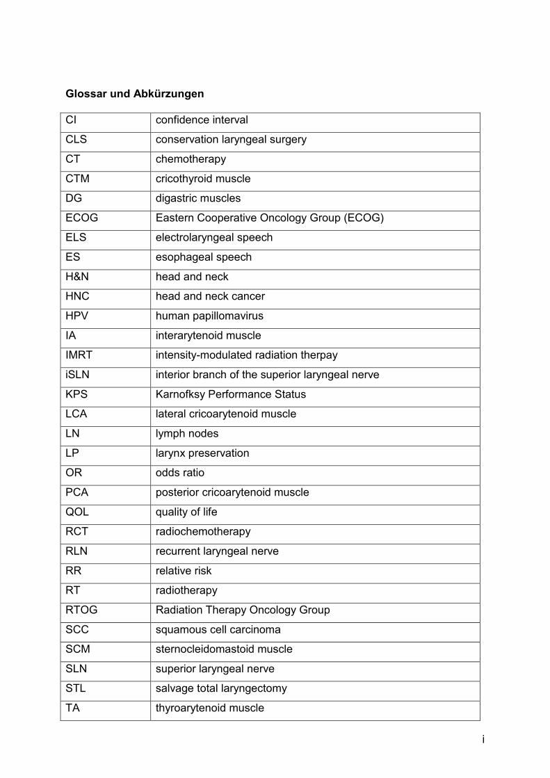
Glossar und Abkürzungen
CI confidence interval
CLS conservation laryngeal surgery
CT chemotherapy
CTM cricothyroid muscle
DG digastric muscles
ECOG Eastern Cooperative Oncology Group (ECOG)
ELS electrolaryngeal speech
ES esophageal speech
H&N head and neck
HNC head and neck cancer
HPV human papillomavirus
IA interarytenoid muscle
IMRT intensity-modulated radiation therpay
iSLN interior branch of the superior laryngeal nerve
KPS Karnofksy Performance Status
LCA lateral cricoarytenoid muscle
LN lymph nodes
LP larynx preservation
OR odds ratio
PCA posterior cricoarytenoid muscle
QOL quality of life
RCT radiochemotherapy
RLN recurrent laryngeal nerve
RR relative risk
RT radiotherapy
RTOG Radiation Therapy Oncology Group
SCC squamous cell carcinoma
SCM sternocleidomastoid muscle
SLN superior laryngeal nerve
STL salvage total laryngectomy
TA thyroarytenoid muscle

ii
TES tracheoesophageal speech
TL total laryngectomy
TLP total laryngopharyngectomy
TO tracheo-esophageal
VA Department of Veteran Affairs
VP voice prosthesis
iii
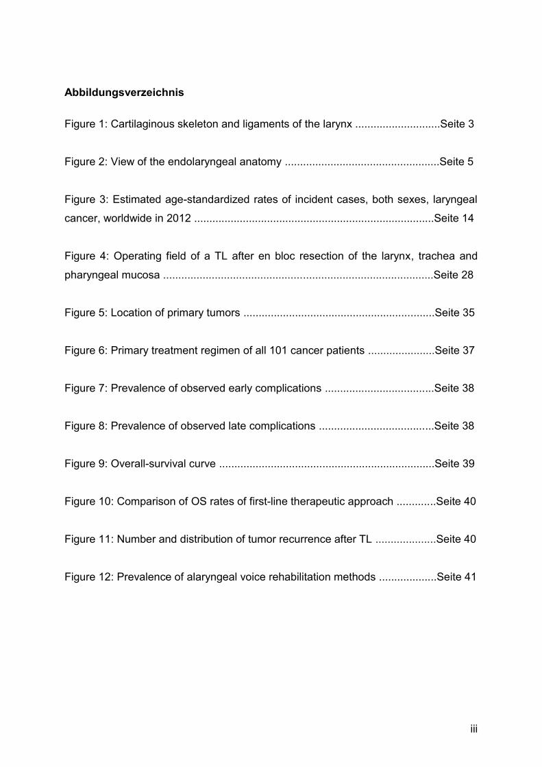
Abbildungsverzeichnis
Figure 1: Cartilaginous skeleton and ligaments of the larynx ............................Seite 3
Figure 2: View of the endolaryngeal anatomy ...................................................Seite 5
Figure 3: Estimated age-standardized rates of incident cases, both sexes, laryngeal
cancer, worldwide in 2012 ...............................................................................Seite 14
Figure 4: Operating field of a TL after en bloc resection of the larynx, trachea and
pharyngeal mucosa .........................................................................................Seite 28
Figure 5: Location of primary tumors ...............................................................Seite 35
Figure 6: Primary treatment regimen of all 101 cancer patients ......................Seite 37
Figure 7: Prevalence of observed early complications ....................................Seite 38
Figure 8: Prevalence of observed late complications ......................................Seite 38
Figure 9: Overall-survival curve .......................................................................Seite 39
Figure 10: Comparison of OS rates of first-line therapeutic approach .............Seite 40
Figure 11: Number and distribution of tumor recurrence after TL ....................Seite 40
Figure 12: Prevalence of alaryngeal voice rehabilitation methods ...................Seite 41
iv
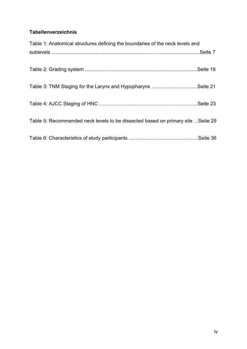
Tabellenverzeichnis Table 1: Anatomical structures defining the boundaries of the neck levels and
sublevels ...........................................................................................................Seite 7
Table 2: Grading system .................................................................................Seite 18
Table 3: TNM Staging for the Larynx and Hypopharynx .................................Seite 21
Table 4: AJCC Staging of HNC .......................................................................Seite 23
Table 5: Recommended neck levels to be dissected based on primary site ...Seite 29
Table 6: Characteristics of study participants ..................................................Seite 36
v
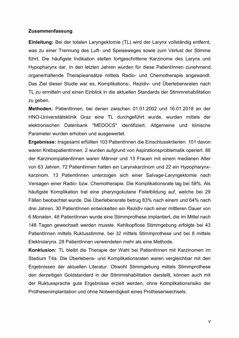
Zusammenfassung Einleitung: Bei der totalen Laryngektomie (TL) wird der Larynx vollständig entfernt,
was zu einer Trennung des Luft- und Speiseweges sowie zum Verlust der Stimme
führt. Die häufigste Indikation stellen fortgeschrittene Karzinome des Larynx und
Hypopharynx dar. In den letzten Jahren wurden für diese PatientInnen zunehmend
organerhaltende Therapieansätze mittels Radio- und Chemotherapie angewandt.
Das Ziel dieser Studie war es, Komplikations-, Rezidiv- und Überlebensraten nach
TL zu ermitteln und einen Einblick in die aktuellen Standards der Stimmrehabilitation
zu geben.
Methoden: PatientInnen, bei denen zwischen 01.01.2002 und 16.01.2018 an der
HNO-Universitätsklinik Graz eine TL durchgeführt wurde, wurden mittels der
elektronischen Datenbank "MEDOCS" identifiziert. Allgemeine und klinische
Parameter wurden erhoben und ausgewertet.
Ergebnisse: Insgesamt erfüllten 103 PatientInnen die Einschlusskriterien. 101 davon
waren KrebspatientInnen, 2 wurden aufgrund von Aspirationsproblematik operiert. 88
der KarzinompatientInnen waren Männer und 13 Frauen mit einem medianen Alter
von 63 Jahren. 72 PatientInnen hatten ein Larynxkarzinom und 22 ein Hypopharynx-
karzinom. 13 PatientInnen unterzogen sich einer Salvage-Laryngektomie nach
Versagen einer Radio- bzw. Chemotherapie. Die Komplikationsrate lag bei 58%. Als
häufigste Komplikation trat eine pharyngokutane Fistelbildung auf, welche bei 29
Fällen beobachtet wurde. Die Überlebensrate betrug 83% nach einem und 64% nach
drei Jahren. 30 PatientInnen entwickelten ein Rezidiv nach einer mittleren Dauer von
6 Monaten. 48 PatientInnen wurde eine Stimmprothese implantiert, die im Mittel nach
148 Tagen gewechselt werden musste. Kehlkopflose Stimmgebung erfolgte bei 43
PatientInnen mittels Ruktusstimme, bei 32 mittels Stimmprothese und bei 8 mittels
Elektrolarynx. 28 PatientInnen verwendeten mehr als eine Methode.
Konklusion: TL bleibt die Therapie der Wahl bei PatientInnen mit Karzinomen im
Stadium T4a. Die Überlebens- und Komplikationsraten waren vergleichbar mit den
Ergebnissen der aktuellen Literatur. Obwohl Stimmgebung mittels Stimmprothese
den derzeitigen Goldstandard in der Stimmrehabilitation darstellt, können auch mit
der Ruktussprache gute Ergebnisse erzielt werden, ohne Komplikationsrisiko der
Prothesenimplantation und ohne Notwendigkeit eines Prothesenwechsels.
vi
Abstract Introduction: Total laryngectomy (TL), the procedure in which the larynx is
completely removed, results in total separation of respiratory and upper digestive
tract and loss of voice. It is most commonly used in the treatment of advanced cancer
of the larynx and hypopharynx. These patients are increasingly being treated with
organ-preserving protocols using radiotherapy and chemotherapy. The purpose of
this study was to review complication, recurrence and survival rates following TL and
gain insight into the recent standards of voice rehabilitation.
Methods: In a retrospective study, patients who had undergone TL between
01.01.2002 and 16.01.2018 were identified and reviewed via the electronic database
"MEDOCS" of the Department of Otolaryngology, University Hospital of Graz.
Clinicopathological data was collected and evaluated.
Results: A total of 103 patients met the inclusion criteria. 101 patients had been
treated due to cancer, and 2 due to chronic aspiration. 88 of the cancer patients were
men and 13 women, with a median age of 63 years. Primary cancer involved the
larynx in 72 and the hypopharynx in 22 cases. 13 patients were treated with salvage
laryngectomy. Overall complication rate was 58%, and the most common
complication was development of pharyngocutaneous fistula in 29 patients. Survival
rate was at 83% after one year and 64% after three years. Recurrence developed in
30 patients after a median of 6 months. In 48 patients a voice prosthesis was
implanted, with a mean time range of 148 days between replacements. Alaryngeal
speech was achieved by use of esophageal speech in 43 patients, of
tracheoesophageal speech in 32, of an electrolarynx in 8 and of more than one
method in 28.
Conclusion: TL remains the preferred initial approach to treat T4a laryngeal and
hypopharyngeal cancer. We found survival and complication rates comparable to
results found in current literature. While tracheo-esophageal speech by voice
prosthesis is considered the gold standard, esophageal speech can achieve good
results without the risk of complications and the frequent prosthesis replacements.
1
1. Introduction
1.1. History Laryngeal cancer has been a known and feared entity since antiquity, and
treament used to be almost impossible due to a lack of knowledge in anatomy and
physiology combined with the limited methods of anesthesia and
instrumentation.(1) Reports show that the first tracheotomies were done on
dyspnoeic patients in ancient egypt.(2) First accounts of a TL performed on
corpses by Watson(3) in the United States stem from 1866. Around 1870,
Czerny(4), one of Billroth's assistants, conducted experimental laryngectomies on
dogs. The results were questionable, and as 80% of the dogs died, the
experiments were terminated.(2,5) The first known successful laryngectomy was
carried out by Billroth(6–8) on a human cancer patient in 1873 in Vienna. The 36-
year old patient had suffered from dysphonia for three years. Upon examination, a
subglottic tumor, located mainly on the left side, was found. Conservative
treatment proved ineffective and the tumor continued to grow, causing stridor and
dyspnea. Billroth practiced a median cricothyroidotomy and endolaryngeal
excision of the tumor in November. One month later, on December 31, an
examination showed extensive recurrence of the tumor, which required Billroth to
perform a TL. The procedure was complicated by pronounced bleeding,
intermittent awakening of the patient from anesthesia and coughing, but was
ultimately successful. The hyoid bone and epiglottis were left in situ. The patient
resumed oral nutrition on the eighth postoperative day, even though he suffered
from a large pharyngocutaneous fistula. The patient was then fitted with a T-
shaped device, which is said to have prevented food from entering the trachea and
also succeeded in giving the patient a clear, if monotonous, voice. The patient was
discharged in March of 1874 and survived for seven months. (1,2,6,9) It was
Gussenbauer(8), one of Billroth's students, that reported the details of the
procedure to the German Surgical Society in 1874, as Billroth was absent at the
time.(6,8,9)
In 1875 in Italy, Enrico Bottini from Turin performed a TL on a patient who survived
for 10 years after the operation.(10) In a retrospective study published in 1880,
2
Gluck and Soerensen found the intraoperative and immediate postoperative
mortality following TL to be at 50%. To decrease postprocedural mortality, he
proposed to perform the procedure in two stages.(10,11) First, larynx and trachea
were separated, followed by TL after a recovery period of two weeks. Soerensen
developed a new technique in 1890 conducting a total TL in a single procedure,
which was very similar to the method of retrograde laryngectomy currently in use.
The American surgeon Crile implemented the radical neck dissection to be done in
the same procedure with TL after understanding the connection between the
lymphatic system and metastasis.(12) Martin and Ogura succeeded in creating a
standardized TL with neck dissection after continous improvements in technique
and equipment around 1950.(5,13,14)
1.2. Anatomy
1.2.1. The Larynx
The important role of the larynx in airway protection, swallowing and speech
production complicates the therapeutic management of cancers of this structure.
The larynx is comprised of a cartilaginous framework, and the vocal folds divide it
vertically into the supraglottic, glottic, and subglottic subsites.(15)
The larynx is located at the anterior aspect of the neck, suspended from the hyoid
bone and continuing inferiorly into the trachea. Spanning between C5 and C6
vertebrae, it conjoins the hypopharynx with the trachea and facilitates the
separation of the upper respiratory and digestive tract. Laterally, it is blanketed by
the lobes of the thyorid gland and related closely to the major blood vessels of the
neck in the course of their ascent to the head. The infrahyoid muscles cover the
larynx anteriorly, and at its back and to the side the hypopharynx and the cervical
part of the oesophagus are situated. The hypopharynx contains the pyriform
sinuses, the postcricoid area and the posterior pharyngeal wall. The larynx is
fixated by its connection to the hyoid and the trachea through the thyrohyoid
membrane and cricotracheal ligament, respectively, as well as by muscular
attachment connecting it to the hyoid and mandibule and the sternum and
clavicule. Physiologically, it is susceptible to considerable movement along the
longitudinal axis during swallowing and phonation axis due to the elasticity of the
3
trachea. The most important functions of the larynx are control of ventilation and
phonation, but also encompass coughing, the Valsalva maneuver and protection
of the airway during passage of food to avoid aspiration.(16–18)
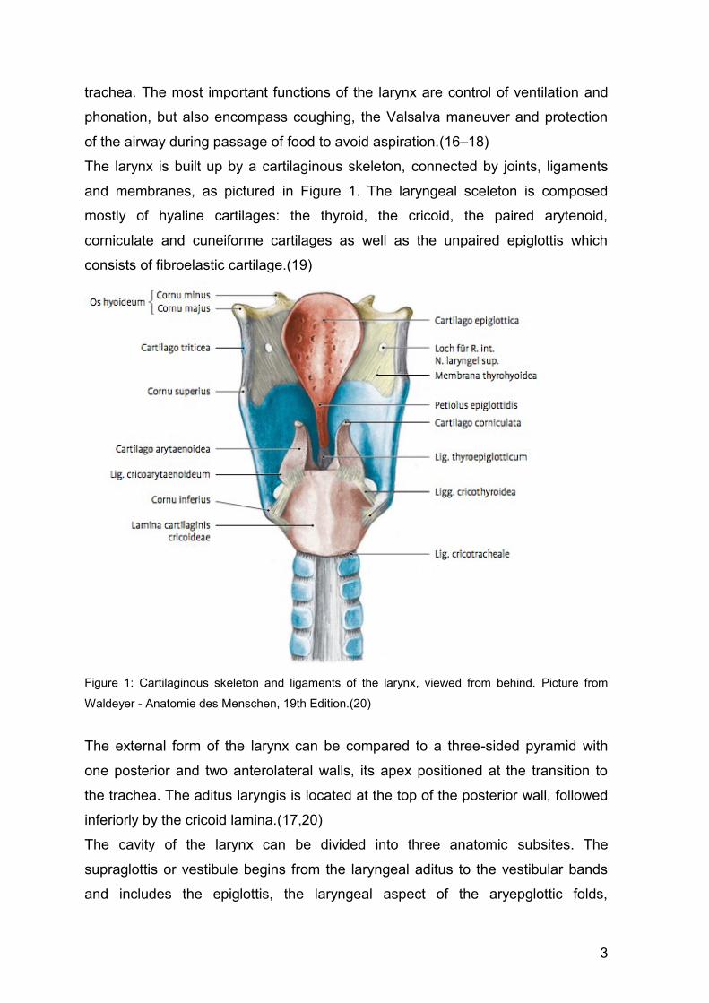
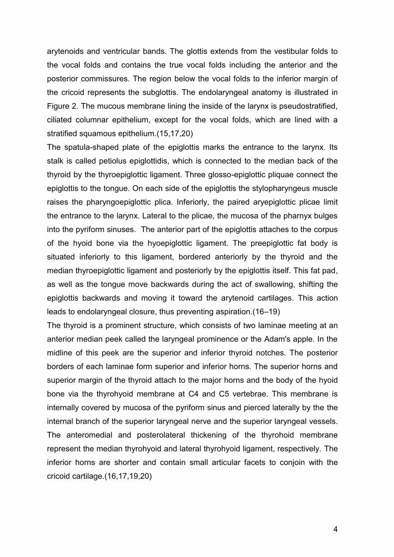
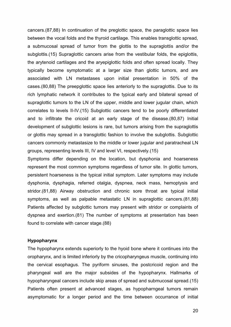
The larynx is built up by a cartilaginous skeleton, connected by joints, ligaments
and membranes, as pictured in Figure 1. The laryngeal sceleton is composed
mostly of hyaline cartilages: the thyroid, the cricoid, the paired arytenoid,
corniculate and cuneiforme cartilages as well as the unpaired epiglottis which
consists of fibroelastic cartilage.(19)
Figure 1: Cartilaginous skeleton and ligaments of the larynx, viewed from behind. Picture from
Waldeyer - Anatomie des Menschen, 19th Edition.(20)
The external form of the larynx can be compared to a three-sided pyramid with
one posterior and two anterolateral walls, its apex positioned at the transition to
the trachea. The aditus laryngis is located at the top of the posterior wall, followed
inferiorly by the cricoid lamina.(17,20)
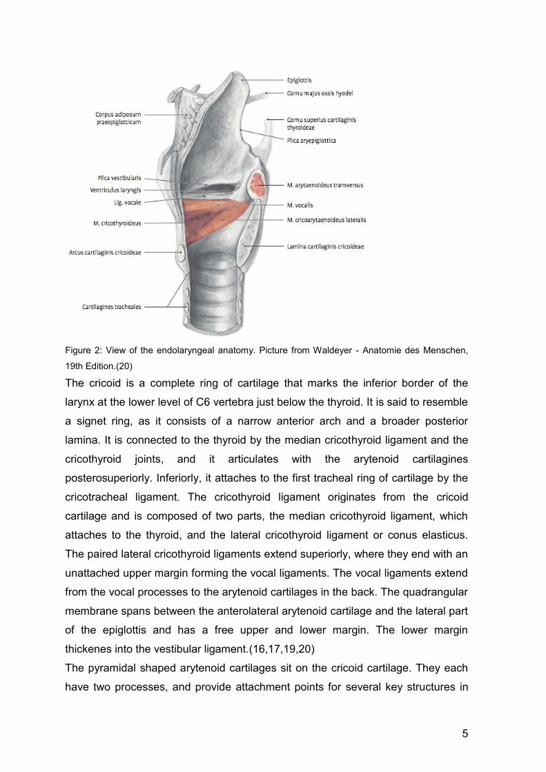
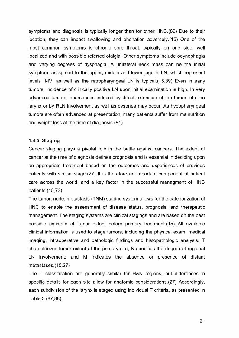
The cavity of the larynx can be divided into three anatomic subsites. The
supraglottis or vestibule begins from the laryngeal aditus to the vestibular bands
and includes the epiglottis, the laryngeal aspect of the aryepglottic folds,
4
arytenoids and ventricular bands. The glottis extends from the vestibular folds to
the vocal folds and contains the true vocal folds including the anterior and the
posterior commissures. The region below the vocal folds to the inferior margin of
the cricoid represents the subglottis. The endolaryngeal anatomy is illustrated in
Figure 2. The mucous membrane lining the inside of the larynx is pseudostratified,
ciliated columnar epithelium, except for the vocal folds, which are lined with a
stratified squamous epithelium.(15,17,20)
The spatula-shaped plate of the epiglottis marks the entrance to the larynx. Its
stalk is called petiolus epiglottidis, which is connected to the median back of the
thyroid by the thyroepiglottic ligament. Three glosso-epiglottic pliquae connect the
epiglottis to the tongue. On each side of the epiglottis the stylopharyngeus muscle
raises the pharyngoepiglottic plica. Inferiorly, the paired aryepiglottic plicae limit
the entrance to the larynx. Lateral to the plicae, the mucosa of the pharnyx bulges
into the pyriform sinuses. The anterior part of the epiglottis attaches to the corpus
of the hyoid bone via the hyoepiglottic ligament. The preepiglottic fat body is
situated inferiorly to this ligament, bordered anteriorly by the thyroid and the
median thyroepiglottic ligament and posteriorly by the epiglottis itself. This fat pad,
as well as the tongue move backwards during the act of swallowing, shifting the
epiglottis backwards and moving it toward the arytenoid cartilages. This action
leads to endolaryngeal closure, thus preventing aspiration.(16–19)
The thyroid is a prominent structure, which consists of two laminae meeting at an
anterior median peek called the laryngeal prominence or the Adam's apple. In the
midline of this peek are the superior and inferior thyroid notches. The posterior
borders of each laminae form superior and inferior horns. The superior horns and
superior margin of the thyroid attach to the major horns and the body of the hyoid
bone via the thyrohyoid membrane at C4 and C5 vertebrae. This membrane is
internally covered by mucosa of the pyriform sinus and pierced laterally by the the
internal branch of the superior laryngeal nerve and the superior laryngeal vessels.
The anteromedial and posterolateral thickening of the thyrohoid membrane
represent the median thyrohyoid and lateral thyrohyoid ligament, respectively. The
inferior horns are shorter and contain small articular facets to conjoin with the
cricoid cartilage.(16,17,19,20)
5
Figure 2: View of the endolaryngeal anatomy. Picture from Waldeyer - Anatomie des Menschen,
19th Edition.(20)
The cricoid is a complete ring of cartilage that marks the inferior border of the
larynx at the lower level of C6 vertebra just below the thyroid. It is said to resemble
a signet ring, as it consists of a narrow anterior arch and a broader posterior
lamina. It is connected to the thyroid by the median cricothyroid ligament and the
cricothyroid joints, and it articulates with the arytenoid cartilagines
posterosuperiorly. Inferiorly, it attaches to the first tracheal ring of cartilage by the
cricotracheal ligament. The cricothyroid ligament originates from the cricoid
cartilage and is composed of two parts, the median cricothyroid ligament, which
attaches to the thyroid, and the lateral cricothyroid ligament or conus elasticus.
The paired lateral cricothyroid ligaments extend superiorly, where they end with an
unattached upper margin forming the vocal ligaments. The vocal ligaments extend
from the vocal processes to the arytenoid cartilages in the back. The quadrangular
membrane spans between the anterolateral arytenoid cartilage and the lateral part
of the epiglottis and has a free upper and lower margin. The lower margin
thickenes into the vestibular ligament.(16,17,19,20)
The pyramidal shaped arytenoid cartilages sit on the cricoid cartilage. They each
have two processes, and provide attachment points for several key structures in
6
the larynx. The apex of the arytenoid articulates with the corniculate cartilage,
while the base connects to the upper border of the cricoid. The posterior and
lateral cricoarytenoid muscles attach to the muscular process, while the vocal
process provides attachment for the vocal ligament. The corniculate cartilages are
small structures that articulate with the apices of the arytenoid cartilages. (16–
18,20) The paired aryepiglottic folds extend from the epiglottic framework to the
arytenoid cartilages in the back and consist of muscle, connective tissue and
mucous membrane. Both the corniculate and the cuneiform cartilages are located
within the aryepiglottic folds. While the cuneiform cartilages do not have direct
attachment, they act to strengthen the folds and add support and stiffness.(19)
1.2.2. Laryngeal Musculature
Muscles that attach to the larynx either move it as a whole (extrinsic muscles), or
alter the position of the cartilages against each other (intrinsic muscles). The
extrinsic muscles move the larynx along the frontal axis superiorly and
inferiorly.(16) The extrinsic group is comprised of the suprahyoid and infrahyoid
muscles, and the stylopharyngeus, which is a pharyngeal muscle. Elevation of the
larynx is achieved by the suprahyoid muscles and the stylopharyngeus, whilst the
infrahyoid muscles depress the larynx.(17,20) In the act of swallowing an elevation
of the larynx happens, faciliating the closure of the laryngeal aperature by the
epiglottis, as well as during voice production. Elevation of the larynx changes the
angle between the cricoid and thyroid cartilage, lengthening the vocal folds.(22)
Phonation and ventilation are controlled by movement of the intrinsic laryngeal
muscles, which alter the position of the muscular and vocal processes of the
arytenoids. This alters the length, tension, shape, and spatial position of the vocal
folds and the shape of the opening between the vocal folds and the arytenoids,
which is called the rima glottidis.(19)
The posterior cricoarytenoid (PCA) is the only muscle capable of widening the
rima glottidis, attachig to the muscular process of the arytenoid and inserting on
the middle and lateral section of the posterior cricoid.(22) It rotates the arytenoids
outward and therefore abducts the vocal folds. The cricothyroid (CTM) muscle
elongates and tenses the vocal folds, controlling forceful speech. The
thyroarytenoid (TA) muscle acts to shorten and relax the vocal folds, allowing for a
softer voice, and also to narrow the rima glottidis. It forms the body of the vocal
7
fold, connecting the arytenoid cartilage to the anterior inner commissure of the
thyroid while enveloping the vocal processes. The lateral cricoarytenoid (LCA)
muscle rotates the arytenoid cartilages to approximate their vocal processes,
which adducts the vocal folds and narrows the rima glottidis, which modulates the
volume and tone of speech. The interarytenoid (IA) muscles act to adduct the
arytenoids, resulting in closure of the posterior part of the rima glottidis.(16,17,20)
1.2.3. Main vessels of the larynx
The main vessels of the larynx are the superior and inferior laryngeal artery and
the accompanying veins. The superior laryngeal artery is a branch of the superior
thyroid artery, which derives from the external carotid artery. It follows the internal
branch of the superior laryngeal nerve into the larynx. The superior laryngeal vein
drains into the superior thyroid vein and then the internal jugular vein. They supply
the larynx above the level of the vocal folds, while the inferior laryngeal vessels
supply the area below the vocal folds. The inferior laryngeal artery derives from
the inferior thyroid artery, which arises from the thyrocervical trunk. It follows the
recurrent laryngeal nerve into the larynx. The inferior laryngeal vein drains to the
inferior thyroid vein, which then joins the bracheocephalic vein.(16,17,20)
1.2.4. Innervation of the larynx
The vagus nerve and sympathetic nerve provide innervation to the larynx, the
vagus nerve sending two branches, the superior and recurrent laryngeal nerve.
The superior laryngeal nerve (SLN) divides into an external and internal branch.
The internal branch (iSLN) is almost entirely sensory, entering the larynx by
passing through the hyothyroid membrane. It then divides into three branches,
supplying the epiglottis, the aryepiglottic fold and the mucous membrane along the
posterior part of the larynx, where it communicates with the recurrent nerve. The
external branch innervates the CT muscle. Beneath the lower margin of the inferior
pharyngeal constrictor muscle, the recurrent nerve (RLN) ascends, providing
sensory innervation to the infraglottis and to all remaining intrinsic muscles of the
larynx via its inferior laryngeal nerve. The mucous membrane is supplied by
subepthelial plexuses formed by sensory branches of both laryngeal
nerves.(16,17,20)
8
1.2.5. Lymphatic system of the larynx
The lymphatic vessels consist of a superficial and a deep network of vessels. Both
of these networks communicate with each other and with the lymphatic network of
the pharynx and trachea. The superior lymphatic vessels collecting the lymph fluid
of the supraglottis and glottis accompany the superior laryngeal artery, passing
through the hyothyroid membrane. They drain mainly to the upper and middle
deep cervial lymph nodes (LN) located cranio- and mediojugularly, along with
lymph fluid from the cranial hypopharynx. Subglottic lymph fluid flows in ventral
direction mainly through the conic ligament and in posterior direction through the
cricotracheal ligament. It is drained to the deep cervical LN, the LN of the recurrent
chain, the prethyroidal LN, the pre- and paratracheal and sporadically also to
prelaryngeal LN. The laryngeal lymphatic plexus continues into the tracheal
lymphatic network without interruption. Lymphathic drainage of the larynx is
characterized by a high degree of variability.(20,23)
In the past, LN of the neck were classified into chains surrounding important
vascular and nervous structures of the neck and divided into superficial, deep,
lateral and medial chains. (16,17) It was Lindberg in 1972 that first described the
distribution of lymphatic metastases related to localization of the primary head and
neck (H&N) tumor.(24) He developed the concept of anatomically correlated
groups of cervical LN, describing nine lymphatic levels on each side of the neck
plus LN of the parotid gland. His studies form the basis for our current knowledge,
allowing to a certain degree of probability the prediction of the direction of
lymphatic spread.(23,25) In 1982, Shan et al. developed a simplified version of
Lindberg's LN classification, dividing the LN regions into seven levels.(25) This
basic structure has not changed significantly since, although it was modified
several times.(23)
The level system describing the location of LN of the neck now consists of seven
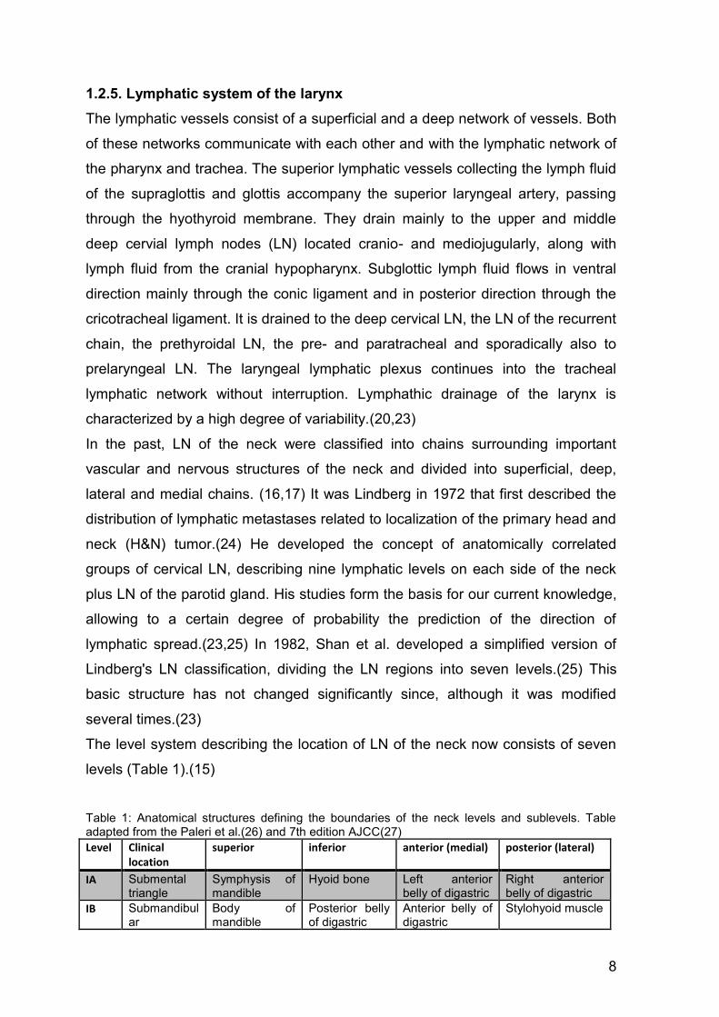
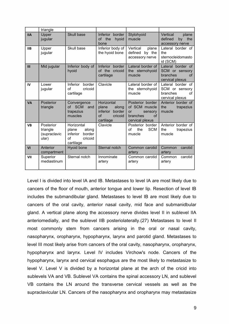
levels (Table 1).(15)
Table 1: Anatomical structures defining the boundaries of the neck levels and sublevels. Table adapted from the Paleri et al.(26) and 7th edition AJCC(27)
Level Clinical location
superior inferior anterior (medial) posterior (lateral)
IA Submental triangle
Symphysis of mandible
Hyoid bone Left anterior belly of digastric
Right anterior belly of digastric
IB Submandibular
Body of mandible
Posterior belly of digastric
Anterior belly of digastric
Stylohyoid muscle
9
triangle
IIA Upper jugular
Skull base Inferior border of the hyoid bone
Stylohyoid muscle
Vertical plane defined by the accessory nerve
IIB Upper jugular
Skull base Inferior body of the hyoid bone
Vertical plane defined by the accessory nerve
Lateral border of the sternocleidomastoid (SCM)
III Mid jugular Inferior body of hyoid
Inferior border of the cricoid cartilage
Lateral border of the sternohyoid muscle
Lateral border of SCM or sensory branches of cervical plexus
IV Lower jugular
Inferior border of cricoid cartilage
Clavicle
Lateral border of the sternohyoid muscle
Lateral border of SCM or sensory branches of cervical plexus
VA Posterior triangle
Convergence of SCM and trapezius muscles
Horizontal plane along inferior border of cricoid cartilage
Posterior border of SCM muscle or sensory branches of cervical plexus
Anterior border of the trapezius muscle
VB Posterior triangle (supraclavicular)
Horizontal plane along inferior border of cricoid cartilage
Clavicle
Posterior border of the SCM muscle
Anterior border of the trapezius muscle
VI Anterior compartment
Hyoid bone
Sternal notch Common carotid artery
Common carotid artery
VII Superior mediastinum
Sternal notch Innominate artery
Common carotid artery
Common carotid artery
Level I is divided into level IA and IB. Metastases to level IA are most likely due to
cancers of the floor of mouth, anterior tongue and lower lip. Resection of level IB
includes the submandibular gland. Metastases to level IB are most likely due to
cancers of the oral cavity, anterior nasal cavity, mid face and submanidbular
gland. A vertical plane along the accessory nerve divides level II in sublevel IIA
anteriomedially, and the sublevel IIB posteriolaterally.(27) Metastases to level II
most commonly stem from cancers arising in the oral or nasal cavity,
nasopharynx, oropharynx, hypopharynx, larynx and parotid gland. Metastases to
level III most likely arise from cancers of the oral cavity, nasopharynx, oropharynx,
hypopharynx and larynx. Level IV includes Virchow's node. Cancers of the
hypopharynx, larynx and cervical esophagus are the most likely to metastasize to
level V. Level V is divided by a horizontal plane at the arch of the cricid into
sublevels VA and VB. Sublevel VA contains the spinal accessory LN, and sublevel
VB contains the LN around the transverse cervical vessels as well as the
supraclavicular LN. Cancers of the nasopharynx and oropharynx may metastasize
10
into LN of sublevel VA, while cancers of the thyroid gland may metastasize into LN
of sublevel VB. A plane defined by the sensory branches of the cervical plexus
constitutes the surgical landmark that marks the lateral border of levels II, III, and
IV and the medial border of level V. Level VI includes the pre- and paratracheal
LN, the precricoid and the prethyroidal LN.(27) Metastases to these levels most
commonly come from cancers of the thyroid gland, glottic and subglottic larynx,
pyriform sinuses and cervical esophagus.(15) Level VII encompasses the
pretracheal, paratracheal, and esophageal groove LN. Metastases to these LN are
most likely due to cancer of the thyroid and of the esophagus. (15,26,27)
1.3. Physiology The larynx serves as a valve in the upper airway, maintaining the opening of the
vocal folds for respiration and closing during deglutition to prevent aspiration of
foreign matter into the lower airway or to stabilize the chest by building up
intrathoracic pressure.(22)
In humans, the larynx serves three important functions: protection of the airway,
control of respiration and phonation.(19) The primary function of the larynx is that
of a sphincter, guarding the lower respiratory tract from the intrusion of foreign
matter. Its secondary function centers around its role in respiration by active
dilatation of the laryngeal aperture. Phonation, the third function of the larynx,
appears to be a late phylogenetic acquisition. Humans alone have developed
complex sound production by using the sphincter of the larynx as a vibratory
source.(21,28)
The upper airway crosses the digestive tract in the region of the pharynx. This
common passageway complicates the sphincteric function of the larynx and also
compromises its respiratory performance by creating ventilatory turbulence and
resistance. The anatomic configuration in adult humans that enables phonation
simultaneously compromises the sphincteric and respiratory function of the larynx,
creating a functional dilemma. This is resolved on a laryngopharyngeal level by
structural adaption and exact coordination of the laryngeal functions by precisely
organized brainstem reflexes, called laryngeal reflexes.(21) The larynx constantly
controls life supporting processes such as breathing, swallowing and coughing by
specific laryngeal reflexes or responses.(22) Basic functions are controlled by
11
complex interactions of polysynaptic brainstem reflexes. The protective function is
involuntary and reflexive, as opposed to the respiratory and phonatory functions
that can be initiated voluntarily but are regulated through an array of involuntary
feedback reflexes.(17,21)
1.3.1. Swallowing
The act of swallowing is elicited by stimulation of mechanoreceptors in the larynx
and pharynx. Deglutition actively suppresses respiration while the upper airway
closes to allow for passing of the bolus into the esophagus. The respiratory apnea
during deglutition is an active cessation, which usually happens on expiration and
resets the respiratory rhythm.(22) During the pharyngeal phase of swallowing, a
sequence of events works to transport the bolus from the oropharynx into the
esophagus without aspiration. The flat configuration of the epiglottis in adults
directs swallowed matter away from the median laryngeal aperature into the
pyriform sinuses. The aryepiglottic folds act as embarkments, guiding food boli to
pass laterally of the epiglottis along the gutter between the fold and the pharyngeal
wall on each side.(21) The larynx and hyoid bone are elevated and moved
anteriorly by extrinsic laryngeal muscles. This elevation as well as the descent of
the epiglottis facilitate closure of the vestibule, with simultaneous closure of the
vocal folds.(22,29) Deglutition is a semireflectory process.(29) The pharyngeal
phase of swallowing is regulated by a central pattern generator in the medulla
oblongata. This phase of swallowing is considered reflexive and can be initiated by
stimulation of sensory triggers in the oropharynx, but it can also be intitiated
volitionally.(30) Deglutition depends on feedback of the SLN to execute and
coordinate the pharyngeal phase of swallowing. Loss of this feedback has not only
shown to induce a sensation of being unable to initiate deglutition but also to
increase the occurrence of aspiration in human adults.(22)
1.3.2. Respiration
To facilitate respiration, the upper airway is dilated for inspiration(31) and relaxes
on expiration. The PCA, which works to open the rima glottidis(32), is constantly
active but tone increases during inspiration to keep the vocal folds apart.(33) This
muscle is essential to life support(34), as bilateral paralysis of the PCA can result
12
in negative pressure between the vocal folds during inspiration, sucking them
toward the midline and thus obstructing air flow.
During expiration, air flow is modulated by muscles closing the vocal folds,
including the TA(35) and the LCA, which actively adducts the vocal folds.The LCA
is most active for phonation and deglutition.(36)
During inspiration, a chain of muscles actively dilate the upper respiratory tract
from the nasal cavity and to the larynx.(37) The laryngeal muscles act to open the
vocal folds to enhance air intake. In tidal breathing, laryngeal muscular activity is
generally low during expiration, with the TA activating to partially close the vocal
folds to reduce air flow.(30,38) Hypoxia or hypercapnia cause stress on the
respiratory system, which activate the laryngeal muscles to a higher degree to
support air flow intake with opening of the vocal folds and activation of the
adductor muscle to reduce expiratory air flow.(39,40) This demonstrates that
respiratory reflexes actively control the laryngeal musculature in adult humans.(40)
1.3.3. Phonation
The most complex and specialized laryngeal function is voice production. Human
speech, phonation, articulation and resonance require precise coordination and
specific mechanical properties of the laryngeal musculature, brainstem reflexes
and high-level cortical control. Lung capacity, thoracic wall compliance, anatomy of
the pharynx, nasal and oral cavity, and mental status also play a role, and
expiratory flow from the lungs must provide sufficient subglottic pressure.(19)
The process begins with inhalation and activation of the laryngeal muscles to
create glottic closure as well as the adequate length, tension and closure of the
vocal folds.(22) Expiratory flow increases subglottic pressure until it overcomes the
glottal closure force and opens the posterior part of the vocal folds. Expiratory
airflow passes between the vocal folds, inducing vibration due to the pliability and
vibratory capacity of the mucosa. This wave-like motion of loose mucosa of the
vocal folds over the denser tissue of the vocal ligaments and vocalis muscles is
described by the body-cover theory. The mucosal wave begins infraglottically and
moves superiorly toward the free edge of the vocal fold. Due to the decrease in
pressure and elastic recoil of the tissue, the inferior edges eventually become
reapproximated. The closure phase is propagated toward the front. Subglottic
13
pressure builds again when the vocal folds are fully approximated, and the cycle
can be repeated.(19,22)
The role of the laryngeal muscles during phonation is to achieve an appropriate
posture the vocal folds. This requires adequate contraction of the TA to shorten
the vocal folds and lower them into the midline. The opposing action of the CTM
lengthens and tenses the vocal folds.(22,41) The ratio of contraction levels of
these muscles changes the fundamental frequency of vibration and thereby the
pitch of the voice. The intrinsic muscles of the larynx are highly specialized for
their specific vector of action during phonation, for the degree of recruitment and
the timing of contraction.(19)
1.4. Laryngeal and Hypopharyngeal Cancer
1.4.1. Epidemiology
Head and neck cancer (HNC) is common all over the world, with large geographic
variations of the incidence and primary site. Chronic exposure of the mucosa of
the upper aero-digestive tract to carcinogenic substances can lead to dysplastic
and ultimately malignant lesions. Primary risk factors for development of HNC are
consumption of tobacco and alcohol as well as human papillomavirus (HPV)
infection.(42) At least 75% of HNC diagnosed in industrialized regions can be
attributed to cigarette smoking and alcohol consumption or a combination of
both.(43) The relative prevalence of these risk factors as well as ethnic and
genetic diversity contributes to observed differences in distribution of HNC.(42)
Worldwide, HNC accounts for more than 550,000 cancer cases and 380,000
deaths annually.(44) In the United States, HNC makes up 3 percent of
malignancies, which correspond to approximately 63,000 Americans developing
HNC and 13,000 HNC related deaths annually.(45) About 14,000 cases of
laryngeal cancer and 3600 laryngeal cancer related deaths occur annually in the
United States.(45,46)
14
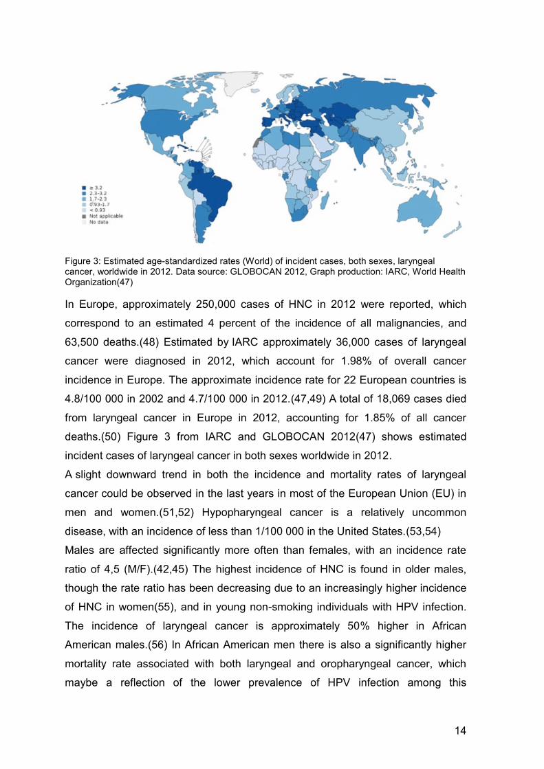
Figure 3: Estimated age-standardized rates (World) of incident cases, both sexes, laryngeal cancer, worldwide in 2012. Data source: GLOBOCAN 2012, Graph production: IARC, World Health Organization(47)
In Europe, approximately 250,000 cases of HNC in 2012 were reported, which
correspond to an estimated 4 percent of the incidence of all malignancies, and
63,500 deaths.(48) Estimated by IARC approximately 36,000 cases of laryngeal
cancer were diagnosed in 2012, which account for 1.98% of overall cancer
incidence in Europe. The approximate incidence rate for 22 European countries is
4.8/100 000 in 2002 and 4.7/100 000 in 2012.(47,49) A total of 18,069 cases died
from laryngeal cancer in Europe in 2012, accounting for 1.85% of all cancer
deaths.(50) Figure 3 from IARC and GLOBOCAN 2012(47) shows estimated
incident cases of laryngeal cancer in both sexes worldwide in 2012.
A slight downward trend in both the incidence and mortality rates of laryngeal
cancer could be observed in the last years in most of the European Union (EU) in
men and women.(51,52) Hypopharyngeal cancer is a relatively uncommon
disease, with an incidence of less than 1/100 000 in the United States.(53,54)
Males are affected significantly more often than females, with an incidence rate
ratio of 4,5 (M/F).(42,45) The highest incidence of HNC is found in older males,
though the rate ratio has been decreasing due to an increasingly higher incidence
of HNC in women(55), and in young non-smoking individuals with HPV infection.
The incidence of laryngeal cancer is approximately 50% higher in African
American males.(56) In African American men there is also a significantly higher
mortality rate associated with both laryngeal and oropharyngeal cancer, which
maybe a reflection of the lower prevalence of HPV infection among this
15
population.(57) Incidence and mortality of laryngeal and hypopharyngeal cancers
is low in people under 40 years of age. Incidence increases rapidly in patients over
40 years old and finally peakes in subjects of 60-75 years. Mortality also increases
with age, peaking at 85 years old.(49,58)
1.4.2. Risk factors
Squamous cell carcinomas (SCC) of the H&N develop from the squamous
mucosal lining of the upper aero-digestive tract mainly in patients with risk factors
like tobacco consumption, alcohol abuse, or HPV infection in their personal
history. However, SCC arise in only 20% of individuals with a history of these risk
factors.(59,60) Squamous carcinogenesis is caused when molecular genetic
changes accumulate in the squamous epithelial lining of the upper aero-digestive
tract. Dysplasia progresses to invasive carcinoma in a complex process, in which
the interaction of epithelium and of host stromal elements trigger the dysregulation
of key pathways which lead to cellular and structural changes.(59,60)
1.4.2.1. Tobacco products
Smoking tobacco products is the most important risk factor for the development of
HNC.(61) Some evidence suggests a genetic predisposition to carcinogenic
effects of tobacco use. Heavy cigarette smokers suffer from a 5- to 25-fold
increased risk of cancer compared with nonsmokers.(42,61,62) There also
appears to be a dose-response relationship, with relative risk (RR) increasing with
frequency, duration and pack-years of cigarette smoking.(42) Cessation of
smoking is associated with a significant decrease in RR, with no excess risk after
20 years.(63) This suggests that smoking primarily affects at a later stage in the
process of carcinogenesis.(43) Cigarette smoking is strongly associated with
higher risk of developing laryngeal and hypopharyngeal cancer, while the
assoctiation to oral cavity or pharyngeal cancers is not as strong.(61,64)
Tobacco consumption other than cigarette smoking also increases the risk of
developing HNC. Smoking of pipes and cigars is associated with a higher risk,
even in patients who never smoked cigarettes.(61) Secondhand smoke exposure
may also result in increased risk. Tan et al.(65) evaluated 59 patients with HNC
who did not consume tobacco and did not abuse alcohol. Non-smoking patients
16
with HNC stood out as predominantely white, female and presented with
significantly more tongue primaries. When compared with the control population
without cancer, these patients showed a significantly higher risk of being exposed
to environmental tobacco smoke.(65)
Evidence showing the association of the development of HNC and marijuana
usage is somewhat conflicting, as the outcome of observational studies is often
limited by reporting and selection bias and confounded by alcohol and tobacco
consumption.(66,67)
1.4.2.2. Alcohol
Consumption of alcohol independently increases the risk of developing cancer in
the upper aero-digestive tract, although the effects of smoking and alcohol are
often confounded.(43) The RR of developing HNC due to alcohol appears to be
dose-dependent.(63,64,68)
While no correlation with duration of alcohol use and risk for HNC is apparent in
the abscence of tobacco use, high doses and high frequency of alcohol use does
increase the risk. Hashibe et al.(64) found that consumption of three or more
alcoholic drinks per day was associated with approximately twice the risk of HNC
than for never-drinkers. Increased frequency of alcohol consumption results in an
increased risk to develop pharyngeal cancer when drinking one to two drinks per
day, and laryngeal cancer when drinking five or more drinks per day.(64) The
increase in risk is higher among those consuming hard liquor or beer than those
consuming wine.(43) Similarly, Lewin et al.(63) reported that alcohol intake of 50
grams or more daily versus less than 10 grams daily led to a RR of 5.5%.
Moderate alcohol consumption of 10-19 grams per day had little or no effect
among nonsmokers.(63)
Alcohol intake combined with tobacco smoking appear to have an interactive and
multiplicative effect on the risk of developing HNC.(43,63,69) The risk is increased
more than 35-fold when two or more packs of cigarettes and more than four
alcoholic drinks were consumed per day according to Blot et al.(43)
17
1.4.2.3. Human papillomavirus infection
HPV infection is associated with increased risk to develop HNC, particularly
oropharyngeal cancer.(57,70) HPV-16 seropositivity was twice as high among
HNC patients as among a control population, while no increased risk was
observed for HPV-18, HPV-33 or HPV-73 by Mork et al.(71) Significantly elevated
OR was detected for cancer of the tonsils and base of tongue. HPV associated
HNC are typically seen in younger, white nonusers of tobacco and alcohol that
present with local T category tumors and poor differentiation at histologic
examination.(42) There is conflicting evidence whether HPV-associated risk of
HNC is different in males and females.(71,72) It is also associated with
significantly better outcome and improved survival in the subsites oropharynx,
hypopharynx, larynx and oral cavity, with the largest benefit in survival in cancers
of the oropharynx and hypopharynx.(72)
Immunohistochemical staining is used to distinguish HPV-related from non-HPV
related cancers, as overexpression of p16, a tumor suppressor protein, is a
surrogate biomarker for HPV-mediated carcinogenesis and also a positive
prognostic factor regarding oropharyngeal carcinomas.(70,73) Overexpression of
p16 is typically localized in tumor cell nuclei and cytoplasm. The cutoff point for
positive p16 expression is at least moderate (+2/3) staining intensity and diffuse
(≥75%) tumor expression.(73)
1.4.2.4. Occupational exposure
Multiple occupational or environmental toxins have been studied for a potential
relationship with HNC. These include the exposure to the dry cleaning agent
perchloroethylene(74), asbestos, pesticides, man-made mineral vitreous fibers
(MMMF)(75), polycyclic aromatic hydrocarbons (76), and cement(77).(42)
1.4.2.5. Diet
An increased intake of fruits and vegetables is associated with a decreased risk for
HNC.(78)
1.4.2.6. Genetic factors
Genetic predisposition plays a modulating role in developing HNC, as not all
18
individuals exposed to the main etiologic factors experience the disease. Multiple
genetic factors and pathways contribute to an increase in the risk of HNC, and
these factors interact with other known risk factors.(79) These factors include
metabolic polymorphisms that affect exposure to the carcinogens in tobacco
smoke, polymorphisms of DNA repair genes, and variations in other pathways
contributing to carcinogenesis.(42)
1.4.3. Pathology
Head and neck squamous cell carcinomas often develop from premalignant
entities caused by carcinogen exposure, such as leukoplakia and
erythroplakia.(80) Histopathologically, dysplasia is defined by the presence of
mitoses and prominent nucleoli. Progression to invasive cancer happens in 15 to
30% of cases of dyplasia.(81) Leukoplakia is a descriptive clinical term for grey to
white lesions characterized by hyperparakeratosis and often associated with
hyperplasia of the underlying epithelium.(80) In the absence of underlying
dysplastic alterations, leukoplakia is associated with malignant change in less than
5%.(82) Erythroplakia describes red patches adjacent to normal mucosa. It is less
common then leukoplakia but more often associated with malignancy.(80) It is
typically associated with epithelial dysplasia and with the presence of carcinoma in
situ or invasive cancer in up to 40% of cases.(81,82)
SCC account for more than 90 percent of the lesions in the hypopharynx and
larynx. In hypopharyngeal lesions, a general pattern is that the more posterior and
the more inferior the site, the worse the prognosis. This is not true for laryngeal
lesions, possibly because of differences in the type and the extent of SCC at the
different locations. Also, anatomical differences between the sites influence
prognosis. The less abundant lymphatic drainage of the larynx compared to the
hypopharyngeal lymphatics may explain the higher metastatic rate and worse
prognosis for hypopharyngeal cancer.(58,83)
SCC are graded depending on their keratinization using the World Health
Organization's standard grading system, although there are distinct grading
schemes used for cancers of different morphologic and anatomic site
groupings.(83,84)
Histopathological grade is reported in registry systems by the grade value (Table
2).
19
Generally, the following grading system is used:
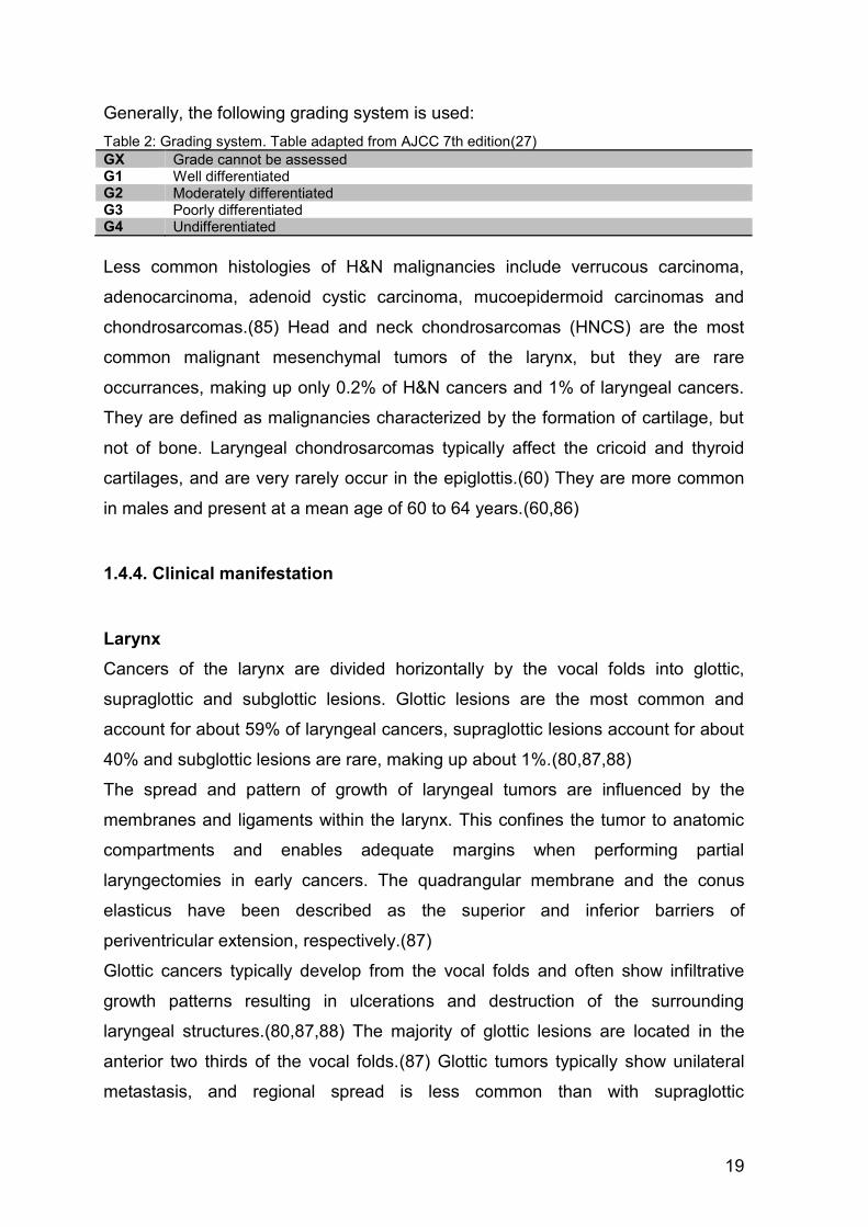
Table 2: Grading system. Table adapted from AJCC 7th edition(27)
GX Grade cannot be assessed G1 Well differentiated G2 Moderately differentiated G3 Poorly differentiated G4 Undifferentiated
Less common histologies of H&N malignancies include verrucous carcinoma,
adenocarcinoma, adenoid cystic carcinoma, mucoepidermoid carcinomas and
chondrosarcomas.(85) Head and neck chondrosarcomas (HNCS) are the most
common malignant mesenchymal tumors of the larynx, but they are rare
occurrances, making up only 0.2% of H&N cancers and 1% of laryngeal cancers.
They are defined as malignancies characterized by the formation of cartilage, but
not of bone. Laryngeal chondrosarcomas typically affect the cricoid and thyroid
cartilages, and are very rarely occur in the epiglottis.(60) They are more common
in males and present at a mean age of 60 to 64 years.(60,86)
1.4.4. Clinical manifestation
Larynx
Cancers of the larynx are divided horizontally by the vocal folds into glottic,
supraglottic and subglottic lesions. Glottic lesions are the most common and
account for about 59% of laryngeal cancers, supraglottic lesions account for about
40% and subglottic lesions are rare, making up about 1%.(80,87,88)
The spread and pattern of growth of laryngeal tumors are influenced by the
membranes and ligaments within the larynx. This confines the tumor to anatomic
compartments and enables adequate margins when performing partial
laryngectomies in early cancers. The quadrangular membrane and the conus
elasticus have been described as the superior and inferior barriers of
periventricular extension, respectively.(87)
Glottic cancers typically develop from the vocal folds and often show infiltrative
growth patterns resulting in ulcerations and destruction of the surrounding
laryngeal structures.(80,87,88) The majority of glottic lesions are located in the
anterior two thirds of the vocal folds.(87) Glottic tumors typically show unilateral
metastasis, and regional spread is less common than with supraglottic
20
cancers.(87,88) In continuation of the preglottic space, the paraglottic space lies
between the vocal folds and the thyroid cartilage. This enables transglottic spread,
a submucosal spread of tumor from the glottis to the supraglottis and/or the
subglottis.(15) Supraglottic cancers arise from the vestibular folds, the epiglottis,
the arytenoid cartilages and the aryepiglottic folds and often spread locally. They
typically become symptomatic at a larger size than glottic tumors, and are
associated with LN metastases upon initial presentation in 50% of the
cases.(80,88) The preepglottic space lies anteriorly to the supraglottis. Due to its
rich lymphatic network it contributes to the typical early and bilateral spread of
supraglottic tumors to the LN of the upper, middle and lower jugular chain, which
correlates to levels II-IV.(15) Subglottic cancers tend to be poorly differentiated
and to infiltrate the cricoid at an early stage of the disease.(80,87) Initial
development of subglottic lesions is rare, but tumors arising from the supraglottis
or glottis may spread in a transglottic fashion to involve the subglottis. Subglottic
cancers commonly metastasize to the middle or lower jugular and paratracheal LN
groups, representing levels III, IV and level VI, respectively.(15)
Symptoms differ depending on the location, but dysphonia and hoarseness
represent the most common symptoms regardless of tumor site. In glottic tumors,
persistent hoarseness is the typical initial symptom. Later symptoms may include
dysphonia, dysphagia, referred otalgia, dyspnea, neck mass, hemoptysis and
stridor.(81,88) Airway obstruction and chronic sore throat are typical initial
symptoms, as well as palpable metastatic LN in supraglottic cancers.(81,88)
Patients affected by subglottic tumors may present with stridor or complaints of
dyspnea and exertion.(81) The number of symptoms at presentation has been
found to correlate with cancer stage.(88)
Hypopharynx
The hypopharynx extends superiorly to the hyoid bone where it continues into the
oropharynx, and is limited inferiorly by the cricopharyngeus muscle, continuing into
the cervical esophagus. The pyriform sinuses, the postcricoid region and the
pharyngeal wall are the major subsides of the hypopharynx. Hallmarks of
hypopharyngeal cancers include skip areas of spread and submucosal spread.(15)
Patients often present at advanced stages, as hypopharngeal tumors remain
asymptomatic for a longer period and the time between occurrance of initial
21
symptoms and diagnosis is typically longer than for other HNC.(89) Due to their
location, they can impact swallowing and phonation adversely.(15) One of the
most common symptoms is chronic sore throat, typically on one side, well
localized and with possible referred otalgia. Other symptoms include odynophagia
and varying degrees of dysphagia. A unilateral neck mass can be the initial
symptom, as spread to the upper, middle and lower jugular LN, which represent
levels II-IV, as well as the retropharyngeal LN is typical.(15,89) Even in early
tumors, incidence of clinically positive LN upon initial examination is high. In very
advanced tumors, hoarseness induced by direct extension of the tumor into the
larynx or by RLN involvement as well as dyspnea may occur. As hypopharyngeal
tumors are often advanced at presentation, many patients suffer from malnutrition
and weight loss at the time of diagnosis.(81)
1.4.5. Staging
Cancer staging plays a pivotal role in the battle against cancers. The extent of
cancer at the time of diagnosis defines prognosis and is essential in deciding upon
an appropriate treatment based on the outcomes and experiences of previous
patients with similar stage.(27) It is therefore an important component of patient
care across the world, and a key factor in the successful managment of HNC
patients.(15,73)
The tumor, node, metastasis (TNM) staging system allows for the categorization of
HNC to enable the assessment of disease status, prognosis, and therapeutic
management. The staging systems are clinical stagings and are based on the best
possible estimate of tumor extent before primary treatment.(15) All available
clinical information is used to stage tumors, including the physical exam, medical
imaging, intraoperative and pathologic findings and histopathologic analysis. T
characterizes tumor extent at the primary site, N specifies the degree of regional
LN involvement; and M indicates the absence or presence of distant
metastases.(15,27)
The T classification are generally similar for H&N regions, but differences in
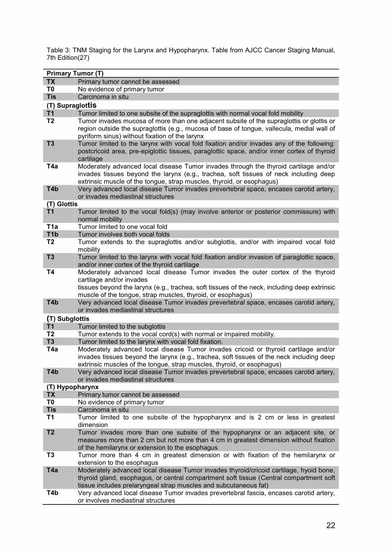
specific details for each site allow for anatomic considerations.(27) Accordingly,
each subdivision of the larynx is staged using individual T criteria, as presented in
Table 3.(87,88)
22
Table 3: TNM Staging for the Larynx and Hypopharynx. Table from AJCC Cancer Staging Manual, 7th Edition(27)
Primary Tumor (T)
TX Primary tumor cannot be assessed T0 No evidence of primary tumor Tis Carcinoma in situ (T) Supraglottis T1 Tumor limited to one subsite of the supraglottis with normal vocal fold mobility T2 Tumor invades mucosa of more than one adjacent subsite of the supraglottis or glottis or
region outside the supraglottis (e.g., mucosa of base of tongue, vallecula, medial wall of pyriform sinus) without fixation of the larynx
T3 Tumor limited to the larynx with vocal fold fixation and/or invades any of the following: postcricoid area, pre-epiglottic tissues, paraglottic space, and/or inner cortex of thyroid cartilage
T4a Moderately advanced local disease Tumor invades through the thyroid cartilage and/or invades tissues beyond the larynx (e.g., trachea, soft tissues of neck including deep extrinsic muscle of the tongue, strap muscles, thyroid, or esophagus)
T4b Very advanced local disease Tumor invades prevertebral space, encases carotid artery, or invades mediastinal structures
(T) Glottis T1 Tumor limited to the vocal fold(s) (may involve anterior or posterior commissure) with
normal mobility T1a Tumor limited to one vocal fold T1b Tumor involves both vocal folds T2 Tumor extends to the supraglottis and/or subglottis, and/or with impaired vocal fold
mobility T3 Tumor limited to the larynx with vocal fold fixation and/or invasion of paraglottic space,
and/or inner cortex of the thyroid cartilage T4 Moderately advanced local disease Tumor invades the outer cortex of the thyroid
cartilage and/or invades tissues beyond the larynx (e.g., trachea, soft tissues of the neck, including deep extrinsic muscle of the tongue, strap muscles, thyroid, or esophagus)
T4b Very advanced local disease Tumor invades prevertebral space, encases carotid artery, or invades mediastinal structures
(T) Subglottis T1 Tumor limited to the subglottis T2 Tumor extends to the vocal cord(s) with normal or impaired mobility. T3 Tumor limited to the larynx with vocal fold fixation. T4a Moderately advanced local disease Tumor invades cricoid or thyroid cartilage and/or
invades tissues beyond the larynx (e.g., trachea, soft tissues of the neck including deep extrinsic muscles of the tongue, strap muscles, thyroid, or esophagus)
T4b Very advanced local disease Tumor invades prevertebral space, encases carotid artery, or invades mediastinal structures
(T) Hypopharynx TX Primary tumor cannot be assessed T0 No evidence of primary tumor Tis Carcinoma in situ T1 Tumor limited to one subsite of the hypopharynx and is 2 cm or less in greatest
dimension T2 Tumor invades more than one subsite of the hypopharynx or an adjacent site, or
measures more than 2 cm but not more than 4 cm in greatest dimension without fixation of the hemilarynx or extension to the esophagus
T3 Tumor more than 4 cm in greatest dimension or with fixation of the hemilarynx or extension to the esophagus
T4a Moderately advanced local disease Tumor invades thyroid/cricoid cartilage, hyoid bone, thyroid gland, esophagus, or central compartment soft tissue (Central compartment soft tissue includes prelaryngeal strap muscles and subcutaneous fat)
T4b Very advanced local disease Tumor invades prevertebral fascia, encases carotid artery, or involves mediastinal structures
23
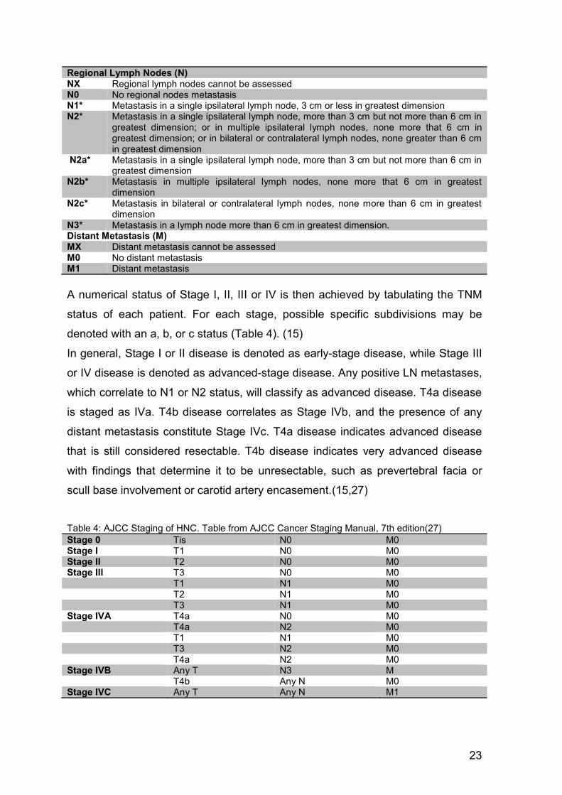
Regional Lymph Nodes (N) NX Regional lymph nodes cannot be assessed N0 No regional nodes metastasis N1* Metastasis in a single ipsilateral lymph node, 3 cm or less in greatest dimension N2* Metastasis in a single ipsilateral lymph node, more than 3 cm but not more than 6 cm in
greatest dimension; or in multiple ipsilateral lymph nodes, none more that 6 cm in greatest dimension; or in bilateral or contralateral lymph nodes, none greater than 6 cm in greatest dimension
N2a* Metastasis in a single ipsilateral lymph node, more than 3 cm but not more than 6 cm in greatest dimension
N2b* Metastasis in multiple ipsilateral lymph nodes, none more that 6 cm in greatest dimension
N2c* Metastasis in bilateral or contralateral lymph nodes, none more than 6 cm in greatest dimension
N3* Metastasis in a lymph node more than 6 cm in greatest dimension. Distant Metastasis (M) MX Distant metastasis cannot be assessed M0 No distant metastasis M1 Distant metastasis
A numerical status of Stage I, II, III or IV is then achieved by tabulating the TNM
status of each patient. For each stage, possible specific subdivisions may be
denoted with an a, b, or c status (Table 4). (15)
In general, Stage I or II disease is denoted as early-stage disease, while Stage III
or IV disease is denoted as advanced-stage disease. Any positive LN metastases,
which correlate to N1 or N2 status, will classify as advanced disease. T4a disease
is staged as IVa. T4b disease correlates as Stage IVb, and the presence of any
distant metastasis constitute Stage IVc. T4a disease indicates advanced disease
that is still considered resectable. T4b disease indicates very advanced disease
with findings that determine it to be unresectable, such as prevertebral facia or
scull base involvement or carotid artery encasement.(15,27)
Table 4: AJCC Staging of HNC. Table from AJCC Cancer Staging Manual, 7th edition(27)
Stage 0 Tis N0 M0 Stage I T1 N0 M0 Stage II T2 N0 M0 Stage III T3 N0 M0 T1 N1 M0 T2 N1 M0 T3 N1 M0 Stage IVA T4a N0 M0 T4a N2 M0 T1 N1 M0 T3 N2 M0 T4a N2 M0 Stage IVB Any T N3 M T4b Any N M0 Stage IVC Any T Any N M1
24
1.4.6. Treatment options The treatment goal for laryngeal cancers is using available treatment options to
achieve a cure while preserving functionality. For T1 and T2 larynx cancers,
organ-preserving treatment such as radiotherapy (RT) and transoral endoscopic
excision is recommended as the initial treatment approach.(88,90) While both
options yield good locoregional control, proponents of each modality often
disagree on the functional sequelea of each option.(91) Appropriate treatment
protocols should be decided using a multidisciplinary approach.(88)
Definitive treatment for advanced stage cancer of the larynx consists of either TL
or organ preserving RCT. The objective of organ preserving therapy in cases of
advanced laryngeal cancer is to offer improved function and quality of life (QOL)
without compromising survival.(92) With the publication of the Department of
Veteran Affairs (VA) Laryngeal Cancer Study(93) and the Radiation Therapy
Oncology Group (RTOG) 91-11 study(94), a major shift in treatment for patients
occured, as it was indicated that organ preservation could be achieved in locally
advanced disease when using concurrant radiochemotheray (RCT) without
compromising survival. This lead to a fall in primary TL and a corresponding rise in
nonoperative larynx-preservation (LP) treatments.(87,92,95) RTOG protocol 91-11
showed that concurrent RCT achieved higher rates of organ perservation than RT
alone or induction CT followed by definitive RT. Primary RCT can achieve high
rates of organ preservation and no decline in survival compared with RT alone or
TL even in advanced laryngeal cancer.(87,95) Primary RT is reserved for patients
with substantial comorbidity who are unsuitable for the safe administration of RCT
or TL.(92) Concurrent RCT is regarded as the gold standard of care for patients
with T3 cancer who prefer LP.(92) Guideline recommendations specify TL as the
initial treatment of choice for T4a larynx cancer, as T4 patients showed lower rates
of successful LP and higher complication rates.(92,96) Concerns have been raised
that LP is being offered too broadly to patients, as a decline in survival and
concurrent rise of LP and the decrease of TL has been demonstrated.(95,96)
Furthermore, an improved overall survival rate was reported for patients with stage
IV disease treated with TL compared with LP.(95,97) The selection of treatment is
complex and multifactorial. In addition to institutional factors including support
systems and surgical expertise, it is also influenced by individual factors such as
25
functional performance status and personal preference of the patient. The choice
of treatment regimen also depends on the overall health of the patient.
Comorbidity can be classified by specific measures of additional medical illnesses.
General performance measures are helpful in predicting survival.(27) To assess
this, the Eastern Cooperative Oncology Group Performance Status (ECOG PS)
scale is used, as it is a standard functional classification in oncology practice.
Each patient is allocated a score on a linear scale between 0 (fully active) and 5
(dead) according to their physical activity. (98,99)
1.4.6.1. Radiotherapy
Radiotherapy (RT) alone and in combination with LP surgery is accepted as an
effective treatment for T1 tumors and also, to a lesser degree, for T2 tumors.
Radiation doses typically range from 60 to 70 Gy. Accelerated regimens using
higher daily fractions and shorter treatment durations have been associated with
improved outcomes in local control.(88)
Complications from RT can be divided into early and late subgroups. Early
complications include mucositis, edema, hoarseness and dysphagia. Fibrosis,
stenosis, xerostomia and hypothyroidism are late complications. Incidence of
xerostomia and dysphagia has been shown to be reduced using intensity-
modulated radiation therapy (IMRT).(88)
In a 2014 study by O'Neill et al. comparing treatment-related complications, RCT
patients suffered from mucositis, xerostomia and dysphagia significantly more
often than TL patients.(92) There was no significant difference in the rates of
patients who experienced esophageal stricture requiring dilation, esophagitis,
pneumonia, sepsis or thrombosis between the groups. While the number of
patients requiring hospital or ER admissions did not differ significantly, RCT
patients required feeding tubes more often and for longer periods of time than TL
patients. Some patients undergoing RCT experience acute, often severe
treatment-related toxicities, which can lead to interruption or modification of RT
delivery. This compromises RCT especially among older patients, patients with
coexisting medical conditions or lower PS.(92)
Ideally, adjuvant RT should begin within 4–6 weeks after primary surgery and neck
dissection, unless wound healing is delayed by postoperative complications. A
delay in administering adjuvant therapy has been shown to significantly decrease
26
ther rate of locoregional control.(15) Concurrent RCT increases local tumor
control, but it does so at the expense of high toxicity. Several clinical and
pretreatment factors were associated with the development of severe late toxicity.
In a study by Machtay et al., 46% of evaluable patients showed symptoms of
severe late toxicity. Development of severe late toxicity correlated most
significantly with older age and also with radiation dose received to the inferior
hypopharynx.(100)
1.4.6.2. Chemotherapy
Chemotherapy (CT) has been evaluated as a monotherapy, which is not the
standard of care, and in combination with either surgery or RT.(88) In concurrent
RCT regimens, CT is given on days 1, 22 and 43 of RT.(15) Concurrent RCT has
been shown to significantly ameliorate local control and LP in advanced cancers. It
has also been shown to significantly increase organ preservation in T1 and T2
cancers.(101) Concomitant RCT used in the postoperative setting should be
instituted within six weeks of the primary surgery. It has been shown to yield
improved disease control, overall survival in patients with positive margins,
positive LN and/or extracapsular spread in cervical LN.(15) Neoadjuvant CT
before TL or induction CT before definite RT are used to decrease tumor
volume.(102) TL is performed after two cycles of ICT when restaging shows a
partial remission of less than 50% of tumor volume.
Typical agents used for CH are cisplatin, carboplatin and 5-fluorouracil. Recurrent
and/or metastatic HNC are incurable, but palliative CT can result in delaying
cancer progression and modestly improve survival.(87)
Epidermal growth factor receptor (EGFR) overexpression is associated with more
advanced tumor and nodal stage, and worse rates of locoregional control and
overall survival. Multiple compounds are the subjects of ongoing research
evaluating their effect on tumor progression. The most widely studied in the
treatment of HNC is the monoclonal antibody cetuximab, which inhibits EGFR.(15)
1.4.6.3. Larynx Preservation Surgery
Options for LP surgery are mostly used in T1 and T2 stage laryngeal cancers and
include transoral endoscopic resection and open procedures. Transoral laser
27
microsurgery (TLM) has similar outcomes to other larynx-preserving modalities in
T1 and T2 glottic tumors.(88) Open procedures include laryngofissure with
cordectomy, vertical hemilaryngectomy, supraglottic laryngectomy and
supracricoid laryngectomy. In a supracricoid laryngectomy, both the true and false
vocal folds are resected, as well as the thyroid cartilage, one partial or full
arytenoid and both paraglottic spaces.(87,88)
In a supraglottic laryngectomy the resection occurs between the preepiglottic
space and the ventricles, which allows for preservation of the true vocal folds, the
arytenoids and the hyoid.(88) Supraglottic laryngectomy is also available for T3
and T4 supraglottic cancers and has shown a local control rate of 70-85%, but
poor predictability for recurrence. Elective bilateral neck dissection should be
considered in T3N0 settings to spare the patient postoperative RT. Supraglottic
laryngectomy is not recommended in patients whose pulmonary function is too low
to tolerate aspiration, or in patients who previously underwent RT due to
associated wound healing complications.(87)
1.4.7. Total Laryngectomy
For many patients with advanced tumors who are not suitable for organ
preservation treatment options, TL remains the standard of care, possibly
combined with partial pharyngectomy and cervical esophagectomy.(87,89)
Primary total laryngectomy is typically performed in T4a tumors, and smaller
tumors in which the functional outcome of primary RCT is expected to be poor.(89)
TL can also become necessary for patients with intractable dysphagia and
aspiration as an ultima ratio treatment approach.(103)
TL describes a complete resection of the larynx, including all laryngeal cartilages,
the intrinsic muscles, hyoid bone as well as the upper tracheal rings. TL results in
a complete separation of the pharynx and the respiratory tract. Subsequently, the
trachea is brought out through the front of the neck and a permanent
tracheostoma is created, through which patients breathe.(104)
The skin incision used for traditional TL is a U-shaped or "apron" incision in the
anterior neck. The incision starts at the apex of the mastoid and continues on the
anterior border of the SCM muscle, and runs symmetrically to the mastoid on the
other side.(5,87) It can encompass a planned stoma located just above the sternal
notch in line with the incision, or a separatedly created tracheostoma below the
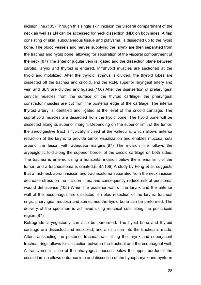
28
incision line.(105) Through this single skin incision the visceral compartment of the
neck as well as LN can be accessed for neck dissection (ND) on both sides. A flap
consisting of skin, subcutaneous tissue and platysma, is dissected up to the hyoid
bone. The blood vessels and nerves supplying the larynx are then separated from
the trachea and hyoid bone, allowing for separation of the visceral compartment of
the neck.(87) The anterior jugular vein is ligated and the dissection plane between
carotid, larynx and thyroid is entered. Infrahyoid muscles are sectioned at the
hyoid and mobilized. After the thyroid isthmus is divided, the thyroid lobes are
dissected off the trachea and cricoid, and the RLN, superior laryngeal artery and
vein and SLN are divided and ligated.(106) After the disinsertion of prelaryngeal
cervical muscles from the surface of the thyroid cartilage, the pharyngeal
constrictor muscles are cut from the posterior edge of the cartilage. The inferior
thyroid artery is identified and ligated at the level of the cricoid cartilage. The
suprahyoid muscles are dissected from the hyoid bone. The hyoid bone will be
dissected along its superior margin. Depending on the superior limit of the tumor,
the aerodigestive tract is typically incised at the valleculla, which allows anterior
retraction of the larynx to provide tumor visualization and enables mucosal cuts
around the lesion with adequate margins.(87) The incision line follows the
aryepiglottic fold along the superior border of the cricoid cartilage on both sides.
The trachea is entered using a horizontal incision below the inferior limit of the
tumor, and a tracheostoma is created.(5,87,106) A study by Feng et al. suggests
that a mid-neck apron incision and tracheostoma separated from the neck incision
decrease stress on the incision lines, and consequently reduce risk of peristomal
wound dehiscence.(105) When the posterior wall of the larynx and the anterior
wall of the oesophagus are dissected, en bloc resection of the larynx, tracheal
rings, pharyngeal mucosa and sometimes the hyoid bone can be performed. The
delivery of the specimen is achieved using mucosal cuts along the postcricoid
region.(87)
Retrograde laryngectomy can also be performed. The hyoid bone and thyroid
cartilage are dissected and mobilized, and an incision into the trachea is made.
After transsecting the posterior tracheal wall, lifting the larynx and superjacent
tracheal rings allows for dissection between the tracheal and the esophageal wall.
A transverse incision of the pharyngeal mucosa below the upper border of the
cricoid lamina allows entrance into and dissection of the hypopharynx and pyriform
29
sinuses. Upward traction on the epiglottis allows for inspection of the pharynx to
ensure sufficient tumor resection. The base of the tongue is cut and sutured and
en bloc resection of the larynx, trachea and pharyngeal mucosa occurs.(5,106)
Figure 4 shows the operating field after resection of the larynx.
Figure 4: Operating field of a TL after en bloc resection of the larynx, trachea and pharyngeal
mucosa. Photo used with permission from the Department of Otorhinolaryngology, University
Hospital Graz.
A pharyngoesophageal myotomy is typically performed to prevent hypertonicity
and optimize speech and swallowing. A tracheoesophageal voice prosthesis can
be placed by creating a fistula about 5-10mm below the upper margin of the
tracheostoma. Then, pharyngeal mucosal sutures are placed with care as tension
sutures can lead to stenosis, and too loose sutures can lead to
pharyngocutaneous fistula. A nasogastric feeding tube should be inserted and
stitched to the nasal septum to allow feeding until oral nourishment can be
resumed.(5,106)
Concurrent neck dissection is performed in case of clinically positive LN or high
possibility of nodal metastasis in laryngeal cancers. Selective ND of levels II-IV is
likely adequate in advanced disease for clinically N0 necks, while modified or
30
radical ND is performed in clinically positive necks.(5,87) As hypopharyngeal
tumors have a high risk for LN metastases, even in clinically N0 necks selective
ND is appropriate. In most cases, levels II-IV should be resected. Level VI LN
should be included for tumors that show esophageal extension or invasion of the
apex of the pyriform sinus.(89)
Hypopharyngeal cancers can be resected by TL with partial pharyngectomy. The
pharyngeal defect can usually be closed by primary closure, but as the
submucosal spread of hypopharyngeal cancers requires wide margins, a pedicle
flap or free-tissue transfer can become necessary. This can also be required in
salvage total laryngectomy (STL) after failure to achieve disease control using
RCT. Single stage closure can then be achieved by using a pectoralis major or a
radial forearm myocutaneous flap.(89)
TL is one of the most feared surgical procedures by patients, as swallow function
and speech as well as body image are significantly influenced by the procedure.
QOL is profoundly and adversely affected if treatment is associated with
dysfunction and disfigurement, making body image reintegration postsurgically
critical to ensure subsequent QOL.(87) TL safely prevents aspiration, but also
significantly alters swallow function. Resection of the larynx leads to weaker bolus
propulsion and higher resistance to bolus clearance. The pharyngeal phase of
swallowing is prolonged. Postsurgically, swallowing soft foods poses less
problems, but patients may progress to a general diet.(104)
As the treatment of cancers of the larynx has moved away from primary TL
towards organ preserving modalities, the use of initial RCT has become more
popular in recent years. In case of subsequent disease recurrence, STL can
become necessary.(107,108) STL constitutes a final curative treatment strategy,
but can be technically complex and can significantly impact patient morbidity.(92)
1.4.8. Neck dissection
Radical neck dissection (RND) refers to the resection of all ipsilateral cervical LN
groups from Levels I through V. The spinal accessory nerve, internal jugular vein,
and SCM muscle are also removed. Modified radical neck dissection (MRND)
describes RND where one or more non-lymphatic structures are spared from
resection. Selective neck dissection (SND) refers to a cervical lymphadenectomy
in which one or more of the LN groups that are routinely removed in RND are
31
preserved. The LN groups resected are based on the patterns of metastasis that
are predictable according to the primary site of tumor (Table 5).(15,26,91)
Table 5: Recommended neck levels to be dissected based on primary site. Table from Paleri et al.(26)
Supraglottis
IIa–III; IIb and IV can be spared. Contralateral SND not indicated for lateralised tumors
Glottis IIa-III, IIb can be spared. Include IV for T3 and T4 primaries
Subglottis II-IV, VI Hypopharynx II-IV
In a clinically N0 neck the choice for elective treatment lies between MRND and
SND. SND seems to be as effective as MRND for most primary sites with
decreased morbidity. In laryngeal and hypopharyngeal tumors, the procedure of
choice is SND of levels II–IV.(26) This refers to the resection of the upper, middle
and the lower jugular LN.(91) Incidence of metastasis is less than two percent at
level IIb LN. Therefore, level IIb resection may be excluded for the majority of
patients, and the designated procedure would be SND of levels IIa, III, IV.(26,91)
Patients with central tumors or tumors involving both sides of the larynx require
bilateral ND.(109) This is especially true for supra- and subglottic tumors.(91)
Tumors of only one side of the larynx are more likely to show lymphatic spread to
the ipsilateral LN, but occult metastatic spread may be found on contralateral LN.
This suggests that elective bilateral ND should always be performed, even if this
increases the number of unnecessary ND.(109)
Elective neck irradiation has been shown to be as effective as elective ND in
controlling subclinical regional disease.(26) Elective ND during salvage
laryngectomy is beneficial in patients with advanced local disease at recurrence
for both disease-free and overall survival.(110)
1.4.9. Complications of total laryngectomy
The overall mortality rate for TL was 0.5% in a 2005 study by Ganly et al.(111),
and 40% of all patients developed postoperative complications after TL. Local
complications occurred most frequently, in 28% of all patients.
TL in the era of the principle of organ preservation has increasingly taken the role
of salvage surgery. Primary RCT has been shown to be an independent predictor
of both local wound complication and PCF. STL following RCT was more
frequently associated with any postoperative complication than PTL due to wound
32
healing difficulties.(92,111) The complication rates following STL are not
consistently reported in the literature. The most common complication of STL is
development of a pharyngocutaneous fistula (PCF), which was found to occur in
28.9% of all patients who undergo STL in a review by Hasan et al.(112) The
overall complication rate was 67.5%, but complications such as wound infection,
dehiscence and bleeding are likely under-reported.(112) Ganly et al.(111) found
that in the group of STL after RCT, a greater number of patients developed local
wound and fistula complications compared to the PTL group. In STL patients, the
most common postoperative complications were local wound healing problems
and the development of PCF.(111)
1.4.9.1. Early Postoperative Complications
The most common early postoperative complications include wound infection,
pharyngeal stenosis, bleeding and dysphagia. Overall complication rate was
67.5% according to the review of Hasan et al.(112)
1.4.9.2. Pharyngocutaneous Fistula
Development of a pharyngocutaneous fistula (PCF) is a common complication in
the early postoperative period after TL. Many predisposing factors to fistula
formation have been identified, including preoperative RT, surgical technique,
concurrent ND, tumor stage, preoperative tracheostomy, early oral feeding,
presence of systemic diseases, and nutritional status.(113)
Pooled incidence of PCF was 28.9% in the meta-analysis of 50 studies by Hasan
et al.(112), and rates varied in individual series between 5.6-73%. In the study by
Ganly et al.(111), PCF occurred in 17% patients. Multivariate analysis showed that
primary RT and RCT were independent predictors of PCF, with no significant
difference in PCF incidence between patients who received RCT compared to RT
only.(111,112) There seems to be no association of ND and elevated incidence of
PCF.(112) Fung et al.(114) showed that free vascularized tissue reinforcement of
primary pharyngeal closure in STL could help prevent major wound complications
by but did not reduce the overall rate of PCF development.
33
1.4.9.3. Pharyngeal stenosis
A typical late complication following TL is stenosis of the neopharynx due to scar
tissue, resulting in difficulty clearing food and liquid to the esophagus. Nasal
regurgitation of liquids can occur in severe cases.(104) Pharyngeal stenosis was
reported in 14.3% of patients in the study by Hasan et al.(112) Stricture formation
was typically located around the pharyngeal anastomosis or the thoracic
esophagus. Flap reconstruction shows an increased risk of stenosis compared to
primary pharyngeal closure.(112) Relief can be provided by serial dilatation, which
can be required to be performed periodically for the rest of the affected patients'
lives. Pharyngoesophageal myotomy is another option to prevent stenosis.(104)
1.4.10. Voice rehabilitation Achieving alaryngeal voice is the most challenging of post-TL rehabilitations, as
the ability to speak and communicate vocally is a unique human characteristic that
is fundamental to many aspects of daily life.(104,115) Nowadays, three options to
achieve voice restoration are in common use: esophageal speech (ES), tracheo-
esophageal speech (TES) using a voice prosthesis and use of an electorlarynx
(ELS). All three methods have potential benefits, but TES is usually described as
the gold standard.(115) Articulation is only affected if the surgery involves base of
tongue resection, which can lead to reduced tongue range of motion, resulting in
distorted articulation.(104) Both adjuvant RT and definite RT which then requires
STL have a significantly negative effect on phonatory and swallowing outcome.
This should be taken into consideration for patients with advanced laryngeal
cancer when deciding upon laryngectomy or organ preservation treatments.(116)
To achieve ES speech, vibration of the pharyngoesophageal segment is used to
create sound, which travels through the pharynx and oral cavity and can be
articulated into speech. Patients must learn to swallow air and to use a process
called glossopress, in which the tongue is used as a pumping mechanism. This
can make this method of voice rehabilitation difficult to learn. The ES voice sounds
natural, but is often lower in pitch.(87,104)
The introduction of TES in 1979 made it possible for many patients to rapidly
recover high quality speech. This facilitates quick rehabilitation of TL patients as it
allows them to return to normal activities of life and work and subsequently to
34
maintain satisfactory QOL.(87,117) Shunting air from the lungs, TES also utilizes
vibration of the pharyngoesophageal segment to create a voice. A surgeon
performs a tracheoesophageal puncture (TEP) and a voice prosthesis (VP) is
implanted. This can happen primarily or secondarily to TL.(115) The VP is a one-
way valve, which lets air travel anterior-posteriorly, but stops food or liquid to travel
posterior-anteriorly. The VP does require periodic replacement and leakage
around the prosthesis is possible. To achieve efficient stoma occlusion and
exhalation strategies, speech therapy is usually required following placement of
the VP.(104) TES has been shown to confer significant benefit in terms of self-
reported voice outcome in a study by Robertson et al.(116)
The electrolarynx is a device that is held against the neck or cheek. Sound is
created by a piston rapidly striking a fixed diaphragm, which travels through the
pharynx and oral cavity and is articulated into speech. Pitch and loudness can be
adjusted. ELS is a very effective mode of communication, but it does require
speech and language therapy. The voice created has a mechanical sound, which
puts off some patients of using ELS.(104)
2. Purpose Total laryngectomy (TL) is the operation in which the larynx is completely
removed, which results in a total and permanent separation of respiratory and
digestive tract and consequent loss of voice and sense of smell. For much of the
20th century, TL was widely regarded as the gold standard of treatment for
advanced laryngeal and hypopharyngeal cancers. In the last years, organ
preservation has become the principle of therapeutic approaches for advanced
lesions that would previously have been treated with TL.(5,118) This was
encouraged by the results of studies that found that larynx preserving treatment
using concurrent radiotherapy and chemotherapy did not reduce survival in
patients with even advanced cancer of the larynx and hypopharynx.(93,94) As
there have only been few reports on the clinical outcome following TL, the purpose
of the present study was to analyse complication, survival and recurrence rates
after TL and to gain insight into postoperative voice rehabilitation.
35
3. Materials and methods The study population consisted of patients who underwent total laryngectomy
between 01.01.2002 and 15.01.2018 at the Department of Otorhinolaryngology, at
the University Hospital Graz. The patients' clinical data and medical history were
collected from medical files in the computerized database MEDOCS. Ethical
approval was obtained from the Ethics comitee at the University Hospital Graz.
The Institute for Medical Informatics, Statistics and Documentation at the Medical
University of Graz provided a table of patients who had received the medical
service "total laryngectomy" at the Department of Otorhinolaryngology in the last
16 years. Of 106 patients who were identified, 103 could be included in the study.
The following parameter were assessed:
Age
Sex (female, male)
Common comorbidities
Date of diagnosis
Location of tumor
T-, N-, M-stage
Pathological type
Grade
First-line therapeutic approach
Positive LN metastasis
Surgical technique
Median postoperative stay
Occurrence of early and late complications
Recurrence
Overall suvival (OS)
Disease-free survival (DFS)
Methods of alaryngeal voice rehabilitation
Date of laryngeal biopsy was considered as the time of diagnosis. The location of
the tumor was classified according to the patient's pathology report into three main
categories, laryngeal, hypopharyngeal and other location (including oral, cervical
esophagus and tracheal). Subcategories for laryngeal cancer were defined as
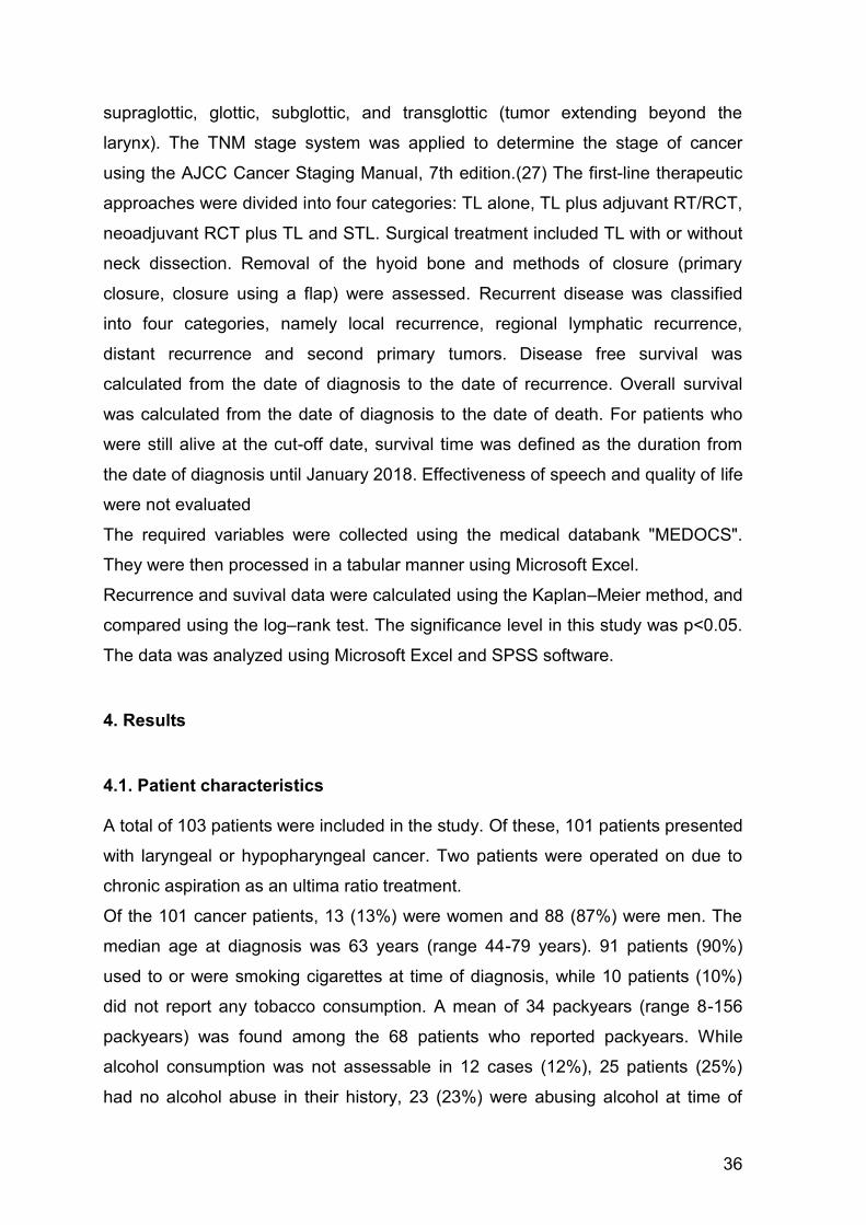
36
supraglottic, glottic, subglottic, and transglottic (tumor extending beyond the
larynx). The TNM stage system was applied to determine the stage of cancer
using the AJCC Cancer Staging Manual, 7th edition.(27) The first-line therapeutic
approaches were divided into four categories: TL alone, TL plus adjuvant RT/RCT,
neoadjuvant RCT plus TL and STL. Surgical treatment included TL with or without
neck dissection. Removal of the hyoid bone and methods of closure (primary
closure, closure using a flap) were assessed. Recurrent disease was classified
into four categories, namely local recurrence, regional lymphatic recurrence,
distant recurrence and second primary tumors. Disease free survival was
calculated from the date of diagnosis to the date of recurrence. Overall survival
was calculated from the date of diagnosis to the date of death. For patients who
were still alive at the cut-off date, survival time was defined as the duration from
the date of diagnosis until January 2018. Effectiveness of speech and quality of life
were not evaluated
The required variables were collected using the medical databank "MEDOCS".
They were then processed in a tabular manner using Microsoft Excel.
Recurrence and suvival data were calculated using the Kaplan–Meier method, and
compared using the log–rank test. The significance level in this study was p<0.05.
The data was analyzed using Microsoft Excel and SPSS software.
4. Results
4.1. Patient characteristics A total of 103 patients were included in the study. Of these, 101 patients presented
with laryngeal or hypopharyngeal cancer. Two patients were operated on due to
chronic aspiration as an ultima ratio treatment.
Of the 101 cancer patients, 13 (13%) were women and 88 (87%) were men. The
median age at diagnosis was 63 years (range 44-79 years). 91 patients (90%)
used to or were smoking cigarettes at time of diagnosis, while 10 patients (10%)
did not report any tobacco consumption. A mean of 34 packyears (range 8-156
packyears) was found among the 68 patients who reported packyears. While
alcohol consumption was not assessable in 12 cases (12%), 25 patients (25%)
had no alcohol abuse in their history, 23 (23%) were abusing alcohol at time of
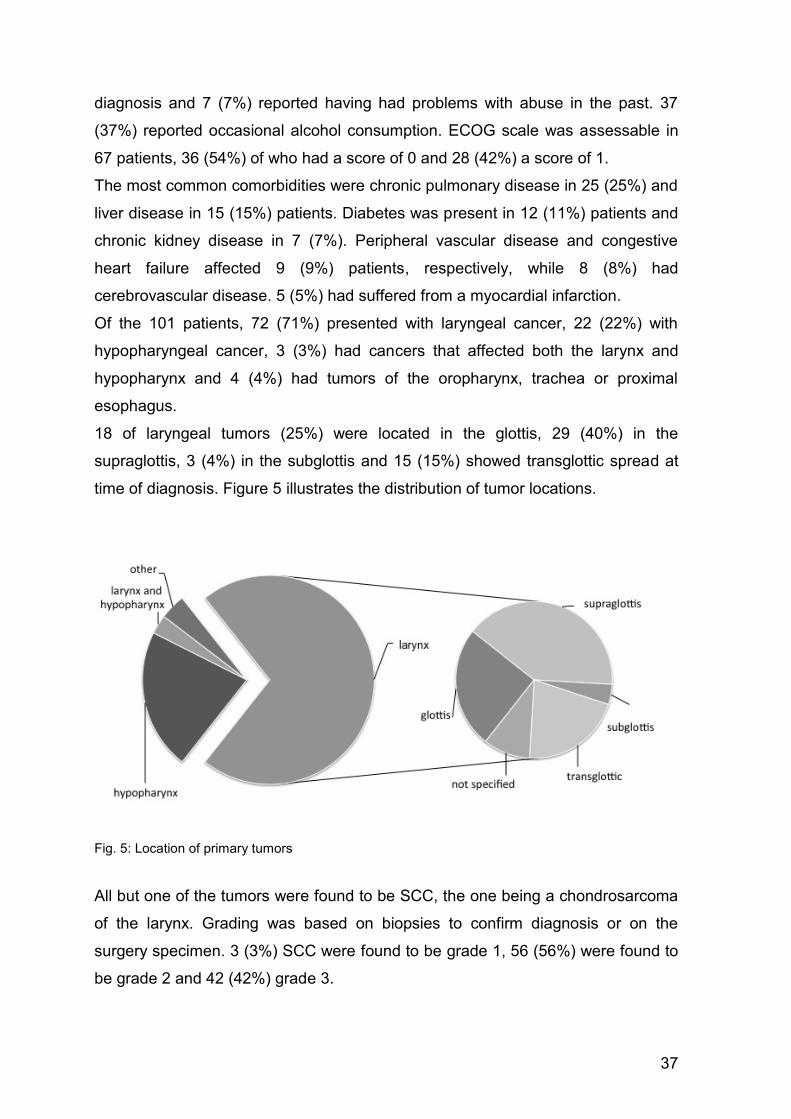
37
diagnosis and 7 (7%) reported having had problems with abuse in the past. 37
(37%) reported occasional alcohol consumption. ECOG scale was assessable in
67 patients, 36 (54%) of who had a score of 0 and 28 (42%) a score of 1.
The most common comorbidities were chronic pulmonary disease in 25 (25%) and
liver disease in 15 (15%) patients. Diabetes was present in 12 (11%) patients and
chronic kidney disease in 7 (7%). Peripheral vascular disease and congestive
heart failure affected 9 (9%) patients, respectively, while 8 (8%) had
cerebrovascular disease. 5 (5%) had suffered from a myocardial infarction.
Of the 101 patients, 72 (71%) presented with laryngeal cancer, 22 (22%) with
hypopharyngeal cancer, 3 (3%) had cancers that affected both the larynx and
hypopharynx and 4 (4%) had tumors of the oropharynx, trachea or proximal
esophagus.
18 of laryngeal tumors (25%) were located in the glottis, 29 (40%) in the
supraglottis, 3 (4%) in the subglottis and 15 (15%) showed transglottic spread at
time of diagnosis. Figure 5 illustrates the distribution of tumor locations.
Fig. 5: Location of primary tumors
All but one of the tumors were found to be SCC, the one being a chondrosarcoma
of the larynx. Grading was based on biopsies to confirm diagnosis or on the
surgery specimen. 3 (3%) SCC were found to be grade 1, 56 (56%) were found to
be grade 2 and 42 (42%) grade 3.
38
The 7th edition AJCC staging system(27) was used to determine cancer stage
according to TNM status. Of all 101 cancer patients, 1 (1%) presented at stage I, 9
(9%) at stage II, 26 (26%) at stage III and 49 (49%) at stage IVa. 2 (2%) were
found to be at stage IVb, 4 (4%) at stage IVc, 6 (6%) were classified as Mx, 1 (1%)
as Nx and 3 (3%) patients could not be classified. An overview of the
characteristics of the study population is presented in Table 6.
Table 6: Characteristics of study participants. SCC= squamos cell carcinoma, DM= diabetes
mellitus, CHF=congestive heart failure, OS=overall survival, DFS= disease-free survival Age at diagnosis (year) 63 (44-79)
Sex (n=101)
Male 88
Female 13
Primary site
Larynx 72
Glottis 18 Supraglottis 29
Subglottis 3 Transglottic 15
Hypopharynx 22
Other 7
Risk Factors
Smoker 91 Packyears 38 (8-156)
Non-smoker 10 Alcohol consumption 67
Stage
II 9 IVb 2
III 26 IVc 4
IVa 49 Mx 6
Histology
SCC 100 Chondrosarcoma 1
Comorbidities
Pulmonary disease 25 DM 12
Myocardial infarct 5 CHF 9
Peripheral vascular
disease
9 Cerebrovascular
disease
8
Liver disease 15 Renal disease 7
ECOG scale
0 36 1 28
2 1 3 2
Survival rates
1-year OS (%) 83 1-year DFS (%) 82
3-year OS (%) 64 3-year DFS (%) 64
4.2. Treatment regimen First-line treatment regimen included TL and adjuvant RT in 35 patients (35%). TL
and adjuvant RCT was implemented in 29 patients (29%), while 21 patients (21%)
underwent only TL. 3 patients (3%) were treated with neoadjuvant RCT. On 13
patients (13%), STL following primary RCT or induction CT and RT was
39
performed. Prior LP surgery had been performed on 14 patients (14%). The
incidence of first-line therapeutic approaches is illustrated in Figure 6.
Figure 6: Primary treatment regimen of all 101 cancer patients. TL= total laryngectomy. RT=
radiotherapy. RCT= radiochemotherapy. ICT= induction chemotherapy.
4.3. Surgical techniques In 68 patients, surgical reports specified whether the hyoid bone had been
removed during the procedure. Of these, removals of the hyoid bone were
performed in 21 cases (31%), while it was left in situ in 47 cases (69%). In 6 cases
(6%) of all 103 TL, surgical closure was performed using a flap - a pectoralis flap
in 5 cases, and a radialis flap in 1 case. Neck dissection was performed in 88 of
the 101 cancer patients (87%). In the majority of the cases, levels II to IV were
resected. Upon pathohistological examination, 46 of these cases were shown to
have positive LN metastasis. Median postoperative stay was 19 days (range 11-88
days). Implantation of a PEG-probe was necessary in 38 patients (37%), while a
jejunal probe was implanted in 9 patients (9%). PEG-probe implantation was
performed before surgery or before RT in 22 of these patients, due to dysphagia
as a result of complications such as a PCF or stenosis in 10 patients and in a
palliative setting in 2 patients.
4.4. Postoperative complications 24 patients (23%) experienced early postoperative complications, the most
common being wound infection (12 patients), bleeding (9 patients), lymphatic
TL+ adjuv. RT; 35
TL+ adjuv. RCT; 30
TL; 20
neodjuv RCT+TL; 3
prim. RCT/ICT; 13
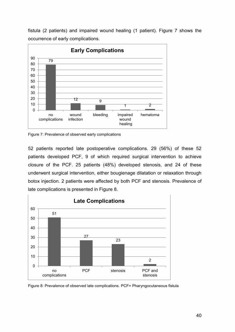
40
fistula (2 patients) and impaired wound healing (1 patient). Figure 7 shows the
occurrence of early complications.
Figure 7: Prevalence of observed early complications
52 patients reported late postoperative complications. 29 (56%) of these 52
patients developed PCF, 9 of which required surgical intervention to achieve
closure of the PCF. 25 patients (48%) developed stenosis, and 24 of these
underwent surgical intervention, either bougienage dilatation or relaxation through
botox injection. 2 patients were affected by both PCF and stenosis. Prevalence of
late complications is presented in Figure 8.
Figure 8: Prevalence of observed late complications. PCF= Pharyngocutaneous fistula
79
12 9
1 2
0
10
20
30
40
50
60
70
80
90
nocomplications
woundinfection
bleeding impairedwound
healing
hematoma
Early Complications
51
2723
2
0
10
20
30
40
50
60
nocomplications
PCF stenosis PCF andstenosis
Late Complications
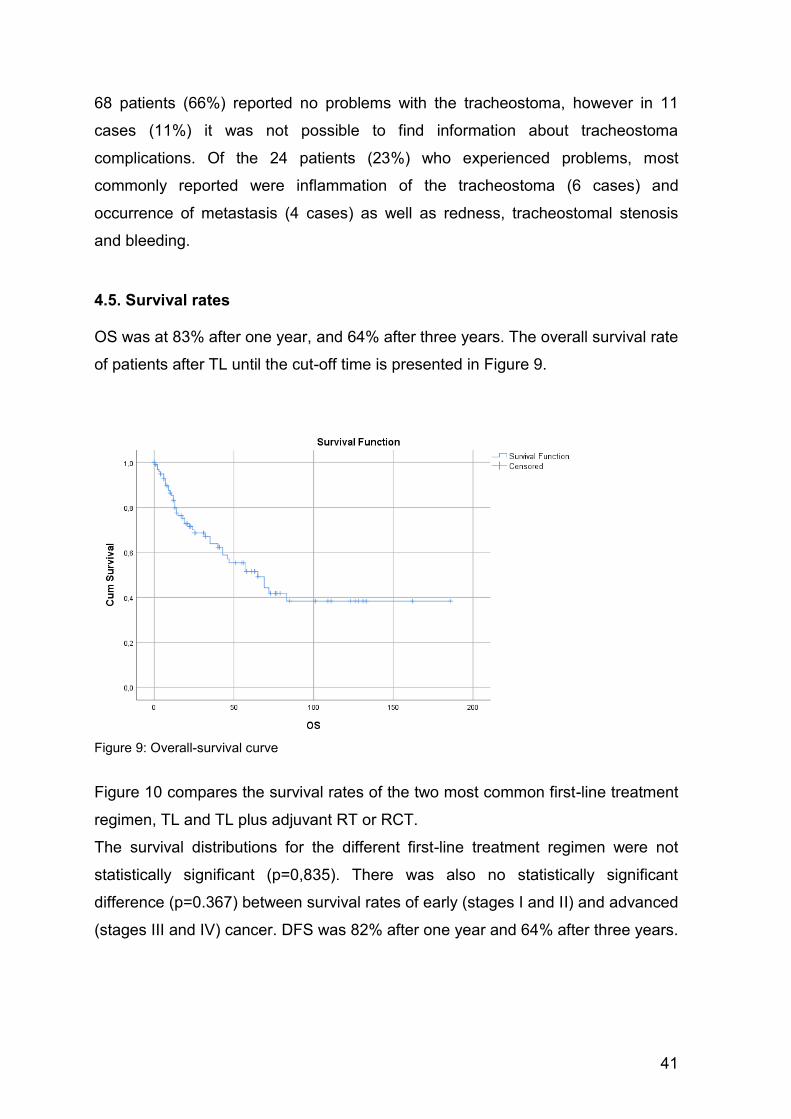
41
68 patients (66%) reported no problems with the tracheostoma, however in 11
cases (11%) it was not possible to find information about tracheostoma
complications. Of the 24 patients (23%) who experienced problems, most
commonly reported were inflammation of the tracheostoma (6 cases) and
occurrence of metastasis (4 cases) as well as redness, tracheostomal stenosis
and bleeding.
4.5. Survival rates OS was at 83% after one year, and 64% after three years. The overall survival rate
of patients after TL until the cut-off time is presented in Figure 9.
Figure 9: Overall-survival curve
Figure 10 compares the survival rates of the two most common first-line treatment
regimen, TL and TL plus adjuvant RT or RCT.
The survival distributions for the different first-line treatment regimen were not
statistically significant (p=0,835). There was also no statistically significant
difference (p=0.367) between survival rates of early (stages I and II) and advanced
(stages III and IV) cancer. DFS was 82% after one year and 64% after three years.
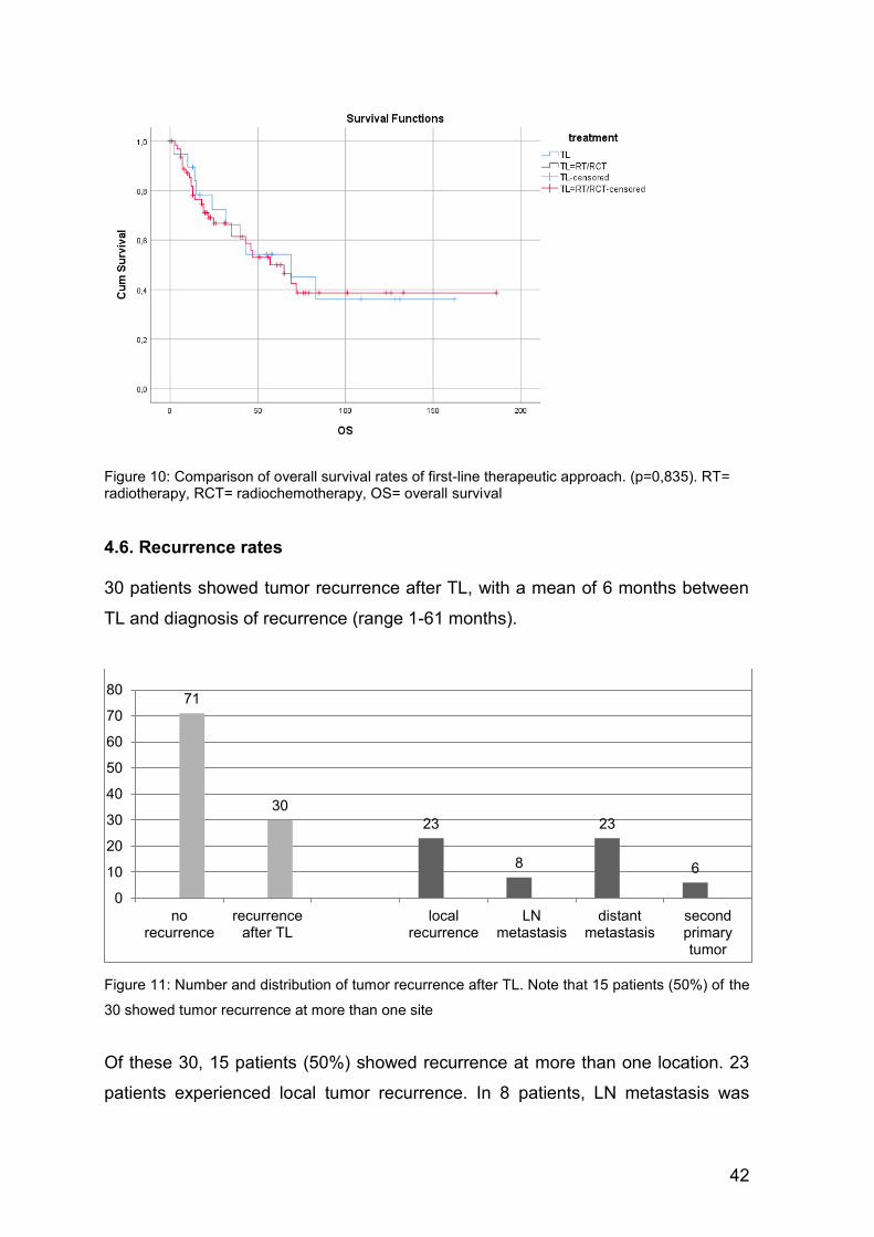
42
Figure 10: Comparison of overall survival rates of first-line therapeutic approach. (p=0,835). RT= radiotherapy, RCT= radiochemotherapy, OS= overall survival
4.6. Recurrence rates 30 patients showed tumor recurrence after TL, with a mean of 6 months between
TL and diagnosis of recurrence (range 1-61 months).
Figure 11: Number and distribution of tumor recurrence after TL. Note that 15 patients (50%) of the
30 showed tumor recurrence at more than one site
Of these 30, 15 patients (50%) showed recurrence at more than one location. 23
patients experienced local tumor recurrence. In 8 patients, LN metastasis was
23
8
23
6
71
30
0
10
20
30
40
50
60
70
80
norecurrence
recurrenceafter TL
localrecurrence
LNmetastasis
distantmetastasis
secondprimarytumor
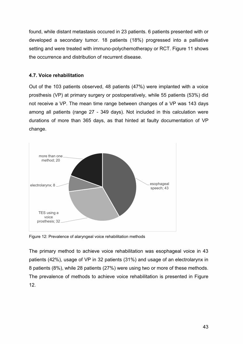
43
found, while distant metastasis occured in 23 patients. 6 patients presented with or
developed a secondary tumor. 18 patients (18%) progressed into a palliative
setting and were treated with immuno-polychemotherapy or RCT. Figure 11 shows
the occurrence and distribution of recurrent disease.
4.7. Voice rehabilitation Out of the 103 patients observed, 48 patients (47%) were implanted with a voice
prosthesis (VP) at primary surgery or postoperatively, while 55 patients (53%) did
not receive a VP. The mean time range between changes of a VP was 143 days
among all patients (range 27 - 349 days). Not included in this calculation were
durations of more than 365 days, as that hinted at faulty documentation of VP
change.
Figure 12: Prevalence of alaryngeal voice rehabilitation methods
The primary method to achieve voice rehabilitation was esophageal voice in 43
patients (42%), usage of VP in 32 patients (31%) and usage of an electrolarynx in
8 patients (8%), while 28 patients (27%) were using two or more of these methods.
The prevalence of methods to achieve voice rehabilitation is presented in Figure
12.
esophageal speech; 43
TES using a voice
prosthesis; 32
electrolarynx; 8
more than one method; 20
44
5. Discussion
The aim of the study was to examine the clinical outcome of patients who were
treated with TL. Of the 103 patients who fit the study criteria, 101 patients
presented with laryngeal or hypopharyngeal cancer. Two patients were operated
on due to chronic aspiration as an ultima ratio treatment.
The typical demographic of laryngeal and hypopharyngeal cancer patients are
males over 65 who have a history of tobacco and or alcohol
consumption.(43,45,58) This fact was represented in our study population of 101
patients with laryngeal or hypopharyngeal cancer. Median age at diagnosis was 63
years. 87% of the patients were males, and 13% were females. The discrepancy
in incidence in men and women can be expected to shrink as women nowadays
smoke and drink alcohol at the same rate as men, and as HPV as an etiologic
factor is on the rise. We were not able to conclusively assess prevalence of HPV-
positive cancers, as HPV status was not recorded in a large number of patients.
90% of patients reported tobacco consumption with a mean of 34 packyears, and
67% disclosed alcohol consumption, ranging from occasional consumption to
abuse. Accordingly, the most common comorbidities were chronic pulmonary
disease in 25% and liver disease in 15% of patients. Diabetes, chronic kidney
disease and peripheral and cerebro-vascular disease were also common.
Laryngeal cancer was most common, affecting 71% of the patients.
Hypopharyngeal cancer is a less common entity, and occurred in 22% of patients.
When classifying the tumors according to the 7th Edition AJCC Cancer Staging
Manual(27) and compartmentalizing stage I and II as early disease and stage III
and IV as advanced disease, only 10% of patients presented with early disease.
81% presented with advanced disease tumors at diagnosis.
Neck dissection was performed in 87% of patients, and 52% of these were shown
to have positive LN metastasis at histological examination.
Median postoperative stay was 19 days (range 11-88 days). Hall et al.(118) also
reported a median of 19 days postoperative stay in their 2003 study.
Overall, the incidence of any postoperative complication was at 58%. Early
postoperative complications occurred in 23% of all patients, most commonly
45
wound infection, bleeding and lymphatic fistula. 51% of all patients reported late
postoperative complications. The 2017 systematic review of complications after
STL by Hasan et al.(112) found overall complication rates of 67.5%. It is difficult to
compare overall complication rates because many studies focus on a specific
complication, such as occurrence of PCF or pharyngeal stenosis, and do not
report incidence of overall complications.
Incidence of development of PCF was 26% of all patients, constituting 56% of late
postoperative complications, 31% of which required surgical closure. Galli et
al.(113) found a 16% rate of PCF development following TL in their study. The
incidence of PCF was 17.7% in the study by Hall et al.(118) Sayles and
Grant(119) found the incidence of PCF to be 14.3% after primary TL, 22.8% after
previous RT and 34.1% after previous RCT in their 2014 review. We did not
assess whether treatment methods influenced incidence of complications. Since
multiple studies showed an association of PCF to previous RT or RCT, this would
be an interesting aspect to examine in further studies. Incidence of pharyngeal
stenosis was 22%, occurring in 48% of late complication cases. 96% of these had
to undergo either bougienage dilatation or relaxation through Botox injection to
regain oral feeding. Hasan et al.(112) found a 14.3% incidence of pharyngeal
stenosis or stricture with a range of 2.3-43.9% in their review.
Comparing survival rates among different studies is complicated by different
methods of recruitment, study design, and assessment of the patients in different
studies. We found OS to be at 83% after one year, and 64% after three years. The
cutoff time for the study was January 2018, which means time of observation
differed massively between patients. There was no statistically significant
difference between early and advanced stage disease, however only a small
fraction of patients in our study population presented with early stage disease.
Daneshi et al.(120) reported the 1-year, 3-year and 5-year year survival rates in
patients with laryngeal carcinoma were 81%, 62% and 53%, respectively. Hall et
al.(118) described a 67% 5-year survival for patients with laryngeal cancer and a
significantly worse 37% 5-year survival for hypopharyngeal cancer. They also
found a correlation of survival with clinical and pathological stage of laryngeal
cancer, while prognosis for hypopharyngeal cancer was poor irrespective of stage.
A study by Ho et al.(121) found that stage influenced outcome, reporting a decline
in survival from 74% to 14% for stages I to IV. Wolf et al.(93) reported a 2-year
46
survival rate of 68% among patients with advanced cancers of the larynx who
were treated with surgery and RT. Foote et al.(122) reported a 5-year survival rate
of 74% among patients with T3 laryngeal cancer treated by surgery only.
Laryngeal and hypopharyngeal cancer constitute a diverse disease, and preferred
therapeutic management is influenced by anatomic subsite, stage of disease, and
patient preference. Organ preserving treatments have been persistently popular
for T1 to T3 disease following the publication of RTOG 91-11(94) and the VA
Laryngeal Cancer(93) studies, but TL is still regarded as standard of care for
individuals with more locally advanced disease. Recent analyses of care patterns
show a trend of LP protocols for patients with T4 disease.(95–97) Many studies
were conducted on the impact of treatment methods on patients' survival, but did
not reach conclusive results, possibly due to differences in study design and
population. Timmermans et al.(123) and Karlsson et al.(124) found no significant
difference in survival between surgical and organ-preserving treatment modalities.
However, Hoffman et al. showed that the survival rate of patients with advanced
laryngeal cancer declined between 1985 to 1996, following the increase in
nonsurgical definite treatment.(96) Carvalho et al. also found a declining 5-year
survival rate. Although the causal factors for this decline were not specified,
evidence suggests that the decline of TL and a consequent increase in organ
preserving treatments occurred during the same time period.(125) A study by
Megwalu and Sikora(126) showed that patients who received surgical treatment
had significantly better survival compared to non-surgically treated patients,
regardless of gender or stage. Chen and Halpern(97) reported higher survival
rates in patients with stage IV laryngeal cancer that had been treated with TL
compared to other methods of treatment. The study by Grover et al.(95) showed a
significant association of TL and survival rate in patients with T4a disease. This
suggests that surgical treatment should remain the standard of care for T4a
cancers as opposed to organ preserving methods, and highlights the risk of
broadly applying results from studies that included a specific patient population,
but were not limited to it.(95)
It was not a purpose of this study to evaluate speech rehabilitation or QOL, but
reports on outcome measures that are not strictly clinical are increasingly
47
appreciated. QOL is an outcome measure whose results sometimes defy clinical
impressions and biases. While TL results in greater measurable dysfunction,
especially of speech, Morton(127) found no major QOL differences in overall QOL
between patients treated with TL or definite RT in his 1997 study. Similarly,
Deleyiannis et al.(128) reported no significant difference in pre- and post-
laryngectomy QOL scores. Though the loss of voice is disabling, the functional
limitations caused by TL may not necessarily translate into a worse overall
QOL.(128)
As TL represents a challenging change in physiology for all patients, proper
rehabilitation is essential to restore functionality and QOL, which relies on
continuous speech and language counselling and therapy. A pre-treatment
assessment of swallowing has been found to significantly improve post-
laryngectomy swallow function and should be implemented in all HNC
patients.(87,129) Continued speech and language therapy is crucial to regain and
optimize voice and swallow function after TL. Krishnan and Maclean also found
dysphagia to be common following TL, compounded by the use of adjuvant RT
and CT.(130) Arenaz Búa et al.(131) reported problems with swallowing in 89% of
patients, and moderate-to-severe voice handicap in 66%. 62% of patients required
additional therapeutic interventions to help with problems with voice and
swallowing.(131) Van Sluis et al.(132) found that all speaker groups after TL report
a degree of voice handicap. Attendance of rehabilitation was associated with
better functional outcome in speech intelligibility in the study by Singer et al.(133);
they also found considerable improvements in intelligibility between 6 months and
1 year after TL.
Out of the 103 patients observed, 47% underwent TEP and placement of a VP at
primary surgery or secondarily, while 53% of patients did not receive a VP. The
mean time range between replacements of a VP was 143 days among all patients
(range 27 - 349 days). Mayo-Yáñez et al. found a mean time range between VP
replacements to be between 106 and 124 days in their study.(134)
It is important to note the challenges faced in assessing mean time range between
VP replacements, which has not been documented in a uniform manner. Several
patients received speech and language therapy and medical attendance at other

48
medical centers, which made a thorough documentation of the VP changes
impossible. These patients, as well as cases reporting durations of more than 365
days, were excluded from calculations.
The primary method to achieve voice rehabilitation was ES in 42% of patients,
TES in 31% and ELS in 8%, while 27% of patients were using two or more
methods. When comparing international figures, TES is the most common form of
voice rehabilitation and considered gold standard in developed countries.(5)
Krishnan and Maclean(130) and Xi et al.(135) described TES as the primary
alaryngeal voice rehabilitation method in their respective studies. Singer et al.(133)
reported the best results regarding subjective and objective speech intelligibility in
patients using TES compared to ES and ELS. Van Sluis et al.(132) reported
significant better outcomes for TES compared to ES for acoustic parameters and
maximum time and intensity of phonation, but no significant difference in patient-
reported outcomes in their review.
We did not investigate intelligibility or quality of speech after TL, but looked at
complications with and mean duration of VP, and found that many patients
reported problems, most importantly leakage around the VP, and consequent VP
replacements. Occurrence of complications and high replacement frequency can
result in numerous hospital visits, which negatively influence QOL while also
constituting a financial burden on the health care system. Furthermore, it is
important to consider that TEP is an interventional procedure with a risk of
complications. Overall complication rate related to TEP and the VP was 42.6%
during a mean follow-up of 15.3 months in a study by Imre et al.(136) Mostly minor
complications were reported, the most common being TEP enlargement and
leakage around prosthesis, which was also the most common cause of complete
closure of TEP. Of three observed major complications, two were life-threatening;
a mediastinitis and paraesophageal abscess.(136)
ES was the main form of vocal rehabilitation in our study population. While this is
the most difficult to learn and requires practice and extensive vocal rehabilitation
training, very good results in intelligibility can be achieved without the risk of
complications of TES.
Limitations of the study were due to its retrospective design and method of data
collection, as database analysis has inherent limitations. This includes the
49
potential for miscoding of variables and selection bias that is not accounted for by
variables available in the database. Many variables were not uniformly
documented, which impeded data collection significantly.
To make a statement about which treatment of advanced cancer of the larynx and
hypopharynx yields better results at the Department of Otorhinolaryngology at the
University Hospital Graz, the clinical outcome of patients treated with organ
preserving protocols must be reviewed and the results compared. Furthermore,
prospectively assessing subjective and objective outcomes of speech
rehabilitation and QOL following TL would provide valuable insight into the impact
TL has on patients' life.
50
References 1. Karamanou M, Markatos K, Lymperi M, Agapitos E, Androutsos G. A
historical overview of laryngeal carcinoma and the first total laryngectomies. J BUON. 22(3):807–11.
2. Glunz Mechthild, Reuß Cornelia, Schmitz Eugen SH. Laryngektomie - Von der Stimmlosigkeit zur Stimme. 2nd ed. Springer Berlin Heidelberg; 2004.
3. Lahav G, Lahav Y, Ciobotaro P, Ziv N, Halperin D. Laryngeal Syphilis. Arch Otolaryngol Neck Surg. 2011 Mar 21;137(3):294.
4. Czerny V. Versuche üeber Kehlfopf-Exstirpation. Wiener Medizinische Wochenschrift. 1870;20:557–61.
5. Ceachir O, Hainarosie R, Zainea V. Total laryngectomy - past, present, future. Maedica (Buchar). 2014 Jun;9(2):210–6.
6. Schwartz AW. Dr. Theodor Billroth and the first laryngectomy. Ann Plast Surg. 1978;1(5):513–6.
7. Weir NF. Theodore Billroth: The first laryngectomy for cancer. J Laryngol Otol. 1973;87(12):1162–70.
8. Gussenbauer C. Ueber die erste durch Th Billroth am Menschen ausgefuhrte Kehlkopf-Exstirpation. Arch Klin Surg. 1874;17:343–56.
9. STELL PM. The First Laryngectomy for Carcinoma. Arch Otolaryngol - Head Neck Surg. 1973 Nov 1;98(5):293–293.
10. Moretti A, Croce A. Total laryngectomy: from hands of the general surgeon to the otolaryngologist. Acta Otorhinolaryngol Ital. 2000 Feb;20(1):16–22.
11. Folz BJ, Silver CE, Rinaldo A, Ferlito A. Themistocles Gluck: Biographic remarks emphasising his contributions to laryngectomy. Eur Arch Oto-Rhino-Laryngology. 2011;268(8):1175–9.
12. Silver CE, Rinaldo A, Ferlito A. Crile’s Neck Dissection. Laryngoscope. 2007 Nov;117(11):1974–7.
13. Martin H, Del Valle B, Ehrlich H, Cahan WG. Neck dissection. Cancer. 1951 May;4(3):441–99.
14. Ogura JH. Supraglottic subtotal laryngectomy and radical neck dissection for carcinoma of the epiglottis. Laryngoscope. 1958;68(6):983–1003.
15. Deschler DG, Moore MG, Smith R V. Quick Reference Guide to TNM Staging of Head and Neck Cancer and Neck Dissection Classification. 4th ed. American Academy of Otolaryngology - Head and Neck Sugery Foundation. Alexandria: American Academy of Otolaryngology - Head and Neck Sugery Foundation; 2014.
16. Hafferl A. Lehrbuch der Topographischen Anatomie. 3rd ed. Thiel W, editor. Berlin, Heidelberg: Springer Berlin Heidelberg; 1969. 249-287 p.
17. Gray H. Anatomy of the Human Body. 20. Warren H. Lewis, editor. Anatomy of the Human Body. Philadelphia: Lea & Febiger; 2000. 1396 p.
18. Gürkov R. BASICS Hals-Nasen-Ohren-Heilkunde. 4th ed. Märtz E, editor. München: Elsevier GmbH, Urban & Fischer Verlag; 2016. 90-91 p.
19. Rosen, Clark A.; Simpson B. Anatomy and Physiology of the Larynx. In: Operative Techniques in Laryngology. 1st ed. Berlin, Heidelberg: Springer Berlin Heidelberg; 2008. p. 1–16.
20. Anderhuber, Friedrich; Pera, Franz; Streicher J. Waldeyer- Anatomie des Menschen. 19th ed. Berlin/Boston: Walter de Gruyter GmbH & Co. KG, Berlin/Boston; 2012. 844-855 p.
21. Sasaki CT. Anatomy and development and physiology of the larynx
51
[Internet]. GI Motility online, Published online: 16 May 2006; | doi:10.1038/gimo7. Nature Publishing Group; 2006 [cited 2018 Mar 10]. Available from: https://www.nature.com/gimo/contents/pt1/full/gimo7.html
22. Ludlow CL. Laryngeal Reflexes: Physiology, Technique, and Clinical Use. J Clin Neurophysiol. 2015 Aug;32(4):284–93.
23. Werner JA, Davis RK. Basics of Lymphology. In: Metastases in Head and Neck Cancer. Berlin, Heidelberg: Springer Berlin Heidelberg; 2004. p. 1–44.
24. Lindberg R. Distribution of cervical lymph node metastases from squamous cell carcinoma of the upper respiratory and digestive tracts. Cancer. 1972 Jun 1;29(6):1446–9.
25. Shah JP. Patterns of cervical lymph node metastasis from squamous carcinomas of the upper aerodigestive tract. Am J Surg. 1990;160(4):405–9.
26. Paleri V, Urbano TG, Mehanna H, Repanos C, Lancaster J, Roques T, et al. Management of neck metastases in head and neck cancer: United Kingdom National Multidisciplinary Guidelines. J Laryngol Otol. 2016 May;130(S2):S161–9.
27. Edge SB, Byrd DR, Compton CC, Fritz AG, Greene FL, Trotti III A. 7th ed 2010 AJCC Cancer Staging Manual Seventh Edition. 7th ed. AJCC Cancer Staging Manual Seventh Edition. Chicago: Springer New York; 2010. 21-57 p.
28. Negus VE. The comparative anatomy and physiology of the larynx. Laryngoscope. 1950 May 1;60(5):516–516.
29. Prosiegel M, Weber S, Thiel MM, Ewerbeck C. Grundlagen der Schluckanatomie und-physiologie. In: Dysphagie. Berlin, Heidelberg: Springer Berlin Heidelberg; 2010. p. 3–10.
30. Ludlow CL. Central nervous system control of the laryngeal muscles in humans. Respir Physiol Neurobiol. 2005 Jul 28;147(2–3):205–22.
31. Amis TC, Brancatisano A, Tully A. Thyroid cartilage movements during breathing. J Appl Physiol. 1995 Feb;78(2):441–8.
32. Tully A, Brancatisano A, Loring SH, Engel LA. Influence of posterior cricoarytenoid muscle activity on pressure-flow relationship of the larynx. J Appl Physiol. 1991 May;70(5):2252–8.
33. Poletto CJ, Verdun LP, Strominger R, Ludlow CL. Correspondence between laryngeal vocal fold movement and muscle activity during speech and nonspeech gestures. J Appl Physiol. 2004 Sep;97(3):858–66.
34. Zealear DL, Billante CR, Courey MS, Netterville JL, Paniello RC, Sanders I, et al. Reanimation of the Paralyzed Human Larynx With an Implantable Electrical Stimulation Device. Laryngoscope. 2003 Jul;113(7):1149–56.
35. Kuna ST, Insalaco G, Woodson GE. Thyroarytenoid muscle activity during wakefulness and sleep in normal adults. J Appl Physiol. 1988 Sep;65(3):1332–9.
36. Hillel AD. The Study of Laryngeal Muscle Activity in Normal Human Subjects and in Patients With Laryngeal Dystonia Using Multiple Fine-Wire Electromyography. Laryngoscope. 2001 Apr;111(S97):1–47.
37. Strohl KP, Hensley MJ, Hallett M, Saunders NA, Ingram RH. Activation of upper airway muscles before onset of inspiration in normal humans. J Appl Physiol. 1980 Oct;49(4):638–42.
38. Kuna ST, Insalaco G, Villeponteaux RD. Arytenoideus muscle activity in normal adult humans during wakefulness and sleep. J Appl Physiol. 1991 Apr;70(4):1655–64.
52
39. Insalaco G, Kuna ST, Cibella F, Villeponteaux RD. Thyroarytenoid muscle activity during hypoxia, hypercapnia, and voluntary hyperventilation in humans. J Appl Physiol. 1990 Jul;69(1):268–73.
40. Kuna ST, Vanoye CR, Griffin JR, Updegrove JD. Effect of hypercapnia on laryngeal airway resistance in normal adult humans. J Appl Physiol. 1994 Dec;77(6):2797–803.
41. Kianicka I, Diaz V, Renolleau S, Canet E, Praud J-P. Laryngeal and abdominal muscle electrical activity during periodic breathing in nonsedated lambs. J Appl Physiol. 1998 Feb;84(2):669–75.
42. Stenson, Kerstin M; Brockstein, Bruce E; Ross ME. Epidemiology and risk factors for head and neck cancer - UpToDate [Internet]. 2018 [cited 2018 May 22]. Available from: https://www.uptodate.com/contents/epidemiology-and-risk-factors-for-head-and-neck-cancer?search=laryngeal cancer&source=search_result&selectedTitle=5~69&usage_type=default&display_rank=5
43. Blot WJ, McLaughlin JK, Winn DM, Austin DF, Greenberg RS, Preston-Martin S, et al. Smoking and drinking in relation to oral and pharyngeal cancer. Cancer Res. 1988 Jun 1;48(11):3282–7.
44. Fitzmaurice C, Allen C, Barber RM, Barregard L, Bhutta ZA, Brenner H, et al. Global, Regional, and National Cancer Incidence, Mortality, Years of Life Lost, Years Lived With Disability, and Disability-Adjusted Life-years for 32 Cancer Groups, 1990 to 2015. JAMA Oncol. 2017 Apr 1;3(4):524.
45. Siegel RL, Miller KD, Jemal A. Cancer Statistics, 2017. CA Cancer J Clin. 2017 Jan;67(1):7–30.
46. Issa MR, Samuels SE, Bellile E, Shalabi FL, Eisbruch A, Wolf G. Tumor Volumes and Prognosis in Laryngeal Cancer. Cancers (Basel). 2015 Nov 10;7(4):2236–61.
47. Ferlay J, Soerjomataram I, Ervik M, Dikshit R, Eser S, Mathers C, et al. GLOBOCAN 2012 v1. 0, cancer incidence and mortality worldwide: IARC CancerBase No. 11 [internet]. International Agency for Research on Cancer, Lyon. globocan iarc fr (accessed 10 Oct 2014) [Internet]. 2013 [cited 2018 Jun 19]; Available from: http://globocan.iarc.fr
48. Gatta G, Botta L, Sánchez MJ, Anderson LA, Pierannunzio D, Licitra L, et al. Prognoses and improvement for head and neck cancers diagnosed in Europe in early 2000s: The EUROCARE-5 population-based study. Eur J Cancer. 2015 Oct 1;51(15):2130–43.
49. Wierzbicka M, Winiarski P, Osuch-Wójcikiewicz E. The incidence of laryngeal cancer in Europe with special regard to Poland in last 2 decades. Otolaryngol Pol. 2016 Jun 30;70(4):16–21.
50. Gómez Bernal GJ, Bernal Perez M, Bezerra de Souza DL, Esteban Rodríguez E, Reboreda Amoedo A, Hernández Diaz R. Trends in Laryngeal Cancer Incidence in a Health Area Between 2007 and 2013. Acta Otorrinolaringol (English Ed. 2015 May;66(3):127–31.
51. Bray F, Sankila R, Ferlay J, Parkin DM. Estimates of cancer incidence and mortality in Europe in 1995. Eur J Cancer. 2002 Jan;38(1):99–166.
52. Simard EP, Torre LA, Jemal A. International trends in head and neck cancer incidence rates: Differences by country, sex and anatomic site. Oral Oncol. 2014 May 1;50(5):387–403.
53. Bradley PJ. Survey of current management of laryngeal and hypopharyngeal cancer. J R Coll Surg Edinb. 1989 Aug;34(4):197–200.
53
54. Helliwell TR. Evidence based pathology: squamous carcinoma of the hypopharynx. J Clin Pathol - Best Pract No 169. 2003 Feb 1;56(2):81–5.
55. DeRienzo DP, Greenberg SD, Fraire AE. Carcinoma of the larynx. Changing incidence in women. Arch Otolaryngol Head Neck Surg. 1991 Jun;117(6):681–4.
56. DeSantis C, Naishadham D, Jemal A. Cancer statistics for African Americans, 2013. CA Cancer J Clin. 2013 May;63(3):151–66.
57. Settle K, Posner MR, Schumaker LM, Tan M, Suntharalingam M, Goloubeva O, et al. Racial survival disparity in head and neck cancer results from low prevalence of human papillomavirus infection in black oropharyngeal cancer patients. Cancer Prev Res (Phila). 2009 Sep 1;2(9):776–81.
58. Hoffman HT, Karnell LH, Shah JP, Ariyan S, Brown GS, Fee WE, et al. Hypopharyngeal cancer patient care evaluation. Laryngoscope. 1997 Aug;107(8):1005–17.
59. El-Naggar AK. Cellular and Molecular Pathology of Head and Neck Tumors. In: Head and Neck Cancer. New York, NY: Springer New York; 2011. p. 57–79.
60. Hammer GP. Carcinogenesis of Laryngeal Tumors. In: Mechanisms of Molecular Carcinogenesis – Volume 1. Cham: Springer International Publishing; 2017. p. 205–23.
61. Wyss A, Hashibe M, Chuang S-C, Lee Y-CA, Zhang Z-F, Yu G-P, et al. Cigarette, cigar, and pipe smoking and the risk of head and neck cancers: pooled analysis in the International Head and Neck Cancer Epidemiology Consortium. Am J Epidemiol. 2013 Sep 1;178(5):679–90.
62. Spitz MR. Epidemiology and risk factors for head and neck cancer. Semin Oncol. 1994 Jun;21(3):281–8.
63. Lewin F, Norell SE, Johansson H, Gustavsson P, Wennerberg J, Biörklund A, et al. Smoking tobacco, oral snuff, and alcohol in the etiology of squamous cell carcinoma of the head and neck: a population-based case-referent study in Sweden. Cancer. 1998 Apr 1;82(7):1367–75.
64. Hashibe M, Brennan P, Benhamou S, Castellsague X, Chen C, Curado MP, et al. Alcohol Drinking in Never Users of Tobacco, Cigarette Smoking in Never Drinkers, and the Risk of Head and Neck Cancer: Pooled Analysis in the International Head and Neck Cancer Epidemiology Consortium. JNCI J Natl Cancer Inst. 2007 May 16;99(10):777–89.
65. Tan EH, Adelstein DJ, Droughton ML, Van Kirk MA, Lavertu P. Squamous cell head and neck cancer in nonsmokers. Am J Clin Oncol. 1997 Apr;20(2):146–50.
66. Aldington S, Harwood M, Cox B, Weatherall M, Beckert L, Hansell A, et al. Cannabis use and cancer of the head and neck: Case-control study. Otolaryngol Neck Surg. 2008 Mar;138(3):374–80.
67. Hashibe M, Morgenstern H, Cui Y, Tashkin DP, Zhang Z-F, Cozen W, et al. Marijuana use and the risk of lung and upper aerodigestive tract cancers: results of a population-based case-control study. Cancer Epidemiol Biomarkers Prev. 2006 Oct 1;15(10):1829–34.
68. De Stefani E, Boffetta P, Oreggia F, Fierro L, Mendilaharsu M. Hard liquor drinking is associated with higher risk of cancer of the oral cavity and pharynx than wine drinking. A case-control study in Uruguay. Oral Oncol. 1998 Mar;34(2):99–104.
69. Tuyns A-J, Esteve J, Raymond L, Berrino F, Benhamou E, Blanchet F, et al.
54
Cancer of the larynx/hypopharynx, tobacco and alcohol: Iarc international case-control study in Turin and Varese (Italy), Zaragoza and Navarra (Spain), Geneva (Switzerland) and Calvados (France). Int J Cancer [Internet]. 1988 Apr 15 [cited 2018 May 30];41(4):483–91. Available from: http://doi.wiley.com/10.1002/ijc.2910410403
70. Kreimer AR, Clifford GM, Boyle P, Franceschi S. Human Papillomavirus Types in Head and Neck Squamous Cell Carcinomas Worldwide: A Systematic Review. Cancer Epidemiol Biomarkers Prev. 2005 Feb 1;14(2):467–75.
71. Mork J, Lie AK, Glattre E, Clark S, Hallmans G, Jellum E, et al. Human Papillomavirus Infection as a Risk Factor for Squamous-Cell Carcinoma of the Head and Neck. N Engl J Med. 2001 Apr 12;344(15):1125–31.
72. Li H, Torabi SJ, Yarbrough WG, Mehra S, Osborn HA, Judson B. Association of Human Papillomavirus Status at Head and Neck Carcinoma Subsites With Overall Survival. JAMA Otolaryngol Neck Surg. 2018 May 10;
73. Lydiatt WM, Patel SG, O’Sullivan B, Brandwein MS, Ridge JA, Migliacci JC, et al. Head and neck cancers-major changes in the American Joint Committee on cancer eighth edition cancer staging manual. CA Cancer J Clin. 2017 Mar;67(2):122–37.
74. Vaughan TL, Stewart PA, Davis S, Thomas DB. Work in dry cleaning and the incidence of cancer of the oral cavity, larynx, and oesophagus. Occup Environ Med. 1997 Sep;54(9):692–5.
75. MAIER H, VRIES ND, SNOW GB. Occupational factors in the aetiology of head and neck cancer. Clin Otolaryngol Allied Sci. 2009 Jan 19;16(4):406–12.
76. Becher H, Ramroth H, Ahrens W, Risch A, Schmezer P, Dietz A. Occupation, exposure to polycyclic aromatic hydrocarbons and laryngeal cancer risk. Int J cancer. 2005 Sep 1;116(3):451–7.
77. Dietz A, Ramroth H, Urban T, Ahrens W, Becher H. Exposure to cement dust, related occupational groups and laryngeal cancer risk: results of a population based case-control study. Int J cancer. 2004 Mar 1;108(6):907–11.
78. Freedman ND, Park Y, Subar AF, Hollenbeck AR, Leitzmann MF, Schatzkin A, et al. Fruit and vegetable intake and head and neck cancer risk in a large United States prospective cohort study. Int J cancer. 2008 May 15;122(10):2330–6.
79. Lacko M, Braakhuis BJM, Sturgis EM, Boedeker CC, Suárez C, Rinaldo A, et al. Genetic susceptibility to head and neck squamous cell carcinoma. Int J Radiat Oncol Biol Phys. 2014 May 1;89(1):38–48.
80. Böcker, W.; Denk, H.; Heitz, U.; Höfler, G.; Kreipe, H.; Moch H. Pathologie. In: Lunk, Bettina; Szczepanek, Susanne; Kortenhaus M, editor. Pathologie. 5. München: Elsevier GmbH, Urban & Fischer Verlag; 2012. p. 129, 469–73.
81. Poon, Clin S; Stenson KM. Overview of the diagnosis and staging of head and neck cancer - UpToDate [Internet]. 2017 [cited 2018 Jun 1]. Available from: https://www.uptodate.com/contents/overview-of-the-diagnosis-and-staging-of-head-and-neck-cancer?search=laryngeal cancer&source=search_result&selectedTitle=3~69&usage_type=default&display_rank=3
82. Silverman S, Gorsky M, Lozada F. Oral leukoplakia and malignant transformation. A follow-up study of 257 patients. Cancer. 1984 Feb
55
1;53(3):563–8. 83. Hoffman HT, Karnell LH, Funk GF, Robinson RA, Menck HR. The National
Cancer Data Base Report on Cancer of the Head and Neck. Arch Otolaryngol Neck Surg. 1998 Sep 1;124(9):951.
84. Barnes L, Eveson JW, Reichart P, Sidransky D. World Health Organization Classification of Tumours. Pathology & Genetics of Head and Neck Tumours [Internet]. Barnes L, Eveson JW, Reichart P, Sidransky D, editors. Lyon: IARCPress International Agency for Research on Cancer (IARC) 69008 Lyon, France; 2005 [cited 2018 Aug 7]. 119-121 p. Available from: www.iarc.fr/IARCPress/pdfs/index1.php
85. Thompson LDR. Squamous cell carcinoma variants of the head and neck. Curr Diagnostics Pathol [Internet]. 2003 [cited 2018 Jul 20];(9):384–96. Available from: http://lesterthompsonmd.com/pdf/Curr Diag Pathol-2003-09_Squamous cell carcinoma variants of the head and neck.pdf
86. Koch BB, Karnell LH, Hoffman HT, Apostolakis LW, Robinson RA, Zhen W, et al. National cancer database report on chondrosarcoma of the head and neck. Head Neck. 2000 Jul;22(4):408–25.
87. Saba NF, Wadsworth JT, Beitler JJ, Khuri FR. Diagnosis and Multidisciplinary Treatment of Laryngeal Cancers. In: Head and Neck Cancer. New York, NY: Springer New York; 2011. p. 471–81.
88. Lewis CM, Laccourreye O, Weber RS, Holsinger FC. Cancers of the Larynx: Tis, T1, T2 Evaluation and Management. In: Head and Neck Cancer. New York, NY: Springer New York; 2011. p. 457–70.
89. Hamoir M, Machiels J-P, Schmitz S, Gregoire V. Multidisciplinary Management of Hypopharyngeal Carcinoma. In: Head and Neck Cancer. New York, NY: Springer New York; 2011. p. 431–55.
90. Pfister DG, Laurie SA, Weinstein GS, Mendenhall WM, Adelstein DJ, Ang KK, et al. American Society of Clinical Oncology Clinical Practice Guideline for the Use of Larynx-Preservation Strategies in the Treatment of Laryngeal Cancer. J Clin Oncol. 2006 Aug 1;24(22):3693–704.
91. Robbins KT, Clayman G, Levine PA, Medina J, Sessions R, Shaha A, et al. Neck Dissection Classification Update. Arch Otolaryngol Neck Surg. 2002;128(7):751–8.
92. O’Neill CB, O’Neill JP, Atoria CL, Baxi SS, Henman MC, Ganly I, et al. Treatment complications and survival in advanced laryngeal cancer: a population-based analysis. Laryngoscope. 2014 Dec;124(12):2707–13.
93. Wolf GT, Fisher SG, Hong WK, Hillman R, Spaulding M, Laramore GE, et al. Induction Chemotherapy plus Radiation Compared with Surgery plus Radiation in Patients with Advanced Laryngeal Cancer. N Engl J Med. 1991 Jun 13;324(24):1685–90.
94. Forastiere AA, Goepfert H, Maor M, Pajak TF, Weber R, Morrison W, et al. Concurrent chemotherapy and radiotherapy for organ preservation in advanced laryngeal cancer. N Engl J Med. 2003;349(22):2091–8.
95. Grover S, Swisher-McClure S, Mitra N, Li J, Cohen RB, Ahn PH, et al. Total Laryngectomy Versus Larynx Preservation for T4a Larynx Cancer: Patterns of Care and Survival Outcomes. Int J Radiat Oncol. 2015 Jul 1;92(3):594–601.
96. Hoffman HT, Porter K, Karnell LH, Cooper JS, Weber RS, Langer CJ, et al. Laryngeal Cancer in the United States: Changes in Demographics, Patterns of Care, and Survival. Laryngoscope. 2006 Sep;116(S111):1–13.
56
97. Chen AY, Halpern M. Factors Predictive of Survival in Advanced Laryngeal Cancer. Arch Otolaryngol Neck Surg. 2007 Dec 1;133(12):1270.
98. Taylor AE, Olver IN, Sivanthan T, Chi M, Purnell C. Observer error in grading performance status in cancer patients. Support Care Cancer. 1999 Sep;7(5):332–5.
99. Kelly CM, Shahrokni A. Moving beyond Karnofsky and ECOG Performance Status Assessments with New Technologies. J Oncol. 2016;2016:6186543.
100. Machtay M, Moughan J, Farach A, Martin-O’Meara E, Galvin J, Garden AS, et al. Hypopharyngeal Dose Is Associated With Severe Late Toxicity in Locally Advanced Head-and-Neck Cancer: An RTOG Analysis. Int J Radiat Oncol. 2012 Nov 15;84(4):983–9.
101. Nishimura G, Tsukuda M, Mikami Y, Matsuda H, Horiuchi C, Taguchi T, et al. Efficacy of concurrent chemoradiotherapy for T1 and T2 laryngeal squamous cell carcinoma regarding organ preservation. Anticancer Res. 2009 Feb;29(2):661–6.
102. Devisetty K, Wong SJ. Neoadjuvant versus induction chemotherapy: more than semantics. J Clin Oncol [Internet]. 2013 Aug 10 [cited 2018 Sep 26];31(23):2971–2. Available from: http://ascopubs.org/doi/10.1200/JCO.2013.50.2674
103. Topf MC, Magaña LC, Salmon K, Hamilton J, Keane WM, Luginbuhl A, et al. Safety and efficacy of functional laryngectomy for end-stage dysphagia. Laryngoscope. 2018 Mar;128(3):597–602.
104. Kammer RE, Robbins J. Rehabilitation of Heavily Treated Head and Neck Cancer Patients. In: Head and Neck Cancer. New York, NY: Springer New York; 2011. p. 629–39.
105. Feng AL, Clark JH, Agrawal N, Moussa W, Richmon JD. Neck incision planning for total laryngectomy: A finite element analysis. J Biomech. 2015;48(15):4149–54.
106. Fagan J. Total Larygectomy [Internet]. Open Access Atlas Of Otolaryngology, Head & Neck Operative Surgery. [cited 2018 Jul 17]. Available from: https://vula.uct.ac.za/access/content/group/ba5fb1bd-be95-48e5-81be-586fbaeba29d/Total laryngectomy.pdf
107. Jing SS, O’Neill T, Clibbon JJ. A comparison between free gracilis muscle flap and pedicled pectoralis major flap reconstructions following salvage laryngectomy. J Plast Reconstr Aesthetic Surg. 2014;67(1):17–22.
108. Aftab S, Rezaee R, Lavertu P. Salvage Therapy in Head and Neck Cancer Patients. In: Head and Neck Cancer. New York, NY: Springer New York; 2011. p. 641–9.
109. Fiorella R, Di Nicola V, Fiorella ML, Russo C. Conditional neck dissection in management of laryngeal carcinoma. Acta Otorhinolaryngol Ital. 2006 Dec;26(6):356–9.
110. Hilly O, Gil Z, Goldhaber D, Amit M, Biadsee A, Popovtzer A, et al. Elective neck dissection during salvage total laryngectomy - a beneficial prognostic effect in locally advanced recurrent tumours. Clin Otolaryngol. 2015;40(1):9–15.
111. Ganly I, Patel S, Matsuo J, Singh B, Kraus D, Boyle J, et al. Postoperative complications of salvage total laryngectomy. Cancer. 2005;103(10):2073–81.
112. Hasan Z, Dwivedi RC, Gunaratne DA, Virk SA, Palme CE, Riffat F. Systematic review and meta-analysis of the complications of salvage total
57
laryngectomy. Eur J Surg Oncol. 2017;43(1):42–51. 113. Galli J, De Corso E, Volante M, Almadori G, Paludetti G. Postlaryngectomy
pharyngocutaneous fistula: Incidence, predisposing factors, and therapy. Otolaryngol - Head Neck Surg. 2005;133(5):689–94.
114. Fung K, Teknos TN, Vandenberg CD, Lyden TH, Bradford CR, Hogikyan ND, et al. Prevention of wound complications following salvage laryngectomy using free vascularized tissue. Head Neck. 2007 May;29(5):425–30.
115. Kaye R, Tang CG, Sinclair CF. The electrolarynx: Voice restoration after total laryngectomy. Vol. 10, Medical Devices: Evidence and Research. 2017. p. 133–40.
116. Robertson SM, Yeo JCL, Dunnet C, Young D, MacKenzie K. Voice, swallowing, and quality of life after total laryngectomy-Results of the west of Scotland laryngectomy audit. Head Neck. 2012;34(1):59–65.
117. Pou AM. Tracheoesophageal voice restoration with total laryngectomy. Vol. 37, Otolaryngologic Clinics of North America. 2004. p. 531–45.
118. Hall FT, O’Brien CJ, Clifford AR, McNeil EB, Bron L, Jackson MA. Clinical outcome following total laryngectomy for cancer. ANZ J Surg. 2003 May 1;73(5):300–5.
119. Sayles M, Grant DG. Preventing pharyngo-cutaneous fistula in total laryngectomy: A systematic review and meta-analysis. Laryngoscope. 2014 May;124(5):1150–63.
120. Daneshi N, Fararouei M, Mohammadianpanah M, Zare-Bandamiri M, Parvin S, Dianatinasab M. Effects of Different Treatment Strategies and Tumor Stage on Survival of Patients with Advanced Laryngeal Carcinoma: A 15-Year Cohort Study. J Cancer Epidemiol [Internet]. 2018 [cited 2018 Sep 3];2018:1–8. Available from: http://www.ncbi.nlm.nih.gov/pubmed/29973955
121. Ho CM, Lam KH, Wei WI, Yuen PW, Lam LK. Squamous cell carcinoma of the hypopharynx--analysis of treatment results. Head Neck. 1993;15(5):405–12.
122. Foote RL, Olsen KD, Buskirk SJ, Stanley RJ, Suman VJ. Laryngectomy alone for T3 glottic cancer. Head Neck. 16(5):406–12.
123. Timmermans AJ, de Gooijer CJ, Hamming-Vrieze O, Hilgers FJM, van den Brekel MWM. T3-T4 laryngeal cancer in The Netherlands Cancer Institute; 10-year results of the consistent application of an organ-preserving/-sacrificing protocol. Head Neck. 2015 Oct;37(10):1495–503.
124. Karlsson TR, Al-Azzawe M, Aziz L, Hurman D, Finizia C. Survival outcome depending on different treatment strategies in advanced stages III and IV laryngeal cancers: an audit of data from two European centres. Eur Arch Oto-Rhino-Laryngology. 2014 Mar 31;271(3):547–54.
125. Carvalho AL, Nishimoto IN, Califano JA, Kowalski LP. Trends in incidence and prognosis for head and neck cancer in the United States: A site-specific analysis of the SEER database. Int J Cancer. 2005 May 1;114(5):806–16.
126. Megwalu UC, Sikora AG. Survival Outcomes in Advanced Laryngeal Cancer. JAMA Otolaryngol Neck Surg. 2014 Sep 1;140(9):855.
127. Morton RP. Laryngeal cancer: quality-of-life and cost-effectiveness. Head Neck. 1997 Jul;19(4):243–50.
128. Deleyiannis FW-B, Weymuller EA, Coltrera MD, Futran N. Quality of life after laryngectomy: Are functional disabilities important? Head Neck. 1999 Jul 1;21(4):319–24.
58
129. Clarke P, Radford K, Coffey M, Stewart M. Speech and swallow rehabilitation in head and neck cancer: United Kingdom National Multidisciplinary Guidelines. J Laryngol Otol. 2016 May 12;130(S2):S176–80.
130. Krishnan S, Maclean J. Practice of laryngectomy rehabilitation interventions. Curr Opin Otolaryngol Head Neck Surg. 2013 May;21(3):1.
131. Arenaz Búa B, Pendleton H, Westin U, Rydell R. Voice and swallowing after total laryngectomy. Acta Otolaryngol. 2018 Feb 5;138(2):170–4.
132. van Sluis KE, van der Molen L, van Son RJJH, Hilgers FJM, Bhairosing PA, van den Brekel MWM. Objective and subjective voice outcomes after total laryngectomy: a systematic review. Eur Arch Oto-Rhino-Laryngology. 2018 Jan 31;275(1):11–26.
133. Singer S, Wollbrück D, Dietz A, Schock J, Pabst F, Vogel H-J, et al. Speech rehabilitation during the first year after total laryngectomy. Head Neck. 2013 Nov;35(11):1583–90.
134. Mayo-Yáñez M, Cabo-Varela I, Dovalo-Carballo L, Calvo-Henríquez C, Martínez-Morán A, Herranz González-Botas J. Provox 2® and Provox Vega® device life-time: a case-crossover study with multivariate analysis of possible influential factors and duration. Eur Arch Oto-Rhino-Laryngology. 2018 Jul 24;275(7):1827–30.
135. Xi S, Li Z, Gui C, Huang X. The effectiveness of voice rehabilitation on vocalization in post-laryngectomy patients: a systematic review. JBI Libr Syst Rev [Internet]. 2009 [cited 2018 Sep 6];7(23):1004–35. Available from: http://www.ncbi.nlm.nih.gov/pubmed/27820501
136. Imre A, Pınar E, Callı C, Sakarya EU, Oztürkcan S, Oncel S, et al. Complications of tracheoesophageal puncture and speech valves: retrospective analysis of 47 patients. Kulak Burun Bogaz Ihtis Derg. 23(1):15–20.


![Analysis of biomechanical effectiveness of valgus-inducing ...patients with knee osteoarthrit is are fitted with a knee brace [2]. The clinical effectiveness of this medical device](https://img.pdfslide.org/doc/110x75/60bbb573327d9f102a77dd05/analysis-of-biomechanical-effectiveness-of-valgus-inducing-patients-with-knee.jpg)

![Transfusionsindikationen bei malignen Erkrankungen · Scenario 1: number of patients Scenario 2: number of patients % of patients (mean) [46] Scenario 1: number of patients Scenario](https://img.pdfslide.org/doc/110x75/605ea3d56540f665aa09ac74/transfusionsindikationen-bei-malignen-scenario-1-number-of-patients-scenario-2.jpg)