Embed Size (px)
Citation preview
Aus dem Institut für Molekulare Immunologie
Leiterin:Prof. Dolores Schendel
HMGU-München
Functional characterization of FMNL1
as potential target for novel anti-tumor therapies
Dissertation
Zum Erwerb des Doktorgrades der Humanbiologie
an der Medizinischen Fakultät der
Ludwig-Maximilians-Universität München
vorgelegt von
Yanyan Han
aus Nanjing, China
2010

Mit Genehmigung der Medizinischen Fakultät
Der Universität München
Berichterstatter: Prof. Dr. Reinhard Zeidler
Mittberichterstatter: Prof. Dr. Christiane J. Bruns
Priv. Doz. Dr. Marion Subklewe
Mitbetreuung durch die
Promovierte Mitarbeiterin: Dr. med. Angela Krackhardt
Dekan: Prof. Dr. med, Dr. h.c. M.Reiser, FACR, FRCR
Tag der mündlichen Prüfung: 10.03.2010
Contents
Contents
1 Abstract .................................................................................. 1
2 Introduction ............................................................................ 5
2.1 Tumor antigens ..................................................................................... 5
2.1.1 Identification of tumor antigens ...................................................... 5
2.1.2 Characterization of tumor antigens ................................................ 7
2.1.3 Cancer immunotherapies based on tumor antigens ....................... 9
2.2 FMNL1 as a tumor associated antigens .............................................. 12
2.2.1 Identification of FMNL1 ................................................................ 12
2.2.2 Formin protein family .................................................................... 13
2.2.3 Diaphanous-related formins (DRFs) ............................................. 16
2.3 Non-Hodgkin's lymphoma (NHL) ........................................................ 18
2.3.1 Chronic lymphocytic leukemia (CLL) ........................................... 19
2.3.2 Therapies for CLL ........................................................................ 20
2.4 Aim of the project ................................................................................ 21
3 Materials................................................................................ 22
3.1 Equipments and Supplies .................................................................. 22
3.2 Chemicals, enzymes and cytokines ................................................... 23
3.3 Kits ...................................................................................................... 26
3.4 Buffers and solutions .......................................................................... 26
3.5 Cell culture medium ............................................................................ 28
3.6 Cell lines and bacteria ......................................................................... 29
3.7 Antibodies and peptides ...................................................................... 30
3.8 DNA-Material ...................................................................................... 31
3.8.1 Vectors ......................................................................................... 31
3.8.2 Primers ......................................................................................... 32
Contents
3.9 Software and website .......................................................................... 32
4 Methods................................................................................. 33
4.1 Cell culture method ............................................................................. 33
4.1.1 General cell culture methods ....................................................... 33
4.1.1.1 Freezing and thawing of cells .............................................. 33
4.1.1.2 Culture of cell lines ............................................................... 33
4.1.1.3 Determination of cell number ............................................... 33
4.1.1.4 Mycoplasma contamination test ........................................... 33
4.1.2 Isolation of PBL from whole blood ................................................ 34
4.1.3 Unspecific stimulation of T cells ................................................... 34
4.1.4 Peptide pulsing of T2 cells ........................................................... 34
4.1.5 Isolation of B cells from PBL using Magnetic Beads .................... 34
4.1.6 Unspecific Stimulation of B cells and CLL cells ............................ 35
4.1.7 Isolation and Maturation of Dendritic cells from PBL .................... 35
4.2 Protein biochemical methods .............................................................. 35
4.2.1 Western Blot ................................................................................. 35
4.2.1.1 Cell lysis and protein concentration mearsurment ............... 35
4.2.1.2 Electrophoresis and transfer ................................................ 36
4.2.1.3 Blotting ................................................................................. 36
4.2.2 [3H]- myristic acid uptake assay.................................................... 36
4.2.2.1 Immunoprecipitation ............................................................ 37
4.2.4.2 Autoradiography .................................................................. 37
4.3 Cell biological assays ......................................................................... 37 4.3.1 Immunofluorescence staining …………………………………….... 37
4.3.2 Apoptosis assay ........................................................................... 38
4.3.3 Proliferation Assay ....................................................................... 38
4.3.3.1 SNARF-1 staining ................................................................ 38
4.3.3.2 BrdU uptake assay .............................................................. 38
4.3.4 Analysis of free intracellular calcium concentration...................... 38
4.4 Protein overexpression ....................................................................... 39
4.4.1 Calcium-phosphate Transfection .................................................. 39
4.4.2 Adenoviral transduction ................................................................ 39
4.4.2.1 LR recombination reaction ................................................... 39
4.4.2.2 Generation and amplification of adenoviral stocks .............. 40
4.4.2.3 Titeration of adenoviral stocks ............................................. 40
4.4.2.4 Cell transduction with adenoviral stocks .............................. 40
4.5 Molecular biology method ................................................................... 41
Contents
4.5.1 Cloning of FMNL1 and FMNL1 into pcDNA 3.1 vector ............. 41
4.5.1.1 Amplification of 3` terminuses of FMNL1 and FMNL141
4.5.1.2 Gel electrophoresis and gel-extraction of the PCR
production ........................................................................... 42
4.5.1.3 Digestion and purification of the inserts and vector ……….. 42
4.5.1.4 Ligation ................................................................................ 43
4.5.1.5 Transformation to bacteria ................................................... 44
4.5.1.6 Selection of the transformed bacteria .................................. 44
4.5.1.7 Plasmid-DNA-extraction and digestion ................................ 44
4.5.1.8 Plasmid sequencing and maxi-preparation .......................... 44
4.5.2 Cloning of Kozak sequence into pcDNA 3.1-FMNL144
4.5.2.1 Amplification of 5` terminuse of FMNL1containing kozak
sequence ............................................................................ 45
4.5.2.2 Digestion and purification of the inserts and vector ............. 46
4.5.2.3 Ligation ................................................................................ 46
4.5.2.4 Plasmid-DNA-extraction and digestion................................. 47
4.5.3 Cloning of FMNL1 / into pACDC entry clone vector ................. 47
4.5.3.1 Amplification of FMNL1 ………………………………….. 48
4.5.3.2 Digestion and purification of the inserts and vector ……….. 49
4.5.3.3 Ligation …………………………………………………………. 49
4.5.3.4 Plasmid-DNA-extraction and digestion ................................ 49
4.5.4 Mutagenesis ................................................................................... 50
4.5.4.1 Mutagenesis of N-termial myristoylation site in
pACDC-FMNL1................................................................. 50
4.5.4.2 Cloning of N-terminal myristoylation site mutant into pcDNA
3.1-FMNL1........................................................................ 51
4.5.5 Cloning of FMNL1ΔDAD and G2TA4TΔDAD into pcDNA 3.1 vector
and pACDC entry clone vector ...................................................... 52
4.5.5.1 Cloning of FMNL1ΔDAD and G2TA4TΔDAD into
pcDNA 3.1 ............................................................................ 52
4.5.5.2 Cloning of FMNL1ΔDAD and G2TA4TΔDAD into pACDC entry
clone vector .......................................................................... 55
5 Results .................................................................................. 58
5.1 Expression and localization of endogenous FMNL1 in hematopoietic
lineage-derived cells and tumor cells .............................................. 58
5.1.1 Expression of FMNL1 in tumor cell lines and in malignant cells
derived from patients with acute lymphatic leukaemia ………….. 58
5.1.2 Expression and localization of endogenous FMNL1 in different
subtypes of unstimulated and stimulated PBLs……..................... 60
5.1.3 The function-associated localization of endogenous FMNL1……. 60

Contents
5.2 FMNL1 splice variants and mutant investigated in this study ……… 62
5.3 Function-associated localization of FMNL1 and FMNL1 lacking the
C-terminal DAD .................................................................................. 63
5.3.1 FMNL1 and FMNL1ΔDAD are located at the cell membrane and
induce membrane blebbing ........................................................ 63
5.3.2 FMNL1 co-localizes with actin, ezrin and myosin IIb on cell
membrane and cortex ................................................................ 65
5.3.3 FMNL1 does not induce apoptosis in K562 cell ………………... 67
5.3.4 Blebbing induced by FMNL1 or FMNL1ΔDAD is independent of
Src and ROCK but depends on myosin, actin and tubulin
Integrity ...................................................................................... 68
5.4 Identification of the mechanism of membranous localization induced by
FMNL1 and FMNL1ΔDAD ............................................................. 71
5.4.1 FMNL1 contains an N-terminal myristoylation site …………….. 72
5.4.2 Membrane localization and blebbing of FMNL1 is mediated by
N-terminal myristoylation ........................................................... 73
5.5 FMNL1 expression moderately increases cell proliferation ………… 75
5.6 FMNL1 increases free intracellular calcium after stimulation ……….. 78
6 Discussion ............................................................................ 81
6.1 Deregulation of autoinhibition in FMNL1........................................... 82
6.2 Regulation of blebbing induced by FMNL1........................................ 85
6.3 N-terminal myristoylation .................................................................... 87
6.4 Acceleration of cell proliferation induced by FMNL1 89
6.5 Intracellular calcium concentration ..................................................... 91
7 References ........................................................................... 93
8 Abbreviations ..................................................................... 103
9 Acknowledgements ........................................................... 105
10 Curriculum Vitae .............................................................. 106

Contents
11 Appendix:
Yanyan Han, Elfriede Eppinger, Ingrid G. Schuster, Luise U. Weigand,
Xiaoling Liang, Elisabeth Kremmer, Christian Peschel and Angela M.
Krackhardt
Formin-like 1 (FMNL1) is regulated by N-terminal myristoylation and induces
polarized membrane blebbing.
The Journal of biological chemistry; 2009; 284(48): 33409–33417
1 Abstract
1
1 Abstract
Formins represent a protein family indispensable for many fundamental
actin-dependent polarized processes including migration, vesicle trafficking,
morphogenesis and cytokinesis (Faix and Grosse 2006). The hematopoietic
lineage-restricted formin protein formin-like 1 (FMNL1) has been previously
demonstrated to be overexpressed in chronic lymphocytic leukemia (CLL),
other leukemias and lymphomas and in cell lines derived from solid tumors. In
healthy tissue, it is almost exclusively expressed in hematopoietic cells. This
restricted expression suggests FMNL1 to be an attractive target for novel
immunotherapies in malignant and inflammatory diseases (Krackhardt,
Witzens et al. 2002; Schuster, Busch et al. 2007). An allorestricted T-cell clone
expressing a single defined T cell receptor recognizing a peptide derived from
FMNL1 has been identified and showed potent antitumor activity against
lymphoma and renal cell carcinoma cell lines, Epstein-Barr virus
(EBV)-transformed B cells and primary tumor samples derived from patients
with CLL (Schuster, Busch et al. 2007). However, the function and regulation of
FMNL1 is recently not well understood which might be necessary for antigen
validation. Previous work has shown involvement of FMNL1 in the
reorientation of the microtubule organizing center (MTOC) towards the
immunological synapse and cytotoxicity of T cells (Gomez, Kumar et al. 2007).
In addition, the murine homologue FRL, which has 85% homology to the
human counterpart, has been shown to be involved in cell adhesion and
motility of macrophages as well as Fc receptor-mediated phagocytosis
(Yayoshi-Yamamoto, Taniuchi et al. 2000; Seth, Otomo et al. 2006).
The aim of this project is to investigate functional characteristics of FMNL1 for
further validation of this protein as potential target for novel anti-tumor
therapies. Moreover, we have identified a novel splice variant (FMNL1)
containing an intron retention at the C-terminal end affecting the DAD and
exhibiting a distinct membranous and cortical localization in diverse cell lines
compared to the cytoplasma localization of other FMNL1 splice variants.
Similar localization of FMNL1 was observed for a mutant lacking the DAD
domain (FMNL1ΔDAD) indicating that deregulation of autoinhibition is
effective in FMNL1which therefore represents a constitutively active form of
FMNL1 potentially playing a role in cellular transformation. Both expression of
FMNL1 and FMNL1ΔDAD could induce polarized non-apoptotic blebbing
which is dependent on myosin, actin and tubulin integrity but independent of
Src and ROCK activity. We have additionally confirmed FMNL1 as a
myristoylated protein and identified the N-terminal myristoylation as an
important regulatory tool for FMNL1 enabling fast and reversible membrane
localization.
Moreover, FMNL1 located at the contractile ring and cortex of FMNL1
1 Abstract
2
transfected mitotic cells and induced slightly accelerated cell proliferation. We
have also observed colocalization of human endogenous FMNL1 with -tublin
at the cortex and mitotic spindles of dividing T cells linking its role to mitosis
and cell growth. FMNL1 could induce higher intracellular free calcium
concentration suggesting its involvement in the calcium signaling pathway.
Thus our results provide novel insights into the regulation and function of
FMNL1 and point to its involvement in diverse polarized processes. The
identification of interaction partners of FMNL1 in diverse hematopoietic-derived
cells as well as the further characterization of splice variants will be highly
interesting to identify key molecules regulating different FMNL1 functions
potentially revealing possibilities for specific therapeutic interaction in
malignant and inflammatory diseases.
1 Abstract
3
1 Zusammenfassung
Formine spielen eine wichtige Bedeutung bei der Regulierung polarisierter
Aktin-gesteuerter Prozesse. Dies betrifft beispielsweise die Zellmigration, den
Vesikeltransport, die Morphogenese und die Zytokinese (Faix und Grosse
2006). In früheren Arbeiten konnte nachgewiesen werden, dass das Protein
Formin-like 1 (FMNL1) in Zellen von Patienten mit chronischer lymphatischer
Leukämie (CLL), anderen Leukämien und Lymphomen und in Zelllinien, die
von soliden Tumoren stammen, überexprimiert wird. Im gesunden Gewebe
wird es fast ausschließlich in hämatopoetischen Zellen exprimiert. Diese
selektive Expression macht FMNL1 zu einem attraktiven Ziel für neuartige
Immuntherapien bei malignen und entzündlichen Erkrankungen (Krackhardt,
Witzens et al. 2002; Schuster, Busch et al. 2007). In Vorarbeiten der Gruppe
wurde ein allorestringierter T-Zellklon mit einem definierten T-Zellrezeptor
identifiziert, der ein Peptid von FMNL1 erkennt und eine starke
Antitumor-Aktivität gegen Lymphom- und Nierenzellkarzinom-Zelllinien,
Epstein-Barr-Virus (EBV)-transformierte B-Zellen und von CLL-Zellen zeigt
(Schuster, Busch et al. 2007). Allerdings sind die Funktion und Regulation
von FMNL1 – beide wichtig für die Validierung dieses Proteins als Antigen –
noch nicht gut untersucht. Frühere Arbeiten haben eine Beteiligung von
FMNL1 bei der Neuausrichtung des MTOC (Mikrotubulin-organisierendes
Zentrum) in Richtung der immunologischen Synapse und zusätzlich bei der
Zytotoxizität von T-Zellen beschrieben (Gomez, Kumar et al gezeigt. 2007).
Darüber hinaus wurde gezeigt, dass das murine FRL, das zu 85% homolog
zum menschlichen FMNL1 ist, an der Zelladhäsion und Motilität von
Makrophagen sowie an der Fc-Rezeptor-vermittelten Phagozytose beteiligt
ist (Yayoshi-Yamamoto, et al Taniuchi hat. 2000; Seth, Otomo et al. 2006).
Das Ziel dieses Projekts war es, die Funktion von FMNL1 für die weitere
Validierung dieses Proteins als mögliche Zielscheibe für neue
Anti-Tumor-Therapien zu untersuchen. Wir haben eine neue Spleißvariante
(FMNL1) identifiziert, die am C-terminalen Ende ein residuelles Intron
aufweist, welches einen Einfluss auf die
Diaphanous-autoinhibierende-Domäne (DAD) hat. Im Gegensatz zu anderen
FMNL1-Spleißvarianten, die eine zytoplasmatischen Lokalisierung aufweisen,
zeigt diese Speißvariante eine kortikale und membranständige Lokalisation in
verschiedenen Zelllinien. Eine FMNL1 Mutante, bei der die DAD-Domäne
fehlt (FMNL1ΔDAD), weist eine ähnliche Lokalisierung auf. Das weist darauf
hin, dass es bei FMNL1 zu einer Deregulierung der Autoinhibition kommt,
die zu einer konstitutiv aktiven Form von FMNL1 führt, die möglicherweise bei
der zellulären Transformation eine Rolle spielen könnte. FMNL1 und
FMNL1ΔDAD können eine polarisierte, nicht mit einer Apoptose-assoziierten
Blasenbildung an der Membran hervorrufen, die von Myosin, Aktin und
1 Abstract
4
Tubulin abhängig ist, aber unabhängig von Src und ROCK zu sein scheint.
Wir haben außerdem nachgewiesen, dass FMNL1 als myristoyliertes Protein
vorliegt und konnten zeigen, dass die N-terminale Myristoylierung wichtig für
die Regulierung der Funktion von FMNL1 ist, indem sie eine schnelle und
reversible Membran-Lokalisierung ermöglicht.
Des Weiteren haben wir gezeigt, dass FMNL1, das auch am kontraktilen
Ring und Kortex von FMNL1-transfizierten mitotischen Zellen lokalsiert ist,
die Zellproliferation moderat verstärkt. Eine gemeinsame Lokalisierung von
menschlichem endogenem FMNL1 und -Tubulin am Kortex und den
mitotischenden Spindeln von sich teilenden T-Zellen weist ebenfalls auf eine
Rolle von FMNL1 in der Mitose und dem Zellwachstum hin. Überexpression
von FMNL1 konnte eine höhere Konzentration von intrazellulärem freiem
Calcium nach Zell-Stimulation induzieren, was auf eine Beteiligung von
FMNL1 am Calcium-Signalweg deutet.
Unsere Ergebnisse eröffnen neue Einblicke in die Regulation und Funktion
von FMNL1 und zeigen dessen Beteiligung an unterschiedlichen
Polarisierungsprozessen. Die Identifizierung von Interaktionspartnern von
FMNL1 in verschiedenen hämatopoetischen Zellen sowie die weitere
funktionelle Charakterisierung der Spleißvarianten wird von besonderer
Bedeutung sein, und möglicherweise zur Entwicklung einer spezifischen
therapeutischen Beinflussung maligner und entzündlicher Erkrankungen
beitragen.
2 Introduction
5
2 Introduction
2.1 Tumor antigens
2.1.1 Identification of tumor antigens
The immune system can respond to cancer cells by T cells recognizing tumor
antigens (TA) on the cancer cells (Finn 2008). However, this mechanism
remained in doubt until the development of methods for identifying and
isolating tumor antigens. The first experimental evidence for the existence of
human tumor antigens was obtained in the late 1960s, when it was found that
lymphocytes from urinary bladder carcinomas patients other than from
unrelated tumours patients or various normal tissues were cytotoxic for bladder
carcinoma cells in vitro, and that serum of some patients contained antibodies
specific to this tumor antigen (Bubenik, Perlmann et al. 1970). From then on,
identifying potential tumor antigens has been essential in the progress towards
the development of successful cancer immunotherapy (Graziano and Finn
2005).
Monoclonal antibodies derived from mice immunized with human tumors
were used to detect potential human tumor antigens (Hellstrom, Hellstrom et
al. 1982). However, it was not clear that if the human immune system could
also recognize these tumor antigens identified by mice monoclonal antibodies
(Finn 2008).
Identification of tumor antigens by tumor-infiltrating lymphocytes (TILs) based
methods
A melanoma-specific antigen, MAGE-1, was the first human tumour antigen
identified by being recognized in vitro by cytotoxic T cells derived from the
tumor-bearing patient (van der Bruggen, Traversari et al. 1991), which proved
that the human immune system could respond to tumor antigens. Furthermore,
to identify the tumor antigens recognized by tumor-infiltrating lymphocytes
(TILs), cDNA libraries prepared from tumor cells were transfected into target
cells that expressed the appropriate major histocompatibility complex (MHC)
molecule. These transfectants were then tested for their specific anti-tumor
reactivity by using cytokine release or lysis by human T cells (Rosenberg 1996;
Rosenberg 1999). Using this strategy, a number of tumor antigens from
melanoma were successfully identified, such as MART-1, gp100, Tyrosinase,
Tyrosinase-associated protein 1 (TRP1), p15 and Beta-Catenin (Rosenberg
1996; Graziano and Finn 2005).
2 Introduction
6
Identification of tumor antigens by biochemical methods
By biochemical methods, peptides were eluted and fractionated from tumor
cells or from MHC molecules purified from tumor cells and loaded onto
antigen-presenting cells (APCs) to prime the tumor-reactive T cells. Fractions
capable of inducing a T cell response are further analyzed using mass
spectrometry (MS) analysis to identify the peptide sequence (Cox, Skipper et
al. 1994). An alternative biochemical approach was performed without using
known tumor-reactive T cells. After elution, peptides were analyzed by MS and
tested for their ability to bind to MHC molecules. Peptides are identified
depending on their natural processing and presentation on tumor cells, without
known immunogenicity (Schirle, Keilholz et al. 2000). Although this approach
allows for the rapid screening of large numbers of tumor genes, it is limited by
the need of highly specialized equipment and the requirement of sufficient
amounts of peptides present on the tumor cell surface (Rosenberg 1999;
Graziano and Finn 2005).
Identification of tumor antigens by dendritic cells (DCs) based method
Tumor antigen proteins are broken down into small peptides by the
proteasomes of normal and neoplastic cells. APCs could then present these
peptides on the cell surface to cytotoxic CD8 T cells by MHC class I molecules.
Dendritic cells (DC) are considered the most potent APCs, expressing high
level of MHC molecules and costimulatory molecules, essential for capturing
and presenting antigens to naive T cells (Banchereau, Briere et al. 2000).The
biggest advantage of this approach is to load peptides extracted from tumors
onto in vitro-generated DCs and prime naive T cells from healthy donors
instead of T cell lines or clones from cancer patients. Peptides which
successfully prime tumor-specific T cells have been sequenced in search for
tumor-specific antigens. This approach has led to the identification of the tumor
antigen cyclin B1 (Kao, Amoscato et al. 2001). Beside peptides derived from
tumors, DCs could also be loaded with proteins or even whole tumor cells and
prime T cells, mimicking what occurs in vivo (Finn 2008). This approach of
identifying new tumor antigens is different from others which relied on the
availability of T cell lines or clones from cancer patients.
Identification of tumor antigens by serological analysis of recombinant cDNA
expression libraries (SEREX)
Another technique for the identification of tumor antigens which also does not
dependent on the prior availability of antitumor T cells is based on the humoral
immune response to an antigen. This approach is used by serological analysis
of recombinant cDNA expression libraries, or SEREX. In this approach, a
cDNA library is constructed from a fresh tumor specimen, packaged into a
lambda-phage vector and expressed in E. coli. The recombinant proteins are
transferred onto nitrocellulose membranes and screened for recognition by
high-titer IgG antibodies present in the patient‟s serum. Any positive clones are
2 Introduction
7
subcloned further to monoclonality, and the sequence of the inserted cDNA is
determined (Sahin, Tureci et al. 1997). Proteins recognized by antibodies in
serum samples from patients with a particular tumor, and not from healthy
controls, were tagged as candidate tumor antigens. This approach is based on
the assumption that antibody production implies that a helper T cell reaction
exists against the detected antigen (Rosenberg 1999). Several new tumor
antigens were identified by SEREX, among which NY-ESO-1 is one of the
most actively studied tumor antigens (Chen, Stockert et al. 1996). NY-ESO-1
belongs to a family of cancer–testis antigens that are expressed by a variety of
human tumors but not by any normal cells or tissues, except for the testis.
2.1.2 Characterization of tumor antigens
To date a number of tumor antigens have been identified through the
previously described approaches. Among several criteria for selecting
particular ones for clinical development, safety is of primary concern. It means
the immune responses against tumor antigens would only destroy tumor cells
but no normal cells (Finn 2008). Beside, the expression of tumor antigens
should be present at the very earliest stages of tumorigenesis, or even during
a pre-malignant stage. In addition to the primary tumor, a specific tumor
antigen should be expressed on metastatic lesions in able to prevent
metastatic growth and recurrence of the tumor after removing the primary
tumor surgically (Graziano and Finn 2005). However, one important issue at
least reagarding adoptive T cell therapies is the question if antigen expression
correlates with peptide ligand presentation which seems not always to be the
case (Weinzierl, Lemmel et al. 2007).
Perfect target tumor antigens would be unique mutant antigens which are
expressed in the malignant but not the normal cell type. An additional
important feature of unique antigens is that the mutated protein may be crucial
to the oncogenic process and indispensable for maintaining the neoplastic
state or required for tumor cell survival. This kind of antigen would overcome
the immunoselection, either by further mutation or loss of expression (Ho,
Blattman et al. 2003; Parmiani, De Filippo et al. 2007). Moreover, since the
majority of human tumor antigens described so far are self-proteins, mutant
proteins which differ from normal self proteins have the advantage to
potentially overcome peripheral tolerance (Theobald, Biggs et al. 1997). The
first description of such a unique antigen was a human melanoma antigen,
resulting from a point mutation of cyclin-dependent kinase (CDK4) in 1995
(Wolfel, Hauer et al. 1995). After that, a series of tumor-specific mutant
proteins have be identified which are encoded by oncogenes or suppressor
genes that have undergone structural mutations resulting from point mutations
(e.g: p21/ras mutations found in multiple malignancies), chromosomal
translocations (e.g: BCR/ABL translocation in chronic myelogenous leukemia),
internal deletions (e.g: the epidermal growth factor receptor gene deletion in
2 Introduction
8
human primary brain tumor) and viral insertional mutagenesis (e.g: HPV
proteins in cervical cancer and EBV proteins in Hodgkin‟s disease and
nasopharyngeal carcinoma) (Urban and Schreiber 1992).
However, most tumor antigens are nonmutated proteins which are over-
expressed or aberrantly expressed by the tumor. These antigens are named
tumor-associated antigens (TAA). Compared to tumor unique antigens, a large
number of peptides from tumor associated antigens which are also produced
from normal cells are much more successfully presented on the cell surface.
That is the reason that the majority of tumor antigens identified as cytotoxic
T-lymphocyte (CTL) targets in cancer patients are tumor associated antigen
(Morris, Bendle et al. 2003).
The tumor-associated antigens which are important for maintaining the
malignant phenotype are considered as the second choice beside the unique
tumor antigens. Because higher expression of such proteins in malignant cells
than in normal tissues may make it possible for immune system to recognize
and eliminate tumors while ignoring the low levels of antigen expressed by
normal tissues. Overexpressed protein antigens associated with tumorigenicity
include the WT1 (Wilms‟ tumor) gene in leukemias and various solid tumors as
well as HER-2/neu in breast and ovarian cancer (Ho, Blattman et al. 2003).
The cancer testis antigens, such as NY-ESO-1, which have unknown functions
are expressed by a variety of human tumors such as ovarian cancer and
melanoma but not by any normal tissues, except for the testis (Chen, Stockert
et al. 1996). The melanoma antigen MAGE-1 also belongs to this category.
The family of cancer testis antigens provides nearly unique potential target
antigens.
Many tumor-associated antigens are derived from malignant melanomas and
have been found not only on melanomas but also on other tumor types. These
normal, non-mutated genes are differentiation antigens which are limited to
melanomas as well as melanocytes, the cell of origin of this tumor, and
pigment-producing cells in retina (Rosenberg 1999). In this category,
MART-1/MelanA antigen which have an unknown function are targets of
tumor-reactive T cells in melanoma patients (Coulie, Brichard et al. 1994).
gp100 antigen encodes an enzyme involved in melatonin synthesis (Kawakami,
Eliyahu et al. 1994) and tyrosinase is an enzyme critical for the synthesis of
melatonin (Brichard, Van Pel et al. 1993).
In conclusion, the classification of tumor antigens was reviewed by Daniel F.
Graziano and Olivera J. Finn as follows:
1. Cancer testis antigens: These antigens are expressed only in tumors and
germ cells of the testes. This group includes a large number of MAGE antigens,
2 Introduction
9
GAGE and the more recently discovered NY-ESO-1.
2. Melanocyte differentiation antigens: This group of antigens is expressed
during melanocyte differentiation, in normal melanocytes and in melanomas.
Included in this group are MART-1/MelanA, tyrosinase and gp100.
3. Tumor-specific mutated gene products: This group includes antigens that
are products of mutated normal genes. These mutations are usually
responsible for oncogenic properties of the tumor cell. CDK-4, β-catenin,
MUM-1, mutated p53 and ras (H- and K-ras) all belong to this group.
4. Overexpressed or widely expressed self-antigens: This class of TA is
encoded by genes that are widely expressed in normal tissues but are also
selectively expressed on tumor cells. This group includes PRAME, SART-1,
P15, wild type p53, MUC1, cyclin B1, Her2-neu and CEA.
5. Viral antigens. Examples are the products of the E6 and E7 genes of the
human papillomavirus and EBNA-1, the Epstein–Barr virus nuclear antigen.
2.1.3 Cancer immunotherapies based on tumor antigens
To date, continuous efforts have been made to improve the conventional
cancer therapy such as chemotherapy and radiotherapy to obtain a long-term
eradication of tumour cells without any side effects. However, the conventional
approaches are limited by both their lack of specificity and toxicity (Morris,
Bendle et al. 2003). The development of immunotherapies based on the
identification of tumor antigens has supplied a promise option of cancer
therapy (Rosenberg 1995). Cancer immunotherapies can be categorized as
passive and active approaches. In passive immunotherapy, immune cells or
monoclonal antibodies with anti-tumor abilities are generated and adoptively
transferred to the patients. In active immunizations, patients are directly
immunized the with the therapeutic cancer vaccines which could induce
endogenous immune responses against tumors (Rosenberg 1996).
Passive immunotherapy:
After the development of monoclonal antibodies in 1975, monoclonal
antibodies against cancer cells have been investigated as an immunotherapy
(Chapman 2004). The infusion of manufactured monoclonal antibodies can
generate an immediate immune response which is independent of MHC
molecules restriction bypassing many of the limitations that impede
endogenous immunity. However, the lack of monoclonal antibodies to
penetrate tissues and the extracellular matrix to reach their target cells limits
the efficiency of the treatment, especially for solid tumors which are
characterized by heterogeneous and tortuous vasculature, high interstitial fluid
pressure and high viscosity of the tumour blood supply. (Chames, Van
Regenmortel et al. 2009). Therefore, although melanoma antigens were the
first targets chosen by investigators, most successfully developed
immunotherapeutic monoclonal antibodies are against haematological
antigens and surface antigens or tumor mediator of other types of tumors. For
2 Introduction
10
example, alemtuzumab anti-CD52 for CLL, gemtuzumab anti-CD33 for AML,
rituximab anti-CD20 for B-cell lymphoma, More recently, antibodies are more
successful also in solid tumors as trastuzumab anti-HER2/neu for breast
cancer, cetuximab anti-epidermal growth factor receptor for colon cancer and
bevacizumab anti-vascular endothelial growth factor for colon cancer (Adams
and Weiner 2005).
Another passive cancer immunotherapy is based on the tumor-specific T-cell
responses which have several advantages over monoclonal antibody therapy.
Specific T cells can penetrate the tissue barriers and home in to
antigen-expressing tumors despite of their location. In addition, T cells can
continue to proliferate in response to tumor antigens until all the tumour cells
are eradicated. Moreover, memorial T cells with specificity for tumor antigens
can be generated in able to eliminate antigen-bearing tumors when they
relapse (Disis, Bernhard et al. 2009). However, there are also some
drawbacks of T-cell immunotherapy such as the restriction of MHC molecules
and undesirable toxicity induced by unspecific reaction. Furthermore, the
majority of T cell-recognized tumour antigens in humans are encoded by
genes that are also present in normal tissues. Low levels of gene expression in
normal cells can lead to the inactivation of high avidity T cells by
immunological tolerance mechanisms.
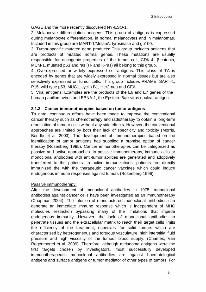
Recently, a number of novel approaches have been developed to circumvent
the tolerance mechanisms to generate high-avidity cytotoxic T cells or
monoclonal antibodies specific for tumour-associated antigens (Figure 2.1).
2 Introduction
11
Figure 2.1 Novel approaches for the generation of high-avidity CTL specific for
tumor-associated antigens. (a) and (b) take advantage of the TCR repertoire of
healthy individuals or transgenic mice that are not tolerant to A2-presented peptide
epitopes of tumor-associated antigens. (c) and (d) take advantage of in vitro
mutagenesis and selection of high affinity TCRs (c) or high-affinity single chain
antibodies specific for the peptide/HLA combination. (e) Strategies to transfer high
avidity HLA-restricted receptors into patient T cells or stem cells to produce
tumor-reactive T cells. (Morris, Bendle et al. 2003)
A tumor associated antigen, p53, has been reported to be an attractive
immunotherapy target, because it is only expressed in normal tissues of low
amounts. However, p53 turns may be not suitable to be targeted by
immunotherapeutic approaches since the low expression of p53 in normal
cells could still sufficiently stimulate T cells activation (Theoret, Cohen et al.
2008). Therefore, the functional characterizations of tumor antigens which are
selected as targets are critical for the development of cancer immunotherapy.
Active immunotherapy:
The active immunotherapy refers to cancer therapeutic vaccines which could
elicit or boost similar tumor antibodies and T cells in the patients other than
infusion of antibodies or T cells (Finn 2008). These strategies are usually
directed against tumor associated antigens, or against an unidentified number
of antigens such as whole tumour cells, cell lysates or heat shock proteins and
RNA isolated from tumour cells. Although vaccines based on multiple
unidentified antigens have shown promise in the experimental setting, it is
difficult to monitor vaccine-induced immune responses without knowing the
specific target antigens (Morris, Bendle et al. 2003). The progressive
improvement of vaccines is more probable with defined antigens, including
immunodominant peptides, peptides modified to increase immunogenicity,
proteins, "naked" DNA encoding cancer antigens, antigen-presenting cells
expressing the antigen or recombinant viruses or bacteria containing the
genes encoding tumor antigens (Rosenberg 1996; Morris, Bendle et al. 2003).
Some examples are vaccines against breast cancer (the HER2 antigen), B-cell
lymphoma (the tumor immunoglobulin idiotype), lung cancer (the MUC1
antigen), melanoma (dendritic cells loaded with tumor peptides or killed tumor
cells), pancreatic cancer (telomerase peptides), and prostate cancer (dendritic
cells loaded with prostatic acid phosphatase).
A predominant limiting factor of therapeutic vaccines is the
immunosuppressive microenvironment both during the induction of immunity
and in the effector phase of the response. One approach to improve the
induction phase is by using antibodies against a negative regulator of the
activation of effector T cells named cytotoxic T-lymphocyte–associated antigen
4 (CTLA-4) (Korman, Peggs et al. 2006; Ribas, Hanson et al. 2007).
2 Introduction
12
Unfortunately, the enhancing tumor immunity induced by anti-CTLA 4 antibody
is accompanied by serious autoimmunity. Combination of therapeutic vaccine
with chemotherapy could also induce the synergistic action of immunotherapy
probably because of the elimination of regulatory T cells by chemotherapy
(Emens and Jaffee 2005).
2.2 FMNL1 as a tumor associated antigens
2.2.1 Identification of FMNL1
The formin-like 1 (FMNL1) protein is a lymphoma-associated antigen identified
by using the SEREX approach in patients with chronic lymphocytic leukemia
(CLL) (Krackhardt, Witzens et al. 2002). It belongs to the formin protein family
indispensable for many fundamental actin-dependent processes including
migration, endocytosis, vesicle trafficking, morphogenesis and cytokinesis
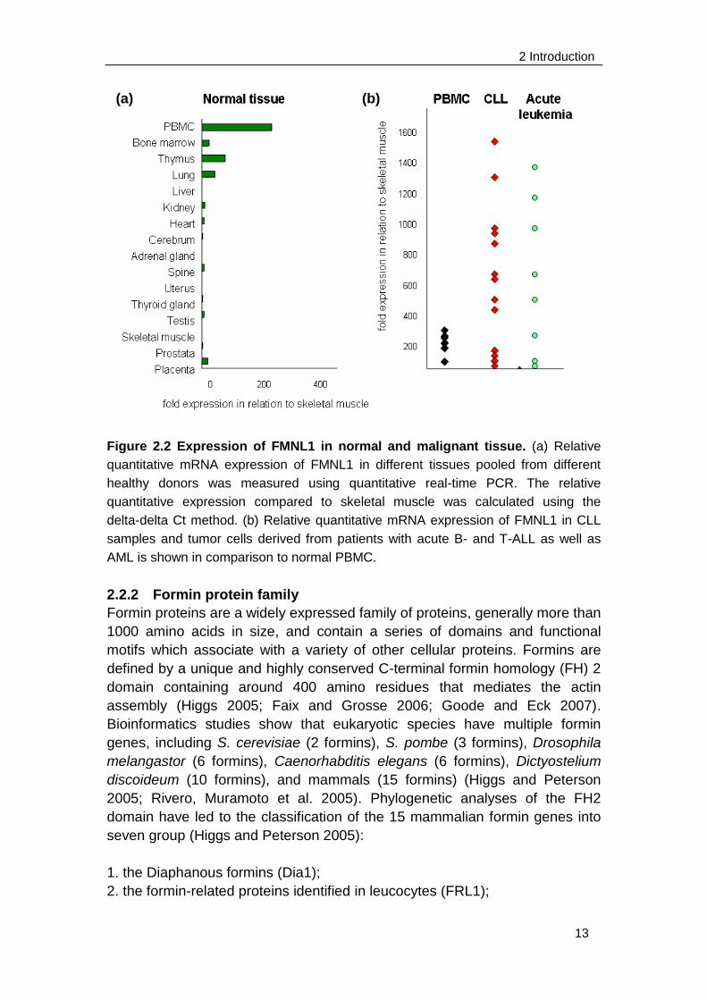
(Faix and Grosse 2006). FMNL1 is restrictedly expressed in
hematopoietic-lineage derived cells and overexpressed in malignant cells of
different origins (Figure 2.2). Detailed expression analysis was performed on
mRNA level. FMNL1 is most highly expressed in peripheral blood leukocytes
(PBL) and seldom expressed in normal tissues with the exception of bone
marrow, thymus and lung where hematopoietic cells accumulate. Moreover,
FMNL1 expression is up-regulated in PBLs derived from patient with CLL and
acute leukemia. This restricted expression suggests FMNL1 to be an attractive
target for novel immunotherapies in malignant and inflammatory diseases
(Krackhardt, Witzens et al. 2002; Schuster, Busch et al. 2007).
However, function and regulation of FMNL1 is less well characterized.
Previous work has shown involvement of FMNL1 in the reorientation of the
microtubule organizing center (MTOC) towards the immunological synapse
and cytotoxicity of T cells (Gomez, Kumar et al. 2007). It demonstrated a
fringe-like localization of FMNL1 at the leading edge of the lamellipod in
spreading T cells. This FMNL1 accumulation was also apparent at the edge of
F-actin structures, which formed during APC recognition, along with a distinct,
„„ring-like‟‟ localization of FMNL1 around the centrosome within the T cell.
Moreover, this FMNL1 ring reoriented along with the MTOC to face stimulating
APCs and regulate centrosome polarization in T cell as the absence of FMNL1
negatively effects MTOC polarization and cytotoxic T cell function. Additionally,
the murine homologue FRL, which has 85% homology to the human
counterpart, has been shown to be involved in cell adhesion and motility of
macrophages as well as Fc receptor-mediated phagocytosis
(Yayoshi-Yamamoto, Taniuchi et al. 2000; Seth, Otomo et al. 2006).
2 Introduction
13
Figure 2.2 Expression of FMNL1 in normal and malignant tissue. (a) Relative
quantitative mRNA expression of FMNL1 in different tissues pooled from different
healthy donors was measured using quantitative real-time PCR. The relative
quantitative expression compared to skeletal muscle was calculated using the
delta-delta Ct method. (b) Relative quantitative mRNA expression of FMNL1 in CLL
samples and tumor cells derived from patients with acute B- and T-ALL as well as
AML is shown in comparison to normal PBMC.
2.2.2 Formin protein family
Formin proteins are a widely expressed family of proteins, generally more than
1000 amino acids in size, and contain a series of domains and functional
motifs which associate with a variety of other cellular proteins. Formins are
defined by a unique and highly conserved C-terminal formin homology (FH) 2
domain containing around 400 amino residues that mediates the actin
assembly (Higgs 2005; Faix and Grosse 2006; Goode and Eck 2007).
Bioinformatics studies show that eukaryotic species have multiple formin
genes, including S. cerevisiae (2 formins), S. pombe (3 formins), Drosophila
melangastor (6 formins), Caenorhabditis elegans (6 formins), Dictyostelium
discoideum (10 formins), and mammals (15 formins) (Higgs and Peterson
2005; Rivero, Muramoto et al. 2005). Phylogenetic analyses of the FH2
domain have led to the classification of the 15 mammalian formin genes into
seven group (Higgs and Peterson 2005):
1. the Diaphanous formins (Dia1);
2. the formin-related proteins identified in leucocytes (FRL1);
(a) (b)
2 Introduction
14
3. the disheveled-associated activators of morphogenesis (DAAM1);
4. Delphilin (Delphilin);
5. the “inverted” formins (INF1);
6. the formin homology domain containing proteins (FHOD);
7. the original “namesake” formins (FMN).
Most eukaryotes have multiple formin isoforms, suggesting diverse cellular
roles. The FH2 core domain and the intervening linker region between the FH1
and FH2 domains are necessary and sufficient to nucleate actin polymerization
from G-actin in vitro (Pruyne, Evangelista et al. 2002; Kovar, Kuhn et al. 2003).
Before FH2 domain is a proline-rich FH1 domain which binds with low
micromolar affinity to profilin (Evangelista, Blundell et al. 1997; Watanabe,
Madaule et al. 1997).
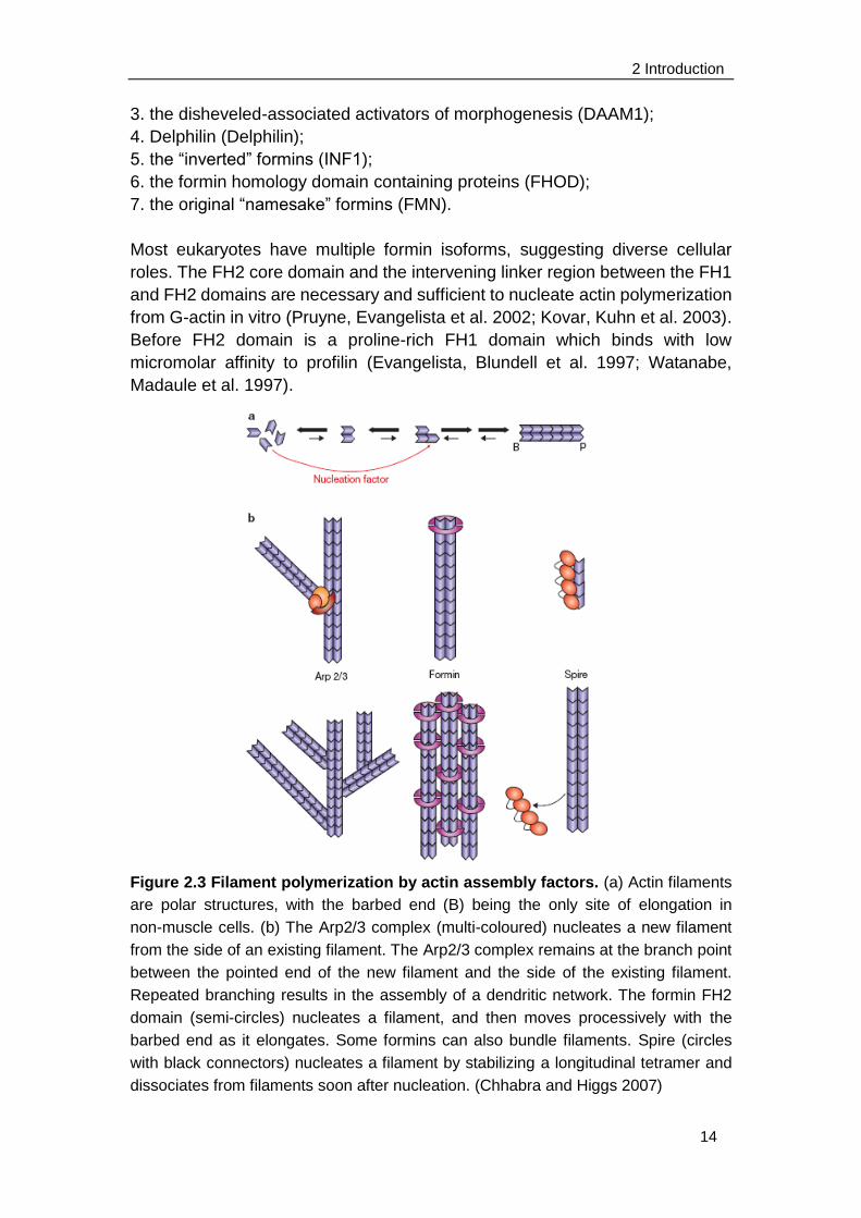
Figure 2.3 Filament polymerization by actin assembly factors. (a) Actin filaments
are polar structures, with the barbed end (B) being the only site of elongation in
non-muscle cells. (b) The Arp2/3 complex (multi-coloured) nucleates a new filament
from the side of an existing filament. The Arp2/3 complex remains at the branch point
between the pointed end of the new filament and the side of the existing filament.
Repeated branching results in the assembly of a dendritic network. The formin FH2
domain (semi-circles) nucleates a filament, and then moves processively with the
barbed end as it elongates. Some formins can also bundle filaments. Spire (circles
with black connectors) nucleates a filament by stabilizing a longitudinal tetramer and
dissociates from filaments soon after nucleation. (Chhabra and Higgs 2007)
2 Introduction
15
Eukaryotic cells nucleate actin filaments from a large pool of monomeric actin
bound to profilin in order to elicit temporal and spatial remodeling of the actin
cytoskeleton. This process underlies several cellular functions such as the
establishment of cell shape, cytokinesis, or cell motility (Faix and Grosse 2006).
The actin filament is a polar structure with a „barbed‟ and a „pointed‟ end, since
all the actin subunits face the same direction. Monomers can add to or
depolymerize from either end, but both processes are much more efficient at
the barbed end (Goode and Eck 2007). The step to assemble dimeric and
trimeric actin complexes limits the rate of actin filament nucleation. So far,
three major classes of actin nucleators are known to nucleate actin filaments in
vivo: the Arp2/3 complex, Spire, and the formin-homology proteins with
different mechanisms to accomplish their tasks (Figure 2.3).
The first identified actin nucleator was the Arp2/3 complex, which binds to the
side of pre-existing actin filaments, and nucleates a branched filament
structure with an angle of 70° between the filaments. The Arp2/3 complex
remains at the branch point between two filaments. Repeated branching leads
to a „dendritic network‟. Therefore, the Arp2/3 complex functions not only as a
nucleation factor, but also provides structure to the network. Spire nucleates
the assembly of unbranched actin filaments and, like the Arp2/3 complex,
remains bound to the pointed ends of the nucleated filaments. Pointed-end
nucleators, such as Arp2/3 and Spire, will experience only limited growth
before being capped at their barbed ends by capping proteins (Faix and
Grosse 2006; Chhabra and Higgs 2007).
Formins were identified more recently as an additional family of actin assembly
factors. All formins studied to date are dimeric because of their FH2 domain,
which initiates actin filament assembly and remains persistently associated
with the fast-growing barbed end, enabling rapid insertion of actin subunits
while protecting the end from capping proteins (Higgs 2005). Central function
of FH2 domain is to nucleate new actin filaments through binding tightly to the
barbed end of the filaments with low nanomolar affinity (Moseley, Sagot et al.
2004). Meanwhile, the FH2 domain moves processively with this elongating
actin filament barbed end. It means, the FH2 domain remains contacting with
actin filaments barbed ends when adding new actin monomers without
dissociation and reassociation. Related to processive barbed-end movement,
FH2 domains antagonize the effects of barbed-end capping proteins which
could completely block monomer addition to barbed ends. Therefore, the
barbed ends of actin filaments retain the ability to elongate even in the
presence of capping protein (Higgs 2005). Formin-mediated elongation is
further enhanced by the association of profilin with the FH1 domain of formin.
The adjacent FH1 domain recruits profilin-actin complexes and accelerates
filament elongation. The profilin–actin complex participates in actin assembly
at the barbed end as fast as free actin monomers do, whereas profilin
2 Introduction
16
interferes with actin nucleation. Normally, free actin monomers exist only at a
submicromolar concentration in cells, and thus profilin prevents spontaneous
actin nucleation while it allows fast actin elongation (Watanabe and Higashida
2004).
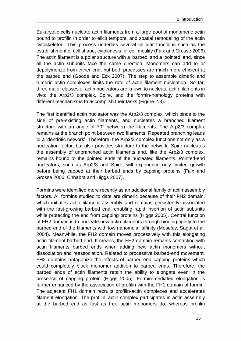
Vavylonis et al. suggested a model of formin-associated actin filament
elongation. They assumed that FH1 domains capture profilin-actin complex
from the bulk solution and transfer the captured subunit to the barbed end of
the filament (Figure 2.4a). The FH1 domains vary in the length and number of
proline-rich stretches, separated by proline-poor „„linker‟‟ stretches. Both of
them serve as the profilin binding sites but differ in affinity. If the profilin binding
sites were immobile at fixed locations relative to the barbed end, an actin unit
would be released and to diffuse to the barbed end. Otomo et al. proposed that
the FH2 dimer exists in equilibrium between two states (Figure 2.4b): the
closed (or blocked) configuration that prevents polymerization and the open (or
accessible) state that allows actin subunit addition. The transition from the
closed to the open state involves movement of the FH2 dimer toward the
barbed end (Otomo, Tomchick et al. 2005; Vavylonis, Kovar et al. 2006).
Figure 2.4 Model of actin filament growth mediated by formin dimer. FH2
domains (red) are shown as a dimer encircling the barbed end of the filament (blue).
The FH1 domain (black, green) is shown unstructured with three profilin binding sites
(green). (a) FH1-mediated polymerization: (1) assembly of profilin-actin to FH1; (2)
transfer of actin from FH1 to the barbed end; (3) detachment of profilin. (b)
Mechanism of FH2 processivity: FH2 in states preventing or allowing actin subunit
incorporation. (Otomo, Tomchick et al. 2005)
2.2.3 Diaphanous-related formins (DRFs)
In a conserved and widely expressed subfamily of formins known as
(a)
(b)
2 Introduction
17
Diaphanous-related formins (DRFs), the FH1 and FH2 domains are flanked by
an array of regulatory domains at the N-terminus and by a single C-terminal
Diaphanous autoregulatory domain (DAD) (Alberts 2001). The large N-terminal
regulatory region includes the GTPase binding domain (GBD) followed by an
adjacent Diaphanous-inhibitory domain (DID) and a dimerization domain (DD)
(Rose, Weyand et al. 2005; Faix and Grosse 2006; Nezami, Poy et al. 2006)
(Figure 2.5a). In the basal state, DRFs exist as autoinhibited proteins via
intramolecular interactions between DID and DAD (Figure 2.5b) (Alberts, 2001;
Li and Higgs, 2003; Watanabe et al., 1999). As revealed by the mDia1 crystal
structure, the DID domain is composed of five armadillo repeats, a structural
motif of three helices arrayed in a superhelical coil. The DAD polypeptide binds
along the entire length of the domain, roughly perpendicular to the B helices,
and establishes an extensive interface through numerous mainly hydrophobic
contacts (Figure 2.5c) (Otomo, Otomo et al. 2005; Rose, Weyand et al. 2005;
Nezami, Poy et al. 2006).
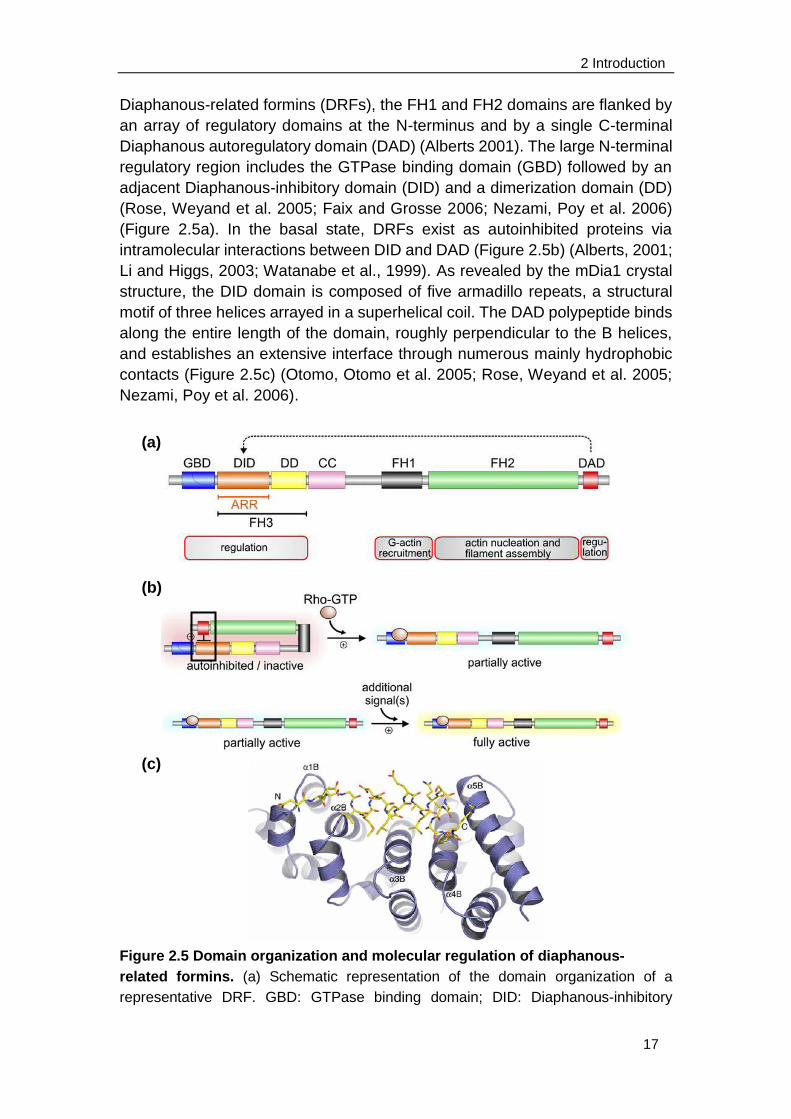
Figure 2.5 Domain organization and molecular regulation of diaphanous-
related formins. (a) Schematic representation of the domain organization of a
representative DRF. GBD: GTPase binding domain; DID: Diaphanous-inhibitory
(a)
(b)
(c)
2 Introduction
18
domain; DD: dimerization domain; CC: coiled coil; FH1: formin homology 1 domain;
FH2: formin homology 2 domain; FH3: formin homology 3 domain; ARR:
armadillo-repeat region. Based on its structure, DID is also referred to as ARR. (b)
Autoinhibition of DRFs caused by the interaction of DAD with DID is relieved by
binding a Rho GTPase to GBD, which releases DAD and lead to a partial activation of
the DRF. An unknown additional signal(s) is required to fully activate the DRF. (c)
Ribbon diagram showing the overall structure of the DID/DAD complex. The DID
domain is shown in blue, and the DAD domain is shown in yellow. The B helices of
armadillo repeats 1–5 are labeled from left to right, and the N and C termini of the
DAD domain are indicated. (Faix and Grosse 2006; Nezami, Poy et al. 2006)
This autoinhibition is relieved after binding of an active Rho GTPase to the
GBD domain. The binding induces displacement of the DAD, because the
GTPase binds to the GBD/DID fragment more tightly than does the DAD
segment (Faix and Grosse 2006; Nezami, Poy et al. 2006). The mammalian
diaphanous 1 (Dia1) as well as the macrophage-enriched murine Formin-like
protein 1 (FRL) are both regulated by autoinhibition in a DAD-dependent
manner (Yayoshi-Yamamoto, Taniuchi et al. 2000; Li and Higgs 2003; Harris, Li
et al. 2004; Seth, Otomo et al. 2006).
In addition to the domain specific functions, formin proteins seem to be
intensively regulated by splicing. Splicing at the N-terminus of human Dia2 has
been demonstrated to be involved in a novel signal transduction pathway, in
which hDia2C and c-Src are sequentially activated by RhoD to regulate the
motility of early endosomes through interactions with the actin cytoskeleton
(Gasman, Kalaidzidis et al. 2003). The DAD domain is also a hotspot of
splicing. Within this area, two splice variants have been characterized for FRL
although functional differences have not been observed (Harris, Li et al. 2004).
In contrast, abrogation of autoinhibition in mutants lacking the C-terminal end
in Dia1 and FRL specifically induces peripheral and plasma membrane
localization (Seth, Otomo et al. 2006; Copeland, Green et al. 2007). The exact
mechanism how DRFs locate to the plasma membrane is, however, currently
unknown.
2.3 Non-Hodgkin's lymphoma (NHL)
FMNL1 has been reported as a tumor-associated antigen in chronic
lymphocytic leukemia and overexpressed in various lymphomas (Krackhardt
et al, 2002b, Schuster et al, 2007). Malignant lymphoma is a group of
malignant neoplasms characterized by accumulation of cells native to the
lymphoid tissues, such as lymphocytes, histiocytes, and their precursors and
derivatives. The group is divided into two major categories: Hodgkin disease
(HD) and non-Hodgkin lymphoma (NHL), with subtypes, classified according
to predominant cell type and degree of differentiation. Hodgkin's disease was
2 Introduction
19
named after Thomas Hodgkin, who first described abnormalities in the lymph
system in 1832. It has a histological feature of the Reed-Sternberg giant cells,
which emerge from the so-called Hodgkin cells. Non-Hodgkin's lymphomas
are defined by the absence of these giant cells of Hodgkin's lymphoma. The
NHL is a group of malignant clonal neoplasms, originating from the B-(80 to
85% of cases) or T-lymphocytes (15 to 20% of cases). They arise from
different lymphoid components of the immune system (Kuppers 2009). One
widely used classification is based on two criteria: cytologic characteristics of
the constituent cells and type of cell growth pattern (defined as either follicular
or diffuse). Another system of classification is based on the cell type of origin:
T- or B-lymphocytes or histiocytes. FMNL1 has been demonstrated to be
expressed in all these subtypes of NHL as follicular NHL, diffuse large B cell
NHL and T cell NHL, and most highly expressed in T cell NHL (Favaro, Traina
et al. 2006) although partially at low levels. The most recent lymphoma
classifications, the 1994 Revised European-American Lymphoma (REAL)
classification and the 2001 WHO classification, abandoned the HD vs. NHL
grouping. Instead, 43 different forms of lymphoma are listed and discussed
separately.
2.3.1 Chronic lymphocytic leukemia (CLL)
Chronic lymphocytic leukemia is the most frequent leukemia in adults in
western countries, accounting for 25% of all leukemias, but fewer than 5% of
the cases in the eastern hemisphere (Kalil and Cheson 1999). It is
characterized by the clonal proliferation and accumulation of neoplastic B
lymphocytes in the blood, bone marrow, lymph nodes, and spleen. The normal
counterpart of CLL cells is a subpopulation of mature CD5+ B lymphocytes
(also known as B-1 cells) present in the mantle zone of lymph nodes and also
in small numbers in blood. These CD5+ B cells are increased in diseases with
an autoimmune basis, such as rheumatoid arthritis, systemic lupus
erythematosus, and Sjoeren‟s syndrome (Rozman and Montserrat 1995). The
median age of patients at diagnosis is 65 years, with only 10 to 15 percent
under 50 years of age. In most series, more men than women are affected.
The course of the disease is variable. Whereas some patients with CLL have a
normal life span, others die within five years after diagnosis. The etiology of
CLL is unknown. Several lines of evidence suggest a genetic component, such
as the increased prevalence of CLL among first-degree relatives.
Environmental factors, such as ionizing radiation, chemicals, and drugs have
shown no apparent relationship (Rozman and Montserrat 1995; Kalil and
Cheson 1999; Montserrat and Moreno 2008).
Patients with CLL have progressive immunodeficiency, which chiefly manifests
as hypogammaglobulinaemia (up to 60% of CLL patients), but involves all
elements of the immune system. Compared with normal peripheral blood B
cells, CLL B cells express relatively low levels of surface-membrane
2 Introduction
20
immunoglobulin, mostly IgM and IgD with a single light chain (kappa or lambda)
(Rozman and Montserrat 1995). Only 15% of patients with CLL have
completely normal serum immunoglobulins. The extent of hypogamma-
globulinaemia depends on the stage and duration of the disease and lead to
bacterial infections of the respiratory tract, skin or urinary tract. They are the
major causes of death in between a quarter and a half of patients with CLL
(Hamblin and Hamblin 2008). The mechanisms of immune defects in CLL
have not been completely elucidated, although several explanations have
been proposed, including abnormalities in T regulatory cells and
non-neoplastic B cells (Montserrat and Moreno 2008). One critical step for the
immune response to antigen is the expression of the CD40 ligand on the
surface of activated T cells. Downregulation of this ligand induced by CLL
lymphocytes results in severe immunodeficiency states. Gene transfer of
CD40 ligand into B cells of CLL patients has been reported to induce cytotoxic
T lymphocytes against autologous leukemic cells (Cantwell, Hua et al. 1997;
Kato, Cantwell et al. 1998). B-CLL cells are resistant to most currently
available gene transfer systems. Several virus vectors such as replication
defective adenovirus vector and recombinant adeno-associated virus (rAAV)
vector have been used to enable efficient transduction of CD40 ligand to
primary B-CLL cells and already entered the clinical trial (Wendtner, Kofler et al.
2004). However, the specificity and safety need to be further confirmed.
2.3.2 Therapies for CLL
CLL is generally considered not curable. The former standard treatment,
chlorambucil only leads to a very low level of complete response (10%). The
more effective agent fludarabine results in a much higher complete response
rate (20–40%) and a longer disease-free interval, however, survival is not
prolonged (Montserrat and Moreno 2008). The target therapy using
monoclonal antibodies rituximab (anti-CD20) and alemtuzumab (anti-CD52)
has brought great improvement in the treatment of CLL. It has dramatically
improved complete response rates and progression-free survival in patients
with both newly-diagnosed and relapsed CLL when incorporating of these
monoclonal antibodies with chemoimmunotherapy, however, the overall
survival has not been changed (Quintas-Cardama and O'Brien 2009). Stem
cell transplants have been performed in an increasing number of subjects with
CLL. Autologous stem cell transplants do not cure CLL but could prolong
survival in extremely selected patients (Dreger, Stilgenbauer et al. 2004).
However, the necessary condition for the success of an autologous transplant
is to achieve complete remission before the procedure, which is rare in
patients failed in chemo or chemo-immunotherapy (Dreger and Montserrat
2002). An additional problem with autotransplant is the risk of secondary
myelodysplasia/acute myeloid leukaemia, particularly in patients having
performed with total body irradiation. In contrast, allogeneic transplants can
cure about 40% of the patients, but leads to a high toxicity and mortality
2 Introduction
21
(Moreno, Villamor et al. 2005; Dreger, Corradini et al. 2007).
Since the beneficial effects of allogeneic stem cell transplantation partly
depend on the graft-versus-leukaemia effects, one promising strategy consists
of the genetic manipulation of effector T lymphocytes with tumor specific T-cell
receptors or chimeric antigen receptors on the surface directed against surface
antigens on the CLL cells. This strategy may produce therapeutic benefits
without the adverse effects of more generalized allo-reactivity (Foster, Brenner
et al. 2008). A number of tumour-associated antigens overexpressed on CLL
cells can be selected as the target of specific CTL responses, including
fibromodulin, MDM2 (murine double minute 2), survivin, oncofetal
antigen-immature laminin receptor protein (OFAiLRP), FMNL1, preferentially
expressed antigen of melanoma (PRAME) and receptor for hyaluronic-acid
mediated motility (RHAMM/CD168) (Giannopoulos and Schmitt 2006).
Moreover, the idiotypic determinants of the unique monoclonal immunoglobulin
on CLL cells may serve as tumour-specific antigens (Trojan, Schultze et al.
2000). It has been already reported that an allorestricted T-cell clone, SK22,
expressing a single defined T cell receptor recognizing a peptide derived from
FMNL1 shows potent antitumor activity against lymphoma and renal cell
carcinoma cell lines, Epstein-Barr virus (EBV)-transformed B cells and primary
tumor samples derived from patients with CLL. SK22 recognizes preferentially
malignant cells and has only limited reaction against normal hematopoietic
cells. (Schuster, Busch et al. 2007). This suggests FMNL1 to be a highly
attractive target antigen in CLL patients and gene-modified TCR-transgenic T
cells might be suitable therapeutic tools in the autologous setting. However,
functional investigation of FMNL1 might be highly important in order to identify
FMNL1-dependent processes of diverse hematopoietic-lineage derived cells
which may be associated with enhanced FMNL1 ligand presentation and
therefore affected by FMNL1-specific immunotherapeutic approaches.
2.4 Aim of the project
The aim of this project is to investigate functional role of FMNL1 in order to
evaluate and validate FMNL1 as potential target antigen for novel
immunotherapies:
1. Testing of novel FMNL1-specific antibodies for potential application in
immunofluorescence analysis and investigation of the localization of
native FMNL1 in diverse hematopoietic-lineage derived cells, especially
focusing on the function-associated localization with the help of confocal
microscopy.
2. Functional analysis of different splice variants of FMNL1 using diverse
cell biochemical and biological assays.
3. Investigation of functional aspects of FMNL1, associated with the newly
identified interaction partners.
3 Materials
22
3 Materials
3.1 Equipments and supplies
• Cell culture bottles, Becton Dickinson, Heidelbuerg
• Centrifuge, Hettich, Kirchlengern, Germany
• Centrifuge 4K15, Sigma Laborzentrifugen GmbH, Osterode an Herz,
Germany
• Centrifuge 5417R , Eppendorf-Netheler-Hinz-GmbH, Hamburg, Germany
• Counting chamber, Brand GmbH & Co., Wertheim, Germany
• Confocal microscopy, LEICA TCS SP2/405, Leica Microsystems GmbH,
Wetzlar, Germany
• Coverslip 10 mm X 10 mm, Josef Peske, Aindling, Germany
• FACS Calibur cell sorter, Becton Dickinson, Heidelbuerg
• Fluorescence Microscope, Leica Microsystems GmbH, Wetzlar, Germany
• Gloves latex, Rösner-Mautby Meditrade GmbH, Kiefersfelden, Germany
• Gloves nitrile, Kimberly-Clark Corporation, Neenah, USA
• Hyperfilm MP, Amersham Bioscience, Freiburg, Germany
• Incubator, Heraeus Holding GmbH, Hanau, Germany
• Incubator for bacteria, Bachofer, Reutlingen, Germany
• Incubator shaker, INFORS AG, Bottmingen, Switzerland
• InGenius gel documentation (gel doc) system, Synoptics Ltd, Cambridge,
England
• Irradiation facility Gammacell 40, Atomic Energy of Canada Limited,
Ottawa, Canada
• Microplate reader ELISA Reader, SLT Labinstruments, Crailsheim,
Germany
• Microscope, Zeiss AG, Jena, Germany
• Midi Electrophoresis chamber, Harnischmacher, Labor- und
Kuststofftechnick, Kassel, Germany
• MilliQ System, Millipore GmbH, Schwalbach, Germany
• MoFlowTM, Dako, Hamburg, Germany
• Multichannel pipets, Eppendorf-Netheler-Hinz-GmbH, Hamburg, Germany
• PCR thermocycler, Biometra Biomedizinische Analytik GmbH, Göttingen,
Germany
• PH-meter 766, Knick Elektronische Messgeräte GmbH & CO. KG, Berlin,
Germany
• Pipets, Stripette 5 ml, 10 ml, 25 ml, Corning, New York, USA
• Pipet boy, Integra Biosciences, Fernwald, Germany
• Pipets, Eppendorf-Netheler-Hinz-GmbH, Hamburg, Germany
• Polystyrene round bottom FACS tubes, Becton Dickinson, Heidelbuerg
• Power supply, Pharmacia GmbH, Erlangen, Germany
• Pursep-A Xpress disinfection agent, Metz Consumer Care GmbH, Frankfurt,
3 Materials
23
Germany
• Slides, Glaswarenfabrik Karl Hecht KG, Sondheim, Germany
• Sterile bench, BDK, Sonnenbühl-Genkingen, Germany
• Sterile polypropylene round bottom FACS tubes with cap, Becton Dickinson,
Heidelbuerg
• Sterile filters, Techno Plastic Products AG, Trasadingen, Switzerland
• Syringes 10ml, 50 ml, BD Perfusion, Becton Dickinson, Heidelbuerg
• Thermomixer, Eppendorf-Netheler-Hinz-GmbH, Hamburg, Germany
• Tips 1–10 μl, 10–200 μl, 100–1000 μl, Greiner Bio-One International AG,
Kremsmünster, Austria
• Ultraspec 1100 pro UV/ vis Spectrophotometer, Biochrom Ltd, Cambridge,
England
• Vortexer, Bender & Hobei AG, Switzerland
• Water bath, Memmert, Schwab Bach
• XCell SureLockTM Mini-Cell, Invitrogen Cooperation, Karlsruhe
• XCell IITM Blot Module, Invitrogen Cooperation, Karlsruhe
• X-ray film, valmex, Augsburg, Germany
• 12-, 24- well tissue culture plates, Becton Dickinson, Heidelbuerg
• 96 well U-bottom tissue culture plates, Becton Dickinson, Heidelbuerg
• 96 well flat-bottom tissue culture plates, Becton Dickinson, Heidelbuerg
• 50 ml, 15ml Falcons, Becton Dickinson, Heidelbuerg
• 1.5 ml, 0.5 ml tubes, Eppendorf-Netheler-Hinz-GmbH, Hamburg, Germany
3.2 Chemicals, enzymes and cytokines
• Ampicillin sodium salt, Sigma-Aldrich Chemie GmbH, Taufkirchen,
Germany
• A23187, Sigma-Aldrich Chemie GmbH, Taufkirchen, Germany
• Agarose, Bio & Sell Nürnberg, Germany
• CaCl2, Merck KGaA, Darmstadt, Germany
• CD40 ligand, Peprotech, London, UK
• Chloroform (1 Bromo-3-choloro-prapan), Sigma Aldrich Chemie GmbH,
Taufkirchen, Germany
• Chloroform: Isoamyl alcohol (CIA) 24:1, Sigma Aldrich Chemie GmbH,
Taufkirchen, Germany
• Ciprofloxacin, Sigma-Aldrich Chemie GmbH, Taufkirchen, Germany
• Cyclosporin A, Sigma-Aldrich Chemie GmbH, Taufkirchen, Germany
• Complete (25 X), Roche, Basel, Switzerland
• 4`,6-Diamidino-2-phenylindole dihydrochloride (DAPI), Sigma-Aldrich
Chemie GmbH, Taufkirchen, Germany
• Diethylpyrocarbonat (DEPC) H2O, Invitrogen Corporation, Karlsruhe,
Germany
• Dulbecco‟s modified Eagle‟s medium (DMEM), Gibco, Invitrogen
Corporation, Karlsruhe, Germany
3 Materials
24
• Dimethyl Sulfoxide (DMSO), Merck KGaA, Darmstadt, Germany
• Desoxinucleosidtriphosphate (dNTP) (2mM/10mM), Fermentas GmbH,
St.Leon-Rot, Germany
• Dithiothreitol (DTT), 0.1M, Fermentas GmbH, St.Leon-Rot, Germany
• EcoR I enzyme, Fermentas GmbH, St.Leon-Rot, Germany
• Ethanol, Merck KGaA, Darmstadt, Germany
• Ethidium Bromide, Invitrogen Corporation, Karlsruhe, Germany
• Fetal calf serum (FCS), PAA Laboratories GmbH, Pasching, Austria
• Ficoll, Biochrom, Berlin, Germany
• First strand buffer (5×), Fermentas GmbH, St.Leon-Rot, Germany
• G418, PAA Laboratories GmbH, Pasching, Austria
• Glucose, Merck KGaA, Darmstadt, Germany
• Granulocyte Macrophage Colony Stimulating Factory (GM-CSF),Peprotech,
London, UK
• [3H]-myristic acid, PerkinElmer, Boston, U.S.A
• HEPES (1M), Gibco, Invitrogen Corporation, Karlsruhe, Germany
• HEPES (powder), Biochrom KG, Berlin, Germany
• HPLC H2O, Fisher Scientific Corporation, UK
• Human serum, Helmholtz Zentrum Munich, healthy male donors
• HyperLadder I, Bioline GmbH, Luckenwalde, Germany
• IL-1β, Peprotech, London, UK
• IL-2, Chiron Vaccines International, Marburg, Germany
• IL-4, Peprotech, London, UK
• IL-6, Peprotech, London, UK
• Iscove‟s Modified Dulbecco‟s Medium (IMDM), Gibco, Invitrogen
Corporation, Karlsruhe, Germany
• Indo-1 AM, Sigma-Aldrich Chemie GmbH, Taufkirchen, Germany
• Ionomycine, Sigma-Aldrich Chemie GmbH, Taufkirchen, Germany
• Isopropyl alcohol, Sigma-Aldrich Chemie GmbH, Taufkirchen, Germany
• Kanamycin, Sigma-Aldrich Chemie GmbH, Taufkirchen, Germany
• KCl, Merck KGaA, Darmstadt, Germany
• Kpn I enzyme, New England BioLabs GmbH, Frankfurt am Main, Germany
• 100bp ladder, New England BioLabs Incorporation
• Lusogeny broth (LB) agar, Sigma-Aldrich Chemie GmbH, Taufkirchen,
Germany
• L-Glutamine, Gibco, Invitrogen Corporation, Karlsruhe, Germany
• Lipopolysoccharide (LPS), Sigma Aldrich Chemie GmbH, Taufkirchen,
Germany
• 6× loading Dye Solution, Fermentas GmbH, St.Leon-Rot, Germany
• LURIA Broth Base, Sigma-Aldrich Chemie GmbH, Taufkirchen, Germany
• Methanol, Merck KGaA, Darmstadt, Germany
• Mfe I enzyme, New England BioLabs GmbH, Frankfurt am Main, Germany
• Milk powder, Frema, Lueneburg, Germany
• Mounting medium, Vector Laboratories, Burlingame, U.S.A
3 Materials
25
• 3-(4,5)-dimethylthiahiazo (-z-y1)-3,5-di- phenytetrazoliumromide (MTT),
Sigma-Aldrich Chemie GmbH, Taufkirchen, Germany
• Na-Acetate, Merck KGaA, Darmstadt, Germany
• Na2HPO4·2H2O, Merck KGaA, Darmstadt, Germany
• NaN3, Merck KGaA, Darmstadt, Germany
• NEB buffer, New England BioLabs GmbH, Frankfurt am Main, Germany
• Nhe I enzyme, New England BioLabs GmbH, Frankfurt am Main, Germany
• Non Essential Amino Acids, Gibco, Invitrogen Corporation, Karlsruhe,
Germany
• NP-40, Sigma-Aldrich Chemie GmbH, Taufkirchen, Germany
• Okt-3, kindly provided by Elisabeth Kremmer
• Olig(dt)15 Primer, Promega Corporation Madison, USA
• Penicilline Streptomycin, Gibco, Invitrogen Corporation, Karlsruhe,
Germany
• Paraformaldehyd (PFA), Sigma-Aldrich Chemie GmbH, Taufkirchen,
Germany
• Phenol, Carl Roth GmbH, Karlsruhe, Germany
• Pluronic, Peprotech, Sigma-Aldrich Chemie GmbH, Taufkirchen, Germany
• Phorbol 12-myristate 13-acetate (PMA) , Sigma-Aldrich Chemie GmbH,
Taufkirchen, Germany
• Prostaglandin (PG) E-2, Sigma-Aldrich Chemie GmbH, Taufkirchen,
Germany
• Pheylmethanesulfonyl (PMSF), Sigma-Aldrich Chemie GmbH, Taufkirchen,
Germany
• Poly-L-lysin, Sigma-Aldrich Chemie GmbH, Taufkirchen, Germany
• Ponceau, Sigma-Aldrich Chemie GmbH, Taufkirchen, Germany
• Prodium Iodide (PI), Sigma-Aldrich Chemie GmbH, Taufkirchen, Germany
• RNaseOUT Recombinant Ribonuclease Inhibitor, Invitrogen Corporation,
Karlsruhe, Germany
• RPMI 1640 (-L-Glutamin), Gibco, Invitrogen Corporation, Karlsruhe,
Germany
• Seminaphtorhodafluor-1 carboxylic acid, acetate and succinimidyl ester
(SNARF-1), Invitrogen Corporation, Karlsruhe, Germany
• Sbf I enzyme, New England BioLabs GmbH, Frankfurt am Main, Germany
• Sodium Carbonate, Sigma Aldrich Chemie GmbH, Taufkirchen, Germany
• Sodium Pyruvate, Gibco, Invitrogen Corporation, Karlsruhe, Germany
• Sodium Chloride, Sigma Aldrich Chemie GmbH, Taufkirchen, Germany
• SuperScript II Reverse Transcriptase, Invitrogen Corporation, Karlsruhe,
Germany
• T4 DNA ligase (10× ligation buffer), Fermentas GmbH, St.Leon-Rot,
Germany
• TAE buffer (10×), Invitrogen Corporation, Karlsruhe, Germany
• Tumor necrosis factor α (TNF α), Peprotech, London, UK
• Trypsin EDTA 0,5 %, Gibco, Invitrogen Corporation, Karlsruhe, Germany
3 Materials
26
• Trypan blue, Gibco, Invitrogen Corporation, Karlsruhe, Germany
• Tri Reagent, Sigma Aldrich Chemie GmbH, Taufkirchen, Germany
• Tween 20, Sigma Aldrich Chemie GmbH, Taufkirchen, Germany
• Western LightingTM Plus-ECL, Amersham Bioscience, Freiburg, Germany
• Xba I enzyme, New England BioLabs GmbH, Frankfurt am Main, Germany
• Xho I enzyme, Fermentas GmbH, St.Leon-Rot, Germany
• ß2-Microglobulin, EMD Biosciences, Darmstadt, Germany
3.3 Kits
• BCATM Protein Assay kit, Pierce, Rockfold, U.S.A
• B cell negative isolation kit, Dynal, Invitrogen Corporation, Karlsruhe,
Germany
• BrdU flow kit, Becton Dickinson, Heidelbuerg, Germany
• KOD Hot Start DNA Polymerase kit, Novagen, EMD Biosciences,
Darmstadt, Germany
• LightCycler® - FastStart DNA MasterPLUS SYBR Green I kit, Roche, Basel,
Schweiz
• Venor®GeM-Mykoplasmen Detektions Kit, MinervaBiolabs, Berlin,
Germany
• NucleoSpin® Extract II kit for PCR gel extraction, Macherey-Nagel
Corporation, Düren, Germany
• Jetstar 2.0 kit for plasmid purification, Genomed GmbH, Löhne, Germany
3.4 Buffers and Solutions
• Dulbecco's Phosphate-Buffered Saline (DPBS), Gibco, Invitrogen
Corporation, Karlsruhe, Germany
• Phosphate buffered saline (PBS), Dulbecco‟s Biochrom AG, Berlin,
Germany
• Binding Buffer (10 X), Becton Dickinson, Heidelbuerg, Germany
• MOPS buffer (20 X), Invitrogen Corporation, Karlsruhe, Germany
• NuPAGE® Transfer Buffer (20 X), Invitrogen Corporation, Karlsruhe,
Germany
• NuPAGE® LDS Sample Buffer (4 X), Invitrogen Corporation, Karlsruhe,
Germany
• Protein G–Sepharose 4 Fast Flow, Amersham Bioscience, Freiburg,
Germany
• AmplifyTM Fluorographic Reagent, Amersham Bioscience, Freiburg,
Germany
• FACS buffer
o PBS
o 1 % Δ FCS
• Ripa Buffer (stock solution):
3 Materials
27
o 10 mM Tris-Cl
o 10 mM NaCl
o 1 % SDS
o 0.5 % Deoxycholic
o 1 % NP-40
• Ripa Buffer (working solution):
o Ripa Buffer (stock solution)
o 1 mM PMSF
o 1 X complete
• Lysate Buffer for immunoprecipitation (stock solution):
o 20 mM Tris-Cl
o 150 mM NaCl
o pH 7.4
• Lysate Buffer for immunoprecipitation (working solution):
o Lysate Buffer (stock solution)
o 150 mM Chaps
o 1 mM Na3VO4
o 1 mM NaF
o 1 mM PMSF
o 1 X complete
• Fixation buffer for SDS/PAGE gel:
o 25% volume isoproponal
o 65% volume water
o 10% volume acetic acid
• Tris buffered saline (TBS) buffer (10 X) pH 8.0:
o 10 mM Tris-base
o 150 mM NaCl
o H2O
• TBST buffer:
o 1 X TBS buffer
o 0.05% Tween 20
• Western blot washing buffer:
o 1 X TBS buffer
o 0.05 % Tween 20
• 5 % or 1 % milk powder in TBST
• Immunofluoresence washing buffer:
o PBS
o 0.5 % NP-40
o 0.01 % NaN3
• Fixation buffer for Immunofluoresence staining:
o PBS
o 10 % PFA
• Saline-sodium citrate (SSC) buffer (20 X) pH 7.0:
o H2O
3 Materials
28
o 0.3 M Na Citrat 2 H2O
o 3 M NaCl
• DAPI solution:
o 1 X SSC buffer
o 5 μg/ul DAPI
• 2× Hanks Balanced Salt Solution (HBSS) Buffer pH 7.06:
o 280 mM NaCl
o 10 mM KCl
o 12 mM Glucose
o 50 mM HEPES
o 1.5 mM Na2HPO4·2H20
3.5 Cell culture medium
• Complete DMEM/ RPMI
o DMEM/RPMI1640
o 10% Δ FCS
o 2 mM L-Glutamine
o Penicillin (100 U/ml)-Streptomycin (100 μg/ml)
o 10 mM Non essential amino acid
o 1 mM Sodium-Pyruvate
• DMEM hunger medium
o DMEM
o 3 % Δ FCS
o 2 mM L-Glutamine
o Penicillin (100 U/ml)-Streptomycin (100 μg/ml)
o 10 mM Non essential amino acid
o 1 mM Sodium-Pyruvate
• T cell medium
o RPMI1640
o 10 % Δ human serum
o 2 mM L-Glutamine
o Penicillin (100 U/ml)-Streptomycin (100 μg/ml)
o 10 mM Non essential amino acid
o 1 mM Sodium-Pyruvate
o 10 mM HEPES (pH 7.4)
o Gentamycin (16.6 μg/ml)
• B cell medium
o IMDM
o 10 % Δ FCS
o 2 mM L-Glutamine
o Insulin-Transferrin (50 μg/ml)
o Gentamycin (16.6 μg/ml)
• AIM-V medium
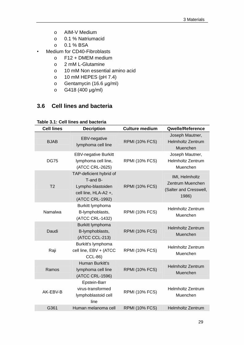
3 Materials
29
o AIM-V Medium
o 0.1 % Natriumacid
o 0.1 % BSA
• Medium for CD40-Fibroblasts
o F12 + DMEM medium
o 2 mM L-Glutamine
o 10 mM Non essential amino acid
o 10 mM HEPES (pH 7.4)
o Gentamycin (16.6 μg/ml)
o G418 (400 μg/ml)
3.6 Cell lines and bacteria
Table 3.1: Cell lines and bacteria
Cell lines Decription Culture medium Qwelle/Reference
BJAB EBV-negative
lymphoma cell line RPMI (10% FCS)
Joseph Mautner,
Helmholtz Zentrum
Muenchen
DG75
EBV-negative Burkitt
lymphoma cell line,
(ATCC CRL-2625)
RPMI (10% FCS)
Joseph Mautner,
Helmholtz Zentrum
Muenchen
T2
TAP-deficient hybrid of
T-and B-
Lympho-blastoiden
cell line, HLA-A2 +,
(ATCC CRL-1992)
RPMI (10% FCS)
IMI, Helmholtz
Zentrum Muenchen
(Salter and Cresswell,
1986)
Namalwa
Burkitt lymphoma
B-lymphoblasts,
(ATCC CRL-1432)
RPMI (10% FCS) Helmholtz Zentrum
Muenchen
Daudi
Burkitt lymphoma
B-lymphoblasts,
(ATCC CCL-213)
RPMI (10% FCS) Helmholtz Zentrum
Muenchen
Raji
Burkitt's lymphoma
cell line, EBV + (ATCC
CCL-86)
RPMI (10% FCS) Helmholtz Zentrum
Muenchen
Ramos
Human Burkitt's
lymphoma cell line
(ATCC CRL-1596)
RPMI (10% FCS) Helmholtz Zentrum
Muenchen
AK-EBV-B
Epstein-Barr
virus-transformed
lymphoblastoid cell
line
RPMI (10% FCS) Helmholtz Zentrum
Muenchen
G361 Human melanoma cell RPMI (10% FCS) Helmholtz Zentrum
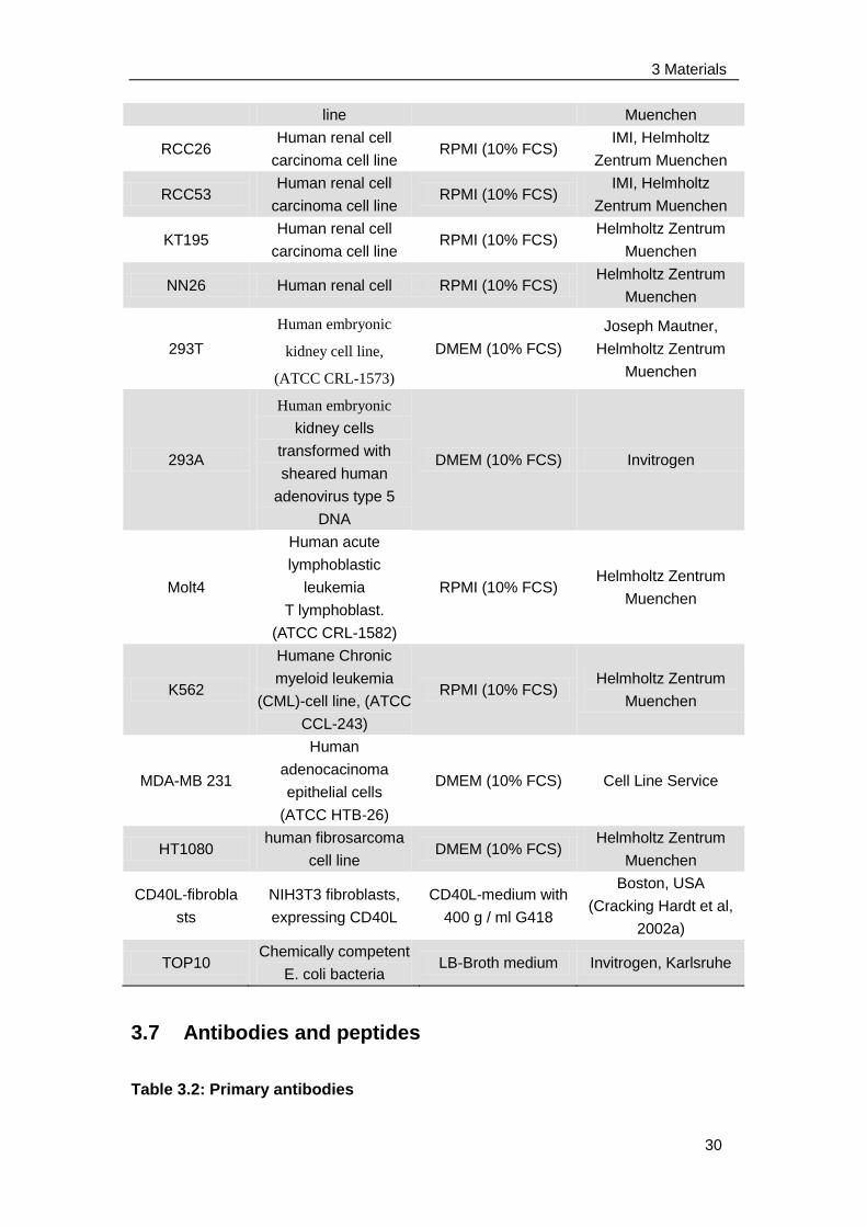
3 Materials
30
line Muenchen
RCC26 Human renal cell
carcinoma cell line RPMI (10% FCS)
IMI, Helmholtz
Zentrum Muenchen
RCC53 Human renal cell
carcinoma cell line RPMI (10% FCS)
IMI, Helmholtz
Zentrum Muenchen
KT195 Human renal cell
carcinoma cell line RPMI (10% FCS)
Helmholtz Zentrum
Muenchen
NN26 Human renal cell RPMI (10% FCS) Helmholtz Zentrum
Muenchen
293T
Human embryonic
kidney cell line,
(ATCC CRL-1573)
DMEM (10% FCS)
Joseph Mautner,
Helmholtz Zentrum
Muenchen
293A
Human embryonic
kidney cells
transformed with
sheared human
adenovirus type 5
DNA
DMEM (10% FCS) Invitrogen
Molt4
Human acute
lymphoblastic
leukemia
T lymphoblast.
(ATCC CRL-1582)
RPMI (10% FCS) Helmholtz Zentrum
Muenchen
K562
Humane Chronic
myeloid leukemia
(CML)-cell line, (ATCC
CCL-243)
RPMI (10% FCS) Helmholtz Zentrum
Muenchen
MDA-MB 231
Human
adenocacinoma
epithelial cells
(ATCC HTB-26)
DMEM (10% FCS) Cell Line Service
HT1080 human fibrosarcoma
cell line DMEM (10% FCS)
Helmholtz Zentrum
Muenchen
CD40L-fibrobla
sts
NIH3T3 fibroblasts,
expressing CD40L
CD40L-medium with
400 g / ml G418
Boston, USA
(Cracking Hardt et al,
2002a)
TOP10 Chemically competent
E. coli bacteria LB-Broth medium Invitrogen, Karlsruhe
3.7 Antibodies and peptides
Table 3.2: Primary antibodies
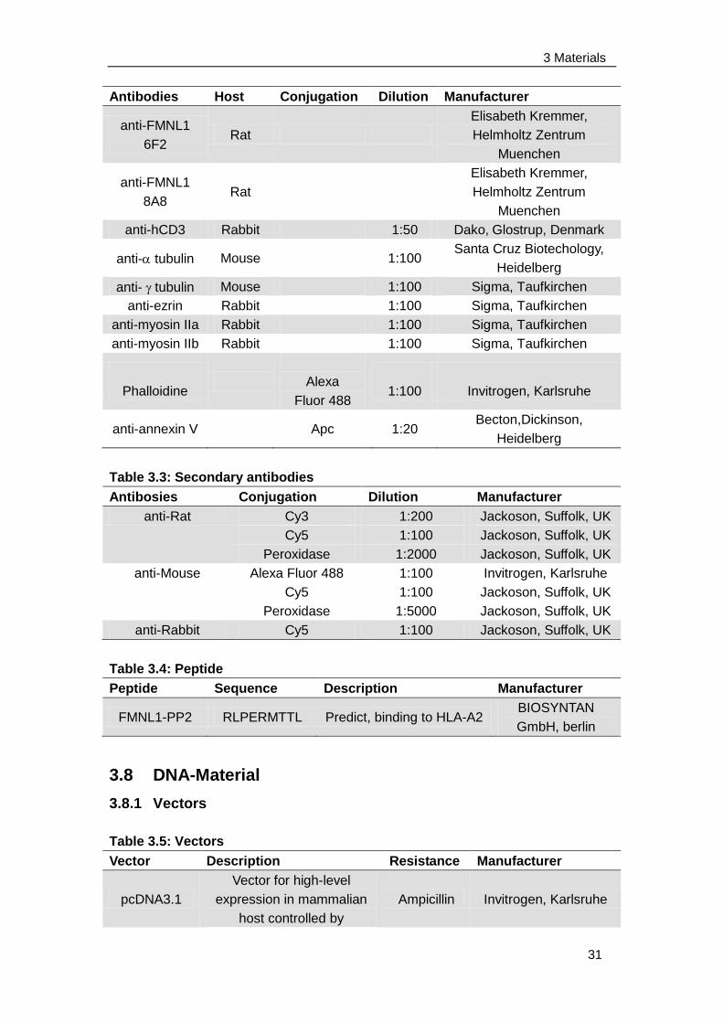
3 Materials
31
Antibodies Host Conjugation Dilution Manufacturer
anti-FMNL1
6F2 Rat
Elisabeth Kremmer,
Helmholtz Zentrum
Muenchen
anti-FMNL1
8A8 Rat
Elisabeth Kremmer,
Helmholtz Zentrum
Muenchen
anti-hCD3 Rabbit 1:50 Dako, Glostrup, Denmark
anti- tubulin Mouse 1:100 Santa Cruz Biotechology,
Heidelberg
anti- tubulin Mouse 1:100 Sigma, Taufkirchen
anti-ezrin Rabbit 1:100 Sigma, Taufkirchen
anti-myosin IIa Rabbit 1:100 Sigma, Taufkirchen
anti-myosin IIb Rabbit 1:100 Sigma, Taufkirchen
Phalloidine
Alexa
Fluor 488
1:100
Invitrogen, Karlsruhe
anti-annexin V Apc 1:20 Becton,Dickinson,
Heidelberg
Table 3.3: Secondary antibodies
Antibosies Conjugation Dilution Manufacturer
anti-Rat Cy3 1:200 Jackoson, Suffolk, UK
Cy5 1:100 Jackoson, Suffolk, UK
Peroxidase 1:2000 Jackoson, Suffolk, UK
anti-Mouse Alexa Fluor 488 1:100 Invitrogen, Karlsruhe
Cy5 1:100 Jackoson, Suffolk, UK
Peroxidase 1:5000 Jackoson, Suffolk, UK
anti-Rabbit Cy5 1:100 Jackoson, Suffolk, UK
Table 3.4: Peptide
Peptide Sequence Description Manufacturer
FMNL1-PP2 RLPERMTTL Predict, binding to HLA-A2 BIOSYNTAN
GmbH, berlin
3.8 DNA-Material
3.8.1 Vectors
Table 3.5: Vectors
Vector Description Resistance Manufacturer
pcDNA3.1
Vector for high-level
expression in mammalian
host controlled by
Ampicillin Invitrogen, Karlsruhe
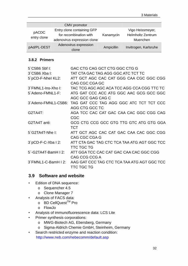
3 Materials
32
CMV promotor
pACDC
entry clone
Entry clone containing GFP
for recombination with
adenovirus expression clone
Kanamycin
Vigo Heissmeyer,
Helmholtz Zentrum
Muenchen
pAd/PL-DEST Adenovirus expression
clone Ampicillin Invitrogen, Karlsruhe
3.8.2 Primers
3.9 Software and website
• Edition of DNA sequence:
o Sequencher 4.5
o Clone Manager 7
• Analysis of FACS data:
o BD CellQuestTM Pro
o FlowJo
• Analysis of immunofluorescence data: LCS Lite
• Primer synthesis corporations:
o MWG-Biotech AG, Ebersberg, Germany
o Sigma-Aldrich Chemie GmbH, Steinheim, Germany
• Search restricted enzyme and reaction condition:
http://www.neb.com/nebecomm/default.asp
5`C5B6 Sbf I:
3`C5B6 Xba I:
5`pCD-F-NheI KL2:
3`FMNL1-Ins-Xho I:
5`Adeno-FMNL1-F:
3`Adeno-FMNL1-C5B6:
G2TA4T:
G2TA4T anti:
5`G2TA4T-Nhe I:
3`pCD-F-C-Xba I 2:
5`-G2TA4T-BamH I 2:
3`FMNL1-C-BamH I 2:
GAC CTG CAG GCT CTG GGC CTG G
TAT CTA GAC TAG AGG GGC ATC TCT TC
ATT GCT AGC CAC CAT GGG CAA CGC GGC CGG
CAG CGC CGA GC
TAC TCG AGC AGC ACA TCC AGG CCA CGG TTC TC
ATG GAT CCC ACC ATG GGC AAC GCG GCC GGC
AGC GCC GAG CAG C
TAG GAT CCC TAG AGG GGC ATC TCT TCT CCC
AGG CTG GCC TC
AGA TCC CAC CAT GAC CAA CAC GGC CGG CAG
CGC
GCG CTG CCG GCC GTG TTG GTC ATG GTG GGA
TCT
ATT GCT AGC CAC CAT GAC CAA CAC GGC CGG
CAG CGC CGA G
ATT CTA GAC TAG CTC TCA TAA ATG AGT GGC TCC
TTC TGC TG
ATT GGA TCC CAC CAT GAC CAA CAC GGC CGG
CAG CCG CCG A
AAG GAT CCC TAG CTC TCA TAA ATG AGT GGC TCC
TTC TGC TG
4 Methods
33
4 Methods
4.1 Cell culture method
4.1.1 General cell culture methods
All cells were incubated at 37 °C and in an incubator with 5 to 6% CO2 and
95% humidity. Cell culture was performed under sterile conditions. Working
with adenoviruses or untested blood (testing for HIV, hepatitis B and C) were
performed under the genetic conditions of the security level S2 and biological
safety conditions L2.
4.1.1.1 Freezing and thawing of cells
Cells were frozen in 1.8ml of 90% FCS and 10% DMSO per tube first at -80 °
C for at least 24 h and then in liquid nitrogen for permanent storage.
For thawing, the cells were first briefly incubated at 37 °C in a water bath and
immediately transferred into medium, washed and cultured in medium
depending on the cell line.
4.1.1.2 Culture of cell lines
Suspension cells were cultured by replacing old medium with fresh medium
every 3-4 days. Adherent cells were placed in new bottles and added with
fresh medium every 3-4 days. To do this, they were washed once with PBS in
the bottle, incubated with trypsin for several minutes and harvested from the
bottle within complete medium into 50 ml tubes. After centrifugation (1800
rpm, 5 min), cells were resuspended in fresh medium and transferred 1:2 to
1:40 split to new culture bottles according to cell types.
4.1.1.3 Determination of cell number
To determine cell numbers, the cells were diluted in trypan blue for living-dead
distinction. Trypan blue can only penetrate dead cells and dye them blue.
Unstained living cells were conducted using a Neubauer-counting chamber
and counted in two to four large squares under microscope, the dead cells
were excluded. The cell number was determined by the following formula:
c [cells / ml] = number of cells / number of large squares X dilution factor X
104
4.1.1.4 Mycoplasma contamination test
Mycoplasma contamination was tested by using Venor®GeM- Mykoplasmen
4 Methods
34
Detektions Kit (Minerva Biolabs). Samples were derived from cell cultures
which are at 90-100 % confluence. Cell culture supernatant or cell pellets
following DNA extraction were tested for mycoplasma C infection according to
the manufacturer. After gel running, the internal control of 191 bp should be
seen in every samples and a 265-278 bp band could be seen only in the
mycoplasma-contaminated samples.
When culturing mycoplasma-contaminated cells, 6-10μg/ml ciprofioxaxin was
added to the cell culture medium.
4.1.2 Isolation of PBL from whole blood
Peripheral blood leukocytes (PBLs) were obtained from donors by ficoll
gradient. Blood samples with heparin were firstly diluted 1:1 with RPMI. Each
25-35 ml mixture was then carefully transferred on the top of 15ml pre-warmed
ficoll and centrifuged at 2000 rpm for 30 min with slow acceleration and brake.
The middle layer buffy coat was carefully taken out and washed with RPMI
with centrifugation (1800 rpm, 5 min). Finally, the PBL were resuspended in
T-cell medium and counted.
4.1.3 Unspecific stimulation of T cells
For unspecific stimulation, T cells were cultured in T-cell medium with IL2 (50
U/ml) and OKT3 (100 ng/ml) for 3-5 days. In some cases, T cells were also
unspecifically stimulated by LPS (1 μg/ml), TNFα (1000 U/ml) or PMA (50
ng/ml) combined with ionomycine (250 ng/ml) for about 16 hours.
4.1.4 Peptide pulsing of T2 cells
T2 cells were washed twice in serum-free RPMI (centrifugation at 1800 rpm 5
min) and then pulsed in AIM-V medium with a concentration of 2 Mio cells/ml
containing 5 μg/ml β2-microglobulin and 100 μM peptide. After 2 hours
incubation at 37 °C, they were washed once in T-cell medium.
4.1.5 Isolation of B cells from PBL using magnetic beads
B cells were isolated from PBLs of healthy donors using B cell negative
isolation kit (Dynal). The cells were incubated with a mixture of monoclonal
antibodies against T cells, NK cells, monocytes, granulocytes and
erythrothytes to mark all the PBLs with the exception of B cells. Magnetic
beads coupled with secondary antibodies binding to the constant part of
primary antibodies could separate the B cells with the help of a magnet to all
other cells in the PBLs. The magnetic beads were washed twice with PBS
4 Methods
35
containing 1% FCS. PBLs were incubated with antibodies for 30 min at 4°C
under rotation, and washed once. Subsequently, the PBLs were gently mixed
and incubated with magnetic beads for another 30 min at 4°C under rotation
and placed in the magnet. B cells in supernatants were collected, washed
and resuspended in B cell medium. Isolated B cells were investigated using
flow cytometry and were about 90% pure.
4.1.6 Unspecific stimulation of B cells and CLL cells
Isolated or thawed B cells or chronic lymphatic leukemia (CLL) cells were
added to irradiated (90 Gy) CD40 ligand expressing fibroblasts which were
cultured in 6-well plates with the addition of IL-4 (5 ng / ml) and cyclosporin A
(0.66 g / ml). After 3-4 days B cells were transferred onto fresh plates
containing CD40 ligand expressing fibroblasts. The CD40 ligand-activated
CLL cells were tested after 3-7 days. B-cells were tested after approximately
14 days, when the purity of the cells was at least 70%. The purity was
determined with an anti-CD19 antibody binding to the surface of B cells.
4.1.7 Isolation and maturation of dendritic cells from PBL
PBLs suspended in B-cell medium (5 Mio/ml) were placed into 6-well plate (3
ml per well). After 2 hours incubation at 37℃ , the medium containing
non-adherent cells were removed and the adherent immature dendritic cells
were stimulated by B-cell medium (3 ml per well) with IL 4 (20 ng/ml) and
GM-CSF (100 ng/ml) and incubated at 37℃. Three days later, the immature
dendritic cells were restimulated under the same condition. After another 3
days, the immature dendritic cells were matured by either CD40 ligand (1
μg/ml) combined with LPS (1 μg/ml) or a cocktail containing GM-CSF (50
μg/ml), IL-4 (20 ng/ml), IL-1β (10 ng/ml), TNF α (10 ng/ml), PGE-2 (1 μg/ML)
and IL-6 (15 ng/ml) for about 7 days.
4.2 Protein biochemical methods
4.2.1 Western blot
4.2.1.1 Cell lysis and protein concentration measurement
The cell pellets of different cell lines were incubated in Ripa lysis buffer for 30
min on ice and vortex 3 times during incubation. The cell lysates were
centrifuged (5000rpm, 10min, 4℃) and the supernatants were taken to
measure the protein concentration with BCATM Protein Assay Kit (Pierce).
4 Methods
36
4.2.1.2 Electrophoresis and transfer
To prepare loading samples, 65μl protein supernatants were mixed with 25μl
4X NuPAGE® LDS Sample Buffer (Invitrogen) and 10μl 0.5mM DTT. These
samples were heated at 95°C for 5 min and loaded equally of the protein
concentration into the NuPAGE® Novex Bis-Tris Gel (Invitrogen). The
electrophoresis was progressed in XCell SureLockTM Mini-Cell (Invitrogen)
using 500 ml MOPS buffer (Invitrogen) under 200V constant for 50 min.
Transfer was performed in the XCell IITM Blot Module (Invitrogen). Briefly, 500
ml of 1X NuPAGE® Transfer Buffer was prepared by adding 25 ml 20X
NuPAGE® Transfer Buffer (Invitrogen) and 50 ml methanol to 425 ml
deionized water for transferring one gel in one blot module. Two buffer
pre-soaked blotting pads were placed into the cathode (-) core of the blot
module. A gel/membrane sandwich was prepared by a pre-soaked filter paper,
the gel, a pre-soaked nitrocellulose or PVDF membrane and another
pre-soaked filter paper. The gel/membrane sandwich was placed on the
blotting pads in the correct orientation, so the gel was closest to the cathode
core. Finally, enough pre-soaked blotting pads were added to rise to 0.5 cm
over rim of cathode core. The anode (+) core was placed on the top of the
pads. Transfer was performed under 30V constant for 1 hour.
If two gels need to be transferred in one blot module, 1X NuPAGE® Transfer
Buffer was prepared by adding 25 ml 20X NuPAGE® Transfer Buffer
(Invitrogen) and 100 ml methanol to 375 ml deionized water. Another
pre-soaked blotting pad need to be placed between two gel/membrane
sandwiches. Other steps were the same as transferring one gel in one blot
module.
4.2.1.3 Blotting
The membrane was stained by ponceau for 1 min and washed by deionized
water to analyse the efficiency of protein transfer. The membrane was then
blocked in 5% fat-free milk for 1 to 2 hours at room temperature. After
washing with TBST buffer, the membrane was incubated with primary
antibodies diluted in 1% fat-free milk for 1 hour at room temperature. The
membrane was washed with TBST buffer for 3 times (10 min for each time)
and incubated with secondary antibodies diluted in 1% fat-free milk for
another 1 hour at room temperature. After another 3 washing steps with TBST
buffer (10 min for each time), the membrane was incubated with Western
LightingTM Plus-ECL buffer (Amersham) for 1 min and exposed to an X-ray
film for half to several minutes. The film was developed using a developing
machine.
4.2.2 [3H]- myristic acid uptake assay
4 Methods
37
293T cells were transfected with FMNL1 and G2TA4T. 1 day later, the cells
were biosynthetically labelled for 16 h with [3H]myristic acid (0.2 mCi/ml)
(PerkinElmer) in DMEM supplemented with 5% heat-inactivated FCS.
4.2.2.1 Immunoprecipitation
[3H]myristic acid -labelled cells were washed with PBS and lysed in lysis buffer.
Probes were centrifuged at 12000 g for 10 min. 1 ml protein supernatants were
cleared by incubation with 50 μl Protein G–Sepharose 4 Fast Flow (Amersham
Biosciences) for 1 hour at 4°C. Meanwhile, the Protein G–Sepharose 4 Fast
Flow was bound to FMNL1 specific antibody 8A8 and isotype control by
coincubation for 1 hour at 4°C. The specific proteins were immunoprecipitated
by mixing precleared supernatants and antibodies coupled to Protein
G–Sepharose 4 Fast Flow while rotating at 4°C for 3 hours. After that,
immunoprecipitated proteins were eluted in LDS buffer through Pall filters
(6000 rpm, 2 min, 3 times) and analyzed by western blot.
4.2.2.2 Autoradiography
Immunoprecipitated proteins were resolved by SDS/PAGE. The gel was
treated first with fixation solution for 30 min and then with AmplifyTM
Fluorographic Reagent (Amersham Biosciences) for another 30 min. After
drying, the gel was exposed for 12 days to Hyperfilm MP (Amersham
Biosciences) at −80°C using an intensifying screen.
4.3 Cell biological assays
4.3.1 Immunofluorescence staining
Adherent cells were settled on sterilized coverslips (1 cm X 1 cm) in 12-well
plate overnight (0.2 Mio cells/well). For non-adherent cells, coverslips were
treated with poly-L-lysine for 15 min at room temperature, washed with
deionized water and then dried in air. Non-adherent cells were then seeded
on the coverslips (0.5 Mio cells/well) and incubated for 40 min at room
temperature. Cells were fixed by fresh prepared 3% PFA and washed with
immunofluoresence washing buffer. After blocking by washing buffer
containing 10% FCS, the coverslips were stained with the primary antibody
for 1 hour at room temperature. The coverslips were washed by washing
buffer containing 10% FCS and incubated with the secondary antibody diluted
in washing buffer containing 10% FCS for 30 min at room temperature. After 2
washing steps with immunofluoresence washing buffer, the coverslips were
stained with DAPI for 1-2 min and washed again with washing buffer. During
fixation, blocking and staining the 12-well plate was kept shaking slowly. The
4 Methods
38
coverslips were dried and put on the slide prepared with a drop of mounting
medium. The edges were sealed with nail polish and stored at -20℃ in dark.
Slides were investigated by the Leica TCS SP20/405 confocal microscopy.
4.3.2 Apoptosis assay
K562 cells were adenovirally transduced with FMNL1 isoforms as well as
vector control. Meanwhile, K562 cells were incubated with vincristine for 48
hours to induce cell apoptosis as a positive control. The cells were washed
twice with cold PBS and resuspended in 1x Binding Buffer (BD) at a
concentration of ~1 Mio cells/ml. 100 μl of the solution (~0.1 Mio cells) were
transferred to a 5 ml culture tube and then gently mixed with 5 μl APC
conjugated annexin V antibody together with 2 μl propidium iodide (PI) and
incubated for 15 min at room temperature. After that, 400 μl 1x Binding Buffer
was added into the tube and the cells were analyzed by flow cytometry using
FACS Calibur (BD) as soon as possible (within 1 hour). The annexin V
positive cells represented apoptotic cells.
4.3.3 Proliferation assay
4.3.3.1 SNARF-1 staining
The cells were stained with SNARF-1 at a working concentration of 5 μM for
15 minutes at 37°C. After removing SNARF-1, cells were incubated in
medium at 37°C for 30 minutes and then adenovirally transduced with
different splice variants of FMNL1 and GFP vector control (Charpter 4.3.2)
and cultured for three additional days. Cells were analyzed by flow cytometry
using a FACS Calibur (BD).
4.3.3.2 BrdU uptake assay
A BrdU flow kit (BD) was used to determine the cell cycle kinetics by
measuring the incorporation of 5`-bromo-2`-deoxyuridine (BrdU) into DNA of
proliferating cells and therefore coupling cell cycle and cell proliferation.
HT1080 cells were seeded overnight and transduced with different FMNL1
splice variants and GFP vector control on the next day (Chapter 4.3.2). After
24 hours 10 μM BrdU was added. 48 hours later, cells were harvested, fixed
and permeabilized followed by DNAse incubation. After washing,
APC-conjugated anti-BrdU antibody was added for 20 min at room
temperature. Cells were then washed and stained with 7- amino-actinomycin
D (7-AAD) followed by flow cytometric analysis using the FACS Calibur (BD).
4.3.4 Analysis of free intracellular Calcium
4 Methods
39
Cells were harvested and adjusted to 1 Mio/ml in prewarmed RPMI
containing 10% FCS in round bottom tubes. Indo-1 AM stain was prepared by
adding 40 μl prewarmed 10% pluronic solution (prepared in DMSO) and 1 ml
prewarmed RPMI containing 10% FCS to an indo-1 AM stock aliquot (1 μg/ul,
20 ul, dried, store in the dark at -20°C). To stain 1 Mio cells in a round bottom
tube, 250 μl indo-1 AM stain was added and the final volume was adjusted to
1 ml with prewarmed RPMI containing 10% FCS. Cells were then incubated
for 30 min at 37°C and mixed every 10 min during incubation. Stained cells
were diluted to 0.1 Mio/ml and kept in the dark until analysis. The free
intracellular calcium was analyzed by flow cytometry at the MoFlow (Dako).
The FL7 channel of violet fluorescence (405 nm) represents the
calcium-bound dye and the FL8 channel of green fluorescence (530 nm)
represents the calscium-free dye. Therefore the ratio FL8/FL7 indicates the
free intracellular calcium concentration. Stained cells were measured within
2-3 hours after labeling.
4.4 Protein overexpression
4.4.1 Calcium-phosphate transfection
293T cells or 293A cells were seeded overnight in 6-well plate (5x10⁵ per 3ml
medium per well). 300 μl 2x HBSS buffer were mixed with 300 μl plasmid
mixture containing 200 mM CaCl2, 253.4 μM chloroquine and 6 μg plasmid.
The plasmid mixture was dropped into 2x HBSS buffer while generating
bubbles with the pipetboy. After 30 min incubation at room temperature, the
slightly turbid mixture (300 μl per well) was transferred drop by drop to cells
incubated in DMEM hunger medium for about 30-60 min before the
transfection. At last, the transfected cells were incubated for another 6 hours
at 37°C, washed 1 time with DPBS and finally cultured in complete DMEM.
4.4.2 Adenoviral transduction
4.4.2.1 LR recombination reaction
pAd/PL-DESTTM Gateway® vector (Invitrogen) was used to induce high-level,
transient protein expression in mammalian cells by the ViraPowerTM
Adenoviral Expression System. 2 μl LR ClonaseTM II enzyme (Invitrogen) was
added to a mixture containing entry clone (50-150 ng/reaction),
pAd/PL-DESTTM Gateway® vector (300 ng/reaction) and TE buffer (pH 8.0,
adjust the total volume to 8μl) and mixed well by pipetting up and down. After
incubation at 25°C for 18 hours followed by another incubation with 1 μl of the
4 Methods
40
Proteinase K solution (Invitrogen) in each reaction for 10 min at 37°C, the
reaction mixture transformed into E.coli host (Chapter 4.4.1.5) and selected
on LB plates containing ampicillin for expression clones.
4.4.2.2 Generation and amplification of adenoviral stocks
The selected pAd/PL-DEST expression clones were digested by Pac I
enzyme for 16 hours at 37°C, purified by phenol-chroloform extraction
(Chapter 4.4.1.3) and then transfected to 293A cell line (Invitrogen) by
calcium-phosphate transfection (Chapter 4.3.1). After around 10-13 days
post-transfection during which culture medium were replaced with fresh,
complete culture medium every 2-3 days, approximately 80% of cells showed
a cytopathic effect (CPE) which is characterized by formation of plaques. The
adenovirus-containing cells were then harvested. To prepare a crude viral
lysate, the harvested cells were frozen at -80°C for at least 30 min and
thawed at 37°C in water bath for 15 min. After repeating freezing and thawing
twice, the cell lysates were centrifuged (3000rpm, 15 min, room temperature)
and the supernatants were taken and stored at -80°C as adenoviral stock
passage one.
The andenoviral stocks were amplified by infecting 293A cells. The infected
cells were harvested 2-3 days after infection when 80-90% cells had rounded
up or were floating. After repeating freezing-thawing, the cell lysates were
centrifuged and the supernatants were collected and stored at -80°C as
adenoviral stock passage two.
4.4.2.3 Titeration of adenoviral stocks
293A cells were seeded in 6-well plate at 37°C overnight (1 Mio cells per well).
Adenoviral stocks were thawed and diluted ranging from 10-4 to 10-9 with
complete culture medium to a final volume of 1 ml. The adenoviral dilutions
were added to cells and incubated at 37°C overnight. The following day, the
medium containing virus was gently replaced with agarose overlay (2 ml per
well). After another 2 days, the cells were gently overlayed with an additional
1 ml of agarose overlay solution per well. Generally 8-12 days post-infection,
MTT solution (5 mg/ml, 1/10 the volume of the agarose overlay) was layered
the on top of the solidified agar and incubated for 3 hours at 37°C.
Plaques were counted to determine the titer of adenoviral stocks. Titers
ranging from 1 x 10⁸ to 1 x 10⁹ pfu/ml are generally suitable for use in most
applications.
4.4.2.4 Cell transduction with adenoviral stocks
The mammalian cells of choice were plated in appropriate complete media. On
the day of transduction, the adenoviral stocks of passage 2 were thawed and
mixed gently by pipetting. The culture medium of the cells were replaced with
the supernatant of passge 2 of diverse adenoviral stocks. The plate was
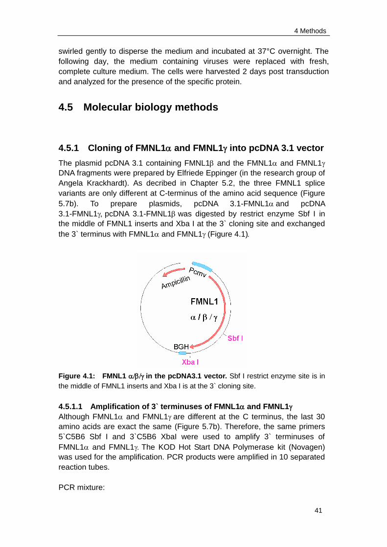
4 Methods
41
swirled gently to disperse the medium and incubated at 37°C overnight. The
following day, the medium containing viruses were replaced with fresh,
complete culture medium. The cells were harvested 2 days post transduction
and analyzed for the presence of the specific protein.
4.5 Molecular biology methods
4.5.1 Cloning of FMNL1 and FMNL1 into pcDNA 3.1 vector
The plasmid pcDNA 3.1 containing FMNL1 and the FMNL1 and FMNL1
DNA fragments were prepared by Elfriede Eppinger (in the research group of
Angela Krackhardt). As decribed in Chapter 5.2, the three FMNL1 splice
variants are only different at C-terminus of the amino acid sequence (Figure
5.7b). To prepare plasmids, pcDNA 3.1-FMNL1and pcDNA
3.1-FMNL1pcDNA 3.1-FMNL1was digested by restrict enzyme Sbf I in
the middle of FMNL1 inserts and Xba I at the 3` cloning site and exchanged
the 3` terminus with FMNL1 and FMNL1(Figure 4.1)
Figure 4.1: FMNL1 in the pcDNA3.1 vector. Sbf I restrict enzyme site is in
the middle of FMNL1 inserts and Xba I is at the 3` cloning site.
4.5.1.1 Amplification of 3` terminuses of FMNL1 and FMNL1
Although FMNL1 and FMNL1are different at the C terminus, the last 30
amino acids are exact the same (Figure 5.7b). Therefore, the same primers
5`C5B6 Sbf I and 3`C5B6 XbaI were used to amplify 3` terminuses of
FMNL1 and FMNL1The KOD Hot Start DNA Polymerase kit (Novagen)
was used for the amplification. PCR products were amplified in 10 separated
reaction tubes.
PCR mixture:
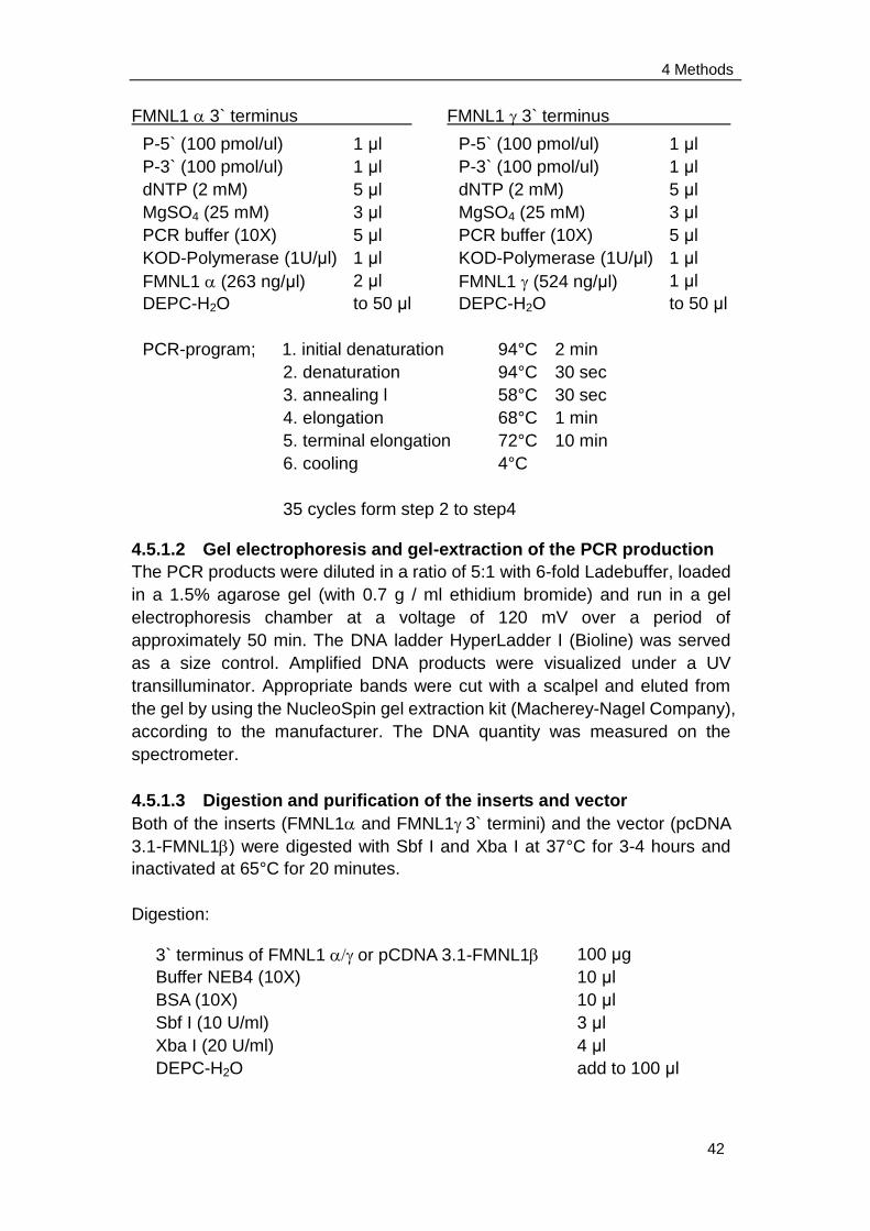
4 Methods
42
FMNL1 3` terminus FMNL1 3` terminus
4.5.1.2 Gel electrophoresis and gel-extraction of the PCR production
The PCR products were diluted in a ratio of 5:1 with 6-fold Ladebuffer, loaded
in a 1.5% agarose gel (with 0.7 g / ml ethidium bromide) and run in a gel
electrophoresis chamber at a voltage of 120 mV over a period of
approximately 50 min. The DNA ladder HyperLadder I (Bioline) was served
as a size control. Amplified DNA products were visualized under a UV
transilluminator. Appropriate bands were cut with a scalpel and eluted from
the gel by using the NucleoSpin gel extraction kit (Macherey-Nagel Company),
according to the manufacturer. The DNA quantity was measured on the
spectrometer.
4.5.1.3 Digestion and purification of the inserts and vector
Both of the inserts (FMNL1 and FMNL13` termini) and the vector (pcDNA
3.1-FMNL1) were digested with Sbf I and Xba I at 37°C for 3-4 hours and
inactivated at 65°C for 20 minutes.
Digestion:
P-5` (100 pmol/ul)
P-3` (100 pmol/ul)
dNTP (2 mM)
MgSO4 (25 mM)
PCR buffer (10X)
KOD-Polymerase (1U/μl)
FMNL1 (263 ng/μl)
DEPC-H2O
1 μl
1 μl
5 μl
3 μl
5 μl
1 μl
2 μl
to 50 μl
P-5` (100 pmol/ul)
P-3` (100 pmol/ul)
dNTP (2 mM)
MgSO4 (25 mM)
PCR buffer (10X)
KOD-Polymerase (1U/μl)
FMNL1 (524 ng/μl)
DEPC-H2O
1 μl
1 μl
5 μl
3 μl
5 μl
1 μl
1 μl
to 50 μl
PCR-program; 1. initial denaturation
2. denaturation
3. annealing l
4. elongation
5. terminal elongation
6. cooling
35 cycles form step 2 to step4
94°C 2 min
94°C 30 sec
58°C 30 sec
68°C 1 min
72°C 10 min
4°C
4
. elongation
5
. terminal
elongation
6
. cooling
3` terminus of FMNL1 or pCDNA 3.1-FMNL1
Buffer NEB4 (10X)
BSA (10X)
Sbf I (10 U/ml)
Xba I (20 U/ml)
DEPC-H2O
100 μg
10 μl
10 μl
3 μl
4 μl
add to 100 μl
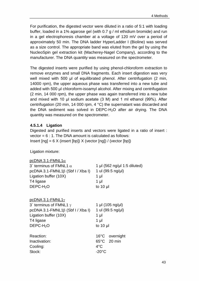
4 Methods
43
For purification, the digested vector were diluted in a ratio of 5:1 with loading
buffer, loaded in a 1% agarose gel (with 0.7 g / ml ethidium bromide) and run
in a gel electrophoresis chamber at a voltage of 120 mV over a period of
approximately 50 min. The DNA ladder HyperLadder I (Bioline) was served
as a size control. The appropriate band was eluted from the gel by using the
NucleoSpin gel extraction kit (Macherey-Nagel Company), according to the
manufacturer. The DNA quantity was measured on the spectrometer.
The digested inserts were purified by using phenol-chloroform extraction to
remove enzymes and small DNA fragments. Each insert digestion was very
well mixed with 500 μl of equilibrated phenol. After centrifugation (2 min,
14000 rpm), the upper aqueous phase was transferred into a new tube and
added with 500 μl chloroform-isoamyl alcohol. After mixing and centrifugation
(2 min, 14 000 rpm), the upper phase was again transferred into a new tube
and mixed with 10 μl sodium acetate (3 M) and 1 ml ethanol (99%). After
centrifugation (20 min, 14 000 rpm, 4 °C) the supernatant was discarded and
the DNA sediment was solved in DEPC-H2O after air drying. The DNA
quantity was measured on the spectrometer.
4.5.1.4 Ligation
Digested and purified inserts and vectors were ligated in a ratio of insert :
vector = 6 : 1. The DNA amount is calculated as follows:
Insert [ng] = 6 X (insert [bp]) X (vector [ng]) / (vector [bp])
Ligation mixture:
pcDNA 3.1-FMNL1
3` terminus of FMNL1
pcDNA 3.1-FMNL1(Sbf I / Xba I)
Ligation buffer (10X)
T4 ligase
DEPC-H2O
1 μl (562 ng/μl 1:5 diluted)
1 ul (99.5 ng/μl)
1 μl
1 μl
to 10 μl
pcDNA 3.1-FMNL1
3` terminus of FMNL1
pcDNA 3.1-FMNL1(Sbf I / Xba I)
Ligation buffer (10X)
T4 ligase
DEPC-H2O
Reaction:
Inactivation:
Cooling:
Stock:
1 μl (105 ng/μl)
1 ul (99.5 ng/μl)
1 μl
1 μl
to 10 μl
16°C overnight
65°C 20 min
4°C
-20°C
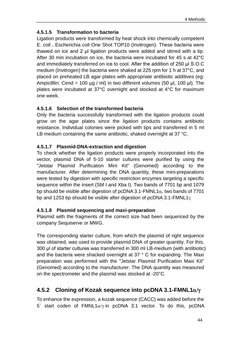
4 Methods
44
4.5.1.5 Transformation to bacteria
Ligation products were transformed by heat shock into chemically competent
E. coli , Escherichia coli One Shot TOP10 (Invitrogen). These bacteria were
thawed on ice and 2 μl ligation products were added and stirred with a tip.
After 30 min incubation on ice, the bacteria were incubated for 45 s at 42°C
and immediately transferred on ice to cool. After the addition of 250 μl S.O.C
medium (Invitrogen) the bacteria were shaked at 225 rpm for 1 h at 37°C, and
placed on preheated LB agar plates with appropriate antibiotic additives (eg:
Ampicilllin: Cend = 100 μg / ml) in two different volumes (50 μl, 100 μl). The
plates were incubated at 37°C overnight and stocked at 4°C for maximum
one week.
4.5.1.6 Selection of the transformed bacteria
Only the bacteria successfully transformed with the ligation products could
grow on the agar plates since the ligation products contains antibiotic
resistance. Individual colonies were picked with tips and transferred in 5 ml
LB medium containing the same antibiotic, shaked overnight at 37 °C.
4.5.1.7 Plasmid-DNA-extraction and digestion
To check whether the ligation products were properly incorporated into the
vector, plasmid DNA of 5-10 starter cultures were purified by using the
"Jetstar Plasmid Purification Mini Kit" (Genomed) according to the
manufacturer. After determining the DNA quantity, these mini-preparations
were tested by digestion with specific restriction enzymes targeting a specific
sequence within the insert (Sbf I and Xba I). Two bands of 7701 bp and 1079
bp should be visible after digestion of pcDNA 3.1-FMNL1two bands of 7701
bp and 1253 bp should be visible after digestion of pcDNA 3.1-FMNL1
4.5.1.8 Plasmid sequencing and maxi-preparation
Plasmid with the fragments of the correct size had been sequenced by the
company Sequiserve or MWG.
The corresponding starter culture, from which the plasmid of right sequence
was obtained, was used to provide plasmid DNA of greater quantity. For this,
300 μl of starter cultures was transferred in 300 ml LB-medium (with antibiotic)
and the bacteria were shacked overnight at 37 ° C for expanding. The Maxi
preparation was performed with the "Jetstar Plasmid Purification Maxi Kit"
(Genomed) according to the manufacturer. The DNA quantity was measured
on the spectrometer and the plasmid was stocked at -20°C.
4.5.2 Cloning of Kozak sequence into pcDNA 3.1-FMNL1
To enhance the expression, a kozak sequence (CACC) was added before the
5` start coden of FMNL1in pcDNA 3.1 vector. To do this, pcDNA
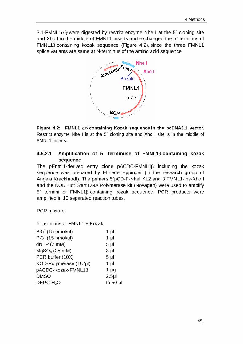
4 Methods
45
3.1-FMNL1were digested by restrict enzyme Nhe I at the 5` cloning site
and Xho I in the middle of FMNL1 inserts and exchanged the 5` terminus of
FMNL1containing kozak sequence (Figure 4.2)since the three FMNL1
splice variants are same at N-terminus of the amino acid sequence.
Figure 4.2: FMNL1 containing Kozak sequencein the pcDNA3.1 vector.
Restrict enzyme Nhe I is at the 5` cloning site and Xho I site is in the middle of
FMNL1 inserts.
4.5.2.1 Amplification of 5` terminuse of FMNL1containing kozak
sequence
The pEntr11-derived entry clone pACDC-FMNL1 including the kozak
sequence was prepared by Elfriede Eppinger (in the research group of
Angela Krackhardt). The primers 5`pCD-F-NheI KL2 and 3`FMNL1-Ins-Xho I
and the KOD Hot Start DNA Polymerase kit (Novagen) were used to amplify
5` termini of FMNL1containing kozak sequence. PCR products were
amplified in 10 separated reaction tubes.
PCR mixture:
5` terminus of FMNL1 + Kozak
P-5` (15 pmol/ul)
P-3` (15 pmol/ul)
dNTP (2 mM)
MgSO4 (25 mM)
PCR buffer (10X)
KOD-Polymerase (1U/μl)
pACDC-Kozak-FMNL1
DMSO
DEPC-H2O
1 μl
1 μl
5 μl
3 μl
5 μl
1 μl
1 μg
2.5μl
to 50 μl
4 Methods
46
The PCR products were used for gel electrophoresis and eluted from the gel
as described before (Chapter 4.5.1.2). The DNA quantity was measured on
the spectrometer.
4.5.2.2 Digestion and purification of the inserts and vector
Both of the inserts (5` terminus of FMNL1+ Kozak) and the vector (pcDNA
3.1-FMNL1) were digested with Nhe I and Xho I at 37°C for 3-4 hours and
inactivated at 65°C for 20 minutes.
Digestion:
Purification of the digested insert and vectors was performed as described
before (Chapter 4.5.1.3). DNA quantity was measured on the spectrometer.
4.5.2.3 Ligation
Digested and purified inserts and vectors were ligated in a ratio of insert:
vector = 6 : 1.
Ligation mixture:
PCR-program; 1. initial denaturation
2. denaturation
3. annealing l
4. elongation
5. terminal elongation
6. cooling
35 cycles form step 2 to step 4
94°C 2 min
94°C 30 sec
68°C 30 sec
68°C 1 min
72°C 10 min
4°C
4
. elongation
5
. terminal
elongation
6
. cooling
5` terminus of FMNL1+ Kozak or pcDNA 3.1-FMNL1
Buffer NEB2 (10X)
BSA (10X)
Nhe I (10 U/ml)
Xho I (20 U/ml)
DEPC-H2O
100 μg
10 μl
10 μl
2 μl
2 μl
add to 100 μl
pcDNA 3.1-Kozak-FMNL1
5` terminus of FMNL1 + Kozak
pcDNA 3.1-FMNL1(Nhe I / Xho I)
Ligation buffer (10X)
T4 ligase
DEPC-H2O
1 μl (52.24 ng/μl)
1.5 ul (64.29 ng/μl)
1 μl
1 μl
to 10 μl
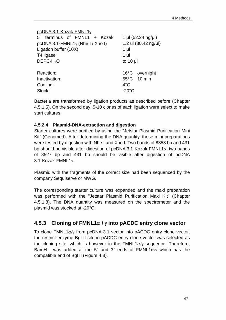
4 Methods
47
Bacteria are transformed by ligation products as described before (Chapter
4.5.1.5). On the second day, 5-10 clones of each ligation were select to make
start cultures.
4.5.2.4 Plasmid-DNA-extraction and digestion
Starter cultures were purified by using the "Jetstar Plasmid Purification Mini
Kit" (Genomed). After determining the DNA quantity, these mini-preparations
were tested by digestion with Nhe I and Xho I. Two bands of 8353 bp and 431
bp should be visible after digestion of pcDNA 3.1-Kozak-FMNL1two bands
of 8527 bp and 431 bp should be visible after digestion of pcDNA
3.1-Kozak-FMNL1.
Plasmid with the fragments of the correct size had been sequenced by the
company Sequiserve or MWG.
The corresponding starter culture was expanded and the maxi preparation
was performed with the "Jetstar Plasmid Purification Maxi Kit" (Chapter
4.5.1.8). The DNA quantity was measured on the spectrometer and the
plasmid was stocked at -20°C.
4.5.3 Cloning of FMNL1 / into pACDC entry clone vector
To clone FMNL1/ from pcDNA 3.1 vector into pACDC entry clone vector,
the restrict enzyme Bgl II site in pACDC entry clone vector was selected as
the cloning site, which is however in the FMNL1 sequence. Therefore,
BamH I was added at the 5` and 3` ends of FMNL1which has the
compatible end of Bgl II (Figure 4.3).
pcDNA 3.1-Kozak-FMNL1
5` terminus of FMNL1 + Kozak
pcDNA 3.1-FMNL1(Nhe I / Xho I)
Ligation buffer (10X)
T4 ligase
DEPC-H2O
Reaction:
Inactivation:
Cooling:
Stock:
1 μl (52.24 ng/μl)
1.2 ul (80.42 ng/μl)
1 μl
1 μl
to 10 μl
16°C overnight
65°C 10 min
4°C
-20°C
4 Methods
48
Figure 4.3: FMNL1 in the pACDC entry clone vector. Restrict enzyme BamH
I at the 5` and 3` ends of FMNL1 was ligated with Bgl II in poly cloning site of
pACDC entry clone.
4.5.3.1 Amplification of FMNL1
The primers 5`Adeno-FMNL1-F and 3`Adeno-FMNL1-C5B6 and the KOD Hot
Start DNA Polymerase kit (Novagen) were used to amplify FMNL1using
pcDNA 3.1-Kozak-FMNL1 as templates. PCR products were amplified in
10 separated reaction tubes.
PCR mixture:
FMNL1
The PCR products were used for gel electrophoresis and eluted from the gel
as described before (Chapter 4.5.1.2). The DNA quantity was measured on
the spectrometer.
P-5` (100 pmol/ul)
P-3` (100 pmol/ul)
dNTP (2 mM)
MgSO4 (25 mM)
PCR buffer (10X)
KOD-Polymerase (1U/μl)
pcDNA 3.1-Kozak-FMNL1
DMSO
DEPC-H2O
1 μl
1 μl
5 μl
3 μl
5 μl
1 μl
1 μg
2.5 μl
to 50 μl
PCR-program; 1. initial denaturation
2. denaturation
3. annealing l
4. elongation
5. terminal elongation
6. cooling
35 cycles form step 2 to step4
94°C 2 min
94°C 30 sec
72°C 30 sec
72°C 4 min
72°C 10 min
4°C
4
. elongation
5
. terminal
elongation
6
. cooling
4 Methods
49
4.5.3.2 Digestion and purification of the inserts and vector
The inserts (FMNL1+ Kozak) was digested by BamH I at 37°C for 3-4
hours and inactivated at 65°C for 20 minutes.
Digestion:
Purification of the digested insert was performed as described before
(Chapter 4.5.1.3). DNA quantity was measured on the spectrometer. The Bgl
II digestion and purification of vector (pACDC) was done by Elfriede Eppinger
(in the research group of Angela Krackhardt).
4.5.3.3 Ligation
Digested and purified inserts and vectors were ligated in a ratio of insert:
vector = 5 : 1.
Ligation mixture:
Ligation products were transformed to bacteria as described before (Chapter
4.5.1.5). On the second day, 5-10 clones of each ligation were select to make
starter cultures.
4.5.3.4 Plasmid-DNA-extraction and digestion
FMNL1
BamH I buffer with BSA (10X)
BamH I (10 U/ml)
DEPC-H2O
100 μg
10 μl
3 μl
to 100 μl
pACDC-FMNL1
FMNL1 (BamH I)
pACDC(Bgl II)
Ligation buffer (10X)
T4 ligase
DEPC-H2O
2.6 μl (117 ng/μl)
0.5 ul (220 ng/μl)
1 μl
1 μl
to 10 μl
pACDC-FMNL1
FMNL1 (BamH I)
pACDC(Bgl II)
Ligation buffer (10X)
T4 ligase
DEPC-H2O
Reaction:
Inactivation:
Cooling:
Stock:
3 μl (104 ng/μl)
0.5 ul (220 ng/μl)
1 μl
1 μl
to 10 μl
16°C overnight
65°C 10 min
4°C
-20°C
4 Methods
50
Starter cultures were purified by using the "Jetstar Plasmid Purification Mini
Kit" (Genomed). After determining the DNA quantity, these mini-preparations
were tested by digestion with EcoR I (in insert) and Kpn I (in vector). Two
bands of 5302 bp and 3165 bp should be visible after digestion of
pACDC-Kozak-FMNL1two bands of 5302 bp and 3339 bp should be visible
after digestion of pACDC-Kozak-FMNL1.
Plasmid with the fragments of the correct size had been sequenced by the
company Sequiserve or MWG.
The corresponding starter culture was expanded and the maxi preparation
was performed with the "Jetstar Plasmid Purification Maxi Kit" (Chapter
4.5.1.8). The DNA quantity was measured on the spectrometer and the
plasmid was stocked at -20°C.
4.5.4 Mutagenesis
Mutagenesis was performed by QuikChange® Site-Directed Mutagenesis Kit
(STRATAGENE) which allows site-specific mutation in virtually any
double-stranded plasmid. The QuikChange® site-directed mutagenesis
method is performed using PfuTurbo DNA polymerase and a temperature
cycler. The basic principle is that PfuTurbo DNA polymerase replicates both
plasmid strands with high fidelity and without displacing the mutant
oligonucleotide primers. The oligonucleotide primers, each complementary to
opposite strands of the plasmid, are extended during temperature cycling to
generate a mutated plasmid containing staggered nicks. Following
temperature cycling, the product is treated with Dpn I (target sequence:
5 -Gm6ATC-3 ) which is specific for methylated and hemimethylated DNA and
is used to digest the parental DNA template and to select for
mutation-containing synthesized DNA. DNA isolated from almost all E. coli
strains is dam methylated and therefore susceptible to Dpn I digestion. The
nicked vector DNA containing the desired mutations is then transformed into
XL1-Blue supercompetent cells.
4.5.4.1 Mutagenesis of N-terminal myristoylation site in pACDC-FMNL1
To mutate the N-terminal myristoylation site (Chapter 5.4, Figure 5.15), the
oligonucleotide primers G2TA4T and G2TA4T anti were designed using
QuikChange® Primer Design Program available online
(http://www.stratagene.com/qcprimerdesign). A four-step procedure to
generate mutants was performed according to the introduction manual.
Reaction mixture:
4 Methods
51
After cycling, 1 μl of the Dpn I restriction enzyme (10 U/μl) was directly added
to the amplified product and incubated at 37°C for 1 hour to digest the parental
supercoiled dsDNA. After that, reaction was transformed into XL1-Blue
supercompetent cells.
DNA was extracted from selected bacteria clones and digested by EcoR I and
Kpn I. Plasmids with the fragments of the correct size were sent for
sequencing by the company Sequiserve or MWG to check if correct mutants
were obtained. The plasmid containing the correct mutant was named
pACDC-G2TA4T.
4.5.4.2 Cloning of the N-terminal myristoylation site mutant into
pcDNA 3.1-FMNL1
In order to clone the mutated N-terminal myristoylation site into pcDNA
3.1-FMNL1, pcNDA 3.1-FMNL1was digested by restrict enzyme Nhe I at
the 5` cloning site and Xho I in the middle of FMNL1 inserts and exchanged
the 5` terminus with G2TA4T containing the mutant N-terminal myristoylation
site (Figure 4.2).
The primers 5`G2TA4T-Nhe I and 3`FMNL1-Ins-Xho I and the KOD Hot Start
DNA Polymerase kit (Novagen) were used to amplify the 5` terminus
sequence of G2TA4T using pACDC-G2TA4T as template. PCR products
were amplified in 10 separated reaction tubes.
PCR mixture:
P-G2TA4T (100 ng/ul)
P-G2TA4T anti (100 ng/ul)
dNTP mix
Reacton buffer (10X)
PfuTurbo DNA polymerase
pACDC-FMNL1
DMSO
DEPC-H2O
1.25 μl
1.25 μl
1 μl
5 μl
1 μl
1.925 μl (25ng)
4 μl (Cend=8%)
to 50 μl
Cycling-program; 1. initial denaturation
2. denaturation
3. annealing l
4. elongation
18 cycles form step 2 to step4
95°C 2 min
95°C 1 min
68°C 1 min
68°C 9 min
5
6
. cooling
4 Methods
52
5` terminus of G2TA4T
The PCR products were used for gel electrophoresis and eluted from the gel.
After digestion with Nhe I and Xho I, the PCR products were ligated with
pcDNA 3.1-FMNL1digested with the same enzymes in a ratio of 6:1.
Bacteria were transformed by ligation products. After DNA extraction, the
plasmids with correct insert were selected by digestion with Nhe I and Xho I
and sequencing. Maxi preparation was performed with the "Jetstar Plasmid
Purification Maxi Kit" (Chapter 4.5.2). The DNA quantity was measured on the
spectrometer and the plasmid was stocked at -20°C.
4.5.5 Cloning of FMNL1ΔDAD and G2TA4TΔDAD into pcDNA
3.1 vector and pACDC entry clone vector
4.5.5.1 Cloning of FMNL1ΔDAD and G2TA4TΔDAD into pcDNA 3.1
To Clone FMNL1ΔDAD into pcDNA 3.1, the restrict enzyme Nhe I and Xba I
were selected as the cloning sites in pcDNA 3.1 vector (Figure 4.4). The
primers 5`pcD-F-NheI KL2 and 3`pcD-F-C-Xba I 2 were used to amplify
FMNL1ΔDAD sequence using pcDNA 3.1-Kozak-FMNL1 as template. The
amplification was performed by KOD Hot Start DNA Polymerase kit
(Novagen). The gel-purified PCR production was used as template for further
PCR reaction to obtain more product. PCR products were amplified in 10
separated reaction tubes.
P-5` (15 pmol/ul)
P-3` (15 pmol/ul)
dNTP (2 mM)
MgSO4 (25 mM)
PCR buffer (10X)
KOD-Polymerase (1U/μl)
pACDC-FMNL1
DMSO
DEPC-H2O
PCR-program; 1. initial denaturation
2. denaturation
3. annealing l
4. elongation
5. terminal elongation
6. cooling
35 cycles form step 2 to step4
94°C 2 min
94°C 30 sec
68°C 30 sec
68°C 1 min
68°C 10 min
4°C
4
. elongation
5
. terminal
elongation
6
. cooling
1 μl
1 μl
5 μl
3 μl
5 μl
1 μl
1 μg
2.5 μl
to 50 μl
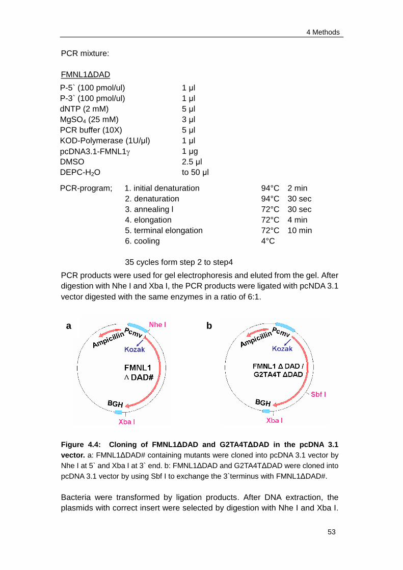
4 Methods
53
PCR mixture:
FMNL1ΔDAD
PCR products were used for gel electrophoresis and eluted from the gel. After
digestion with Nhe I and Xba I, the PCR products were ligated with pcNDA 3.1
vectordigested with the same enzymes in a ratio of 6:1.
Figure 4.4: Cloning of FMNL1ΔDAD and G2TA4TΔDAD in the pcDNA 3.1
vector. a: FMNL1ΔDAD# containing mutants were cloned into pcDNA 3.1 vector by
Nhe I at 5` and Xba I at 3` end. b: FMNL1ΔDAD and G2TA4TΔDAD were cloned into
pcDNA 3.1 vector by using Sbf I to exchange the 3`terminus with FMNL1ΔDAD#.
Bacteria were transformed by ligation products. After DNA extraction, the
plasmids with correct insert were selected by digestion with Nhe I and Xba I.
P-5` (100 pmol/ul)
P-3` (100 pmol/ul)
dNTP (2 mM)
MgSO4 (25 mM)
PCR buffer (10X)
KOD-Polymerase (1U/μl)
pcDNA3.1-FMNL1
DMSO
DEPC-H2O
PCR-program; 1. initial denaturation
2. denaturation
3. annealing l
4. elongation
5. terminal elongation
6. cooling
35 cycles form step 2 to step4
94°C 2 min
94°C 30 sec
72°C 30 sec
72°C 4 min
72°C 10 min
4°C
4
. elongation
5
. terminal
elongation
6
. cooling
1 μl
1 μl
5 μl
3 μl
5 μl
1 μl
1 μg
2.5 μl
to 50 μl
a b
4 Methods
54
However, several mutatations were found in the FMNL1ΔDAD sequence
before Sbf I restriction site by sequencing. Maxi preparation of pcDNA 3.1-
FMNL1ΔDAD containing mutatations (pcDNA 3.1-FMNL1ΔDAD#) was
performed with the "Jetstar Plasmid Purification Maxi Kit". The 3` terminus
lacking DAD was cut out from FMNL1ΔDAD# by Sbf I and Xba I and
exchanged with the 3`terminuses of FMNL1 and G2TA3T in pcDNA 3.1
vector (Figure 4.4).
Digestion1:
The small fragment of 956 bp was extracted from the gel and used as insert
for ligation (3`terminus of FMNL1ΔDAD).
Digestion2:
The big fragments of 7705 bp were extracted from the gel and used as cut
vector for ligation (pcDNA 3.1-FMNL1 / G2TA4T (Sbf I / Xba I)).
Ligation (insert : vector = 6 : 1)
pcDNA 3.1-FMNL1ΔDAD#
Buffer NEB4 (10X)
BSA (10X)
Sbf I (10 U/ml)
Xba I (20 U/ml)
DEPC-H2O
10 μg
10 μl
10 μl
3 μl
3 μl
to 100 μl
pcDNA 3.1-FMNL1 / G2TA4T
Buffer NEB4 (10X)
BSA (10X)
Sbf I (10 U/ml)
Xba I (20 U/ml)
DEPC-H2O
20 μg
10 μl
10 μl
3 μl
3 μl
add to 100 μl
pcDNA 3.1-FMNL1ΔDAD:
3`terminus of FMNL1ΔDAD
pcDNA 3.1-FMNL1 (Sbf I / Xba I)
Ligation buffer (10X)
T4 ligase
DEPC-H2O
0.83 μl (90.9 ng/μl)
1.55 ul (64.4 ng/μl)
1 μl
1 μl
to 10 μl
pcDNA 3.1-GTA4TΔDAD:
3`terminus of FMNL1ΔDAD
pcDNA 3.1-G2TA4T (Sbf I / Xba I)
Ligation buffer (10X)
T4 ligase
DEPC-H2O
0.83 μl (90.9 ng/μl)
0.67 ul (148.5 ng/μl)
1 μl
1 μl
to 10 μl

4 Methods
55
Bacteria were transformed by ligation products. After DNA extraction, the
plasmids with correct insert were selected by digestion with Sbf I (in insert)
and Mfe I (in vector). Two bands of 5723 bp and 2938 bp should be visible
after digestion and gel-electrophoresis of both pcDNA 3.1-FMNL1ΔDAD and
pcDNA 3.1-G2TA4TΔDAD.
Plasmids with the fragments of the correct size had been sequenced by the
company Sequiserve or MWG.
The corresponding starter culture was expanded and the maxi preparation
was performed with the "Jetstar Plasmid Purification Maxi Kit". The DNA
quantity was measured on the spectrometer and the plasmid was stocked at
-20°C.
4.5.5.2 Cloning of FMNL1ΔDAD and G2TA4TΔDAD into pACDC entry
clone vector
To clone FMNL1ΔDAD and G2TA4TΔDAD from pcDNA 3.1 vector into
pACDC entry clone vector, similar procedures have been performed as
described in chapter 4.5.3.
The primers 5`Adeno-FMNL1-F and 3`FMNL1-C-BamH I 2 were used to
amplify the FMNL1ΔDAD, while the FMNL1ΔDAD fragment was used as
template which was cut out from pcDNA 3.1-FMNL1ΔDAD by Nhe I and Xba I
and purified by gel extraction. The primers 5`-G2TA4T-BamH I 2 and
3`FMNL1-C-BamH I 2 were used to amplify G2TA4TΔDAD while the
G2TA4TΔDAD fragment was used as template which was cut out from
pcDNA 3.1- G2TA4TΔDAD by Nhe I and Xba I and purified by gel extraction.
The amplification was performed by using the KOD Hot Start DNA
Polymerase kit (Novagen). The product was amplified in 10 separated
reaction tubes.
PCR mixture:
Reaction:
Inactivation:
Cooling:
16°C overnight
65°C 10 min
4°C

4 Methods
56
FMNL1ΔDAD: G2TA4TΔDAD:
The PCR products were used for gel electrophoresis and eluted from the gel
and digested by BamH I at 37°C for 3-4 hours and inactivated at 65°C for 20
minutes.
Digestion:
After purification, the digested inserts were ligated with the Bgl II digested and
purified pACDC entry clone vector with a ratio of 6 : 1.
Ligation mixture:
P-5` (100 pmol/ul)
P-3` (100 pmol/ul)
dNTP (2 mM)
MgSO4 (25 mM)
PCR buffer (10X)
KOD-Polymerase (1U/μl)
insert
DMSO
DEPC-H2O
1 μl
1 μl
5 μl
3 μl
5 μl
1 μl
1 μl
2.5 μl
to 50 μl
PCR-program; 1. initial denaturation
2. denaturation
3. annealing l
4. elongation
5. terminal elongation
6. cooling
35 cycles form step 2 to step4
94°C 2 min
94°C 30 sec
72°C 30 sec
72°C 4 min
72°C 10 min
4°C
4
. elongation
5
. terminal
elongation
6
. cooling
FMNL1ΔDAD or G2TA4TΔDAD
BamH I buffer with BSA (10X)
BamH I (10 U/ml)
DEPC-H2O
100 μg
10 μl
3 μl
to 100 μl
pACDC-FMNL1ΔDAD
FMNL1ΔDAD (BamH I)
pACDC(Bgl II)
Ligation buffer (10X)
T4 ligase
DEPC-H2O
0.35 μl (1034 ng/μl)
0.5 ul (220 ng/μl)
1 μl
1 μl
to 10 μl
P-5` (100 pmol/ul)
P-3` (100 pmol/ul)
dNTP (2 mM)
MgSO4 (25 mM)
PCR buffer (10X)
KOD-Polymerase (1U/μl)
G2TA4T insert
DMSO
DEPC-H2O
1 μl
1 μl
5 μl
3 μl
5 μl
1 μl
1 μl
2.5 μl
to 50 μl
4 Methods
57
Bacteria were transformed by ligation products. After DNA extraction, the
plasmids with correct insert were selected by double digestion.
Plasmid Sbf I (ins)/Mfe I (vec) EcoR I (ins)/Kpn I (vec)
pACDC-Kozak-FMNL1ΔDAD 5572 bp/2756 bp 5286 bp/3042 bp
pACDC-Kozak-G2TA4TΔDAD 5572 bp/2756 bp 5286 bp/3042 bp
Plasmid with the fragments of the correct size had been sequenced by the
company Sequiserve or MWG. The corresponding starter culture was
expanded and the maxi preparation was performed with the "Jetstar Plasmid
Purification Maxi Kit". The DNA quantity was measured on the spectrometer
and the plasmid was stocked at -20°C.
pACDC-G2TA4TΔDAD
GTA4TΔDAD (BamH I)
pACDC(Bgl II)
Ligation buffer (10X)
T4 ligase
DEPC-H2O
1.3 μl (289 ng/μl)
0.5 ul (220 ng/μl)
1 μl
1 μl
to 10 μl
Reaction:
Inactivation:
Cooling:
Stock:
16°C overnight
65°C 10 min
4°C
-20°C
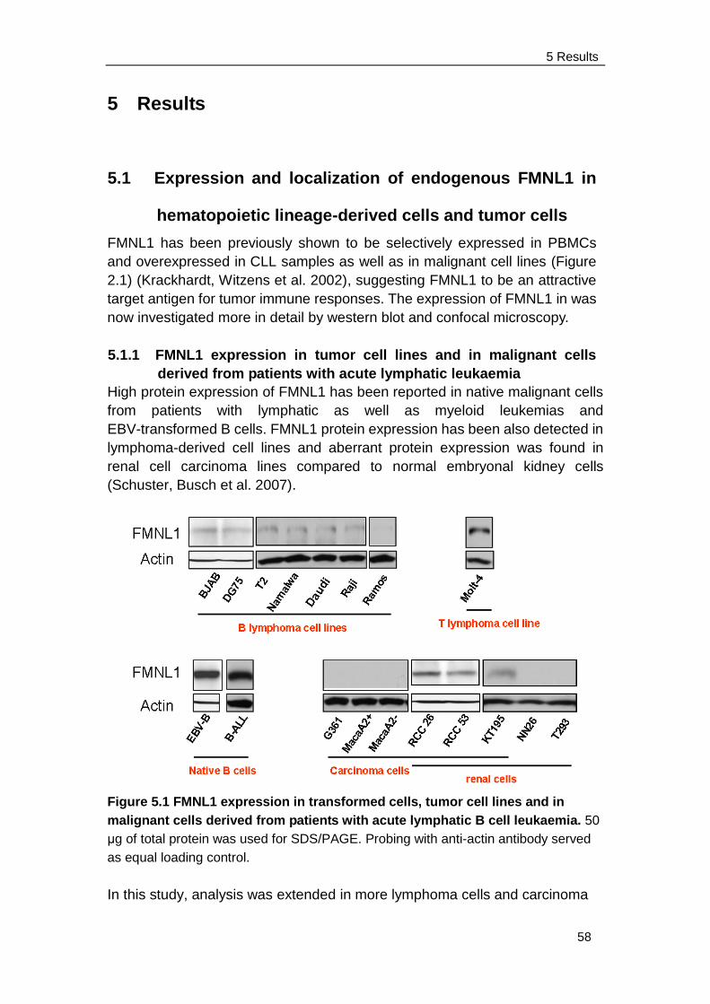
5 Results
58
5 Results
5.1 Expression and localization of endogenous FMNL1 in
hematopoietic lineage-derived cells and tumor cells
FMNL1 has been previously shown to be selectively expressed in PBMCs
and overexpressed in CLL samples as well as in malignant cell lines (Figure
2.1) (Krackhardt, Witzens et al. 2002), suggesting FMNL1 to be an attractive
target antigen for tumor immune responses. The expression of FMNL1 in was
now investigated more in detail by western blot and confocal microscopy.
5.1.1 FMNL1 expression in tumor cell lines and in malignant cells
derived from patients with acute lymphatic leukaemia
High protein expression of FMNL1 has been reported in native malignant cells
from patients with lymphatic as well as myeloid leukemias and
EBV-transformed B cells. FMNL1 protein expression has been also detected in
lymphoma-derived cell lines and aberrant protein expression was found in
renal cell carcinoma lines compared to normal embryonal kidney cells
(Schuster, Busch et al. 2007).
Figure 5.1 FMNL1 expression in transformed cells, tumor cell lines and in
malignant cells derived from patients with acute lymphatic B cell leukaemia. 50
μg of total protein was used for SDS/PAGE. Probing with anti-actin antibody served
as equal loading control.
In this study, analysis was extended in more lymphoma cells and carcinoma
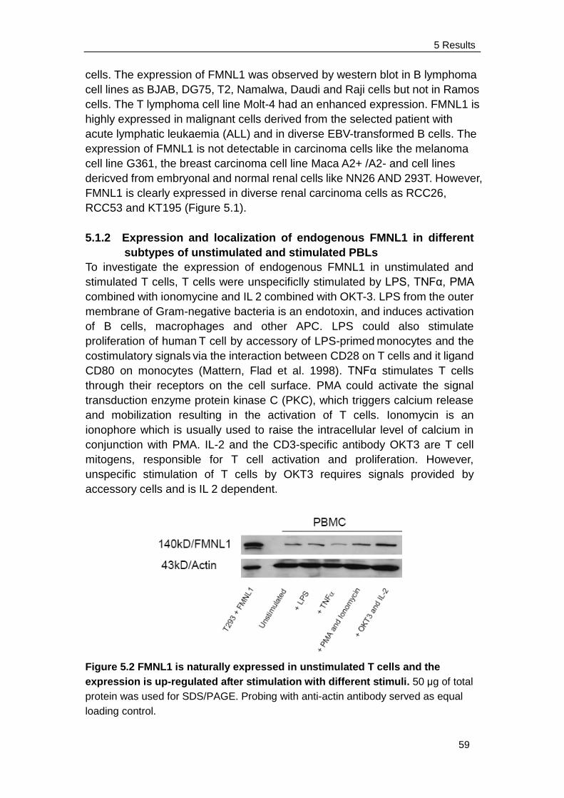
5 Results
59
cells. The expression of FMNL1 was observed by western blot in B lymphoma
cell lines as BJAB, DG75, T2, Namalwa, Daudi and Raji cells but not in Ramos
cells. The T lymphoma cell line Molt-4 had an enhanced expression. FMNL1 is
highly expressed in malignant cells derived from the selected patient with
acute lymphatic leukaemia (ALL) and in diverse EBV-transformed B cells. The
expression of FMNL1 is not detectable in carcinoma cells like the melanoma
cell line G361, the breast carcinoma cell line Maca A2+ /A2- and cell lines
dericved from embryonal and normal renal cells like NN26 AND 293T. However,
FMNL1 is clearly expressed in diverse renal carcinoma cells as RCC26,
RCC53 and KT195 (Figure 5.1).
5.1.2 Expression and localization of endogenous FMNL1 in different
subtypes of unstimulated and stimulated PBLs
To investigate the expression of endogenous FMNL1 in unstimulated and
stimulated T cells, T cells were unspecificlly stimulated by LPS, TNFα, PMA
combined with ionomycine and IL 2 combined with OKT-3. LPS from the outer
membrane of Gram-negative bacteria is an endotoxin, and induces activation
of B cells, macrophages and other APC. LPS could also stimulate
proliferation of human T cell by accessory of LPS-primed monocytes and the
costimulatory signals via the interaction between CD28 on T cells and it ligand
CD80 on monocytes (Mattern, Flad et al. 1998). TNFα stimulates T cells
through their receptors on the cell surface. PMA could activate the signal
transduction enzyme protein kinase C (PKC), which triggers calcium release
and mobilization resulting in the activation of T cells. Ionomycin is an
ionophore which is usually used to raise the intracellular level of calcium in
conjunction with PMA. IL-2 and the CD3-specific antibody OKT3 are T cell
mitogens, responsible for T cell activation and proliferation. However,
unspecific stimulation of T cells by OKT3 requires signals provided by
accessory cells and is IL 2 dependent.
Figure 5.2 FMNL1 is naturally expressed in unstimulated T cells and the
expression is up-regulated after stimulation with different stimuli. 50 μg of total
protein was used for SDS/PAGE. Probing with anti-actin antibody served as equal
loading control.
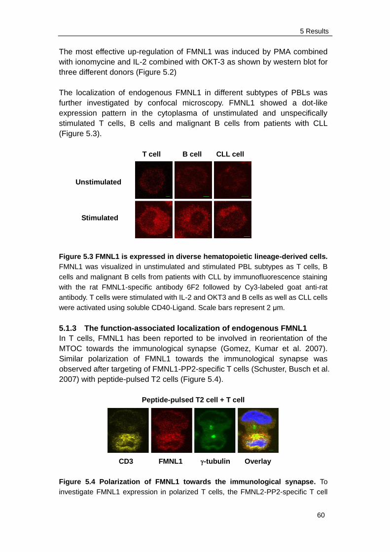
5 Results
60
The most effective up-regulation of FMNL1 was induced by PMA combined
with ionomycine and IL-2 combined with OKT-3 as shown by western blot for
three different donors (Figure 5.2)
The localization of endogenous FMNL1 in different subtypes of PBLs was
further investigated by confocal microscopy. FMNL1 showed a dot-like
expression pattern in the cytoplasma of unstimulated and unspecifically
stimulated T cells, B cells and malignant B cells from patients with CLL
(Figure 5.3).
T cell B cell CLL cell
Figure 5.3 FMNL1 is expressed in diverse hematopoietic lineage-derived cells.
FMNL1 was visualized in unstimulated and stimulated PBL subtypes as T cells, B
cells and malignant B cells from patients with CLL by immunofluorescence staining
with the rat FMNL1-specific antibody 6F2 followed by Cy3-labeled goat anti-rat
antibody. T cells were stimulated with IL-2 and OKT3 and B cells as well as CLL cells
were activated using soluble CD40-Ligand. Scale bars represent 2 μm.
5.1.3 The function-associated localization of endogenous FMNL1
In T cells, FMNL1 has been reported to be involved in reorientation of the
MTOC towards the immunological synapse (Gomez, Kumar et al. 2007).
Similar polarization of FMNL1 towards the immunological synapse was
observed after targeting of FMNL1-PP2-specific T cells (Schuster, Busch et al.
2007) with peptide-pulsed T2 cells (Figure 5.4).
Peptide-pulsed T2 cell + T cell
CD3 FMNL1 -tubulin Overlay
Figure 5.4 Polarization of FMNL1 towards the immunological synapse. To
investigate FMNL1 expression in polarized T cells, the FMNL2-PP2-specific T cell
Unstimulated
Stimulated
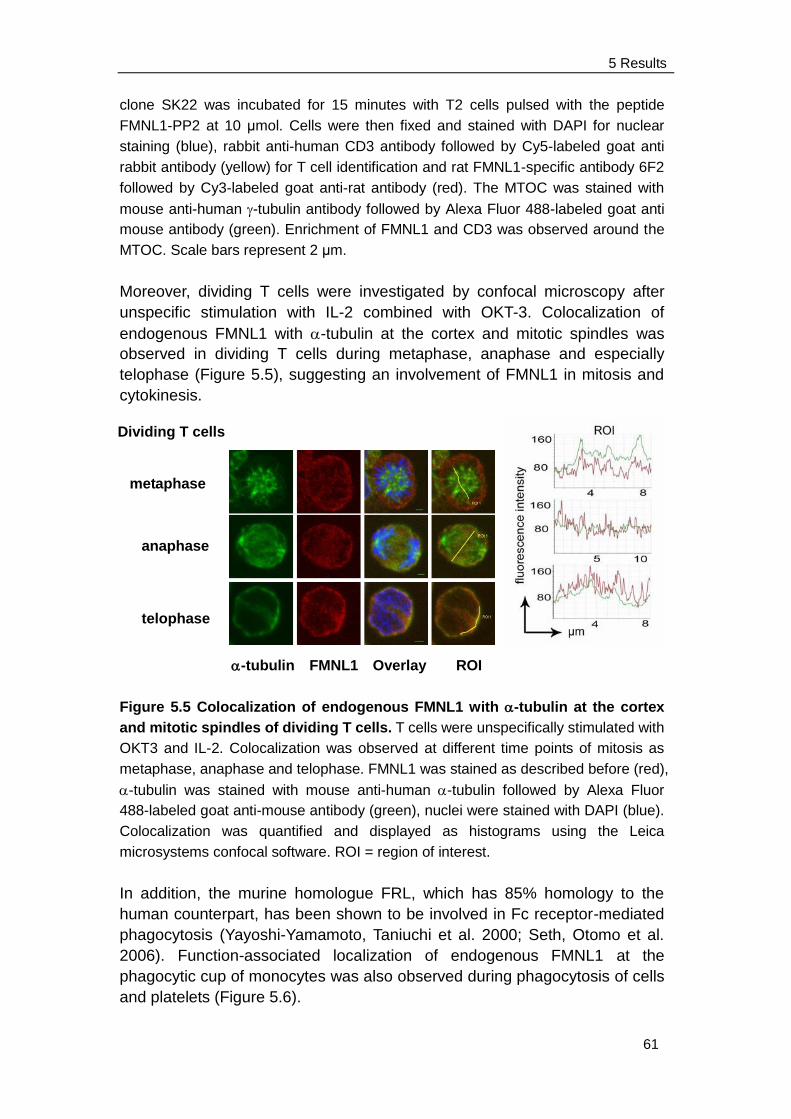
5 Results
61
clone SK22 was incubated for 15 minutes with T2 cells pulsed with the peptide
FMNL1-PP2 at 10 μmol. Cells were then fixed and stained with DAPI for nuclear
staining (blue), rabbit anti-human CD3 antibody followed by Cy5-labeled goat anti
rabbit antibody (yellow) for T cell identification and rat FMNL1-specific antibody 6F2
followed by Cy3-labeled goat anti-rat antibody (red). The MTOC was stained with
mouse anti-human -tubulin antibody followed by Alexa Fluor 488-labeled goat anti
mouse antibody (green). Enrichment of FMNL1 and CD3 was observed around the
MTOC. Scale bars represent 2 μm.
Moreover, dividing T cells were investigated by confocal microscopy after
unspecific stimulation with IL-2 combined with OKT-3. Colocalization of
endogenous FMNL1 with -tubulin at the cortex and mitotic spindles was
observed in dividing T cells during metaphase, anaphase and especially
telophase (Figure 5.5), suggesting an involvement of FMNL1 in mitosis and
cytokinesis.
-tubulin FMNL1 Overlay ROI
Figure 5.5 Colocalization of endogenous FMNL1 with -tubulin at the cortex
and mitotic spindles of dividing T cells. T cells were unspecifically stimulated with
OKT3 and IL-2. Colocalization was observed at different time points of mitosis as
metaphase, anaphase and telophase. FMNL1 was stained as described before (red),
-tubulin was stained with mouse anti-human -tubulin followed by Alexa Fluor
488-labeled goat anti-mouse antibody (green), nuclei were stained with DAPI (blue).
Colocalization was quantified and displayed as histograms using the Leica
microsystems confocal software. ROI = region of interest.
In addition, the murine homologue FRL, which has 85% homology to the
human counterpart, has been shown to be involved in Fc receptor-mediated
phagocytosis (Yayoshi-Yamamoto, Taniuchi et al. 2000; Seth, Otomo et al.
2006). Function-associated localization of endogenous FMNL1 at the
phagocytic cup of monocytes was also observed during phagocytosis of cells
and platelets (Figure 5.6).
metaphase
anaphase
telophase
Dividing T cells
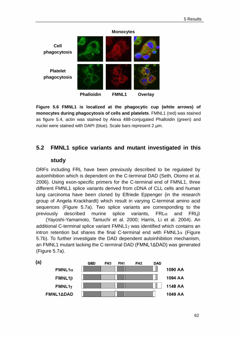
5 Results
62
Monocytes
Phalloidin FMNL1 Overlay
Figure 5.6 FMNL1 is localized at the phagocytic cup (white arrows) of
monocytes during phagocytosis of cells and platelets. FMNL1 (red) was stained
as figure 5.4, actin was stained by Alexa 488-conjugated Phalloidin (green) and
nuclei were stained with DAPI (blue). Scale bars represent 2 μm.
5.2 FMNL1 splice variants and mutant investigated in this
study
DRFs including FRL have been previously described to be regulated by
autoinhibition which is dependent on the C-terminal DAD (Seth, Otomo et al.
2006). Using exon-specific primers for the C-terminal end of FMNL1, three
different FMNL1 splice variants derived from cDNA of CLL cells and human
lung carcinoma have been cloned by Elfriede Eppenger (in the research
group of Angela Krackhardt) which result in varying C-terminal amino acid
sequences (Figure 5.7a). Two splice variants are corresponding to the
previously described murine splice variants, FRL and FRL
(Yayoshi-Yamamoto, Taniuchi et al. 2000; Harris, Li et al. 2004). An
additional C-terminal splice variant FMNL1 was identified which contains an
intron retention but shares the final C-terminal end with FMNL1 (Figure
5.7b). To further investigate the DAD dependent autoinhibition mechanism,
an FMNL1 mutant lacking the C-terminal DAD (FMNL1ΔDAD) was generated
(Figure 5.7a).
Cell
phagocytosis
Platelet
phagocytosis
(a)
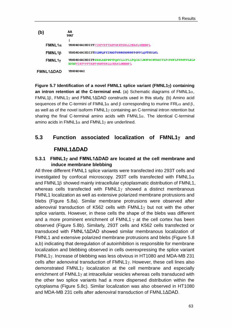
5 Results
63
Figure 5.7 Identification of a novel FMNL1 splice variant (FMNL1) containing
an intron retention at the C-terminal end. (a) Schematic diagrams of FMNL1,
FMNL1, FMNL1 and FMNL1ΔDAD constructs used in this study. (b) Amino acid
sequences of the C-termini of FMNL1 and corresponding to murine FRL and ,
as well as of the novel isoform FMNL1 containing an C-terminal intron retention but
sharing the final C-terminal amino acids with FMNL1. The identical C-terminal
amino acids in FMNL1 and FMNL1 are underlined.
5.3 Function associated localization of FMNL1 and
FMNL1ΔDAD
5.3.1 FMNL1 and FMNL1ΔDAD are located at the cell membrane and
induce membrane blebbing
All three different FMNL1 splice variants were transfected into 293T cells and
investigated by confocal microscopy. 293T cells transfected with FMNL1
and FMNL1 showed mainly intracellular cytoplasmatic distribution of FMNL1,
whereas cells transfected with FMNL1 showed a distinct membranous
FMNL1 localization as well as extensive polarized membrane protrusions and
blebs (Figure 5.8a). Similar membrane protrusions were observed after
adenoviral transduction of K562 cells with FMNL1 but not with the other
splice variants. However, in these cells the shape of the blebs was different
and a more prominent enrichment of FMNL1 at the cell cortex has been
observed (Figure 5.8b). Similarly, 293T cells and K562 cells transfected or
transduced with FMNL1ΔDAD showed similar membranous localization of
FMNL1 and extensive polarized membrane protrusions and blebs (Figure 5.8
a,b) indicating that deregulation of autoinhibition is responsible for membrane
localization and blebbing observed in cells overexpressing the splice variant
FMNL1. Increase of blebbing was less obvious in HT1080 and MDA-MB 231
cells after adenoviral transduction of FMNL1. However, these cell lines also
demonstrated FMNL1 localization at the cell membrane and especially
enrichment of FMNL1 at intracellular vesicles whereas cells transduced with
the other two splice variants had a more dispersed distribution within the
cytoplasma (Figure 5.8c). Similar localization was also observed in HT1080
and MDA-MB 231 cells after adenoviral transduction of FMNL1ΔDAD.
(b)
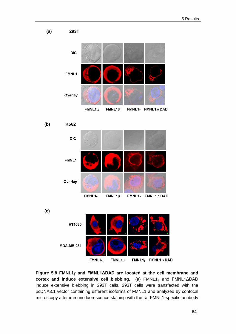
5 Results
64
(a) 293T
(b) K562
(c)
Figure 5.8 FMNL1 and FMNL1ΔDAD are located at the cell membrane and
cortex and induce extensive cell blebbing. (a) FMNL1 and FMNL1ΔDAD
induce extensive blebbing in 293T cells. 293T cells were transfected with the
pcDNA3.1 vector containing different isoforms of FMNL1 and analyzed by confocal
microscopy after immunofluorescence staining with the rat FMNL1-specific antibody
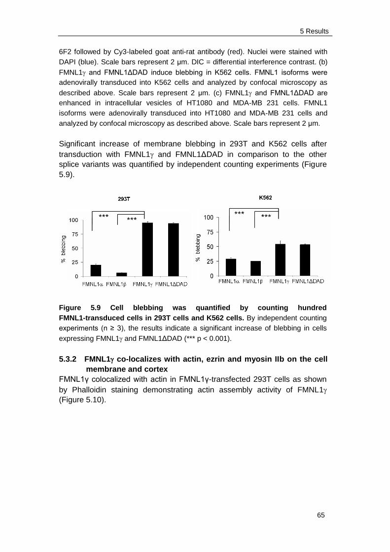
5 Results
65
6F2 followed by Cy3-labeled goat anti-rat antibody (red). Nuclei were stained with
DAPI (blue). Scale bars represent 2 μm. DIC = differential interference contrast. (b)
FMNL1 and FMNL1ΔDAD induce blebbing in K562 cells. FMNL1 isoforms were
adenovirally transduced into K562 cells and analyzed by confocal microscopy as
described above. Scale bars represent 2 μm. (c) FMNL1 and FMNL1ΔDAD are
enhanced in intracellular vesicles of HT1080 and MDA-MB 231 cells. FMNL1
isoforms were adenovirally transduced into HT1080 and MDA-MB 231 cells and
analyzed by confocal microscopy as described above. Scale bars represent 2 μm.
Significant increase of membrane blebbing in 293T and K562 cells after
transduction with FMNL1 and FMNL1ΔDAD in comparison to the other
splice variants was quantified by independent counting experiments (Figure
5.9).
Figure 5.9 Cell blebbing was quantified by counting hundred
FMNL1-transduced cells in 293T cells and K562 cells. By independent counting
experiments (n ≥ 3), the results indicate a significant increase of blebbing in cells
expressing FMNL1 and FMNL1ΔDAD (*** p < 0.001).
5.3.2 FMNL1 co-localizes with actin, ezrin and myosin IIb on the cell
membrane and cortex
FMNL1γ colocalized with actin in FMNL1γ-transfected 293T cells as shown
by Phalloidin staining demonstrating actin assembly activity of FMNL1
(Figure 5.10).
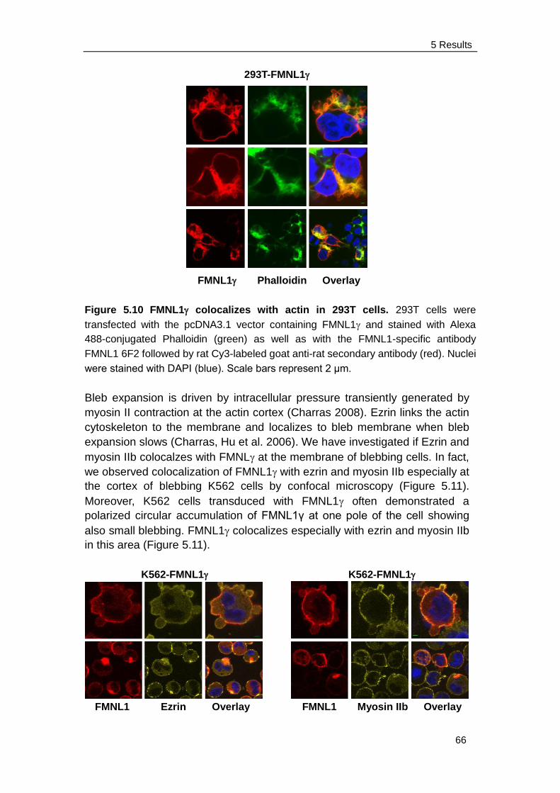
5 Results
66
293T-FMNL1
FMNL1 Phalloidin Overlay
Figure 5.10 FMNL1 colocalizes with actin in 293T cells. 293T cells were
transfected with the pcDNA3.1 vector containing FMNL1 and stained with Alexa
488-conjugated Phalloidin (green) as well as with the FMNL1-specific antibody
FMNL1 6F2 followed by rat Cy3-labeled goat anti-rat secondary antibody (red). Nuclei
were stained with DAPI (blue). Scale bars represent 2 μm.
Bleb expansion is driven by intracellular pressure transiently generated by
myosin II contraction at the actin cortex (Charras 2008). Ezrin links the actin
cytoskeleton to the membrane and localizes to bleb membrane when bleb
expansion slows (Charras, Hu et al. 2006). We have investigated if Ezrin and
myosin IIb colocalzes with FMNL at the membrane of blebbing cells. In fact,
we observed colocalization of FMNL1 with ezrin and myosin IIb especially at
the cortex of blebbing K562 cells by confocal microscopy (Figure 5.11).
Moreover, K562 cells transduced with FMNL1 often demonstrated a
polarized circular accumulation of FMNL1γ at one pole of the cell showing
also small blebbing. FMNL1 colocalizes especially with ezrin and myosin IIb
in this area (Figure 5.11).
K562-FMNL1K562-FMNL1
FMNL1 Ezrin Overlay FMNL1 Myosin IIb Overlay
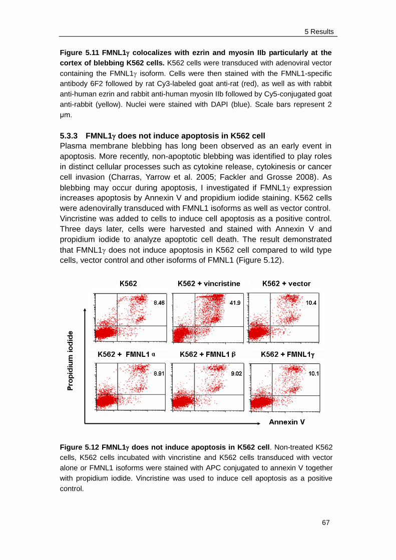
5 Results
67
Figure 5.11 FMNL1 colocalizes with ezrin and myosin IIb particularly at the
cortex of blebbing K562 cells. K562 cells were transduced with adenoviral vector
containing the FMNL1 isoform. Cells were then stained with the FMNL1-specific
antibody 6F2 followed by rat Cy3-labeled goat anti-rat (red), as well as with rabbit
anti-human ezrin and rabbit anti-human myosin IIb followed by Cy5-conjugated goat
anti-rabbit (yellow). Nuclei were stained with DAPI (blue). Scale bars represent 2
μm.
5.3.3 FMNL1 does not induce apoptosis in K562 cell
Plasma membrane blebbing has long been observed as an early event in
apoptosis. More recently, non-apoptotic blebbing was identified to play roles
in distinct cellular processes such as cytokine release, cytokinesis or cancer
cell invasion (Charras, Yarrow et al. 2005; Fackler and Grosse 2008). As
blebbing may occur during apoptosis, I investigated if FMNL1 expression
increases apoptosis by Annexin V and propidium iodide staining. K562 cells
were adenovirally transduced with FMNL1 isoforms as well as vector control.
Vincristine was added to cells to induce cell apoptosis as a positive control.
Three days later, cells were harvested and stained with Annexin V and
propidium iodide to analyze apoptotic cell death. The result demonstrated
that FMNL1 does not induce apoptosis in K562 cell compared to wild type
cells, vector control and other isoforms of FMNL1 (Figure 5.12).
Figure 5.12 FMNL1 does not induce apoptosis in K562 cell. Non-treated K562
cells, K562 cells incubated with vincristine and K562 cells transduced with vector
alone or FMNL1 isoforms were stained with APC conjugated to annexin V together
with propidium iodide. Vincristine was used to induce cell apoptosis as a positive
control.
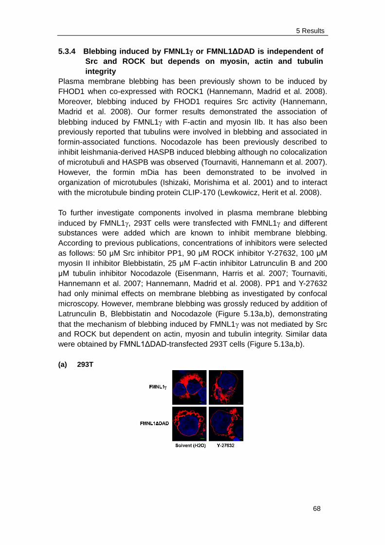
5 Results
68
5.3.4 Blebbing induced by FMNL1 or FMNL1ΔDAD is independent of
Src and ROCK but depends on myosin, actin and tubulin
integrity
Plasma membrane blebbing has been previously shown to be induced by
FHOD1 when co-expressed with ROCK1 (Hannemann, Madrid et al. 2008).
Moreover, blebbing induced by FHOD1 requires Src activity (Hannemann,
Madrid et al. 2008). Our former results demonstrated the association of
blebbing induced by FMNL1 with F-actin and myosin IIb. It has also been
previously reported that tubulins were involved in blebbing and associated in
formin-associated functions. Nocodazole has been previously described to
inhibit leishmania-derived HASPB induced blebbing although no colocalization
of microtubuli and HASPB was observed (Tournaviti, Hannemann et al. 2007).
However, the formin mDia has been demonstrated to be involved in
organization of microtubules (Ishizaki, Morishima et al. 2001) and to interact
with the microtubule binding protein CLIP-170 (Lewkowicz, Herit et al. 2008).
To further investigate components involved in plasma membrane blebbing
induced by FMNL1, 293T cells were transfected with FMNL1 and different
substances were added which are known to inhibit membrane blebbing.
According to previous publications, concentrations of inhibitors were selected
as follows: 50 μM Src inhibitor PP1, 90 μM ROCK inhibitor Y-27632, 100 μM
myosin II inhibitor Blebbistatin, 25 μM F-actin inhibitor Latrunculin B and 200
μM tubulin inhibitor Nocodazole (Eisenmann, Harris et al. 2007; Tournaviti,
Hannemann et al. 2007; Hannemann, Madrid et al. 2008). PP1 and Y-27632
had only minimal effects on membrane blebbing as investigated by confocal
microscopy. However, membrane blebbing was grossly reduced by addition of
Latrunculin B, Blebbistatin and Nocodazole (Figure 5.13a,b), demonstrating
that the mechanism of blebbing induced by FMNL1 was not mediated by Src
and ROCK but dependent on actin, myosin and tubulin integrity. Similar data
were obtained by FMNL1ΔDAD-transfected 293T cells (Figure 5.13a,b).
(a) 293T
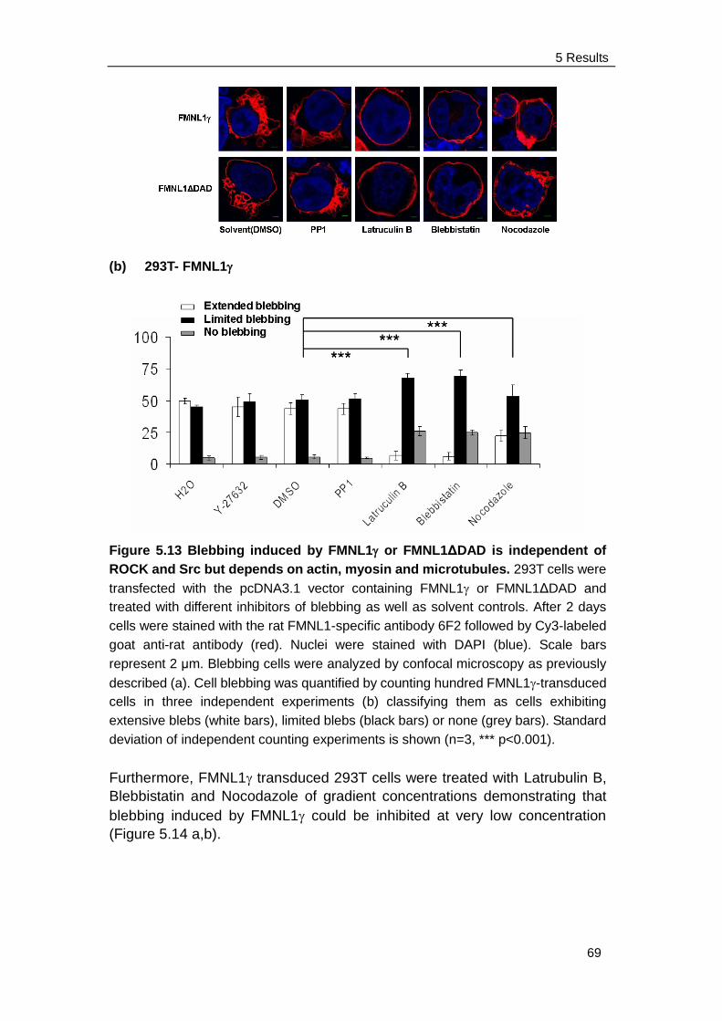
5 Results
69
(b) 293T- FMNL1
Figure 5.13 Blebbing induced by FMNL1 or FMNL1ΔDAD is independent of
ROCK and Src but depends on actin, myosin and microtubules. 293T cells were
transfected with the pcDNA3.1 vector containing FMNL1 or FMNL1ΔDAD and
treated with different inhibitors of blebbing as well as solvent controls. After 2 days
cells were stained with the rat FMNL1-specific antibody 6F2 followed by Cy3-labeled
goat anti-rat antibody (red). Nuclei were stained with DAPI (blue). Scale bars
represent 2 μm. Blebbing cells were analyzed by confocal microscopy as previously
described (a). Cell blebbing was quantified by counting hundred FMNL1-transduced
cells in three independent experiments (b) classifying them as cells exhibiting
extensive blebs (white bars), limited blebs (black bars) or none (grey bars). Standard
deviation of independent counting experiments is shown (n=3, *** p<0.001).
Furthermore, FMNL1 transduced 293T cells were treated with Latrubulin B,
Blebbistatin and Nocodazole of gradient concentrations demonstrating that
blebbing induced by FMNL1 could be inhibited at very low concentration
(Figure 5.14 a,b).
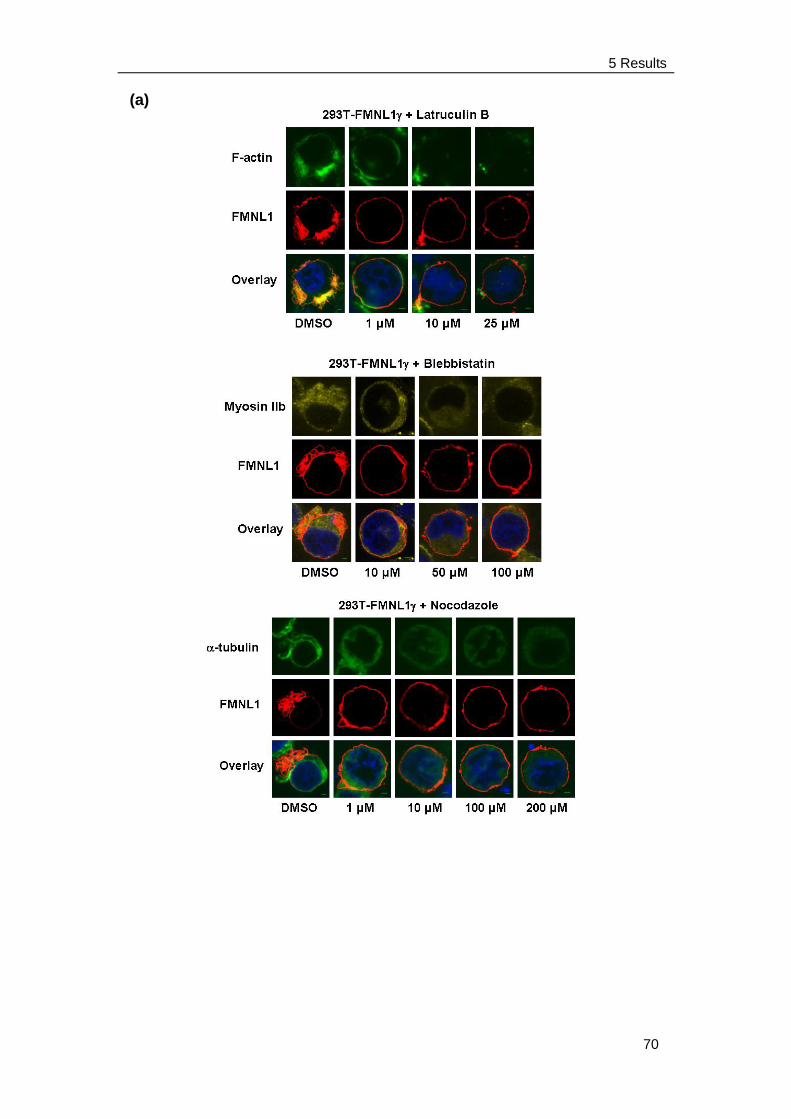
5 Results
70
(a)
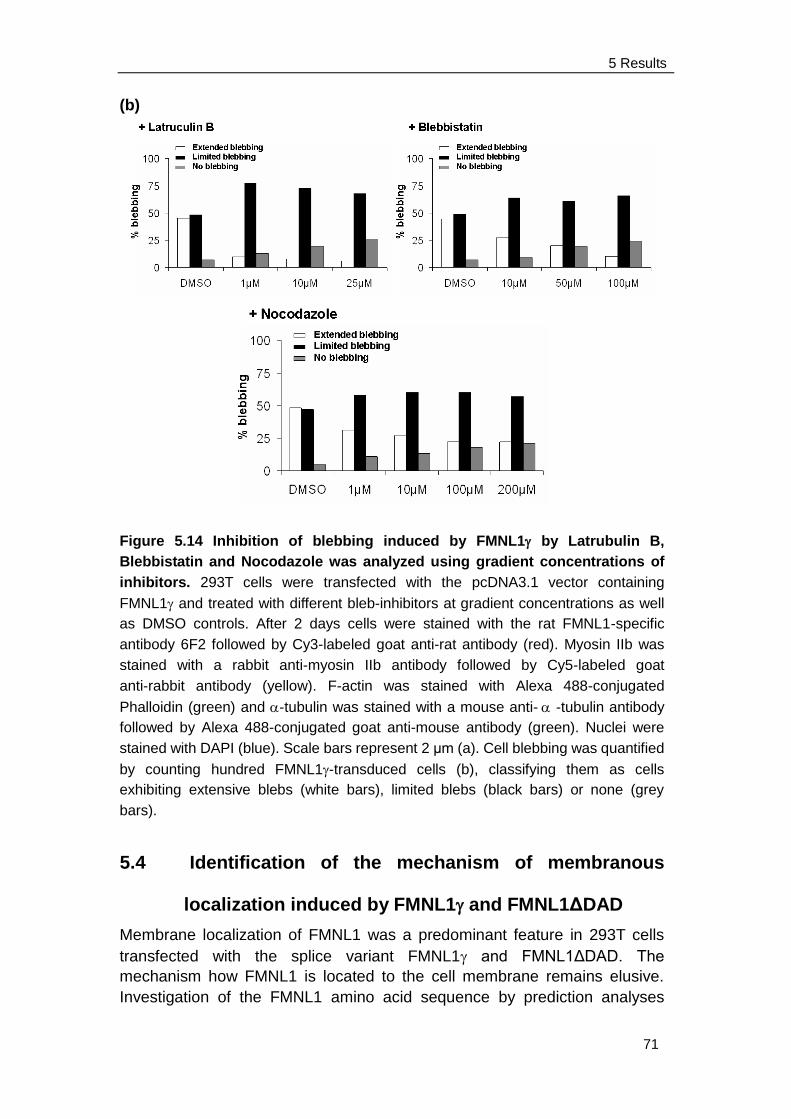
5 Results
71
(b)
Figure 5.14 Inhibition of blebbing induced by FMNL1 by Latrubulin B,
Blebbistatin and Nocodazole was analyzed using gradient concentrations of
inhibitors. 293T cells were transfected with the pcDNA3.1 vector containing
FMNL1 and treated with different bleb-inhibitors at gradient concentrations as well
as DMSO controls. After 2 days cells were stained with the rat FMNL1-specific
antibody 6F2 followed by Cy3-labeled goat anti-rat antibody (red). Myosin IIb was
stained with a rabbit anti-myosin IIb antibody followed by Cy5-labeled goat
anti-rabbit antibody (yellow). F-actin was stained with Alexa 488-conjugated
Phalloidin (green) and -tubulin was stained with a mouse anti- -tubulin antibody
followed by Alexa 488-conjugated goat anti-mouse antibody (green). Nuclei were
stained with DAPI (blue). Scale bars represent 2 μm (a). Cell blebbing was quantified
by counting hundred FMNL1-transduced cells (b), classifying them as cells
exhibiting extensive blebs (white bars), limited blebs (black bars) or none (grey
bars).
5.4 Identification of the mechanism of membranous
localization induced by FMNL1 and FMNL1ΔDAD
Membrane localization of FMNL1 was a predominant feature in 293T cells
transfected with the splice variant FMNL1 and FMNL1ΔDAD. The
mechanism how FMNL1 is located to the cell membrane remains elusive.
Investigation of the FMNL1 amino acid sequence by prediction analyses
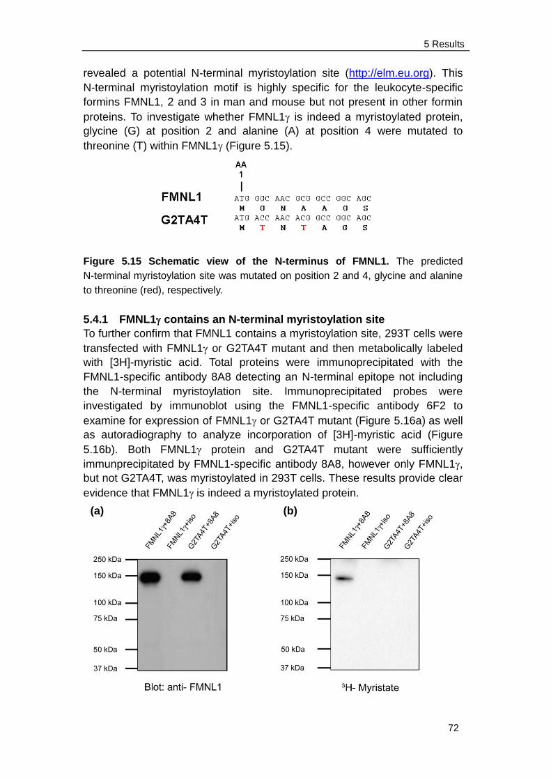
5 Results
72
revealed a potential N-terminal myristoylation site (http://elm.eu.org). This
N-terminal myristoylation motif is highly specific for the leukocyte-specific
formins FMNL1, 2 and 3 in man and mouse but not present in other formin
proteins. To investigate whether FMNL1 is indeed a myristoylated protein,
glycine (G) at position 2 and alanine (A) at position 4 were mutated to
threonine (T) within FMNL1 (Figure 5.15).
Figure 5.15 Schematic view of the N-terminus of FMNL1. The predicted
N-terminal myristoylation site was mutated on position 2 and 4, glycine and alanine
to threonine (red), respectively.
5.4.1 FMNL1 contains an N-terminal myristoylation site
To further confirm that FMNL1 contains a myristoylation site, 293T cells were
transfected with FMNL1 or G2TA4T mutant and then metabolically labeled
with [3H]-myristic acid. Total proteins were immunoprecipitated with the
FMNL1-specific antibody 8A8 detecting an N-terminal epitope not including
the N-terminal myristoylation site. Immunoprecipitated probes were
investigated by immunoblot using the FMNL1-specific antibody 6F2 to
examine for expression of FMNL1 or G2TA4T mutant (Figure 5.16a) as well
as autoradiography to analyze incorporation of [3H]-myristic acid (Figure
5.16b). Both FMNL1 protein and G2TA4T mutant were sufficiently
immunprecipitated by FMNL1-specific antibody 8A8, however only FMNL1,
but not G2TA4T, was myristoylated in 293T cells. These results provide clear
evidence that FMNL1 is indeed a myristoylated protein.
(a) (b)
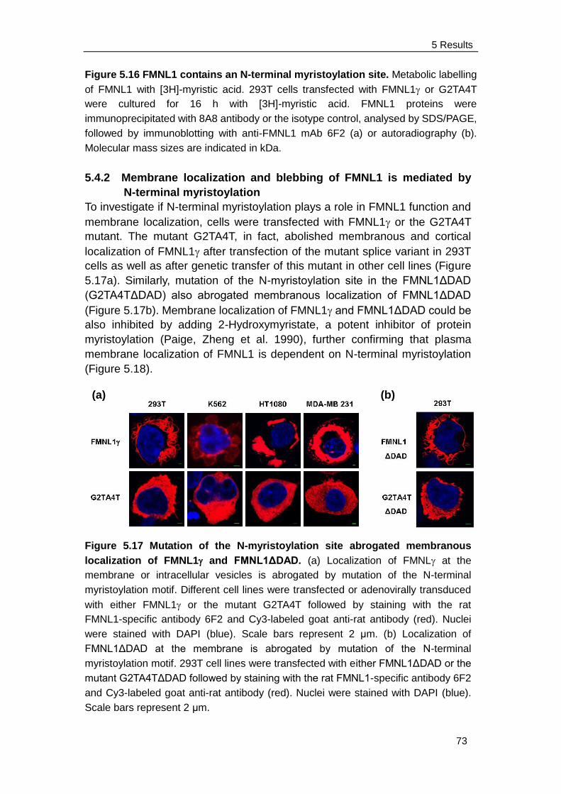
5 Results
73
Figure 5.16 FMNL1 contains an N-terminal myristoylation site. Metabolic labelling
of FMNL1 with [3H]-myristic acid. 293T cells transfected with FMNL1 or G2TA4T
were cultured for 16 h with [3H]-myristic acid. FMNL1 proteins were
immunoprecipitated with 8A8 antibody or the isotype control, analysed by SDS/PAGE,
followed by immunoblotting with anti-FMNL1 mAb 6F2 (a) or autoradiography (b).
Molecular mass sizes are indicated in kDa.
5.4.2 Membrane localization and blebbing of FMNL1 is mediated by
N-terminal myristoylation
To investigate if N-terminal myristoylation plays a role in FMNL1 function and
membrane localization, cells were transfected with FMNL1 or the G2TA4T
mutant. The mutant G2TA4T, in fact, abolished membranous and cortical
localization of FMNL1 after transfection of the mutant splice variant in 293T
cells as well as after genetic transfer of this mutant in other cell lines (Figure
5.17a). Similarly, mutation of the N-myristoylation site in the FMNL1ΔDAD
(G2TA4TΔDAD) also abrogated membranous localization of FMNL1ΔDAD
(Figure 5.17b). Membrane localization of FMNL1 and FMNL1ΔDAD could be
also inhibited by adding 2-Hydroxymyristate, a potent inhibitor of protein
myristoylation (Paige, Zheng et al. 1990), further confirming that plasma
membrane localization of FMNL1 is dependent on N-terminal myristoylation
(Figure 5.18).
Figure 5.17 Mutation of the N-myristoylation site abrogated membranous
localization of FMNL1 and FMNL1ΔDAD. (a) Localization of FMNL at the
membrane or intracellular vesicles is abrogated by mutation of the N-terminal
myristoylation motif. Different cell lines were transfected or adenovirally transduced
with either FMNL1 or the mutant G2TA4T followed by staining with the rat
FMNL1-specific antibody 6F2 and Cy3-labeled goat anti-rat antibody (red). Nuclei
were stained with DAPI (blue). Scale bars represent 2 μm. (b) Localization of
FMNL1ΔDAD at the membrane is abrogated by mutation of the N-terminal
myristoylation motif. 293T cell lines were transfected with either FMNL1ΔDAD or the
mutant G2TA4TΔDAD followed by staining with the rat FMNL1-specific antibody 6F2
and Cy3-labeled goat anti-rat antibody (red). Nuclei were stained with DAPI (blue).
Scale bars represent 2 μm.
(a)
(b)
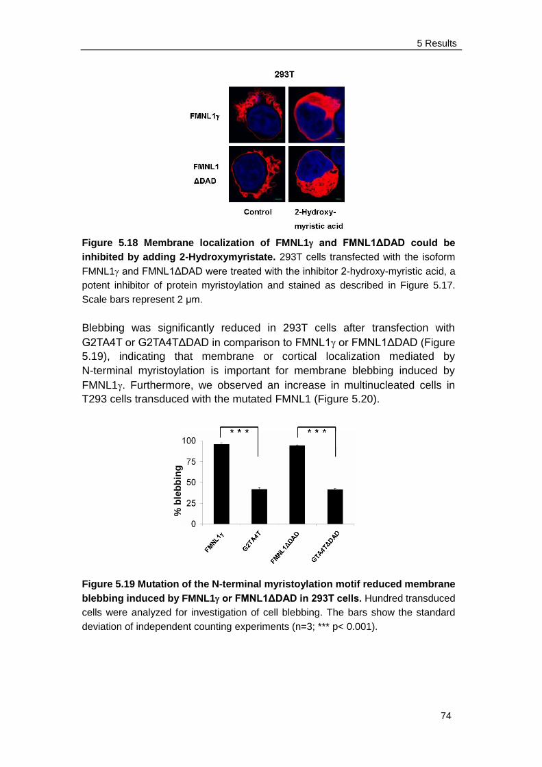
5 Results
74
Figure 5.18 Membrane localization of FMNL1 and FMNL1ΔDAD could be
inhibited by adding 2-Hydroxymyristate. 293T cells transfected with the isoform
FMNL1 and FMNL1ΔDAD were treated with the inhibitor 2-hydroxy-myristic acid, a
potent inhibitor of protein myristoylation and stained as described in Figure 5.17.
Scale bars represent 2 μm.
Blebbing was significantly reduced in 293T cells after transfection with
G2TA4T or G2TA4TΔDAD in comparison to FMNL1 or FMNL1ΔDAD (Figure
5.19), indicating that membrane or cortical localization mediated by
N-terminal myristoylation is important for membrane blebbing induced by
FMNL1. Furthermore, we observed an increase in multinucleated cells in
T293 cells transduced with the mutated FMNL1 (Figure 5.20).
Figure 5.19 Mutation of the N-terminal myristoylation motif reduced membrane
blebbing induced by FMNL1 or FMNL1ΔDAD in 293T cells. Hundred transduced
cells were analyzed for investigation of cell blebbing. The bars show the standard
deviation of independent counting experiments (n=3; *** p< 0.001).
% b
leb
bin
g
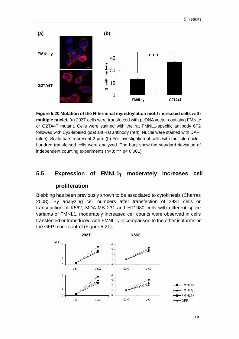
5 Results
75
Figure 5.20 Mutation of the N-terminal myristoylation motif increased cells with
multiple nuclei. (a) 293T cells were transfected with pcDNA vector contiaing FMNL
or G2TA4T mutant. Cells were stained with the rat FMNL1-specific antibody 6F2
followed with Cy3-labeled goat anti-rat antibody (red). Nuclei were stained with DAPI
(blue). Scale bars represent 2 μm. (b) For investigation of cells with multiple nuclei,
hundred transfected cells were analyzed. The bars show the standard deviation of
independent counting experiments (n=3; *** p< 0.001).
5.5 Expression of FMNL1 moderately increases cell
proliferation
Blebbing has been previously shown to be associated to cytokinesis (Charras
2008). By analyzing cell numbers after transfection of 293T cells or
transduction of K562, MDA-MB 231 and HT1080 cells with different splice
variants of FMNL1, moderately increased cell counts were observed in cells
transfected or transduced with FMNL1 in comparison to the other isoforms or
the GFP mock control (Figure 5.21).
(a)
(b)
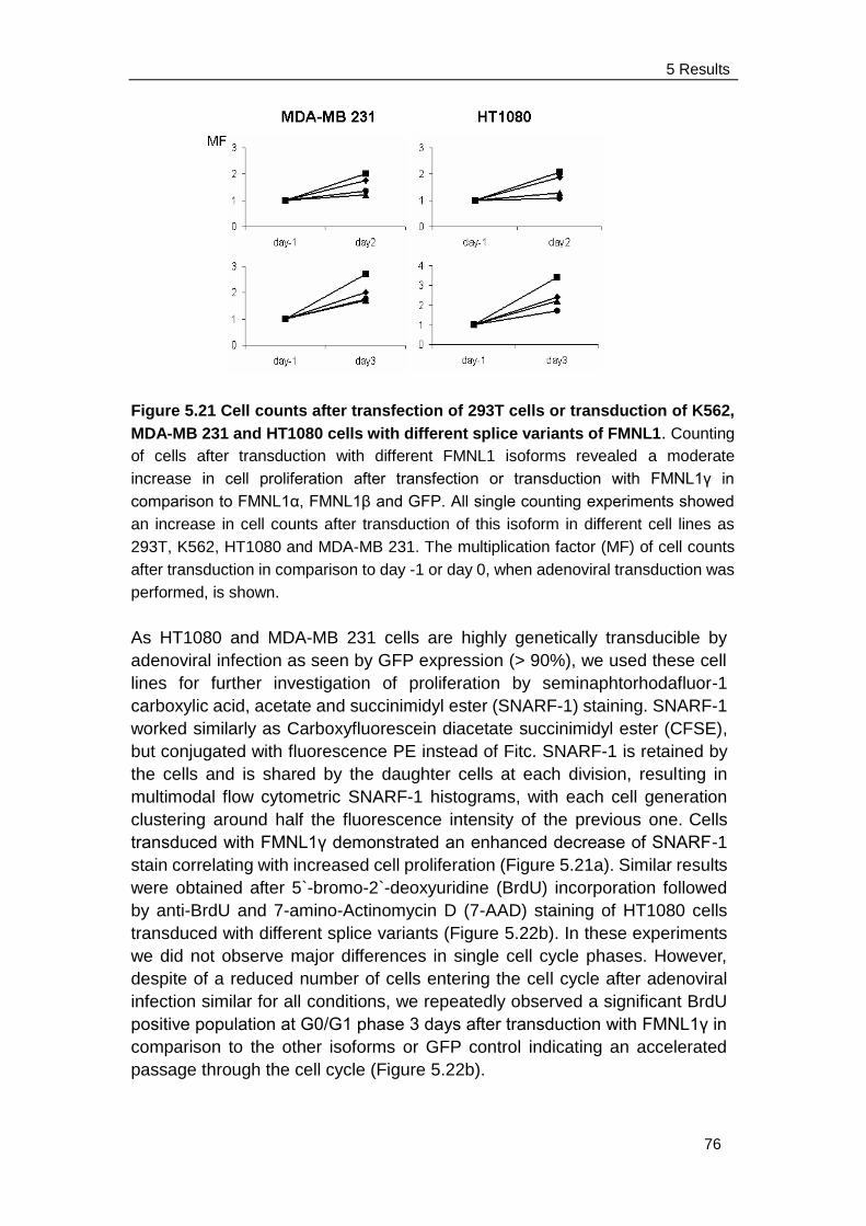
5 Results
76
Figure 5.21 Cell counts after transfection of 293T cells or transduction of K562,
MDA-MB 231 and HT1080 cells with different splice variants of FMNL1. Counting
of cells after transduction with different FMNL1 isoforms revealed a moderate
increase in cell proliferation after transfection or transduction with FMNL1γ in
comparison to FMNL1α, FMNL1β and GFP. All single counting experiments showed
an increase in cell counts after transduction of this isoform in different cell lines as
293T, K562, HT1080 and MDA-MB 231. The multiplication factor (MF) of cell counts
after transduction in comparison to day -1 or day 0, when adenoviral transduction was
performed, is shown.
As HT1080 and MDA-MB 231 cells are highly genetically transducible by
adenoviral infection as seen by GFP expression (> 90%), we used these cell
lines for further investigation of proliferation by seminaphtorhodafluor-1
carboxylic acid, acetate and succinimidyl ester (SNARF-1) staining. SNARF-1
worked similarly as Carboxyfluorescein diacetate succinimidyl ester (CFSE),
but conjugated with fluorescence PE instead of Fitc. SNARF-1 is retained by
the cells and is shared by the daughter cells at each division, resulting in
multimodal flow cytometric SNARF-1 histograms, with each cell generation
clustering around half the fluorescence intensity of the previous one. Cells
transduced with FMNL1γ demonstrated an enhanced decrease of SNARF-1
stain correlating with increased cell proliferation (Figure 5.21a). Similar results
were obtained after 5`-bromo-2`-deoxyuridine (BrdU) incorporation followed
by anti-BrdU and 7-amino-Actinomycin D (7-AAD) staining of HT1080 cells
transduced with different splice variants (Figure 5.22b). In these experiments
we did not observe major differences in single cell cycle phases. However,
despite of a reduced number of cells entering the cell cycle after adenoviral
infection similar for all conditions, we repeatedly observed a significant BrdU
positive population at G0/G1 phase 3 days after transduction with FMNL1γ in
comparison to the other isoforms or GFP control indicating an accelerated
passage through the cell cycle (Figure 5.22b).
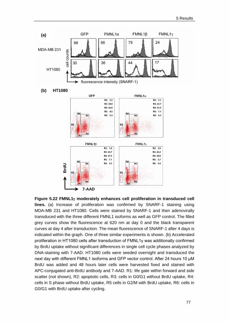
5 Results
77
(b) HT1080
Figure 5.22 FMNL1 moderately enhances cell proliferation in transduced cell
lines. (a) Increase of proliferation was confirmed by SNARF-1 staining using
MDA-MB 231 and HT1080. Cells were stained by SNARF-1 and then adenovirally
transduced with the three different FMNL1 isoforms as well as GFP control. The filled
grey curves show the fluorescence at 620 nm at day 0 and the black transparent
curves at day 4 after transduction. The mean fluorescence of SNARF-1 after 4 days is
indicated within the graph. One of three similar experiments is shown. (b) Accelerated
proliferation in HT1080 cells after transduction of FMNL1γ was additionally confirmed
by BrdU uptake without significant differences in single cell cycle phases analyzed by
DNA-staining with 7-AAD. HT1080 cells were seeded overnight and transduced the
next day with different FMNL1 isoforms and GFP vector control. After 24 hours 10 μM
BrdU was added and 48 hours later cells were harvested fixed and stained with
APC-conjugated anti-BrdU antibody and 7-AAD. R1: life gate within forward and side
scatter (not shown), R2: apoptotic cells, R3: cells in G0/G1 without BrdU uptake, R4:
cells in S phase without BrdU uptake, R5 cells in G2/M with BrdU uptake, R6: cells in
G0/G1 with BrdU uptake after cycling.
(a)
Brd
U
7-AAD
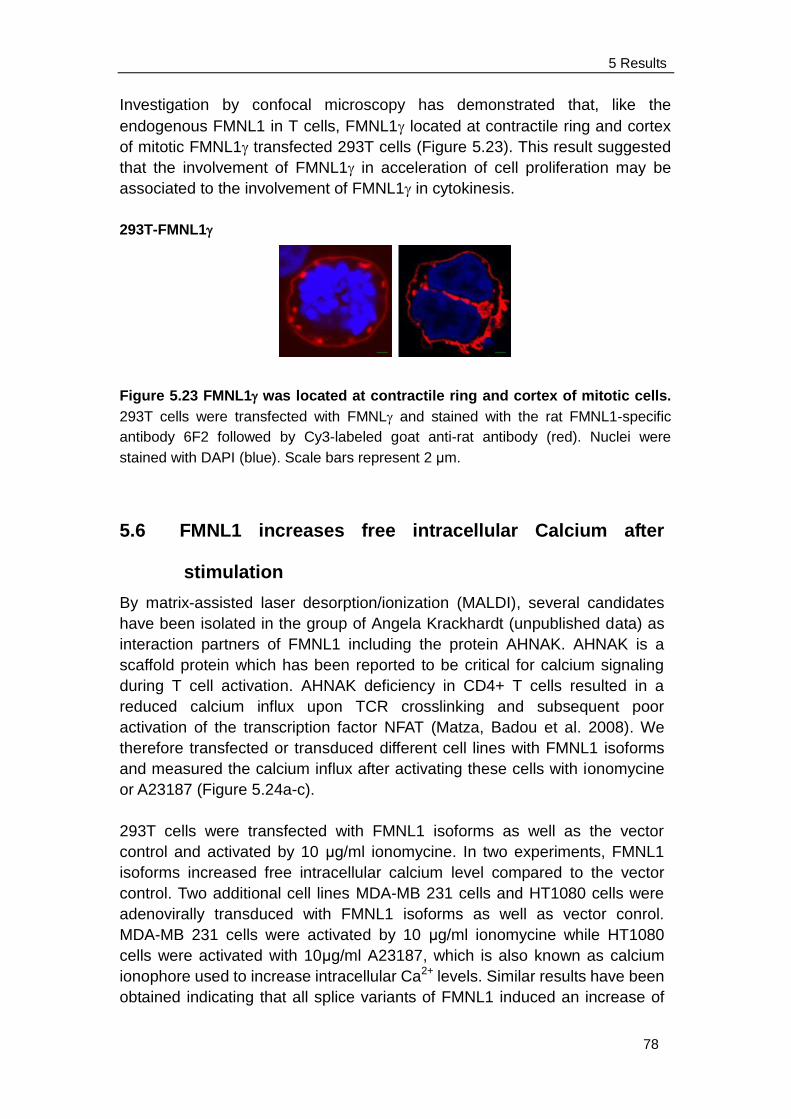
5 Results
78
Investigation by confocal microscopy has demonstrated that, like the
endogenous FMNL1 in T cells, FMNL1 located at contractile ring and cortex
of mitotic FMNL1 transfected 293T cells (Figure 5.23). This result suggested
that the involvement of FMNL1 in acceleration of cell proliferation may be
associated to the involvement of FMNL1 in cytokinesis.
293T-FMNL1
Figure 5.23 FMNL1 was located at contractile ring and cortex of mitotic cells.
293T cells were transfected with FMNL and stained with the rat FMNL1-specific
antibody 6F2 followed by Cy3-labeled goat anti-rat antibody (red). Nuclei were
stained with DAPI (blue). Scale bars represent 2 μm.
5.6 FMNL1 increases free intracellular Calcium after
stimulation
By matrix-assisted laser desorption/ionization (MALDI), several candidates
have been isolated in the group of Angela Krackhardt (unpublished data) as
interaction partners of FMNL1 including the protein AHNAK. AHNAK is a
scaffold protein which has been reported to be critical for calcium signaling
during T cell activation. AHNAK deficiency in CD4+ T cells resulted in a
reduced calcium influx upon TCR crosslinking and subsequent poor
activation of the transcription factor NFAT (Matza, Badou et al. 2008). We
therefore transfected or transduced different cell lines with FMNL1 isoforms
and measured the calcium influx after activating these cells with ionomycine
or A23187 (Figure 5.24a-c).
293T cells were transfected with FMNL1 isoforms as well as the vector
control and activated by 10 μg/ml ionomycine. In two experiments, FMNL1
isoforms increased free intracellular calcium level compared to the vector
control. Two additional cell lines MDA-MB 231 cells and HT1080 cells were
adenovirally transduced with FMNL1 isoforms as well as vector conrol.
MDA-MB 231 cells were activated by 10 μg/ml ionomycine while HT1080
cells were activated with 10μg/ml A23187, which is also known as calcium
ionophore used to increase intracellular Ca2+ levels. Similar results have been
obtained indicating that all splice variants of FMNL1 induced an increase of
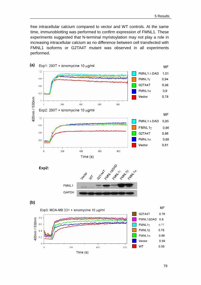
5 Results
79
free intracellular calcium compared to vector and WT controls. At the same
time, immunoblotting was performed to confirm expression of FMNL1. These
experiments suggested that N-terminal myristoylation may not play a role in
increasing intracellular calcium as no difference between cell transfected with
FMNL1 isoforms or G2TA4T mutant was observed in all experiments
performed.
(a)
(b)
Exp2:
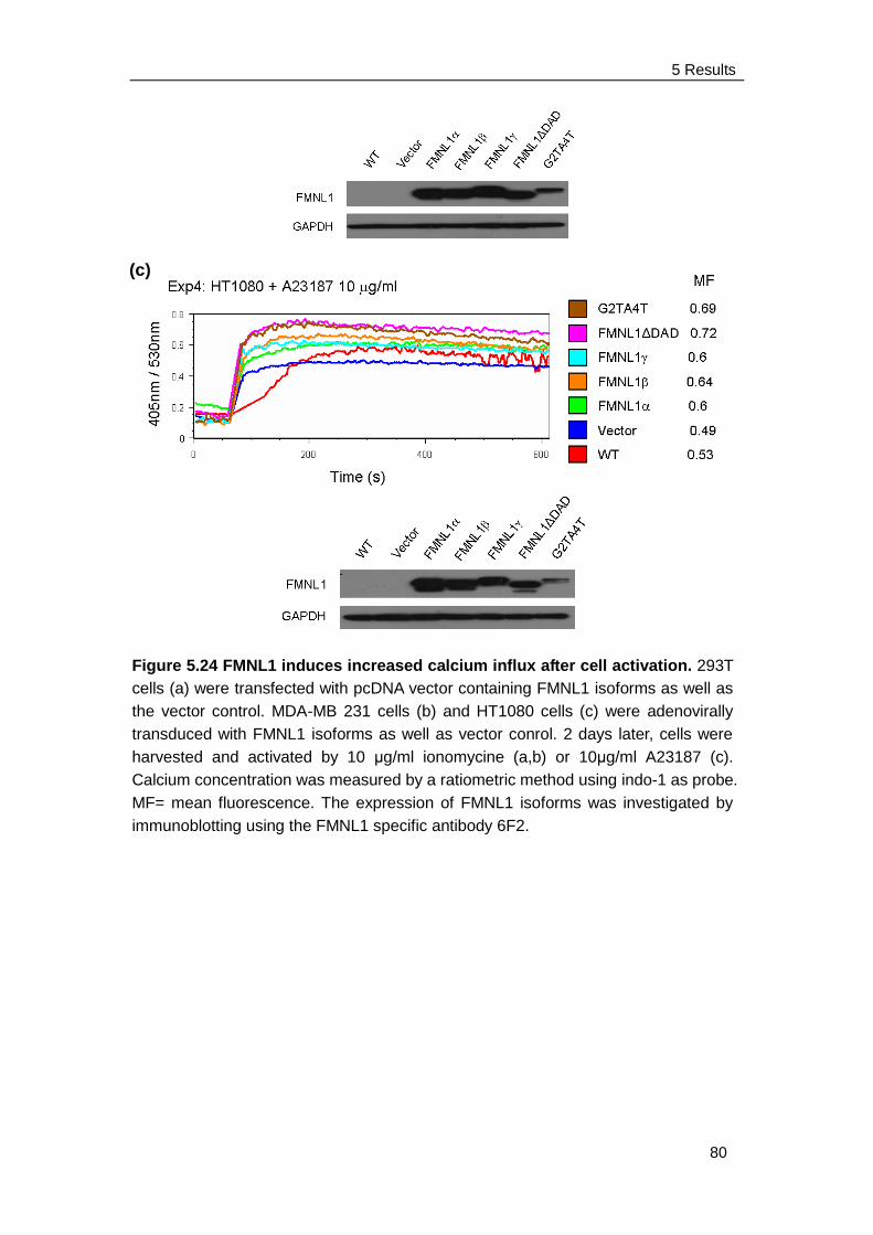
5 Results
80
Figure 5.24 FMNL1 induces increased calcium influx after cell activation. 293T
cells (a) were transfected with pcDNA vector containing FMNL1 isoforms as well as
the vector control. MDA-MB 231 cells (b) and HT1080 cells (c) were adenovirally
transduced with FMNL1 isoforms as well as vector conrol. 2 days later, cells were
harvested and activated by 10 μg/ml ionomycine (a,b) or 10μg/ml A23187 (c).
Calcium concentration was measured by a ratiometric method using indo-1 as probe.
MF= mean fluorescence. The expression of FMNL1 isoforms was investigated by
immunoblotting using the FMNL1 specific antibody 6F2.
(c)
6 Discussion
81
6 Discussion
Immunotherapies against cancer using T cells or antibodies specific for tumor
antigens are attractive alternative and complementary treatment modalities of
malignant diseases. One major advantage is the high specificity and limited
toxicity of these agents. Several therapeutic monoclonal antibodies have been
already approved by the US Food and Drug Administration (FDA) for use in
oncology (Adams and Weiner 2005; Weiner, Dhodapkar et al. 2009). Adoptive
cell therapy (ACT) using autologous tumor-infiltrating lymphocytes has been
considered as the most effective treatment for patients with metastatic
melanoma since it can induce approximate 50% regression in patients with
target cancer (Rosenberg, Restifo et al. 2008). The emphasis of the
development of immunotherapies has shifted from discovery and identification
of tumor antigens to the utilizating these antigens to mediate the destruction of
tumor in humans (Rosenberg 1999). However, it might be also important to
carefully investigate expression and function of tumor-associated antigens in
order to estimate potential side effect when targeting them (Theoret et.
al.2008).
The hematopoietic lineage-specific formin FMNL1 is a tumor-associated
antigen which has been previously identified using the SEREX technology.
FMNL1 is overexpressed in malignant cells derived from patients with CLL
and other lymphomas as well as some solid tumors. In healthy tissue, it is
almost exclusively expressed in hematopoietic cells suggesting this TAA to
be a suitable target antigen for immune-mediated therapies in a transplant
setting. However, function and role of FMNL1 have not been well
characterized. Formin proteins have been described to promote the formation
of actin networks and to be involved in regulation of essential cellular
functions as cell division, migration, adhesion and intracellular trafficking.
In this work, we first investigated the expression of FMNL1 by western blot and
confirmed the previous data that FMNL1 could be detected in native malignant
cells from patients with lymphatic as well as myeloid leukemias and
EBV-transformed B cells. FMNL1 is also expressed in lymphoma-derived cells
and renal carcinoma cells (Schuster, Busch et al. 2007). Moreover, we
continued to investigate the localization of FMNL1 by confocal microscopy. We
found a dot-like cytoplasmic localization of native human FMNL1 in different
hematopoietic lineage-derived cells and observed stimulation-specific
polarization of endogenous FMNL1 to the immunological synapse of T cells
and to the phagocytotic cup of monocytes which is in common with previous
observations for human FMNL1 and the murine counterpart
(Yayoshi-Yamamoto, Taniuchi et al. 2000; Seth, Otomo et al. 2006; Gomez,
Kumar et al. 2007). We additionally noticed colocalization of FMNL1 at the
mitotic spindle and cortex of dividing T cells, especially at the telephase,
6 Discussion
82
suggesting involvement of FMNL1 involved in mitosis and cytokinesis
(discussed in Chapter 6.4).
To further investigate the functional role of FMNL1, we have isolated three
different FMNL1 splice variants from cDNA of CLL cells and human lung
carcinoma, which result in varying C-terminal amino acid sequences (Figure
5.7a). FMNL1 and FMNL1 are corresponding to the previously described
murine splice variants, FRL and FRL (Yayoshi-Yamamoto, Taniuchi et
al. 2000; Harris, Li et al. 2004). However, the novel identified splice variant
(FMNL1) contains an intron retention of 58 amino acids at the C-terminal end
affecting the DAD but sharing the last 30 amino acids of C-terminal end with
FMNL1 (Figure 5.7b). FMNL1 showed increased mRNA-expression in bone
marrow, thymus, fetal brain and diverse hematopoietic lineage-derived cells.
We also observed a high expression of FMNL1 in malignant cells of a subset
of CLL patients. Detailed analyses of systematically isolated and processed
patient samples need to be performed in order to clarify if overexpression of
FMNL1 in CLL samples is associated with an activated malignant cell status
and/or patient prognosis and will be a focus of further studies.
Overexpression of FMNL1 shows that it is specifically located at the cell
membrane and cortex in diverse cell lines which is similar as observed for a
mutant lacking the DAD domain (FMNL1∆DAD) indicating that deregulation of
autoinhibition is effective in FMNL1. Both expression of FMNL1 and
FMNL1∆DAD induces polarized non-apoptotic blebbing which is dependent on
actin, tubulin and myosin integrity but independent of Src and ROCK activity,
which are reported to be crucial for bleb formation induced by other formins
(discussed in Chapter 6.2). Moreover, we described N-terminal myristoylation
as a regulative mechanism of FMNL1 responsible for membrane trafficking
potentially involved in a diversity of polarized processes of cells including
proliferation.
6.1 Deregulation of autoinhibition in FMNL1
Although a role of the novel isolated splice variant FMNL1 in malignant
transformation could not be ascertained in this study, it showed distinct
functional properties similar to the FMNL1 mutant missing the DAD sequence
with deregulated autoinhibition. Both of them showed membrane localization
and distinct functional properties as actin assembly activity and blebbing. This
is in common with a previous publication demonstrating that deregulation of
autoinhibition by deletion of the DAD region induces membrane localization
and actin assembly activity in FRLα (Seth, Otomo et al. 2006).
A complex regulation network seems to be responsible for activation and
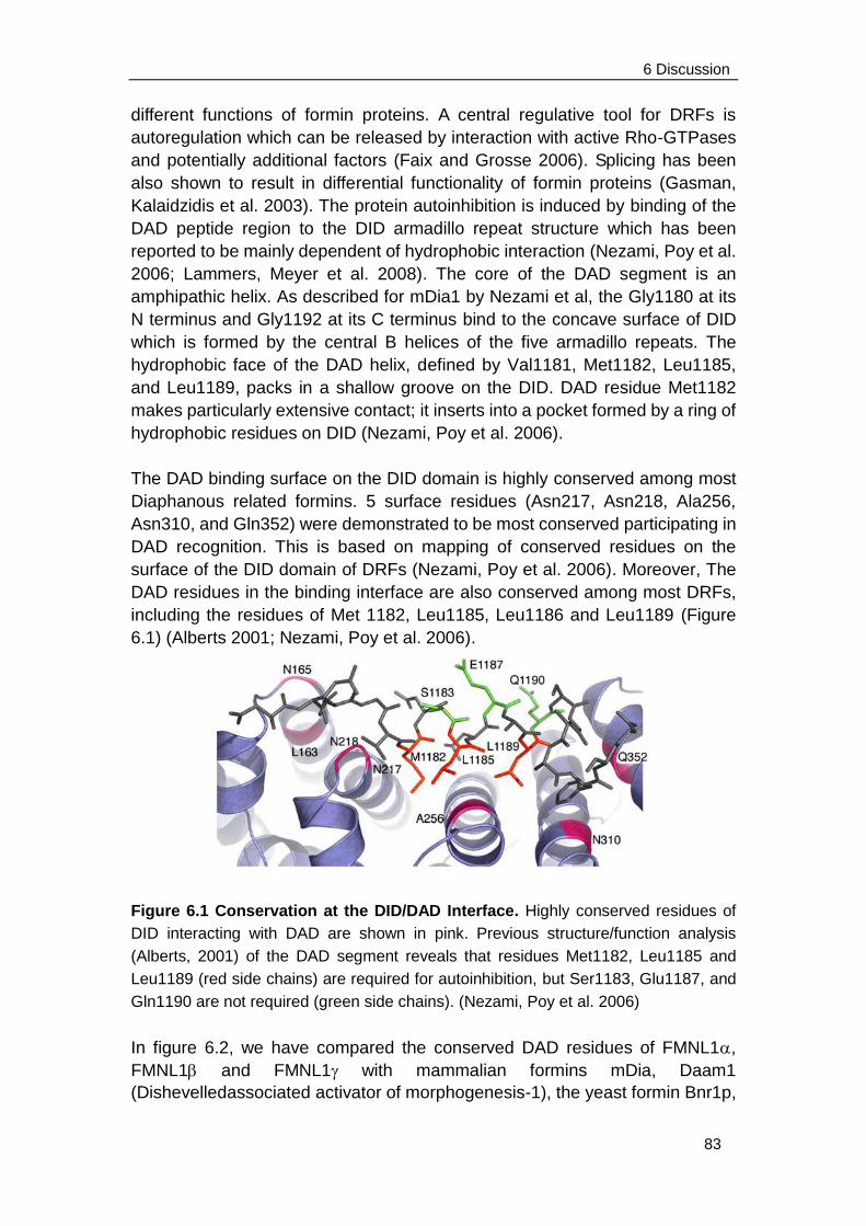
6 Discussion
83
different functions of formin proteins. A central regulative tool for DRFs is
autoregulation which can be released by interaction with active Rho-GTPases
and potentially additional factors (Faix and Grosse 2006). Splicing has been
also shown to result in differential functionality of formin proteins (Gasman,
Kalaidzidis et al. 2003). The protein autoinhibition is induced by binding of the
DAD peptide region to the DID armadillo repeat structure which has been
reported to be mainly dependent of hydrophobic interaction (Nezami, Poy et al.
2006; Lammers, Meyer et al. 2008). The core of the DAD segment is an
amphipathic helix. As described for mDia1 by Nezami et al, the Gly1180 at its
N terminus and Gly1192 at its C terminus bind to the concave surface of DID
which is formed by the central B helices of the five armadillo repeats. The
hydrophobic face of the DAD helix, defined by Val1181, Met1182, Leu1185,
and Leu1189, packs in a shallow groove on the DID. DAD residue Met1182
makes particularly extensive contact; it inserts into a pocket formed by a ring of
hydrophobic residues on DID (Nezami, Poy et al. 2006).
The DAD binding surface on the DID domain is highly conserved among most
Diaphanous related formins. 5 surface residues (Asn217, Asn218, Ala256,
Asn310, and Gln352) were demonstrated to be most conserved participating in
DAD recognition. This is based on mapping of conserved residues on the
surface of the DID domain of DRFs (Nezami, Poy et al. 2006). Moreover, The
DAD residues in the binding interface are also conserved among most DRFs,
including the residues of Met 1182, Leu1185, Leu1186 and Leu1189 (Figure
6.1) (Alberts 2001; Nezami, Poy et al. 2006).
Figure 6.1 Conservation at the DID/DAD Interface. Highly conserved residues of
DID interacting with DAD are shown in pink. Previous structure/function analysis
(Alberts, 2001) of the DAD segment reveals that residues Met1182, Leu1185 and
Leu1189 (red side chains) are required for autoinhibition, but Ser1183, Glu1187, and
Gln1190 are not required (green side chains). (Nezami, Poy et al. 2006)
In figure 6.2, we have compared the conserved DAD residues of FMNL1,
FMNL1 and FMNL1 with mammalian formins mDia, Daam1
(Dishevelledassociated activator of morphogenesis-1), the yeast formin Bnr1p,

6 Discussion
84
the mammalian formin FRL1, FRL2, and FRL3 (Higgs and Peterson 2005;
Nezami, Poy et al. 2006). It shows that Ile1059, Ile1062 and Ile1066 in
FMNL1 and Ile1059, Ile1062 and Leu1066 in FMNL1 are corresponding to
Met1182, Leu1185, and Leu1189 in mDia, which are considered to be
responsible for protein autoinhibition. However, FMNL1 contains an intron
retention of 58 amino acids at the C-terminus but shares the last 30 amino
acids with FMNL1. This intron retention affects the DAD sequence and
results in a positive charged polar amino acid Lys1066 instead of the non polar
amino acids Ile1066 or Leu1066 present in FMNL1 or FMNL1. This polar
lysine could be responsible for release of hydrophobic reaction between DID
and DAD and further release of autoinhibition of FMNL1 resulting in a
constitutively activated form.
Figure 6.2 Multiple sequence alignment of residues at the DAD interface in DRF
formins. Residues involved in the autoinhibitory interaction are indicated in red. High
conservation of these residues in all mDia formins as well as mammalian Daam1,
FRL1, FRL2, FRL3, FMNL1 splice variants and yeast Bnr1p points to a similar
mechanism of autoinhibition in these formins. However, FMNL1 contains an intron
retention affecting the DAD sequence and resulting in a positive charged polar amino
acid Lys1066 instead (in blue), which may be responsible for the release of
autoinhibition in FMNL1.
In fact, the splice variant FMNL1, in common with FMNL1ΔDAD, was
specifically located at the membrane and cortex of different cell lines and
induced bleb formation. It points to the fact that the incapability of DID binding
to DAD is responsible for deregulation of autoinhibition and membrane
localization and bleb formation by FMNL1. It is very interesting that FRL2 has
been also reported to be a constitutively activated formin protein because of its
capability to accumulate actin stress fibers. However, FRL2 is located in the
cytoplasma as its autoinhibited counterpart FRL3. The DAD of FRL2 could still
6 Discussion
85
bind to the DID without any inhibition (Vaillant, Copeland et al. 2008). It could
be explained by the presence of only non polar amino acids at the highly
conserved position of DAD in FRL2, which insures the interaction between the
two domains. However, this interaction failed to inhibit FRL2 activity suggesting
an additional regulator for formin autoinhibition.
6.2 Regulation of blebbing induced by FMNL1
Blebbing is a common feature of cell physiology during cell movement,
cytokinesis, cell spreading and apoptosis. Blebs are spherical protrusions of
the cell membrane which form and disappear in seconds (Charras, Yarrow et
al. 2005). Usually, the blebs induced by apoptosis are larger and more
transparent and their growth is independent of actomyosin contractions
(Barros, Kanaseki et al. 2003). Unlike them, other blebs depend on actomyosin
contractions of the cortex, which cause either detachment of the cell
membrane from the actin cortex (Charras, Yarrow et al. 2005) or a local
rupture in the actin cortex (Paluch, Piel et al. 2005) to form blebbs. In the
former way, a local shortening of the actin cortex caused by myosin contraction
gives rises to a centripetal compression of the cytoplasm and induces a
transient increase in intra-cellular pressure. This high local intra-cellular
pressure tears the membrane from the actin cortex and pushed cytosol from
cell body by into blebs. Alternatively, myosin contraction stretchs the actin
cortex and increases local tension, leading to the final rupture of actin cortex
and the expelling of cytosol which inflates the cell membrane. During growth,
the plasma membrane is transiently devoid of an actin cytoskeleton and lipids
flow into the bleb through the neck of blebs. Furthermore, after growth has
stalled, an actin cortex is reconstituted under the plasma membrane (Charras
2008). During the blebs expansion and cortex regrowth, regulatory proteins of
the actin cytoskeleton are present at the cell membrane, the first of which is the
ERM protein ezrin, which links the actin cytoskeleton to the cell membrane.
Shortly after the recruitment of ezrin, actin appeared at the bleb membrane
forming a cage-like structure underneath the bleb membrane. Finally, myosin II
motors powered bleb retraction (Charras, Hu et al. 2006; Charras 2008).
In this work, we have observed that FMNL1 could induce non-apoptotic cell
blebbing in 293T cells and K562 cells. Moreover, FMNL1 colocalized with the
indicated regulatory proteins such as actin, ezrin and myosin IIb on cell
membrane and cortex to blebs. The colocalization FMNL1 with F-actin in bleb
protrusions suggests the property of FMNL1 to induce actin assembly.
Blebbing but not membrane localization of FMNL1 was totally abrogated by
Latrunculin B further confirming regulation of actin assembly and
actin-mediated polarized bleb formation by FMNL1. To further investigate the
mechanism of bleb formation induced by FMNL1, FMNL1 transduced 293T
cells were incubated in medium containing Myosin II-ATPase inhibitor
6 Discussion
86
Blebbistatin and microtubuli inhibitor Nocodazole. Bleb formation was
significantly inhibited by these inhibitors confirming a substantial role of myosin
and microtubules in FMNL1-induced blebbing. These results are in common
with previous reports demonstrating that bleb induction by the Leishmania
parasite virulence factor HASPB depends on the integrity of F-actin and
requires myosin II function as well as microtubule networks (Tournaviti,
Hannemann et al. 2007). Tournaviti also demonstrated that although blebbing
induced by HASPB is dependent on the integrity of microtubule, no specific
association of microtubule with the blebs was observed (Tournaviti,
Hannemann et al. 2007). In contrast, we additionally observed colacolization of
-tubulin with FMNL1 to FMNL1-induced blebs in 293T cells. It suggested
that, unlike HASPB, FMNL1 may directly associate with microtubule to induce
bleb formation. However, it needs to be confirmed by protein
immunoprecipitation.
Moreover, the formins FHOD1, mDia1 and mDia2 have been also associated
to blebbing (Eisenmann, Harris et al. 2007; Kitzing, Sahadevan et al. 2007;
Hannemann, Madrid et al. 2008). Bleb formation induced by these formins
required Rho-kinase (ROCK) and Src activity. It has been previously reported
that the small GTPase Rho and its downstream effector ROCK play an
essential role in governing cell cortex contraction (Coleman, Sahai et al. 2001;
Etienne-Manneville and Hall 2002). The activation of GTPase Rho and ROCK
leads to both actin polymerization and myosin II recruitment during cytokinesis
and chemotaxis (Lee, Katakai et al. 2004; Bement, Benink et al. 2005; Kamijo,
Ohara et al. 2006). Rho directly activates formins, which as an actin-nucleating
protein hold onto growing barbed ends (Higashida, Miyoshi et al. 2004), and
activates myosin II by regulation of its phosphorylation state through
Rho-kinase (Totsukawa, Yamakita et al. 2000). How might Src signalling
regulate blebbing? Ezrin which is a membrane-actin crosslinks protein, is
subject to regulation by Src (Elliott, Qiao et al. 2004; Srivastava, Elliott et al.
2005). Additionally, Src functionally cooperates with formin protein family
which might also govern bleb retraction (Tominaga, Sahai et al. 2000; Gasman,
Kalaidzidis et al. 2003; Koka, Minick et al. 2005). However, in contrast to
former publications, blebbing induced by FMNL1 is independent of Src and
ROCK as the Src inhibitor PP1 and the ROCK inhibitor Y-27632 did not reduce
blebbing in our experiments. This might be explained by the constitutive
activation of FMNL1 and suggest a novel regulation of blebbing induced by
formin proteins beside the activation of Rho GTPase, ROCK and Src.
Futrthermore, the shape and extent of blebs varied in different cell lines.
FMNL1-induced blebbing was less obvious in HT1080 and MDA-MB 231 cells.
However, these cell lines especially demonstrated enrichment of FMNL1 at
intracellular vesicles beside the localization at the cell membrane. HT1080 has
mesenchymal characteristics and MDA-MB 231 cells are epithelial cells,
6 Discussion
87
indicating that cell type-specific interaction partners additionally play an
important role in function and bleb shape. In these cell lines, FMNL1 could play
a role in vesicles transferring and autophagocytosis. Unlike them, the K562
cells are hematopoietic origin, which could probably represent a more natural
recipient cell for investigation of FMNL1. K562 cells transduced with FMNL1
often demonstrated a polarized uropod-like circular. Uropod formation of
leukocytes has been reported to be involved in cell migration, activation and
cell-cell interaction. This polarized structure has distinct function in the immune
response as it could help T cell to form the immunological synapse when
interacting with target cells (Fais and Malorni 2003). The involvement of
FMNL1 in the cell polarization is in common with the investigation of
endogenous FMNL1 in specific targeted T cell.
6.3 N-terminal myristoylation
To further evaluate the mechanism of membrane localization and
bleb-induction by FMNL1, we analyzed the protein sequence motifs of FMNL1
and identified a myristoylation site at the N-terminus of human FMNL1 and
murine FRL with glycine at position 2 representing an essential requirement for
myristoylation.
N-terminal myristoylation of proteins stand for the covalent attachment of
myristate, a 14-carbon saturated fatty acid, to the N-terminal glycine of
eukaryotic and viral proteins (Farazi, Waksman et al. 2001). This transfer is
catalyzed by myristoyl-CoA:protein N-myristoyltransferase (Nmt) (Towler,
Adams et al. 1987). N-terminal myristoylation sites have been reported to
direct membrane association by interaction with negatively charged
phospholipids enriched in the cytoplasmic leaflet of cellular membranes
(Peitzsch and McLaughlin 1993). The model of how the N-terminal
myristoylated protein, tyrosine kinase c-Src, anchors to the membrane is
shown in Figure 6.3 (Murray, Ben-Tal et al. 1997): 10 carbon fatty acids
penetrate the hydrocarbon core of the membrane while the other four insert
into the polar head group region. The N-terminal glycine is located just outside
the envelope of the polar head group region.
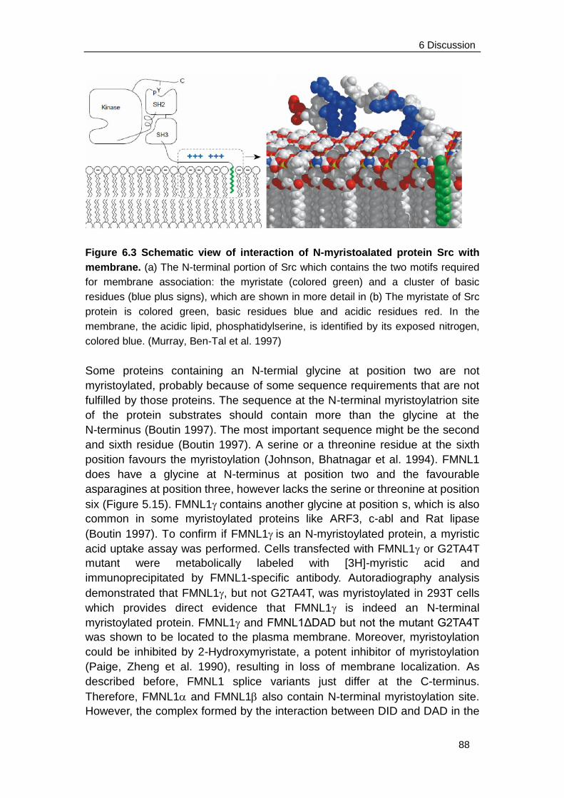
6 Discussion
88
Figure 6.3 Schematic view of interaction of N-myristoalated protein Src with
membrane. (a) The N-terminal portion of Src which contains the two motifs required
for membrane association: the myristate (colored green) and a cluster of basic
residues (blue plus signs), which are shown in more detail in (b) The myristate of Src
protein is colored green, basic residues blue and acidic residues red. In the
membrane, the acidic lipid, phosphatidylserine, is identified by its exposed nitrogen,
colored blue. (Murray, Ben-Tal et al. 1997)
Some proteins containing an N-termial glycine at position two are not
myristoylated, probably because of some sequence requirements that are not
fulfilled by those proteins. The sequence at the N-terminal myristoylatrion site
of the protein substrates should contain more than the glycine at the
N-terminus (Boutin 1997). The most important sequence might be the second
and sixth residue (Boutin 1997). A serine or a threonine residue at the sixth
position favours the myristoylation (Johnson, Bhatnagar et al. 1994). FMNL1
does have a glycine at N-terminus at position two and the favourable
asparagines at position three, however lacks the serine or threonine at position
six (Figure 5.15). FMNL1contains another glycine at position s, which is also
common in some myristoylated proteins like ARF3, c-abl and Rat lipase
(Boutin 1997). To confirm if FMNL1is an N-myristoylated protein, a myristic
acid uptake assay was performed. Cells transfected with FMNL1 or G2TA4T
mutant were metabolically labeled with [3H]-myristic acid and
immunoprecipitated by FMNL1-specific antibody. Autoradiography analysis
demonstrated that FMNL1, but not G2TA4T, was myristoylated in 293T cells
which provides direct evidence that FMNL1 is indeed an N-terminal
myristoylated protein. FMNL1 and FMNL1ΔDAD but not the mutant G2TA4T
was shown to be located to the plasma membrane. Moreover, myristoylation
could be inhibited by 2-Hydroxymyristate, a potent inhibitor of myristoylation
(Paige, Zheng et al. 1990), resulting in loss of membrane localization. As
described before, FMNL1 splice variants just differ at the C-terminus.
Therefore, FMNL1 and FMNL1 also contain N-terminal myristoylation site.
However, the complex formed by the interaction between DID and DAD in the
6 Discussion
89
autoinhibited state may impede membrane localization regulated by N-terminal
myristoylation.
N-terminal myristoylated proteins have diverse functions and intracellular
destinations. They are crucial in signal transduction, cellular transformation,
and oncogenesis (Shrivastav, Varma et al. 2008). This motif is typically
present in the SH4 domain of Src tyrosine kinases and myristoylation has been
shown to be critical for malignant transformation of v-Src (Cross, Garber et al.
1984; Kamps, Buss et al. 1985). Myristate is required for Src membrane
binding which, in turn, is required for Src to function, since nonmyristoylated
v-Src mutants are found in the cytoplasm but do not transform cells, even
though the kinase activity of the protein is unaffected. It may because some
critical targets for Src activity are located at or near the cell membrane (Kamps,
Buss et al. 1985; Buss, Kamps et al. 1986). In this work, we investigated that
blebbing induced by FMNL1γ is dependent on N-terminal myristoylation
since the mutation of glycine at position 2 significantly reduced blebbing. We
therefore propose that activation of FMNL1 is regulated not only by binding of
a Rho-GTPase but additionally by an increase of intracellular Ca2+ or a specific
ligand binding. This may cause a myristoyl switch and therefore induces
extrusion of the myristoyl group enabling it to quickly and reversibly interact
with lipid bilayer membranes as previously described for other myristoylated
proteins (Seykora, Myat et al. 1996; Ames, Ishima et al. 1997). It was proposed
that the myristoyl group is sequestered in the calcium-free form of the protein
and becomes exposed in the calcium-bound form, enabling it to insert into a
lipid bilayer of membrane or a hydrophobic site of a target protein (Zozulya and
Stryer 1992; Ames, Ishima et al. 1997). These data suggested that myristoyl
groups are not simply hydrophobic anchors, but can serve as dynamic
partners in signaling (Ames, Ishima et al. 1997). We have observed that
calcium binding slightly increases the membrane localization of the
autoinhibited FMNL1 and FMNL1(preliminary data) suggesting that FMNL1
could also be activated through the calcium- myristoyl switch. Furthermore,
stimulation of ionomycine will induce higher concentration of intracellular
calcium in FMNL1 transduced cells potentially reprtesenting a self-activating
loop, which will be discussed later in chapter 6.5.
6.4 Acceleration of cell proliferation induced by FMNL1
As we have shown by Annexin V and propidium iodide staining that blebbing
induced by FMNL1 was not associated to apoptosis (Figure 5.12). We
continued to investigate other cellular processes which have been described
to be involved in cell blebbing such as cytokinesis. By analyzing cell numbers
in independent counting experiments, we observed that cells derived from
different cell lines transfected or transduced with FMNL1γ revealed slightly
6 Discussion
90
enhanced cell numbers compared to the other splice variants and the mock
control. Moderately accelerated cell proliferation induced by FMNL1 was
confirmed by SNARF-1 staining in two cell lines, HT1080 and MDA-MB 231,
which are highly genetically transducible by adenoviral infection as seen by
GFP expression (> 90%). Similar results were obtained through a cell cycle
experiment by BrdU incorporation followed by anti-BrdU and 7-AAD staining
of HT1080 cells transduced with different FMNL1 splice variants
demonstrating a significant BrdU positive population at G0/G1 phase 3 days
after transduction with FMNL1γ which was not present in the other isoforms
or GFP control. This experiment did not show major differences in single cell
cycle phases, however indicated an accelerated passage of cells transduced
by FMNL through the cell cycle.
Murine formin FRL has been shown to be involved in regulation of cell
survival (Yayoshi-Yamamoto et al., 2000). This group demonstrated that
macrophages overexpressing the FRL-FH3 truncated protein (only contains
N-terminal FH3 domain but not FH1 and FH2 domians) were inhibited in cell
growth and were prone to cell apoptosis. This may be explained by the direct
binding of FRL-FH3 truncated protein to Rho GTPase Rac, which interfered
with the Rac binding to endogenous FRL; thus, the normal function of FRL
was inhibited. However, a direct effect on proliferation has not been
demonstrated so far. In this work, we have observed by confocal microscopy
that FMNL1 located at contractile ring and cortex of mitotic FMNL1
transfected 293T cells. Futhermore, proliferation of cells transduced with
G2TA4T was still enhanced although slightly reduced in comparison to the
non-mutated FMNL1 (data not shown). Meanwhile, cells transduced with
G2TA4T showed an enhancement of multinucleated cells compared to cells
transduced with non-mutated FMNL1. Multinucleation was also observed in
spermatids of male flies, when the formin diaphanous mutants resulted in a
failure in cell division during spermatogenesis (Goode and Eck 2007). Our
relsults give rise for the hypothsis that the accelerated cell proliferation
induced by FMNL1 may because of its involvement in cell cytokinesis and
N-terminal myristoylation may play a role in cell seperation.
The effect on cell proliferation induced by FMNL1 was moderate but
constantly observed. This may correlate with the slow increase in malignant
cell burden often observed in patients with CLL and highly encourages the
further investigation of the role of FMNL1 in CLL cells. In addition, we
observed colocalization of endogenous FMNL1 to the microtubules and
midzone cortex of dividing T cells pointing to a possible involvement of
FMNL1 in mitosis of T cells.
To further confirm the involvement of FMNL1 in cell proliferation, a FMNL1
specific siRNA would be required to knockdown the endogenous FMNL1 in
6 Discussion
91
hematopoietic lineage-derived cells, for example T cells, and then analyzes
cell proliferation. A FMNL1 specific antiboby would also be required to detect
the colocalization of endogenous FMNL1 to the microtubuli at the cortex and
mitotic spindles in dividing cells. On the other hand, the endogenous FMNL1
may be generally involved in enhanced cell proliferation since knock down of
FMNL1 in T cells showed slight inhibition of T cell proliferation (unpublished
data of the group of Dr. Angela Krackhardt). Furthermore, FMNL1 could
increase intracellular calcium concentration after stimulation which is a
sufficient signal for cell activation and subsequent gene expression and
proliferation through the nuclear factor-B (NF-B) pattway (Dolmetsch,
Lewis et al. 1997). Analysis of interaction partners of FMNL1 could also be
required to characterize the complete sigaling pathway.
6.5 Intracellular calcium concentration
The intracellular calcium is one of the most fundamental signalling substances
which controls diverse cell functions, including adhesion, motility, gene
expression and proliferation. It has also been reported to be involved in cell
morphological oscillations such as blebbing (Pletjushkina, Rajfur et al. 2001;
Blaser, Reichman-Fried et al. 2006). Cyclic intra-cellular calcium changes are
accompanied with cortical contractility, but whether these calcium oscillations
are the cause or a consequence of the enhanced contractility is unclear.
Chelation of extracellular calcium could inhibit blebbing. Therefore elevated
intra-cellular calcium may cause the myosin contractions that give rise to blebs.
Conversely, calcium could also enter the cells as a consequence of increased
membrane tension in blebs.
On the other hand, a cooperation partner of FMNL1, AHNAK, which has been
identified as an interaction partner of FMNL1 by our group, has been reported
to be critical for calcium signaling during T cell activation. AHNAK deficiency in
CD4+ T cells resulted in a reduced calcium influx upon TCR crosslinking and
subsequent poor activation of the transcription factor NFAT (Matza, Badou et al.
2008). In our preliminary experiments, AHNAK, like FMNL1, was polarized at
the immunosynapse in specifically stimulated T cells and colocalized with CD3.
Moreover, AHNAK was colocalized with FMNL1 at cell membrane and blebs
in FMNL1 transduced cells.
In this work, different cell lines were transfected or transduced with FMNL1
isoforms and stained with indo-1 AM. Immediately after activation with
ionomycine or A23187, the intracellular calcium concentrations in these cells
were measured during a time of 10 minutes. The results demonstrated a
higher intracellular calcium concentration in cells transfected or transduced
with different isoforms of FMNL1 compared to vector and wildtype control.
6 Discussion
92
However, there is no difference of enhancement of intracellular calcium
concentration among different isoforms of FMNL1 including the mutant
G2TA4T. Thus, the increase of intracellular calcium concentration seems to be
independent of N-terminal myristoylation. Further experiments need to be
performed to investigate the role of FMNL1 in calcium signaling pathways.
In conclusion, this project provides novel information about expression and
specific localization of human endogenous FMNL1 in a diversity of polarized
processes of hematopoietic lineage-derived cells suggesting FMNL1 to be a
pluripotent effector protein. Moreover, the newly identified splice variant
FMNL1 represents a constitutively active form of FMNL1 which localizes at
the membrane and induces bleb formation. FMNL1 also associates FMNL1
to mitosis and cell growth. We identified N-terminal myristoylation as an
important regulatory tool for FMNL1 necessary for bleb formation, enabling
fast and reversible membrane localization compatible with diverse functions
of hematopoietic lineage-derived cells. Thus our results suggest FMNL1 to be
a promise potential target antigen for anti-tumor immunotherapy as it is
potentially involved in cellular transformation and tumor progression. The
identification of interaction partners of FMNL1 in diverse
hematopoietic-derived cells as well as the further characterization of splice
variants will be highly interesting to identify key molecules regulating different
FMNL1 functions potentially revealing possibilities for specific therapeutic
interaction in malignant and inflammatory disease.
7 Reference
93
7 Reference
Adams, G. P. and L. M. Weiner (2005). "Monoclonal antibody therapy of cancer." Nat
Biotechnol 23(9): 1147-57.
Alberts, A. S. (2001). "Identification of a carboxyl-terminal diaphanous-related formin
homology protein autoregulatory domain." J Biol Chem 276(4): 2824-30.
Ames, J. B., R. Ishima, et al. (1997). "Molecular mechanics of calcium-myristoyl
switches." Nature 389(6647): 198-202.
Banchereau, J., F. Briere, et al. (2000). "Immunobiology of dendritic cells." Annu Rev
Immunol 18: 767-811.
Barros, L. F., T. Kanaseki, et al. (2003). "Apoptotic and necrotic blebs in epithelial cells
display similar neck diameters but different kinase dependency." Cell Death
Differ 10(6): 687-97.
Bement, W. M., H. A. Benink, et al. (2005). "A microtubule-dependent zone of active
RhoA during cleavage plane specification." J Cell Biol 170(1): 91-101.
Blaser, H., M. Reichman-Fried, et al. (2006). "Migration of zebrafish primordial germ cells:
a role for myosin contraction and cytoplasmic flow." Dev Cell 11(5): 613-27.
Boutin, J. A. (1997). "Myristoylation." Cell Signal 9(1): 15-35.
Brichard, V., A. Van Pel, et al. (1993). "The tyrosinase gene codes for an antigen
recognized by autologous cytolytic T lymphocytes on HLA-A2 melanomas." J
Exp Med 178(2): 489-95.
Bubenik, J., P. Perlmann, et al. (1970). "Cellular and humoral immune responses to
human urinary bladder carcinomas." Int J Cancer 5(3): 310-9.
Buss, J. E., M. P. Kamps, et al. (1986). "The absence of myristic acid decreases
membrane binding of p60src but does not affect tyrosine protein kinase activity."
J Virol 58(2): 468-74.
Cantwell, M., T. Hua, et al. (1997). "Acquired CD40-ligand deficiency in chronic
lymphocytic leukemia." Nat Med 3(9): 984-9.
Chames, P., M. Van Regenmortel, et al. (2009). "Therapeutic antibodies: successes,
limitations and hopes for the future." Br J Pharmacol 157(2): 220-33.
Chapman, P. B. (2004). "T-cell chauvinists versus antibody advocates--can't we all just
7 Reference
94
get along?" J Clin Oncol 22(22): 4446-8.
Charras, G. T. (2008). "A short history of blebbing." J Microsc 231(3): 466-78.
Charras, G. T., C. K. Hu, et al. (2006). "Reassembly of contractile actin cortex in cell
blebs." J Cell Biol 175(3): 477-90.
Charras, G. T., J. C. Yarrow, et al. (2005). "Non-equilibration of hydrostatic pressure in
blebbing cells." Nature 435(7040): 365-9.
Chen, Y. T., E. Stockert, et al. (1996). "Serological analysis of Melan-A(MART-1), a
melanocyte-specific protein homogeneously expressed in human melanomas."
Proc Natl Acad Sci U S A 93(12): 5915-9.
Chhabra, E. S. and H. N. Higgs (2007). "The many faces of actin: matching assembly
factors with cellular structures." Nat Cell Biol 9(10): 1110-21.
Coleman, M. L., E. A. Sahai, et al. (2001). "Membrane blebbing during apoptosis results
from caspase-mediated activation of ROCK I." Nat Cell Biol 3(4): 339-45.
Copeland, S. J., B. J. Green, et al. (2007). "The diaphanous inhibitory
domain/diaphanous autoregulatory domain interaction is able to mediate
heterodimerization between mDia1 and mDia2." J Biol Chem 282(41): 30120-30.
Coulie, P. G., V. Brichard, et al. (1994). "A new gene coding for a differentiation antigen
recognized by autologous cytolytic T lymphocytes on HLA-A2 melanomas." J
Exp Med 180(1): 35-42.
Cox, A. L., J. Skipper, et al. (1994). "Identification of a peptide recognized by five
melanoma-specific human cytotoxic T cell lines." Science 264(5159): 716-9.
Cross, F. R., E. A. Garber, et al. (1984). "A short sequence in the p60src N terminus is
required for p60src myristylation and membrane association and for cell
transformation." Mol Cell Biol 4(9): 1834-42.
Disis, M. L., H. Bernhard, et al. (2009). "Use of tumour-responsive T cells as cancer
treatment." Lancet 373(9664): 673-83.
Dolmetsch, R. E., R. S. Lewis, et al. (1997). "Differential activation of transcription factors
induced by Ca2+ response amplitude and duration." Nature 386(6627): 855-8.
Dreger, P., P. Corradini, et al. (2007). "Indications for allogeneic stem cell transplantation
in chronic lymphocytic leukemia: the EBMT transplant consensus." Leukemia
21(1): 12-7.
7 Reference
95
Dreger, P. and E. Montserrat (2002). "Autologous and allogeneic stem cell
transplantation for chronic lymphocytic leukemia." Leukemia 16(6): 985-92.
Dreger, P., S. Stilgenbauer, et al. (2004). "The prognostic impact of autologous stem cell
transplantation in patients with chronic lymphocytic leukemia: a risk-matched
analysis based on the VH gene mutational status." Blood 103(7): 2850-8.
Eisenmann, K. M., E. S. Harris, et al. (2007). "Dia-interacting protein modulates
formin-mediated actin assembly at the cell cortex." Curr Biol 17(7): 579-91.
Elliott, B. E., H. Qiao, et al. (2004). "Co-operative effect of c-Src and ezrin in deregulation
of cell-cell contacts and scattering of mammary carcinoma cells." J Cell Biochem
92(1): 16-28.
Emens, L. A. and E. M. Jaffee (2005). "Leveraging the activity of tumor vaccines with
cytotoxic chemotherapy." Cancer Res 65(18): 8059-64.
Etienne-Manneville, S. and A. Hall (2002). "Rho GTPases in cell biology." Nature
420(6916): 629-35.
Evangelista, M., K. Blundell, et al. (1997). "Bni1p, a yeast formin linking cdc42p and the
actin cytoskeleton during polarized morphogenesis." Science 276(5309): 118-22.
Fackler, O. T. and R. Grosse (2008). "Cell motility through plasma membrane blebbing."
J Cell Biol 181(6): 879-84.
Fais, S. and W. Malorni (2003). "Leukocyte uropod formation and
membrane/cytoskeleton linkage in immune interactions." J Leukoc Biol 73(5):
556-63.
Faix, J. and R. Grosse (2006). "Staying in shape with formins." Dev Cell 10(6): 693-706.
Farazi, T. A., G. Waksman, et al. (2001). "The biology and enzymology of protein
N-myristoylation." J Biol Chem 276(43): 39501-4.
Favaro, P. M., F. Traina, et al. (2006). "High expression of FMNL1 protein in T
non-Hodgkin's lymphomas." Leuk Res 30(6): 735-8.
Finn, O. J. (2008). "Cancer immunology." N Engl J Med 358(25): 2704-15.
Foster, A. E., M. K. Brenner, et al. (2008). "Adoptive T-cell immunotherapy of chronic
lymphocytic leukaemia." Best Pract Res Clin Haematol 21(3): 375-89.
Gasman, S., Y. Kalaidzidis, et al. (2003). "RhoD regulates endosome dynamics through
7 Reference
96
Diaphanous-related Formin and Src tyrosine kinase." Nat Cell Biol 5(3): 195-204.
Giannopoulos, K. and M. Schmitt (2006). "Targets and strategies for T-cell based
vaccines in patients with B-cell chronic lymphocytic leukemia." Leuk Lymphoma
47(10): 2028-36.
Gomez, T. S., K. Kumar, et al. (2007). "Formins regulate the actin-related protein 2/3
complex-independent polarization of the centrosome to the immunological
synapse." Immunity 26(2): 177-90.
Goode, B. L. and M. J. Eck (2007). "Mechanism and function of formins in the control of
actin assembly." Annu Rev Biochem 76: 593-627.
Graziano, D. F. and O. J. Finn (2005). "Tumor antigens and tumor antigen discovery."
Cancer Treat Res 123: 89-111.
Hamblin, A. D. and T. J. Hamblin (2008). "The immunodeficiency of chronic lymphocytic
leukaemia." Br Med Bull 87: 49-62.
Hannemann, S., R. Madrid, et al. (2008). "The Diaphanous-related Formin FHOD1
associates with ROCK1 and promotes Src-dependent plasma membrane
blebbing." J Biol Chem 283(41): 27891-903.
Harris, E. S., F. Li, et al. (2004). "The mouse formin, FRLalpha, slows actin filament
barbed end elongation, competes with capping protein, accelerates
polymerization from monomers, and severs filaments." J Biol Chem 279(19):
20076-87.
Hellstrom, K. E., I. Hellstrom, et al. (1982). "Human tumor-associated antigens identified
by monoclonal antibodies." Springer Semin Immunopathol 5(2): 127-46.
Higashida, C., T. Miyoshi, et al. (2004). "Actin polymerization-driven molecular
movement of mDia1 in living cells." Science 303(5666): 2007-10.
Higgs, H. N. (2005). "Formin proteins: a domain-based approach." Trends Biochem Sci
30(6): 342-53.
Higgs, H. N. and K. J. Peterson (2005). "Phylogenetic analysis of the formin homology 2
domain." Mol Biol Cell 16(1): 1-13.
Ho, W. Y., J. N. Blattman, et al. (2003). "Adoptive immunotherapy: engineering T cell
responses as biologic weapons for tumor mass destruction." Cancer Cell 3(5):
431-7.
7 Reference
97
Ishizaki, T., Y. Morishima, et al. (2001). "Coordination of microtubules and the actin
cytoskeleton by the Rho effector mDia1." Nat Cell Biol 3(1): 8-14.
Johnson, D. R., R. S. Bhatnagar, et al. (1994). "Genetic and biochemical studies of
protein N-myristoylation." Annu Rev Biochem 63: 869-914.
Kalil, N. and B. D. Cheson (1999). "Chronic lymphocytic leukemia." Oncologist 4(5):
352-69.
Kamijo, K., N. Ohara, et al. (2006). "Dissecting the role of Rho-mediated signaling in
contractile ring formation." Mol Biol Cell 17(1): 43-55.
Kamps, M. P., J. E. Buss, et al. (1985). "Mutation of NH2-terminal glycine of p60src
prevents both myristoylation and morphological transformation." Proc Natl Acad
Sci U S A 82(14): 4625-8.
Kao, H., A. A. Amoscato, et al. (2001). "A new strategy for tumor antigen discovery based
on in vitro priming of naive T cells with dendritic cells." Clin Cancer Res 7(3
Suppl): 773s-780s.
Kato, K., M. J. Cantwell, et al. (1998). "Gene transfer of CD40-ligand induces autologous
immune recognition of chronic lymphocytic leukemia B cells." J Clin Invest 101(5):
1133-41.
Kawakami, Y., S. Eliyahu, et al. (1994). "Identification of a human melanoma antigen
recognized by tumor-infiltrating lymphocytes associated with in vivo tumor
rejection." Proc Natl Acad Sci U S A 91(14): 6458-62.
Kitzing, T. M., A. S. Sahadevan, et al. (2007). "Positive feedback between Dia1, LARG,
and RhoA regulates cell morphology and invasion." Genes Dev 21(12): 1478-83.
Koka, S., G. T. Minick, et al. (2005). "Src regulates the activity of the mammalian formin
protein FHOD1." Biochem Biophys Res Commun 336(4): 1285-91.
Korman, A. J., K. S. Peggs, et al. (2006). "Checkpoint blockade in cancer
immunotherapy." Adv Immunol 90: 297-339.
Kovar, D. R., J. R. Kuhn, et al. (2003). "The fission yeast cytokinesis formin Cdc12p is a
barbed end actin filament capping protein gated by profilin." J Cell Biol 161(5):
875-87.
Krackhardt, A. M., M. Witzens, et al. (2002). "Identification of tumor-associated antigens
in chronic lymphocytic leukemia by SEREX." Blood 100(6): 2123-31.
Kuppers, R. (2009). "The biology of Hodgkin's lymphoma." Nat Rev Cancer 9(1): 15-27.
7 Reference
98
Lammers, M., S. Meyer, et al. (2008). "The specificity of interactions between
mdia-isoforms and rho proteins." J Biol Chem.
Lee, J. H., T. Katakai, et al. (2004). "Roles of p-ERM and Rho-ROCK signaling in
lymphocyte polarity and uropod formation." J Cell Biol 167(2): 327-37.
Lewkowicz, E., F. Herit, et al. (2008). "The microtubule-binding protein CLIP-170
coordinates mDia1 and actin reorganization during CR3-mediated
phagocytosis." J Cell Biol 183(7): 1287-98.
Li, F. and H. N. Higgs (2003). "The mouse Formin mDia1 is a potent actin nucleation
factor regulated by autoinhibition." Curr Biol 13(15): 1335-40.
Mattern, T., H. D. Flad, et al. (1998). "Stimulation of human T lymphocytes by LPS is
MHC unrestricted, but strongly dependent on B7 interactions." J Immunol 160(7):
3412-8.
Matza, D., A. Badou, et al. (2008). "A scaffold protein, AHNAK1, is required for calcium
signaling during T cell activation." Immunity 28(1): 64-74.
Montserrat, E. and C. Moreno (2008). "Chronic lymphocytic leukaemia: a short
overview." Ann Oncol 19 Suppl 7: vii320-5.
Moreno, C., N. Villamor, et al. (2005). "Allogeneic stem-cell transplantation may
overcome the adverse prognosis of unmutated VH gene in patients with chronic
lymphocytic leukemia." J Clin Oncol 23(15): 3433-8.
Morris, E. C., G. M. Bendle, et al. (2003). "Prospects for immunotherapy of malignant
disease." Clin Exp Immunol 131(1): 1-7.
Moseley, J. B., I. Sagot, et al. (2004). "A conserved mechanism for Bni1- and
mDia1-induced actin assembly and dual regulation of Bni1 by Bud6 and profilin."
Mol Biol Cell 15(2): 896-907.
Murray, D., N. Ben-Tal, et al. (1997). "Electrostatic interaction of myristoylated proteins
with membranes: simple physics, complicated biology." Structure 5(8): 985-9.
Nezami, A. G., F. Poy, et al. (2006). "Structure of the autoinhibitory switch in formin
mDia1." Structure 14(2): 257-63.
Otomo, T., C. Otomo, et al. (2005). "Structural basis of Rho GTPase-mediated activation
of the formin mDia1." Mol Cell 18(3): 273-81.
Otomo, T., D. R. Tomchick, et al. (2005). "Structural basis of actin filament nucleation and
processive capping by a formin homology 2 domain." Nature 433(7025): 488-94.
7 Reference
99
Paige, L. A., G. Q. Zheng, et al. (1990). "Metabolic activation of 2-substituted derivatives
of myristic acid to form potent inhibitors of myristoyl CoA:protein
N-myristoyltransferase." Biochemistry 29(46): 10566-73.
Paluch, E., M. Piel, et al. (2005). "Cortical actomyosin breakage triggers shape
oscillations in cells and cell fragments." Biophys J 89(1): 724-33.
Parmiani, G., A. De Filippo, et al. (2007). "Unique human tumor antigens: immunobiology
and use in clinical trials." J Immunol 178(4): 1975-9.
Peitzsch, R. M. and S. McLaughlin (1993). "Binding of acylated peptides and fatty acids
to phospholipid vesicles: pertinence to myristoylated proteins." Biochemistry
32(39): 10436-43.
Pletjushkina, O. J., Z. Rajfur, et al. (2001). "Induction of cortical oscillations in spreading
cells by depolymerization of microtubules." Cell Motil Cytoskeleton 48(4):
235-44.
Pruyne, D., M. Evangelista, et al. (2002). "Role of formins in actin assembly: nucleation
and barbed-end association." Science 297(5581): 612-5.
Quintas-Cardama, A. and S. O'Brien (2009). "Targeted therapy for chronic lymphocytic
leukemia." Target Oncol 4(1): 11-21.
Ribas, A., D. C. Hanson, et al. (2007). "Tremelimumab (CP-675,206), a cytotoxic T
lymphocyte associated antigen 4 blocking monoclonal antibody in clinical
development for patients with cancer." Oncologist 12(7): 873-83.
Rivero, F., T. Muramoto, et al. (2005). "A comparative sequence analysis reveals a
common GBD/FH3-FH1-FH2-DAD architecture in formins from Dictyostelium,
fungi and metazoa." BMC Genomics 6(1): 28.
Rose, R., M. Weyand, et al. (2005). "Structural and mechanistic insights into the
interaction between Rho and mammalian Dia." Nature 435(7041): 513-8.
Rosenberg, S. A. (1995). "The development of new cancer therapies based on the
molecular identification of cancer regression antigens." Cancer J Sci Am 1(2):
90-100.
Rosenberg, S. A. (1996). "Development of cancer immunotherapies based on
identification of the genes encoding cancer regression antigens." J Natl Cancer
Inst 88(22): 1635-44.
Rosenberg, S. A. (1999). "A new era for cancer immunotherapy based on the genes that
encode cancer antigens." Immunity 10(3): 281-7.
7 Reference
100
Rosenberg, S. A., N. P. Restifo, et al. (2008). "Adoptive cell transfer: a clinical path to
effective cancer immunotherapy." Nat Rev Cancer 8(4): 299-308.
Rozman, C. and E. Montserrat (1995). "Chronic lymphocytic leukemia." N Engl J Med
333(16): 1052-7.
Sahin, U., O. Tureci, et al. (1997). "Serological identification of human tumor antigens."
Curr Opin Immunol 9(5): 709-16.
Schirle, M., W. Keilholz, et al. (2000). "Identification of tumor-associated MHC class I
ligands by a novel T cell-independent approach." Eur J Immunol 30(8): 2216-25.
Schuster, I. G., D. H. Busch, et al. (2007). "Allorestricted T cells with specificity for the
FMNL1-derived peptide PP2 have potent antitumor activity against hematologic
and other malignancies." Blood 110(8): 2931-9.
Seth, A., C. Otomo, et al. (2006). "Autoinhibition regulates cellular localization and actin
assembly activity of the diaphanous-related formins FRLalpha and mDia1." J
Cell Biol 174(5): 701-13.
Seykora, J. T., M. M. Myat, et al. (1996). "Molecular determinants of the
myristoyl-electrostatic switch of MARCKS." J Biol Chem 271(31): 18797-802.
Shrivastav, A., S. Varma, et al. (2008). "Requirement of N-myristoyltransferase 1 in the
development of monocytic lineage." J Immunol 180(2): 1019-28.
Srivastava, J., B. E. Elliott, et al. (2005). "Src-dependent ezrin phosphorylation in
adhesion-mediated signaling." Mol Biol Cell 16(3): 1481-90.
Theobald, M., J. Biggs, et al. (1997). "Tolerance to p53 by A2.1-restricted cytotoxic T
lymphocytes." J Exp Med 185(5): 833-41.
Theoret, M. R., C. J. Cohen, et al. (2008). "Relationship of p53 Overexpression on
Cancers and Recognition by Anti-p53 T Cell Receptor-Transduced T Cells." Hum
Gene Ther 19(11): 1219-32.
Tominaga, T., E. Sahai, et al. (2000). "Diaphanous-related formins bridge Rho GTPase
and Src tyrosine kinase signaling." Mol Cell 5(1): 13-25.
Totsukawa, G., Y. Yamakita, et al. (2000). "Distinct roles of ROCK (Rho-kinase) and
MLCK in spatial regulation of MLC phosphorylation for assembly of stress fibers
and focal adhesions in 3T3 fibroblasts." J Cell Biol 150(4): 797-806.
Tournaviti, S., S. Hannemann, et al. (2007). "SH4-domain-induced plasma membrane
7 Reference
101
dynamization promotes bleb-associated cell motility." J Cell Sci 120(Pt 21):
3820-9.
Towler, D. A., S. P. Adams, et al. (1987). "Purification and characterization of yeast
myristoyl CoA:protein N-myristoyltransferase." Proc Natl Acad Sci U S A 84(9):
2708-12.
Trojan, A., J. L. Schultze, et al. (2000). "Immunoglobulin framework-derived peptides
function as cytotoxic T-cell epitopes commonly expressed in B-cell
malignancies." Nat Med 6(6): 667-72.
Urban, J. L. and H. Schreiber (1992). "Tumor antigens." Annu Rev Immunol 10: 617-44.
Vaillant, D. C., S. J. Copeland, et al. (2008). "Interaction of the N- and C-terminal
autoregulatory domains of FRL2 does not inhibit FRL2 activity." J Biol Chem
283(48): 33750-62.
van der Bruggen, P., C. Traversari, et al. (1991). "A gene encoding an antigen
recognized by cytolytic T lymphocytes on a human melanoma." Science
254(5038): 1643-7.
Vavylonis, D., D. R. Kovar, et al. (2006). "Model of formin-associated actin filament
elongation." Mol Cell 21(4): 455-66.
Watanabe, N. and C. Higashida (2004). "Formins: processive cappers of growing actin
filaments." Exp Cell Res 301(1): 16-22.
Watanabe, N., P. Madaule, et al. (1997). "p140mDia, a mammalian homolog of
Drosophila diaphanous, is a target protein for Rho small GTPase and is a ligand
for profilin." EMBO J 16(11): 3044-56.
Weiner, L. M., M. V. Dhodapkar, et al. (2009). "Monoclonal antibodies for cancer
immunotherapy." Lancet 373(9668): 1033-40.
Weinzierl, A. O., C. Lemmel, et al. (2007). "Distorted relation between mRNA copy
number and corresponding major histocompatibility complex ligand density on
the cell surface." Mol Cell Proteomics 6(1): 102-13.
Wendtner, C. M., D. M. Kofler, et al. (2004). "The potential of gene transfer into primary
B-CLL cells using recombinant virus vectors." Leuk Lymphoma 45(5): 897-904.
Wolfel, T., M. Hauer, et al. (1995). "A p16INK4a-insensitive CDK4 mutant targeted by
cytolytic T lymphocytes in a human melanoma." Science 269(5228): 1281-4.
Yayoshi-Yamamoto, S., I. Taniuchi, et al. (2000). "FRL, a novel formin-related protein,

7 Reference
102
binds to Rac and regulates cell motility and survival of macrophages." Mol Cell
Biol 20(18): 6872-81.
Zozulya, S. and L. Stryer (1992). "Calcium-myristoyl protein switch." Proc Natl Acad Sci
U S A 89(23): 11569-73.
8 Abbreviations
103
8 Abbreviations
7- amino-Actinomycin D
Adoptive cell therapy
acute lymphatic leukaemia
antigen-presenting cells
5`-bromo-2`-deoxyuridine
cluster of differentiation
complememtary-DNA
Carboxyfluorescein diacetate succinimidyl
ester
chronic lymphocytic leukemia
Curie
cytopathic effect
cytotoxic T-lymphocyte
cytotoxic T-lymphocyte–associated
antigen 4
Diaphanous autoregulatory domain
Dendritic cells
dimerization domain
Diethylpyrocarbonat
Diaphanous-inhibitory domain
Dulbecco‟s modified Eagle‟s medium
Dimethyl Sulfoxide
Desoxinucleosidtriphosphate
diaphanous-related forming
dithiothreitol
Epstein-Barr virus
fluorescence-activated cell sorting
Fetal calf serum
formin homology
formin-like 1
GTPase binding domain
Green fluorescent protein
Granulocyte Macrophage Colony Stimulating
Factory
Hanks Balanced Salt Solution
Hodgkin disease
Iscove‟s Modified Dulbecco‟s Medium
Interferone-γ
Immunoglobulin
Interleukin
Lipopolysaccharide
7-AAD
ACT
ALL
APCs
BrdU
CD
cDNA
CFSE
CLL
Ci
CPE
CTL
CTLA-4
DAD
DC
DD
DEPC
DID
DMEM
DMSO
dNTP
DRF
DTT
EBV
FACS
FCS
FH
FMNL1
GBD
GFP
GM-CSF
HBSS
HD
IMDM
INF
Ig
IL
LPS
8 Abbreviations
104
MALDI
mDia1
MHC
min
Mio
MTOC
NHL
PBL
PBMC
PBS
PCR
PFA
PG
PI
PMA
PMSF
RCC
rpm
SNARF-1
TA
TAA
TAE
TBS
TILs
TNF α
matrix-assisted laser desorption/ionization
mammalian diaphanous 1
major histocompatibility complex
minute
miollion
microtubule organizing center
non-Hodgkin lymphoma
peripheral blood leukocytes
Peripheral blood mononuclear cells
Phosphate buffered saline
polymerase chain reaction
Paraformaldehyd
Prostaglandin
Prodium Iodide
phorbol 12-myristate 13-acetate
Pheylmethanesulfonyl
renal cell carcinoma
rounds per minute
Seminaphtorhodafluor-1carboxylic acid,
acetate and succinimidyl ester
tumor antigens
tumor-associated antigens
Tris-acetat-EDTA buffer
Tris buffered saline
tumor-infiltrating lymphocytes
Tumor necrosis factor α

9 Acknowledgments
105
9 Acknowledgments
First of all I would like to sincerely thank Dr. Angela Krackhardt for entrusting
me and providing me with such an exciting opportunity to accomplish my
doctor thesis. She has opened a new academic field for me and assisted me to
go deeply into this field step by step. During the past three years, she always
gave me help in time and answered my questions kindly and patiently. Her
instruction helped me get more knowledge the immunology and biological
chemistry. And by her supports, I had the chances to learn many advanced
experiments technology. In addition to her help on my research, she has also
helped a lot on my daily life. She picked me up at the Munich airport at
dawning, accompanied me to my temporary residence and helped me to find
an apartment. Moreover, she gave me quite a lot encouragements and
supports to apply the visa of my husband to come to Munich and live together
with me. I will always keep these in my heart.
I highly appreciate Prof. Dolores Schendel to give me this chance to work in
institute of molecular immunology in Helmholtz Zentrum Muenchen for my
doctor thesis. I would like to acknowledge the laboratories of Dr. Elisabeth
Kremmer and Dr. Vigo Heissmeyer for kindly providing antibodies, the entry
clone vectors and instruction for adenoviral transduction technology. I also
would thank Dr. Joachim Ellwart for performing the intracellular calcium
concentration measurement by the MoFlowTM and Manuel Deutsch for
operating the confocal microscopy.
I am very happy to work together with all the members in our laboratory. They
were so nice to me and helped me a lot. Elfriede Eppinger taught me many
methods of technology with great patience; Ingrid Schuster, Luise Weigand
and Xiaoling Liang helped me perform many experiments, encouraged me and
gave me many advices both in experiments and daily life. And the members
from other laboratory also were friendly to me. They gave me useful
suggestions and advices.
Furthermore, I would like to give special thanks to Prof. Reinhard Zeidler from
the University Munich for agreeing to be my doctor supervisor.
My husband Nan Luo is the most important support for me. He took good care
of me and encouraged me which gave me power to overcome all the
difficulties I faced in my experiments and living in a foreign country. My parents
also helped me a lot. I would also thank my American friend Mr. Ely gilliam who
read my whole dissertation carefully and revised mistakes of English. Last but
not least I want to thank my respecting friend in Germany Mrs.Traute Aharia
who made my life in Munich warm and happy.
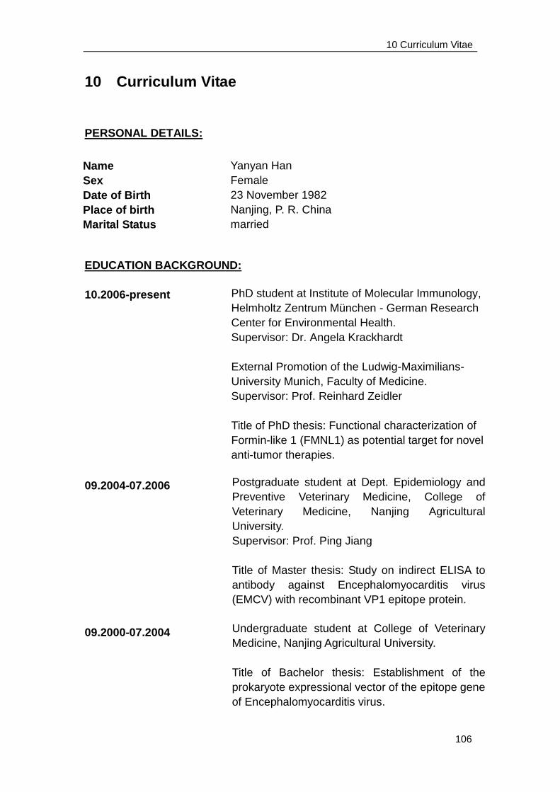
10 Curriculum Vitae
106
10 Curriculum Vitae
PERSONAL DETAILS:
EDUCATION BACKGROUND:
10.2006-present
09.2004-07.2006
09.2000-07.2004
Yanyan Han
Female
23 November 1982
Nanjing, P. R. China
married
Name
Sex
Date of Birth
Place of birth
Marital Status
PhD student at Institute of Molecular Immunology,
Helmholtz Zentrum München - German Research
Center for Environmental Health.
Supervisor: Dr. Angela Krackhardt
External Promotion of the Ludwig-Maximilians-
University Munich, Faculty of Medicine.
Supervisor: Prof. Reinhard Zeidler
Title of PhD thesis: Functional characterization of
Formin-like 1 (FMNL1) as potential target for novel
anti-tumor therapies.
Postgraduate student at Dept. Epidemiology and
Preventive Veterinary Medicine, College of
Veterinary Medicine, Nanjing Agricultural
University.
Supervisor: Prof. Ping Jiang
Title of Master thesis: Study on indirect ELISA to
antibody against Encephalomyocarditis virus
(EMCV) with recombinant VP1 epitope protein.
Undergraduate student at College of Veterinary
Medicine, Nanjing Agricultural University.
Title of Bachelor thesis: Establishment of the
prokaryote expressional vector of the epitope gene
of Encephalomyocarditis virus.
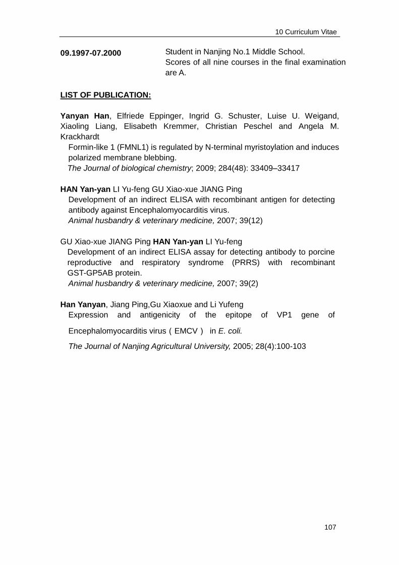
10 Curriculum Vitae
107
09.1997-07.2000
LIST OF PUBLICATION:
Yanyan Han, Elfriede Eppinger, Ingrid G. Schuster, Luise U. Weigand,
Xiaoling Liang, Elisabeth Kremmer, Christian Peschel and Angela M.
Krackhardt
Formin-like 1 (FMNL1) is regulated by N-terminal myristoylation and induces
polarized membrane blebbing.
The Journal of biological chemistry; 2009; 284(48): 33409–33417
HAN Yan-yan LI Yu-feng GU Xiao-xue JIANG Ping
Development of an indirect ELISA with recombinant antigen for detecting
antibody against Encephalomyocarditis virus.
Animal husbandry & veterinary medicine, 2007; 39(12)
GU Xiao-xue JIANG Ping HAN Yan-yan LI Yu-feng
Development of an indirect ELISA assay for detecting antibody to porcine
reproductive and respiratory syndrome (PRRS) with recombinant
GST-GP5AB protein.
Animal husbandry & veterinary medicine, 2007; 39(2)
Han Yanyan, Jiang Ping,Gu Xiaoxue and Li Yufeng
Expression and antigenicity of the epitope of VP1 gene of
Encephalomyocarditis virus(EMCV) in E. coli.
The Journal of Nanjing Agricultural University, 2005; 28(4):100-103
Student in Nanjing No.1 Middle School.
Scores of all nine courses in the final examination
are A.
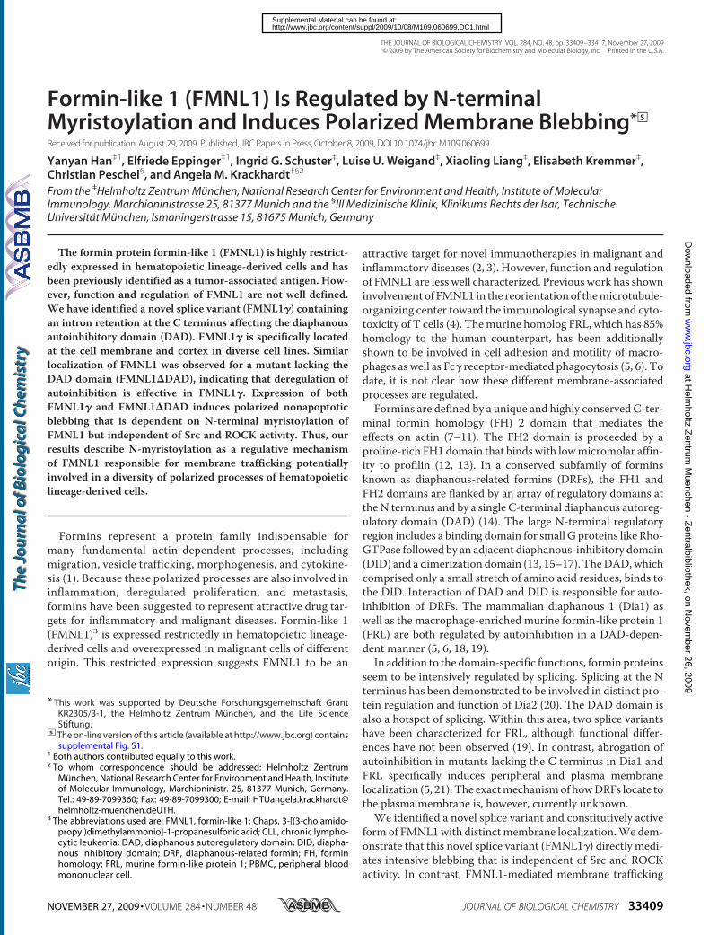
Formin-like 1 (FMNL1) Is Regulated by N-terminalMyristoylation and Induces Polarized Membrane Blebbing*□S
Received for publication, August 29, 2009 Published, JBC Papers in Press, October 8, 2009, DOI 10.1074/jbc.M109.060699
Yanyan Han‡1, Elfriede Eppinger‡1, Ingrid G. Schuster‡, Luise U. Weigand‡, Xiaoling Liang‡, Elisabeth Kremmer‡,Christian Peschel§, and Angela M. Krackhardt‡§2
From the ‡Helmholtz Zentrum Munchen, National Research Center for Environment and Health, Institute of MolecularImmunology, Marchioninistrasse 25, 81377 Munich and the §III Medizinische Klinik, Klinikums Rechts der Isar, TechnischeUniversitat Munchen, Ismaningerstrasse 15, 81675 Munich, Germany
The formin protein formin-like 1 (FMNL1) is highly restrict-edly expressed in hematopoietic lineage-derived cells and hasbeen previously identified as a tumor-associated antigen. How-ever, function and regulation of FMNL1 are not well defined.We have identified a novel splice variant (FMNL1�) containingan intron retention at the C terminus affecting the diaphanousautoinhibitory domain (DAD). FMNL1� is specifically locatedat the cell membrane and cortex in diverse cell lines. Similarlocalization of FMNL1 was observed for a mutant lacking theDAD domain (FMNL1�DAD), indicating that deregulation ofautoinhibition is effective in FMNL1�. Expression of bothFMNL1� and FMNL1�DAD induces polarized nonapoptoticblebbing that is dependent on N-terminal myristoylation ofFMNL1 but independent of Src and ROCK activity. Thus, ourresults describe N-myristoylation as a regulative mechanismof FMNL1 responsible for membrane trafficking potentiallyinvolved in a diversity of polarized processes of hematopoieticlineage-derived cells.
Formins represent a protein family indispensable formany fundamental actin-dependent processes, includingmigration, vesicle trafficking, morphogenesis, and cytokine-sis (1). Because these polarized processes are also involved ininflammation, deregulated proliferation, and metastasis,formins have been suggested to represent attractive drug tar-gets for inflammatory and malignant diseases. Formin-like 1(FMNL1)3 is expressed restrictedly in hematopoietic lineage-derived cells and overexpressed in malignant cells of differentorigin. This restricted expression suggests FMNL1 to be an
attractive target for novel immunotherapies in malignant andinflammatory diseases (2, 3). However, function and regulationof FMNL1 are less well characterized. Previous work has showninvolvement of FMNL1 in the reorientation of themicrotubule-organizing center toward the immunological synapse and cyto-toxicity of T cells (4). Themurine homolog FRL, which has 85%homology to the human counterpart, has been additionallyshown to be involved in cell adhesion and motility of macro-phages as well as Fc� receptor-mediated phagocytosis (5, 6). Todate, it is not clear how these different membrane-associatedprocesses are regulated.Formins are defined by a unique and highly conserved C-ter-
minal formin homology (FH) 2 domain that mediates theeffects on actin (7–11). The FH2 domain is proceeded by aproline-rich FH1 domain that bindswith lowmicromolar affin-ity to profilin (12, 13). In a conserved subfamily of forminsknown as diaphanous-related formins (DRFs), the FH1 andFH2 domains are flanked by an array of regulatory domains attheN terminus and by a single C-terminal diaphanous autoreg-ulatory domain (DAD) (14). The large N-terminal regulatoryregion includes a binding domain for small G proteins like Rho-GTPase followed by an adjacent diaphanous-inhibitory domain(DID) and a dimerization domain (13, 15–17). TheDAD,whichcomprised only a small stretch of amino acid residues, binds tothe DID. Interaction of DAD and DID is responsible for auto-inhibition of DRFs. The mammalian diaphanous 1 (Dia1) aswell as the macrophage-enriched murine formin-like protein 1(FRL) are both regulated by autoinhibition in a DAD-depen-dent manner (5, 6, 18, 19).In addition to the domain-specific functions, formin proteins
seem to be intensively regulated by splicing. Splicing at the Nterminus has been demonstrated to be involved in distinct pro-tein regulation and function of Dia2 (20). The DAD domain isalso a hotspot of splicing. Within this area, two splice variantshave been characterized for FRL, although functional differ-ences have not been observed (19). In contrast, abrogation ofautoinhibition in mutants lacking the C terminus in Dia1 andFRL specifically induces peripheral and plasma membranelocalization (5, 21). The exactmechanismof howDRFs locate tothe plasma membrane is, however, currently unknown.We identified a novel splice variant and constitutively active
form of FMNL1with distinctmembrane localization.We dem-onstrate that this novel splice variant (FMNL1�) directly medi-ates intensive blebbing that is independent of Src and ROCKactivity. In contrast, FMNL1-mediated membrane trafficking
* This work was supported by Deutsche Forschungsgemeinschaft GrantKR2305/3-1, the Helmholtz Zentrum Munchen, and the Life ScienceStiftung.
□S The on-line version of this article (available at http://www.jbc.org) containssupplemental Fig. S1.
1 Both authors contributed equally to this work.2 To whom correspondence should be addressed: Helmholtz Zentrum
Munchen, National Research Center for Environment and Health, Instituteof Molecular Immunology, Marchioninistr. 25, 81377 Munich, Germany.Tel.: 49-89-7099360; Fax: 49-89-7099300; E-mail: [email protected].
3 The abbreviations used are: FMNL1, formin-like 1; Chaps, 3-[(3-cholamido-propyl)dimethylammonio]-1-propanesulfonic acid; CLL, chronic lympho-cytic leukemia; DAD, diaphanous autoregulatory domain; DID, diapha-nous inhibitory domain; DRF, diaphanous-related formin; FH, forminhomology; FRL, murine formin-like protein 1; PBMC, peripheral bloodmononuclear cell.
THE JOURNAL OF BIOLOGICAL CHEMISTRY VOL. 284, NO. 48, pp. 33409 –33417, November 27, 2009© 2009 by The American Society for Biochemistry and Molecular Biology, Inc. Printed in the U.S.A.
NOVEMBER 27, 2009 • VOLUME 284 • NUMBER 48 JOURNAL OF BIOLOGICAL CHEMISTRY 33409
at Helm
holtz Zentrum
Muenchen - Z
entralbibliothek, on Novem
ber 26, 2009w
ww
.jbc.orgD
ownloaded from
http://www.jbc.org/content/suppl/2009/10/08/M109.060699.DC1.htmlSupplemental Material can be found at:
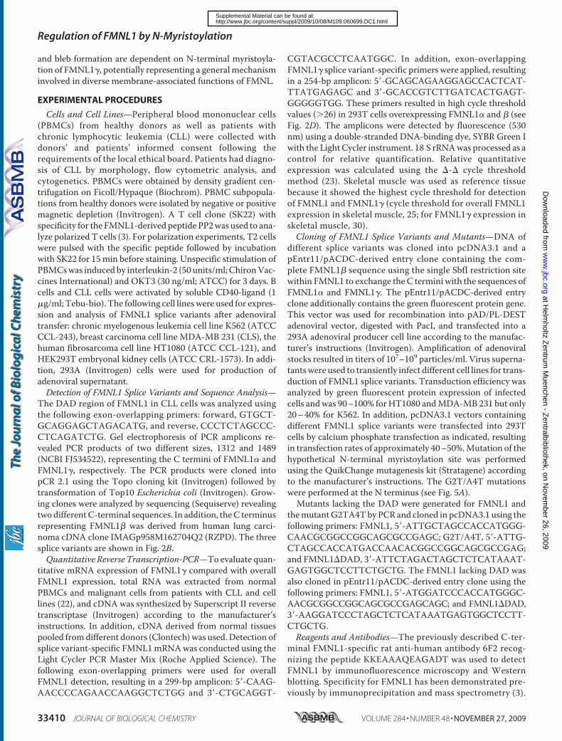
and bleb formation are dependent on N-terminal myristoyla-tion of FMNL1�, potentially representing a general mechanisminvolved in diverse membrane-associated functions of FMNL.
EXPERIMENTAL PROCEDURES
Cells and Cell Lines—Peripheral blood mononuclear cells(PBMCs) from healthy donors as well as patients withchronic lymphocytic leukemia (CLL) were collected withdonors’ and patients’ informed consent following therequirements of the local ethical board. Patients had diagno-sis of CLL by morphology, flow cytometric analysis, andcytogenetics. PBMCs were obtained by density gradient cen-trifugation on Ficoll/Hypaque (Biochrom). PBMC subpopula-tions from healthy donors were isolated by negative or positivemagnetic depletion (Invitrogen). A T cell clone (SK22) withspecificity for the FMNL1-derived peptide PP2was used to ana-lyze polarized T cells (3). For polarization experiments, T2 cellswere pulsed with the specific peptide followed by incubationwith SK22 for 15min before staining. Unspecific stimulation ofPBMCswas induced by interleukin-2 (50 units/ml; ChironVac-cines International) and OKT3 (30 ng/ml; ATCC) for 3 days. Bcells and CLL cells were activated by soluble CD40-ligand (1�g/ml; Tebu-bio). The following cell lineswere used for expres-sion and analysis of FMNL1 splice variants after adenoviraltransfer: chronic myelogenous leukemia cell line K562 (ATCCCCL-243), breast carcinoma cell line MDA-MB 231 (CLS), thehuman fibrosarcoma cell line HT1080 (ATCC CCL-121), andHEK293T embryonal kidney cells (ATCC CRL-1573). In addi-tion, 293A (Invitrogen) cells were used for production ofadenoviral supernatant.Detection of FMNL1 Splice Variants and Sequence Analysis—
The DAD region of FMNL1 in CLL cells was analyzed usingthe following exon-overlapping primers: forward, GTGCT-GCAGGAGCTAGACATG, and reverse, CCCTCTAGCCC-CTCAGATCTG. Gel electrophoresis of PCR amplicons re-vealed PCR products of two different sizes, 1312 and 1489(NCBI FJ534522), representing the C termini of FMNL1� andFMNL1�, respectively. The PCR products were cloned intopCR 2.1 using the Topo cloning kit (Invitrogen) followed bytransformation of Top10 Escherichia coli (Invitrogen). Grow-ing clones were analyzed by sequencing (Sequiserve) revealingtwodifferentC-terminal sequences. In addition, theC terminusrepresenting FMNL1� was derived from human lung carci-noma cDNA clone IMAGp958M162704Q2 (RZPD). The threesplice variants are shown in Fig. 2B.Quantitative Reverse Transcription-PCR—Toevaluate quan-
titative mRNA expression of FMNL1� compared with overallFMNL1 expression, total RNA was extracted from normalPBMCs and malignant cells from patients with CLL and celllines (22), and cDNA was synthesized by Superscript II reversetranscriptase (Invitrogen) according to the manufacturer’sinstructions. In addition, cDNA derived from normal tissuespooled fromdifferent donors (Clontech)was used. Detection ofsplice variant-specific FMNL1mRNAwas conducted using theLight Cycler PCR Master Mix (Roche Applied Science). Thefollowing exon-overlapping primers were used for overallFMNL1 detection, resulting in a 299-bp amplicon: 5�-CAAG-AACCCCAGAACCAAGGCTCTGG and 3�-CTGCAGGT-
CGTACGCCTCAATGGC. In addition, exon-overlappingFMNL1� splice variant-specific primers were applied, resultingin a 254-bp amplicon: 5�-GCAGCAGAAGGAGCCACTCAT-TTATGAGAGC and 3�-GCACCGTCTTGATCACTGAGT-GGGGGTGG. These primers resulted in high cycle thresholdvalues (�26) in 293T cells overexpressing FMNL1� and � (seeFig. 2D). The amplicons were detected by fluorescence (530nm) using a double-stranded DNA-binding dye, SYBR Green Iwith the Light Cycler instrument. 18 S rRNAwas processed as acontrol for relative quantification. Relative quantitativeexpression was calculated using the �-� cycle thresholdmethod (23). Skeletal muscle was used as reference tissuebecause it showed the highest cycle threshold for detectionof FMNL1 and FMNL1� (cycle threshold for overall FMNL1expression in skeletal muscle, 25; for FMNL1� expression inskeletal muscle, 30).Cloning of FMNL1 Splice Variants and Mutants—DNA of
different splice variants was cloned into pcDNA3.1 and apEntr11/pACDC-derived entry clone containing the com-plete FMNL1� sequence using the single SbfI restriction sitewithin FMNL1 to exchange the C termini with the sequences ofFMNL1� and FMNL1�. The pEntr11/pACDC-derived entryclone additionally contains the green fluorescent protein gene.This vector was used for recombination into pAD/PL-DESTadenoviral vector, digested with PacI, and transfected into a293A adenoviral producer cell line according to the manufac-turer’s instructions (Invitrogen). Amplification of adenoviralstocks resulted in titers of 107–109 particles/ml. Virus superna-tantswere used to transiently infect different cell lines for trans-duction of FMNL1 splice variants. Transduction efficiency wasanalyzed by green fluorescent protein expression of infectedcells andwas 90–100% forHT1080 andMDA-MB 231 but only20–40% for K562. In addition, pcDNA3.1 vectors containingdifferent FMNL1 splice variants were transfected into 293Tcells by calcium phosphate transfection as indicated, resultingin transfection rates of approximately 40–50%.Mutation of thehypothetical N-terminal myristoylation site was performedusing the QuikChange mutagenesis kit (Stratagene) accordingto the manufacturer’s instructions. The G2T/A4T mutationswere performed at the N terminus (see Fig. 5A).Mutants lacking the DAD were generated for FMNL1 and
themutantG2TA4Tby PCR and cloned in pcDNA3.1 using thefollowing primers: FMNL1, 5�-ATTGCTAGCCACCATGGG-CAACGCGGCCGGCAGCGCCGAGC; G2T/A4T, 5�-ATTG-CTAGCCACCATGACCAACACGGCCGGCAGCGCCGAG;and FMNL1�DAD, 3�-ATTCTAGACTAGCTCTCATAAAT-GAGTGGCTCCTTCTGCTG. The FMNL1 lacking DAD wasalso cloned in pEntr11/pACDC-derived entry clone using thefollowing primers: FMNL1, 5�-ATGGATCCCACCATGGGC-AACGCGGCCGGCAGCGCCGAGCAGC; and FMNL1�DAD,3�-AAGGATCCCTAGCTCTCATAAATGAGTGGCTCCTT-CTGCTG.Reagents and Antibodies—The previously described C-ter-
minal FMNL1-specific rat anti-human antibody 6F2 recog-nizing the peptide KKEAAAQEAGADT was used to detectFMNL1 by immunofluorescence microscopy and Westernblotting. Specificity for FMNL1 has been demonstrated pre-viously by immunoprecipitation and mass spectrometry (3).
Regulation of FMNL1 by N-Myristoylation
33410 JOURNAL OF BIOLOGICAL CHEMISTRY VOLUME 284 • NUMBER 48 • NOVEMBER 27, 2009
at Helm
holtz Zentrum
Muenchen - Z
entralbibliothek, on Novem
ber 26, 2009w
ww
.jbc.orgD
ownloaded from
http://www.jbc.org/content/suppl/2009/10/08/M109.060699.DC1.htmlSupplemental Material can be found at:
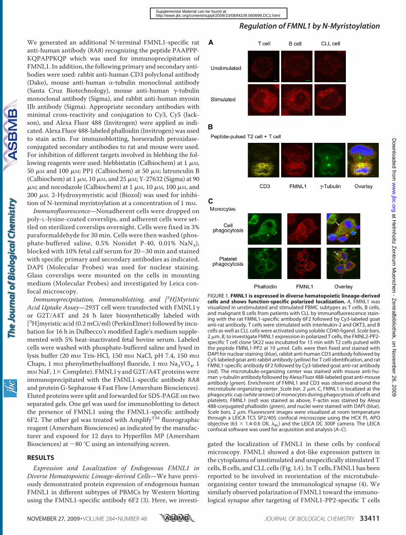
We generated an additional N-terminal FMNL1-specific ratanti-human antibody (8A8) recognizing the peptide PAAPPP-KQPAPPKQP which was used for immunoprecipitation ofFMNL1. In addition, the following primary and secondary anti-bodies were used: rabbit anti-human CD3 polyclonal antibody(Dako), mouse anti-human �-tubulin monoclonal antibody(Santa Cruz Biotechnology), mouse anti-human �-tubulinmonoclonal antibody (Sigma), and rabbit anti-human myosinIIb antibody (Sigma). Appropriate secondary antibodies withminimal cross-reactivity and conjugation to Cy3, Cy5 (Jack-son), and Alexa Fluor 488 (Invitrogen) were applied as indi-cated. Alexa Fluor 488-labeled phalloidin (Invitrogen) was usedto stain actin. For immunoblotting, horseradish peroxidase-conjugated secondary antibodies to rat and mouse were used.For inhibition of different targets involved in blebbing the fol-lowing reagents were used: blebbistatin (Calbiochem) at 1 �M,50 �M and 100 �M; PP1 (Calbiochem) at 50 �M; latrunculin B(Calbiochem) at 1�M, 10�M, and 25�M; Y-27632 (Sigma) at 90�M; and nocodazole (Calbiochem) at 1 �M, 10 �M, 100 �M, and200 �M. 2-Hydroxymyristic acid (Biozol) was used for inhibi-tion of N-terminal myristoylation at a concentration of 1 mM.Immunofluorescence—Nonadherent cells were dropped on
poly-L-lysine-coated coverslips, and adherent cells were set-tled on sterilized coverslips overnight. Cells were fixed in 3%paraformaldehyde for 30 min. Cells were then washed (phos-phate-buffered saline, 0.5% Nonidet P-40, 0.01% NaN3),blocked with 10% fetal calf serum for 20–30 min and stainedwith specific primary and secondary antibodies as indicated.DAPI (Molecular Probes) was used for nuclear staining.Glass coverslips were mounted on the cells in mountingmedium (Molecular Probes) and investigated by Leica con-focal microscopy.Immunoprecipitation, Immunoblotting, and [3H]Myristic
Acid Uptake Assay—293T cell were transfected with FMNL1�or G2T/A4T and 24 h later biosynthetically labeled with[3H]myristic acid (0.2mCi/ml) (PerkinElmer) followed by incu-bation for 16 h in Dulbecco’s modified Eagle’s medium supple-mented with 5% heat-inactivated fetal bovine serum. Labeledcells were washed with phosphate-buffered saline and lysed inlysis buffer (20 mM Tris-HCl, 150 mM NaCl, pH 7.4, 150 mM
Chaps, 1 mM phenylmethylsulfonyl fluoride, 1 mM Na3VO4, 1mMNaF, 1�Complete). FMNL1� andG2T/A4Tproteins wereimmunoprecipitated with the FMNL1-specific antibody 8A8and protein G-Sepharose 4 Fast Flow (Amersham Biosciences).Eluted proteinswere split and forwarded for SDS-PAGEon twoseparated gels. One gel was used for immunoblotting to detectthe presence of FMNL1 using the FMNL1-specific antibody6F2. The other gel was treated with AmplifyTM fluorographicreagent (Amersham Biosciences) as indicated by the manufac-turer and exposed for 12 days to Hyperfilm MP (AmershamBiosciences) at �80 °C using an intensifying screen.
RESULTS
Expression and Localization of Endogenous FMNL1 inDiverse Hematopoietic Lineage-derived Cells—We have previ-ously demonstrated protein expression of endogenous humanFMNL1 in different subtypes of PBMCs by Western blottingusing the FMNL1-specific antibody 6F2 (3). Here, we investi-
gated the localization of FMNL1 in these cells by confocalmicroscopy. FMNL1 showed a dot-like expression pattern inthe cytoplasma of unstimulated and unspecifically stimulatedTcells, B cells, andCLL cells (Fig. 1A). InT cells, FMNL1has beenreported to be involved in reorientation of the microtubule-organizing center toward the immunological synapse (4). Wesimilarly observed polarization of FMNL1 toward the immuno-logical synapse after targeting of FMNL1-PP2-specific T cells
FIGURE 1. FMNL1 is expressed in diverse hematopoietic lineage-derivedcells and shows function-specific polarized localization. A, FMNL1 wasvisualized in unstimulated and stimulated PBMC subtypes as T cells, B cells,and malignant B cells from patients with CLL by immunofluorescence stain-ing with the rat FMNL1-specific antibody 6F2 followed by Cy3-labeled goatanti-rat antibody. T cells were stimulated with interleukin-2 and OKT3, and Bcells as well as CLL cells were activated using soluble CD40-ligand. Scale bars,2 �m. B, to investigate FMNL1 expression in polarized T cells, the FMNL2-PP2-specific T cell clone SK22 was incubated for 15 min with T2 cells pulsed withthe peptide FMNL1-PP2 at 10 �mol. Cells were then fixed and stained withDAPI for nuclear staining (blue), rabbit anti-human CD3 antibody followed byCy5-labeled goat anti-rabbit antibody (yellow) for T cell identification, and ratFMNL1-specific antibody 6F2 followed by Cy3-labeled goat anti-rat antibody(red). The microtubule-organizing center was stained with mouse anti-hu-man �-tubulin antibody followed by Alexa Fluor 488-labeled goat anti-mouseantibody (green). Enrichment of FMNL1 and CD3 was observed around themicrotubule-organizing center. Scale bar, 2 �m. C, FMNL1 is localized at thephagocytic cup (white arrows) of monocytes during phagocytosis of cells andplatelets. FMNL1 (red) was stained as above, F-actin was stained by Alexa488-conjugated phalloidin (green), and nuclei were stained with DAPI (blue).Scale bars, 2 �m. Fluorescent images were visualized at room temperaturethrough a LEICA TCS SP2/405 confocal microscope using the HCX PL APOobjective (63 � 1.4-0.6 OIL �BL) and the LEICA DC 300F camera. The LEICAconfocal software was used for acquisition and analysis (A–C).
Regulation of FMNL1 by N-Myristoylation
NOVEMBER 27, 2009 • VOLUME 284 • NUMBER 48 JOURNAL OF BIOLOGICAL CHEMISTRY 33411
at Helm
holtz Zentrum
Muenchen - Z
entralbibliothek, on Novem
ber 26, 2009w
ww
.jbc.orgD
ownloaded from
http://www.jbc.org/content/suppl/2009/10/08/M109.060699.DC1.htmlSupplemental Material can be found at:
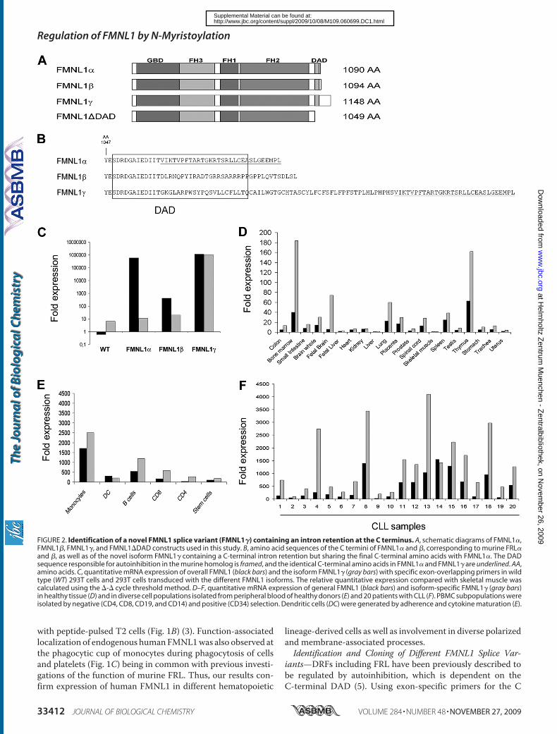
with peptide-pulsed T2 cells (Fig. 1B) (3). Function-associatedlocalization of endogenous humanFMNL1was also observed atthe phagocytic cup of monocytes during phagocytosis of cellsand platelets (Fig. 1C) being in common with previous investi-gations of the function of murine FRL. Thus, our results con-firm expression of human FMNL1 in different hematopoietic
lineage-derived cells as well as involvement in diverse polarizedand membrane-associated processes.Identification and Cloning of Different FMNL1 Splice Var-
iants—DRFs including FRL have been previously described tobe regulated by autoinhibition, which is dependent on theC-terminal DAD (5). Using exon-specific primers for the C
FIGURE 2. Identification of a novel FMNL1 splice variant (FMNL1�) containing an intron retention at the C terminus. A, schematic diagrams of FMNL1�,FMNL1�, FMNL1�, and FMNL1�DAD constructs used in this study. B, amino acid sequences of the C termini of FMNL1� and �, corresponding to murine FRL�and �, as well as of the novel isoform FMNL1� containing a C-terminal intron retention but sharing the final C-terminal amino acids with FMNL1�. The DADsequence responsible for autoinhibition in the murine homolog is framed, and the identical C-terminal amino acids in FMNL1� and FMNL1� are underlined. AA,amino acids. C, quantitative mRNA expression of overall FMNL1 (black bars) and the isoform FMNL1� (gray bars) with specific exon-overlapping primers in wildtype (WT) 293T cells and 293T cells transduced with the different FMNL1 isoforms. The relative quantitative expression compared with skeletal muscle wascalculated using the �-� cycle threshold method. D–F, quantitative mRNA expression of general FMNL1 (black bars) and isoform-specific FMNL1� (gray bars)in healthy tissue (D) and in diverse cell populations isolated from peripheral blood of healthy donors (E) and 20 patients with CLL (F). PBMC subpopulations wereisolated by negative (CD4, CD8, CD19, and CD14) and positive (CD34) selection. Dendritic cells (DC) were generated by adherence and cytokine maturation (E).
Regulation of FMNL1 by N-Myristoylation
33412 JOURNAL OF BIOLOGICAL CHEMISTRY VOLUME 284 • NUMBER 48 • NOVEMBER 27, 2009
at Helm
holtz Zentrum
Muenchen - Z
entralbibliothek, on Novem
ber 26, 2009w
ww
.jbc.orgD
ownloaded from
http://www.jbc.org/content/suppl/2009/10/08/M109.060699.DC1.htmlSupplemental Material can be found at:
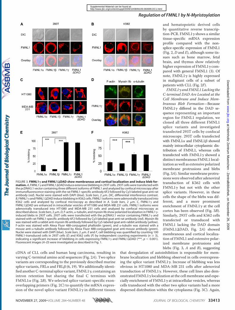
cDNA of CLL cells and human lung carcinoma, resulting invarying C-terminal amino acid sequences (Fig. 2A). Two splicevariants are corresponding to the previously described murinesplice variants, FRL� and FRL� (6, 19). We additionally identi-fied anotherC-terminal splice variant, FMNL1�, containing anintron retention but sharing the final C terminus withFMNL1� (Fig. 2B). We selected splice variant-specific exon-overlapping primers (Fig. 2C) to quantify the mRNA expres-sion of the novel splice variant FMNL1� in different tissues
and hematopoietic derived cellsby quantitative reverse transcrip-tion-PCR. FMNL1� shows a similartissue-specific mRNA expressionprofile compared with the non-splice-specific expression of FMNL1(Fig. 2, D and E), although some tis-sues such as bone marrow, fetalbrain, and thymus show relativelyhigher expression of FMNL1� com-pared with general FMNL1 (3). Ofnote, FMNL1� is highly expressedin malignant cells of a subset ofpatients with CLL (Fig. 2F).FMNL1� and FMNL1 Lacking the
C-terminal DAD Are Located at theCell Membrane and Induce Mem-branous Bleb Formation—BecauseFMNL1� differed in the DAD se-quence representing an importantregion for FMNL1 regulation, wecloned all three different FMNL1splice variants and investigatedtransfected 293T cells by confocalmicroscopy. 293T cells transfectedwith FMNL1� and FMNL1� showedmainly intracellular cytoplasmic dis-tribution of FMNL1, whereas cellstransfected with FMNL1� showed adistinctmembranous FMNL1 local-ization aswell as extensive polarizedmembrane protrusions and blebs(Fig. 3A). Similarmembrane protru-sionswere observed after adenoviraltransduction of K562 cells withFMNL1� but not with the othersplice variants. However, in thesecells the shape of the blebs was dif-ferent, and a more prominentenrichment of FMNL1� at the cellcortex has been observed (Fig. 3B).Similarly, 293T cells and K562 cellstransfected or transduced withFMNL1 lacking the DAD domain(FMNL1�DAD, Fig. 2A) showedmembranous and cortical localiza-tion of FMNL1 and extensive polar-ized membrane protrusions andblebs (Fig. 3, A and B), suggesting
that deregulation of autoinhibition is responsible for mem-brane localization and blebbing observed in cells overexpress-ing the splice variant FMNL1�. Increase of blebbing was lessobvious in HT1080 and MDA-MB 231 cells after adenoviraltransduction of FMNL1�. However, these cell lines also dem-onstrated FMNL1� localization at the cell membrane and espe-cially enrichment of FMNL1� at intracellular vesicles, whereascells transduced with the other two splice variants had a moredispersed distribution within the cytoplasma (Fig. 3C). Again,
FIGURE 3. FMNL1� and FMNL1�DAD show membranous and cortical localization and induce bleb for-mation. A, FMNL1� and FMNL1�DAD induce extensive blebbing in 293T cells. 293T cells were transfected withthe pcDNA3.1 vector containing three different isoforms of FMNL1 and analyzed by confocal microscopy afterimmunofluorescence staining with the rat FMNL1-specific antibody 6F2 followed by Cy3-labeled goat anti-ratantibody (red). Nuclei were stained with DAPI (blue). Scale bars, 2 �m. DIC, differential interference contrast.B, FMNL1� and FMNL1�DAD induce blebbing in K562 cells. FMNL1 isoforms were adenovirally transduced intoK562 cells and analyzed by confocal microscopy as described in A. Scale bars, 2 �m. C, FMNL1� andFMNL1�DAD are enhanced in intracellular vesicles of HT1080 and MDA-MB 231 cells. FMNL1 isoforms wereadenovirally transduced into HT1080 and MDA-MB 231 cells and analyzed by confocal microscopy asdescribed above. Scale bars, 2 �m. D, F-actin, �-tubulin, and myosin IIb show polarized localization in FMNL1�-induced blebs in 293T cells. 293T cells were transfected with the pcDNA3.1 vector containing FMNL1� andstained with rat FMNL1-specific antibody 6F2 followed by Cy3-labeled goat anti-rat antibody (red). Myosin IIbwas stained with a rabbit anti-myosin IIb antibody followed by Cy5-labeled goat anti-rabbit antibody (yellow).F-actin was stained with Alexa Fluor 488-conjugated phalloidin (green), and �-tubulin was stained with amouse anti-�-tubulin antibody followed by Alexa Fluor 488-conjugated goat anti-mouse antibody (green).Nuclei were stained with DAPI (blue). Scale bars, 2 �m. E and F, cell blebbing was quantified by counting 100FMNL1-transduced cells in 293T cells (E) and K562 cells (F) by independent counting experiments (n � 3),indicating a significant increase of blebbing in cells expressing FMNL1� and FMNL1�DAD (***, p � 0.001).Fluorescent images (A–D) were investigated as described in Fig. 1.
Regulation of FMNL1 by N-Myristoylation
NOVEMBER 27, 2009 • VOLUME 284 • NUMBER 48 JOURNAL OF BIOLOGICAL CHEMISTRY 33413
at Helm
holtz Zentrum
Muenchen - Z
entralbibliothek, on Novem
ber 26, 2009w
ww
.jbc.orgD
ownloaded from
http://www.jbc.org/content/suppl/2009/10/08/M109.060699.DC1.htmlSupplemental Material can be found at:
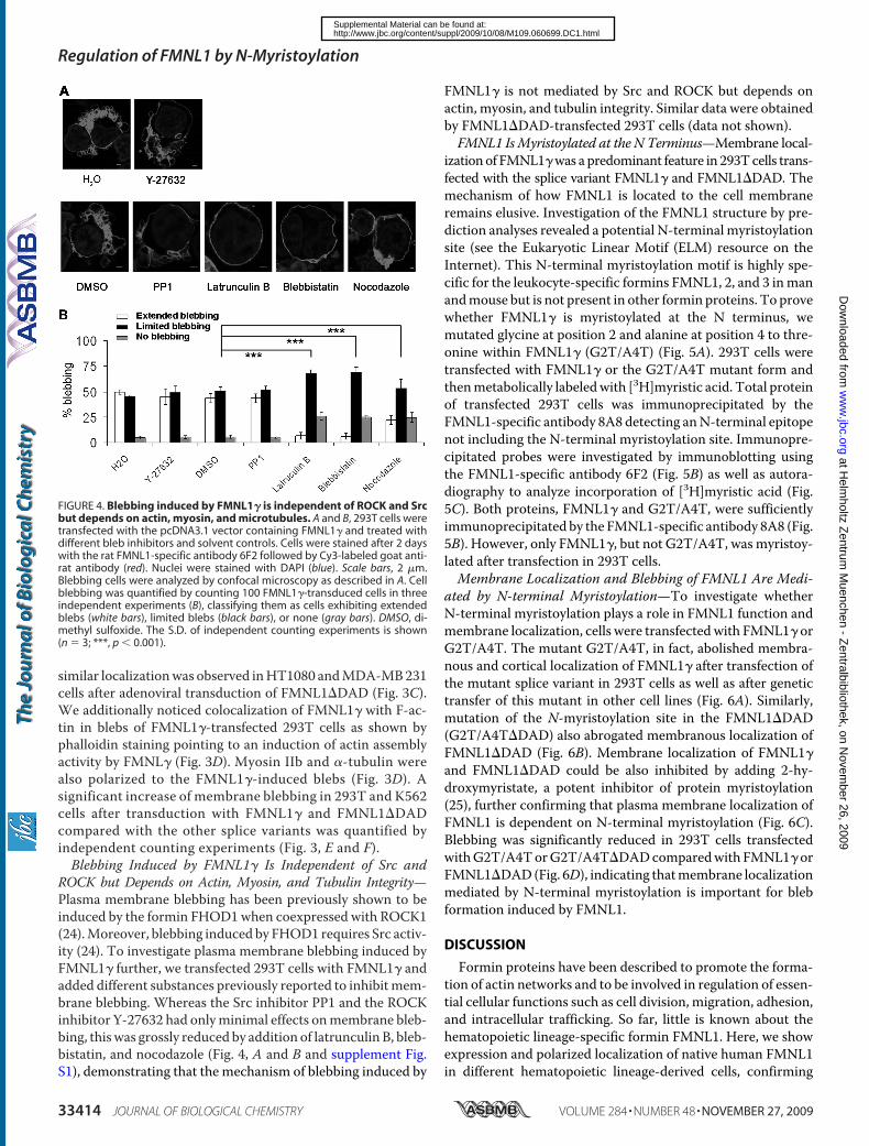
similar localizationwas observed inHT1080 andMDA-MB231cells after adenoviral transduction of FMNL1�DAD (Fig. 3C).We additionally noticed colocalization of FMNL1� with F-ac-tin in blebs of FMNL1�-transfected 293T cells as shown byphalloidin staining pointing to an induction of actin assemblyactivity by FMNL� (Fig. 3D). Myosin IIb and �-tubulin werealso polarized to the FMNL1�-induced blebs (Fig. 3D). Asignificant increase of membrane blebbing in 293T and K562cells after transduction with FMNL1� and FMNL1�DADcompared with the other splice variants was quantified byindependent counting experiments (Fig. 3, E and F).Blebbing Induced by FMNL1� Is Independent of Src and
ROCK but Depends on Actin, Myosin, and Tubulin Integrity—Plasma membrane blebbing has been previously shown to beinduced by the formin FHOD1when coexpressed with ROCK1(24).Moreover, blebbing induced by FHOD1 requires Src activ-ity (24). To investigate plasma membrane blebbing induced byFMNL1� further, we transfected 293T cells with FMNL1� andadded different substances previously reported to inhibitmem-brane blebbing. Whereas the Src inhibitor PP1 and the ROCKinhibitor Y-27632 had onlyminimal effects onmembrane bleb-bing, thiswas grossly reduced by addition of latrunculin B, bleb-bistatin, and nocodazole (Fig. 4, A and B and supplement Fig.S1), demonstrating that the mechanism of blebbing induced by
FMNL1� is not mediated by Src and ROCK but depends onactin, myosin, and tubulin integrity. Similar data were obtainedby FMNL1�DAD-transfected 293T cells (data not shown).FMNL1 IsMyristoylated at the N Terminus—Membrane local-
izationof FMNL1�was apredominant feature in 293Tcells trans-fected with the splice variant FMNL1� and FMNL1�DAD. Themechanism of how FMNL1 is located to the cell membraneremains elusive. Investigation of the FMNL1 structure by pre-diction analyses revealed a potential N-terminal myristoylationsite (see the Eukaryotic Linear Motif (ELM) resource on theInternet). This N-terminal myristoylation motif is highly spe-cific for the leukocyte-specific formins FMNL1, 2, and 3 inmanandmouse but is not present in other formin proteins. To provewhether FMNL1� is myristoylated at the N terminus, wemutated glycine at position 2 and alanine at position 4 to thre-onine within FMNL1� (G2T/A4T) (Fig. 5A). 293T cells weretransfected with FMNL1� or the G2T/A4T mutant form andthenmetabolically labeledwith [3H]myristic acid. Total proteinof transfected 293T cells was immunoprecipitated by theFMNL1-specific antibody 8A8 detecting anN-terminal epitopenot including the N-terminal myristoylation site. Immunopre-cipitated probes were investigated by immunoblotting usingthe FMNL1-specific antibody 6F2 (Fig. 5B) as well as autora-diography to analyze incorporation of [3H]myristic acid (Fig.5C). Both proteins, FMNL1� and G2T/A4T, were sufficientlyimmunoprecipitated by the FMNL1-specific antibody 8A8 (Fig.5B). However, only FMNL1�, but not G2T/A4T, wasmyristoy-lated after transfection in 293T cells.Membrane Localization and Blebbing of FMNL1 Are Medi-
ated by N-terminal Myristoylation—To investigate whetherN-terminal myristoylation plays a role in FMNL1 function andmembrane localization, cells were transfectedwith FMNL1� orG2T/A4T. The mutant G2T/A4T, in fact, abolished membra-nous and cortical localization of FMNL1� after transfection ofthe mutant splice variant in 293T cells as well as after genetictransfer of this mutant in other cell lines (Fig. 6A). Similarly,mutation of the N-myristoylation site in the FMNL1�DAD(G2T/A4T�DAD) also abrogated membranous localization ofFMNL1�DAD (Fig. 6B). Membrane localization of FMNL1�and FMNL1�DAD could be also inhibited by adding 2-hy-droxymyristate, a potent inhibitor of protein myristoylation(25), further confirming that plasma membrane localization ofFMNL1 is dependent on N-terminal myristoylation (Fig. 6C).Blebbing was significantly reduced in 293T cells transfectedwithG2T/A4TorG2T/A4T�DADcomparedwith FMNL1� orFMNL1�DAD(Fig. 6D), indicating thatmembrane localizationmediated by N-terminal myristoylation is important for blebformation induced by FMNL1.
DISCUSSION
Formin proteins have been described to promote the forma-tion of actin networks and to be involved in regulation of essen-tial cellular functions such as cell division, migration, adhesion,and intracellular trafficking. So far, little is known about thehematopoietic lineage-specific formin FMNL1. Here, we showexpression and polarized localization of native human FMNL1in different hematopoietic lineage-derived cells, confirming
FIGURE 4. Blebbing induced by FMNL1� is independent of ROCK and Srcbut depends on actin, myosin, and microtubules. A and B, 293T cells weretransfected with the pcDNA3.1 vector containing FMNL1� and treated withdifferent bleb inhibitors and solvent controls. Cells were stained after 2 dayswith the rat FMNL1-specific antibody 6F2 followed by Cy3-labeled goat anti-rat antibody (red). Nuclei were stained with DAPI (blue). Scale bars, 2 �m.Blebbing cells were analyzed by confocal microscopy as described in A. Cellblebbing was quantified by counting 100 FMNL1�-transduced cells in threeindependent experiments (B), classifying them as cells exhibiting extendedblebs (white bars), limited blebs (black bars), or none (gray bars). DMSO, di-methyl sulfoxide. The S.D. of independent counting experiments is shown(n � 3; ***, p � 0.001).
Regulation of FMNL1 by N-Myristoylation
33414 JOURNAL OF BIOLOGICAL CHEMISTRY VOLUME 284 • NUMBER 48 • NOVEMBER 27, 2009
at Helm
holtz Zentrum
Muenchen - Z
entralbibliothek, on Novem
ber 26, 2009w
ww
.jbc.orgD
ownloaded from
http://www.jbc.org/content/suppl/2009/10/08/M109.060699.DC1.htmlSupplemental Material can be found at:
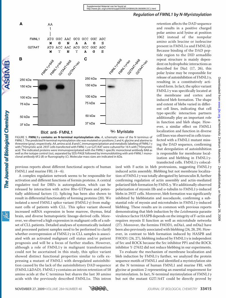
previous reports about different functional aspects of humanFMNL1 and murine FRL (4–6).A complex regulation network seems to be responsible for
activation and different functions of formin proteins. A centralregulative tool for DRFs is autoregulation, which can bereleased by interaction with active Rho-GTPases and poten-tially additional factors (1). Splicing has been also shown toresult in differential functionality of forming proteins (20). Weisolated a novel FMNL1 splice variant (FMNL1�) from malig-nant cells of patients with CLL. This splice variant showedincreased mRNA expression in bone marrow, thymus, fetalbrain, and diverse hematopoietic lineage-derived cells. More-over, we observed a high expression in malignant cells of a sub-set of CLL patients. Detailed analyses of systematically isolatedand processed patient samples need to be performed to clarifywhether overexpression of FMNL1� in CLL samples is associ-ated with an activated malignant cell status and/or patientprognosis and will be a focus of further studies. However,although a role of FMNL1� in malignant transformationcould not be ascertained in this study, this splice variantshowed distinct functional properties similar to cells ex-pressing a mutant of FMNL1 with deregulated autoinhibi-tion caused by the lack of the autoinhibitory DAD sequence(FMNL1�DAD). FMNL1� contains an intron retention of 58amino acids at the C terminus but shares the last 30 aminoacids with the previously described FMNL1�. This intron
retention affects the DAD sequenceand results in a positive chargedpolar amino acid lysine at position1062 instead of the nonpolaramino acids leucine or isoleucinepresent in FMNL1� and FMNL1�.Because binding of the DAD pep-tide region to the DID armadillorepeat structure is mainly depen-dent on hydrophobic interactions asdescribed for Dia1 (17, 26), thispolar lysine may be responsible forrelease of autoinhibition of FMNL1�,resulting in a constitutively acti-vated form. In fact, the splice variantFMNL1� was specifically located atthe membrane and cortex andinduced bleb formation. The shapeand extent of blebs varied in differ-ent cell lines, indicating that celltype-specific interaction partnersadditionally play an important rolein function and bleb shape. How-ever, a similar effect on FMNL1localization and function in diversecell lines was observed in cells trans-fected with a FMNL1 mutant miss-ing the DAD sequence, confirmingthat deregulation of autoinhibitionis responsible for membrane local-ization and blebbing in FMNL1�-transfected cells. FMNL1� colocal-
ized with F-actin in bleb protrusions, suggesting FMNL1�induced actin assembly. Blebbing but not membrane localiza-tion of FMNL1� was totally abrogated by latrunculin B, furtherconfirming regulation of actin assembly and actin-mediatedpolarized bleb formation by FMNL�. We additionally observedpolarization of myosin IIb and �-tubulin to FMNL1�-inducedblebs in 293T cells. Moreover, bleb formation was significantlyinhibited by blebbistatin and nocodazole, confirming a sub-stantial role of myosin and microtubules in FMNL1�-inducedblebbing. These results are in common with previous reportsdemonstrating that bleb induction by the Leishmania parasitevirulence factor HASPB depends on the integrity of F-actin andrequires myosin II function as well as microtubule networks(27). Moreover, the formins FHOD1, mDia1, and mDia2 havebeen also previously associatedwith blebbing (24, 28, 29). How-ever, in contrast to bleb formation induced by HASPB andFHOD1 (24, 27), blebbing induced by FMNL1� is independentof Src and ROCK because the Src inhibitor PP1 and the ROCKinhibitor Y-27632 did not reduce blebbing in our experiments.To evaluate the mechanism of membrane localization and
bleb induction by FMNL1� further, we analyzed the proteinsequence motifs of FMNL1 and identified a myristoylation siteat the N terminus of human FMNL1 and murine FRL withglycine at position 2 representing an essential requirement formyristoylation. In fact, N-terminal myristoylation of FMNL1�but not the mutant G2T/A4T could be directly proved by
FIGURE 5. FMNL1� contains an N-terminal myristoylation site. A, schematic view of the N terminus ofFMNL1. The predicted N-terminal myristoylation site was mutated on positions 2 and 4, glycine and alanine tothreonine (gray), respectively. AA, amino acid. B and C, immunoprecipitation and metabolic labeling of FMNL1�with [3H]myristic acid. 293T cells transfected with FMNL1� or G2T/A4T were cultured for 16 h with [3H]myristicacid. The indicated proteins were immunoprecipitated with the FMNL1-specific monoclonal antibody 8A8 aswell as the isotype control (iso), separated by SDS-PAGE followed by immunoblotting with anti-FMNL1 mono-clonal antibody 6F2 (B) or fluorography (C). Molecular mass sizes are indicated in kDa.
Regulation of FMNL1 by N-Myristoylation
NOVEMBER 27, 2009 • VOLUME 284 • NUMBER 48 JOURNAL OF BIOLOGICAL CHEMISTRY 33415
at Helm
holtz Zentrum
Muenchen - Z
entralbibliothek, on Novem
ber 26, 2009w
ww
.jbc.orgD
ownloaded from
http://www.jbc.org/content/suppl/2009/10/08/M109.060699.DC1.htmlSupplemental Material can be found at:
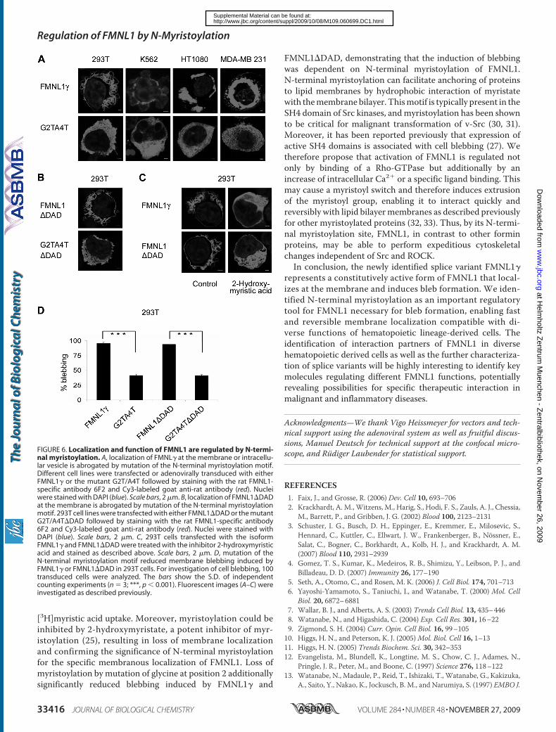
[3H]myristic acid uptake. Moreover, myristoylation could beinhibited by 2-hydroxymyristate, a potent inhibitor of myr-istoylation (25), resulting in loss of membrane localizationand confirming the significance of N-terminal myristoylationfor the specific membranous localization of FMNL1. Loss ofmyristoylation by mutation of glycine at position 2 additionallysignificantly reduced blebbing induced by FMNL1� and
FMNL1�DAD, demonstrating that the induction of blebbingwas dependent on N-terminal myristoylation of FMNL1.N-terminal myristoylation can facilitate anchoring of proteinsto lipid membranes by hydrophobic interaction of myristatewith themembrane bilayer. Thismotif is typically present in theSH4 domain of Src kinases, andmyristoylation has been shownto be critical for malignant transformation of v-Src (30, 31).Moreover, it has been reported previously that expression ofactive SH4 domains is associated with cell blebbing (27). Wetherefore propose that activation of FMNL1 is regulated notonly by binding of a Rho-GTPase but additionally by anincrease of intracellular Ca2� or a specific ligand binding. Thismay cause a myristoyl switch and therefore induces extrusionof the myristoyl group, enabling it to interact quickly andreversibly with lipid bilayermembranes as described previouslyfor other myristoylated proteins (32, 33). Thus, by its N-termi-nal myristoylation site, FMNL1, in contrast to other forminproteins, may be able to perform expeditious cytoskeletalchanges independent of Src and ROCK.In conclusion, the newly identified splice variant FMNL1�
represents a constitutively active form of FMNL1 that local-izes at the membrane and induces bleb formation. We iden-tified N-terminal myristoylation as an important regulatorytool for FMNL1 necessary for bleb formation, enabling fastand reversible membrane localization compatible with di-verse functions of hematopoietic lineage-derived cells. Theidentification of interaction partners of FMNL1 in diversehematopoietic derived cells as well as the further characteriza-tion of splice variants will be highly interesting to identify keymolecules regulating different FMNL1 functions, potentiallyrevealing possibilities for specific therapeutic interaction inmalignant and inflammatory diseases.
Acknowledgments—We thank Vigo Heissmeyer for vectors and tech-nical support using the adenoviral system as well as fruitful discus-sions, Manuel Deutsch for technical support at the confocal micro-scope, and Rudiger Laubender for statistical support.
REFERENCES1. Faix, J., and Grosse, R. (2006) Dev. Cell 10, 693–7062. Krackhardt, A. M.,Witzens, M., Harig, S., Hodi, F. S., Zauls, A. J., Chessia,
M., Barrett, P., and Gribben, J. G. (2002) Blood 100, 2123–21313. Schuster, I. G., Busch, D. H., Eppinger, E., Kremmer, E., Milosevic, S.,
Hennard, C., Kuttler, C., Ellwart, J. W., Frankenberger, B., Nossner, E.,Salat, C., Bogner, C., Borkhardt, A., Kolb, H. J., and Krackhardt, A. M.(2007) Blood 110, 2931–2939
4. Gomez, T. S., Kumar, K., Medeiros, R. B., Shimizu, Y., Leibson, P. J., andBilladeau, D. D. (2007) Immunity 26, 177–190
5. Seth, A., Otomo, C., and Rosen, M. K. (2006) J. Cell Biol. 174, 701–7136. Yayoshi-Yamamoto, S., Taniuchi, I., and Watanabe, T. (2000) Mol. Cell
Biol. 20, 6872–68817. Wallar, B. J., and Alberts, A. S. (2003) Trends Cell Biol. 13, 435–4468. Watanabe, N., and Higashida, C. (2004) Exp. Cell Res. 301, 16–229. Zigmond, S. H. (2004) Curr. Opin. Cell Biol. 16, 99–10510. Higgs, H. N., and Peterson, K. J. (2005)Mol. Biol. Cell 16, 1–1311. Higgs, H. N. (2005) Trends Biochem. Sci. 30, 342–35312. Evangelista, M., Blundell, K., Longtine, M. S., Chow, C. J., Adames, N.,
Pringle, J. R., Peter, M., and Boone, C. (1997) Science 276, 118–12213. Watanabe, N., Madaule, P., Reid, T., Ishizaki, T., Watanabe, G., Kakizuka,
A., Saito, Y., Nakao, K., Jockusch, B. M., and Narumiya, S. (1997) EMBO J.
FIGURE 6. Localization and function of FMNL1 are regulated by N-termi-nal myristoylation. A, localization of FMNL� at the membrane or intracellu-lar vesicle is abrogated by mutation of the N-terminal myristoylation motif.Different cell lines were transfected or adenovirally transduced with eitherFMNL1� or the mutant G2T/A4T followed by staining with the rat FMNL1-specific antibody 6F2 and Cy3-labeled goat anti-rat antibody (red). Nucleiwere stained with DAPI (blue). Scale bars, 2 �m. B, localization of FMNL1�DADat the membrane is abrogated by mutation of the N-terminal myristoylationmotif. 293T cell lines were transfected with either FMNL1�DAD or the mutantG2T/A4T�DAD followed by staining with the rat FMNL1-specific antibody6F2 and Cy3-labeled goat anti-rat antibody (red). Nuclei were stained withDAPI (blue). Scale bars, 2 �m. C, 293T cells transfected with the isoformFMNL1� and FMNL1�DAD were treated with the inhibitor 2-hydroxymyristicacid and stained as described above. Scale bars, 2 �m. D, mutation of theN-terminal myristoylation motif reduced membrane blebbing induced byFMNL1� or FMNL1�DAD in 293T cells. For investigation of cell blebbing, 100transduced cells were analyzed. The bars show the S.D. of independentcounting experiments (n � 3; ***, p � 0.001). Fluorescent images (A–C) wereinvestigated as described previously.
Regulation of FMNL1 by N-Myristoylation
33416 JOURNAL OF BIOLOGICAL CHEMISTRY VOLUME 284 • NUMBER 48 • NOVEMBER 27, 2009
at Helm
holtz Zentrum
Muenchen - Z
entralbibliothek, on Novem
ber 26, 2009w
ww
.jbc.orgD
ownloaded from
http://www.jbc.org/content/suppl/2009/10/08/M109.060699.DC1.htmlSupplemental Material can be found at:
16, 3044–305614. Alberts, A. S. (2001) J. Biol. Chem. 276, 2824–283015. Rose, R., Weyand, M., Lammers, M., Ishizaki, T., Ahmadian, M. R., and
Wittinghofer, A. (2005) Nature 435, 513–51816. Otomo, T., Otomo, C., Tomchick, D. R., Machius, M., and Rosen, M. K.
(2005)Mol. Cell 18, 273–28117. Nezami, A. G., Poy, F., and Eck, M. J. (2006) Structure 14, 257–26318. Li, F., and Higgs, H. N. (2003) Curr. Biol. 13, 1335–134019. Harris, E. S., Li, F., and Higgs, H. N. (2004) J. Biol. Chem. 279,
20076–2008720. Gasman, S., Kalaidzidis, Y., andZerial,M. (2003)Nat. Cell Biol.5, 195–20421. Copeland, S. J., Green, B. J., Burchat, S., Papalia, G. A., Banner, D., and
Copeland, J. W. (2007) J. Biol. Chem. 282, 30120–3013022. Chomczynski, P., and Sacchi, N. (1987) Anal. Biochem. 162, 156–15923. Livak, K. J., and Schmittgen, T. D. (2001)Methods 25, 402–40824. Hannemann, S., Madrid, R., Stastna, J., Kitzing, T., Gasteier, J.,
Schonichen, A., Bouchet, J., Jimenez, A., Geyer, M., Grosse, R., Benichou,S., and Fackler, O. T. (2008) J. Biol. Chem. 283, 27891–27903
25. Paige, L. A., Zheng, G. Q., DeFrees, S. A., Cassady, J.M., andGeahlen, R. L.
(1990) Biochemistry 29, 10566–1057326. Lammers, M., Meyer, S., Kuhlmann, D., and Wittinghofer, A. (2008)
J. Biol. Chem. 283, 35236–3524627. Tournaviti, S., Hannemann, S., Terjung, S., Kitzing, T. M., Stegmayer, C.,
Ritzerfeld, J., Walther, P., Grosse, R., Nickel, W., and Fackler, O. T. (2007)J. Cell Sci. 120, 3820–3829
28. Kitzing, T. M., Sahadevan, A. S., Brandt, D. T., Knieling, H., Hannemann,S., Fackler, O. T., Grosshans, J., and Grosse, R. (2007) Genes Dev. 21,1478–1483
29. Eisenmann, K. M., Harris, E. S., Kitchen, S. M., Holman, H. A., Higgs,H. N., and Alberts, A. S. (2007) Curr. Biol. 17, 579–591
30. Cross, F. R., Garber, E. A., Pellman, D., and Hanafusa, H. (1984)Mol. Cell.Biol. 4, 1834–1842
31. Kamps, M. P., Buss, J. E., and Sefton, B. M. (1985) Proc. Natl. Acad. Sci.U.S.A. 82, 4625–4628
32. Ames, J. B., Ishima, R., Tanaka, T., Gordon, J. I., Stryer, L., and Ikura, M.(1997) Nature 389, 198–202
33. Seykora, J. T., Myat, M. M., Allen, L. A., Ravetch, J. V., and Aderem, A.(1996) J. Biol. Chem. 271, 18797–18802
Regulation of FMNL1 by N-Myristoylation
NOVEMBER 27, 2009 • VOLUME 284 • NUMBER 48 JOURNAL OF BIOLOGICAL CHEMISTRY 33417
at Helm
holtz Zentrum
Muenchen - Z
entralbibliothek, on Novem
ber 26, 2009w
ww
.jbc.orgD
ownloaded from
http://www.jbc.org/content/suppl/2009/10/08/M109.060699.DC1.htmlSupplemental Material can be found at: