Embed Size (px)
Citation preview

An International Journal of Palaeontology and Geobiology
Series A /Reihe AMitteilungen der Bayerischen Staatssammlung
für Paläontologie und Geologie
48/49
München 2009
Zitteliana
Zit
telia
na
An
Inte
rnat
iona
l Jou
rnal
of P
alae
onto
logy
and
Geo
biol
ogy
Seri
es A
/Rei
he A
48
/49
Zitteliana
contents/InHALt
In memoriam † Prof. Dr. Volker fahlbusch 3
DhirenDra k. PanDey, franz T. fürsich & rosemarie baron-szabo
Jurassic corals from the Jaisalmer Basin, western Rajasthan, India 13
Joachim GrünDel
Zur Kenntnis der Gattung Metriomphalus cossmann, 1916 (Gastropoda, Vetigastropoda) 39
WolfGanG WiTT
Zur Ostracodenfauna des Ottnangs (Unteres Miozän) der Oberen Meeresmolasse Bayerns 49
neriman rückerT-ülkümen
Erstnachweis eines fossilen Vertreters der Gattung Naslavcea in der Türkei: Naslavcea oengenae n. sp., Untermiozän von Hatay (östliche Paratethys) 69
Jérôme PrieTo & michael rummel
The genus Collimys Daxner-höck, 1972 (Rodentia, Cricetidae) in the Middle Miocene fissure fillings of the Frankian Alb (Germany) 75
Jérôme PrieTo & michael rummel
Small and medium-sized Cricetidae (Mammalia, Rodentia) from the Middle Miocene fissure filling Petersbuch 68 (southern Germany) 89
Jérôme PrieTo & michael rummel
Erinaceidae (Mammalia, Erinaceomorpha) from the Middle Miocene fissure filling Petersbuch 68 (southern Germany) 103
Josef boGner
The free-floating Aroids (Araceae) – living and fossil 113
rainer buTzmann, Thilo c. fischer & ernsT rieber
Makroflora aus dem inneralpinen Fächerdelta der Häring-Formation (Rupelium) vom Duxer Köpfl bei Kufstein/Unterinntal, Österreich 129
michael krinGs, nora DoTzler & Thomas n. Taylor
Globicultrix nugax nov. gen. et nov. spec. (Chytridiomycota), an intrusive microfungus in fungal spores from the Rhynie chert 165
michael krinGs, Thomas n. Taylor & Jean GalTier
An enigmatic microorganism from the Upper Pennsylvanian Grand-Croix cherts (Saint-Etienne Basin, France) 171
Instructions for Authors 175
Zitteliana München, 30.09.2009 Issn 1612-412XA 48/49 176 seiten
An International Journal of Palaeontology and Geobiology
Series A/Reihe A
Mitteilungen der Bayerischen Staatssammlung für Paläontologie und Geologie
48/49
Editors-in-Chief/Herausgeber: Gert Wörheide, Michael Krings
Production and Layout/Bildbearbeitung und Layout: Martine Focke, Manuela Schellenberger
Bayerische Staatssammlung für Paläontologie und Geologie
Bayerische Staatssammlung für Paläontologie und GeologieRichard-Wagner-Str. 10, D-80333 München, Deutschland
http://www.palmuc.de/zittelianaemail: [email protected]
Für den Inhalt der Arbeiten sind die Autoren allein verantwortlich.
Authors are solely responsible for the contents of their articles.
Copyright © 2009 Bayerische Staassammlung für Paläontologie und Geologie, München
Die in der Zitteliana veröffentlichten Arbeiten sind urheberrechtlich geschützt. Nachdruck, Vervielfältigungen auf photomechanischem, elektronischem oder anderem Wege
sowie die Anfertigung von Übersetzungen oder die Nutzung in Vorträgen, für Funk und Fernsehen oder im Internet bleiben – auch auszugsweise – vorbehalten und bedürfen der schriftlichen Genehmigung
durch die Bayerische Staatssammlung für Paläontologie und Geologie, München.
ISSN 1612-412X
Druck: Gebr. Geiselberger GmbH, Altötting
editorial Board
A. Altenbach, MünchenB.J. Axsmith, Mobile, AL
F.T. Fürsich, ErlangenK. Heißig, München
H. Kerp, MünsterJ. Kriwet, Stuttgart
J.H. Lipps, Berkeley, CAT. Litt, Bonn
A. Nützel, MünchenO.W.M. Rauhut, MünchenB. Reichenbacher, München
J.W. Schopf, Los Angeles, CAG. Schweigert, StuttgartF. Steininger, Eggenburg
cover illustration: Cover illustration: The floating plant Cobbania corrugata (lesquereux) sTockey et al. from the Upper Cretaceous of North America inspected by an Ornithomimus dinosaur. The quarry in the Dinosaur Provincial Park, Alberta (Canada), produced numerous complete specimens of this plant and the most complete skeleton of the dinosaur (Reconstruction by Marjorie leGin). For details, see boGner, J.: The free-floating Aroids (Araceae) – living and fossil, pp. 113–128 in this issue.
Umschlagbild: Umschlagbild: Ein Ornithomimus Dinosaurier betrachtet die Schwimmpflanze Cobbania corrugata (lesquereux) sTockey et al. aus der Oberkreide Nordamerikas. Im Steinbruch des Dinosaur Provincial Park, Alberta (Kanada), wurden mehrere komplette Exemplare dieser Pflanze und ein nahezu vollständiges Skelett des Dinosauriers gefunden (Rekonstruktion Marjorie leGin). Für weitere Informationen siehe boGner, J.: The free-floating Aroids (Araceae) – living and fossil, S. 113–128 in diesem Heft.
165
Abstract
Fungal spores from the Lower Devonian Rhynie Chert are known to harbor a wide variety of parasitic and saprotrophic microfungi. However, only a few of these intrusive organisms have been documented in detail. This paper describes a previ-ously unknown microfungus contained in fungal spores from the Rhynie chert; it consists of tenuous branched filaments and terminal, globose, usually apophysate sporangia with a single discharge pore or papilla. This complement of features is similar to the rhizomycelial and zoosporangial morphology seen in certain extant polycentric chytrids, and thus the fossil is provisionally placed in the Chytridiomycota.
Key words: Apophysis, Chydridiomycota, fossil fungi, Early Devonian, Rhynie chert, zoosporangium
Zusammenfassung
Pilzsporen aus dem unterdevonischen Rhynie chert beherbergen ein Reihe parasitischer und saprotropher Mi-kropilze, von denen allerdings nur wenige bislang detailliert dokumentiert worden sind. In dieser Arbeit wird ein bislang unbeschriebener Mikropilz aus Pilzsporen im Rhynie chert vorgestellt, der aus zarten verzweigten Filamenten und end-ständigen, kugeligen, meist apophysaten Sporangien mit einer einzelnen Austrittspore oder -papille besteht. Die Morphologie dieses fossilen Pilzes ähnelt der des Rhizomyzels und der Zoosporangien einiger moderner polyzentrischer Chytridien; auf Grund dessen wird das Fossil vorläufig zu den Chytridio-mycota gestellt.
Schlüsselwörter: Apophyse, Chytridiomycota, fossile Pilze, Rhynie Chert, Unterdevon, Zoosporangium
1. Introduction
The Early Devonian Rhynie chert has preserved a diversity of microorganisms, including bacteria (Kidston & Lang 1921), cyanobacteria (e.g., Croft & george 1959; Krings et al. 2007, 2009), microalgae (edwards & Lyon 1983; dotzLer et al. 2007), peronosporomycetes (tayLor et al. 2006), and fungi (surveyed in tayLor et al. 2004). Many of these life forms were fossilized so that associations and interactions with other organisms can be directly examined. This is especially true of the fungi, which, as heterotrophs, are intricately involved with other organisms in saprotrophic, parasitic, and/or mutualistic associations (tayLor & tayLor 2000). While some of the fungal associations from the Rhynie chert are known in great detail (e.g., endomycorrhizae; see tayLor et al. 1995, 2005), others continue to be incompletely understood because some of the morphology, life history, spatial distribution, systematic affinities, and/or diversity levels of the fungal partner(s) cannot be reconstructed in sufficient detail or demonstrated on a consistent basis.
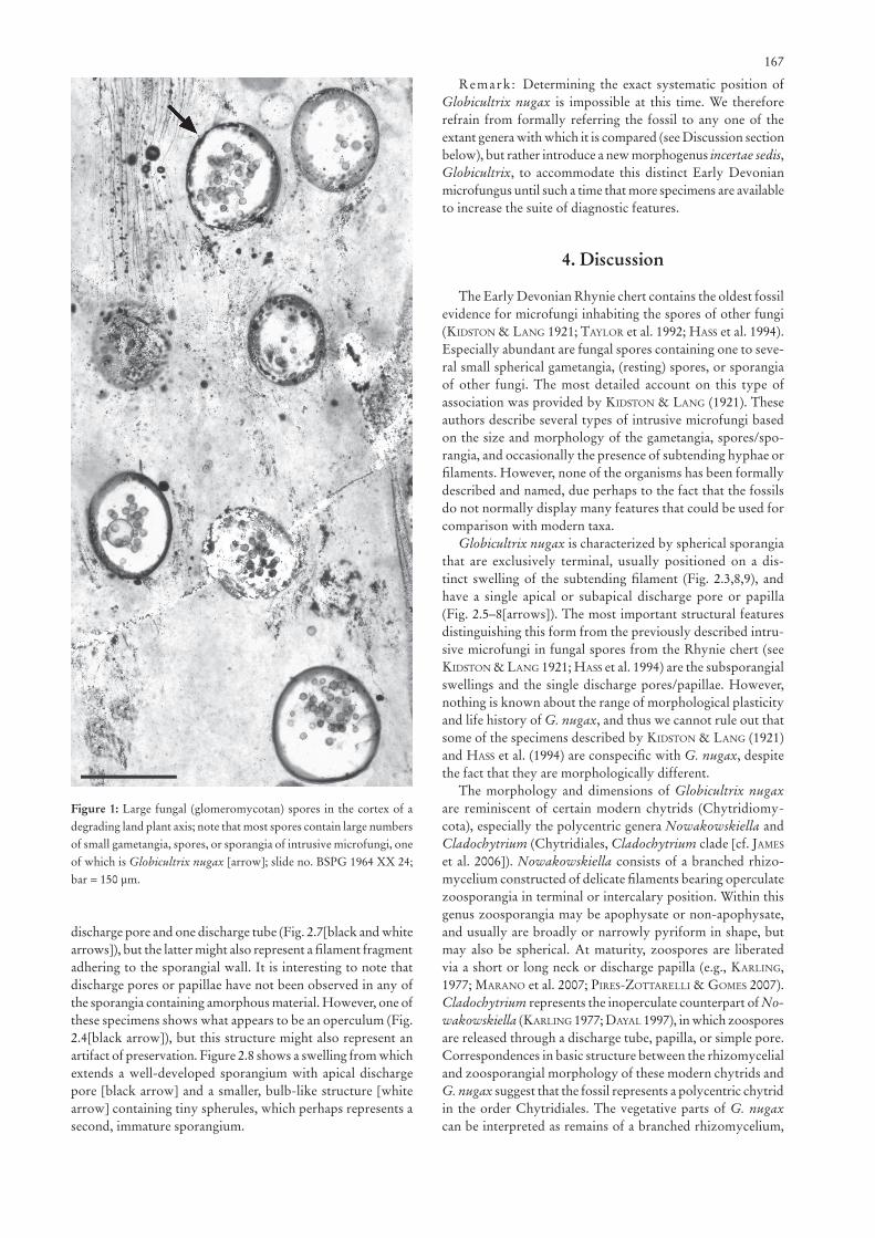
One of the more frequently encountered fungal associations in the Rhynie chert are microfungi inhabiting the spores of other fungi. Several examples of these interfungal associations have been described (Kidston & Lang 1921; tayLor et al. 1992; Hass et al. 1994), one of which consists of glomeromycotan spores containing varying numbers of small gametangia, (rest-ing) spores, or sporangia of other, intrusive fungi (e.g., Fig. 1). The systematic affinities of most of the intrusive fungi remain elusive. However, differences in size and wall composition of the gametangia, spores, or sporangia, together with differences in the morphology of occasionally present subtending hyphae or filaments, suggest that a wide variety of microfungi in the Rhynie paleoecosystem lived, reproduced, and/or produced resting stages inside the spores of other fungi (Kidston & Lang 1921). Thus, detailed knowledge about these organisms represents an important component of fully understanding the roles that fungi played in the Rhynie paleoecosystem.
*Author for correspondence and reprint requests; E-mail: [email protected]
Globicultrix nugax nov. gen. et nov. spec. (Chytridiomycota), an intrusive microfungus in fungal spores from the Rhynie chert
ByMichael Krings1,2*, Nora Dotzler1 & Thomas N. Taylor2
1Bayerische Staatssammlung für Paläontologie und Geologie und GeoBio-CenterLMU, Richard-Wagner-Straße 10, 80333 Munich, Germany
2Department of Ecology and Evolutionary Biology, and Natural History Museum and Biodiversity Research Center, The University of Kansas, Lawrence KS 66045-7534, U.S.A.
Manuscript received August 14, 2008; revised manuscript accepted September 27, 2008.
Zitteliana 165 - 170 2 Figs München, 30.09.2009 ISSN 1612 - 412XA48/49
166
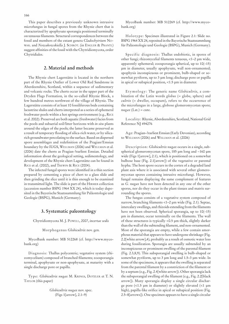
This paper describes a previously unknown intrusive microfungus in fungal spores from the Rhynie chert that is characterized by apophysate sporangia positioned terminally on tenuous filaments. Structural correspondences between the fossil and members of the extant genera Cladochytrium no-waK. and Nowakowskiella J. sCHröt. (in engLer & PrantL) suggest affinities of the fossil with the Chytridiomycota, order Chytridiales.
2. Material and methods
The Rhynie chert Lagerstätte is located in the northern part of the Rhynie Outlier of Lower Old Red Sandstone in Aberdeenshire, Scotland, within a sequence of sedimentary and volcanic rocks. The cherts occur in the upper part of the Dryden Flags Formation, in the so-called Rhynie Block, a few hundred metres northwest of the village of Rhynie. The Lagerstätte consists of at least 10 fossiliferous beds containing lacustrine shales and cherts interpreted as a series of ephemeral freshwater pools within a hot springs environment (e.g., riCe et al. 2002). Preserved are both aquatic (freshwater) facies from the pools and subaerial soil/litter horizons with in situ plants around the edges of the pools; the latter became preserved as a result of temporary flooding of silica-rich water, or by silica-rich groundwater percolating to the surface. Based on dispersed spore assemblages and redefinition of the Pragian/Emsian boundary by the IUGS, weLLman (2006) and weLLman et al. (2006) date the cherts as Pragian-?earliest Emsian. Detailed information about the geological setting, sedimentology, and development of the Rhynie chert Lagerstätte can be found in riCe et al. (2002), and trewin & riCe (2004).
The infected fungal spores were identified in a thin section prepared by cementing a piece of chert to a glass slide and then grinding the slice until it is thin enough to be examined in transmitted light. The slide is part of the Hirmer collection (accession number BSPG 1964 XX 24), which is today depo-sited in the Bayerische Staatssammlung für Paläontologie und Geologie (BSPG), Munich (Germany).
3. Systematic paleontology
Chytridiomycota M. J. PoweLL, 2007, incertae sedis
Morphogenus Globicultrix nov. gen.
MycoBank number: MB 512268 (cf. http://www.myco-bank.org)
Diagnosis : Thallus polycentric; vegetative system (rhi-zomycelium) composed of branched filaments; zoosporangia terminal, apophysate or non-apophysate, at maturity with a single discharge pore or papilla.
Type: Globicultrix nugax M. Krings, dotzLer et T. N. tayLor (this paper)
Globicultrix nugax nov. spec.(Figs 1[arrow], 2.1–9)
MycoBank number: MB 512269 (cf. http://www.myco-bank.org)
Holotype: Specimen illustrated in Figure 2.1: Slide no. BSPG 1964 XX 24, reposited in the Bayerische Staatssammlung für Paläontologie und Geologie (BSPG), Munich (Germany).
Spec i f ic d iagnosis : Thallus endobiotic, in spores of other fungi; rhizomycelial filaments tenuous, <1–2 μm wide, apparently ephemeral; zoosporangia spherical, up to 10(–15) μm in diameter, usually apophysate, wall non-ornamented; apophysis inconspicuous or prominent, bulb-shaped or so-mewhat pyriform, up to 3 μm long; discharge pore or papilla in apical or subapical position, <1.5 μm in diameter.
Etymology : The generic name Globicultrix, a com-bination of the Latin words globus (= globe, sphere) and cultrix (= dweller, occupant), refers to the occurrence of the microfungus in a large, globose glomeromycotan spore; nugax (Lat.) = cute.
Locality: Rhynie, Aberdeenshire, Scotland, National Grid Reference NJ 494276
Age: Pragian–?earliest Emsian (Early Devonian), according to weLLman (2006) and weLLman et al. (2006)
Description: Globicultrix nugax occurs in a single, sub-spherical glomeromycotan spore, 185 μm long and ~160 μm wide (Figs 1[arrow], 2.1), which is positioned on a somewhat bulbose base (Fig. 2.1[arrow]) of the vegetative or parental hypha. The host spore occurs in the cortex of a degraded land plant axis where it is associated with several other glomero-mycotan spores containing intrusive microfungi. However, fungal remains displaying the same complement of features as G. nugax have not been detected in any one of the other spores, nor do they occur in the plant tissues and matrix sur-rounding the spores.
The fungus consists of a vegetative system composed of narrow, branching filaments <1–2 μm wide (Fig. 2.1). Septae, intercalary swellings, and rhizoids extending from the filaments have not been observed. Spherical sporangia, up to 10(–15) μm in diameter, occur terminally on the filaments. The wall of these structures is typically <0.5 μm thick, slightly darker than the wall of the subtending filament, and non-ornamented. Most of the sporangia are empty, while a few contain amor-phous material that appears to have undergone shrinkage (Fig. 2.2[white arrow],4), probably as a result of osmotic water loss during fossilization. Sporangia are usually subtended by an inconspicuous or prominent swelling of the parental filament (Fig. 2.3,8,9). This subsporangial swelling is bulb-shaped or somewhat pyriform, up to 3 μm long and 1.5–3 μm wide. In some of the specimens, it appears that the swelling is separated from the parental filament by a constriction of the filament or by a septum (e.g., Fig. 2.4[white arrow]). Other sporangia lack the subsporangial swelling of the filament (e.g., Fig. 2.2[black arrow]). Many sporangia display a single circular dischar-ge pore (<1.5 μm in diameter) or slightly elevated (<1 μm high), papilla-like orifice in apical or subapical position (Fig. 2.5–8[arrows]). One specimen appears to have a single circular
167
discharge pore and one discharge tube (Fig. 2.7[black and white arrows]), but the latter might also represent a filament fragment adhering to the sporangial wall. It is interesting to note that discharge pores or papillae have not been observed in any of the sporangia containing amorphous material. However, one of these specimens shows what appears to be an operculum (Fig. 2.4[black arrow]), but this structure might also represent an artifact of preservation. Figure 2.8 shows a swelling from which extends a well-developed sporangium with apical discharge pore [black arrow] and a smaller, bulb-like structure [white arrow] containing tiny spherules, which perhaps represents a second, immature sporangium.
Remark: Determining the exact systematic position of Globicultrix nugax is impossible at this time. We therefore refrain from formally referring the fossil to any one of the extant genera with which it is compared (see Discussion section below), but rather introduce a new morphogenus incertae sedis, Globicultrix, to accommodate this distinct Early Devonian microfungus until such a time that more specimens are available to increase the suite of diagnostic features.
4. Discussion
The Early Devonian Rhynie chert contains the oldest fossil evidence for microfungi inhabiting the spores of other fungi (Kidston & Lang 1921; tayLor et al. 1992; Hass et al. 1994). Especially abundant are fungal spores containing one to seve-ral small spherical gametangia, (resting) spores, or sporangia of other fungi. The most detailed account on this type of association was provided by Kidston & Lang (1921). These authors describe several types of intrusive microfungi based on the size and morphology of the gametangia, spores/spo-rangia, and occasionally the presence of subtending hyphae or filaments. However, none of the organisms has been formally described and named, due perhaps to the fact that the fossils do not normally display many features that could be used for comparison with modern taxa.
Globicultrix nugax is characterized by spherical sporangia that are exclusively terminal, usually positioned on a dis-tinct swelling of the subtending filament (Fig. 2.3,8,9), and have a single apical or subapical discharge pore or papilla (Fig. 2.5–8[arrows]). The most important structural features distinguishing this form from the previously described intru-sive microfungi in fungal spores from the Rhynie chert (see Kidston & Lang 1921; Hass et al. 1994) are the subsporangial swellings and the single discharge pores/papillae. However, nothing is known about the range of morphological plasticity and life history of G. nugax, and thus we cannot rule out that some of the specimens described by Kidston & Lang (1921) and Hass et al. (1994) are conspecific with G. nugax, despite the fact that they are morphologically different.
The morphology and dimensions of Globicultrix nugax are reminiscent of certain modern chytrids (Chytridiomy-cota), especially the polycentric genera Nowakowskiella and Cladochytrium (Chytridiales, Cladochytrium clade [cf. James et al. 2006]). Nowakowskiella consists of a branched rhizo-mycelium constructed of delicate filaments bearing operculate zoosporangia in terminal or intercalary position. Within this genus zoosporangia may be apophysate or non-apophysate, and usually are broadly or narrowly pyriform in shape, but may also be spherical. At maturity, zoospores are liberated via a short or long neck or discharge papilla (e.g., KarLing, 1977; marano et al. 2007; Pires-zottareLLi & gomes 2007). Cladochytrium represents the inoperculate counterpart of No-wakowskiella (KarLing 1977; dayaL 1997), in which zoospores are released through a discharge tube, papilla, or simple pore. Correspondences in basic structure between the rhizomycelial and zoosporangial morphology of these modern chytrids and G. nugax suggest that the fossil represents a polycentric chytrid in the order Chytridiales. The vegetative parts of G. nugax can be interpreted as remains of a branched rhizomycelium,
Figure 1: Large fungal (glomeromycotan) spores in the cortex of a degrading land plant axis; note that most spores contain large numbers of small gametangia, spores, or sporangia of intrusive microfungi, one of which is Globicultrix nugax [arrow]; slide no. BSPG 1964 XX 24; bar = 150 μm.
168
the spherical sporangia as terminal zoosporangia with a single discharge pore or papilla, and the subsporangial swellings of the subtending filaments as apophyses. If these interpretations are correct, the zoosporangia with a well-developed discharge pore/papilla (Fig. 2.5–8[arrows]) may appear empty becau-se zoospore liberation had occurred prior to fossilization. Conversely, the presence of amorphous material in sporangia lacking discharge openings (e.g., Fig. 2.2[arrow],4) may indicate that zoospores had not (yet) developed and/or been released at the time of fossilization.
An alternative interpretation views Globicultrix nugax as a member of the Peronosporomycetes (Oomycota). In this
scenario, the sporangia represent terminal oogonia, while the swellings at the base of the sporangia are collar-like, amphigy-nous antheridia. However, the swellings appear to represent enlargements of the subtending filament, rather than being formed by an oogonial hypha growing through an antheri-dium. Moreover, oospores and parental hyphae giving rise to the antheridia have not been observed in any of the specimens. As a result, it is much more likely that G. nugax represents a member of the Chytridiomycota.
Although Globicultrix nugax is similar in basic structure to the modern chytrid genera Nowakowskiella and Cladochytri-um, there are some basic differences. The rhizomycelia in both
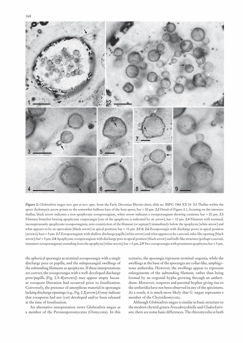
Figure 2: Globicultrix nugax nov. gen et nov. spec. from the Early Devonian Rhynie chert; slide no. BSPG 1964 XX 24. 2.1 Thallus within the spore (holotype); arrow points to the somewhat bulbose base of the host spore; bar = 50 μm. 2.2 Detail of Figure 2.1, focusing on the intrusive thallus; black arrow indicates a non-apophysate zoosporangium, white arrow indicates a zoosporangium showing contents; bar = 20 μm. 2.3 Filament branches bearing apophysate zosporangia [one of the apophyses is indicated by an arrow]; bar = 10 μm. 2.4 Filament with terminal, inconspicuously apophysate zoosporangium; note constriction of the filament (or septum?) immediately below the apophysis [white arrow] and what appears to be an operculum [black arrow] in apical position; bar = 10 μm. 2.5 & 2.6 Zoosporangia with discharge pores in apical position [arrows]; bars = 5 μm. 2.7 Zoosporangium with shallow discharge papilla [white arrow] and what appears to be a second, tube-like opening [black arrow]; bar = 5 μm. 2.8 Apophysate zoosporangium with discharge pore in apical position [black arrow] and bulb-like structure (perhaps a second, immature zoosporangium) extending from the apophysis [white arrow]; bar = 5 μm. 2.9 Two zoosporangia with prominent apophyses; bar = 5 μm.
169
extant taxa typically bear rhizoids and conspicuous intercalary swellings (KarLing 1977; dayaL 1997). These structures have not been observed in the fossil. However, this may be due to the fact that the vegetative system is incompletely preserved in the fossil. The vegetative system of G. nugax was perhaps ephemeral and disintegrated rapidly upon maturation of zo-osporangia. Alternatively, the main portion of the vegetative system may have been too delicate to become preserved in a recognizable manner. In addition, it cannot be determined whether the zoosporangia of G. nugax were operculate or inoperculate. A bona fide operculum has not been observed in any of the specimens, but the consistent absence of this structure may also be a result of preservation.
It is difficult to assess the nature of the association between Globicultrix nugax and its host. If the microfungus colonized the glomeromycotan spore while it was viable, this association would represent a form of mycoparasitism. Mycoparasites are fungi that derive the majority of their nutrients from other fungi that are alive at the time of infection (Jeffries & young 1994; Purin & riLLig 2008). Interpreting G. nugax as a mycoparasite seems reasonable, as parasitic interfungal interactions appear to have been widespread in the Rhynie paleoecosystem (Hass et al. 1994). Moreover, glomeromycotan spores, which are among the largest spores known in the fungal kingdom, certainly represented particularly suitable habitats for parasitic microfungi, because they contain abundant and easily accessible nutrients. However, there is no indication of a host response in the form of structural alterations or modi-fications of the spore wall, which would indicate evidence of a parasitic relationship between G. nugax and its host. In the absence of host responses, fossil mycoparasites are difficult to distinguish from saprotrophs, which colonize and utilize dead organic matter as a carbon source (see dix & webster 1995). Therefore, it is also possible that G. nugax represents a saprotroph that colonized non-viable spores or spores that had already germinated. Most extant members of Nowakowskiella and Cladochytrium are aquatic and soil saprotrophs that thrive on/in decaying plant material (sParrow 1960; KarLing 1977), but at least one species of Cladochytrium (i.e. C. aneurae tHi-rum.) has been described as a parasite of liverworts from the genus Aneura dumort. (tHirumaLaCHar 1947).
The discovery of Globicultrix nugax in the Rhynie chert adds to our understanding of the biodiversity of late Paleozoic microorganisms, and contributes to a more sharply focused concept of the complexity of ancient non-marine ecosystems. As more information is obtained about this and other microbial life forms from the Rhynie chert, it will be possible to offer more detailed hypotheses that can be used in association with those described from modern communities to more accurately depict the role of fungi and their interactions with other organisms in the ecology and evolution of non-marine paleoecosystems.
Acknowledgments
This study was supported by funds from the National Science Foundation (EAR-0542170 to T.N.T. and M.K.) and the Alexander von Humboldt-Foundation (V-3.FLF-DEU/1064359 to M.K.). We thank Brian J. axsmitH (Mobile AL, U.S.A.) for his insightful comments and suggestions.
5. References
Croft, W. N. & george, E. A. (1959): Blue-green algae from the Middle Devonian of Rhynie, Aberdeenshire. – Bulletin of the British Museum of Natural History, Geology, 3: 341–353.
dayaL, r. (1997): Chytrids of India; New Delhi (M.D. Publications Pvt. Ltd.), xiii + 316 pp.
dix, N. J. & webster, J. (1995): Fungal Ecology; New York (Chapman & Hall), 560 pp.
dotzLer, N., tayLor, T. N. & Krings, M. (2007): A prasinophycean alga of the genus Cymatiosphaera in the Early Devonian Rhynie chert. – Review of Palaeobotany and Palynology, 147: 106–111.
edwards, D. S. & Lyon, A. G. (1983): Algae from the Rhynie chert. – Botanical Journal of the Linnean Society, 86: 37–55.
Hass, H., tayLor, T. N. & remy, W. (1994): Fungi from the Lower Devonian Rhynie chert: mycoparasitism. – American Journal of Botany, 81: 29–37.
James, T. Y., LetCHer, P. M., LongCore, J. E., mozLey-standridge, S. E., Porter, D., PoweLL, M. J., griffitH, G. W. & ViLgaLys, R. (2006): A molecular phylogeny of the flagellated fungi (Chytridi-omycota) and description of a new phylum (Blastocladiomycota). – Mycologia, 98: 860–871.
Jeffries, P. & young, T. W. K. (1994): Interfungal Parasitic Relation-ships; Wallingford UK (CAB International), xvi + 296 pp.
KarLing, J. s. (1977): Chytridiomycetarum Iconographia. An Illus-trated and Brief Descriptive Guide to the Chytridiomycetous Genera with a Supplement of the Hyphochytridiomycetes; Vaduz (J. Cramer), xiii + 414 pp.
Kidston, R. & Lang, W. H. (1921): On Old Red Sandstone plants showing structure, from the Rhynie chert bed, Aberdeenshire. Part V. The Thallophyta occurring in the peat-bed; the succession of the plants through a vertical section of the bed, and the conditions of accumulation and preservation of the deposit. – Transactions of the Royal Society of Edinburgh, 52: 855–902.
Krings, m., Hass, H., KerP, H., tayLor, t. n., agerer, r. & dotz-Ler, n. (2009): Endophytic cyanobacteria in a 400-million-yr-old land plant: A scenario for the origin of a symbiosis? – Review of Palaeobotany and Palynology, 153: 62–69.
Krings, m., KerP, H., Hass, H., tayLor, t. n. & dotzLer, n. (2007): A filamentous cyanobacterium showing structured colonial growth from the Early Devonian Rhynie chert. – Review of Pa-laeobotany and Palynology, 146: 265–276.
marano, A. V., steCiow, M. M., areLLano, M. L., arambarri, A. M. & sierra, M. V. (2007): El género Nowakowskiella (Cla-dochytriaceae, Chytridiomycota) en ambientes de la Pcia, de Buenos Aires (Argentina): taxonomía, frequencia y abundancia de las especies encontradas. – Boletín de la Sociedad Argentina de Botánica, 42: 13–24.
Pires-zottareLLi, C. L. A. & gomes, A. L. (2007): Contribuição para o conhecimento de Chytridiomycota da “Reserva Biología de Paranapiacaba”, Santo André, SP, Brazil. – Biota Neotropica, 7: 309–329.
Purin, S. & riLLig, M. C. (2008): Parasitism of arbuscular mycorrhizal fungi: reviewing the evidence. – FEMS Microbiology Letters, 279: 8–14.
riCe, C. M., trewin, N. H. & anderson, L. I. (2002): Geological setting of the Early Devonian Rhynie cherts, Aberdeenshire, Scotland: an early terrestrial hot spring system. – Journal of the Geological Society of London, 159: 203–214.
sParrow, f. K. (1960): Aquatic Phycomycetes; Ann Arbor (The Uni-versity of Michigan Press), xxv + 1187 pp.
tayLor, T. N., Hass, H. & KerP, H. (2005): Life history biology of early land plants: deciphering the gametophyte phase. – Procee-dings of the National Academy of Sciences of the United States of America, 102: 5892–5897.
tayLor, T. N., KLaVins, S. D., Krings, M., tayLor, E. L., KerP, H. & Hass, H. (2004): Fungi from the Rhynie chert: A view from the dark side. – Transactions of the Royal Society of Edinburgh, Earth Sciences, 94: 457–473.
tayLor, T. N., Krings, M. & KerP, H. (2006): Hassiella monospora nov. gen. et sp., a new microfungus from the 400 million year old

170
Rhynie chert. – Mycological Research, 110: 628–632.tayLor, T. N., remy, W. & Hass, H. (1992): Fungi from the Lower
Devonian Rhynie chert: Chytridiomycetes. – American Journal of Botany, 79: 1233–1241.
tayLor, T. N., remy, W., Hass, H. & KerP, H. (1995): Fossil arbus-cular mycorrhizae from the Early Devonian. – Mycologia, 87: 560–573.
tayLor, T. N. & tayLor, E. L. (2000): The Rhynie chert ecosystem: a model for understanding fungal interactions. – In: C. W. baCon & J. F. wHite (Eds), Microbial Endophytes; New York (Marcel Dekker), pp. 31–47.
tHirumaLaCHar, m. J. (1947): Some fungal diseases of bryophytes in Mysore. – Transactions of the British Mycological Society, 31: 7–12.
trewin, N. H. & riCe, C. M. [Eds] (2004): The Rhynie Hot Springs System: Geology, Biota and Mineralisation. – Transactions of the Royal Society of Edinburgh, Earth Sciences 94; Edinburgh (The Royal Society of Edinburgh Scotland Foundation, on behalf of the Royal Society of Edinburgh), 246 pp.
weLLman, C. H. (2006): Spore assemblages from the Lower Devonian “Lower Old Red Sandstone” deposits of the Rhynie Outlier, Scotland. – Transactions of the Royal Society of Edinburgh, Earth Sciences, 97: 167–211.
weLLman, C. H., KerP, H. & Hass, H. (2006): Spores of the Rhy-nie chert plant Aglaophyton (Rhynia) major (Kidston & Lang) Edwards 1986. – Review of Palaeobotany and Palynology, 142: 229–250.