Embed Size (px)
Citation preview
Human IgG+ Plasma Cells in
Patients with Systemic Lupus Erythematosus
vorgelegt von
Diplom-Ingenieurin
Juliane Kofer
aus Pasewalk
Von der Fakultät III - Prozesswissenschaften
der Technischen Universität Berlin
zur Erlangung des akademischen Grades
Doktor der Ingenieurwissenschaften
- Dr.-Ing. -
genehmigte Dissertation
Promotionsausschuss:
Vorsitzender: Prof. Dipl.-Ing. Dr. Ulf Stahl
Gutachter: Prof. Dr. rer. nat. Roland Lauster
Gutachter: Dr. rer. nat. Hedda Wardemann
Tag der wissenschaftlichen Aussprache: 22.03.2011
Berlin 2011
D 83


Whenever they say it can’t be done,
remind them that they make a jellybean that
tastes exactly like popcorn.
John C. Mayer

ABSTRACT i
ABSTRACT
Systemic Lupus Erythematosus (SLE) is a chronic inflammatory autoimmune disease
that is associated with a major breakdown in B cell self-tolerance as reflected by elevated serum
immunoglobulin G (IgG) levels of predominantly anti-nuclear antibodies (ANAs). Serum
antibody titers are maintained by short-lived antibody-secreting plasmablasts and long-lived
plasma cells, the latter residing in survival niches of the bone marrow. Experimental evidence
from mouse models suggests that treatment-resistant bone marrow plasma cells are the major
contributor to serum IgG autoantibodies in SLE. However, the frequency of self-reactive and
potentially pathogenic antibodies in the bone marrow plasma cell compartment of SLE patients
has not been determined.
The aim of this study was to characterize the human bone marrow IgG+ plasma cell
compartment in SLE patients. The Ig genes of 196 bone marrow plasma cells from five SLE
patients were cloned and expressed in vitro by using a single cell approach that facilitates the
unbiased analysis of human plasma cells both on the Ig molecular level and antibody reactivity
level. Antibody reactivity testing demonstrated significantly increased frequencies of poly- and
self-reactive bone marrow plasma cells in SLE patients with an active disease.
In summary, the data provide direct evidence that the bone marrow harbors high
frequencies of self-reactive IgG+ plasma cells that may directly contribute to disease
pathogenesis in SLE. Thus, self-reactive IgG+ bone marrow plasma cells in SLE patients may
represent an important therapeutic target in SLE.

ii
iii
ZUSAMMENFASSUNG
Der systemische Lupus erythematodes (SLE) ist eine chronisch entzündliche
Autoimmunerkrankung, die durch eine Fehlfunktion der körpereigenen Abwehr verursacht
wird. Plasmazellen und Plasmablasten produzieren unter normalen Bedingungen protektive
Antikörper, welche einen wesentlichen Bestandteil der Immunantwort auf fremde Erreger
darstellen. Charakteristisch für SLE ist die Bildung hochaffiner IgG Antikörper, welche gegen
DNA und weitere Bestandteile des Zellkerns im eigenen Organismus gerichtet sind. Diese
Autoantikörper werden von kurzlebigen Plasmablasten und langlebigen Plasmazellen
sezerniert und führen in Körperorganen wie der Niere zu chronischen Entzündungen.
Langlebige Plasmazellen leisten den größten Anteil an IgG Antikörpern im Serum, überleben
in Nischen des Knochenmarks für Jahre und sind im Gegensatz zu kurzlebigen Plasmablasten
weitgehend therapieresistent. Die Häufigkeit von autoreaktiven und damit möglicherweise
pathogenen Antikörpern in der langlebigen Plasmazellpopulation des Knochenmarks wurde in
SLE Patienten jedoch bisher nicht analysiert.
Das Ziel dieser Arbeit war daher die Charakterisierung der IgG+ Plasmazellpopulation
des Knochenmarks in SLE Patienten. Um die Häufigkeit autoreaktiver Plasmazellen im
Knochenmark von SLE Patienten bestimmen zu können, wurde eine Methode verwendet, die
es erlaubt einzelne Zellen zu isolieren und ihre jeweiligen IgH und IgL Gene zu klonieren. Mit
dieser Information konnten dann Antikörper rekombinant in vitro hergestellt werden, welche
die gleiche Antigenspezifität aufweisen, wie sie in der ursprünglichen Plasmazelle produziert
wurde. Auf diese Weise wurden die Immunglobulingene von 196 Plasmazellen aus
Knochenmarkproben von fünf verschiedenen SLE Patienten kloniert und in vitro exprimiert.
Die Untersuchung der monoklonalen Antikörper zeigte, dass die Anzahl an autoreaktiven
Plasmazellen in SLE Patienten mit aktiver Krankheit signifikant erhöht ist und krankheits-
assoziierte antinukleäre Antikörper (ANAs) durch somatische Mutation entstanden sind.
Zusammenfassend kann die vorliegende Arbeit zeigen, dass Plasmazellen aus dem
Knochenmark von SLE Patienten antinukleäre Antikörper sezernieren und damit direkt zum
Krankheitsverlauf beitragen können. Die erhöhte Frequenz von autoreaktiven Plasmazellen im
Knochenmark von SLE Patienten unterstreicht zudem die Rolle von langlebigen Plasmazellen
als therapeutisches Ziel.

iv

v
Eidesstattliche Erklärung
Hiermit erkläre ich an Eides statt, dass ich die vorliegende Dissertation in allen Teilen
selbständig verfasst habe. Bei der Anfertigung der Dissertation wurde keine Hilfe Dritter in
Anspruch genommen und genutzte Hilfsmittel vollständig angegeben. Veröffentlichungen von
Teilen der vorliegenden Dissertation sind von mir nicht vorgenommen worden. Weiterhin
wurde diese Dissertation nicht anderweitig für eine Prüfung oder Promotion zur Beurteilung
eingereicht. Ich versichere, dass ich die vorstehenden Angaben nach bestem Wissen
vollständig und der Wahrheit entsprechend getätigt habe.
Juliane Kofer Berlin, 30.04.2011

vi
Table of Contents vii
Table of Contents
ABSTRACT i
ZUSAMMENFASSUNG iii
ABBREVIATIONS x
1 INTRODUCTION 1
1.1 The Immune System 1
1.2 Immunoglobulins 1
1.3 Generation of Immunoglobulin Diversity 2
1.4 Immunological Memory and Plasma Cells 4
1.5 Central and Peripheral B Cell Tolerance 6
1.6 Autoimmunity 7
1.7 Systemic Lupus Erythematosus and Autoantibodies 7
1.7.1 Systemic Lupus Erythematosus 7
1.7.2 The Role of B cells in SLE 9
2 AIM 11
3 METHODS 13
3.1 Strategy Outline 13
3.2 Fluorescence-Activated Cell Sorting (FACS) 14
3.3 Reverse Transciption Polymerase Chain Reaction (PCR) 14
3.3.1 cDNA Synthesis 14
3.3.2 Nested PCR for Amplification of Human Ig Gene Transcripts 15
3.3.3 Reversion Strategy for Mutated Ig Heavy and Light Chain Genes 16
3.4 Ig Gene Sequence Analysis 16
3.5 Directional Cloning into Expression Vectors 17
3.5.1 Preparation of Eukaryotic Expression Vectors 17
3.5.2 Restriction Endonuclease Digestion 17
Table of Contents viii
3.5.3 Ligation 18
3.5.4 Preparation of Competent Bacteria 18
3.5.5 Transformation of Competent Bacteria 18
3.5.6 Screening Bacterial Colonies by PCR 19
3.5.7 Purification of Plasmid DNA 19
3.6 Recombinant Antibody Production 20
3.6.1 Cell Culture 20
3.6.2 Polyethylenimine- Mediated DNA Transfection of HEK 293T Cells 20
3.7 Enzyme-Linked Immunofluorescence Assays 20
3.7.1 Determination of Recombinant Ig Concentrations by ELISA 20
3.7.2 Purification of Recombinant Antibodies 21
3.7.3 Determination of Polyreactive Antibodies by ELISA 21
3.8 Immunofluorescence Assays 22
3.9 Statistical Analysis 22
4 RESULTS 23
4.1 Clinical Features of Five Analyzed SLE Patients 23
4.2 Ig Gene Features of Human IgG+ Bone Marrow Plasma Cells from SLE Patients 23
4.3 Polyreactive IgG+ Bone Marrow Plasma Cells in SLE Patients 27
4.4 Self-Reactive IgG+ Bone Marrow Plasma Cells in SLE Patients 29
4.5 Reversion of Somatic Hypermutations Abolishes Reactivity in Poly- and Self-Reactive IgG+ Bone Marrow Plasma Cells from SLE Patients 32
5 DISCUSSION 35
5.1 Unbiased Analysis of the Plasma Cell Antibody Repertoire by Single Cell Ig Gene Cloning Implies a Rigid Selection Process in Healthy Humans 35
5.2 Serum Autoantibodies are a Hallmark of Disease in SLE Patients 36
5.3 The Ig Gene Repertoire in IgG+ Bone Marrow Plasma Cells from SLE Patients 36
5.4 The Bone Marrow Contributes to Serum ANA Production in SLE Patients 37
Table of Contents ix
5.5 The Role of Somatic Hypermutations in the Generation of Self-Reactive Antibodies 38
5.6 Genetic and Environmental Factors Generate Disease Diversity in SLE Patients 39
5.7 Therapeutic Options to Eliminate Autoreactive Plasma Cells 40
5.8 Conclusion and Outlook 41
6 REFERENCES 43
7 SUPPLEMENTAL INFORMATION 59
7.1 Supplemental Figures 59
7.2 Supplemental Tables 63
7.3 Supplemental Material 71
ACKNOWLEDGMENTS 81
ABBREVIATIONS x
ABBREVIATIONS
aa amino acid(s)
ABTS 2, 2’-Azino-bis (3-ethylBenzThiazoline-6-Sulfonic acid)
AID Activation-Induced Cytidine Deaminase
ANA Anti-Nuclear Antibody
APC AlloPhycoCyanin
BAFF B cell Activating Factor belonging to the TNF family
B cell Bursal or Bone marrow derived cell
BCR B Cell Antigen Receptor
bp base pair
CD Cluster of Differentiation
CDR Complementary Determining Region
cDNA complementary DesoxyriboNucleid Acid
CSR Class-Switch Recombination
D Diversity
DC Dendritic Cell
DMEM Dulbecco’s Modified Eagle Medium
dNTP desoxyriboNucleosid TriPhospate
dsDNA double-stranded DesoxyriboNucleid Acid
DTT DiThioTreitol E.coli Escherichia ccoli
EDTA EthyleneDiamineTetraacetic Acid
ELISA Enzyme-Linked ImmunoSorbent Assay
Fab Fragment antigen binding
FACSTM Fluorescence Activated Cell SortingTM
Fc Fragment crystalline
FCS Fetal Calf Serum
FDC Follicular Dendritic Cell
FSC Forward Scatter
FWR FrameWorkRegion
gav average surface ggravity (approx. 9.81 m/s2)
GC Germinal Center
HEPES N-2-HydroxyEthylPiperazine-N'-2-EthaneSulfonic Acid
HEK 293T Human Embryonic Kidney 293 transformed with SV40 large T-antigen
HRP HorseRadish Peroxidase
IFA ImmunoFluorescence Assay(s)
IgBLAST Immunoglobulin Basic Local Alignment Search Tool
Ig Immunoglobulin
IgA,D,E,G,M Immunoglobulin A, D, E, G, M
IgH Immunoglobulin Heavy chain Ig Immunoglobulin gamma heavy chain
Ig Immunoglobulin kappa light chain
Ig Immunoglobulin lambda light chain
ABBREVIATIONS xi
IL Interleukin
IMGT ImMunoGeneTics information system
J Joining
kb kilo base
LB Luria Bertani
LPS LipoPolySaccharide
mAb monoclonal Antibody
mRNA messenger RiboNucleic Acid
n numbers
NCBI National Center for Biotechnology Information
NP-40 Nonidet P-40
OD Optical Density
PBS Phosphate Buffered Saline
PCR Polymerase Chain Reaction
PE PhycoErythrin
PEI PolyEthylenImine
RAG 1 and 2 recombination activating gene 1 and 2
rpm rounds per minute
RPMI Roosevelt Park Memorial Institute
RT Room Temperature
RT-PCR Reverse Transcription - Polymerase Chain Reaction
SHM Somatic HyperMutation
SLE Systemic Lupus Erythematosus
SSC Side SCatter
ssDNA single-stranded DesoxyriboNucleid Acid
Taq polymerase Thermus aquaticus polymerase
TAE buffer Tris-Acetate-EDTA buffer
TB Terrific Broth
T cell Thymus-derived cell
Temp Temperature
TLR Toll-Like Receptor
U Units
UV UltraViolet
V Variable

xii
INTRODUCTION 1
1 INTRODUCTION
1.1 The Immune System
Vertebrates have evolved an elaborate protective immune system consisting of an
innate part and an adaptive part to successfully eliminate disease-causing pathogens. Both
components provide a dynamic network of cells and molecules that discriminate between self
and foreign to efficiently defend the organism against invading pathogens. Innate immune
mechanisms are activated immediately during an immune response and preformed, non-
specific receptors recognize common surface patterns on pathogens. These innate immune
responses activate the adaptive immune system, where lymphocytes detect pathogen-specific
epitopes via antigen-specific receptors. The millions of different lymphocyte clones are able to
detect virtually all foreign antigens in a highly specific way. Specific antigen recognition is
based on clonal selection, where an antigen selectively stimulates those cells that express
complementary antigen-specific receptors. Under normal immune conditions, the immune
system provides an immediate, specific and efficient immune response against pathogens.
However, alterations in this complex network might foster autoimmune reactions, eventually
leading to the development of autoimmune diseases.
1.2 Immunoglobulins
Immunoglobulins (Igs) are glycoproteins that are synthesized and expressed by
B lymphocytes either on their cell surface as part of the B cell antigen receptor (BCR) or in a
soluble form secreted by plasma cells as so-called antibodies. Each B cell has approximately 105
such BCRs in its plasma membrane (Alberts et al., 2002) and all Igs expressed by one
B lymphocyte have the same antigen specificity (Nossal and Lederberg, 1958). Membrane-
bound Igs form the antigen-binding unit of the BCR and are associated with the transmembrane
signaling proteins Ig and Ig (Reth, 1995).
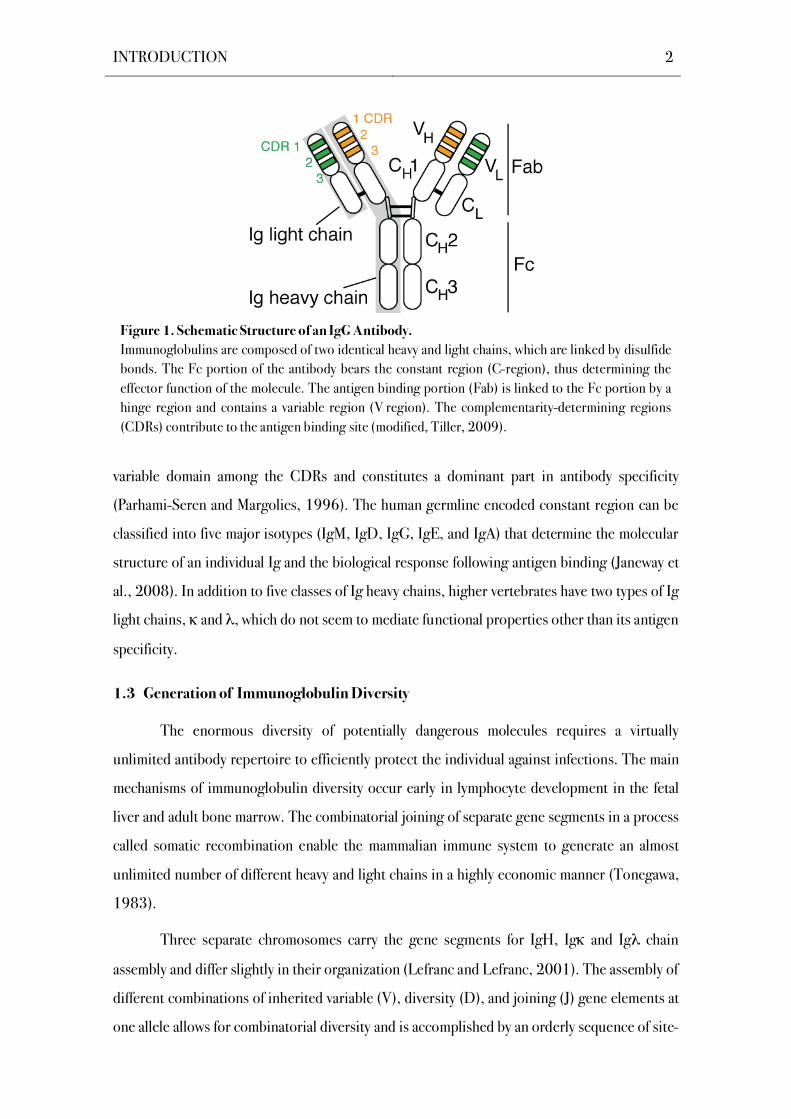
The basic structural unit of an Ig consists of two identical heavy (H) chains that are
covalently linked by disulfide bonds to two identical light (L) chains. Each heavy and light chain
can be divided into a variable region (V) that determines the antigen specificity and a constant
region that mediates distinctive effector functions (Figure 1). The diversity in the V regions is
mostly attributed to three hypervariable, complementarity-determining regions (CDRs) in each
chain, while the framework regions (FWRs) provide the structural backbone. CDR3 is the most
INTRODUCTION 2
variable domain among the CDRs and constitutes a dominant part in antibody specificity
(Parhami-Seren and Margolies, 1996). The human germline encoded constant region can be
classified into five major isotypes (IgM, IgD, IgG, IgE, and IgA) that determine the molecular
structure of an individual Ig and the biological response following antigen binding (Janeway et
al., 2008). In addition to five classes of Ig heavy chains, higher vertebrates have two types of Ig
light chains, and , which do not seem to mediate functional properties other than its antigen
specificity.
1.3 Generation of Immunoglobulin Diversity
The enormous diversity of potentially dangerous molecules requires a virtually
unlimited antibody repertoire to efficiently protect the individual against infections. The main
mechanisms of immunoglobulin diversity occur early in lymphocyte development in the fetal
liver and adult bone marrow. The combinatorial joining of separate gene segments in a process
called somatic recombination enable the mammalian immune system to generate an almost
unlimited number of different heavy and light chains in a highly economic manner (Tonegawa,
1983).
Three separate chromosomes carry the gene segments for IgH, Ig and Ig chain
assembly and differ slightly in their organization (Lefranc and Lefranc, 2001). The assembly of
different combinations of inherited variable (V), diversity (D), and joining (J) gene elements at
one allele allows for combinatorial diversity and is accomplished by an orderly sequence of site-
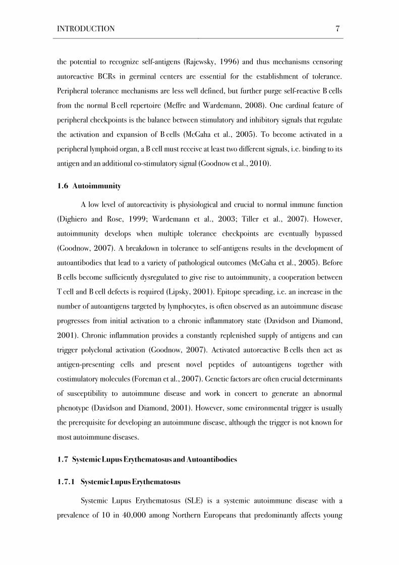
Figure 1. Schematic Structure of an IgG Antibody.
Immunoglobulins are composed of two identical heavy and light chains, which are linked by disulfide
bonds. The Fc portion of the antibody bears the constant region (C-region), thus determining the
effector function of the molecule. The antigen binding portion (Fab) is linked to the Fc portion by a
hinge region and contains a variable region (V region). The complementarity-determining regions
(CDRs) contribute to the antigen binding site (modified, Tiller, 2009).

INTRODUCTION 3
specific rearrangement mediated by the recombination activating gene (RAG) encoded
enzymes RAG1 and RAG2 (Oettinger et al., 1990). The IgH chain locus on chromosome 14
contains about 46 functional variable (V) region gene segments, 27 diversity (D) gene
segments and 6 joining (J) gene segments allowing 7,452 different functional IgH chain
rearrangements. The Ig locus is mapped to chromosome 2 and harbours approximately 37
functional V gene segments and 5 J gene segments facilitating 185 different functional Ig
chain recombinations. The Ig locus on chromosome 22 contains about 35 functional V gene
segments and 4 J gene segments facilitating 140 different functional Ig chain rearrangements.
The pairing of a functional Ig heavy chain with any functional Ig light chain thus allows the
generation of about 2.4 million different BCRs during early B cell development. Ig gene
rearrangement starts at the heavy chain locus and is terminated as soon as a productive IgH
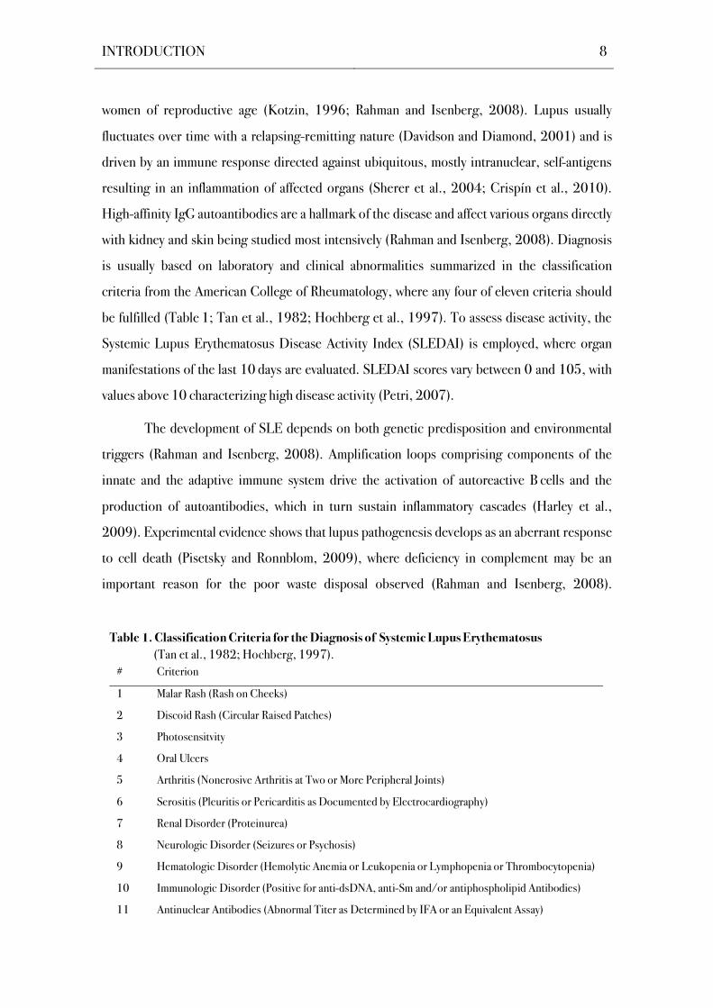
Figure 2. Assembly of an Immunoglobulin.
The heavy chain V region (VH) consists of three gene segments. After linking a D element to a J part, the
DJ gene segment is fused to the V gene element. The C regions, encoded by one or more exons, are joined
to the VH or VL exon by RNA splicing. To assemble a complete light chain gene (VL) from genomic DNA, a
V gene segment is combined with a J gene portion (modified, Janeway et al., 2008).
INTRODUCTION 4
chain is expressed (Figure 2; Alt et al., 1984). IgL chain rearrangement usually occurs first at a
allele and if that fails it either takes place at the second allele or at a allele (Hieter et al.,
1980; Hieter et al., 1981a ; Hieter et al., 1981b; Korsmeyer et al., 1981; Nemazee and
Weigert, 2000; van der Burg et al., 2001). During V(D)J recombination, recombination signal
sequences flanking each gene segment ensure that only appropriate gene segments combine
(Tonegawa, 1983). Additional diversity is provided by imprecise joining due to random loss
and gain of nucleotides at the gene segment joining sites. This junctional diversification
facilitates a high level of diversity in heavy and light chain CDR3s, which has evolved to be
especially important in antibody specificity.
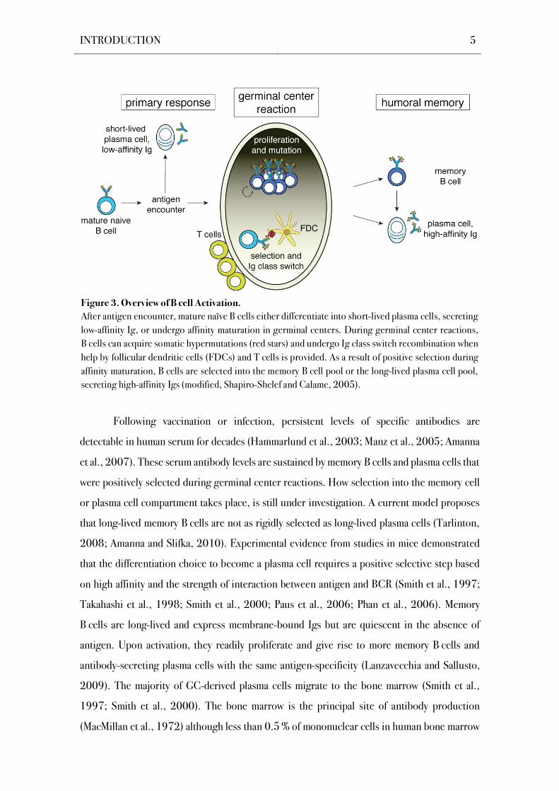
1.4 Immunological Memory and Plasma Cells
The most important biological consequence of adaptive immunity is the establishment
of immunological memory (Sallusto et al., 2010). Immunological memory is generated by both
lymphocyte differentiation and clonal expansion, where activated lymphocytes establish a
tailor-made immune response to a particular pathogen and mount an accelerated and enhanced
response upon antigen reencounter (Nossal et al., 1965).
When a naïve B cell binds to a foreign antigen and receives an additional co-stimulatory
signal in a peripheral lymphoid organ, it either proliferates and differentiates into a short-lived
plasma cell secreting low-affinity antibodies or is recruited into a germinal center (GC;
Figure 3; Shapiro-Shelef and Calame, 2005). Germinal center B cells undergo antigen-
dependent proliferation in response to antigen presentation by follicular dendritic cells (FDCs)
and co-stimulatory signals by T helper cells. During the GC reaction, the enzyme activation-
induced cytidine deaminase (AID) alters the B cell receptor affinity for an antigen by
introducing somatic hypermutations (SHM; Berek and Milstein, 1988). This process is termed
affinity maturation and enables positive selection based on the BCR affinity for the respective
antigen. However, affinity maturation increases the BCR affinity only in a few B cells. These few
B cell clones with higher affinity are preferentially stimulated by an antigen, proliferate and
eventually differentiate either into memory B cells or into plasma cells (Klein and Dalla-Favera,
2008). Class-switch recombination (CSR) of the IgH chain, again mediated by AID, changes
the Ig isotype and enables a more distinctive effector function of the secreted antibody while
retaining its specificity (Muramatsu et al., 2000).
INTRODUCTION 5
Following vaccination or infection, persistent levels of specific antibodies are
detectable in human serum for decades (Hammarlund et al., 2003; Manz et al., 2005; Amanna
et al., 2007). These serum antibody levels are sustained by memory B cells and plasma cells that
were positively selected during germinal center reactions. How selection into the memory cell
or plasma cell compartment takes place, is still under investigation. A current model proposes
that long-lived memory B cells are not as rigidly selected as long-lived plasma cells (Tarlinton,
2008; Amanna and Slifka, 2010). Experimental evidence from studies in mice demonstrated
that the differentiation choice to become a plasma cell requires a positive selective step based
on high affinity and the strength of interaction between antigen and BCR (Smith et al., 1997;
Takahashi et al., 1998; Smith et al., 2000; Paus et al., 2006; Phan et al., 2006). Memory
B cells are long-lived and express membrane-bound Igs but are quiescent in the absence of
antigen. Upon activation, they readily proliferate and give rise to more memory B cells and
antibody-secreting plasma cells with the same antigen-specificity (Lanzavecchia and Sallusto,
2009). The majority of GC-derived plasma cells migrate to the bone marrow (Smith et al.,
1997; Smith et al., 2000). The bone marrow is the principal site of antibody production
(MacMillan et al., 1972) although less than 0.5 % of mononuclear cells in human bone marrow
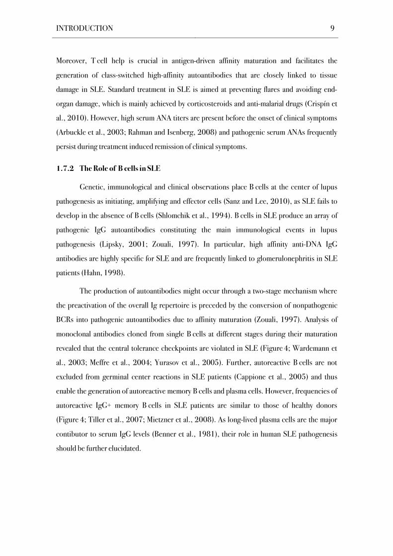
Figure 3. Overview of B cell Activation.
After antigen encounter, mature naïve B cells either differentiate into short-lived plasma cells, secreting
low-affinity Ig, or undergo affinity maturation in germinal centers. During germinal center reactions,
B cells can acquire somatic hypermutations (red stars) and undergo Ig class switch recombination when
help by follicular dendritic cells (FDCs) and T cells is provided. As a result of positive selection during
affinity maturation, B cells are selected into the memory B cell pool or the long-lived plasma cell pool,
secreting high-affinity Igs (modified, Shapiro-Shelef and Calame, 2005).
INTRODUCTION 6
from healthy individuals are plasma cells (Terstappen et al., 1990; Hiepe et al., 2011).
Terminally differentiated plasma cells selected in germinal center reactions synthesize and
secrete up 30,000 antibody molecules per second (Hibi and Dosch, 1986), but do not
proliferate, and lose the ability to sense antigenic changes or T cells in their environment
(Amanna and Slifka, 2010). Moreover, human terminally differentiated plasma cells are
demonstrated to be long-lived (Hammarlund et al., 2003; Amanna et al., 2007) and protect
from infection through the production of high affinity antigen-specific serum antibodies (Manz
et al., 2005; Radbruch et al., 2006).
1.5 Central and Peripheral B Cell Tolerance
The enormous diversity of the Ig repertoire acquired by random somatic V(D)J
recombination and affinity maturation also generates BCRs that recognize self-antigens from
the individual and have the potential of becoming pathogenic (Burnet, 1972). In healthy
individuals, a series of checkpoints purge autoreactive B cells from the repertoire, both
centrally in the bone marrow during B cell development and in peripheral lymphoid tissues
(Goodnow et al., 1995). The outcome of these selection processes is a marked narrowing of
the Ig repertoire (Melchers et al., 1995; Loder et al., 1999).
Central tolerance is established in the fetal liver and adult bone marrow prior to the first
antigen encounter, where immature B cells are censored for reactivity with ubiquitous
membrane-bound self-antigens. To prevent autoimmunity, self-reactive B cells are induced to
either undergo clonal deletion, BCR editing or anergy. Clonal deletion is characterized by self-
antigen induced apoptosis of autoreactive B cells (Nemazee and Burki, 1989). B cell receptor
editing is the main mechanism of central tolerance (Retter and Nemazee, 1998; Casellas et al.,
2001; Halverson et al., 2004) and is conducted by secondary Ig gene rearrangements that
generate a new antigen receptor with an innocuous specificity (Gay et al., 1993; Radic et al.,
1993; Tiegs et al., 1993). Immature B cells can become functionally unresponsive to antigens
due to recurrent exposure to an antigen (Nossal and Pike, 1980; Goodnow et al., 1988). These
anergic B cells can emigrate from the bone marrow into T cell zones of secondary lymphoid
tissues (Cornall et al., 1995).
The processes of central tolerance remain incomplete, allowing some self-reactive
B cells to escape into the periphery (Nemazee and Sato, 1983; Souroujon et al., 1988;
Shlomchik et al., 1993). Further, affinity maturation in germinal centers generates BCRs with
INTRODUCTION 7
the potential to recognize self-antigens (Rajewsky, 1996) and thus mechanisms censoring
autoreactive BCRs in germinal centers are essential for the establishment of tolerance.
Peripheral tolerance mechanisms are less well defined, but further purge self-reactive B cells
from the normal B cell repertoire (Meffre and Wardemann, 2008). One cardinal feature of
peripheral checkpoints is the balance between stimulatory and inhibitory signals that regulate
the activation and expansion of B cells (McGaha et al., 2005). To become activated in a
peripheral lymphoid organ, a B cell must receive at least two different signals, i.e. binding to its
antigen and an additional co-stimulatory signal (Goodnow et al., 2010).
1.6 Autoimmunity
A low level of autoreactivity is physiological and crucial to normal immune function
(Dighiero and Rose, 1999; Wardemann et al., 2003; Tiller et al., 2007). However,
autoimmunity develops when multiple tolerance checkpoints are eventually bypassed
(Goodnow, 2007). A breakdown in tolerance to self-antigens results in the development of
autoantibodies that lead to a variety of pathological outcomes (McGaha et al., 2005). Before
B cells become sufficiently dysregulated to give rise to autoimmunity, a cooperation between
T cell and B cell defects is required (Lipsky, 2001). Epitope spreading, i.e. an increase in the
number of autoantigens targeted by lymphocytes, is often observed as an autoimmune disease
progresses from initial activation to a chronic inflammatory state (Davidson and Diamond,
2001). Chronic inflammation provides a constantly replenished supply of antigens and can
trigger polyclonal activation (Goodnow, 2007). Activated autoreactive B cells then act as
antigen-presenting cells and present novel peptides of autoantigens together with
costimulatory molecules (Foreman et al., 2007). Genetic factors are often crucial determinants
of susceptibility to autoimmune disease and work in concert to generate an abnormal
phenotype (Davidson and Diamond, 2001). However, some environmental trigger is usually
the prerequisite for developing an autoimmune disease, although the trigger is not known for
most autoimmune diseases.
1.7 Systemic Lupus Erythematosus and Autoantibodies
1.7.1 Systemic Lupus Erythematosus
Systemic Lupus Erythematosus (SLE) is a systemic autoimmune disease with a
prevalence of 10 in 40,000 among Northern Europeans that predominantly affects young
INTRODUCTION 8
women of reproductive age (Kotzin, 1996; Rahman and Isenberg, 2008). Lupus usually
fluctuates over time with a relapsing-remitting nature (Davidson and Diamond, 2001) and is
driven by an immune response directed against ubiquitous, mostly intranuclear, self-antigens
resulting in an inflammation of affected organs (Sherer et al., 2004; Crispín et al., 2010).
High-affinity IgG autoantibodies are a hallmark of the disease and affect various organs directly
with kidney and skin being studied most intensively (Rahman and Isenberg, 2008). Diagnosis
is usually based on laboratory and clinical abnormalities summarized in the classification
criteria from the American College of Rheumatology, where any four of eleven criteria should
be fulfilled (Table 1; Tan et al., 1982; Hochberg et al., 1997). To assess disease activity, the
Systemic Lupus Erythematosus Disease Activity Index (SLEDAI) is employed, where organ
manifestations of the last 10 days are evaluated. SLEDAI scores vary between 0 and 105, with
values above 10 characterizing high disease activity (Petri, 2007).
The development of SLE depends on both genetic predisposition and environmental
triggers (Rahman and Isenberg, 2008). Amplification loops comprising components of the
innate and the adaptive immune system drive the activation of autoreactive B cells and the
production of autoantibodies, which in turn sustain inflammatory cascades (Harley et al.,
2009). Experimental evidence shows that lupus pathogenesis develops as an aberrant response
to cell death (Pisetsky and Ronnblom, 2009), where deficiency in complement may be an
important reason for the poor waste disposal observed (Rahman and Isenberg, 2008).
Table 1. Classification Criteria for the Diagnosis of Systemic Lupus Erythematosus
(Tan et al., 1982; Hochberg, 1997).
# Criterion
1 Malar Rash (Rash on Cheeks)
2 Discoid Rash (Circular Raised Patches)
3 Photosensitvity
4 Oral Ulcers
5 Arthritis (Nonerosive Arthritis at Two or More Peripheral Joints)
6 Serositis (Pleuritis or Pericarditis as Documented by Electrocardiography)
7 Renal Disorder (Proteinurea)
8 Neurologic Disorder (Seizures or Psychosis)
9 Hematologic Disorder (Hemolytic Anemia or Leukopenia or Lymphopenia or Thrombocytopenia)
10 Immunologic Disorder (Positive for anti-dsDNA, anti-Sm and/or antiphospholipid Antibodies)
11 Antinuclear Antibodies (Abnormal Titer as Determined by IFA or an Equivalent Assay)
INTRODUCTION 9
Moreover, T cell help is crucial in antigen-driven affinity maturation and facilitates the
generation of class-switched high-affinity autoantibodies that are closely linked to tissue
damage in SLE. Standard treatment in SLE is aimed at preventing flares and avoiding end-
organ damage, which is mainly achieved by corticosteroids and anti-malarial drugs (Crispín et
al., 2010). However, high serum ANA titers are present before the onset of clinical symptoms
(Arbuckle et al., 2003; Rahman and Isenberg, 2008) and pathogenic serum ANAs frequently
persist during treatment induced remission of clinical symptoms.
1.7.2 The Role of B cells in SLE
Genetic, immunological and clinical observations place B cells at the center of lupus
pathogenesis as initiating, amplifying and effector cells (Sanz and Lee, 2010), as SLE fails to
develop in the absence of B cells (Shlomchik et al., 1994). B cells in SLE produce an array of
pathogenic IgG autoantibodies constituting the main immunological events in lupus
pathogenesis (Lipsky, 2001; Zouali, 1997). In particular, high affinity anti-DNA IgG
antibodies are highly specific for SLE and are frequently linked to glomerulonephritis in SLE
patients (Hahn, 1998).
The production of autoantibodies might occur through a two-stage mechanism where
the preactivation of the overall Ig repertoire is preceded by the conversion of nonpathogenic
BCRs into pathogenic autoantibodies due to affinity maturation (Zouali, 1997). Analysis of
monoclonal antibodies cloned from single B cells at different stages during their maturation
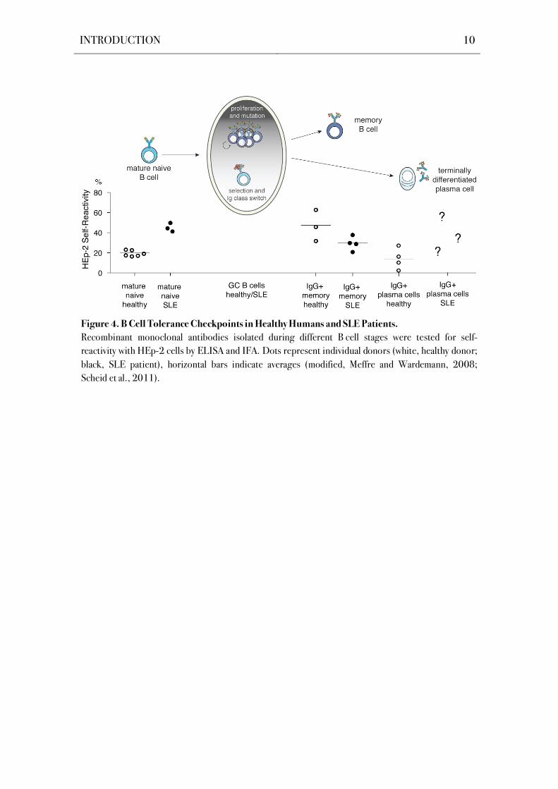
revealed that the central tolerance checkpoints are violated in SLE (Figure 4; Wardemann et
al., 2003; Meffre et al., 2004; Yurasov et al., 2005). Further, autoreactive B cells are not
excluded from germinal center reactions in SLE patients (Cappione et al., 2005) and thus
enable the generation of autoreactive memory B cells and plasma cells. However, frequencies of
autoreactive IgG+ memory B cells in SLE patients are similar to those of healthy donors
(Figure 4; Tiller et al., 2007; Mietzner et al., 2008). As long-lived plasma cells are the major
contibutor to serum IgG levels (Benner et al., 1981), their role in human SLE pathogenesis
should be further elucidated.
INTRODUCTION 10
Figure 4. B Cell Tolerance Checkpoints in Healthy Humans and SLE Patients.
Recombinant monoclonal antibodies isolated during different B cell stages were tested for self-
reactivity with HEp-2 cells by ELISA and IFA. Dots represent individual donors (white, healthy donor;
black, SLE patient), horizontal bars indicate averages (modified, Meffre and Wardemann, 2008;
Scheid et al., 2011).
AIM 11
2 AIM
Long-term humoral immunity is sustained by the formation of memory B cells and long-
lived antibody-secreting plasma cells that have undergone antigen-mediated selection during
germinal center responses. Previous single cell studies have elegantly dissected the
immunoglobulin (Ig) gene repertoire and antibody reactivity profile of circulating IgG+
memory B cells and have demonstrated an enrichment for self-reactive antibodies in this
compartment that mainly arise from non-reactive or polyreactive precursors by somatic
mutations. Immunological niches for human long-lived plasma cells are primarily located in the
bone marrow, where plasma cells survive for decades and continuously secrete large amounts of
protective antibodies. The Ig gene molecular features and the frequency of self-reactivity of this
B cell compartment were recently assessed in healthy individuals. The data suggest that in
contrast to the development of memory B cells, entry into the bone marrow plasma cell
compartment is tightly controlled by self-tolerance checkpoints that thereby prevent the
production of self- and polyreactive serum IgG antibodies.
Antibody secreting plasma cells may be directly involved in the pathogenesis of
systemic lupus erythematosus (SLE) by secreting high affinity self-reactive antibodies, but little
is known about the molecular features and antibody reactivity profiles of long-lived plasma cells
in SLE patients. The aim of this work was to analyze the Ig gene repertoire and the extent of
self-reactivity in the IgG+ bone marrow plasma cell compartment in SLE patients. Antibodies
from single IgG+ bone marrow plasma cells of five patients were cloned and expressed to assess
the reactivity profiles. To evaluate the contribution of somatic hypermutation to antibody
reactivity, poly- and self-reactive antibodies from one patient were reverted into their Ig
germline configuration and their reactivity was compared to the mutated counterparts. The
results provide new insights into the properties of terminally differentiated IgG+ plasma cells in
SLE and might foster further understanding of the disease.

AIM 12
METHODS 13
3 METHODS
3.1 Strategy Outline
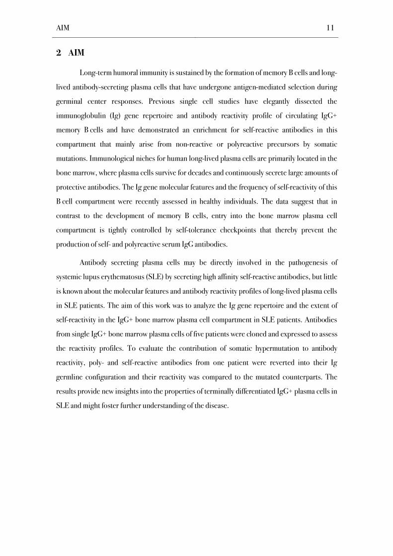
To analyze the Ig gene features and antibody reactivity profiles of human plasma cells, a
library of recombinant monoclonal antibodies was generated from single isolated primary IgG+
plasma cells from bone marrow of five SLE patients. Single bone marrow plasma cells were
sorted by flow cytometry according to cell surface markers (Figure 5). For each individually
sorted plasma cell, IgH and IgL chain variable region genes were amplified by nested RT-PCR
with primer mixes (Figure S4). Human Ig gene sequences were analyzed and classified
according to the international ImMunoGeneTics nomenclature by using the National Center
for Biotechnology IgBLAST database. Defined restriction sites were introduced by PCR for
IgG+ plasma cells to allow convenient ligation of the obtained IgH and IgL PCR products into
Figure 5. Experimental Strategy. Single plasma cells from human bone marrow were isolated by
fluorescence activated cell sorting (FACS). IgH chain and corresponding IgL chain transcripts were
amplified for each individual plasma cell by nested RT-PCR, classified by Ig gene sequence analysis
and cloned into eukaryotic expression vectors. The vectors were co-transfected into HEK 293T
cells to produce monoclonal antibodies of the same specificity in vitro. Recombinant antibodies were
tested for reactivity with diverse self- and non-self antigens by ELISA and IFA experiments and the
frequency of poly- and self-reactive antibodies was determined (Tiller et al., 2008).
METHODS 14
eukaryotic expression vectors containing the appropriate human immunoglobulin constant
regions. After amplification of expression vectors in Escherichia coli and subsequent plasmid
DNA purification, the plasmids encoding for the Ig chain and the corresponding Ig or light
chains were transiently co-transfected into human embryonic kidney 293T cells (HEK 293T).
Cell culture supernatants containing the recombinant human immunoglobulins were collected
and the antibodies were purified. To test for the reactivity of the monoclonal antibodies,
binding analyses such as enzyme-linked immunosorbent assay (ELISA) and indirect
immunofluorescence assays (IFAs) were conducted.
3.2 Fluorescence-Activated Cell Sorting (FACS)
Bone marrow aspirate from five SLE patients was collected after signed informed
consent in accordance with protocols reviewed by the Institutional Review Board of the Charité
University Medical Center. Mononuclear Cells were purified from bone marrow by Ficoll-
Paque® density gradient centrifugation and plasma cells from patients SLE1 and SLE2 were
pre-enriched using CD138 magnetic beads (Miltenyi Biotec). Due to small sample sizes and
low absolute mononuclear counts, CD138-pre-enrichment was not conducted for patients
SLE3-SLE5. Bone marrow mononuclear cells or pre-enriched CD138+ bone marrow cells
were stained on ice with anti-CD19-PECy7, anti-CD27-FITC, anti-CD38-APC and anti-
CD138-PE antibodies (Becton Dickinson) according to standard staining protocols. Single
plasma cells were sorted into 96-well PCR plates containing 4 μl lysis solution (0.5 x PBS
containing 10 mM DTT, 8 U RNAsin (Promega)) using a FACSVantage cell sorter with Diva
configuration (Becton Dickinson). PCR plates were sealed with Microseal ‘F’ film (Bio-Rad),
immediately frozen on dry ice and stored at -80 °C until further processing.
3.3 Reverse Transciption Polymerase Chain Reaction (PCR)
3.3.1 cDNA Synthesis
Total RNA from single cells was reverse transcribed in nuclease-free water (Eppendorf)
using 150 ng random hexamer primer (pd(N)6, Roche Applied Science), 0.5 mM dNTP
(Invitrogen), 10 mM DTT, 0.5 % v/v Igepal CA-630 (Sigma), 14 U RNAsin (Promega) and
50 U Superscript® III Reverse Transcriptase (Invitrogen). cDNA was synthesized in a total
METHODS 15
volume of 14 μl in the original 96-well sorting plate. Reverse transcription reaction was
performed for 60 min at 50 °C.
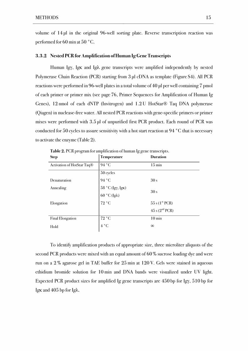
3.3.2 Nested PCR for Amplification of Human Ig Gene Transcripts
Human Ig , Ig and Ig gene transcripts were amplified independently by nested
Polymerase Chain Reaction (PCR) starting from 3 μl cDNA as template (Figure S4). All PCR
reactions were performed in 96-well plates in a total volume of 40 μl per well containing 7 pmol
of each primer or primer mix (see page 76, Primer Sequences for Amplification of Human Ig
Genes), 12 nmol of each dNTP (Invitrogen) and 1.2 U HotStar® Taq DNA polymerase
(Qiagen) in nuclease-free water. All nested PCR reactions with gene-specific primers or primer
mixes were performed with 3.5 l of unpurified first PCR product. Each round of PCR was
conducted for 50 cycles to assure sensitivity with a hot start reaction at 94 °C that is necessary
to activate the enzyme (Table 2).
Table 2. PCR program for amplification of human Ig gene transcripts.
Step Temperature Duration
Activation of HotStar Taq® 94 °C 15 min
50 cycles
Denaturation 94 °C 30 s
Annealing 58 °C (Ig , Ig )
60 °C (Ig ) 30 s
Elongation 72 °C 55 s (1st PCR)
45 s (2nd PCR)
Final Elongation 72 °C 10 min
Hold 4 °C
To identify amplification products of appropriate size, three microliter aliquots of the
second PCR products were mixed with an equal amount of 60 % sucrose loading dye and were
run on a 2 % agarose gel in TAE buffer for 25 min at 120 V. Gels were stained in aqueous
ethidium bromide solution for 10 min and DNA bands were visualized under UV light.
Expected PCR product sizes for amplified Ig gene transcripts are 450 bp for Ig , 510 bp for
Ig and 405 bp for Ig .
METHODS 16
3.3.3 Reversion Strategy for Mutated Ig Heavy and Light Chain Genes
An overlap-PCR strategy was applied to revert mutated Ig heavy and light chain
transcripts into their unmutated germline counterparts (Figure S6; Tiller et al., 2008).
Unmutated germline V genes were amplified from previously cloned unmutated templates with
gene-specific forward primers containing the AgeI restriction site and individual gene-specific
reverse primers that anneal to the 3’ end of the FWR3. Mutated CDR3-J sequences were
reverted independently by PCR using individual primers containing a minimal complementarity
of 10 nucleotides to the germline V gene transcript. Reverse J gene-specific primers included
the respective restriction sites as indicated (Figure S6). PCRs were performed at 94 °C for
30 s, 58 °C for 30 s and 72 °C for 45 s for 30 cycles (see also Table 2; according to conditions
for , and , 2nd PCR). Equal amounts of the reverted V and CDR3-J gene PCR products
were fused under the same conditions in a 20-cycle overlap PCR under the same conditions.
Full-length reverted V(D)J gene PCR products were gel extracted before digestion and purified
before cloning into the respective expression vectors. Gene identitiy and the successful
reversion of somatic mutations were confirmed by sequence analysis of the cloned products, as
described below.
3.4 Ig Gene Sequence Analysis
All samples were sequenced by Eurofins MWG Operon
(http://www.eurofinsdna.com). Four microliter aliquots of the Ig , Ig and Ig chain second
PCR products were sequenced in a final volume of 20 l. Respective reverse primer for
sequencing (see page 76, Primer Sequences for Amplification of Human Ig Genes) were
provided at a concentration of 10 M according to the instructions of Eurofins MWG Operon.
Obtained human Ig gene sequences were analyzed for Ig gene usage and CDR3 analysis, the
number of V gene mutations by IgBLAST comparison with Genbank
(http://www.ncbi.nlm.nih.gov/igblast/) and the IgG isotype subclass (http://imgt.cines.fr).
IgH CDR3 length was determined as indicated in the IgBLAST result by counting the amino
acid residues following framework region (FWR)3 up to the conserved tryptophan-glycine
motif in all JH segments or up to the conserved phenylalanin-glycine motif in J and J
segments (Kabat and Wu, 1991). In addition, the number of positively charged (histidine,
METHODS 17
arginine, lysine) and negatively charged (aspartate, glutamate) amino acids were determined for
each Ig heavy and Ig light chain CDR3.
3.5 Directional Cloning into Expression Vectors
3.5.1 Preparation of Eukaryotic Expression Vectors
Ig , Ig , or Ig expression vectors (Figure S5) with human Ig heavy chain gamma1
( 1), Ig light chain kappa ( ) or Ig light chain lambda ( ) constant regions, respectively, and an
ampicillin resistance gene were cloned in Escherichia coli (E. coli) DH10B bacteria. Vectors
were prepared using the NucleoBond Xtra Maxi Kit from Macherey-Nagel. The manufacturer’s
instructions were followed. After plasmid purification, the DNA concentration was determined
by NanoDrop spectrophotometer and 25 g of circular vector DNA were linearized using the
appropriate restriction enzymes.
3.5.2 Restriction Endonuclease Digestion
Second PCR products for Ig genes contained restriction sites allowing direct cloning
into expression vectors (Tiller et al., 2008). For Ig and Ig genes, restriction sites were
introduced after sequencing by gene-specific primers and first PCR products as template.
Purified PCR products or circular vector DNA were digested in two successive steps in a total
volume of 40 l with restriction enzymes (NEB) under optimal buffer conditions for the
indicated duration (Table 3).
Table 3. Conditions for Endonuclease Digestion.
1st Enzyme Temp Duration 2nd Enzyme Temp Duration
Heavy Chain Age I 37 °C 2 h Sal I 37 °C 2 h
Light Chain Age I 37 °C 2 h BsiW I 55 °C 2 h
Light Chain Age I 37 °C 2 h Xho I 37 °C 2 h
To test for successful and complete linearization of the digested expression vectors, gel
electrophoresis was performed using an 0.8 % agarose gel. Linearized expression vectors and
digested PCR products were purified by using the NucleoSpin Extract II Kit (Macherey-Nagel)
to obtain pure DNA as basic requirement for an efficient ligation reaction.
METHODS 18
3.5.3 Ligation
The ligation was conducted for 90 min at room temperature in 96-well plates in a total
volume of 10 μl per well with 200 U T4 DNA ligase (NEB), 8 μl of digested, purified PCR
product and 25 ng linearized expression vector. Ligation products were immediately
transformed into competent E. coli DH10B or stored at -20 °C.
3.5.4 Preparation of Competent Bacteria
The E.coli strain DH10B (Invitrogen) was used for transformation of recombinant
expression vector constructs. Competent bacteria were prepared by inoculating one freshly
grown E.coli DH10B colony in 5 ml Luria Bertani (LB) medium and were allowed to grow in a
bacteria shaker incubator overnight at 37 °C and 240 rpm. This preculture was transferred
into 500 ml LB medium and bacteria were cultivated to an optical density (OD600) between
0.6 and 0.8, where E.coli bacteria are in the exponential growth phase. Bacteria were kept on
ice for 30 min and after 10 min of centrifugation at 1500 gav, bacterial pellets were
resuspended in ice-cold sterile 180 ml 0.1 M CaCl2 solution. The cells were centrifuged again
for 10 min at 1500 gav and resuspended in 5 ml 0.1 M CaCl2 containing 15% glycerol.
Aliquots of competent bacteria suspensions were stored at -80 °C. Transformation efficiency
of bacteria was determined by calculating how many colonies were grown per g of added DNA.
Bacteria were transformed with different amounts of DNA in the range from 0.1 ng to 20 ng
following the protocol described below. The number of obtained colonies was divided by the
amount of DNA ( g) and multiplied with the ratio of the final volume (ml) at recovery and the
volume (ml) plated. Typical transformation efficiencies ranged from 4 x 10⁵ to 106 colonies per
g DNA.
3.5.5 Transformation of Competent Bacteria
Five microliter of competent E. coli DH10B bacteria were transformed with 3 l of the
ligation product in 96-well plates. After 30 min incubation on ice, a heat shock was performed
at 42 °C for 45 seconds and bacteria were allowed to grow in 200 l LB medium for 30 min
under moderate shaking at 37 °C. 100 l were plated on LB plates containing 100 g/ml
ampicillin and plates were incubated overnight at 37 °C.
METHODS 19
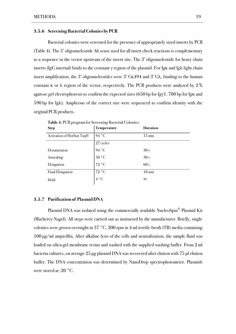
3.5.6 Screening Bacterial Colonies by PCR
Bacterial colonies were screened for the presence of appropriately sized inserts by PCR
(Table 4). The 5’ oligonucleotide Ab sense used for all insert check reactions is complementary
to a sequence in the vector upstream of the insert site. The 3’ oligonucleotide for heavy chain
inserts (IgG internal) binds to the constant region of the plasmid. For Ig and Ig light chain
insert amplification, the 3’ oligonucleotides were 3’ C 494 and 3’ C , binding to the human
constant or region of the vector, respectively. The PCR products were analyzed by 2 %
agarose gel electrophoresis to confirm the expected sizes (650 bp for Ig 1, 700 bp for Ig and
590 bp for Ig ). Amplicons of the correct size were sequenced to confirm identity with the
original PCR products.
Table 4. PCR program for Screening Bacterial Colonies.
Step Temperature Duration
Activation of HotStar Taq® 94 °C 15 min
27 cycles
Denaturation 94 °C 30 s
Annealing 58 °C 30 s
Elongation 72 °C 60 s
Final Elongation 72 °C 10 min
Hold 4 °C
3.5.7 Purification of Plasmid DNA
Plasmid DNA was isolated using the commercially available NucleoSpin® Plasmid Kit
(Macherey-Nagel). All steps were carried out as instructed by the manufacturer. Briefly, single
colonies were grown overnight at 37 °C, 200 rpm in 4 ml terrific broth (TB) media containing
100 g/ml ampicillin. After alkaline lysis of the cells and neutralization, the sample fluid was
loaded on silica-gel membrane resins and washed with the supplied washing buffer. From 2 ml
bacteria cultures, on average 25 g plasmid DNA was recovered after elution with 75 l elution
buffer. The DNA concentration was determined by NanoDrop spectrophotometer. Plasmids
were stored at -20 °C.
METHODS 20
3.6 Recombinant Antibody Production
3.6.1 Cell Culture
Human embryonic kidney (HEK) 293T (ATCC, No. CRL-11268) cells were cultured
in 150 mm culture plates (Becton Dickinson) under standard conditions (37 °C, 5 % CO2) in
Dulbecco's Modified Eagle's Medium (D-MEM; GibcoBRL) supplemented with 10 % heat-
inactivated fetal calf serum (FCS; Biochrom), 100 g/ml streptomycin, 100 U/ml penicillin G
and 0.25 g/ml amphotericin (100x Antibiotic-Antimycotic mix; GibcoBRL).
Transient transfections of exponentially growing cells were performed at 80% cell
confluency by cationic polymer polyethylenimine (PEI) transfection (Boussif et al., 1995) with
equal amounts (10–15 g each) of IgH and corresponding IgL chain encoding plasmid DNA.
3.6.2 Polyethylenimine- Mediated DNA Transfection of HEK 293T Cells
For PEI-mediated transfection, HEK 293T cells were washed with 10 ml serum-free
DMEM and 25 ml DMEM supplemented with 1% Nutridoma-SP (Roche) was added. Equal
amounts of IgH and IgL chain expression vector DNA were mixed in 150 mM sterile NaCl2
solution. The 3-fold weight amount of PEI was added to the plasmid solution and the mixture
was immediately vortexed for 10 s. A subsequent incubation step of 10 min at room
temperature allowed formation of DNA-polymer complexes. The mixture was added dropwise
to the culture dish. Cells were cultured for 4 days in 25 ml DMEM supplemented with 1 %
Nutridoma-SP (Roche) before supernatants were harvested and stored with 0.01 % sodium
azide at 4 °C until further use.
3.7 Enzyme-Linked Immunofluorescence Assays
3.7.1 Determination of Recombinant Ig Concentrations by ELISA
The concentration of recombinant IgG molecules in the harvested supernatants and
purified antibody eluates were determined by antibody sandwich ELISA using a goat anti-
human IgG Fc fragment antibody (Jackson ImmunoResearch) as capture antibody. High-
binding capacity microtiter plates (Costar) were coated with 100 ng capture antibody in 50 μl
PBS. Plates were sealed with Parafilm® and incubated over night at room temperature. The
plates were then washed three times with deionized water and incubated with 200 μl/well
METHODS 21
blocking buffer for 1 h to reduce unspecific binding sites. After another three washing steps,
each well was incubated with 50 μl of the supernatants with unknown IgG concentrations at
eight serial 1:3 dilutions in PBS starting with 1:3 dilutions for the supernatants and
1:100 dilutions for the purified antibody eluates. A human monoclonal IgG1 antibody (Sigma-
Aldrich) was used as standard in serial dilutions beginning with a concentration of 5 μg/ml.
After 2 h of incubation, the plates were washed three times to remove unbound antibodies and
per well 50 ng HRP-conjugated goat anti human IgG Fc fragment antibody (Jackson
ImmunoResearch) in blocking buffer were added and incubated for 1.5 h. After washing, brief
blocking with blocking solution and three additional washing steps, the samples were
developed by adding 100 μl of a 2,2'-Azino-di-[3-ethylbenzthiazoline sulfonate (6)]
diammonium salt (ABTS) solution (Roche) as HRP substrate and analyzed using a microplate
reader at a wavelength of 405 nm. Antibody concentrations were calculated using SoftMax ®
software (Molecular Devices).
3.7.2 Purification of Recombinant Antibodies
Recombinant antibodies were purified from the supernatants with Protein G beads (GE
Healthcare) according to the manufacturer’s instructions. In brief, 25 ml cell culture
supernatant were incubated with 25 μl Protein G beads under agitation for at least 14 h at
4 °C. After centrifugation at 800 gav for 10 min at 4 °C, supernatants were removed and the
beads were transferred to chromatography spin columns (Bio-Rad) that were equilibrated with
PBS before use. After two rounds of washing with PBS, the antibodies were eluted in three
fractions (200 μl each) with 0.1 M glycine (pH 3.0) and collected in tubes containing 20 μl 1 M
Tris (pH 8.0) with 0.1 % sodium azide.
3.7.3 Determination of Polyreactive Antibodies by ELISA
ELISAs were performed as described above (3.7.1) except high-binding capacity
microtiter plates were coated with 250 ng (insulin) or 500 ng (dsDNA, ssDNA,
lipopolysaccharide (LPS)) of individual antigens in PBS. ssDNA was prepared from salmon
sperm dsDNA (Sigma-Aldrich) by boiling at 95 °C for 30 min and aliquots were immediately
frozen at –20 °C. Human recombinant insulin solution (Sigma-Aldrich) and LPS from E. coli
Serotype 055:B5 (Sigma-Aldrich) were stored at 4 °C. Antibody concentrations in
supernatants were adjusted to 1 g/ml and three consecutive 1:4 dilutions in PBS were
prepared. Polyreactivity controls were the recombinant human monoclonal antibodies mGO53
METHODS 22
(negative; Wardemann et al., 2003), eiJB40 (low polyreactive; Wardemann et al., 2003), and
ED38 (highly polyreactive, Meffre et al., 2004) that were included on each plate. Bound
antibodies were detected using ABTS (Roche) as substrate. The cut-off OD405 at which
antibodies were considered reactive was determined for each experiment based on the OD405
minus 2x the standard deviation for the low positive control antibody eiJB40 at a concentration
of 0.25 g/ml. A minimum of 3 controls was included in each experiment to allow calculation
of standard deviations. Antibodies were considered polyreactive, when binding to at least two
structurally different types of tested antigens and if positive reactivity was confirmed in at least
two independent experiments.
3.8 Immunofluorescence Assays
HEp-2 cell coated slides (Bios) were incubated in a moist chamber at room temperature
with 20 l purified antibodies for 30 min at a concentration of 100 g/ml, washed twice in PBS
and incubated for additonal 30 min with Cy3-labeled goat anti-human IgG (Jackson
ImmunoResearch) according to the manufacturer’s instructions. Slides were washed again
twice in PBS before mounting with FluoromountG (Southern Biotech). Samples were
examined on a Zeiss Axioplan 2 fluorescence microscope. Control stainings with PBS, ANA-
negative and ANA-positive control sera were performed as suggested by the manufacturer and
were included in all experiments. Positive staining was determined by comparison to the
controls at equal exposure times.
3.9 Statistical Analysis
P-values for Ig gene repertoire analysis, analysis of positive charges in IgH CDR3, and
antibody reactivity were calculated by Fisher Exact test or Chi Square Test. P-values for IgH
CDR3 length and V gene mutations were calculated by non-paired two-tailed Student’s t test
using the GraphPad Prism Software.
RESULTS 23
4 RESULTS
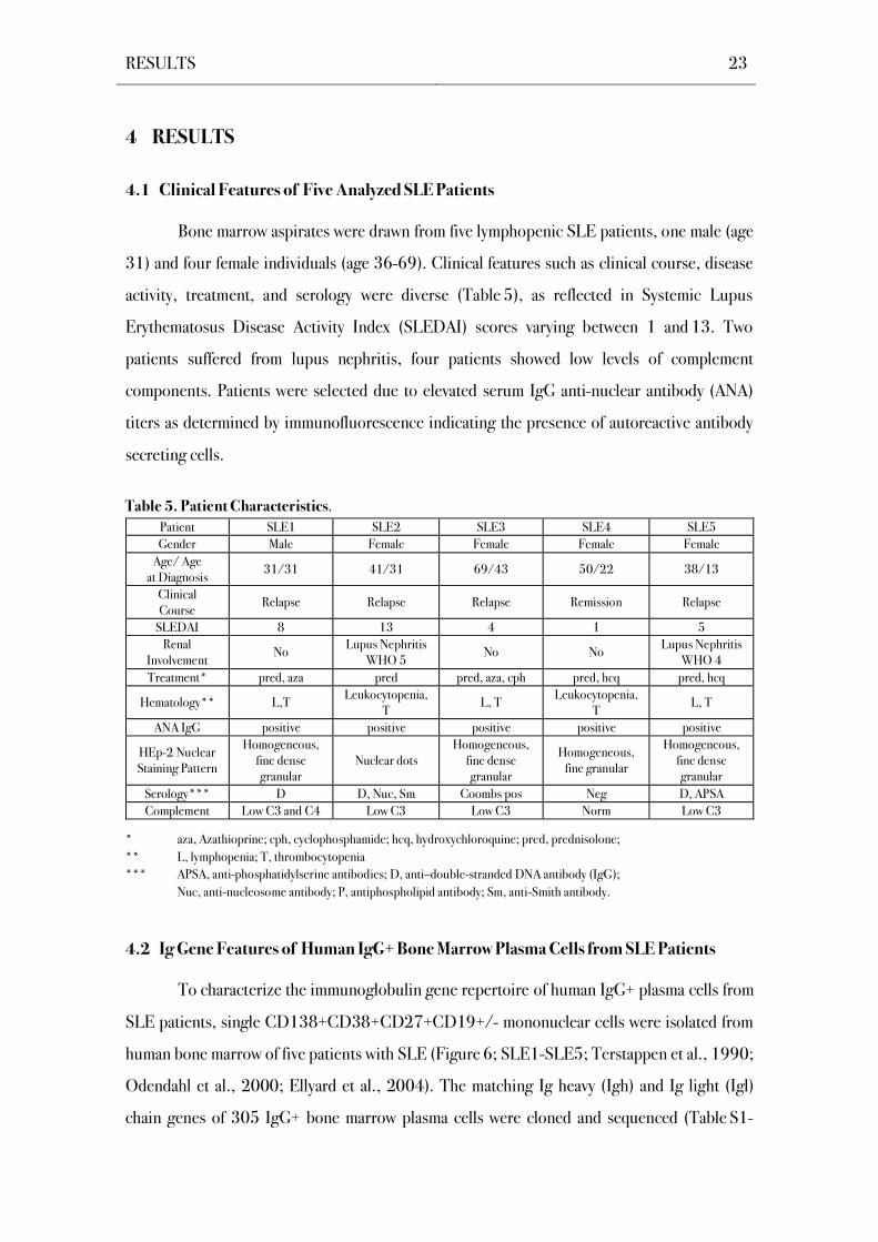
4.1 Clinical Features of Five Analyzed SLE Patients
Bone marrow aspirates were drawn from five lymphopenic SLE patients, one male (age
31) and four female individuals (age 36-69). Clinical features such as clinical course, disease
activity, treatment, and serology were diverse (Table 5), as reflected in Systemic Lupus
Erythematosus Disease Activity Index (SLEDAI) scores varying between 1 and 13. Two
patients suffered from lupus nephritis, four patients showed low levels of complement
components. Patients were selected due to elevated serum IgG anti-nuclear antibody (ANA)
titers as determined by immunofluorescence indicating the presence of autoreactive antibody
secreting cells.
4.2 Ig Gene Features of Human IgG+ Bone Marrow Plasma Cells from SLE Patients
To characterize the immunoglobulin gene repertoire of human IgG+ plasma cells from
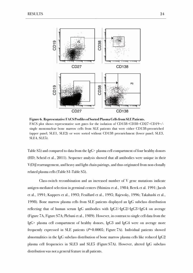
SLE patients, single CD138+CD38+CD27+CD19+/- mononuclear cells were isolated from
human bone marrow of five patients with SLE (Figure 6; SLE1-SLE5; Terstappen et al., 1990;
Odendahl et al., 2000; Ellyard et al., 2004). The matching Ig heavy (Igh) and Ig light (Igl)
chain genes of 305 IgG+ bone marrow plasma cells were cloned and sequenced (Table S1-
Table 5. Patient Characteristics.
Patient SLE1 SLE2 SLE3 SLE4 SLE5
Gender Male Female Female Female Female
Age/ Age at Diagnosis
31/31 41/31 69/43 50/22 38/13
Clinical Course
Relapse Relapse Relapse Remission Relapse
SLEDAI 8 13 4 1 5
Renal Involvement
No Lupus Nephritis
WHO 5 No No
Lupus Nephritis WHO 4
Treatment* pred, aza pred pred, aza, cph pred, hcq pred, hcq
Hematology** L,T Leukocytopenia,
T L, T
Leukocytopenia, T
L, T
ANA IgG positive positive positive positive positive
HEp-2 Nuclear Staining Pattern
Homogeneous, fine dense granular
Nuclear dots Homogeneous,
fine dense granular
Homogeneous, fine granular
Homogeneous, fine dense granular
Serology*** D D, Nuc, Sm Coombs pos Neg D, APSA
Complement Low C3 and C4 Low C3 Low C3 Norm Low C3
* aza, Azathioprine; cph, cyclophosphamide; hcq, hydroxychloroquine; pred, prednisolone;
** L, lymphopenia; T, thrombocytopenia
*** APSA, anti-phosphatidylserine antibodies; D, anti–double-stranded DNA antibody (IgG);
Nuc, anti-nucleosome antibody; P, antiphospholipid antibody; Sm, anti-Smith antibody.
RESULTS 24
Table S5) and compared to data from the IgG+ plasma cell compartment of four healthy donors
(HD; Scheid et al., 2011). Sequence analysis showed that all antibodies were unique in their
V(D)J rearrangement, and heavy and light chain pairings, and thus originated from non-clonally
related plasma cells (Table S1-Table S5).
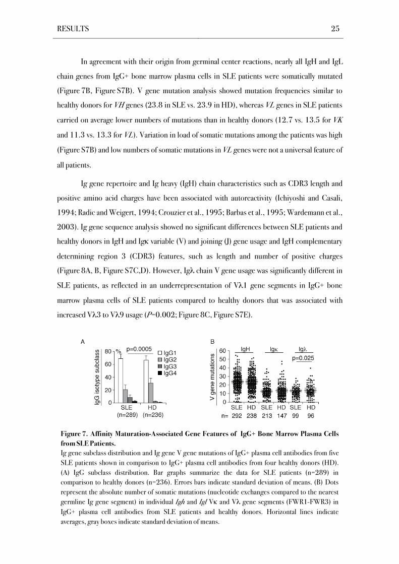
Class-switch recombination and an increased number of V gene mutations indicate
antigen-mediated selection in germinal centers (Shimizu et al., 1984; Berek et al. 1991; Jacob
et al., 1991; Kuppers et al., 1993; Feuillard et al., 1995; Rajewsky, 1996; Takahashi et al.,
1998). Bone marrow plasma cells from SLE patients displayed an IgG subclass distribution
reflecting that of human serum IgG antibodies with IgG1>IgG2>IgG3>IgG4 on average
(Figure 7A, Figure S7A; Plebani et al., 1989). However, in contrast to single cell data from the
IgG+ plasma cell compartment of healthy donors, IgG3 and IgG4 were on average more
frequently expressed in SLE patients (P=0.0005; Figure 7A). Individual patients showed
abnormalities in the IgG subclass distribution of bone marrow plasma cells like reduced IgG2
plasma cell frequencies in SLE3 and SLE5 (Figure S7A). However, altered IgG subclass
distribution was not a general feature in all patients.
Figure 6. Representative FACS Profiles of Sorted Plasma Cells from SLE Patients.
FACS plot shows representative sort gates for the isolation of CD138+CD38+CD27+CD19+/-
single mononuclear bone marrow cells from SLE patients that were either CD138-preenriched
(upper panel; SLE1, SLE2) or were sorted without CD138 preenrichment (lower panel; SLE3,
SLE4, SLE5).
RESULTS 25
In agreement with their origin from germinal center reactions, nearly all IgH and IgL
chain genes from IgG+ bone marrow plasma cells in SLE patients were somatically mutated
(Figure 7B, Figure S7B). V gene mutation analysis showed mutation frequencies similar to
healthy donors for VH genes (23.8 in SLE vs. 23.9 in HD), whereas VL genes in SLE patients
carried on average lower numbers of mutations than in healthy donors (12.7 vs. 13.5 for VK
and 11.3 vs. 13.3 for VL). Variation in load of somatic mutations among the patients was high
(Figure S7B) and low numbers of somatic mutations in VL genes were not a universal feature of
all patients.
Ig gene repertoire and Ig heavy (IgH) chain characteristics such as CDR3 length and
positive amino acid charges have been associated with autoreactivity (Ichiyoshi and Casali,
1994; Radic and Weigert, 1994; Crouzier et al., 1995; Barbas et al., 1995; Wardemann et al.,
2003). Ig gene sequence analysis showed no significant differences between SLE patients and
healthy donors in IgH and Ig variable (V) and joining (J) gene usage and IgH complementary
determining region 3 (CDR3) features, such as length and number of positive charges
(Figure 8A, B, Figure S7C,D). However, Ig chain V gene usage was significantly different in
SLE patients, as reflected in an underrepresentation of V 1 gene segments in IgG+ bone
marrow plasma cells of SLE patients compared to healthy donors that was associated with
increased V 3 to V 9 usage (P=0.002; Figure 8C, Figure S7E).
Figure 7. Affinity Maturation-Associated Gene Features of IgG+ Bone Marrow Plasma Cells
from SLE Patients.
Ig gene subclass distribution and Ig gene V gene mutations of IgG+ plasma cell antibodies from five
SLE patients shown in comparison to IgG+ plasma cell antibodies from four healthy donors (HD).
(A) IgG subclass distribution. Bar graphs summarize the data for SLE patients (n=289) in
comparison to healthy donors (n=236). Errors bars indicate standard deviation of means. (B) Dots
represent the absolute number of somatic mutations (nucleotide exchanges compared to the nearest
germline Ig gene segment) in individual Igh and Igl V and V gene segments (FWR1-FWR3) in
IgG+ plasma cell antibodies from SLE patients and healthy donors. Horizontal lines indicate
averages, gray boxes indicate standard deviation of means.
RESULTS 26
Figure 8. Ig Gene Features of IgG+ Plasma Cells from SLE Patients.
Ig gene features of IgG+ plasma cell antibodies from five SLE patients are shown in
comparison to IgG+ plasma cell antibodies from four healthy donors (HD). VH/JH gene
family usage and IgH CDR3 positive charges and length (A), and V /J (B) and V /J (C)
gene family usage are shown. Bar graphs display the data obtained from lupus patients ( ,
n=305; , n=226; , n=103) in comparison to healthy donors ( , n=238; , n=147; ,
n=97). (E) Ratio of VH to VL somatic mutations in IgG+ plasma cells from SLE patients.
Dots represent individual antibodies from SLE patients (SLE1-SLE5) and healthy donors
(HD15, HD16, HD20, HD21) and pooled data (SLE1-5, HD). VL mutation values were
set from 0 to 1, in cases where mutated IgH chain transcripts were paired with unmutated
IgL chains. (A)-(D) Error bars indicate standard deviation of means.
RESULTS 27
Alterations in Ig light chain recombination were also detected in the Ig light chain
isotype distribution, as IgG+ plasma cells displayed on average a significantly lower frequency
of Ig functional transcripts in SLE patients (Figure 8D, Figure S7E). Elevated levels of Ig
and Ig light chain double positive plasma cells were observed in all SLE patients (P=0.002;
Figure 8D, Figure S7F).
The average number of mutations is typically higher in VH than in VL gene segments,
both in healthy donors and SLE patients (de Wildt et al., 1999; de Wildt et al., 2000; Mietzner
et al., 2008). The mutation ratio of IgG+ bone marrow plasma cells from three SLE patients
was comparable to healthy donors, whereas patients with large numbers of Ig and Ig light
chain included functional transcripts showed elevated ratios of VH to VL somatic mutations
(3.2 in SLE3 and SLE5 as compared to 2.0- 2.4 in the other SLE patients and 2.0- 2.7 in HD;
Figure E).
Thus in summary, SLE is associated with alterations in subclass distribution, V gene
usage and Ig light chain isotype expression in IgG+ bone marrow plasma cells.
4.3 Polyreactive IgG+ Bone Marrow Plasma Cells in SLE Patients
Entry into the bone marrow plasma cell compartment appears to be strongly selective
and requires high antigen specificity (Smith et al., 1997; Smith et al., 2000; Takahashi et al.,
1998; Phan et al., 2000; Scheid et al., 2011). In SLE patients however, pathogenic
autoantibodies are frequently polyreactive, which might be important in disease pathogenesis
(Spatz et al., 1997; Sabbaga et al., 1990). Between 2-16 % of IgG+ bone marrow plasma cells
from healthy donors express polyreactive antibodies that react with structurally diverse self- and
non-self antigens (Scheid et al., 2011). To determine if SLE is associated with an impaired
exclusion of polyreactive plasma cells, the cloned IgG and IgL genes of 196 bone marrow
plasma cells were recombinantly expressed in vitro and the frequency of polyreactive antibodies
in the IgG+ bone marrow plasma cell compartment of SLE patients was measured. The
recombinant monoclonal antibodies were thus tested by ELISA for binding to a small panel of
self- and non-self antigens comprising single-stranded and double-stranded DNA (ssDNA and
dsDNA), lipopolysaccharide (LPS), and insulin (Figure 9; Table S1-Table S5). Between 3-
30 % of IgG+ bone marrow plasma cells reacted with at least two structurally distinct antigens
RESULTS 28
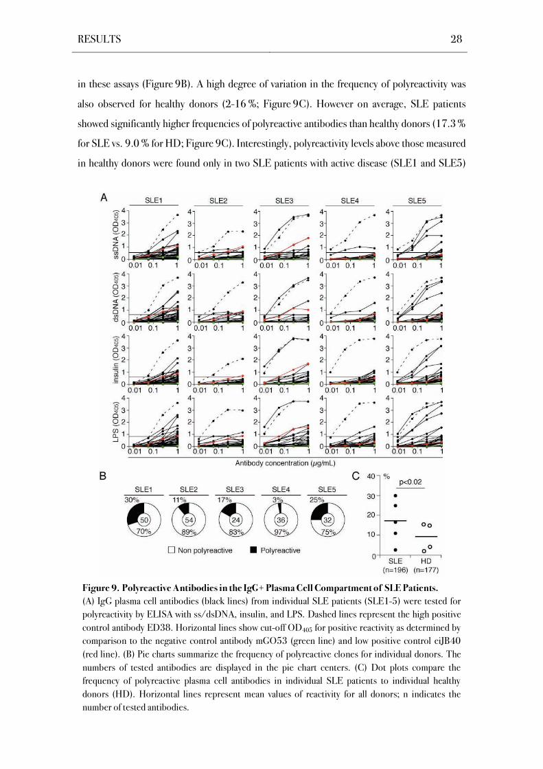
in these assays (Figure 9B). A high degree of variation in the frequency of polyreactivity was
also observed for healthy donors (2-16 %; Figure 9C). However on average, SLE patients
showed significantly higher frequencies of polyreactive antibodies than healthy donors (17.3 %
for SLE vs. 9.0 % for HD; Figure 9C). Interestingly, polyreactivity levels above those measured
in healthy donors were found only in two SLE patients with active disease (SLE1 and SLE5)
Figure 9. Polyreactive Antibodies in the IgG+ Plasma Cell Compartment of SLE Patients.
(A) IgG plasma cell antibodies (black lines) from individual SLE patients (SLE1-5) were tested for
polyreactivity by ELISA with ss/dsDNA, insulin, and LPS. Dashed lines represent the high positive
control antibody ED38. Horizontal lines show cut-off OD405 for positive reactivity as determined by
comparison to the negative control antibody mGO53 (green line) and low positive control eiJB40
(red line). (B) Pie charts summarize the frequency of polyreactive clones for individual donors. The
numbers of tested antibodies are displayed in the pie chart centers. (C) Dot plots compare the
frequency of polyreactive plasma cell antibodies in individual SLE patients to individual healthy
donors (HD). Horizontal lines represent mean values of reactivity for all donors; n indicates the
number of tested antibodies.
RESULTS 29
and the lowest level of polyreactivity was observed in patient SLE4 with low disease activity in
clinical remission. Thus, exclusion of polyreactive IgG+ plasma cells from the bone marrow
compartment is impaired in some but not all SLE patients.
4.4 Self-Reactive IgG+ Bone Marrow Plasma Cells in SLE Patients
Self-reactive IgG+ bone marrow plasma cells are efficiently excluded from the bone
marrow compartment of healthy donors (Scheid et al., 2011). In SLE patients, self-reactive IgG
serum antibodies display a cardinal feature of the disease and are a diagnostic marker (Kotzin,
1996). To determine if SLE is associated with a defect in self-tolerance that allows self-reactive
plasma cells to accumulate in the bone marrow, all antibodies were tested for self-reactivity with
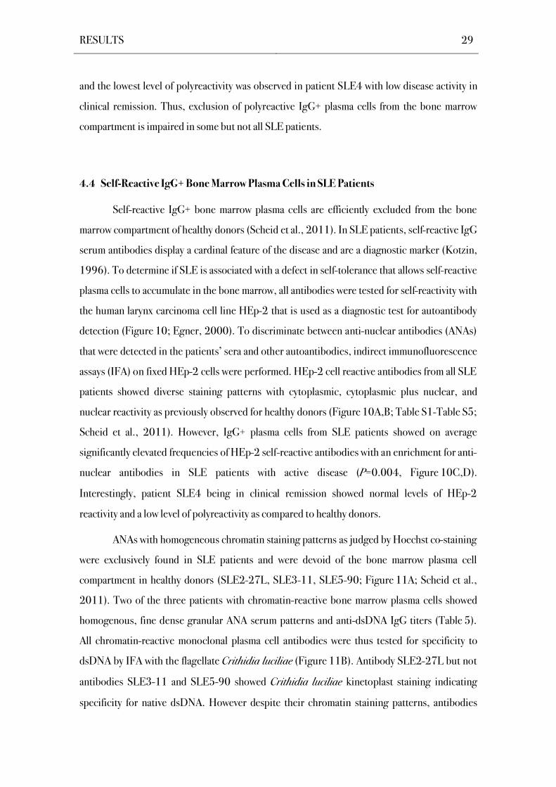
the human larynx carcinoma cell line HEp-2 that is used as a diagnostic test for autoantibody
detection (Figure 10; Egner, 2000). To discriminate between anti-nuclear antibodies (ANAs)
that were detected in the patients’ sera and other autoantibodies, indirect immunofluorescence
assays (IFA) on fixed HEp-2 cells were performed. HEp-2 cell reactive antibodies from all SLE
patients showed diverse staining patterns with cytoplasmic, cytoplasmic plus nuclear, and
nuclear reactivity as previously observed for healthy donors (Figure 10A,B; Table S1-Table S5;
Scheid et al., 2011). However, IgG+ plasma cells from SLE patients showed on average
significantly elevated frequencies of HEp-2 self-reactive antibodies with an enrichment for anti-
nuclear antibodies in SLE patients with active disease (P=0.004, Figure 10C,D).
Interestingly, patient SLE4 being in clinical remission showed normal levels of HEp-2
reactivity and a low level of polyreactivity as compared to healthy donors.
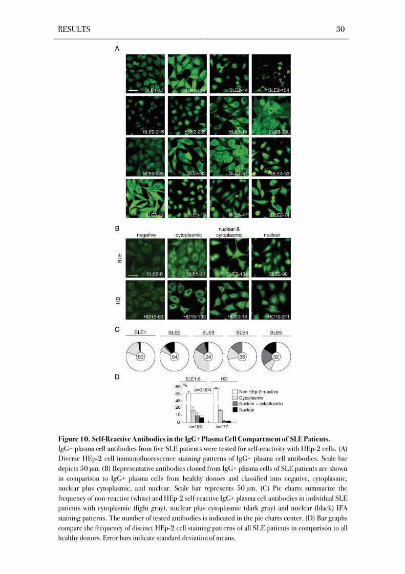
ANAs with homogeneous chromatin staining patterns as judged by Hoechst co-staining
were exclusively found in SLE patients and were devoid of the bone marrow plasma cell
compartment in healthy donors (SLE2-27L, SLE3-11, SLE5-90; Figure 11A; Scheid et al.,
2011). Two of the three patients with chromatin-reactive bone marrow plasma cells showed
homogenous, fine dense granular ANA serum patterns and anti-dsDNA IgG titers (Table 5).
All chromatin-reactive monoclonal plasma cell antibodies were thus tested for specificity to
dsDNA by IFA with the flagellate Crithidia luciliae (Figure 11B). Antibody SLE2-27L but not
antibodies SLE3-11 and SLE5-90 showed Crithidia luciliae kinetoplast staining indicating
specificity for native dsDNA. However despite their chromatin staining patterns, antibodies
RESULTS 30
Figure 10. Self-Reactive Antibodies in the IgG+ Plasma Cell Compartment of SLE Patients.
IgG+ plasma cell antibodies from five SLE patients were tested for self-reactivity with HEp-2 cells. (A)
Diverse HEp-2 cell immunofluorescence staining patterns of IgG+ plasma cell antibodies. Scale bar
depicts 50 μm. (B) Representative antibodies cloned from IgG+ plasma cells of SLE patients are shown
in comparison to IgG+ plasma cells from healthy donors and classified into negative, cytoplasmic,
nuclear plus cytoplasmic, and nuclear. Scale bar represents 50 μm. (C) Pie charts summarize the
frequency of non-reactive (white) and HEp-2 self-reactive IgG+ plasma cell antibodies in individual SLE
patients with cytoplasmic (light gray), nuclear plus cytoplasmic (dark gray) and nuclear (black) IFA
staining patterns. The number of tested antibodies is indicated in the pie charts center. (D) Bar graphs
compare the frequency of distinct HEp-2 cell staining patterns of all SLE patients in comparison to all
healthy donors. Error bars indicate standard deviation of means.
RESULTS 31
SLE3-11 and SLE5-90 lacked reactivity with nucleosomes and histones as determined by
ELISA (data not shown).
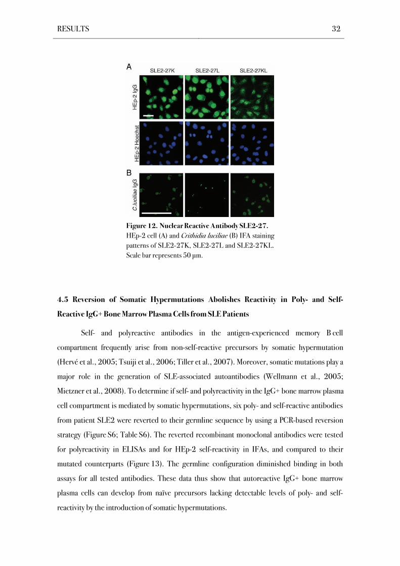
Anti-nuclear antibody SLE2-27 originated from an Ig and Ig light chain included
plasma cell (Table S2). The anti-nuclear reactivity of SLE2-27 was independent of whether the
IgH chain was co-expressed with the Ig light chain, Ig light chain or with both light chains
(Figure 12A). However, anti-dsDNA specificity as determined by IFA with the flagellate
Crithidia luciliae was dependent on sole expression of the Ig light chain and was not detected
when the Ig light chain was co-expressed (Figure 12B). However, analyzing the antibody
repertoire of all SLE patients did not show a correlation between Ig isotype inclusion and
antibody reactivity, as double Ig light chain positive plasma cells were identified where one,
both or none of the two IgL chains mediated antibody self-reactivity when expressed with the
respective IgH chain (Table S1- Table S5).
In summary, the bone marrow compartment in SLE patients with active disease is
enriched for self-reactive IgG+ plasma cells and disease-associated IgG+ ANAs including anti-
chromatin specific plasma cells fail to be excluded.
Figure 11. Chromatin-Reactive Antibodies in the IgG+ Plasma Cell Compartment of SLE
Patients.
IgG+ plasma cell antibodies from five lupus patients were tested for dsDNA reactivity with HEp-2 and
Crithidia luciliae cells. (A) Strong homogenous nuclear HEp-2 staining patterns of IgG+ plasma cell
antibodies SLE2-27L, SLE3-11, and SLE5-90. Nuclei are visualized by Hoechst staining. Scale bar
represents 50 μm. (B) Crithidia luciliae IFA staining pattern of the same antibodies as in A. Nuclei and
kinetoplasts are visualized by Hoechst staining. Scale bar represents 20 μm.
RESULTS 32
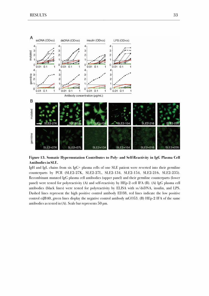
4.5 Reversion of Somatic Hypermutations Abolishes Reactivity in Poly- and Self-
Reactive IgG+ Bone Marrow Plasma Cells from SLE Patients
Self- and polyreactive antibodies in the antigen-experienced memory B cell
compartment frequently arise from non-self-reactive precursors by somatic hypermutation
(Hervé et al., 2005; Tsuiji et al., 2006; Tiller et al., 2007). Moreover, somatic mutations play a
major role in the generation of SLE-associated autoantibodies (Wellmann et al., 2005;
Mietzner et al., 2008). To determine if self- and polyreactivity in the IgG+ bone marrow plasma
cell compartment is mediated by somatic hypermutations, six poly- and self-reactive antibodies
from patient SLE2 were reverted to their germline sequence by using a PCR-based reversion
strategy (Figure S6; Table S6). The reverted recombinant monoclonal antibodies were tested
for polyreactivity in ELISAs and for HEp-2 self-reactivity in IFAs, and compared to their
mutated counterparts (Figure 13). The germline configuration diminished binding in both
assays for all tested antibodies. These data thus show that autoreactive IgG+ bone marrow
plasma cells can develop from naïve precursors lacking detectable levels of poly- and self-
reactivity by the introduction of somatic hypermutations.
Figure 12. Nuclear Reactive Antibody SLE2-27.
HEp-2 cell (A) and Crithidia luciliae (B) IFA staining
patterns of SLE2-27K, SLE2-27L and SLE2-27KL.
Scale bar represents 50 μm.
RESULTS 33
Figure 13. Somatic Hypermutation Contributes to Poly- and Self-Reactivity in IgG Plasma Cell
Antibodies in SLE.
IgH and IgL chains from six IgG+ plasma cells of one SLE patient were reverted into their germline
counterparts by PCR (SLE2-27K, SLE2-27L, SLE2-134, SLE2-154, SLE2-216, SLE2-235).
Recombinant mutated IgG plasma cell antibodies (upper panel) and their germline counterparts (lower
panel) were tested for polyreactivity (A) and self-reactivity by HEp-2 cell IFA (B). (A) IgG plasma cell
antibodies (black lines) were tested for polyreactivity by ELISA with ss/dsDNA, insulin, and LPS.
Dashed lines represent the high positive control antibody ED38, red lines indicate the low positive
control eiJB40, green lines display the negative control antibody mGO53. (B) HEp-2 IFA of the same
antibodies as tested in (A). Scale bar represents 50 μm.

RESULTS 34
DISCUSSION 35
5 DISCUSSION
5.1 Unbiased Analysis of the Plasma Cell Antibody Repertoire by Single Cell Ig Gene
Cloning Implies a Rigid Selection Process in Healthy Humans
Immunoglobulin (Ig) gene sequence analysis of distinct B cell populations allowed a
basic understanding of the expressed human antibody repertoire at different stages during
B cell development (Huang and Stollar, 1991; Huang et al., 1992; Brezinschek et al., 1995;
Wang and Stollar, 2000). However, sequence analysis alone does not permit predictions on
antibody reactivities. Human monoclonal antibodies can be produced by different methods
such as immortalization of B cells with Ebstein-Barr virus (Steinitz et al., 1977; Lanzavecchia
et al., 2007), the production of B-cell hybridomas (Kohler and Milstein, 1975), using phage
display libraries (McCafferty et al., 1990) or the humanization of antibodies from other
mammalian species (Jones et al., 1986). Still, immortalization and fusion efficiencies are low,
largely depend on the maturation status of the B cell and frequently require additional B cell
stimulation. Phage display technology on the other hand relies on random pairing of human Ig
heavy and light chains in vitro, thus not providing information on the epitope specificities that
humans generate in vivo.
This study takes advantage of an unbiased and efficient approach that combines Ig gene
repertoire analysis and Ig reactivity profiling at the single cell level (Wardemann et al., 2003;
Tiller et al., 2008). The applied single-cell strategy facilitates the isolation of single B cells at
any stage of their development and requires only small cell numbers. Using this methodology it
was demonstrated that selection into the human bone marrow plasma cell compartment
requires a positive selection step after the germinal center reaction under normal immune
conditions (Scheid et al., 2011), as was elegantly demonstrated in mouse models (Smith et al.,
1997; Takahashi et al., 1998; Phan et al., 2006; Paus et al., 2006). This counterselection
against poly- and self-reactivity in the bone marrow plasma cell compartment may be essential
to avoid the production of poly- and self-reactive serum IgG antibodies that might lead to the
induction of inflammatory autoimmune reactions and the development of autoimmune disease.
DISCUSSION 36
5.2 Serum Autoantibodies are a Hallmark of Disease in SLE Patients
B cell lymphopenia and B cell overactivity are among the most striking abnormalities
encountered in systemic lupus erythematosus (Rahman and Isenberg, 2008; Crispín et al.,
2010) and were a characteristic of the analyzed SLE patients. Patient selection was based on
elevated serum ANA IgG levels indicating the presence of autoantibody secreting cells.
Enhanced B cell activity results in the production of characteristic patterns of autoantibodies
and has been postulated to be the defining pathogenic event in human SLE (Lipsky, 2001;
Rahman and Isenberg, 2008). Serum IgG autoantibodies against dsDNA and nucleosomes, as
detected in the analyzed lupus patients with SLEDAI scores above 4, indicate disease activity
and vanish during remission (Grammer and Lipsky, 2003; Vallerskog et al., 2007; Hiepe et al.,
2011). However, a recent study analyzing serum autoantibodies at low dilutions demonstrated
a persisting SLE antibody profile independently of disease activity (Fattal et al., 2010). This
specific antibody profile revealed increased anti-dsDNA IgG antibody reactivities in all tested
patients. Thus, baseline anti-dsDNA serum IgG reactivity may be sustained by terminally
differentiated plasma cells during remission and plasmablasts may contribute to elevated anti-
dsDNA serum IgG levels during flares.
5.3 The Ig Gene Repertoire in IgG+ Bone Marrow Plasma Cells from SLE Patients
The present study demonstrates that SLE is associated with variable abnormalities in
subclass distribution, V gene usage and Ig light chain isotype expression in IgG+ bone
marrow plasma cells, whereas somatic mutation levels and IgH and Ig gene usage appear
normal. Previous studies have reported molecular abnormalities in the Ig gene repertoire of
peripheral B cells from SLE patients, including altered IgH and IgL V gene usage, abnormal
IgH CDR3 features, altered somatic mutation frequencies and differences in IgG subclass
distribution (Demaison et al, 1994; Manheimer-Lory et al., 1997; Dorner et al., 1998; Dorner
et al., 1999; Jacobi et al., 2000; Odendahl et al., 2000; Yurasov et al., 2005; Yurasov et al.,
2006; Mietzner et al., 2008). The present study demonstrates that some of these SLE-
associated Ig gene features, such as underrepresentation of IgG2 and V gene biases extend to
the bone marrow plasma cell compartment. The data moreover confirms previous findings on a
high variability in the Ig gene repertoire among individual SLE patients (Yurasov et al., 2005;
DISCUSSION 37
Yurasov et al., 2006; Foreman et al., 2007; Mietzner et al., 2008) and this diversity of the Ig
gene repertoire likely reflects the high degree of disease diversity in SLE.
B cell receptor editing has been proposed to be the main mechanism in establishing
self-tolerance and is mainly conferred by secondary Ig gene rearrangements that generate a new
antigen receptor with an innocuous specificity (Gay et al., 1993; Radic et al., 1993; Tiegs et
al., 1993). Abnormalities in Ig gene usage indicating secondary rearrangement were not
detected in the IgG+ bone marrow plasma cell compartment and alterations in V gene usage
were not linked to chromosomal location, as was also reported in previous studies on peripheral
B cells (Bensimon et al., 1994; Dorner et al., 1999; Mietzner et al., 2008). However, other
studies have demonstrated that SLE is associated with abnormalities in Ig light chain editing in
peripheral B cells (Suzuki et al., 1996; Dorner et al., 1998; Dorner et al., 1999; Yurasov et
al., 2006).
High frequencies of Ig and Ig double positive B cells were detected in the IgG+ bone
marrow plasma cell compartment of two SLE patients, as previously reported for IgG+ memory
B cells from one SLE patient (Mietzner et al., 2008). Evidence from experimental mouse
models has proven that editing of self-reactive B cell antigen receptors may be associated with
Ig light chain inclusion (Nemazee and Weigert, 2000; Li et al., 2001; Li et al., 2002; Liu et
al., 2005; Doyle et al., 2006; Casellas et al., 2007; Zouali, 2008). Moreover, the two patients
with an increased frequency of allelically included light chains also displayed elevated ratios of
VH to VL gene somatic mutations, suggesting that secondary Ig gene recombination in these
individuals occurred after antigen-mediated activation in the periphery and after initial affinity
maturation.
5.4 The Bone Marrow Contributes to Serum ANA Production in SLE Patients
Exclusion of polyreactive IgG+ plasma cells from the bone marrow compartment is
impaired in some SLE patients. In agreement with this observation, previous studies
demonstrated that pathogenic lupus autoantibodies are frequently polyreactive (Sabbaga et al.,
1990; Spatz, 1997) and naïve B cells of SLE patients are enriched for polyreactive antibodies
both during relapse and remission (Yurasov et al., 2005; Yurasov et al., 2006). In healthy
donors, polyreactivity is a common feature of IgG+ memory B cells (Tiller et al., 2007) but rare
DISCUSSION 38
in the long-lived bone marrow plasma cell compartment (Scheid et al., 2011). As observed in
two SLE patients, polyreactive antibodies can accumulate in the plasma cell compartment to a
higher extent than detected in healthy donor plasma cells. This increase in polyreactivity was
not associated with an increase in self-reactivity in one patient, which indicates that chronic
inflammation provides a constant supply of antigen to stimulate lymphocytes in this patient
(Goodnow, 2007). In summary, the data suggest that an accumulation of polyreactive
antibodies in the plasma cell compartment is not a universal feature of the disease, but likely
reflects differences in genetic and environmental factors that both are important in the
development of SLE (Rahman and Isenberg, 2008).
The bone marrow in SLE patients harbors IgG+ plasma cells expressing disease-
associated ANAs including anti-chromatin IgGs. Anti-nuclear IgG antibodies with chromatin
reactivity represent a hallmark of SLE (Kotzin, 1996) and may arise from autoreactive B cells
that fail to be excluded from germinal center reactions (Cappione et al., 2005). One of the
identified chromatin-reactive antibodies in this study could be further characterized to be
specific for native dsDNA. Both anti-dsDNA and anti-nucleosome antibodies have been
demonstrated to mediate lupus nephritis (Rahman and Isenberg, 2008; van Bavel et al., 2008).
However, chromatin-specific antibodies detected in immunofluorescence assays frequently
show no or very low reactivity against individual histones or native dsDNA (van Bavel et al.,
2008). Nevertheless, the present study clearly demonstrates that the bone marrow plasma cell
compartment contributes to serum ANA production in SLE patients as previously reported for
an autoimmune lupus mouse model (Hoyer et al., 2004).
5.5 The Role of Somatic Hypermutations in the Generation of Self-Reactive Antibodies
In agreement with previously published data on the impact of somatic hypermutation in
the generation of disease-associated SLE autoantibodies (Shlomchik et al., 1987; van Es et al.,
1991; Radic et al., 1993; Manheimer-Lory et al., 1997; Wellmann et al., 2005; Mietzner et
al., 2008; Guo et al., 2010), the germline forms of all tested SLE plasma cell antibodies lacked
detectable levels of self- and polyreactivity. This finding implies that these antibody specificities
developed by somatic mutations introduced during affinity maturation in germinal center
reactions. The data thus confirm previous findings demonstrating the importance of somatic
mutations for the development of SLE autoantibodies.
DISCUSSION 39
5.6 Genetic and Environmental Factors Generate Disease Diversity in SLE Patients
All patients differed in clinical features as well as serology and hematology. Further,
differences among SLE patients were detected at both the Ig molecular level and the antibody
reactivity level in the bone marrow plasma cell compartment. This high degree of disease
diversity is one of the main obstacles in lupus treatment. Many different pathways, processes
and cell types of the immune system are involved in the generation of the SLE phenotype
(Harley et al., 2009).
All SLE patients with active disease had low complement serum levels. These low
complement levels indicate poor waste disposal due to deficiency in complement components
or an increase in cell death rates that may be one of the main factors triggering the onset of the
disease (Rahman and Isenberg, 2008; Muñoz et al., 2010). Further, the IgG+ bone marrow
plasma cell compartment is enriched for self-reactive antibodies with nuclear specificity. Due to
defective clearance mechanisms, cellular debris containing nuclear antigens is exposed and can
activate immune cells. Nuclear antigens like chromatin contain both B cell and T cell epitopes
and additionally activate immune cells through TLR9 specific for DNA (Goodnow, 2007;
Harley et al., 2009). This simultaneous activation of different pathways subsequently enhances
inflammatory cascades and provides stimuli for chronic inflammation (Isenberg et al., 1990;
Rahman et al., 2002).
The presence of autoantibody titers like those detected in the analyzed patients
correlates with elevated BAFF serum levels (Cambridge et al., 2008; Tieng and Peeva, 2008).
Alterations in the expression of components involved in BCR signaling can affect thresholds for
B cell survival and activation. The threshold for naïve and memory B cell activation,
proliferation and antibody secretion is lowered by the presence of both IL-17 and BAFF
(Doreau et al., 2009) and may also influence the bone marrow plasma cell compartment. In
situations of B cell hyperresponsiveness, affinity maturation would be censored less stringently
(Tarlinton and Smith, 2000) and selection into the long-lived plasma cell compartment may be
impaired.
Elevated levels of poly- and self-reactive plasma cells are probably a result of B cell
hyperresponsiveness. The IgG inhibitory Fc gamma receptor IIB (Fc RIIB) is the only
inhibitory Fc receptor on B cells (Nimmerjahn and Ravetch, 2008) and has been shown to be
involved in SLE pathogenesis of mice and man at both genetic and functional levels (Morel,
DISCUSSION 40
2010; Tiller et al., 2010). The frequency of Fc RIIB on circulating memory B cells is
significantly decreased in SLE patients (Mackay et al., 2006), thus may also be impaired on
terminally differentiated plasma cells. A decrease of IgG inhibitory (Fc RIIB) is suggested to
promote B cell hyperresponsiveness and thus nourishes an unregulated expansion of activated
autoreactive B cells and the production of pathogenic autoantibodies (Morel, 2010; Mackay et
al., 2006).
5.7 Therapeutic Options to Eliminate Autoreactive Plasma Cells
SLE is associated with high frequencies of IgG ANA-secreting bone marrow plasma
cells that may represent an important therapeutic target in patients refractory to conventional
therapies. Self-reactive high-affinity germinal center B cells are difficult to target once they
have been selected into the bone marrow plasma pool. As soon as plasma cells have reached a
survival niche in the bone marrow, they lose the ability to respond to antigenic changes in their
environment (Amanna and Slifka, 2010) and are thus resistant to glucocorticoids,
immunosuppressive and cytotoxic treatments (Hoyer et al., 2004; Hiepe et al., 2011).
However, targeting plasma cells is possible but universal depletion of plasma cells also
eliminates the main source of protective antimicrobial antibodies and is accompanied with
severe side effects (Neubert et al., 2008; Hoyer et al., 2008; Sanz and Lee, 2010).
Therapeutic drugs abolishing autoantibodies and preventing flares in SLE patients are
still missing in disease treatment. Approaches of the last decades focused on specific
immunomodulation to alter B cell populations and several strategies targeting B cells are
currently available (Sanz and Lee, 2010). Current approaches comprise direct B cell death
using depleting antibodies, inhibition of factors involved in B cell differentiation or survival and
interruption of signaling through the B cell receptor or co-stimulatory receptors (Sanz and Lee,
2010). For example, CD20-mediated depletion eliminates autoreactive mature B cells but
does not target plasmablasts and plasma cells and thus, protective as well as autoreactive
antibodies secreted by plasma cells are still detected in the patients’ serum (Traggiai et al.,
2003; Vallerskog et al., 2007; Cambridge et al., 2008; Lipsky and Dorner, 2010). On the
other hand, using a tolerogen to selectively target DNA-reactive B cells has not proven to be
clinically beneficial (Rahman and Isenberg, 2008). An expansion of regulatory B cells, a
recently discovered B cell subpopulation, might provide an alternative treatment strategy
DISCUSSION 41
(Yanaba et al., 2008), as regulatory B cells are capable of inhibiting proinflammatory cascades
by secretion of the regulatory cytokines IL-10 and TGF- .
The formation of a stable pool of autoreactive plasma cells that contribute to serum IgG
production may represent an important therapeutic target in patients refractory to conventional
therapies (Hiepe et al., 2011). A clinical long-term benefit for patients with refractory SLE has
been demonstrated for autologous stem cell transplantation along with non-specific
immunosuppressive agents. This approach does not specifically target plasma cells, but resets
adaptive immunity and thereby eliminates autoreactive lymphocytes (Alexander et al., 2009).
However, this therapeutic approach goes along with a high mortality (Jayne et al., 2004;
Alexander et al., 2009) and thus requires further optimization.
5.8 Conclusion and Outlook
Long-lived plasma cells do not divide after selection into the bone marrow
compartment (Manz et al., 1997; Slifka et al., 1998; Amanna and Slifka, 2010) and long-term
antibody production after infection or vaccination is often maintained by bone marrow plasma
cells (Benner et al., 1981). Physical space in survival niches is limited (Radbruch et al., 2006)
and once detached from this microenvironment, plasma cells do not relocate (Hoyer et al.,
2004). However, specific antibody titers are stable for decades (Amanna et al., 2007) and are
shown to sustain for more than 70 years even without antigenic challenge (Hammarlund et al.,
2003).
The checkpoint omitting poly- and self-reactive specificities in human bone marrow
plasma cells appears to be disturbed in SLE patients with active disease, as reflected by elevated
levels of poly- and self-reactivity. As the circulating memory B cell compartment in the majority
of SLE patients with active disease is not enriched for poly- or selfreactive antibodies, the bone
marrow might be the major source for high-affinity serum IgG antibodies in SLE patients.
Differences in the frequency and specificity of autoreactive bone marrow plasma cells
among individual SLE patients likely reflects the high degree of disease diversity. The
identification of patient subgroups by B cell profiling could thus provide additional information
that might affect the choice of therapeutic treatment. CD20-mediated B cell depletion trials
DISCUSSION 42
have already shown that patients with distinct B cell characteristics are more likely to benefit
from such regimens (Lipsky and Dorner, 2010).
Inflamed tissues provide additional survival niches for plasma cells (Cassese et al.,
2001; Scheel et al., 2011). The analysis of plasma cell frequencies of affected organs during
immunosuppressive treatment should elucidate the impact of these plasma cells on disease
pathogenesis. Further, experiments analyzing plasma cell frequencies and properties in the
blood and affected organs in the course of different immunomodulatory treatments might foster
further understanding of the disease. However, the latter experiments are not feasible in
humans but have to be conducted in an autoimmune lupus mouse model.
The analysis of B cells participating in germinal center reactions should provide further
insights into the proposed checkpoint that controls selection into the long-lived plasma cell
compartment in healthy individuals but fails to exclude self-reactive specificities in SLE
patients with active disease. Mouse studies have demonstrated that affinity maturation in a
primary immune response occurs by early selective differentiation of high-affinity variants into
plasma cells that subsequently persist in the bone marrow (Smith et al., 1997; Smith et al,
2000). Further, analysis of plasma cells in an autoimmune lupus mouse model suggests that
recruitment of self-reactive plasma cells into the bone marrow compartment occurs early in life
(Hoyer et al., 2004). Thus, optimizing the treatment of refractory SLE will require thorough
research investigating the critical components of plasma cell biology during development,
proliferation and maturation, and elucidating the integral parts of the plasma cell survival niche
(Tarlinton and Hodgkin, 2004). Future work should moreover focus on safer strategies for
resetting adaptive immunity or targeting autoreactive effector and memory cells specifically
while compromising protective immunity as little as possible.
REFERENCES 43
6 REFERENCES
Alberts, B, Johnson, A, Walter, P, Lewis, J, and Raff, M (2002). Molecular Biology of
the Cell. 5th edition. Taylor & Francis Ltd., London, ISBN 0-8153-4072-9
Alexander, T, Thiel, A, Rosen, O, Massenkeil, G, Sattler, A, Kohler, S, Mei, H, Radtke,
H, Gromnica-Ihle, E, Burmester, GR, Arnold, R, Radbruch, A, and Hiepe, F (2009).
Depletion of autoreactive immunologic memory followed by autologous hematopoietic stem
cell transplantation in patients with refractory SLE induces long-term remission through de
novo generation of a juvenile and tolerant immune system. Blood 113, 214-23.
Alt, FW, Yancopoulos, GD, Blackwell, TK, Wood, C, Thomas, E, Boss, M, Coffman,
R, Rosenberg, N, Tonegawa, S, and Baltimore, D (1984). Ordered rearrangement of
immunoglobulin heavy chain variable region segments. EMBO J 3, 1209-1219.
Amanna, IJ, Carlson, NE, and Slifka, MK (2007). Duration of humoral immunity to
common viral and vaccine antigens. N Engl J Med 357,1903-1915.
Amanna, IJ and Slifka, MK (2010). Mechanisms that determine plasma cell lifespan and
the duration of humoral immunity. Immunol Rev 236, 125-138.
Arbuckle, MR, McClain, MT, Rubertone, MV, Scofield, RH, Dennis, GJ, James, JA,
and Harley, JB (2003). Development of autoantibodies before the clinical onset of systemic
lupus erythematosus, N Engl J Med 349, 1526-33.
Barbas, SM, Ditzel, HJ, Salonen, EM, Yang, WP, Silverman, GJ, and Burton, DR
(1995). Human autoantibody recognition of DNA. Proc Natl Acad Sci U S A 92, 2529-2533.
Benner, R, Hijmans, W, and Haaijman, JJ (1981). The bone marrow: the major source
of serum immunoglobulins, but still a neglected site of antibody formation. Clin Exp Immunol
46, 1-8.
Bensimon, C, Chastagner, P, and Zouali, M (1994). Human lupus anti-DNA
autoantibodies undergo essentially primary V kappa gene rearrangements. EMBO J 13, 2951–
2962.
Berek, C, Berger, A, and Apel, M (1991). Maturation of the immune response in
germinal centers. Cell 67, 1121-1129.
Berek, C and Milstein, C (1988). The dynamic nature of the antibody repertoire.
Immunol Rev 105, 5-26.
REFERENCES 44
Boussif, O, Lezoualc'h, F, Zanta, MA, Mergny, MD, Scherman, D, Demeneix, B, and
Behr, JP (1995). A versatile vector for gene and oligonucleotide transfer into cells in culture
and in vivo: polyethylenimine. Proceedings of the National Academy of Sciences of the United
States of America 92, 7297-7301.
Brezinschek, HP, Foster, SJ, Dorner, T, Brezinschek, RI, and Lipsky, PE (1998). Pair-
ing of variable heavy and variable kappa chains in individual naive and memory B cells. J
Immunol 160, 4762-4767.
Burnet FM (1972). Auto-immunity and Auto-immune Disease. Medical and Technical
Publishing Co Ltd, Lancaster, UK.
Cambridge, G, Isenberg, DA, Edwards, JC, Leandro, MJ, Migone, TS, Teodorescu, M,
and Stohl, W (2008). B cell depletion therapy in systemic lupus erythematosus: relationships
among serum B lymphocyte stimulator levels, autoantibody profile and clinical response. Ann
Rheum Dis 67, 1011-1016.
Cappione, A 3rd, Anolik, JH, Pugh-Bernard, A., Barnard, J., Dutcher, P., Silverman,
G., and Sanz, I. (2005). Germinal center exclusion of autoreactive B cells is defective in human
systemic lupus erythematosus. J Clin Invest 115, 3205-3216.
Casellas, R, Shih, TA, Kleinewietfeld, M, Rakonjac, J, Nemazee, D, Rajewsky, K, and
Nussenzweig, MC (2001). Contribution of receptor editing to the antibody repertoire. Science
291, 1541-1544.
Casellas, R, Zhang, Q, Zheng, NY, Mathias, MD, Smith, K, and Wilson, PC (2007).
Igk allelic inclusion is a consequence of receptor editing. J Exp Med 204, 153–160.
Cassese, G, Lindenau, S, de Boer, B, Arce, S, Hauser, A, Riemekasten, G, Berek, C,
Hiepe, F, Krenn, V, Radbruch, A, and Manz, RA (2001). Inflamed kidneys of NZB / W mice
are a major site for the homeostasis of plasma cells. Eur J Immunol 31, 2726-2732.
Cornall, RJ, Goodnow, CC, and Cyster, JG (1995). The regulation of self-reactive
B cells. Curr Opin Immunol 7, 804-811.
Crispín, JC, Liossis, SN, Kis-Toth, K, Lieberman, LA, Kyttaris, VC, Juang, YT, and
Tsokos, GC (2010). Pathogenesis of human systemic lupus erythematosus: recent advances.
Trends Mol Med 16, 47-57.
Crouzier, R, Martin, T, and Pasquali, JL (1995). Heavy chain variable region, light
chain variable region, and heavy chain CDR3 influences on the mono- and polyreactivity and on
the affinity of human monoclonal rheumatoid factors. J Immunol 154, 4526-35.
REFERENCES 45
Davidson, A and Diamond, B (2001). Autoimmune Diseases. N Engl J Med 345, 340-
350.
de Wildt, RM, Hoet, RM, van Venrooij, WJ, Tomlinson, IM, and Winter, G (1999).
Analysis of heavy and light chain pairings indicates that receptor editing shapes the human
antibody repertoire. J Mol Biol 285, 895-901.
de Wildt, RM, Tomlinson, IM, van Venrooij, WJ, Winter, G, and Hoet, RM (2000).
Comparable heavy and light chain pairings in normal and systemic lupus erythematosus IgG(+)
B cells. Eur J Immunol 30, 254-261.
Demaison, C, Chastagner, P, Thèze, J, and Zouali, M (1994). Somatic diversification
in the heavy chain variable region genes expressed by human autoantibodies bearing a lupus-
associated nephritogenic anti-DNA idiotype. Proc Natl Acad Sci U S A. 91, 514-518.
Demaison, C, David, D, Fautrel, B, and Theze, J (1996). V-H genefamily
representation in peripheral activated B cells from systemic lupus erythematosus (SLE)
patients. Clin Exp Immunol 104, 439–445.
Dighiero, G and Rose, NR (1999). Critical self-epitopes are key to the understanding
of self-tolerance and autoimmunity. Immunol Today 20, 423-428.
Doreau, A, Belot, A, Bastid, J, Riche, B, Trescol-Biemont, MC, Ranchin, B, Fabien, N,
Cochat, P, Pouteil-Noble, C, Trolliet, P, Durieu, I, Tebib, J, Kassai, B, Ansieau, S, Puisieux, A,
Eliaou, JF, and Bonnefoy-Berard, N (2009). Interleukin 17 acts in synergy with B cell-
activating factor to influence B cell biology and the pathophysiology of systemic lupus
erythematosus. Nat Immunol 10, 778-785.
Dorner, T, Foster, SJ, Farner, NL, and Lipsky, PE (1998). Immunoglobulin kappa
chain receptor editing in systemic lupus erythematosus. J Clin Invest 102, 688–694.
Dorner, T, Farner, NL, and Lipsky, PE (1999). Ig lambda and heavy chain gene usage
in early untreated systemic lupus erythematosus suggests intensive B cell stimulation. J
Immunol 163, 1027-1036.
Dorner, T, Heimbacher, C, Farner, NL, and Lipsky, PE (1999). Enhanced mutational
activity of Vkappa gene rearrangements in systemic lupus erythematosus. Clin Immunol 92,
188-196.
Dorner, T and Lipsky, PE (2001). Immunoglobulin variable-region gene usage in
systemic autoimmune diseases, Arthritis Rheum 44, 2715-2727.
REFERENCES 46
Doyle, CM, Han, J, Weigert, MG, and Prak, ET (2006). Consequences of receptor
editing at the lambda locus: multireactivity and light chain secretion. Proc Natl Acad Sci U S A
103, 11264-11269.
Egner, W (2000). The use of laboratory tests in the diagnosis of SLE. J Clin Pathol 53,
424-432.
Ellyard, JI, Avery, DT, Phan, TG, Hare, NJ, Hodgkin, PD, and Tangye, SG (2004).
Antigen-selected, immunoglobulin-secreting cells persist in human spleen and bone marrow.
Blood 103, 3805-3812.
Fattal, I, Shental, N, Mevorach, D, Anaya, JM, Livneh, A, Langevitz, P, Zandman-
Goddard, G, Pauzner, R, Lerner, M, Blank, M, Hincapie, ME, Gafter, U, Naparstek, Y,
Shoenfeld, Y, Domany, E, and Cohen, IR (2010). An antibody profile of systemic lupus
erythematosus detected by antigen microarray. Immunology 130, 337-343.
Feuillard, J, Taylor, D, Casamayor-Palleja, M, Johnson, GD, and MacLennan, IC
(1995). Isolation and characteristics of tonsil centroblasts with reference to Ig class switching.
Int Immunol 7, 121-130.
Foreman, AL, Van de Water, J, Gougeon, ML, and Gershwin, ME (2007). B cells in
autoimmune diseases: insights from analyses of immunoglobulin variable (Ig V) gene usage.
Autoimmun Rev 6, 387-401.
Gaipl, US, Munoz, LE, Grossmayer, G, Lauber, K, Franz, S, Sarter, K, Voll, RE,
Winkler, T, Kuhn, A, Kalden, J, Kern, P, and Herrmann, M (2007). Clearance deficiency and
systemic lupus erythematosus (SLE). J Autoimmun 28, 114-121.
Gay, D, Saunders, T, Camper, S, and Weigert, M (1993). Receptor editing: an
approach by autoreactive B cells to escape tolerance. J Exp Med 177, 999-1008.
Goodnow, CC, Crosbie, J, Adelstein, S, Lavoie, TB, Smith-Gill, SJ. Brink, RA,
Pritchard-Briscoe, H, Wotherspoon, JS, Loblay, RH, Raphael, K, Trent, RJ, and Basten, A
(1988). Altered immunoglobulin expression and functional silencing of self-reactive B
lymphocytes in transgenic mice. Nature 334, 676-682.
Goodnow, CC, Cyster JG, Hartley SB, Bell SE, Cooke MP, Healy JI, Akkaraju S,
Rathmell JC, Pogue SL, and Shokat KP (1995). Self-Tolerance Checkpoints in B Lymphocyte
Development. Adv Immunol 59, 279-368.
Goodnow, CC (2007), Multistep pathogenesis of autoimmune disease, Cell 130, 25-
35.
REFERENCES 47
Goodnow, CC, Vinuesa, CG, Randall, KL, Mackay, F, and Brink, R (2010). Control
systems and decision making for antibody production. Nat Immunol 11, 681-688.
Graham, FL, and van der Eb, AJ (1973). A new technique for the assay of infectivity of
human adenovirus 5 DNA. Virology 52, 456-467.
Grammer, AC and Lipsky, PE (2003). Arthritis Res Ther 5 (Suppl 4), S22-S27.
Guo, W, Smith, D, Aviszus, K, Detanico, T, Heiser, RA, and Wysocki, LJ (2010).
Somatic hypermutation as a generator of antinuclear antibodies in a murine model of systemic
autoimmunity. J Exp Med 207, 2225-2237.
Hahn, BH (1998). Antibodies to DNA. N Engl J Med 338, 1359-1368.
Halverson, R, Torres, RM, and Pelanda, R (2004): Receptor editing is the main mecha-
nism of B cell tolerance toward membrane antigens. Nat Immunol 5, 645-650.
Hammarlund, E, Lewis, MW, Hansen, SG, Strelow, LI, Nelson, JA, Sexton, GJ,
Hanifin, JM, and Slifka, MK (2003). Duration of antiviral immunity after smallpox vaccination.
Nat Med 9, 1131-1137.
Harley, ITW, Kaufman, KM , Langefeld, CD, Harley, JB, and Kelly, JA (2009).
Genetic susceptibility to SLE: new insights from fine mapping and genome-wide association
studies. Nature Reviews Genetics 10, 285-290.
Herve, M, Xu, K, Ng, YS, Wardemann, H, Albesiano, E, Messmer, BT, Chiorazzi, N,
and Meffre, E (2005). Unmutated and mutated chronic lymphocytic leukemias derive from self-
reactive B cell precursors despite expressing different antibody reactivity. J Clin Invest 115,
1636-1643.
Hibi, T and Dosch, HM (1986). Limiting dilution analysis of the B cell compartment in
human bone marrow. Eur J Immunol 16, 139-145.
Hiepe, F, Dorner, T, Hauser, AE, Hoyer, BF, Mei, H, and Radbruch, A (2011). Long-
lived autoreactive plasma cells drive persistent autoimmune inflammation. Nat Rev Rheumatol
Epub, DOI 10.1038/nrrheum.2011.1.
Hieter, PA, Max, EE, Seidman, JG, Maizel, JV Jr, and Leder, P (1980). Cloned human
and mouse kappa immunoglobulin constant and J region genes conserve homology in
functional segments. Cell 22, 197-207.
Hieter, PA, Korsmeyer, SJ,Waldmann, TA, and Leder, P (1981a). Human
immunoglobulin kappa light-chain genes are deleted or rearranged in lambda-producing B
cells. Nature 290, 368-372.
REFERENCES 48
Hieter, PA, Hollis, GF, Korsmeyer, SJ, Waldmann, TA, and Leder, P (1981b).
Clustered arrangement of immunoglobulin lambda constant region genes in man. Nature 294,
536-540.
Hochberg, MC (1997). Updating the American College of Rheumatology revised
criteria for the classification of systemic lupus erythematosus. Arthritis Rheum 40, 1725.
Hoyer, BF, Moser, K, Hauser, AE, Peddinghaus, A, Voigt, C, Eilat, D, Radbruch, A,
Hiepe, F, Manz, RA (2004). Short-lived plasmablasts and long-lived plasma cells contribute to
chronic humoral autoimmunity in NZB/W mice. J Exp Med 199, 1577-1584
Hoyer, BF, Mumtaz, IM, Yoshida, T, Hiepe, F, and Radbruch, A (2008). How to cope
with pathogenic long-lived plasma cells in autoimmune diseases. Ann Rheum Dis 67 Suppl 3,
iii87-9.
Huang, C and Stollar, BD (1991). Construction of representative immunoglobulin
variable region cDNA libraries from human peripheral blood lymphocytes without in vitro
stimulation. J Immunol Methods 141, 227-236.
Huang, C, Stewart, AK, Schwartz, RS and Stollar, BD (1992). Immunoglobulin heavy
chain gene expression in peripheral blood B lymphocytes. J Clin Invest 89, 1331-1343.
Ichiyoshi, Y and Casali, P (1994). Analysis of the structural correlates for antibody
polyreactivity by multiple reassortments of chimeric human immunoglobulin heavy and light
chain V segments. J Exp Med 180, 885-895.
Isenberg, D, Williams, W, Axford, J, Bakimer, R, Bell, D, Casaseca-Grayson, T,
Diamond, B, Ebling, F, Hahn, B, Harkiss, G, Mackworth-Young, C, Le Page, S, Massicotte, H,
Rauch, J, Ravirajan, C, Schwartz, R, Shoenfeld, Y, Staines, NA, Todd-Pokropek, A, Tucker, L,
Watts, R and Zouali, M (1990). Comparison of DNA antibody idiotypes in human sera: an
international collaborative study of 19 idiotypes from 11 different laboratories. J Autoimmun
3, 393-414.
Jacob J, Kelsoe G, Rajewsky K, and Weiss U (1991). Intraclonal generation of
antibody mutants in germinal centres. Nature 354, 389-392.
Jacobi, AM, Hansen, A, Burmester, GR, Dorner, T, and Lipsky, PE (2000). Enhanced
mutational activity and disturbed selection of mutations in V(H) gene rearrangements in a
patient with systemic lupus erythematosus. Autoimmunity 33 , 61-76.
Janeway, CA, Murphy, K, Travers, P, and Walport, M (2008). Immunobiology, 7th
edition, Garland Science, New York and London, ISBN: 0-8153-4123-7.
REFERENCES 49
Jayne, D, Passweg, J, Marmont, A, Farge, D, Zhao, X, Arnold, R, Hiepe, F, Lisukov, I,
Musso, M, Ou-Yang, J, Marsh, J, Wulffraat, N, Besalduch, J, Bingham, SJ, Emery, P, Brune, M,
Fassas, A, Faulkner, L, Ferster, A, Fiehn, C, Fouillard, L, Geromin, A, Greinix, H, Rabusin, M,
Saccardi, R, Schneider, P, Zintl, F, Gratwohl, A, and Tyndall, A (2004). Autologous stem cell
transplantation for systemic lupus erythematosus, Lupus 13, 168-76.
Jones, PT, Dear, PH, Foote, J, Neuberger, MS, and Winter, G (1986). Replacing the
complementarity-determining regions in a human antibody with those from a mouse. Nature
321, 522-525.
Kabat, EA, and Wu, TT (1991). Identical V region amino acid sequences and
segments of sequences in antibodies of different specificities. Relative contributions of VH and
VL genes, minigenes, and complementarity-determining regions to binding of antibody-
combining sites. J Immunol 147, 1709-1719.
Klein, U and Dalla-Favera, R (2008). Germinal centres: role in B-cell physiology and
malignancy. Nat Rev Immunol 8, 22-33.
Klein U, Kuppers R, and Rajewsky K (1993). Human IgM+IgD+ B cells, the major B
cell subset in the peripheral blood, express V kappa genes with no or little somatic mutation
throughout life. Eur J Immunol 23, 3272-3277.
Kohler, G and Milstein, C (1975). Continuous cultures of fused cells secreting
antibody of predefined specificity. Nature 256, 495-497.
Korsmeyer, SJ, Hieter, PA, Ravetch, JV, Poplack, DG, Waldmann, TA, and Leder, P
(1981). Developmental hierarchy of immunoglobulin gene rearrangements in human leukemic
pre-B-cells. Proc Natl Acad Sci U S A 78, 7096-7100.
Kotzin, BL (1996). Systemic lupus erythematosus, Cell 85, 303-306.
Kuppers R, Zhao M, Hansmann ML, & Rajewsky K (1993). Tracing B cell
development in human germinal centres by molecular analysis of single cells picked from
histological sections. EMBO J 12, 4955-4967.
Lanzavecchia, A, Corti, D, and Sallusto, F (2007). Human monoclonal antibodies by
immortalization of memory B cells. Current opinion in Biotechnology 18, 523-528.
Lanzavecchia A and Sallusto F (2009). Human B cell memory. Curr Opin Immunol 21,
298-304.
Lefranc, MP and Lefranc G (2001). The Immunoglobulin FactsBook. Academic Press,
ISBN: 012441351X
REFERENCES 50
Li, H, Jiang, Y, Cao, H, Radic, M, Prak, EL, and Weigert, M (2001). Editors and edi-
ting of anti-DNA receptors. Immunity 15, 947–957.
Li, Y, Louzoun, Y, and Weigert, M (2002). Autoreactive B cells in the marginal zone
that express dual receptors. J Exp Med 195, 181–188.
Liu, S, Velez, MG, Humann, J, Rowland, S, Conrad, FJ, Halverson, R, Torres, RM, and
Pelanda, R (2005). Receptor editing can lead to allelic inclusion and development of B cells
that retain antibodies reacting with high avidity autoantigens. J Immunol 175, 5067–5076.
Lipsky, PE (2001): Systemic lupus erythematosus: an autoimmune disease of B cell
hyperactivity, Nat Immunol 2, 764-766.
Lipsky, P E, and Dorner, T (2010). The red wolf remains a wily foe. Nat Rev
Rheumatol 6, 307-308.
Loder, F, Mutschler, B, Ray, RJ, Paige, CJ, Sideras, P, Torres, R, Lamers, MC, and
Carsetti, R (1999). B cell development in the spleen takes place in discrete steps and is
determined by the quality of B cell receptor-derived signals. J Exp Med 190, 75-89.
Mackay, M, Stanevsky, A, Wang, T, Aranow, C, Li, M, Koenig, S, Ravetch, JV, and
Diamond, B (2006). Selective dysregulation of the FcgammaIIB receptor on memory B cells in
SLE. J Exp Med 203, 2157-2164.
Manheimer-Lory, AJ, Zandman-Goddard, G, Davidson, A, Aranow, C, and Diamond, B
(1997). Lupus-specific antibodies reveal an altered pattern of somatic mutation. J Clin Invest
100, 2538-2546.
Manz, RA, Hauser, AE, Hiepe, F, and Radbruch, A (2005). Maintenance of serum
antibody levels. Annu Rev Immunol 23, 367-386.
Manz, RA, Thiel, A and Radbruch, A (1997). Lifetime of plasma cells in the bone
marrow. Nature 388, 133-134.
McCafferty, J, Griffiths, AD, Winter, G, and Chiswell, DJ (1990). Phage antibodies:
filamentous phage displaying antibody variable domains. Nature 348, 552-554.
McClain, MT, Heinlen, LD, Dennis, GJ, Roebuck, J, Harley, JB, and James, JA (2005).
Early events in lupus humoral autoimmunity suggest initiation through molecular mimicry. Nat
Med 11, 85-89.
McGaha, TL, Sorrentino, B and Ravetch, JV (2005). Restoration of tolerance in lupus
by targeted inhibitory receptor expression. Science 307, 590-593.
REFERENCES 51
McMillan, R, Longmire, RL, Yelenosky, R, Lang, JE, Heath, V, and Craddock, CG
(1972). Immunoglobulin synthesis by human lymphoid tissues: normal bone marrow as a major
site of IgG production. J Immunol 109, 1386-1394.
Meffre, E, Schaefer, A, Wardemann, H, Wilson, P, Davis, E, and Nussenzweig, MC
(2004). Surrogate light chain expressing human peripheral B cells produce self-reactive
antibodies. J Exp Med 199, 145-150.
Meffre, E and Wardemann, H (2008). B-cell tolerance checkpoints in health and
autoimmunity. Curr Opin Immunol 20, 632-638.
Melchers, F, Rolink, A, Grawunder, U, Winkler, TH, Karasuyama, H, Ghia, P, and
Andersson, J (1995). Positive and negative selection events during B lymphopoiesis. Curr
Opin Immunol 7, 214-227.
Mietzner, B, Tsuiji, M, Scheid, J, Velinzon, K, Tiller, T, Abraham, K, Gonzalez, JB,
Pascual, V, Stichweh, D, Wardemann, H, and Nussenzweig, MC (2008). Autoreactive IgG
memory antibodies in patients with systemic lupus erythematosus arise from nonreactive and
polyreactive precursors. Proc Natl Acad Sci U S A 105, 9727-9732.
Morel, L (2010). Genetics of SLE: evidence from mouse models. Nat Rev Rheumatol
6, 348-357.
Munakata, Y, Saito, S, Hoshino, A, Muryoi, T, Hirabayashi, Y, Shibata, S, Miura, T,
Ishii, T, Funato, T and Sasaki, T (1998). Somatic mutation in autoantibody-associated VH
genes of circulating IgM+IgD+ B cells. Eur J Immunol 28, 1435–1444.
Muñoz, LE, Lauber, K, Schiller, M, Manfredi, AA, and Herrmann, M (2010). The role
of defective clearance of apoptotic cells in systemic autoimmunity. Nat Rev Rheumatol 6, 280-
289.
Muramatsu, M, Kinoshita, K, Fagarasan, S, Yamada, S, Shinkai, Y, and Honjo, T
(2000). Class switch recombination and hypermutation require activation-induced cytidine
deaminase (AID), a potential RNA editing enzyme. Cell 102, 553-563.
Nemazee, D and Burki, K (1989). Clonal deletion of autoreactive B lymphocytes in
bone marrow chimeras. Proc Natl Acad Sci U S A 86, 8039-8043.
Nemazee, DA and Sato, VL (1983). Induction of rheumatoid antibodies in the mouse.
Regulated production of autoantibody in the secondary humoral response. J Exp Med 158,
529-545.
REFERENCES 52
Nemazee, D and Weigert, M (2000). Revising B cell receptors. J Exp Med 191, 1813-
1817.
Neubert, K, Meister, S, Moser, K, Weisel, F, Maseda, D, Amann, K, Wiethe, C,
Winkler, TH, Kalden, JR, Manz, RA, and Voll, RE (2008). The proteasome inhibitor
bortezomib depletes plasma cells and protects mice with lupus-like disease from nephritis. Nat
Med 14, 748-755.
Nimmerjahn, F, and Ravetch, JV (2008). Fcgamma receptors as regulators of immune
responses. Nat Rev Immunol 8, 34-47.
Nossal, GJV, Austin, CM, and Ada, GL (1965). Antigens in immunity, VII Analysis of
immunological memory. Immunology 9. 333-348.
Nossal, GJ and Lederberg, J (1958). Antibody production by single cells. Nature 181,
1419-1420.
Nossal, GJ and Pike, BL (1980). Clonal anergy: persistence in tolerant mice of antigen-
binding B lymphocytes incapable of responding to antigen or mitogen. Proc Natl Acad Sci U S
A 77, 1602-1606.
Odendahl, M, Jacobi, A, Hansen, A, Feist, E, Hiepe, F, Burmester, GR, Lipsky, PE,
Radbruch, A, and Dorner, T (2000). Disturbed peripheral B lymphocyte homeostasis in
systemic lupus erythematosus. J Immunol 165, 5970-5979.
Oettinger, MA, Schatz, DG, Gorka, C, and Baltimore, D (1990). RAG-1 and RAG-2,
adjacent genes that synergistically activate V(D)J recombination. Science 248, 1517-1523.
Parhami-Seren, B and Margolies, MN (1996). Contribution of heavy chain junctional
amino acid diversity to antibody affinity among p-azophenylarsonate-specific antibodies. J
Immunol 157, 2066-2072.
Paus, D, Phan, TG, Chan, TD, Gardam, S, Basten, A, and Brink, R. (2006) Antigen
recognition strength regulates the choice between extrafollicular plasma cell and germinal
center B cell differentiation. J Exp Med 203,1081-1091.
Phan, TG, Paus, D, Chan, TD, Turner, ML, Nutt, SL, Basten, A, and Brink, R (2006).
High affinity germinal center B cells are actively selected into the plasma cell compartment. J
Exp Med 203, 2419-2424.
Pisetsky, DS and Ronnblom, L (2009). Systemic lupus erythematosus: a matter of life
and death. Arthritis Rheum 60, 1567-1570.
REFERENCES 53
Plebani, A, Ugazio, AG, Avanzini, MA, Massimi, P, Zonta, L, Monafo, V, and Burgio,
GR (1989). Serum IgG subclass concentrations in healthy subjects at different age: age normal
percentile charts. Eur J Pediatr 149, 164-167.
Prak, EL and Weigert, M (1995). Light chain replacement: a new model for antibody
gene rearrangement. J Exp Med 182, 541-548.
Radbruch, A, Muehlinghaus, G, Luger, EO, Inamine, A, Smith, KG, Dorner, T, and
Hiepe, F (2006). Competence and competition: the challenge of becoming a long-lived plasma
cell. Nat Rev Immunol 6, 741-750.
Radic, MZ, Mackle, J, Erikson, J, Mol, C, Anderson, WF, and Weigert, M (1993).
Residues that mediate DNA binding of autoimmune antibodies. J Immunol 150, 4966-4977.
Radic, MZ, and Weigert, M (1994). Genetic and structural evidence for antigen selec-
tion of anti-DNA antibodies. Annu Rev Immunol 12, 487-520.
Radic, MZ and Zouali, M (1996). Receptor editing, immune diversification, and self-
tolerance. Immunity 5, 505–511.
Rahman, A, Giles, I, Haley, J, and Isenberg, D (2002). Systematic analysis of
sequences of anti-DNA antibodies--relevance to theories of origin and pathogenicity. Lupus
11, 807-823.
Rahman, A and Isenberg, DA (2008). Systemic lupus erythematosus. N Engl J Med
358, 929-939.
Rajewsky K (1996). Clonal selection and learning in the antibody system. Nature 381,
751-758.
Reth (1995). The B-cell antigen receptor complex and co-receptors. Immunol Today
16, 310-313.
Retter, MW and Nemazee, D (1998). Receptor editing occurs frequently during
normal B cell development. J Exp Med 188, 1231-1238.
Sabbaga, J, Pankewycz, OG, Lufft, V, Schwartz, RS, and Madaio, MP (1990). Cross-
reactivity distinguishes serum and nephritogenic anti-DNA antibodies in human lupus from
their natural counterparts in normal serum. J Autoimmun 3, 215-235.
Sallusto, F, Lanzavecchia, A, Araki, K, and Ahmed, R (2010). From vaccines to
memory and back. Immunity 33, 451-463.
REFERENCES 54
Sanz, I and Lee, FE (2010). B cells as therapeutic targets in SLE. Nat Rev Rheumatol
6, 326-337.
Scheel, T, Gursche, A, Zacher, J, Haupl, T, and Berek, C (2011). V-region gene
analysis of locally defined synovial B and plasma cells reveals selected B cell expansion and
accumulation of plasma cell clones in rheumatoid arthritis. Arthritis Rheum 63, 63-72.
Scheid, J, Mouquet, H, Kofer, J, Yurasov, S, Nussenzweig, MC, and Wardemann, H
(2011). A checkpoint for autoreactivity between memory B cells and bone marrow plasma cells
in humans. manuscript submitted
Sherer, Y, Gorstein, A, Fritzler, MJ, and Shoenfeld, Y (2004). Autoantibody explosion
in systemic lupus erythematosus: more than 100 different antibodies found in SLE patients.
Semin Arthritis Rheum 34, 501-537.
Shapiro-Shelef, M and Calame, K (2005). Regulation of plasma-cell development. Nat
Rev Immunol 5, 230-242.
Shimizu, A, and Honjo, T (1984). Immunoglobulin class switching. Cell 36, 801-803.
Shlomchik, MJ, Marshak-Rothstein, A, Wolfowicz, CB, Rothstein, TL, and Weigert,
MG (1987). The role of clonal selection and somatic mutation in autoimmunity. Nature 328,
805-811.
Shlomchik, MJ, Zharhary, D, Saunders, T, Camper, SA, and Weigert, MG (1993). A
rheumatoid factor transgenic mouse model of autoantibody regulation. Int Immunol 5, 1329-
1341.
Shlomchik, MJ, Madaio, MP, Ni, D, Trounstein, M, and Huszar, D (1994). The role of
B cells in lpr/lpr-induced autoimmunity. J Exp Med 180, 1295-1306.
Slifka MK, Antia R, Whitmire JK, and Ahmed R (1998). Humoral immunity due to
long-lived plasma cells. Immunity 8, 363-372
Smith, KGC, Light, A, Nossal, GJV, and Tarlinton, DM (1997). The extent of affinity
maturation differs between the memory and antibody-forming cell compartments in the primary
immune response. EMBO J 16, 2996–3006.
Smith, KG, Light, A, O'Reilly, LA, Ang, SM, Strasser, A, and Tarlinton, D (2000).
bcl-2 transgene expression inhibits apoptosis in the germinal center and reveals differences in
the selection of memory B cells and bone marrow antibody-forming cells. J Exp Med 191, 475-
484.
REFERENCES 55
Souroujon, M, White-Scharf, ME, Andreschwartz, J, Gefter, ML, and Schwartz, RS
(1988). Preferential autoantibody reactivity of the preimmune B cell repertoire in normal mice.
J Immunol 140, 4173-4179.
Spatz, L, Iliev, A, Saenko, V, Jones, L, Irigoyen, M, Manheimer-Lory, A, Gaynor, B,
Putterman, C, Bynoe, M, Kowal, C, Kuo, P, Newman, J, and Diamond, B (1997). Studies on
the structure, regulation, and pathogenic potential of anti-dsDNA antibodies. Methods 11, 70-
78.
Steinitz, M, Klein, G, Koskimies, S, and Makel, O (1977). EB virus-induced B
lymphocyte cell lines producing specific antibody. Nature 269, 420-422.
Suzuki, N, Harada, T, Mihara, S, and Sakane, T (1996). Characterization of a germline
Vk gene encoding cationic anti-DNA antibody and role of receptor editing for development of
the autoantibody in patients with systemic lupus erythematosus. J Clin Invest 98, 1843-1850.
Takahashi Y, Dutta PR, Cerasoli DM, and Kelsoe G (1998). In situ studies of the pri-
mary immune response to (4-hydroxy-3-nitrophenyl)acetyl. V. Affinity maturation develops in
two stages of clonal selection. J Exp Med 187, 885-895.
Tan EM, Cohen AS, Fries JF, Masi AT, McShane DJ, Rothfield NF, Schaller, JG, Talal,
N, and Winchester, RJ (1982). The 1982 revised criteria for the classification of systemic
lupus erythematosus. Arthritis Rheum 25, 1271-1277.
Tarasenko, T, Dean, JA, and Bolland, S (2007). Fc RIIB as a modulator of
autoimmune disease susceptibility. Autoimmunity 40, 409-417.
Tarlinton, DM (2008). Evolution in miniature: selection, survival and distribution of
antigen reactive cells in the germinal centre. Immunol Cell Biol 86, 133-138
Tarlinton, DM and Hodgkin, PD (2004). Targeting Plasma Cells in Autoimmune
Diseases. J Exp Med 199, 1451-1454.
Tarlinton, DM and Smith, KG (2000). Dissecting affinity maturation: a model
explaining selection of antibody-forming cells and memory B cells in the germinal centre.
Immunol Today 21, 436-441.
Terstappen LW, Johnsen S, Segers-Nolten IM, and Loken MR (1990). Identification
and characterization of plasma cells in normal human bone marrow by high-resolution flow
cytometry. Blood 76, 1739-1747.
Tiegs, SL, Russell, DM and Nemazee, D (1993). Receptor editing in self-reactive bone
marrow B cells. J Exp Med 177, 1009-1020.
REFERENCES 56
Tieng, AT and Peeva, E (2008). B-cell-directed therapies in systemic lupus
erythematosus. Semin Arthritis Rheum 38, 218-227.
Tiller, T (2009). The Frequency of Self-Reactive Human and Mouse IgG+ B Lineage
Cells in Health and Autoimmunity. Thesis. Humboldt Universität zu Berlin.
Tiller, T, Kofer, J, Kreschel, C, Busse, CE, Riebel, S, Wickert, S, Oden, F, Mertes,
MM, Ehlers, M, and Wardemann, H (2010). Development of self-reactive germinal center B
cells and plasma cells in autoimmune Fc gammaRIIB-deficient mice. J Exp Med 207, 2767-
2778.
Tiller, T, Meffre, E, Yurasov, S, Tsuiji, M, Nussenzweig, MC, and Wardemann, H
(2008). Efficient generation of monoclonal antibodies from single human B cells by single cell
RT-PCR and expression vector cloning. J Immunol Methods 329, 112-124.
Tiller, T, Tsuiji, M, Yurasov, S, Velinzon, K, Nussenzweig, MC, and Wardemann, H
(2007). Autoreactivity in human IgG+ memory B cells. Immunity 26, 205-213.
Tonegawa, S (1983). Somatic generation of antibody diversity. Nature 302, 575-581.
Traggiai, E., Puzone, R., and Lanzavecchia, A. (2003). Antigen dependent and
independent mechanisms that sustain serum antibody levels. Vaccine 21, Suppl 2:S35-37.
Tsuiji, M, Yurasov, S, Velinzon, K.; Thomas, S, Nussenzweig, MC, and Wardemann, H
(2006). A checkpoint for autoreactivity in human IgM+ memory B cell development. J Exp Med
203, 393-400.
Vallerskog, T, Gunnarsson, I, Widhe, M, Risselada, A, Klareskog, L, van Vollenhoven,
R, Malmstrom, V, and Trollmo, C (2007). Treatment with rituximab affects both the cellular
and the humoral arm of the immune system in patients with SLE. Clin Immunol 122, 62-74.
van Bavel, CC, Fenton, KA, Rekvig, OP, van der Vlag, J, and Berden, JH (2008).
Glomerular targets of nephritogenic autoantibodies in systemic lupus erythematosus. Arthritis
Rheum 58, 1892-1899
van der Burg, M, Tumkaya, T, Boerma, M, de Bruin-Versteeg, S, Langerak, AW, and
van Dongen, JJ (2001). Ordered recombination of immunoglobulin light chain genes occurs at
the IGK locus but seems less strict at the IGL locus. Blood 97, 1001-1008.
van Es, JH, Gmelig Meyling, FH, van de Akker, WR, Aanstoot, H, Derksen, RH, and
Logtenberg, T (1991). Somatic mutations in the variable regions of a human IgG anti-double-
stranded DNA autoantibody suggest a role for antigen in the induction of systemic lupus
erythematosus. J Exp Med 173, 461-470.
REFERENCES 57
Wang, X and Stollar, BD (2000). Human immunoglobulin variable region gene
analysis by single cell RT-PCR. J Immunol Methods 244, 217-225.
Wardemann, H, Yurasov, S, Schaefer, A, Young, J, Meffre, E, and Nussenzweig, MC
(2003). Predominant autoantibody production by early human B cell precursors. Science 301,
1374-1377.
Wellmann, U, Letz, M, Herrmann, M, Angermuller, S, Kalden, JR, and Winkler, TH
(2005). The evolution of human anti-double-stranded DNA autoantibodies. Proc Natl Acad Sci
U S A 102, 9258-9263.
Yanaba K, Bouaziz JD, Haas KM, Poe JC, Fujimoto M, and Tedder TF (2008). A
regulatory B cell subset with a unique CD1dhiCD5+ phenotype controls T cell-dependent
inflammatory responses. Immunity 28, 639-50.
Yurasov, S, Tiller, T, Tsuiji, M, Velinzon, K, Pascual, V, Wardemann, H, and
Nussenzweig, MC (2006). Persistent expression of autoantibodies in SLE patients in
remission. J Exp Med 203, 2255-2261.
Yurasov, S, Wardemann, H, Hammersen, J, Tsuiji, M, Meffre, E, Pascual, V, and
Nussenzweig, MC (2005). Defective B cell tolerance checkpoints in systemic lupus
erythematosus. J Exp Med 201, 703-711.
Zouali, M (1997). The structure of human lupus anti-DNA antibodies. Methods 11,
27–35.
Zouali, M (2008). Receptor editing and receptor revision in rheumatic autoimmune
diseases. Trends Immunol 29, 103-109.

REFERENCES 58
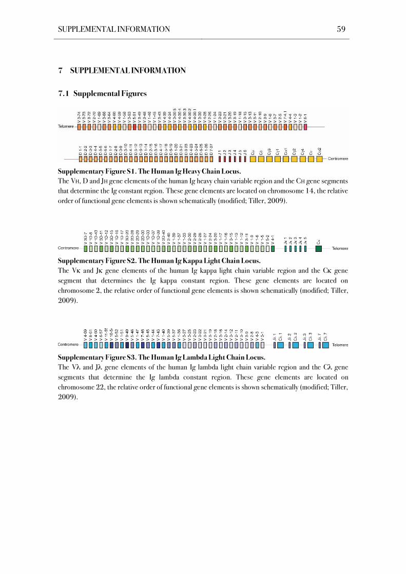
SUPPLEMENTAL INFORMATION 59
7 SUPPLEMENTAL INFORMATION
7.1 Supplemental Figures
Supplementary Figure S1. The Human Ig Heavy Chain Locus.
The VH, D and JH gene elements of the human Ig heavy chain variable region and the CH gene segments
that determine the Ig constant region. These gene elements are located on chromosome 14, the relative
order of functional gene elements is shown schematically (modified; Tiller, 2009).
Supplementary Figure S2. The Human Ig Kappa Light Chain Locus.
The V and J gene elements of the human Ig kappa light chain variable region and the C gene
segment that determines the Ig kappa constant region. These gene elements are located on
chromosome 2, the relative order of functional gene elements is shown schematically (modified; Tiller,
2009).
Supplementary Figure S3. The Human Ig Lambda Light Chain Locus.
The V and J gene elements of the human Ig lambda light chain variable region and the C gene
segments that determine the Ig lambda constant region. These gene elements are located on
chromosome 22, the relative order of functional gene elements is shown schematically (modified; Tiller,
2009).
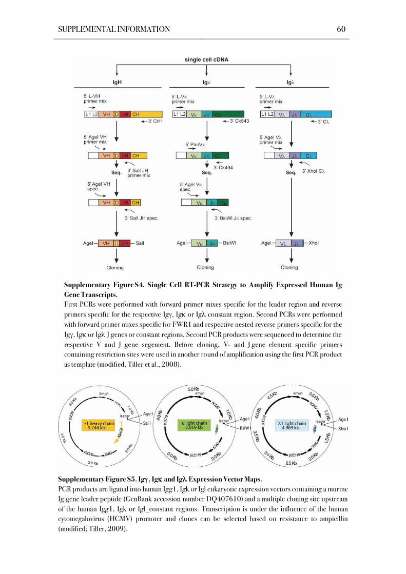
SUPPLEMENTAL INFORMATION 60
Supplementary Figure S4. Single Cell RT-PCR Strategy to Amplify Expressed Human Ig
Gene Transcripts.
First PCRs were performed with forward primer mixes specific for the leader region and reverse
primers specific for the respective Ig , Ig or Ig constant region. Second PCRs were performed
with forward primer mixes specific for FWR1 and respective nested reverse primers specific for the
Ig , Ig or Ig J genes or constant regions. Second PCR products were sequenced to determine the
respective V and J gene segement. Before cloning, V- and J gene element specific primers
containing restriction sites were used in another round of amplification using the first PCR product
as template (modified, Tiller et al., 2008).
Supplementary Figure S5. Ig , Ig and Ig Expression Vector Maps.
PCR products are ligated into human Igg1, Igk or Igl eukaryotic expression vectors containing a murine
Ig gene leader peptide (GenBank accession number DQ407610) and a multiple cloning site upstream
of the human Igg1, Igk or Igl_constant regions. Transcription is under the influence of the human
cytomegalovirus (HCMV) promoter and clones can be selected based on resistance to ampicillin
(modified; Tiller, 2009).
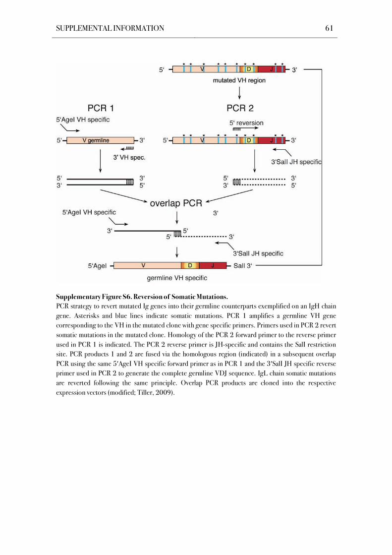
SUPPLEMENTAL INFORMATION 61
Supplementary Figure S6. Reversion of Somatic Mutations.
PCR strategy to revert mutated Ig genes into their germline counterparts exemplified on an IgH chain
gene. Asterisks and blue lines indicate somatic mutations. PCR 1 amplifies a germline VH gene
corresponding to the VH in the mutated clone with gene specific primers. Primers used in PCR 2 revert
somatic mutations in the mutated clone. Homology of the PCR 2 forward primer to the reverse primer
used in PCR 1 is indicated. The PCR 2 reverse primer is JH-specific and contains the SalI restriction
site. PCR products 1 and 2 are fused via the homologous region (indicated) in a subsequent overlap
PCR using the same 5 AgeI VH specific forward primer as in PCR 1 and the 3 SalI JH specific reverse
primer used in PCR 2 to generate the complete germline VDJ sequence. IgL chain somatic mutations
are reverted following the same principle. Overlap PCR products are cloned into the respective
expression vectors (modified; Tiller, 2009).
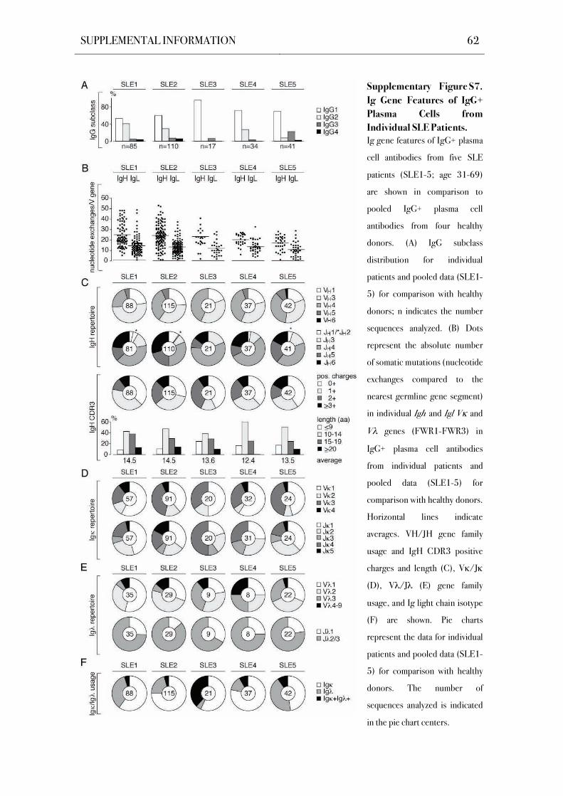
SUPPLEMENTAL INFORMATION 62
Supplementary Figure S7.
Ig Gene Features of IgG+
Plasma Cells from
Individual SLE Patients. Ig gene features of IgG+ plasma
cell antibodies from five SLE
patients (SLE1-5; age 31-69)
are shown in comparison to
pooled IgG+ plasma cell
antibodies from four healthy
donors. (A) IgG subclass
distribution for individual
patients and pooled data (SLE1-
5) for comparison with healthy
donors; n indicates the number
sequences analyzed. (B) Dots
represent the absolute number
of somatic mutations (nucleotide
exchanges compared to the
nearest germline gene segment)
in individual Igh and Igl V and
V genes (FWR1-FWR3) in
IgG+ plasma cell antibodies
from individual patients and
pooled data (SLE1-5) for
comparison with healthy donors.
Horizontal lines indicate
averages. VH/JH gene family
usage and IgH CDR3 positive
charges and length (C), V /J
(D), V /J (E) gene family
usage, and Ig light chain isotype
(F) are shown. Pie charts
represent the data for individual
patients and pooled data (SLE1-
5) for comparison with healthy
donors. The number of
sequences analyzed is indicated
in the pie chart centers.
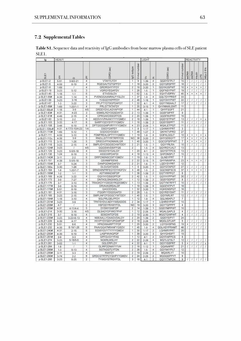
SUPPLEMENTAL INFORMATION 63
7.2 Supplemental Tables
Table S1. Sequence data and reactivity of IgG antibodies from bone marrow plasma cells of SLE patient
SLE1.
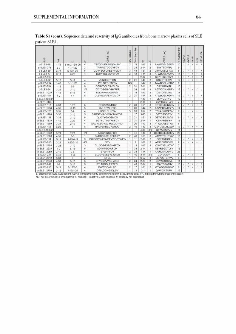
SUPPLEMENTAL INFORMATION 64
Table S1 (cont). Sequence data and reactivity of IgG antibodies from bone marrow plasma cells of SLE
patient SLE1.
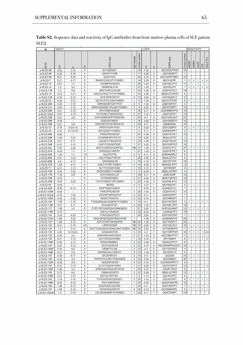
SUPPLEMENTAL INFORMATION 65
Table S2. Sequence data and reactivity of IgG antibodies from bone marrow plasma cells of SLE patient
SLE2.
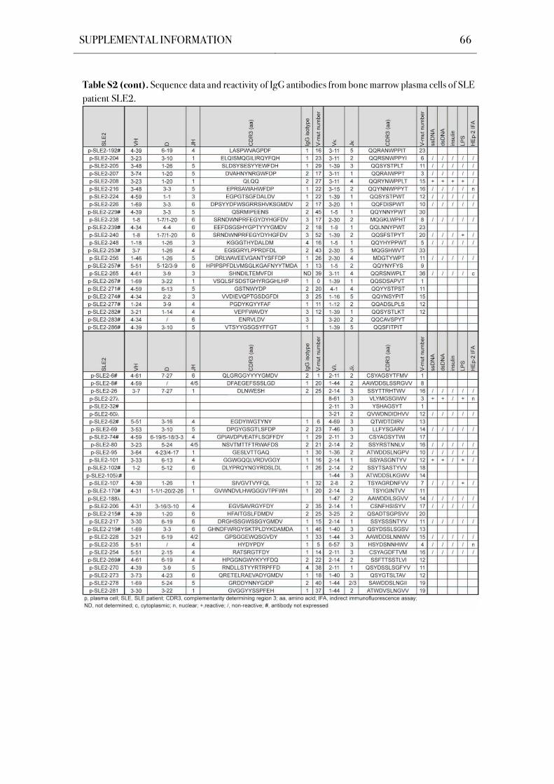
SUPPLEMENTAL INFORMATION 66
Table S2 (cont). Sequence data and reactivity of IgG antibodies from bone marrow plasma cells of SLE
patient SLE2.
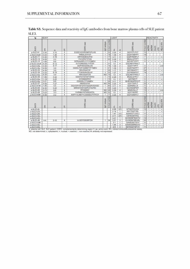
SUPPLEMENTAL INFORMATION 67
Table S3. Sequence data and reactivity of IgG antibodies from bone marrow plasma cells of SLE patient
SLE3.
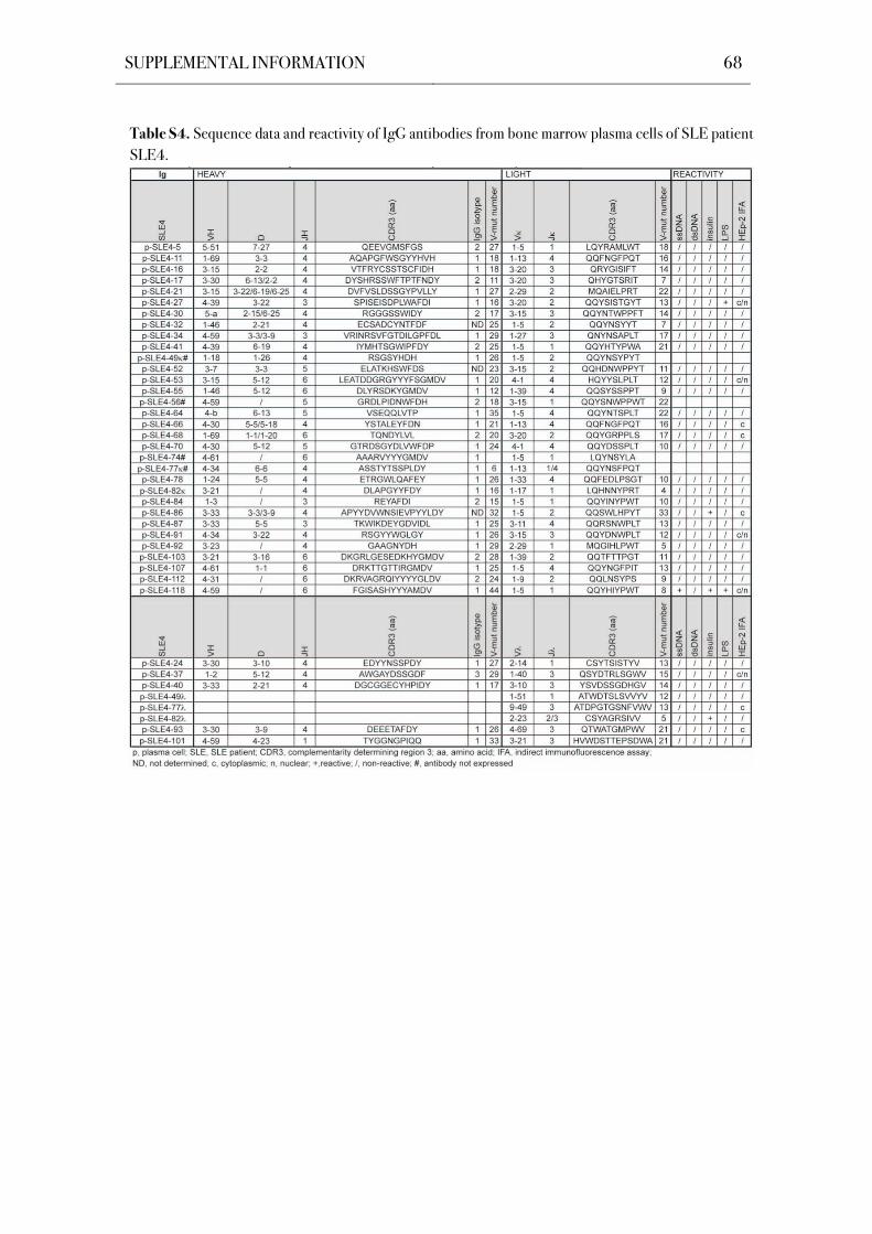
SUPPLEMENTAL INFORMATION 68
Table S4. Sequence data and reactivity of IgG antibodies from bone marrow plasma cells of SLE patient
SLE4.
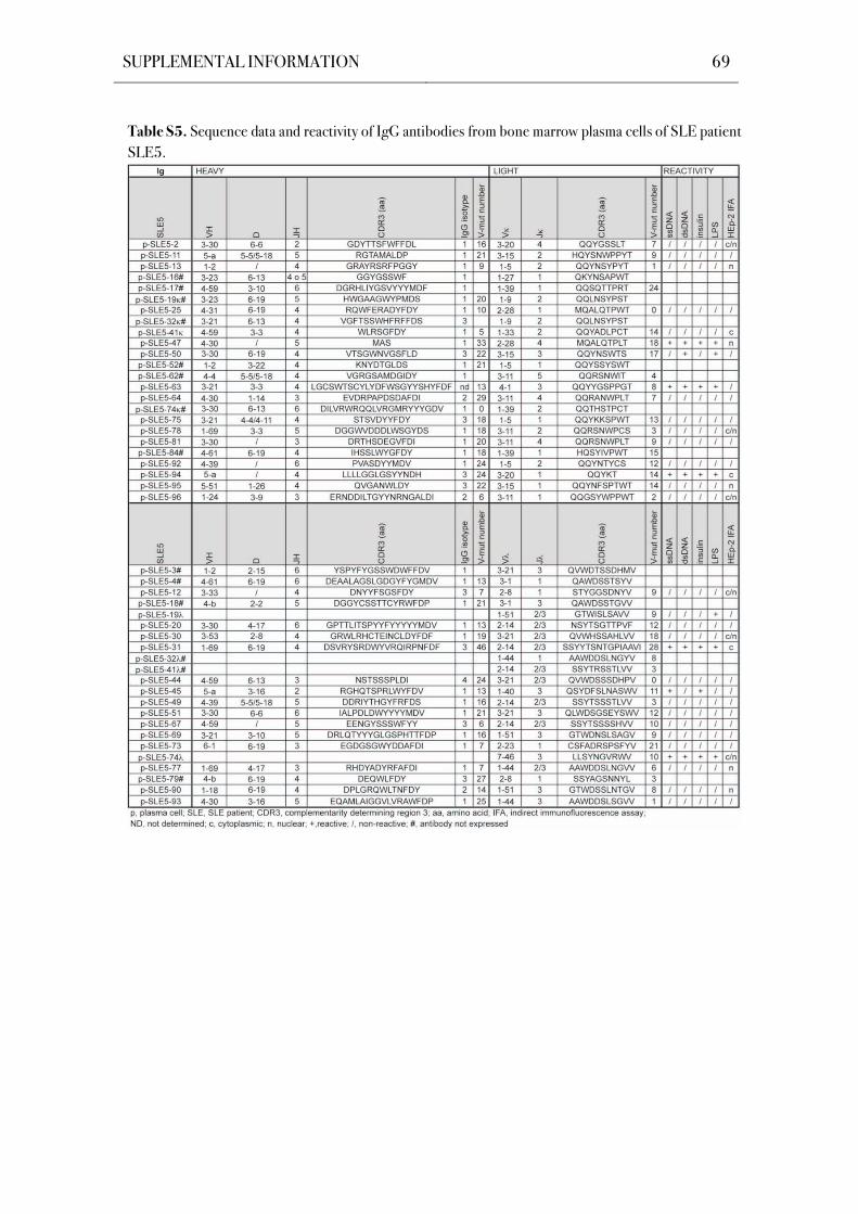
SUPPLEMENTAL INFORMATION 69
Table S5. Sequence data and reactivity of IgG antibodies from bone marrow plasma cells of SLE patient
SLE5.
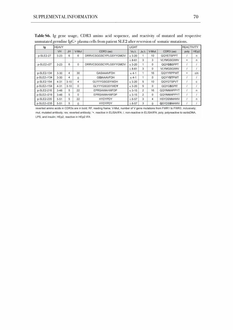
SUPPLEMENTAL INFORMATION 70
Table S6. Ig gene usage, CDR3 amino acid sequence, and reactivity of mutated and respective
unmutated germline IgG+ plasma cells from patient SLE2 after reversion of somatic mutations.
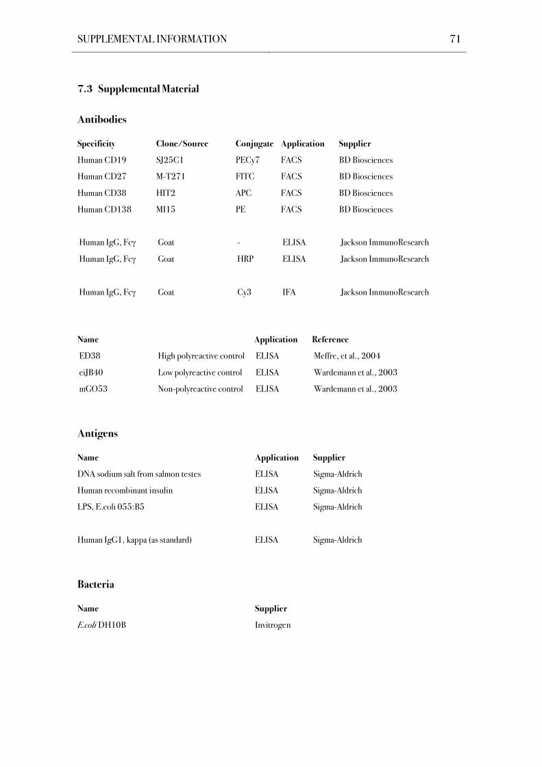
SUPPLEMENTAL INFORMATION 71
7.3 Supplemental Material
Antibodies
Specificity Clone/Source Conjugate Application Supplier
Human CD19 SJ25C1 PECy7 FACS BD Biosciences
Human CD27 M-T271 FITC FACS BD Biosciences
Human CD38 HIT2 APC FACS BD Biosciences
Human CD138 MI15 PE FACS BD Biosciences
Human IgG, Fc Goat - ELISA Jackson ImmunoResearch
Human IgG, Fc Goat HRP ELISA Jackson ImmunoResearch
Human IgG, Fc Goat Cy3 IFA Jackson ImmunoResearch
Name Application Reference
ED38 High polyreactive control ELISA Meffre, et al., 2004
eiJB40 Low polyreactive control ELISA Wardemann et al., 2003
mGO53 Non-polyreactive control ELISA Wardemann et al., 2003
Antigens
Name Application Supplier
DNA sodium salt from salmon testes ELISA Sigma-Aldrich
Human recombinant insulin ELISA Sigma-Aldrich
LPS, E.coli 055:B5 ELISA Sigma-Aldrich
Human IgG1, kappa (as standard) ELISA Sigma-Aldrich
Bacteria
Name Supplier
E.coli DH10B Invitrogen
SUPPLEMENTAL INFORMATION 72
Bacterial Culture Media
Name Compounds
1 % (w/v) Bacto-Tryptone
0.5 % (w/v) yeast extract
85.5 mM NaCl were dissolved in water under stirring,
the pH was adjusted to 7.5 and the medium was autoclaved (15 min at 121 °C),
if required, ampicillin Na salt (Roche) was used at a concentration of 100 μg/ml,
Luria Bertani (LB)
medium
1.5 % (w/v) Agar-agar was added prior to autoclaving (15 min at 121 °C) for LB- plates
47.6 % (w/v) TB powder were dissolved in water under stirring,
4 ml Glycerol was added prior to autoclaving (15 min at 121 °C)
Terrific Broth (TB)
medium
Buffers and Solutions
Name Compounds
50 mM HEPES
10 mM KCl
12 mM Dextrose
280 mM NaCl
1.5 mM Na2HPO4-7H2O
2x HEPES-buffered saline, pH 7.05
60 % (w/v) Sucrose 5x loading dye for DNA gels
1 mM Cresol Red
2 M Tris Base
0.05 % (v/v) Acetic Acid
50x Tris Acetate EDTA (TAE) buffer
0.05 M EDTA (pH 8.0)
29.4 mM Na2HPO4-7H2O
41.2 mM Citric Acid, anhydrous
ABTS buffer, pH 4.2
0.05 % Tween® 20
1 mM EDTA
ELISA blocking buffer
1x PBS
1x Phosphate buffered Saline (PBS) 137 mM NaCl
SUPPLEMENTAL INFORMATION 73
2.7 mM KCl
10 mM Na2HPO4
1.76 mM KH2PO4
the pH was adjusted to 7.4 prior to was autoclaving
1.37 M NaCl
27 mM KCl
100 mM Na2HPO4
17.6 mM KH2PO4
10x Phosphate buffered Saline (PBS)
Cell Line
Name Supplier
HEK293T ATCC® No. CRL-11268™ Invitrogen
Cell Culture Media
Name Supplier
Antibiotic-Antimycotic (100x) Gibco BRL
Dulbecco’s Modified Eagle Medium
(D-MEM) - GlutaMAX™
Gibco BRL
Ultra Low IgG FCS Biochrom
Nutridoma-SP Roche
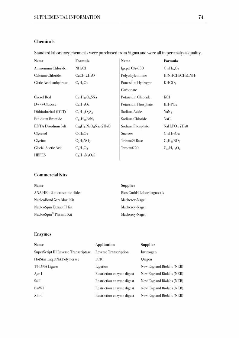
SUPPLEMENTAL INFORMATION 74
Chemicals
Standard laboratory chemicals were purchased from Sigma and were all in per analysis quality.
Name Formula Name Formula
Ammonium Chloride NH4Cl Igepal CA-630 C16H26O2
Calcium Chloride CaCl2·2H2O Polyethylenimine H(NHCH2CH2)nNH2
Citric Acid, anhydrous C6H8O7 Potassium Hydrogen
Carbonate
KHCO3
Cresol Red C21H17O5SNa Potassium Chloride KCl
D-(+)-Glucose C6H12O6 Potassium Phosphate KH2PO4
Dithiothreitol (DTT) C4H10O2S2 Sodium Azide NaN3
Ethidium Bromide C21H20BrN3 Sodium Chloride NaCl
EDTA Disodium Salt C10H14N2O8Na2·2H2O Sodium Phosphate NaH2PO4·7H20
Glycerol C3H8O3 Sucrose C12H22O11
Glycine C2H5NO2 Trizma® Base C4H11NO3
Glacial Acetic Acid C2H4O2 Tween®20 C58H114O2
HEPES C8H18N2O4S
Commercial Kits
Name Supplier
ANA HEp-2 microscopic slides Bios GmbH Labordiagnostik
NucleoBond Xtra Maxi Kit Macherey-Nagel
NucleoSpin Extract II Kit Macherey-Nagel
NucleoSpin® Plasmid Kit Macherey-Nagel
Enzymes
Name Application Supplier
SuperScript III Reverse Transcriptase Reverse Transcription Invitrogen
HotStar Taq DNA Polymerase PCR Qiagen
T4 DNA Ligase Ligation New England Biolabs (NEB)
Age I Restriction enzyme digest New England Biolabs (NEB)
Sal I Restriction enzyme digest New England Biolabs (NEB)
BsiW I Restriction enzyme digest New England Biolabs (NEB)
Xho I Restriction enzyme digest New England Biolabs (NEB)
SUPPLEMENTAL INFORMATION 75
Expression Vectors
Eukaryotic expression vectors with human 1, or constant regions were a kind gift of
Prof. Dr. Michel Nussenzweig, The Rockefeller University, New York, USA.
Human Samples
Bone marrow samples from SLE patients were collected after patients signed informed consent
in accordance with protocols reviewed by the Institutional Review Board of the Charité
University Medical Center.
Nucleotides and Nucleic Acids
Name Supplier
100 bp DNA Ladder New England Biolabs
1 kb DNA Ladder Invitrogen
dATP Invitrogen
dCTP Invitrogen
dGTP Invitrogen
dTTP Invitrogen
Random Hexamer Primer Roche Applied Science
SUPPLEMENTAL INFORMATION 76
Primer Sequences for Amplification of Human Ig Genes
Primer Sequences for Amplification of Human Ig Genes.
1st Heavy Chain PCR
Forward Primer 5’-3’ Sequence
5’ L-VH 1 ACAGGTGCCCACTCCCAGGTGCAG
5’ L-VH 3 AAGGTGTCCAGTGTGARGTGCAG
5’ L-VH 4/6 CCCAGATGGGTCCTGTCCCAGGTGCAG
5’ L-VH 5 CAAGGAGTCTGTTCCGAGGTGCAG
Reverse Primer 5’-3’ Sequence
3’ C CH1 GGAAGGTGTGCACGCCGCTGGTC
2nd Heavy Chain PCR
Forward Primer 5’-3’ Sequence
5’ AgeI VH1 CTGCAACCGGTGTACATTCCCAGGTGCAGCTGGTGCAG
5’ AgeI VH1/5 CTGCAACCGGTGTACATTCCGAGGTGCAGCTGGTGCAG
5’ AgeI VH 1-18 CTGCAACCGGTGTACATTCCCAGGTTCAGCTGGTGCAG
5’ AgeI VH 1-24 CTGCAACCGGTGTACATTCCCAGGTCCAGCTGGTACAG
5’ AgeI VH3 CTGCAACCGGTGTACATTCTGAGGTGCAGCTGGTGGAG
5’ AgeI VH3-23 CTGCAACCGGTGTACATTCTGAGGTGCAGCTGTTGGAG
5’AgeI VH3-33 CTGCAACCGGTGTACATTCTCAGGTGCAGCTGGTGGAG
5’ AgeI VH 3-9 CTGCAACCGGTGTACATTCTGAAGTGCAGCTGGTGGAG
5’ AgeI VH4 CTGCAACCGGTGTACATTCCCAGGTGCAGCTGCAGGAG
5’ AgeI VH 4-34 CTGCAACCGGTGTACATTCCCAGGTGCAGCTACAGCAGTG
5’AgeI VH4-39 CTGCAACCGGTGTACATTCCCAGCTGCAGCTGCAGGAG
5’ AgeI VH 6-1 CTGCAACCGGTGTACATTCCCAGGTACAGCTGCAGCAG
Reverse Primer 5’-3’ Sequence
3’ IgG (internal) GTTCGGGGAAGTAGTCCTTGAC
3’ SalI JH 1/2/4/5 TGCGAAGTCGACGCTGAGGAGACGGTGACCAG
3’ SalI JH 3 TGCGAAGTCGACGCTGAAGAGACGGTGACCATTG
3’ SalI JH 6 TGCGAAGTCGACGCTGAGGAGACGGTGACCGTG
1st Light Chain PCR
Forward Primer 5’-3’ Sequence
5’ L V 1/2 ATGAGGSTCCCYGCTCAGCTGCTGG
5’ L V 3 CTCTTCCTCCTGCTACTCTGGCTCCCAG
5’ L V 4 ATTTCTCTGTTGCTCTGGATCTCTG
Reverse Primer 5’-3’ Sequence
SUPPLEMENTAL INFORMATION 77
3’ C 543 GTTTCTCGTAGTCTGCTTTGCTCA
2nd Light Chain PCR
Forward Primer 5’-3’ Sequence
5’ Pan V ATGACCCAGWCTCCABYCWCCCTG
Reverse Primer 5’-3’ Sequence
3’ C 494 GTGCTGTCCTTGCTGTCCTGCT
Gene-specific Light Chain PCR
Forward Primer 5’-3’ Sequence
5’ AgeI V 1-5 CTGCAACCGGTGTACATTCTGACATCCAGATGACCCAGTC
5’ AgeI V 1-9 TTGTGCTGCAACCGGTGTACATTCAGACATCCAGTTGACCCAGTCT
5’ AgeI V 1D-43 CTGCAACCGGTGTACATTGTGCCATCCGGATGACCCAGTC
5’ AgeI V 2-24 CTGCAACCGGTGTACATGGGGATATTGTGATGACCCAGAC
5’ AgeI V 2-28 CTGCAACCGGTGTACATGGGGATATTGTGATGACTCAGTC
5’ AgeI V 2-30 CTGCAACCGGTGTACATGGGGATGTTGTGATGACTCAGTC
5’ Age V 3-11 TTGTGCTGCAACCGGTGTACATTCAGAAATTGTGTTGACACAGTC
5’ Age V 3-15 CTGCAACCGGTGTACATTCAGAAATAGTGATGACGCAGTC
5’ Age V 3-20 TTGTGCTGCAACCGGTGTACATTCAGAAATTGTGTTGACGCAGTCT
5’ Age V 4-1 CTGCAACCGGTGTACATTCGGACATCGTGATGACCCAGTC
Reverse Primer 5’-3’ Sequence
3’ BsiWI J 1/4 GCCACCGTACGTTTGATYTCCACCTTGGTC
3’ BsiWI J 2 GCCACCGTACGTTTGATCTCCAGCTTGGTC
3’ BsiWI J 3 GCCACCGTACGTTTGATATCCACTTTGGTC
3’ BsiWI J 5 GCCACCGTACGTTTAATCTCCAGTCGTGTC
1st Light Chain PCR
Forward Primer 5’-3’ Sequence
5’ L V 1 GGTCCTGGGCCCAGTCTGTGCTG
5’ L V 2 GGTCCTGGGCCCAGTCTGCCCTG
5’ L V 3 GCTCTGTGACCTCCTATGAGCTG
5’ L V 4/5 GGTCTCTCTCSCAGCYTGTGCTG
5’ L V 6 GTTCTTGGGCCAATTTTATGCTG
5’ L V 7 GGTCCAATTCYCAGGCTGTGGTG
5’ L V 8 GAGTGGATTCTCAGACTGTGGTG
Reverse Primer 5’-3’ Sequence
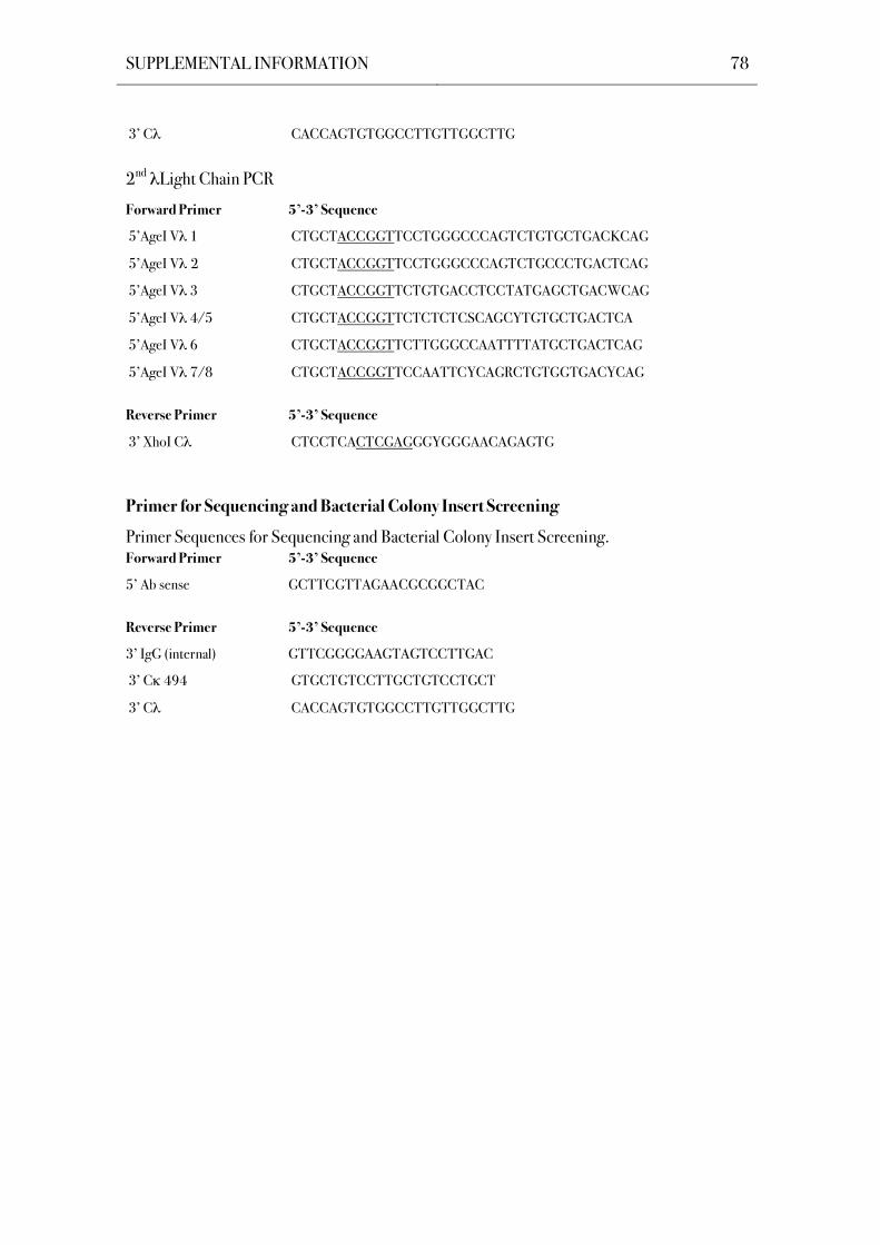
SUPPLEMENTAL INFORMATION 78
3’ C CACCAGTGTGGCCTTGTTGGCTTG
2nd Light Chain PCR
Forward Primer 5’-3’ Sequence
5’AgeI V 1 CTGCTACCGGTTCCTGGGCCCAGTCTGTGCTGACKCAG
5’AgeI V 2 CTGCTACCGGTTCCTGGGCCCAGTCTGCCCTGACTCAG
5’AgeI V 3 CTGCTACCGGTTCTGTGACCTCCTATGAGCTGACWCAG
5’AgeI V 4/5 CTGCTACCGGTTCTCTCTCSCAGCYTGTGCTGACTCA
5’AgeI V 6 CTGCTACCGGTTCTTGGGCCAATTTTATGCTGACTCAG
5’AgeI V 7/8 CTGCTACCGGTTCCAATTCYCAGRCTGTGGTGACYCAG
Reverse Primer 5’-3’ Sequence
3’ XhoI C CTCCTCACTCGAGGGYGGGAACAGAGTG
Primer for Sequencing and Bacterial Colony Insert Screening
Primer Sequences for Sequencing and Bacterial Colony Insert Screening. Forward Primer 5’-3’ Sequence
5’ Ab sense GCTTCGTTAGAACGCGGCTAC
Reverse Primer 5’-3’ Sequence
3’ IgG (internal) GTTCGGGGAAGTAGTCCTTGAC
3’ C 494 GTGCTGTCCTTGCTGTCCTGCT
3’ C CACCAGTGTGGCCTTGTTGGCTTG
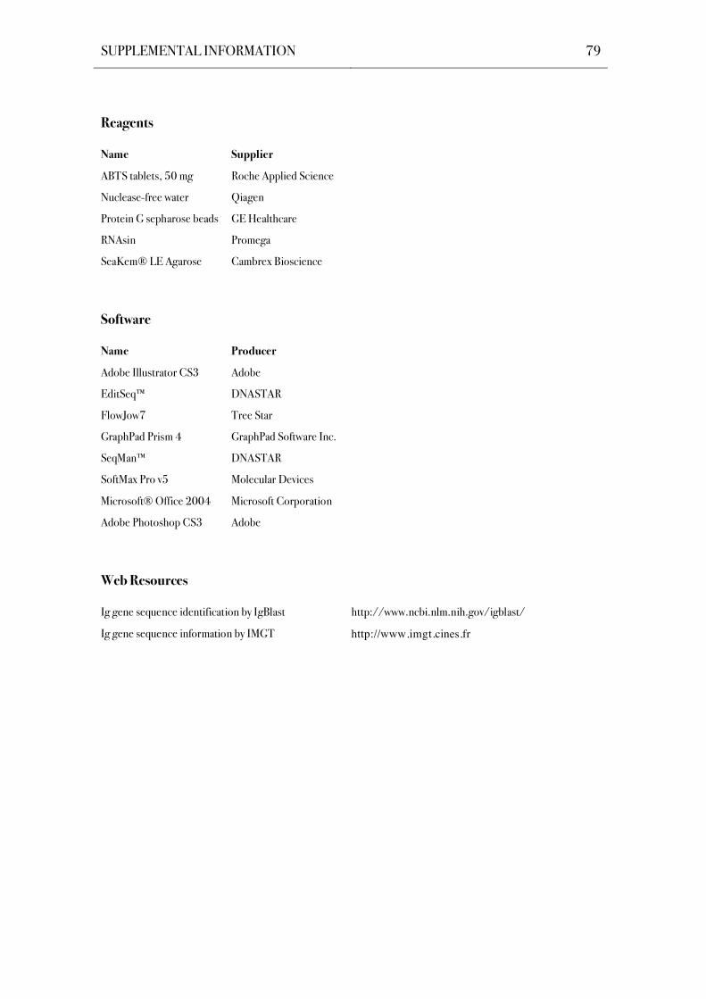
SUPPLEMENTAL INFORMATION 79
Reagents
Name Supplier
ABTS tablets, 50 mg Roche Applied Science
Nuclease-free water Qiagen
Protein G sepharose beads GE Healthcare
RNAsin Promega
SeaKem® LE Agarose Cambrex Bioscience
Software
Name Producer
Adobe Illustrator CS3 Adobe
EditSeq™ DNASTAR
FlowJow7 Tree Star
GraphPad Prism 4 GraphPad Software Inc.
SeqMan™ DNASTAR
SoftMax Pro v5 Molecular Devices
Microsoft® Office 2004 Microsoft Corporation
Adobe Photoshop CS3 Adobe
Web Resources
Ig gene sequence identification by IgBlast http://www.ncbi.nlm.nih.gov/igblast/
Ig gene sequence information by IMGT http://www.imgt.cines.fr

SUPPLEMENTAL INFORMATION 80
ACKNOWLEDGMENTS 81
ACKNOWLEDGMENTS
This PhD thesis would not have been possible without the generous intellectual and
emotional support provided by innumerous colleagues, friends and family.
First and foremost, I want to express my deep appreciation to Dr. Hedda Wardemann
for her excellent guidance and support, for helpful and inspiring discussions, and her never
ending patience. Thank you for having me around!
Moreover, I wish to thank Prof. Dr. Roland Lauster for supervising and reviewing my
PhD thesis on behalf of the TU Berlin.
I would like to thank all present and past members of our lab crew at the MPI for
Infection Biology for a wonderful time and a great working atmosphere. In particular, I want to
thank Dr. Thomas Tiller for introducing me into this exciting field of research and his special
sense of humor. Further, I want to express my special gratitude to my lab family Conny,
Friederike, Julia, Lydie and Markus.
I am also indebted to Prof. Dr. Falk Hiepe, Dr. Bimba Hoyer and Prof. Dr. Annett
Jacobi for providing us with genuine gold- bone marrow aspirates from SLE patients.
I would like to thank Prof. Dr. Michel Nussenzweig and Johannes Scheid for letting us
take care of the healthy plasma cell data and also John Pietzsch for helping us whenever needed.
I want to acknowledge Dr. Marc Ehlers and his group at the DRFZ for the incredibly
friendly collaboration, for fruitful discussions and technical advice.
Furthermore, I want to thank the laboratory of Prof. Dr. Arturo Zychlinsky, especially
Viola, Yvonne and Steven, for being our first aid guardians at any time.
Most importantly, none of this would have been possible without the constant support,
encouragement and understanding of my family and friends. I cannot adequately express my
gratitude to them. Thank you for everything.