Embed Size (px)
Citation preview
Stellenwert der Biopsie
Carsten Tschöpe
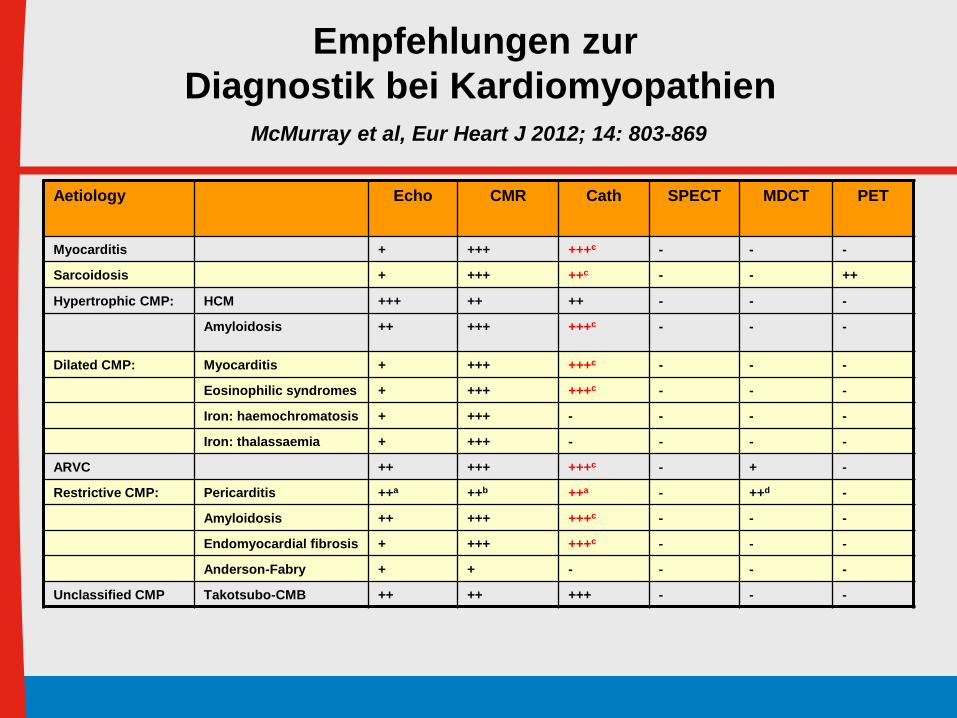
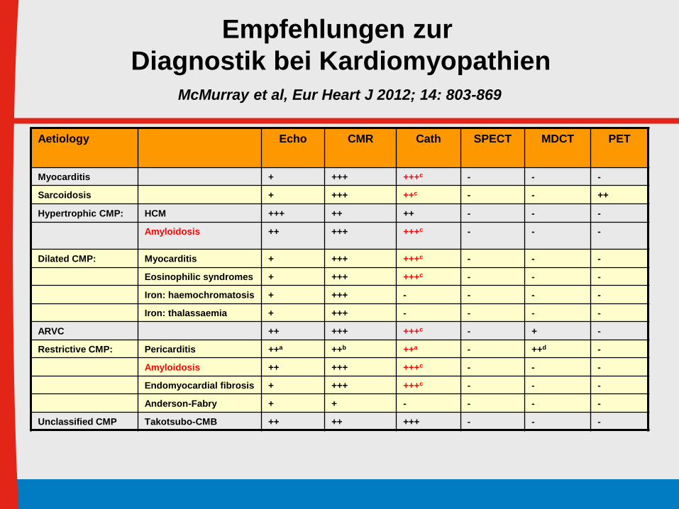
Aetiology Echo CMR Cath SPECT MDCT PET
Myocarditis + +++ +++c - - -
Sarcoidosis + +++ ++c - - ++
Hypertrophic CMP: HCM +++ ++ ++ - - -
Amyloidosis ++ +++ +++c - - -
Dilated CMP: Myocarditis + +++ +++c - - -
Eosinophilic syndromes + +++ +++c - - -
Iron: haemochromatosis + +++ - - - -
Iron: thalassaemia + +++ - - - -
ARVC ++ +++ +++c - + -
Restrictive CMP: Pericarditis ++a ++b ++a - ++d -
Amyloidosis ++ +++ +++c - - -
Endomyocardial fibrosis + +++ +++c - - -
Anderson-Fabry + + - - - -
Unclassified CMP Takotsubo-CMB ++ ++ +++ - - -
Empfehlungen zur
Diagnostik bei KardiomyopathienMcMurray et al, Eur Heart J 2012; 14: 803-869
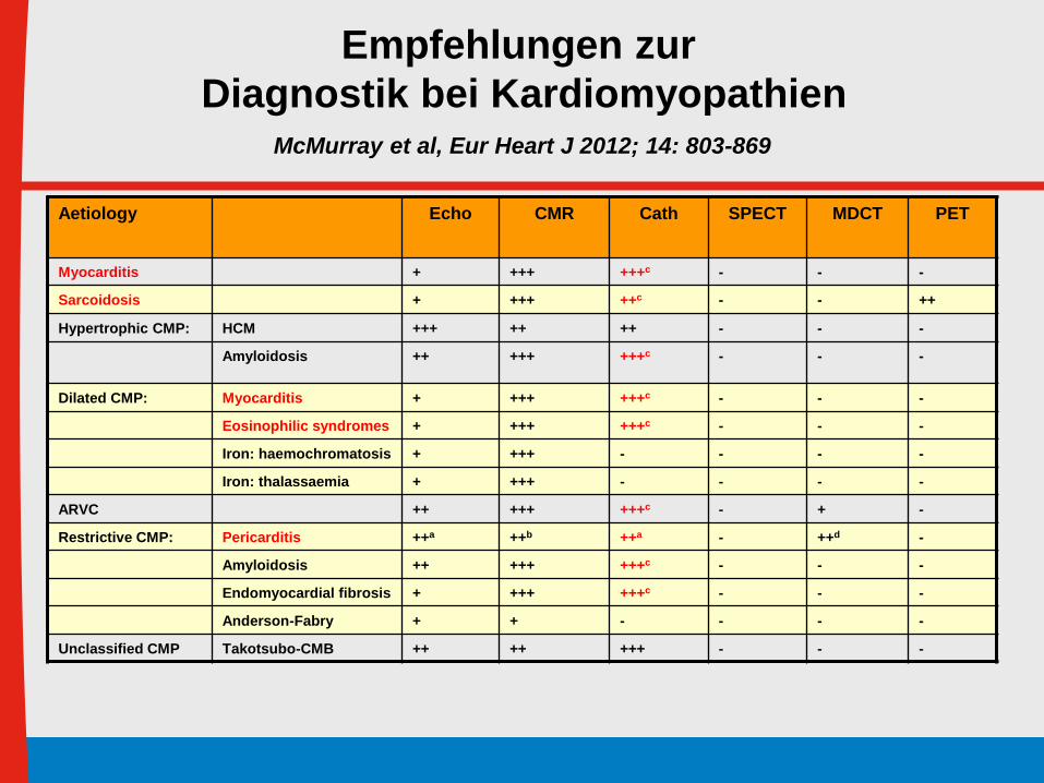
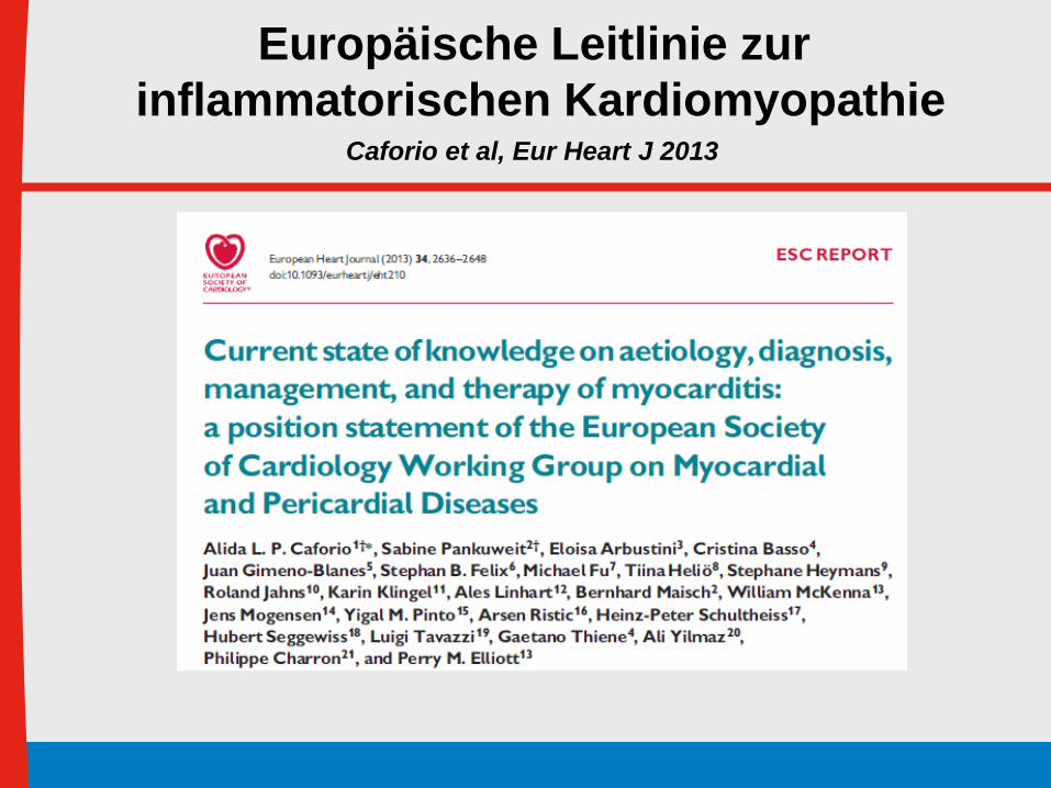
Europäische Leitlinie zur
inflammatorischen KardiomyopathieCaforio et al, Eur Heart J 2013
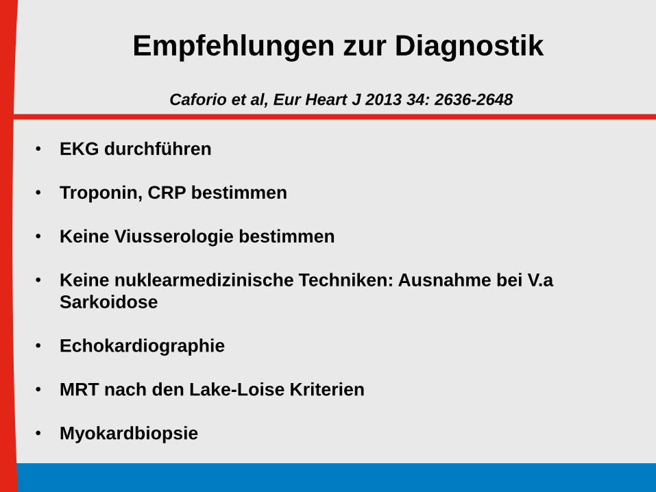
Empfehlungen zur Diagnostik
• EKG durchführen
• Troponin, CRP bestimmen
• Keine Viusserologie bestimmen
• Keine nuklearmedizinische Techniken: Ausnahme bei V.a
Sarkoidose
• Echokardiographie
• MRT nach den Lake-Loise Kriterien
• Myokardbiopsie
Caforio et al, Eur Heart J 2013 34: 2636-2648
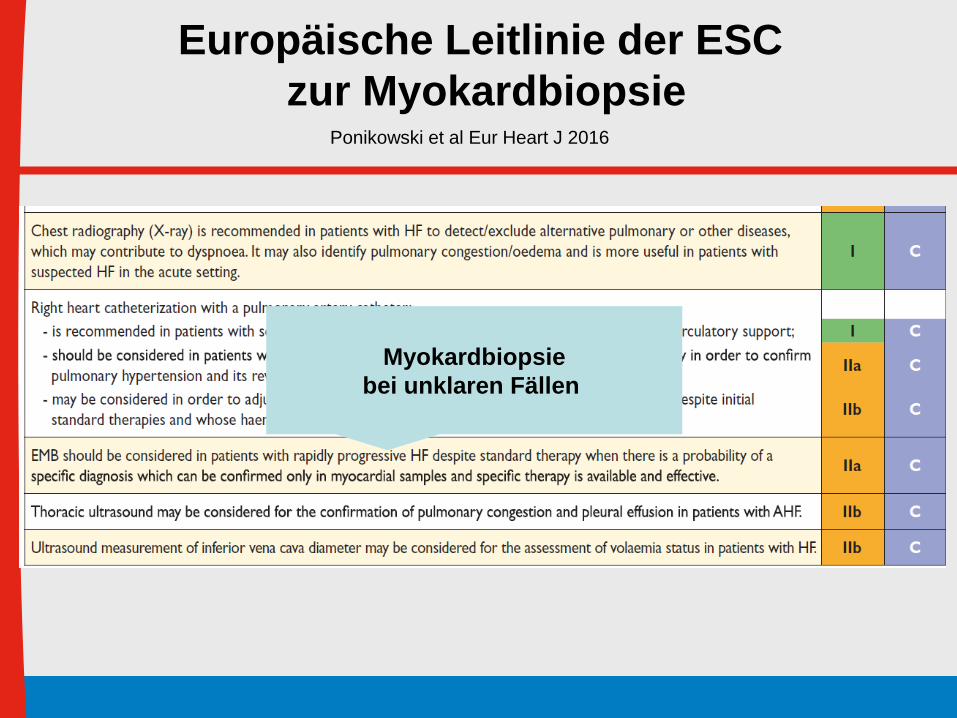
Europäische Leitlinie der ESC
zur MyokardbiopsiePonikowski et al Eur Heart J 2016
Myokardbiopsie
bei unklaren Fällen
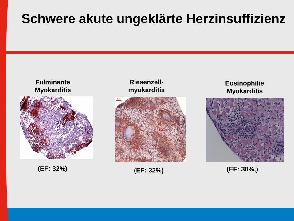
Eosinophilie
Myokarditis
(EF: 30%,)
Fulminante
Myokarditis
(EF: 32%) (EF: 32%)
Riesenzell-
myokarditis
Schwere akute ungeklärte Herzinsuffizienz
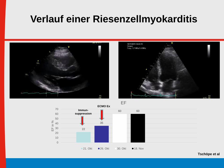
Verlauf einer Riesenzellmyokarditis
22
35
60 60
0
10
20
30
40
50
60
70
EF
in %
EF
21. Okt 26. Okt 30. Okt 18. Nov
Immun-
suppression
ECMO Ex
ECMO Ex
Tschöpe et al
Circulation December 2016

1. Schock
2. Herzinsuffizinez, die nicht adäquat auf die
Standardtherpaie reagiert
3. AV- Block Mobitz oder AV Block III
4. Symptomatische Tachykardien
(Level of Evidence C)
Indikation zur Biopsie
Circulation December 2016
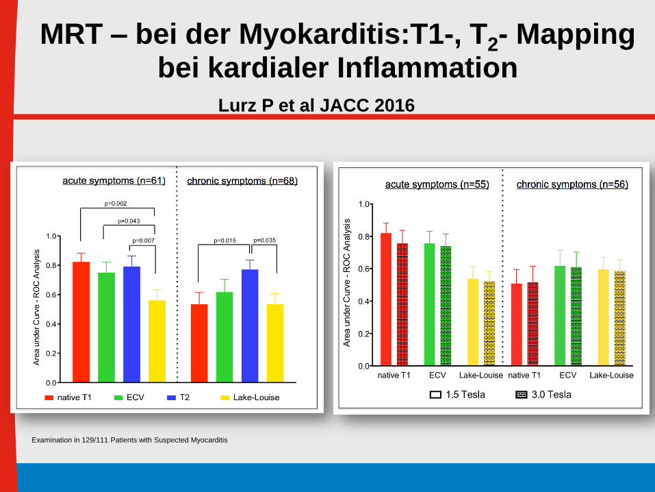
MRT – bei der Myokarditis:T1-, T2- Mapping bei kardialer Inflammation
Examination in 129/111 Patients with Suspected Myocarditis
Lurz P et al JACC 2016
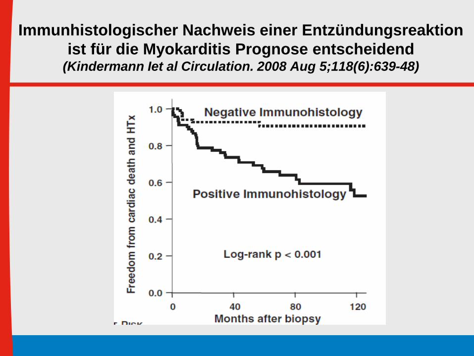
Immunhistologischer Nachweis einer Entzündungsreaktion
ist für die Myokarditis Prognose entscheidend(Kindermann Iet al Circulation. 2008 Aug 5;118(6):639-48)

Vorraussetzung:
Adäquate Diagnostik
Coxsackie
Caforio et al, Eur Heart J 2013
Histologie Immuno-
Histologie
Molecular-
biologie
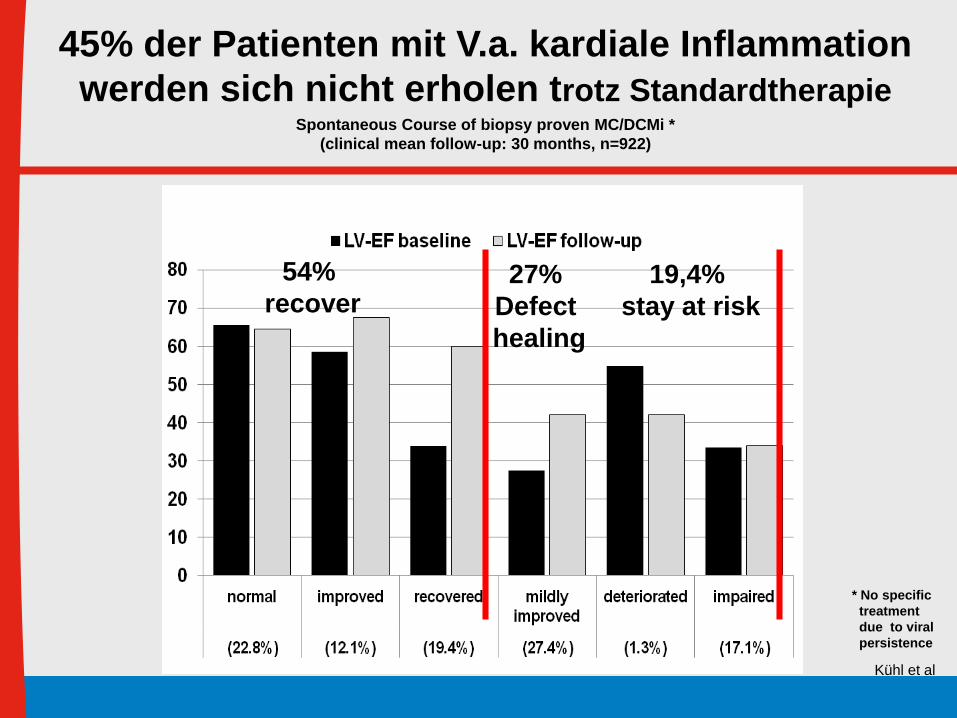
54%
recover
* No specific
treatment
due to viral
persistence
19,4%
stay at risk
27%
Defect
healing
Kühl et al
Spontaneous Course of biopsy proven MC/DCMi *
(clinical mean follow-up: 30 months, n=922)
45% der Patienten mit V.a. kardiale Inflammation
werden sich nicht erholen trotz Standardtherapie
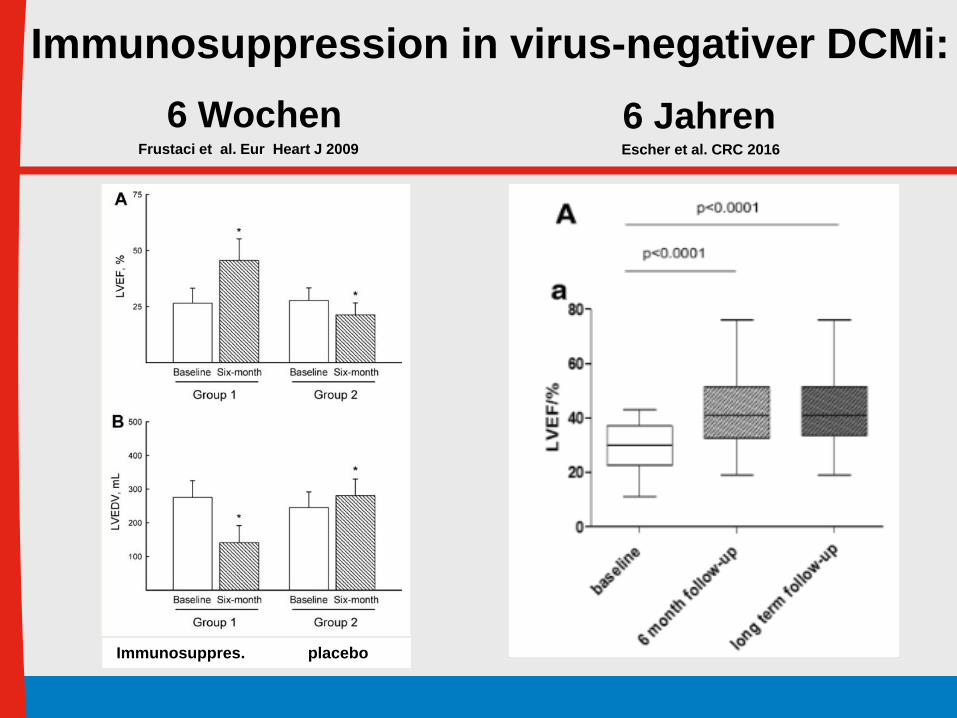
Immunosuppression in virus-negativer DCMi:
Immunosuppres. placebo
Frustaci et al. Eur Heart J 2009
6 WochenEscher et al. CRC 2016
6 Jahren

Caforio et al, Eur Heart J 2013
Immunsuppression
nach Ausschluss einer Entero/Adenovirus
Persistenz
ESC Empfehlungen

1. Schock
2. AV- Block Mobitz oder AV Block III
3. Symptomatische Tachykardien
4. Herzinsuffizinez, die nicht adäquat auf die
Standardtherpaie reagiert
(Level of Evidence C)
Indikation zur Biopsie
Circulation December 2016

Moykarditis
Zusammenfassung:
- Meist gute Prognose unter Schonung und
Standard-HF Therapie
- Biopsie: Schock (IIa)
- ca. 2- Wochen - 3 Monaten ohne Besserung (IIb)
- Biopsie-gesteuerte Immunsuppressive Therapie
- Kontroll-Biopsie bei Therapieversager

1. Schock
2. Herzinsuffizinez, die nicht adäquat auf die
Standardtherpaie reagiert
3. AV- Block Mobitz oder AV Block III
4. Symptomatische Tachykardien
(Level of Evidence C)
Indikation zur Biopsie
Circulation December 2016
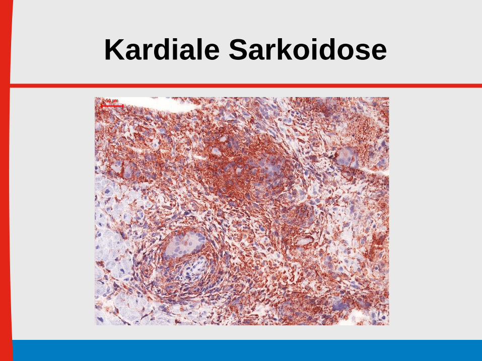
Kardiale Sarkoidose

Costabel et al, Pneumologie 2014; 68: 124-132
Klinische Manifestationen bei kardialer Sarkoidose und
ihre Prävalenz
- 10% der Sarkoidose Patienten erleiden eine kardiale Beteiligung
- Meist ist dabei der linke Ventrikel und das Reizleitungssystem von
der Granulombildung betroffen
Costabel et al, Pneumologie 2014; 68: 124-132
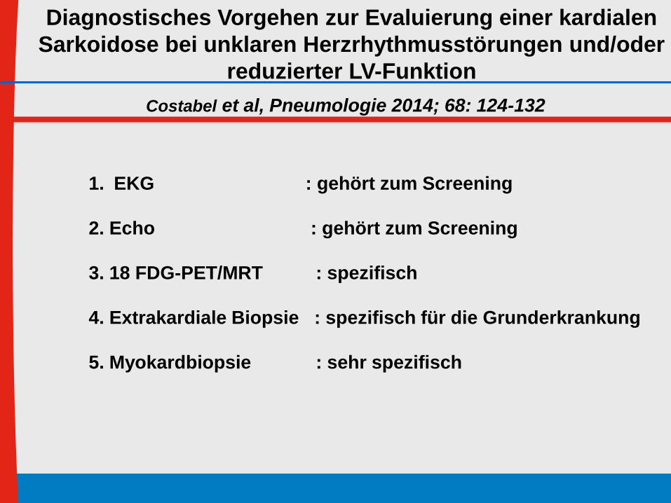
Diagnostisches Vorgehen zur Evaluierung einer kardialen
Sarkoidose bei unklaren Herzrhythmusstörungen und/oder
reduzierter LV-Funktion
1. EKG : gehört zum Screening
2. Echo : gehört zum Screening
3. 18 FDG-PET/MRT : spezifisch
4. Extrakardiale Biopsie : spezifisch für die Grunderkrankung
5. Myokardbiopsie : sehr spezifisch

CT -Thorax
Bihiläre Lymphadenopathie und/oder andere Sarkoidose-typische Befunde
Keine indikative
Histologie
Endomyokardiale Biopsie
Kardiale Sarkoidose
wahrscheinlich
Kardiale Sarkoidose
möglich
Kardiale Sarkoidose
unwahrscheinlich
Extrakardiale Biopsien Kardio-MRT/FDG-PET
Indikative
Befunde
Unauffällige
Befunde
Sarkoidose-
typische Granulome
ja nein
Costabel et al, Pneumologie 2014; 68: 124-132
Diagnostisches Vorgehen zur Evaluierung einer kardialen
Sarkoidose bei unklaren Herzrhythmusstörungen und/oder
reduzierter LV-Funktion
Kardiale Sarkoidose
gesichert
CT -Thorax
Bihiläre Lymphadenopathie und/oder andere Sarkoidose-typische Befunde
Keine indikative
Histologie
Endomyokardiale Biopsie
Kardiale Sarkoidose
wahrscheinlich
Kardiale Sarkoidose
möglich
Kardiale Sarkoidose
unwahrscheinlich
Extrakardiale Biopsien Kardio-MRT/FDG-PET
Indikative
Befunde
Unauffällige
Befunde
Sarkoidose-
typische Granulome
ja nein
Costabel et al, Pneumologie 2014; 68: 124-132
Diagnostisches Vorgehen zur Evaluierung einer kardialen
Sarkoidose bei unklaren Herzrhythmusstörungen und/oder
reduzierter LV-Funktion
Problem:
Sampling Error
Diff-Diagnose

Myokarditiden
Borreliose
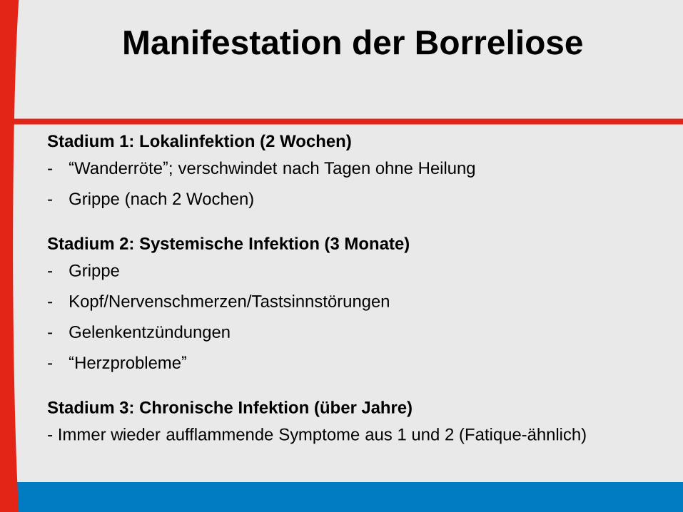
Stadium 1: Lokalinfektion (2 Wochen)
- “Wanderröte”; verschwindet nach Tagen ohne Heilung
- Grippe (nach 2 Wochen)
Stadium 2: Systemische Infektion (3 Monate)
- Grippe
- Kopf/Nervenschmerzen/Tastsinnstörungen
- Gelenkentzündungen
- “Herzprobleme”
Stadium 3: Chronische Infektion (über Jahre)
- Immer wieder aufflammende Symptome aus 1 und 2 (Fatique-ähnlich)
Manifestation der Borreliose
www.borreliose-nachrichten.de.
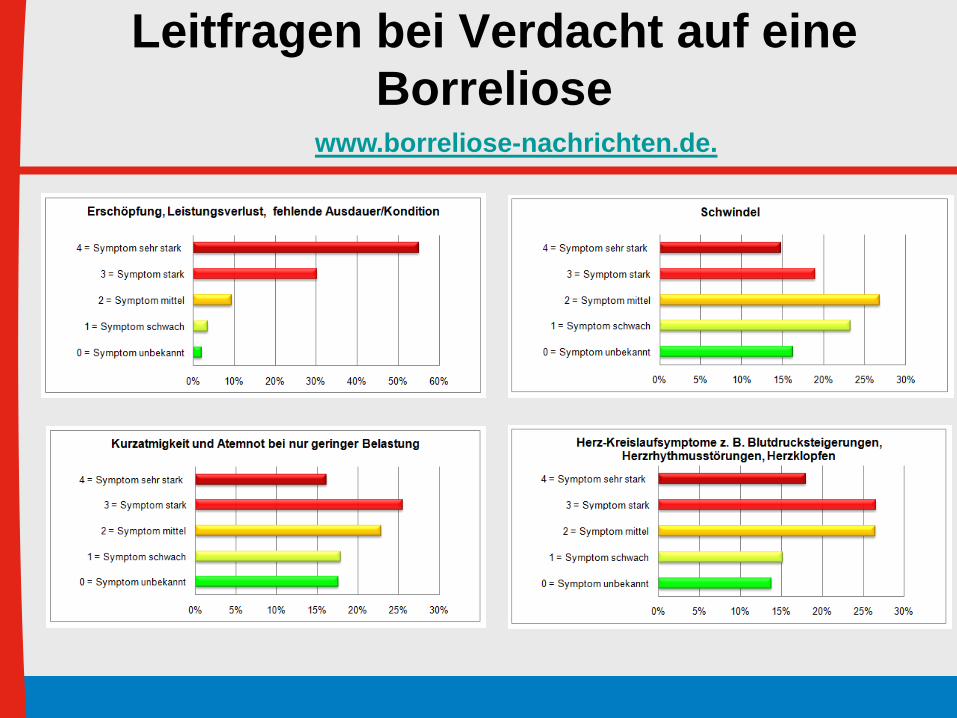
Leitfragen bei Verdacht auf eine
Borreliose
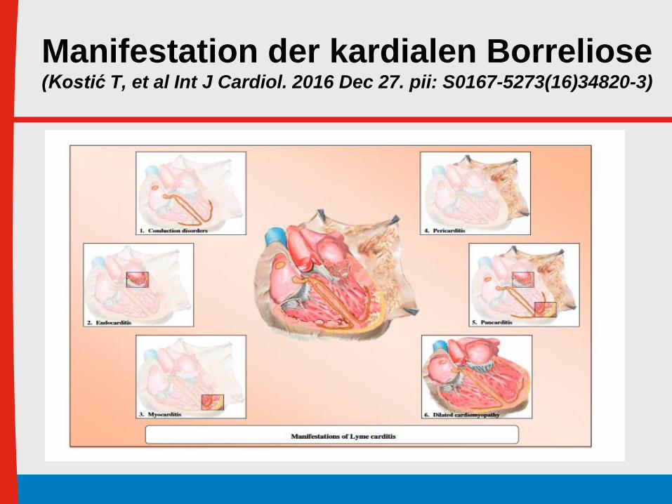
Manifestation der kardialen Borreliose (Kostić T, et al Int J Cardiol. 2016 Dec 27. pii: S0167-5273(16)34820-3)

www.borreliose-nachrichten.de.
Leitfragen bei Verdacht auf eine
Borreliose
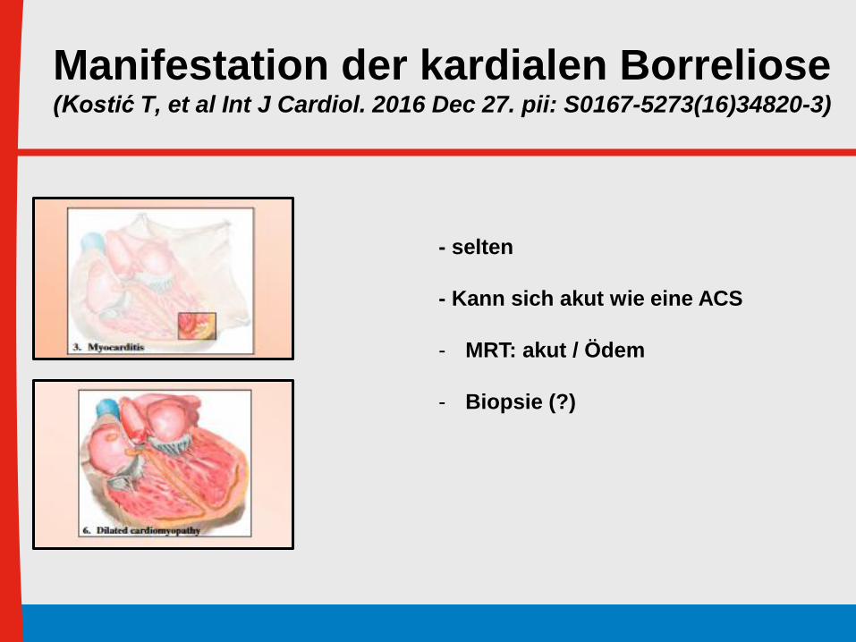
Manifestation der kardialen Borreliose (Kostić T, et al Int J Cardiol. 2016 Dec 27. pii: S0167-5273(16)34820-3)
- selten
- Kann sich akut wie eine ACS
- MRT: akut / Ödem
- Biopsie (?)
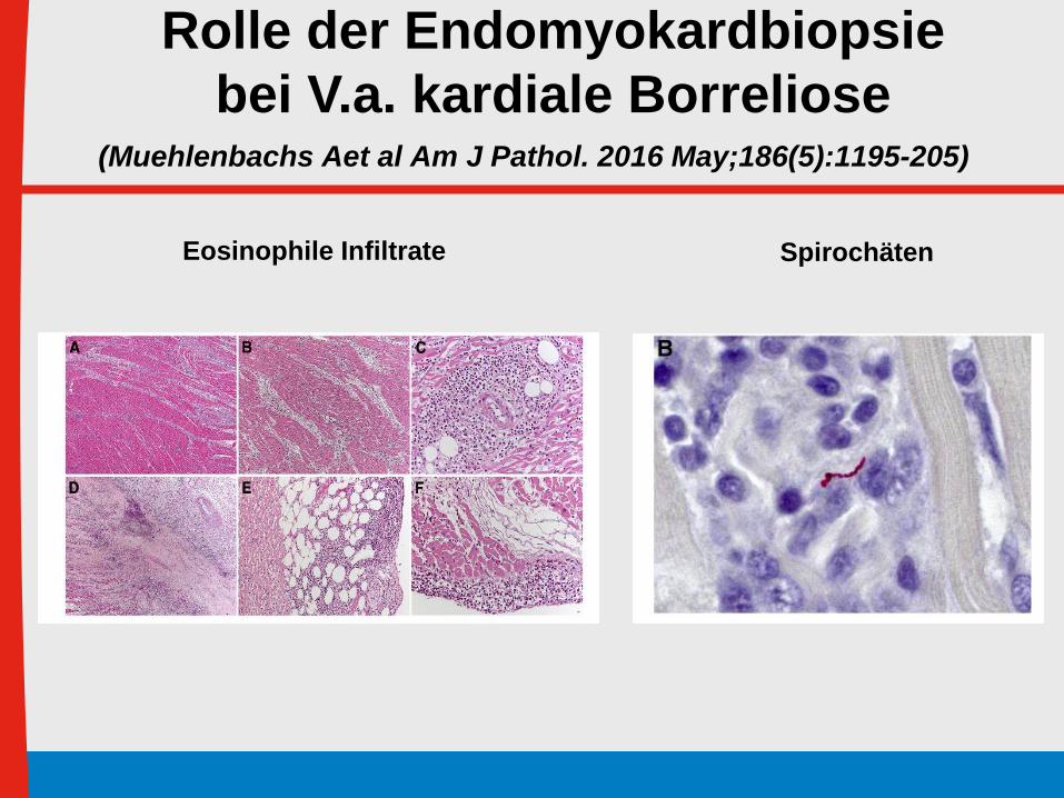
(Muehlenbachs Aet al Am J Pathol. 2016 May;186(5):1195-205)
Rolle der Endomyokardbiopsie
bei V.a. kardiale Borreliose
Eosinophile Infiltrate Spirochäten
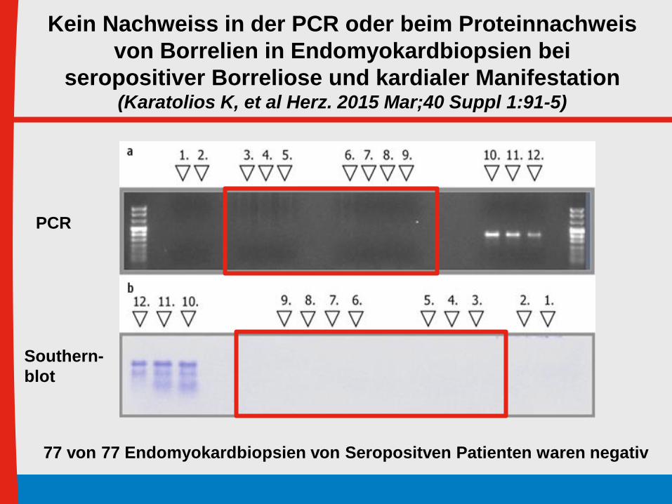
Kein Nachweiss in der PCR oder beim Proteinnachweis
von Borrelien in Endomyokardbiopsien bei
seropositiver Borreliose und kardialer Manifestation(Karatolios K, et al Herz. 2015 Mar;40 Suppl 1:91-5)
PCR
Southern-
blot
77 von 77 Endomyokardbiopsien von Seropositven Patienten waren negativ
Borreliose
Diagnostik
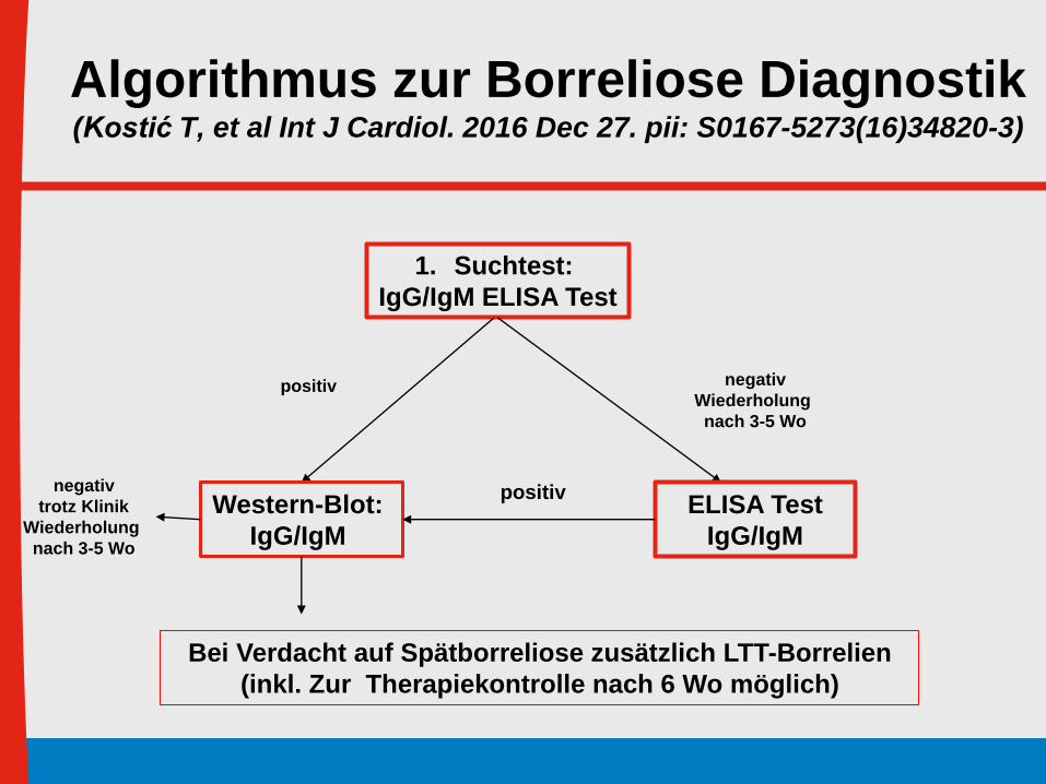
Algorithmus zur Borreliose Diagnostik (Kostić T, et al Int J Cardiol. 2016 Dec 27. pii: S0167-5273(16)34820-3)
1. Suchtest:
IgG/IgM ELISA Test
ELISA Test
IgG/IgM
Western-Blot:
IgG/IgM
negativ
Wiederholung
nach 3-5 Wo
Bei Verdacht auf Spätborreliose zusätzlich LTT-Borrelien
(inkl. Zur Therapiekontrolle nach 6 Wo möglich)
positiv
positivnegativ
trotz Klinik
Wiederholung
nach 3-5 Wo
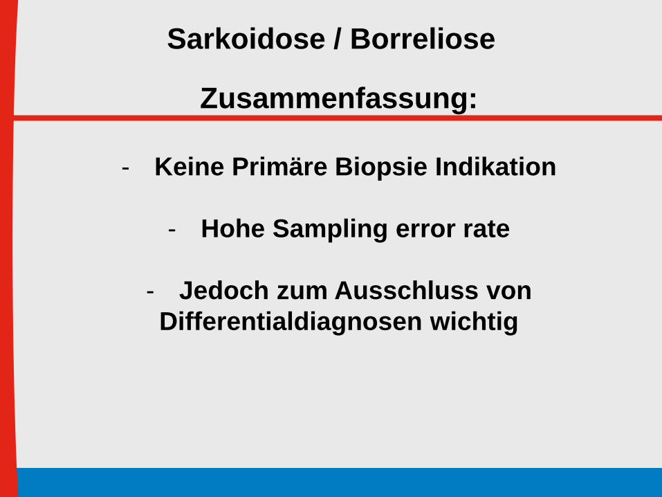
Sarkoidose / Borreliose
Zusammenfassung:
- Keine Primäre Biopsie Indikation
- Hohe Sampling error rate
- Jedoch zum Ausschluss von
Differentialdiagnosen wichtig
Aetiology Echo CMR Cath SPECT MDCT PET
Myocarditis + +++ +++c - - -
Sarcoidosis + +++ ++c - - ++
Hypertrophic CMP: HCM +++ ++ ++ - - -
Amyloidosis ++ +++ +++c - - -
Dilated CMP: Myocarditis + +++ +++c - - -
Eosinophilic syndromes + +++ +++c - - -
Iron: haemochromatosis + +++ - - - -
Iron: thalassaemia + +++ - - - -
ARVC ++ +++ +++c - + -
Restrictive CMP: Pericarditis ++a ++b ++a - ++d -
Amyloidosis ++ +++ +++c - - -
Endomyocardial fibrosis + +++ +++c - - -
Anderson-Fabry + + - - - -
Unclassified CMP Takotsubo-CMB ++ ++ +++ - - -
Empfehlungen zur
Diagnostik bei KardiomyopathienMcMurray et al, Eur Heart J 2012; 14: 803-869
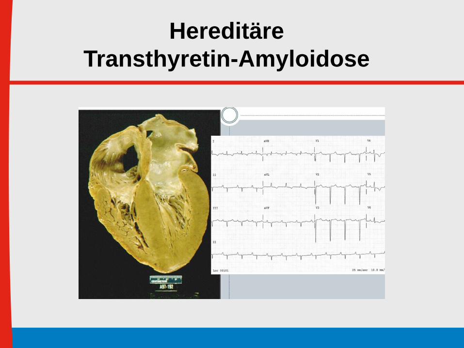
Hereditäre
Transthyretin-Amyloidose

TYPE OF
AMYLOID
PRECURSOR
PROTEIN
MAJOR ORGANS
INVOLVED
COMMENT
AL Immunoglobulin light chain
Heart, kidney, liver, nerves, skin, bowel
Rapidly Progressive. Related to multiple myeloma
TTRm Variant transthyretin Nerve Heart Autosomal dominant with variable penetrance. High gene
prevalence in African Americans
TTRwt Wild-type TTR Heart Slowly progressive. Rapidly increasing prevalence.
Disease of men.
Isolated atrial
amyloidosis
ANP Atrium Little clinicalsignificance
Klassifizierung der Amyloidosen1. Ando Y et al., Arch Neurol 2005;62:1057–1062. 2. Merlini G et al., J Intern Med 2004;255:159–178.
3. Falk R et al., N Engl J Med 1997;337:898–909. 4. Sekijima Y et al. Curr Pharm Des 2008;14:3219–3230.
1.Hammarström P et al. Science. 2003;299:713–716
2.Sekijima Y et al. Curr Pharm Des. 2008;14:3219–3230
3.Benson MD et al. Mus Nerve 2007;36:411–423
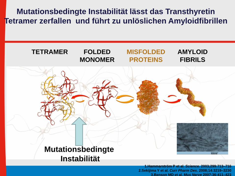
TETRAMER FOLDED
MONOMER
AMYLOID
FIBRILS
MISFOLDED
PROTEINS
Mutationsbedingte Instabilität lässt das Transthyretin
Tetramer zerfallen und führt zu unlöslichen Amyloidfibrillen
Mutationsbedingte
Instabilität
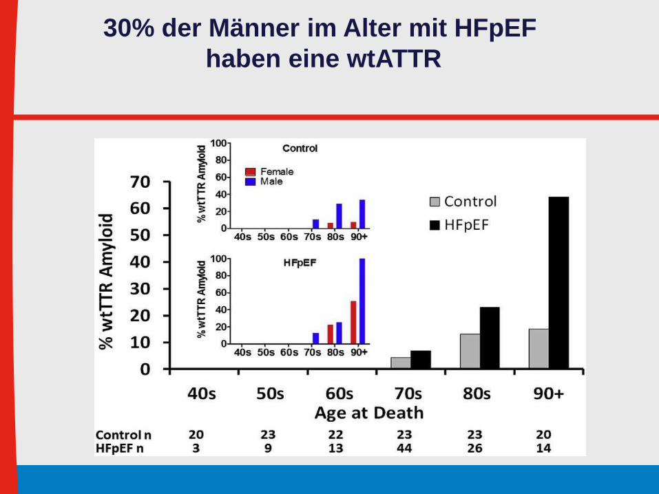
30% der Männer im Alter mit HFpEF
haben eine wtATTR

MRT Diagnostik bei
Amyloidosen
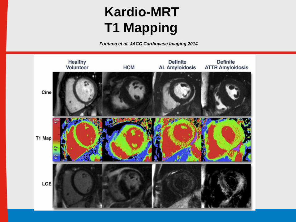
Fontana et al. JACC Cardiovasc Imaging 2014
Kardio-MRT
T1 Mapping
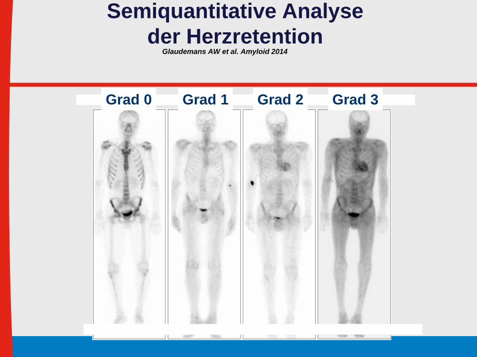
Semiquantitative Analyse
der Herzretention
mutation. Other TTR mutations (non-Val30Met) found were
Val71Ala (10 patients), Tyr114Cys (2), Gly47Glu (1),
Glu89Lys (1), Val122Ile (1), whereas no mutation was
found in six patients (wild-type ATTR). In total, 17 patients
underwent liver transplantation, 13 because of diagnosed
ATTR amyloidosis, and four for other reasons. Of interest,
these last four patients received a donor liver of a patient with
ATTR amyloidosis; three patients are considered as carriers
without clinical signs of amyloidosis, but one patient with
clinical signs has been histologically diagnosed to have
systemic ATTR amyloidosis.
Laboratory findings, echocardiography and ECG
Characteristics of cardiac biomarkers, eGFR, echocardiog-
raphy and ECG are shown in Table 1 for the three groups.
In summary, TnT, log NT-proBNP, many echocardiographic
features (LV ejection fraction, mean LV wall thickness,
septum thickness, LV posterior wall thickness, LV mass
index, E/A ratio, E’-lat, E’ -sep and E/e’ ratio) and ECG
features (PR duration, QRS duration and QTc duration)
differed signif icantly between the ATTR group with echo-
cardiographically-defined amyloidotic cardiomyopathy
(AC) and each of the two other groups, respectively.
Furthermore, NT-proBNP also differed significantly between
carriers and ATTR group without echocardiographically-
defined AC.
Bone scintigraphy
Visual and semi-quantitative scintigraphic findings, both on
planar imaging and with SPECT-CT, are summarized in
Table 1. None of the 11 carriers tested positive at visual score
analysis on planar images, and only one of 11 carriers showed
slightly elevated (grade 1) uptake on SPECT-CT. Among the
19 patients with ATTR without echocardiographically-
defined AC, eight patients on planar imaging showed
uptake grade 2 or 3. In these eight patients with cardiac
uptake, NT-proBNP, TnT, echocardiographic and ECG
findings did not signif icantly differ from the other patients
(n¼11) in this group without cardiac uptake on the bone
scan. All 11 patients with ATTR and echocardiographically-
defined AC showed high uptake (grade 2 or 3), both on planar
images and on SPECT-CT.
At semi-quantitative analysis, H/WB ratio, H/S ratio and
SPECT LV/bloodpool ratio, were signif icantly higher in
patients with ATTR and echocardiographically-defined AC,
compared to both carriers and ATTR patients without
echocardiographically-defined AC. Concerning the H/S
ratio, there was also a significant difference between
Figure 1. Visual grading scale for cardiac uptake on planar imaging (from left to right: grade 0, 1, 2 and 3).
4 A. W. J. M. Glaudemans et al. Amyloid, Early Online: 1–10
Am
ylo
id D
ow
nlo
aded
fro
m i
nfo
rmah
ealt
hca
re.c
om
by U
B H
eidel
ber
g o
n 0
1/2
8/1
4F
or
pers
on
al u
se o
nly
.
Grad 0 Grad 1 Grad 2 Grad 3
Glaudemans AW et al. Amyloid 2014

ATTRAL
Korrelation einer positiven 99mTc-DPD
Szintigraphie bei WT ATTR vs. AL
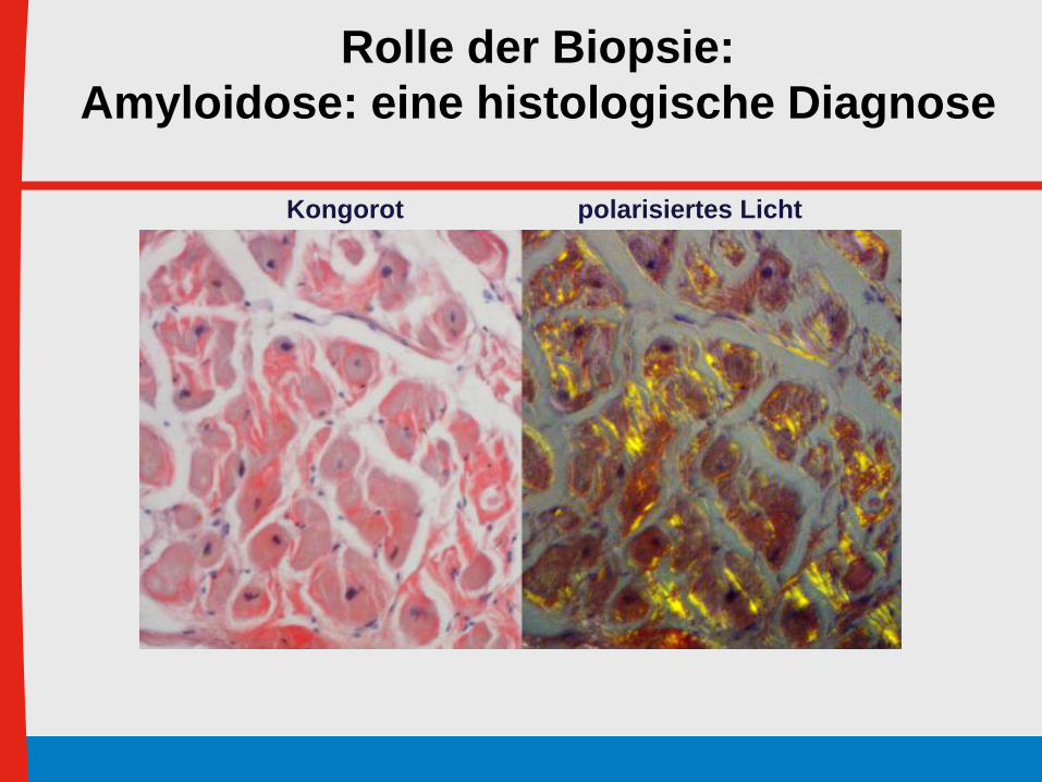
Rolle der Biopsie:
Amyloidose: eine histologische Diagnose
Kongorot polarisiertes Licht
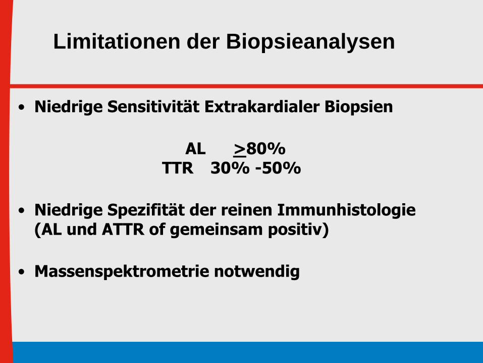
Limitationen der Biopsieanalysen
• Niedrige Sensitivität Extrakardialer Biopsien
AL >80%TTR 30% -50%
• Niedrige Spezifität der reinen Immunhistologie (AL und ATTR of gemeinsam positiv)
• Massenspektrometrie notwendig
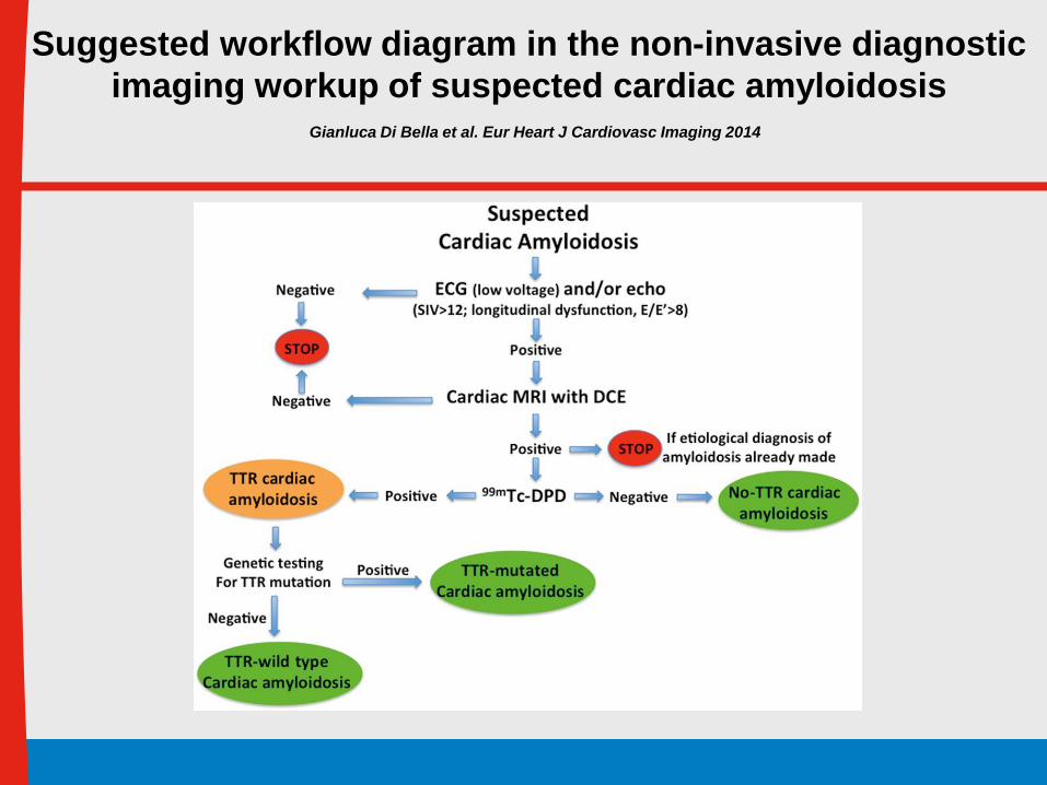
Suggested workflow diagram in the non-invasive diagnostic
imaging workup of suspected cardiac amyloidosis Gianluca Di Bella et al. Eur Heart J Cardiovasc Imaging 2014
- 30% der LVH oder HFpEF Pat haben eine Amyloidose
- Früherkennung ist entscheidend (AL vs ATTR)
- Low VolatageEKG, apikaler Strain,
Szinitigraphie, Biopsie wie für ATTR
- Therapie bei WT-ATTR: in Studien zu Small
Molecules/Tafamides/Grünem Tee Extrakt
- Lebertransplantation bei Mutations-ATTR
Fazit für die Praxis

Aetiology Echo CMR Cath SPECT MDCT PET
Myocarditis + +++ +++c - - -
Sarcoidosis + +++ ++c - - ++
Hypertrophic CMP: HCM +++ ++ ++ - - -
Amyloidosis ++ +++ +++c - - -
Dilated CMP: Myocarditis + +++ +++c - - -
Eosinophilic syndromes + +++ +++c - - -
Iron: haemochromatosis + +++ - - - -
Iron: thalassaemia + +++ - - - -
ARVC ++ +++ +++c - + -
Restrictive CMP: Pericarditis ++a ++b ++a - ++d -
Amyloidosis ++ +++ +++c - - -
Endomyocardial fibrosis + +++ +++c - - -
Anderson-Fabry + + - - - -
Unclassified CMP Takotsubo-CMB ++ ++ +++ - - -
Empfehlungen zur
Diagnostik bei KardiomyopathienMcMurray et al, Eur Heart J 2012; 14: 803-869