Embed Size (px)
Citation preview

The T2TM Tibia Nail A Prospective Multicenter Clinical Study
Inaugural-Dissertation
zur Erlangung des Doktorgrades
der Hohen Medizinischen Fakultät
der Rheinischen Friedrich-Wilhelms-Universität
Bonn
Ramasankerpersad Jairam
aus Distrikt Nickerie, Suriname/South America
2016

Angefertigt mit der Genehmigung der
Medizinischen Fakultät der Universität Bonn
Gutachter: Prof. Dr. med. C. N. Kraft
Gutachter: Prof. Dr. med. W. A. Willinek
Tag der Mündlichen Prüfung: 03.12.2015
Aus der Klinik für Orthopädie, Unfall- und Handchirurgie in Krefeld
Direktor: Prof. Dr. med. Clayton N. Kraft

„Every surgeon carries about him a little cemetery, in which from time to time he goes
to pray, a cemetery of bitterness and regret, of which he seeks the reason for certain
of his failures.” (Rene Leriche, 1879-1955)


– 5 –
Inhaltsverzeichnis
1 Abbreviation Directory ............................................................................................. 7
2 Deutsche Zusammenfassung .................................................................................. 8
2.1 Einleitung ................................................................................................................. 8
2.2 Konservative Behandlung / Frakturreposition .......................................................... 8
2.3 Operative Behandlung ............................................................................................. 9
2.3.1 Konventionelle Plattenosteosynthese (ORIF) ....................................................... 9
2.3.2 Biologische überbrückende Plattenosteosynthese ............................................... 9
2.3.3 MIPO - Minimally Invasive Plate Osteosynthesis ................................................. 9
2.4 Marknagelosteosynthese ....................................................................................... 10
2.4.1 Unaufgebohrte Marknagelosteosynthese ........................................................... 10
2.4.2 Aufgebohrte Marknagelosteosynthese ............................................................... 10
2.5 Ziel der Arbeit ......................................................................................................... 10
2.6 Materialien und Methoden ..................................................................................... 11
2.7 Ergebnisse ............................................................................................................. 12
2.8 Diskussion .............................................................................................................. 13
2.9 Zusammenfassung ................................................................................................ 15
2.10 Literaturverzeichnis der deutschen Zusammenfassung ...................................... 16
3 Introduction ............................................................................................................. 19
3.1 Conservative treatment/fracture reduction ............................................................. 19
3.2 Plate osteosynthesis .............................................................................................. 24
3.3 Intramedullar nailing .............................................................................................. 29
3.3.1 Unreamed nailing ................................................................................................ 30
3.3.2 Reamed nailing ................................................................................................... 32
3.4 General treatment considerations .......................................................................... 38
3.5 Aim of the study ..................................................................................................... 40

– 6 –
4 Material and Methods ............................................................................................. 41
4.1 Patients .................................................................................................................. 41
4.1.1 Inclusion criteria .................................................................................................. 41
4.1.2 Exclusion criteria ................................................................................................. 41
4.2 Surgery .................................................................................................................. 42
4.2.1 Operative procedure ........................................................................................... 42
4.2.2 Description of the device: T2TM Tibial Nailing System ........................................ 43
4.3 Follow-up ............................................................................................................... 45
4.3.1 Clinical assessment ............................................................................................ 45
4.3.2 Radiographic assessment .................................................................................. 45
4.4 Statistics ................................................................................................................. 45
5 Results ..................................................................................................................... 46
5.1 Preoperative ........................................................................................................... 46
5.2 Perioperative .......................................................................................................... 50
5.3 Postoperative assessments: 4-6 weeks, 4 months, 12 months ............................. 55
5.4 Postoperative complications .................................................................................. 64
5.5 Dynamisation of the nail ........................................................................................ 65
5.6 Revision surgery .................................................................................................... 65
5.7 Material removal .................................................................................................... 65
6 Discussion ............................................................................................................... 66
7 Comments (criticism) on this study ...................................................................... 76
8 Summary .................................................................................................................. 77
9 Bibliography ............................................................................................................ 79
10 List of figures ........................................................................................................ 90
11 List of tables .......................................................................................................... 91
12 Appendix - Clinical Review Form (CRF) ............................................................. 92
13 Acknowledgement ................................................................................................ 93
– 7 –
1 Abbreviation Directory
AO Arbeitsgemeinschaft für Osteosynthesefragen
ARDS Adult Respiratory Distress Syndrome
CRF Clinical Review Form
CRPS Complex Regional Pain Syndrome
CT Computer Tomogram
e.g. exempli gratia (for example)
Fig. Figure
ICN Interlocking Compression Nail
kg kilogram
LCP Locking Compression Plate
LIF Locked Internal Fixation
Lig Ligamentum
LISS Less Invasive Stabilization System
LP Locking Plate
min Minute
MIPO Minimal Invasive Percutaneos Osteosynthesis
N Number
ORIF Open Reduction Internal Fixation
OTA Orthopaedic Trauma Association
sec Second
SPSS Superior Performing Software System
T2 nail Titanium nail
VAS Visual Analog Scale

– 8 –
2 Deutsche Zusammenfassung
2.1 Einleitung
Es gibt verschiedene Verfahren, mit denen Tibiafrakturen behandelt werden können. Es
stehen sowohl konservative, als auch operative Verfahren zur Verfügung. Die operativen
Verfahren können in extramedulläre, z.B. Plattenosteosynthesen und externe Fixateure,
und intramedulläre Verfahren, wie z. B. aufgebohrte und unaufgebohrte Marknägel,
unterteilt werden.
2.2 Konservative Behandlung / Frakturreposition
Nachdem im Jahr 1852 der Gipsverband vom holländischen Arzt Mathijsen erfunden
wurde, konnten hölzerne Schienen zur Immobilisierung von Gliedmaßen zunehmend
ersetzt werden. Mit dem Gipsverband soll die verletzte Region mit dem Ziel der
Knochenheilung ruhig gestellt werden.
Im Ersten Weltkrieg wurde der Algorithmus „Reposition, Retention und Rehabilitation“
durch Lorenz Böhler geprägt und so ein standardisiertes Behandlungsschema von
Frakturen geschaffen (Trojan, 1984). Das sogenannte „Drei-Punkte-Prinzip“ erhöht die
Stabilität im Gipsverband dadurch, dass die Ligamentotaxis auf der konkaven Seite der
Fraktur die Fragmente in der korrekten Stellung hält (Bayne et al., 2006). Nach
Sarmiento et al. (1989) kann über diesen Effekt ein Großteil der geschlossenen
Frakturen frühfunktionell beübt werden. Eine Voraussetzung hierfür ist, dass eine
Verkürzung der Fraktur von weniger als 10 mm und eine Achsabweichung kleiner als 5°
vorliegt.
Durch Verbesserungen in der operativen Versorgung hat die konservative Behandlung
von Tibiafrakturen in den Industrienationen stark abgenommen. Gleichwohl gibt es für
diese noch immer Indikationen. Auch wenn die oben genannten Verfahren inzwischen
eher selten zur Anwendung kommen, kann eine Tibiafraktur grundsätzlich erfolgreich
konservativ behandelt werden.

– 9 –
2.3 Operative Behandlung
Zur operativen Behandlung stehen verschiedene Verfahren der
plattenosteosynthetischen Versorgung und Marknagelosteosynthesen zur Verfügung.
Die Grundprinzipien der operativen Frakturversorgung mit einer konventionellen
Plattenosteosynthese sind einerseits die direkte anatomische Reposition, andererseits
die stabile Fixierung der Fragmente (ORIF = Open Reduction and Internal Fixation).
Hierzu ist nicht selten eine weite chirurgische Exploration der Frakturerforderlich, um
gute Sicht auf die zu reponierenden Fragmente zu erlangen.
Folgende Verfahren und Techniken der plattenosteosynthetischen Versorgung werden
angewandt:
2.3.1 Konventionelle Plattenosteosynthese (ORIF)
Direkte, offene Reposition und stabile Osteosynthese mit Platten (winkelstabile
Verfahren zeigen eine größere absolute Stabilität) konnten sich als ein Verfahren zur
erfolgreichen chirurgischen Behandlung von Frakturen etablieren.
2.3.2 Biologische überbrückende Plattenosteosynthese
Indirekte, geschlossene oder offene, aber weniger invasive (“no-touch technique”)
Reposition und biologisch überbrückende Plattenosteosynthese zeigten eine größere
relative Stabilität (Gautier et al., 1994; Leunig et al., 2001).
2.3.3 MIPO - Minimally Invasive Plate Osteosynthesis
Das Ziel dieses Verfahrens ist, der Fraktur durch eine geschlossene Reposition und
Plattenosteosynthese gute relative Stabilität zu geben ohne dabei den Weichteilmantel
über der Fraktur zu verletzen (Perren, 1995, 2001, 2002; Sandelmaier et al., 1999; Tepic
et al., 1995).
– 10 –
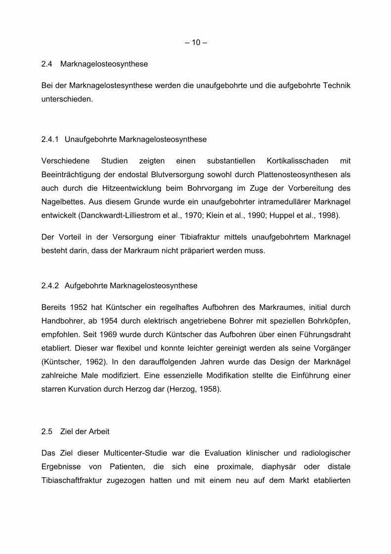
2.4 Marknagelosteosynthese
Bei der Marknagelostesynthese werden die unaufgebohrte und die aufgebohrte Technik
unterschieden.
2.4.1 Unaufgebohrte Marknagelosteosynthese
Verschiedene Studien zeigten einen substantiellen Kortikalisschaden mit
Beeinträchtigung der endostal Blutversorgung sowohl durch Plattenosteosynthesen als
auch durch die Hitzeentwicklung beim Bohrvorgang im Zuge der Vorbereitung des
Nagelbettes. Aus diesem Grunde wurde ein unaufgebohrter intramedullärer Marknagel
entwickelt (Danckwardt-Lilliestrom et al., 1970; Klein et al., 1990; Huppel et al., 1998).
Der Vorteil in der Versorgung einer Tibiafraktur mittels unaufgebohrtem Marknagel
besteht darin, dass der Markraum nicht präpariert werden muss.
2.4.2 Aufgebohrte Marknagelosteosynthese
Bereits 1952 hat Küntscher ein regelhaftes Aufbohren des Markraumes, initial durch
Handbohrer, ab 1954 durch elektrisch angetriebene Bohrer mit speziellen Bohrköpfen,
empfohlen. Seit 1969 wurde durch Küntscher das Aufbohren über einen Führungsdraht
etabliert. Dieser war flexibel und konnte leichter gereinigt werden als seine Vorgänger
(Küntscher, 1962). In den darauffolgenden Jahren wurde das Design der Marknägel
zahlreiche Male modifiziert. Eine essenzielle Modifikation stellte die Einführung einer
starren Kurvation durch Herzog dar (Herzog, 1958).
2.5 Ziel der Arbeit
Das Ziel dieser Multicenter-Studie war die Evaluation klinischer und radiologischer
Ergebnisse von Patienten, die sich eine proximale, diaphysär oder distale
Tibiaschaftfraktur zugezogen hatten und mit einem neu auf dem Markt etablierten

– 11 –
Marknagelsystem versorgt wurden. Durch einen Vergleich der erhobenen Resultate mit
den Ergebnissen von anderen internationalen Studien sollte die Effektivität dieser neuen
Marknagelosteosynthese gegenüber bereits etablierten Verfahren bei der Versorgung
spezifischer Tibiafrakturen untersucht werden.
2.6 Materialien und Methoden
Von Januar 2003 bis Dezember 2004 wurden 102 Patienten mit einer proximalen,
diaphysären oder distalen geschlossen Tibiafraktur behandelt (AO 41,A-C 1-3, AO 42 A-
C 1-3, AO 43 A-C 1-3) behandelt. Drei europäische Level 1-Traumazentren waren in die
Akquisition der Daten involviert: Vrije Universiteit Medical Center, Amsterdam, The
Netherlands, Hospital Universitario Ramon y Cajal, Madrid, Spanien und Klinikum
Hannover Nordstadt, Hannover, Deutschland. Es wurden demographische (z.B. Alter
und Geschlecht), präoperative (z.B. Traumaursache und Frakturtyp), allgemeine
operative (z.B. Operationszeit und Blutverlust) sowie postoperative Daten (z.B.
radiologische Knochenheilung, Belastung, Aktivitäten des täglichen Lebens,
Wiederaufnahme der Arbeit, anteriorer Knieschmerz) erfasst.
Bei allen Patienten wurde das neue T2®-Tibia Marknagel System der Fa. Stryker an-
gewandt. Dieses System ist Europa CE- gekennzeichnet und von der Amerikanischen
Food and Drug Administration (FDA) genehmigt.
Alle Daten wurden prospektiv erhoben und mittels eines standardisierten klinisch-
wissenschaftlichen Formulars dokumentiert. Zudem wurden alle Patienten radiologisch
nachverfolgt. Studienpatienten wurden präoperativ, perioperativ und zu drei festgelegten
postoperativen Zeitpunkten (4-6 Wochen, 4 Monaten und 12 Monaten) klinisch und
radiologisch nachuntersucht. Es wurde gewährleistet, dass alle Patienten in den
entsprechenden Ambulanzen von den gleichen betreuenden Ärzte gesehen wurden, die
bereits initial mit der Dokumentation begonnen hatten. Die visuelle Analogskala (VAS)
wurde zur subjektiven Einschätzung der Schmerzen benutzt. 0 Punkte wiesen hierbei
auf eine komplette Schmerzfreiheit hin, während 10 Punkte den größten vorstellbaren
– 12 –
Schmerz darstellten (Downie et al., 1978). Die radiologische Auswertung erfolgte
gemeinsam durch Chirurgen und Radiologen.
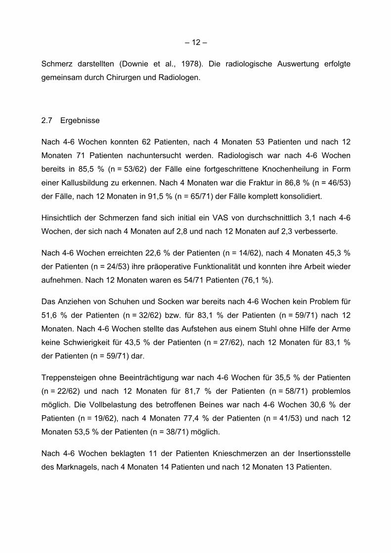
2.7 Ergebnisse
Nach 4-6 Wochen konnten 62 Patienten, nach 4 Monaten 53 Patienten und nach 12
Monaten 71 Patienten nachuntersucht werden. Radiologisch war nach 4-6 Wochen
bereits in 85,5 % (n = 53/62) der Fälle eine fortgeschrittene Knochenheilung in Form
einer Kallusbildung zu erkennen. Nach 4 Monaten war die Fraktur in 86,8 % (n = 46/53)
der Fälle, nach 12 Monaten in 91,5 % (n = 65/71) der Fälle komplett konsolidiert.
Hinsichtlich der Schmerzen fand sich initial ein VAS von durchschnittlich 3,1 nach 4-6
Wochen, der sich nach 4 Monaten auf 2,8 und nach 12 Monaten auf 2,3 verbesserte.
Nach 4-6 Wochen erreichten 22,6 % der Patienten (n = 14/62), nach 4 Monaten 45,3 %
der Patienten (n = 24/53) ihre präoperative Funktionalität und konnten ihre Arbeit wieder
aufnehmen. Nach 12 Monaten waren es 54/71 Patienten (76,1 %).
Das Anziehen von Schuhen und Socken war bereits nach 4-6 Wochen kein Problem für
51,6 % der Patienten (n = 32/62) bzw. für 83,1 % der Patienten (n = 59/71) nach 12
Monaten. Nach 4-6 Wochen stellte das Aufstehen aus einem Stuhl ohne Hilfe der Arme
keine Schwierigkeit für 43,5 % der Patienten (n = 27/62), nach 12 Monaten für 83,1 %
der Patienten (n = 59/71) dar.
Treppensteigen ohne Beeinträchtigung war nach 4-6 Wochen für 35,5 % der Patienten
(n = 22/62) und nach 12 Monaten für 81,7 % der Patienten (n = 58/71) problemlos
möglich. Die Vollbelastung des betroffenen Beines war nach 4-6 Wochen 30,6 % der
Patienten (n = 19/62), nach 4 Monaten 77,4 % der Patienten (n = 41/53) und nach 12
Monaten 53,5 % der Patienten (n = 38/71) möglich.
Nach 4-6 Wochen beklagten 11 der Patienten Knieschmerzen an der Insertionsstelle
des Marknagels, nach 4 Monaten 14 Patienten und nach 12 Monaten 13 Patienten.

– 13 –
Hinsichtlich postoperativer Komplikationen zeigte nach 4-6 Wochen von 62 Patienten 1
Patient ein Hämatom, dieses war oberflächlich und wurde konservativ behandelt, 3
Patienten beklagten Gefühlsstörungen im Unterschenkel, bei 1 Patienten bestand eine
tiefe und bei 3 Patienten eine oberflächliche Infektion, die konservativ mit Antibiose
behandelt wurden. Zwei Patienten entwickelten ein CRPS Typ 1.
Nach 4 Monaten hatten von 53 Patienten 6 Patienten Probleme mit den Schrauben
(Lockerung oder Perforation durch die Haut). Nach 1 Jahr bestanden bei 9 von 71
Patienten mechanische Probleme mit der Osteosynthese, 4 Patienten hatten
Gefühlsstörungen im Unterschenkel, 3 Patienten zeigten oberflächliche Infektionen, 4
Patienten entwickelten ein CRPS Typ 1. Bei 1 Patienten wurden heterotope
Ossifikationen im Bereich des Frakturspaltes nachgewiesen. Diese war nicht weiter
gradiert.
Eine Dynamisierung des Nagels erfolgte bei 4/62 Patienten nach 4-6 Wochen, bei 5/53
Patienten nach 4 Monaten und bei 5/71 Patienten 12 Monaten. Die Gründe für die
Dynamisierung waren eine verzögerte Knochenheilung oder Probleme mit den
Schrauben. Revisionen (wegen Malrotation, Malposition oder Migration des Nagels nach
proximal) erfolgten bei insgesamt 7 von 102 Patienten.
Daten zur Metallentfernung lagen von 47/102 Patienten (46,1 %) vor: Die Gründe für die
Metallentfernung stellten 34 Mal die Konsolidierung der Fraktur, 11 Mal anteriore
Knieschmerzen, 2 Mal gebrochene Schrauben und einmal der Wunsch des Patienten
dar.
2.8 Diskussion
Das T2®- Tibia Marknagel System der Fa. Stryker sorgt durch sein Design mit
proximaler und distaler Verriegelung und Aufbohrung vor Nagelinsertion für eine hohe
Stabilität der Fraktur und fördert somit die Konsolidierung der Fraktur. Dieses ist nicht
neu. Folgt man der Literatur, werden Konsolidierungsraten von über 90 % nach

– 14 –
aufgebohrter Marknagelosteosynthese bei der Versorgung von Tibiafrakturen berichtet
(Klemm et al., 1986; Court-Brown et al., 1991; Alho et al., 1990).
Bei allen in dieser Studie eingeschlossenen Patienten wurde der Markraum aufgebohrt.
Dieses Verfahren hat zwei Vorteile. Zum einen wirkt das Bohrmehl, welches durch das
Aufbohren entsteht, in der Frakturzone wie eine autologe Spongiosaplastik (Reynders et
al., 2000). Zum anderen sorgt das Aufbohren für einen besseren kortikalen Kontakt
zwischen Knochen und Marknagel, der wiederum durch einen dickeren Durchmesser
eine höhere Primärstabilität gewährleistet (Chapman, 1998).
Durch die hier genutzten zusätzlichen Kompressionsschrauben konnte die Stabilität des
Marknagels noch weiter erhöht werden. Experimentell ist dies schon gezeigt worden
(Hutter et al., 1977; Gonschorek et al., 1998.), diese Daten scheinen sich nun auch im
klinischen Alltag zu bestätigen. Welcher der einzelnen Faktoren in welchem Maße zu
der guten Konsolidierungsrate in unserem Patientengut beitrug, lässt sich im klinischen
Setting kaum bestimmen, dennoch spricht vieles dafür, dass es die Kombination
derselben ist, die die Knochenheilung positiv beeinflusst. Da der unaufgebohrte
Marknagel vor allem bei offenen Tibiafrakturen Verwendung findet, lassen sich die
beiden Nagelsysteme hinsichtlich der Ergebnisse im klinischen Alltag kaum miteinander
vergleichen. Auch eine offene Reposition und interne Fixation mittels Platte ist
heutzutage in den meisten Kliniken anderen Indikationen als der „einfachen“ Tibiafraktur
vorbehalten, sodass der Vergleich zwischen unserem Nagel und einem solchen
Verfahren hinsichtlich klinischem Ergebnis (z.B. back-to-work) und radiologischer
Konsolidierungsrate von vorneherein hinkt.
Unsere Ergebnisse zeigten, dass 75 % der Patienten ihrer präoperativen Arbeit
nachgehen konnten. Die Aktivitäten des täglichen Lebens (Sockenanziehen,
Treppensteigen, Vollbelastung und maximal mögliche Gehstrecke) verbesserten sich im
Verlauf, auch wenn nicht alle Patienten ihre volle präoperative Funktion erreichten.
Diese Ergebnisse sind kongruent mit Daten aus der Literatur, in der ebenfalls nicht alle
Patienten ihre volle präoperative Funktion erreichten (Keating et al., 1997; Karladani et
al., 2000).
– 15 –
Der postoperative vordere Knieschmerz kristallisierte sich als häufigste und
signifikanteste Komplikation unserer Versorgungen heraus. Er stellte in unserem
Patientengut die häufigste Indikation zur Entfernung des Implantats dar. Auch hier deckt
sich unsere Datenlage mit der Literatur. Court-Brown et al. (1990) präsentierten die
Ergebnisse einer prospektiven Studie von 125 geschlossenen bzw. offenen
Tibiafrakturen des Typs 1 nach Gustilo und Andersen. (1976), die mittels aufgebohrten
Grosse-Kempf-Tibianagel versorgt wurden. Auch hier klagten über 40 % der Patienten
postoperativ über einen vorderen Knieschmerz, vor allem bei knieenden Tätigkeiten. Bei
den meisten dieser Patienten ließ der anteriore Knieschmerz nach Entfernung des
Nagels nach, wenngleich dies mehrere Wochen dauerte. Folgt man der Literatur,
scheint es sich also um ein verfahrenimmanentes und nicht um ein
implantatspezifisches Problem zu handeln, über das auch andere Autoren berichten,
die andere „Nageltypen“ verwenden. Dieses spiegelt auch den klinischen Alltag wieder.
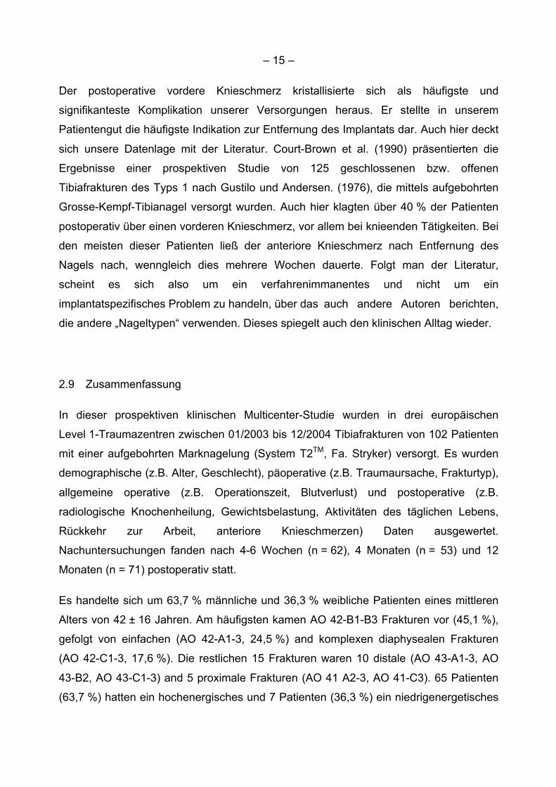
2.9 Zusammenfassung
In dieser prospektiven klinischen Multicenter-Studie wurden in drei europäischen
Level 1-Traumazentren zwischen 01/2003 bis 12/2004 Tibiafrakturen von 102 Patienten
mit einer aufgebohrten Marknagelung (System T2TM, Fa. Stryker) versorgt. Es wurden
demographische (z.B. Alter, Geschlecht), päoperative (z.B. Traumaursache, Frakturtyp),
allgemeine operative (z.B. Operationszeit, Blutverlust) und postoperative (z.B.
radiologische Knochenheilung, Gewichtsbelastung, Aktivitäten des täglichen Lebens,
Rückkehr zur Arbeit, anteriore Knieschmerzen) Daten ausgewertet.
Nachuntersuchungen fanden nach 4-6 Wochen (n = 62), 4 Monaten (n = 53) und 12
Monaten (n = 71) postoperativ statt.
Es handelte sich um 63,7 % männliche und 36,3 % weibliche Patienten eines mittleren
Alters von 42 ± 16 Jahren. Am häufigsten kamen AO 42-B1-B3 Frakturen vor (45,1 %),
gefolgt von einfachen (AO 42-A1-3, 24,5 %) and komplexen diaphysealen Frakturen
(AO 42-C1-3, 17,6 %). Die restlichen 15 Frakturen waren 10 distale (AO 43-A1-3, AO
43-B2, AO 43-C1-3) and 5 proximale Frakturen (AO 41 A2-3, AO 41-C3). 65 Patienten
(63,7 %) hatten ein hochenergisches und 7 Patienten (36,3 %) ein niedrigenergetisches

– 16 –
Trauma erlitten. Die Unfälle hatten sich meistens im Verkehr (31,4 %), auf der Straße
als Fußgänger (22,5 %) oder zuhause (22,5 %) ereignet. Nur bei 36 Patienten (35,2%)
lag eine singuläre Fraktur vor, bei 51 Patienten (50,0 %) war auch die Fibula frakturiert
und 15 Patienten (14,7 %) waren polytraumatisiert.
Nach 12 Monaten wurde die Knochenheilung radiologisch bei 91,5 % (n = 65/71)
bestätigt. 76,1 % (n = 54/71) hatte ihre Arbeit wieder aufgenommen und 74,7 %
(n = 53/71) konnten mit ihrer früheren Kapazität arbeiten. Der mittlere Schmerzscore
nahm von 3,1 ± 1,2 (4-6 Wochen) auf 2,6 ± 2,2 (4 Monate) und 2,3 ± 1,7 (12 Monate)
ab. Nach 12 Monaten litten 13 Patienten unter anterioren Knieschmerzen, der bei 12
Patienten so leicht war, dass sie arbeiten und das Bein belasten konnten. Ein
polytraumatisierter Patient hatte wegen einer Pseudarthrose einen Schmerzscore von 8
Punkten und benötigte mehrere Reoperationen.
Bei insgesamt 14 Patienten wurde eine Dynamisierung des Nagels wegen verzögerter
Heilung und/oder Schraubenproblemen vorgenommen. Eine Revisionsoperation wurde
bei 7/102 Patienten vorgenommen. Bei 3 Patienten war die Revision wegen einer
Malrotation nach Schraubenbruch nötig, bei 2 Patienten wegen Schmerzen durch die
proximalen Schrauben und bei 2 Patienten wegen einer Schraubenlockerung mit der
Gefahr der Hautperforation.
Die Ergebnisse unserer Studie über das T2TM-System sind mit anderen Studien über die
aufgebohrte Marknagelung vergleichbar und belegen die Hypothese, dass diese
Osteosynthesemethode effektiv ist und relativ wenig Komplikationen aufweist.
2.10 Literaturverzeichnis der deutschen Zusammenfassung
Alho A, Ekeland A, Stromsoe K, Folleras G, Thoresen BO. Locked inramedullary nailing for displaced tibial shaft fractures. J Bone Joint Surg Br 1990; 72: 805
Bayne G, Turner RG. Closed fracture manipulation - improving Charnley`s three point fixation technique. Ann R Coll Surg Engl 2006; 88: 504
Chapman MW. The effect of reamed and non reamed intramedullary nailing on fracture healing. Clin Orthop 1998; 355 (Suppl): 230-238

– 17 –
Court-Brown CM, McQueen, MM, Quaba AA, Christie J. Locked intramedullary nailing of open tibial fracture. J Bone Joint Surg Br. 1991; 73: 959
Court-Brown CM, Christie J, McQueen MM. Closed intramedullary tibial nailing: its use in closed and type 1 open fractures. J Bone Joint Surg Br. 1990; 72: 605-611
Danckwardt-Lilliestrom G, Lorenzi L, Olerud S. Intracortical circulation after intramedullary reaming with reduction of pressure in the medullary cavity. J Bone Joint Surg Am 1970; 52: 1390-1394
Downie WW, Leatman PA, Rhind VM, Wright V, Branco JA, Anderson JA. Studies with pain rating scales. Ann Rheum Dis 1978; 37: 378-381
Gautier E, Ganz R. Die biologische Plattenosteosynthese (The biological plateosteosynthesis). Zentralbl Chir. 1994; 119: 564-572
Gonschorek O, Hofmann GO, Buhren V. Interlocking compression nailing: a report on 402 applications. Arch Ortho Trauma Surg 1998; 117: 430-437
Herzog K. Die Technik der geschlossenen Marknagelung frischer Tibiafrakturen mit dem Rohrschlitznagel. Chirurg 1958; 29: 501-506
Hupel TM, Aksenov SA, Schemitsch EH. Effect of limited and standard reaming on cortical bone blood flow and early strenght of union following segmental fracture. J Orthop Trauma 1998; 12: 400-406
Hutter CG, Oden R, Kirk R. The intramedullary compression rod. Clin Orthop 1977; 122: 165-173
Karladani HA, Granhed H, Edshage B, Jerre R, Styf J. Displaced tibial shaft fractures - a prospective randomised study of closed intramedullary nailing versus cast treat- ment in 53 patient. Acta Orthop Scand 2000; 71: 160-167
Keating JF, O`Brien PJ, Blachut PA, Meek RN, Broekhuyse HM .Locking intramedullary nailing with and without reaming for open fractures of the tibial shaft: a prospective, randomised study. J Bone Joint Surg Am 1997; 79: 334-341
Klein MP, Rahn BA, Frigg R, Kessler S, Perren SM. Reaming versus non-reaming in medullary nailing: interference with cortical circulation of the canine tibia. Arch Orthop Trauma Surg 1990; 109: 314-316
Klemm KW, Borner M. Interlocking nailing of complex fracture of femur and tibia. Clin Orthop Relat Res 1986; 212: 89-100
Küntscher G. Praxis der Marknagelung. Schattauer, Stuttgart, 1962
Leuning M, Hertel R, Siebenrock K. The evaluation of indirect reduction techniques for the treatment of fractures. Clin Orthop 2001; 375: 307-314
– 18 –
Perren SM. Evolution and rational of locked internal fixator technology. Introductory remarks. Injury 2001; 32 (Suppl 2): B3-9
Perren SM. Evolution of the internal fixation of long bone fractures. The scientific basis of biological internal fixation: choosing a new balance between stability and biology. J Bone Joint Surg Br. 2002; 84: 1093-1110
Perren SM. Point contact fixator: part 1. Scientific background, design and application. Injury. 1995; 22 (Suppl 1): 1-10
Reynders PA, Broos PLO. Healing of closed femoral shaft fracture treated with the AO undreamed femoral nailing. A comparative study with the AO reamed femoral nail. Injury 2000; 31: 367-371
Sandelmaier P, Stephan C, Reimers N. LISS osteosynthesis for distal fractures of the femur.Trauma Berufskrankh, 1999; 1: 392-297
Sarmiento A, Gertsen LM, Sobol PA, Shankwiler JA; Vangsness CT. Tibial shaft fractures treated with functional braces. J.Bone Joint Surg Br 1989; 71: 602-609
Tepic S, Perren SM. The biomechanics of the PC-Fix internal Fixator.Injury 1995; 26 (Suppl 2): 5-10
Trojan EA. Die konservative Behandlung des frischen geschlossen Unterschenkelschaftbruches nach Lorenz Böhler. Orthopäde 1984; 13: 256-261

– 19 –
3 Introduction
There are different ways on how a tibia fracture can be treated. It can be treated
conservatively, surgically by extramedullar procedures of osteosynthesis e.g. plates and
external fixator, or surgically by intramedullar procedures of osteosynthesis, e.g. reamed
and unreamed nails.
3.1 Conservative treatment/fracture reduction
From the year 1852, the plaster cast introduced by the Dutch medical officer Mathijsen
replaced the use of wooden splints in numerous modifications in order to enable bone
healing by immobilisation of the fractured leg. Additionally, the Steinmann pin, presented
by Steinmann in 1907 in Bern, facilitated the traction and reposition of the fracture ends
and prevented displacement, but pin track infections wer common. In the course of the
First World War Lorenz Böhler introduced reposition, traction and plaster fixation as a
standardized treatment technique, but satisfying treatment results could not be achieved
(Trojan, 1984). At the beginning of the 1950s Charnley in England recognized the
importance of the soft tissue for the fracture stability. In his so-called three point principle
he pointed out that the intact soft tissue on the concave side of the fracture deformation
enhances the relative stability in plaster by taking the fragments to the right position
(Bayne and Turner, 2006). Fracture reduction technique can be done by calculated
pressure and counter pressure.
Fig. 1: Fracture reduction technique( Habermayer et al , 1990)
– 20 –
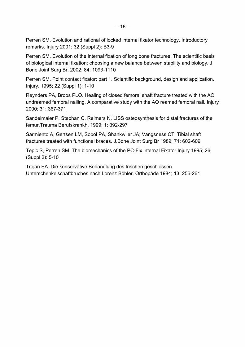
Böhler suggested that axis deviations between 3° and 15° can be corrected by wedging
the plaster (Böhler, 1965). This procedure is not possible in case of an intact fibula.
Based on the x-ray, it is possible to transfer and to mark the axis position on the plaster.
The cut into the plaster takes place at the intersection of the axes on the concave side of
the dislocation. The plaster has to be cut and wedged open by more than half of the
circumference. It is wedged and opened until the marked axes correspond. The position
is fixed by pieces of cork and is plastered again after x-ray control.
Fig. 2: Technique of wedging open the plaster (Habermeyer et al., 1990)
To reduce transverse fractures the fragment reduction requires traction e.g. supported
by a Steinmann pin. The right technique of traction treatment starts with a correct
positioning of the calcaneus wire to avoid failure in correction of malalignments. The
calcaneus wiremust be placed vertically to the distal tibia proportion. According to Jahna
and Wittich (1985) one marks the correct point of incision on the inside ankle 2 cross
fingers in the extended tibia axis and 2 cross fingers dorsally in adults. The lateral exit
point of the wire is one cross finger beneath and dorsal to the tip of the lateral malleolus.
After an exact marking the extension wire can be drilled from medial to lateral (Fig. 3).
For traction of the lower leg a weight of 3-4 kg is usually sufficient.

– 21 –
Fig. 3: Traction treatment technique (Habermeyer et al., 1990)
When a dislocation appears during traction treatment, it requires correction. Valgus and
varus dislocation can be avoided by an absolute correct setting of the traction pin wire
and can be corrected by a modified point of start at the extension clamp. Antecurvation
faults can be adjusted if the direction of the traction runs upwards parallel to the proximal
tibia proportion. Conversely a recurvation position requires a correction of the traction
downwards (Fig. 4).
Fig. 4: Correction of axis positions during traction treatment (Habermeyer et al., 1990)
– 22 –
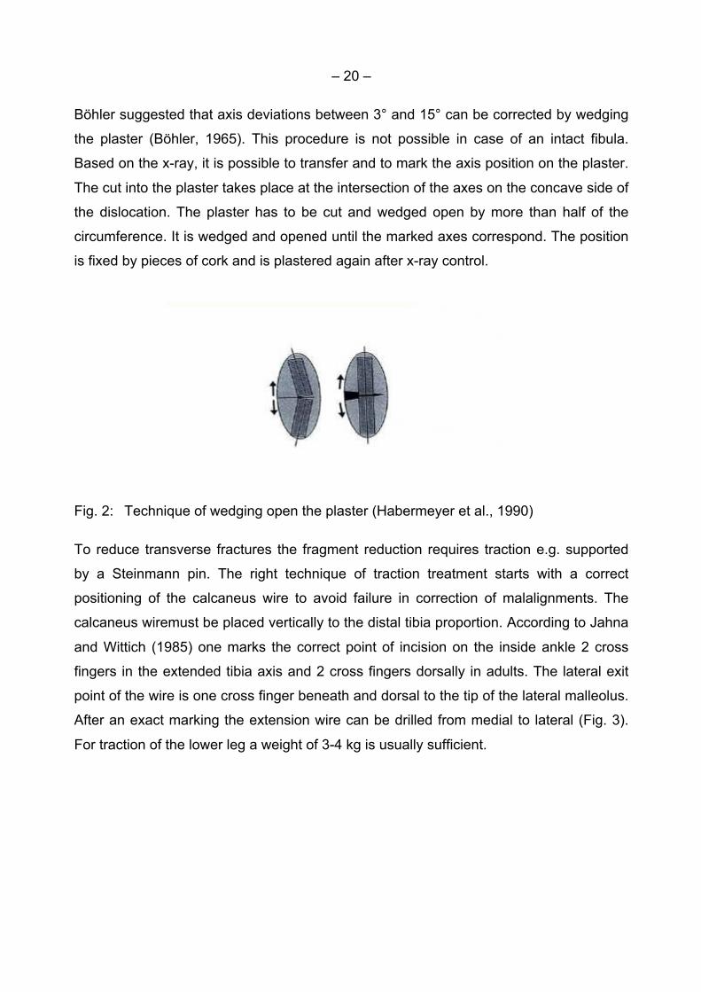
One common mistake is the plastering in the talipes equinus position. In case of weight
bearing, the recurvation of the lower leg results as a consequence (Fig. 5).
Fig. 5: Avoidance of the talipes equinus position (Habermeyer et al., 1990)
Based on the knowledge of the fracture stabilization by means of soft tissue, particularly
of the membrane interossea, Dehne et al. (1961) introduced the early weight bearing in
upper extremity plaster. In 1967, the introduction of ‘brace treatment” by Sarmiento
described the development of this early functional treatment. The external splinting of
the fractured extremity by a brace creates a hydraulic system. This hydraulic mechanism
of soft tissue in brace prevents a further shortening of the fracture fragments (Sarmiento
et al., 1989). A controlled motion in the fracture gap leads to an osteokinetic stimulus by
thermoelectric and vascular changes. Studies have shown that this results in a faster
callus formation (Hulth, 1989; Cornell and Lane, 1992; Aro and Chao, 1993; Lacroix and
Prendergast, 2002; Isaksson et al., 2006; Jagodzinski and Krettek, 2007; Gonzalez-
Torres et al., 2010; Gomez-Benito et al., 2011).
In adults with closed, not dislocated and uncomplicated fracture types the healing of
conservatively treated tibia fractures usually takes between the 10th and 13th week.
Dislocated fractures heal between the 13th and 16th week and open fractures as well as
fractures in fragments between the 16th and 26th week (Leach, 1984).

– 23 –
Drawbacks of the conservative therapy
In the following cases, the conservative therapy is contraindicated and a surgical
procedure preferable:
A. Fractures that cannot be reduced
1) with a displacement of more than shaft width
2) angle larger than 10
3) primary diastase
4) primary shortening larger than 10 mm
B. Instable fracture forms
1) with redislocation
C. accompanying ispsilateral femur fracture
In case of an intact fibula and a dislocated tibia fracture (and thus a ruptured membrana
interossea) a blocking mechanism and a delayed fracture healing or rather a formation
of a pseudarthrosis is frequently seen, thus surgical management is justified, too.
Conversely in the case of an intact fibula without dislocated tibia fracture an above knee
plaster with a stretched knee may be indicated because an intact fibula increases the
stability of the tibia and reduces the degree of the dislocation of tibial fragments (Nicoll,
1964). Without doubt an intact fibula may lead to varus deformity. For this reason
Sarmiento recommended the fibula-osteotomy at the level of the tibia fracture
(Sarmiento et al., 1989). Varus and Valgus deformities as well as ante and recurvation
deformities are described especially by oblique and transverse fractures (den Outer et
al., 1990).
All fractures with an open or closed soft tissue damage of 2nd-3rd grade that are initially
stabilized surgically by external fixation are not ideal for further conservative
management. Per definition, the threatening and manifest compartment syndrome is
among those that require the opening of all four loges and thus urgent surgery is
necessary (Rammelt et al., 2004).

– 24 –
In general, a high rate of complications is seen in conservatively treated tibia fractures.
Leach (1984), Oni et al. (1988), and Puno et al. (1986) found 61 % of complications with
an intact fibula, among them 26 % with delayed union.
In a study on the longterm outcome, 572 subjects who had sustained a tibial shaft
fracture and were treated with cast immobilisation more than 27 years ago were
compared with matched controls without fractures. It was shown that the functional
status was worse in the fracture group than in the control group with regard to knee pain,
functional tests such as climbing stairs, and the physical function score on the SF-36
(Greenwood et al., 1997).
Indications
The Böhler school recommended the conservative treatment of all closed lower leg shaft
fractures as long as they are stable and reducible (Böhler, 1965). According to
Sarmiento et al. (1989) most of the closed fractures can be treated functionally at an
early stage if the leg shortening is less than 10 mm and the axis deviation is smaller than
5° after reposition. First-grade open fractures with slight soft tissue damage can also be
treated by the Sarmiento-technique. A further indication arises in cases where there is a
change of procedure after external fixation immobilization.
In view of the improvements in surgical fracture management, conservative treatment of
tibial fractures has become increasingly more seldom in industrialized countries.
Nonetheless, there are indications for conservative treatment, for example in patients
with an increased operative risk or in patients who refuse a surgical therapy.
3.2 Plate osteosynthesis
Developments in plate osteosynthesis
Robert Danis is regarded as the founder of modern osteosynthesis. In 1949 he
developed plates which provide compression through the implant and narrow the

– 25 –
fracture gap. Fractures treated by those plates healed “directly”, i.e. without callus
formation. In 1958 Danis’ principles were taken over by the Swiss “Arbeitsgemeinschaft
für Osteosynthese” (AO) which propagated precise reduction and absolutely stable
fixation mainly using compression to allow a solid reunion of fragments (Li et al., 2012).
Standard plates produce compression between the implant and the bone and only work,
if they are pressed to the bone. With conventional plate osteosynthesis, wide exposure
of the bone is usually necessary to gain access to and provide good visibility of the
fracture zone to allow reduction and fixation of the plate. This procedure requires pre-
contouring of the plate to match the anatomy of the bone. The screws are tightened to
fix the plate onto the bone. The actual stability results from the friction between the plate
and the bone. The so-called Open Reduction and Internal Fixation (ORIF) by means of
plates and screws has established itself as a standard and successful technique for
treating bone fractures.
However, the biological shortcomings of direct open reduction and conventional
compression plating are damage to the blood supply (compression of the periosteum) to
the bone, which can lead to biological complications. Compression plating needs good
bone quality and precise anatomical reduction is often not possible without the risk of an
iatrogenic bone and soft tissue trauma (Perren, 1995; Rüedi and Murphy, 2000; Perren,
2002).
Therefore, new surgical techniques and devices that aim to preserve the blood supply,
reduce the contact area between plate and bone and alter the load of the plate to
provide pure tension forces on the plate were developed. Examples include the wave
plate (Brunner and Weber, 1981) and bridge plate (Heitemeyer and Hierholzer, 1985).
”Biological bridge plating“ means indirect, closed or open but less invasive (no touch
technique) reduction with biological bridging and delivers greater relative stability
(Gautier and Ganz, 1994; Leunig et al., 2001).
An indirect, closed reduction and bridging of the fracture zone was realized by the
Locked Internal Fixators (LIF) where the screw can be locked in the plate. Plate and
screws form one stable system and the stability of the fracture depends on the stiffness
of the construct. Locking the screw into the plate to ensure angular as well as axial

– 26 –
stability eliminates the possibility for the screw to toggle, slide or be dislodged which
leads to a secondary loss of reduction (Wagner, 2003).
Since the 1990s a paradigm shift has taken place: Rather than rely on absolute rigid
fixation by compression, the “biological osteosynthesis” focuses on the biological
characteristics of the bone. “The basic idea of biological osteosynthesis is, during
fracture reduction and the process of fixation, maximized protection should be done to
preserve the regional blood supply therefore healing of fractures becomes faster and
prevent many complications” (Li et al., 2012). This Less Invasive Stabilization System
(LISS) minimizes the compressive forces exerted by the plate on the bone, the damage
to soft tissue and blood supply is less extensive, and more rapid fracture healing can be
achieved (Wagner, 2003).
The LISS plates are precontoured to match the average anatomical form of the relevant
site and do not have to be further adapted intraoperatively. The development of the
locked internal fixator method has been based on scientific insights into bone biology
especially with reference to its blood supply. The basic locked internal fixation technique
aims at flexible elastic fixationto initiate spontaneous healing, including induction of
callus formation. The method of screw fixation without the plate-bone contact is of
particular advantage in Minimal Invasive Percutaneous Osteosynthesis (MIPO) which
describes indirect, closed reduction and submuscular/subcutaneous sliding techniques.
The aim of MIPO is to bridge the fracture zone using the plate as an internal fixator, and
to give greater relative stability (Perren, 1995; Tepic and Perren, 1995; Schandelmaier
et al., 1999; Perren, 2001, 2002). This in turn allows the use of an aiming handle which
maintains congruency with the implant. It is therefore possible to insert the internal
fixator through a small incision remote to the site of the fracture with blind application of
the self-drilling screws. Avoids a traumatizing surgical approach and allows the
treatment of fractures with contused skin in which the remote skin incision should be an
advantage (Perren, 2002).
– 27 –
Wagner (2003) summarizes the prerequisites for successful internal fixation by MIPO as
follows:
1) Indirect closed reduction without exposure of the fracture.
2) Small incisions for the insertion of the implants.
3) Elastic bridging of the fracture zone with a locked internal fixator (LISS, LCP).
4) Implants with minimal bone contact. Slightly elevated plate from the bone surface to
eliminate any mismatch of the pre-contoured plate to the anatomy of the bone.
5) Self-drilling and self-tapping locking head screws for mono or bicortical insertion.
6) Only for LISS: A geometrical correlation between aiming handle and plate for
”closed” application.
7) Relative stability (elastic fixation) increases callus formation.
Rationale of Locking Plate (LP)
The development of the Locking Compression Plate (LCP) is based on the experience
gained with the LISS (Wagner, 2003). The LCP system has the advantage of allowing
the pre- or intraoperative decision whether or not to use conventional screws, locked
screws or a combination of both. This led to the development of the combination hole for
the LCP (Wagner and Frigg, 2000; Frigg, 2001, 2003; Wagner, 2003).
LP refers to the screw heads that are threaded and, when tightened, locked into threads
in the plate. A fixed angle construct is created. Such constructs are much less prone to
loosening or toggle than traditional non LPs (Cantu and Koval, 2006). The precise
anatomic shape of the LP prevents primary dislocation of the fracture caused by inexact
contouring of a normal plate and allows a better distribution of the angular and axial
loading around the plate (Frigg, 2001, 2003). Minimally invasive surgery using LP uses
indirect reduction and maintains alignment by bridging the fracture without compression.
Percutaneous plating maintains arterial vascularity by preserving the soft tissue
envelope and periosteum. Surgical trauma is minimized. Moreover, screw locking
minimizes the compressive forces exerted by the plate on the bone, and thus avoids
disturbance of bone blood supply (Frigg, 2001, 2003). LP is best described as ”internally
placed external fixators” or “locked internal fixators“. This construct converts axial load

– 28 –
into compression force rather than shear force as in dynamic compression plates. The
system works as a flexible elastic fixation that stimulates callus formation (Wagner,
2003) based on evidence that bone continuity after a fracture can be restored by primary
and secondary healing (Carter et al., 1998). Some flexibility is therefore desirable in the
final fixation to stimulate callus formation and secondary bone healing. Low fracture
strain results in minimal to no callus formation and at best primary bone healing. As the
fracture strain increases, secondary healing or callus formation occurs (Greiwe and
Archdeacon, 2007) while moderate strain is advantageous. There is a level where it
becomes counter productive. Studies have shown that strain between 10 % and 30 %
would result in bone resorption and nonunion (Hente et al., 2004).
The LCP in tibia is indicated as an alternative method to intramedullary nailing in cases
of:
1. Extension of the fracture into the joint.
2. Multifragmentary shaft and metaphyseal fractures.
3. Narrow as well as very large medullary canals.
4. Preexisting bone deformity.
5. Shaft fracture in children.
6. Polytrauma with severe brain or thoracic injury.
7. Simple shaft/metaphyseal fractures with soft tissue compromise.
Surgical technique (Ronga et al., 2009)
Depending on the skin condition, surgery has to be planned when the ankle swelling has
subsided and the “wrinkle sign“ is present. In the wrinkle sign, the ankle is dorsiflexed
while the anterior aspect of the ankle is observed, the absence of a skin crease or
wrinkle suggests severe swelling (Tull and Borrelli, 2003). Temporary skeletal
stabilization can be achieved by simple splintage or bridging external fixation until
surgery is performed. Good quality plain radiographs (antero-posterior, lateral and lateral
alignment views), if necessary, CT scans are obtained to determine optimal plate
location. Identification of the size and location of possible articular fragments is essential
before reconstruction. In the distal tibia the plate is normally applied on the antero

– 29 –
medial aspect of the bone. Several precontoured plates specifically designed for these
locations are commercially available. Anatomical LP should not be bent because
bending alters the biomechanical properties of the plate, possibly leading to fatigue
failure (Ahmad et al., 2007). Great care should be taken to ensure that the fracture can
be clearly visualized on anteroposterior and lateral views. Both legs are prepared an
draped above the knee, thus allowing intraoperative alignment to be checked against the
normal limb. Using manual traction, or through a single Steinmann pin inserted into the
calcaneus, the fracture is reduced. Depending on the quality of tibial fracture reduction
reached, a fibula fracture, if present, can be plated first using a one third tubular plate to
provide lateral stability and restoration of the correct length and to prevent over
distraction at the fracture site. The main fracture fragments of the distal tibia are aligned
and reduced percutaneously or through separate stab incisions and are then fixed with
individual lag screws. With the fracture adequately reduced, an adequate transverse
incision is made distal to the medial malleolus and a subcutaneous tunnel is created. An
LP is then passed along the tunnel, bridging the fracture site. The plate has to be long
enough to bridge the metaphyseal zone and to allow at least two bicortical screws
insertions proximal to the fracture. It is critical at this stage to make a thorough
assessment of the limb alignment and to establish that the correct rotation has been
achieved by comparison with the other limb. At either end of the fracture, there must be
at least 2 bicortical screws.
3.3 Intramedullar nailing
As early as in the 19th and the beginning 20th century, surgeons from Europe saw the
advantages of nailing. Bircher (1886) and König (1913) described the use of metal pegs.
Lambotte (1913) from Belgium was the first to use the metal nailing. According to Peltier
(1990), intramedullary nailing that is familiar today was introduced about 1930 in
England by Heygroves, in America by Rush and Rush and in Germany by Küntscher.
Two different approaches of tibia nailing are discussed controversially until today: the
insertion of the nail with and without preceding drilling, respectively. The drilling of the
intramedullary canal, described by Küntscher, should fixate the elastic nail into the stiff
bone and enlarge the contact area between the implant and the bone. Therefore, the
– 30 –
application of a nail could be expanded to more complex, as well as to proximal and
distal, fractures (Küntscher, 1959). In 1962, Küntscher reported possible risks of drilling
the intramedullary canal in terms of pulmonary complications (Küntscher, 1962), and in
the 1990s, the Arbeitsgemeinschaft für Osteosynthesefragen (AO) developed the
unreamed nail as an alternative to the external fixateur for the first primary care of open
fractures.
Special interest in compression nailing was reported for the first time in the late 1960s
(Hutter et al., 1977) as a reaction to the then innovative method of compression plating.
The initial compression nail had a tie rod placed within a Küntscher nail, which was
anchored to the distal fracture fragment by cross pinning. An external system was used
to achieve compression that was maintained by a collar locker with a set screw (Hutter
et al., 1977).
The first Interlocking Compression Nail (ICN) was described by Gonschorek et al.
(1998). It had a low complication rate and could also be used for the treatment of
pseudarthrosis, malalignment and arthrodesis.
3.3.1 Unreamed nailing
Due to reports on the substantial damage of the corticalis by interference with the
endostal blood flow and heat development during the drilling procedure, a solid
unreamed nail was developed (Danckwardt-Lilliestrom et al., 1970; Klein et al., 1990;
Hupel et al., 1998). By additional damage to the bone on the one hand and a remaining
“dead area” in a cannulized nail on the other hand a heightened risk of infection in case
of open fractures was postulated (Klemm and Borner, 1986; Gustilo et al., 1990). By
repeated drilling of the femoral canal, an increased washing in of fat and particles from
the marrow area into the lungs was also shown (Pape et al., 1992; Pape et al., 1995;
Wenda et al., 1995). Especially for polytraumatized patients with a restricted lung
function and lowered immune resistance, this fact was connected with fat embolism
syndrome and ARDS (Adult Respiratory Distress Syndrome) potentially with lethal
ending. These considerations lead to the development of solid tibia nails that are
inserted undrilled and are protected from rotation by screw fixation in the proximal and

– 31 –
distal part of the nail (Attal and Blauth, 2010). The following advantages were described,
too: Lower intraoperative blood loss and shorter operation time, diminished risk of bone
necroses by excessive reaming, reduced risk of osteomyelitis development caused by
bone sequesters as well as decreased damage of endostal blood flow (Attal and Blauth,
2010).
In contrast, the mechanical principle of the unreamed nail is the intramedullar splinting
without tight fixation in the bone. Therefore, it provides less stability of the implant-bone-
construct with an increased risk of material failure, and an increased rate of delayed
healing and pseudarthroses (Attal and Blauth, 2010).
Consecutive studies showed that the unreamed nailing technique was not only suitable
for open fractures but also achieved good results in the care of closed fractures
(Gregory and Sanders, 1995; Krettek et al., 1995; Riemer et al., 1995; Schandelmaier et
al., 1995; Runkel et al., 1996; Tornetta and Tiburzi, 1997). Yet one of the first
prospective randomized studies concerning the tibia showed no advantages for the
unreamed nailing technique, except the shorter operation time, but complications in
terms of delayed bone healing and implant failures with the unreamed nailing were seen
(Blachut et al., 1997). In a randomized prospective study, Clatworthy et al. (1998)
compared the use of new titanium nails in the femur in reamed and unreamed
technique and found a significantly longer healing period and a higher rate of
implant failure in the unreamed group. This forced the groups to abolish the studies early.
To compensate for the disadvantages of the lower stability of the unreamed nails
compared to the reamed procedure, the Angular Stable Locking System (ASLS)
was developed. The locking screws are supplied with tubes made of bioresorbable
polylactide which extend and tighten the nails corresponding to the new osteosynthesis
principle of “intramedullary fixators”. A significantly increased stability compared to the
conventional locking could be proven in a biomechanical study (Horn et al., 2009).
Whether or not the increased stability results in fewer pseudarthroses and a lower rate
of delayed healing needs to be shown by means of future randomized prospective
studies.

– 32 –
Fig. 6: Angular Stable Locking System (ASLS) (Attal et al., 2010)
3.3.2 Reamed nailing
In 1950, Küntscher recommended routine reaming, at first by already existing hand
reamers, then from 1954 by means of electrically driven reamers with shafts and heads.
In 1969 Küntscher suggested reaming over a wire that was flexible and could be
cleaned more easily than its predecessors (Küntscher, 1962). In the following years the
nail design received several modifications. Essential developments were the introduction
of a fixed curvature of the tibia nail by Herzog (1958) and the invention of nails with a
proximal screw thread to facilitate the insertion and removal (Schneider, 1961). The so-
called “locking nails” by Klemm and Schellmann (1972) and Kempf et al. (1978) should
prevent a rotation of the fragments against each other and - in case of compound and
oblique fractures - the shortening in the fracture area by means of bolts which were
inserted perpendicularly to the axis. A compression at the fracture was made possible by
special compression aids or by putting weight on the extremity in case of a “dynamic”
arrangement (Pfister, 2010).
3.3.2.1 Mechanical effects of reaming
Theoretically, reaming a long bone produces a canal of the same width and length as
the nail. Practically however, this is not possible because of the different mechanical
qualities of spongious bone near the joint and in diaphyseal area. The diameter of the

– 33 –
applied drilling head is therefore especially important: A nail with a diameter of the
drilling head or smaller cannot result in an elastic locking in the horizontal direction. In
case of a nail with a slit and a larger diameter this seems to be possible, but is limited by
the existing risk of fracture dislocation due to too much pressure. Using straight femur
nails when nailing the tibia, however, an elastic 3-point locking in longitudinal direction
can be achieved. This depends significantly on the fracture type and localisation.
Anatomically formed nails result in a pure splinting function (Rehm and Übing, 1963;
Kempf et al., 1978; Pfister and Frigg, 1980). Understandably a crosswise locking of the
nail by screws offers more protection from rotation, a tilting of fragments near a joint in
case of an unfavourable fracture course and from a compression of fragments (long
oblique fracture defects and comminuted fractures).
Fracture stabilization achieved by nailing is understood in the sense of splinting and is
therefore called “relative stability”, in contrast to the classical plates or compression
screws osteosynthesis that should lead to “absolute stability” (Pfister, 2010).
3.3.2.2 Effects of reaming on blood flow and fracture healing
The corticalis of long bones is fed in the inner part by the intramedullary, in the outer part by
the periosteal and paraosseal vascular system (Schneider, 1961; Rhinelander, 1968).
Numerous animal experimental studies showed that the reaming procedure causes
a considerable damage of cortical circulation (Danckwardt-Lilliestrom et al., 1970;
Rhinelander, 1974; Stürmer and Schuchardt, 1980; Klein et al., 1990; Hupel et al.,
1998), although this effect reverses within 8 weeks, and no negative influence on the
callus formation could be proven (Schemitsch et al., 1998).
Reaming the intramedullary canal results in the risk of loss of vascularity of the inner
cortex. The extraosseal and the periosteal blood flow increase and the centrifugally
directed blood flow into the corticalis reverses to a centripetal direction. Blood vessels
from the outer cortex grow into the inner cortical stratum, and non-vascularised cortex
is the basis for new formed osteons. The newly formed intramedullar vascular system
grows into the necrotic bone and hereby revascularization and remodeling occurs
(Danckwardt-Lilliestrom, 1969; Pfister et al., 1979; Rahn, 1995; Pfister, 2010). The

– 34 –
persisting damage of the inner stratum is clearly visible if an infection arises after
intramedullary nailing. The whole inner stratum remains unsupplied by blood and acts
as a sequester. In extreme situations a ring sequesters around the nail (Trueta and
Cavadias, 1955; Rhinelander, 1968; Danckwardt-Lilliestrom, 1969; Pfister et al., 1979;
Rahn, 1995).
Apart from this direct destruction of the intramedullary vascular system, reaming leads
to an indirect damaging by causing pressure to the medullary canal (Wehner et al.,
1966; Stürmer and Schuchardt, 1980). The contents of the marrow is pressed into the
Volkmann ducts and the Havers systems of the corticalis and thereby blocks the
vessels (Danckwardt-Lilliestrom, 1969; Olerud and Danckwardt-Lilliestrom, 1971). A
further reason of vascular obstruction seems to be an activation of clotting
because a majority of the vessels are filled with microemboli (Müller et al., 2009).
During reaming the arising debris settles in the groove of the drilling head which blocks
the medullary canal proximally, and pushing the reamer forward further increases the
pressure in the distal medullary canal. This can be particularly dangerous in case of a
well reduced fracture and during the reaming of the distal fragment as the pressure
cannot be relieved over the fracture. Therefore new reaming systems have been
developed. To avoid a stamp pressure effect, the removal of the debris was facilitated by
redesigning the lamellae of the reamer head, reducing the diameter of the drilling shaft,
and by sucking and irrigation during the reaming procedure by means of the RIA
(Reaming Irrigation Aspiration) principle (Müller et al., 1993; Wieling et al., 1999; Müller,
2003; Joist et al., 2004; Husebye et al., 2006; Müller et al., 2009). Experimental studies
showed that intramedullary pressures are significantly lower or even negative as
compared to the pressure when inserting an unreamed nail (Stürmer and Tammen,
1986; Müller et al., 1996; Müller, 2003).
By reaming, a thermal damage of the corticalis is created. A rise in temperature occurs
by friction of the drilling head against the corticalis that exceeds the heat tolerance of the
bone. A durable damage is assumed if a temperature of > 470 C lasts longer than 1
min., but normally such values are not reached (Krause et al., 1982; Eriksson und
Albrektsson, 1983; Henry et al., 1987; Herzig et al., 2001). Dangerous rises in

– 35 –
temperature occur in case of a hard corticalis. Reaming procedure is prolonged, too, in a
narrow canal if blunt reaming heads are used and if the procedure is not performed in
stages but all at once with a reaming head which is too large in relation to the diameter
of the medullary canal (Povacz, 1979; Ochsner et al., 1998). In case of narrow canal
passage and pseudarthroses, the medullary canal should be opened by hand before
applying the smallest machine driven reaming head size. To avoid thermal damage
sharp reaming heads should be used and only little forward pressure utilized (Herzig et
al., 2001; Müller, 2003). An increase of the periosteal and paraosseal blood flow after
reaming stimulates periosteal callus formation which is important for the primary
stabilization (Chapman, 1998; Larsen et al., 2004; Forster et al., 2005; Bong et al.,
2007). Apart from the described local effects on the long bone, a washing in of marrow
material into the big veins and into the pulmonary circulation is known. In the
transoesophageal echocardiogram, emboli of significant size have been shown (Wenda
et al., 1990; Wenda et al., 1995; Coles und Gross, 2000). These are mixed thrombi that
develop by the aggregation of blood components around an element of the
intramedullary canal (Wenda et al., 1990; Wenda et al., 1995; Coles und Gross, 2000).
In case of unreamed nailing an increased pressure in the intramedullary canal during the
placement of the nail leads to the washing in of contents into veins and the lung
circulation. In reamed nailing, a repeated increase of pressure occurs and quantity and
size of an embolization are raised (Wenda et al., 1990; Pape et al., 1992; Strecker et al.,
1993; Pape et al., 1995)
The vascular damage together with actual trauma leads to further weakening of bone
vitality and is assumed to increase the risk of infection during the care of open fractures.
Therefore, open fractures were generally considered to be contraindications to the
reamed nailing technique (Klemm and Borner, 1986; Gustilo et al., 1990). Wiss and
Stetson (1995) reported an infection rate of 21 % with reamed nailing technique of open
tibial fractures..
First grade open fractures do not show any increased infection rates after reaming. The
risk of infection after reaming of second and third grade open fractures is estimated
differently. The intramedullary nailing carried out after reaming in case of open fractures
of second and third grade is seldom applied in European countries, and if, then after

– 36 –
primary stabilization by external fixation and control of the soft tissue damage as a
secondary procedure. In North America it is used as a primary procedure in case of
higher grade open fractures (Gustilo 111A) (Bhandari et al., 2000; Bhandari et al.,
2008).
3.3.2.3 Operation technique in intramedullar reamed tibia nailing
The positioning of the patient is a decisive step during the preparation of the
operation. Incorrect positioning can complicate the fracture reduction and nail
insertion, and lead to considerable perioperative complications. The positioning on
the extension table is recommended for reamed nailing. It has the advantage that the
reduced fracture does not dislocate during reaming or needs to be reduced repeatedly.
In case of unreamed nailing, an exact reduction is only necessary once while the nail is
inserted and so the extension table is not necessary. The positioning on the extension
table is time-consuming and also leads to complications, such as pressure damages
of the soft tissue, nerve damages due to traction and pressure, and an increase of the
compartment pressure (Pfister, 2010). It is difficult to change the traction after sterile
draping, an intraoperative rotation control before locking and the distal locking itself are
only possible after the removal of the draping and the traction.
It has to be taken into account that the knee of the patient, positioned on his back on
the extension table, is bent > 90°, so that the later manipulations are possible without
damaging the soft tissue around the nail insertion site (Pfister, 2010). The comparison of
the axes of the knee joint and of the foot fixed in the extension shoe or in the heel wire
extension allows a good rotation control.

– 37 –
Fig. 7: Intramedullary nailing of the tibia (Pfister, 2010)
In Europe the Ligg. patellae are mostly split by incision to open the intramedullary
canal. In contrast, the whole ligament is predominantly held lateral in North
America. The transligamental access allows a more exact presentation of the access
point. In the lateral approach the reamer is often pushed away by the ligament.
Follow-up studies show no differences in relation to the function and the pain
sensitiveness of both accesses (Orfaly et al., 1995; Pfister, 2010). The opening of the
entry point is performed in the direct axis of the intramedullary canal, the access
point is about 1 cm medial of the palpable front edge of the tibia and proximal of the
tuberositas tibiae. The access point has to be met precisely, especially in case of
proximal lying fractures, as otherwise a dislocation of the proximal fragment during the
nailing can result. The first reaming is carried out with a drilling head that cuts towards the
front and side. After this, the canal is reamed in 0.5 mm steps. A very strong reaming
should be avoided. Because of the curvation of the medullary canal, the drilling pin is
always situated at the dorsal corticalis and so there is the danger of perforating the
dorsal cortex. The nail is inserted about one cm above the knee joint. It should be
hammered in so far that its end is just palpable and can be found easily in case of a later
removal of the metal.

– 38 –
3.3.2.4 Indications of intramedullar reamed nailing
The standard indications for intramedullar reamed nailing are given as follows:
• Horizontal and short oblique fractures in the middle third of the shaft (AO 32-
A1-A3, 42-A1-A3),
• fractures with a small wedge in the middle third of the shaft (AO 32-B1-B3, 42-
B1-B3),
• pseudarthrosis in the middle third of the shaft.
An extended indication may be considered in the following cases:
• Horizontal, short oblique fractures and pseudarthrosis at the junction to the me-
taphyseal third,
• fractures in the middle third with a larger wedge (AO 32-B1-B3, 42-B1-B3),
• segmental fractures (AO 32 C1-C3, 42 C1-C3),
• pathological fractures,
• comminuted fractures in the middle shaft area (Weller and Knapp, 1975; Kre-
ttek, 2001).
3.4 General treatment considerations
The optimal management of distal tibial fractures remains controversial. External fixation
may result in inaccurate reduction, malunion or nonunion and pin tract infection (Ram-
melt et al., 2004). Classic open reduction and internal plate fixation require extensive
soft tissue dissection and periosteal stripping with high rates of complication, including
infection, delayed union and nonunion (Olerud et al., 1972; Fisher et al., 1978). Several
minimally invasive plate osteosynthesis techniques have been developed, with good
results at medium-term follow-up (Helfet et al., 1997; Francois et al., 2004; Maffulli et al.,
2004). These techniques aim to reduce surgical trauma and to maintain a biologically
favorable environment for the fracture healing.
Intramedullary nailing is considered the standard method for surgically managing
diaphyseal fractures of the tibia, but the distal tibia poses concerns regarding the
stability of fixation, the risk for secondary displacement of the fracture on insertion of the
– 39 –
nail, breakage of the nails and locking screws, and final alignment of the tibia (Boenisch et
al., 1996; Vallier et al., 2008).
Initial clinical series using these methods for distal tibia fractures demonstrated
favorable results with low rates of infection and nonunion (Helfet et al., 1997;
Collinge et al., 2000; Maffulli et al., 2004; Redfern et al., 2004). Several complications,
such as angular deformities greater than 7°, implant failure, and nonunion have been
reported (Helfet et al., 1997; Francois et al., 2004; Maffulli et al., 2004).
Coles and Gross (2000) published a meta-analysis on the care of closed tibial shaft
fracture. Plaster treatment, plate osteosynthesis and nailing were compared; 13
studies with 895 fractures were examined for bone healing, dislocation and infection.
The authors found a rate of delayed healing and pseudarthrosis of 8.0 % related to the
reaming technique and 16.7 % with the unreamed technique. Superficial infections were
more frequent (2.9%) in the reamed group compared with the unreamed technique
(0.5 %). The rate of infections in the plate osteosynthesis group was significantly
higher with 9.0 % superficial infections. There was no difference with regard to deep
infections.
In a further meta-analysis, Bhandari et al. (2000) found a significant advantage for the
reamed nailing technique concerning bone healing and implant failure over the non
reamed technique. Larsen et al. (2004) provided a prospective randomized study
concerning the comparison of both procedures in open and closed tibia fractures. A
significantly longer healing period was found for the unreamed technique. Furthermore,
a tendency towards more dislocation and follow-up operations was noted.
A large multicenter randomized study at 29 clinics in Canada, the USA and the
Netherlands compared reamed and unreamed nailing of the tibia shaft in 1314
patients (Bhandari et al., 2008). In closed fractures there predominated the
advantages of reamed nailing, whereas there was no difference regarding bone healing
and infection rate of open tibia fractures.
– 40 –
3.5 Aim of the study
The objective of this multicenter study was to evaluate the clinical and radiographic
outcome of patients treated with a new nailing system after simple and complex
proximal, diaphyseal and distal tibial fractures (AO 41, 42, 43).
In particular this study should answer the following questions:
• Is the new nailing system appropriate for the intramedullar reamed nailing of
tibia fractures of all types?
• Do perioperative data reveal some distinct prognostic factors?
• Is bone healing comparable to other treatment methods?
• Do postoperative parameters such as activities of daily living and pain show
advantages as compared to other treatment methods?
• Which complications arise after reamed nailing with the new system?
Problem
In industrialized countries, more than 90 % of diaphyseal fractures are treated by
internal implants. These fractures are prone to complications such as nonunion. These
nonunions require secondary operations and additional rehabilitation and time off work.
These additional operations cost a lot of money and there are also indirect costs due to
decreased productivity. Certain management strategies might best minimize these
frequent complications. One of the strategies is the use of the T2 TM intramedullary
tibial nailing system.
The advantages of this nailing system are:
1. Three different nail designs dedicated to proximal, distal or shaft fractures,
2. the possibility to control the bone fragment apposition/compression,
3. not limiting the approach to a certain nailing technique,
4. providing locking options for all types of fractures, plus the advanced Locking
Mode for increased rotational stability.
– 41 –
4 Material and Methods
4.1 Patients
From January 2003 to December 2004 the simple or complex proximal, diaphyseal and distal tibial fractures (AO 41-A2-3 AO 41-C3, AO 42-A1-3, AO 42-B1-3, AO 42-C1-3, AO 43-A1-3, AO 43-B2, AO 43-C1-3) of 102 patients were treated with reamed nailing by means of a T2TM tibial nailing system (Fa. Stryker, Schönkirchen/Germany). Three European Level 1 Traumacenters were involved in this study: Vrije Universiteit medical center, Amsterdam/The Netherlands; Klinikum Hannover Nordstadt, Hannover/Germany; Hospital Universitario Ramon y Cajal, Madrid/Spain.
4.1.1 Inclusion criteria
1) The patient is 18 years or older.
2) The patient has at least one cortical contact at the site of the fracture.
3) The patient agrees to comply with postoperative scheduled clinical and radiogra-
phic evaluation and rehabilitation.
4) The patient does not have an ipsilateral condylar fracture.
5) The patient does not have an ipsilateral foot fracture.
6) The patient does not have an unstable spine fracture.
7) The patient has a fixed address and does not plan to move out of the region in
the next year.
4.1.2 Exclusion criteria
1) The patient has neuromuscular or neurosensory deficiency that could limit the
ability to assess the performance of the device.
2) The patient has pulmonary dysfunction.
3) The patient is physically or mentally compromised in anyway that would affect the
results.
4) The patient is convicted of any crime.
5) The patient is taking long term therapy drugs that could alter bone metabolism.

– 42 –
4.2 Surgery
4.2.1 Operative procedure
The operations were performed on a standard or orthopaedic table with or without
traction. When traction was applied, the patient`s hip and knee were flexed and the foot
was placed in a boot, or calcaneus traction was applied. In case of manual traction the
patient was supine on a radiolucent table with the ability to flex the knee> 90° over an
aluminium triangle or pile of blankets. This method avoids the use of traction pins, which
reduces the operative time and removes the risk of iatrogenic nerve injury or nerve
compression from the bolster. It also avoids elevated compartment pressures seen with
prolonged traction. After appropriate fracture reduction a good AP (anteroposterior) and
lateral view was obtained with the fluoroscopy. The operation was performed under
sterile conditions. The proximal incision was through the midline of the patellar tendon,
1/3 from the midline of the patellar tendon or parapatellar. The location of the starting
point was distal on the anterior tibial cortex. In the AP view the entry point was in line
with the axis of the intramedullary canal and with the lateral tubercle on the intercondylar
eminence. In lateral view the entry point is at the ventral edge of the tibia plateau. An awl
was inserted perpendicular to the cortex and the position was gradually adjusted more
parallel to the cortex as it was advanced. A ball-tipped guide wire was placed through
the entry portal into the medullary canal. The guide wire was advanced across the
fracture site with C-arm assistance and impacted into the distal subchondral bone.
Sequential reaming took place with the knee in flexion to avoid damage to the intra-
articular structure or the anterior cortex. After reaming, the nail length was measured
appropriately. The nail was attached to the introducer and the aiming guide for the
proximal locking screws and inserted over the guide wire. The nail should be
countersunk 0.5 to 1 cm to allow nail backslap and fracture compression and avoid soft
tissue irritation. The proximal locking screws were placed with the assistance of a jig and
soft tissue protector. Distal locking screws were inserted using a freehand technique.

– 43 –
4.2.2 Description of the device: T2TM Tibial Nailing System
The T2TM tibial system (Stryker Howmedica Osteonics (Stryker Orthopaedics) - Mahway,
NY) is the realisation of good biomechanical intramedullary stabilisation using small
caliber, high tensile strength, and strong cannulated implants for internal fixation of long
bones. According to the fracture type the system offers the option of different locking
modes. Next to static locking is a controlled apposition/compression of bone fragments
that can be applied by introducing a compression screw from the top of the nail. To
further increase rotational stability the nail can be locked statically after using the
controlled dynamization and apposition/compression option. The beneficial effect of
apposition/compression in treating long bones in cases involving transverse and short
oblique fractures that are axially stable is well documented (Gonschorek et al., 1998).
The compression screw is pushed against the proximal locking screw that has been
placed in the oblong hole, drawing the distal segment towards the fracture site. In stable
fractures, this has the biomechanical advances of creating active circumferential
compression at the fracture site, transferring axial load to the bone, and reducing the
function of the nail as a load bearing device (Richardson et al., 1995). This ability to
transfer load back to the bone reduces the incidence of implant failure secondary to
fatigue. Typical statically locked nails function as load bearing devices and failure rates
in excess of 20 % have been reported (Hutson et al., 1995). For very distal tibia fracture,
there is a T2 distal tibia nail and is available in only 10 mm diameter with 2 distal locking
holes at 5 and 13 mm from the distal tip. The T2TM tibia proximal nail is used for very
proximal tibia fractures. This nail does not have an oblong hole for optional controlled
dynamisation and compression. All implants in the T2TM tibial nailing system were gun
drilled and made of Type II anodized titanium alloy (Ti6AL4V) for enhanced
biomechanical and biomedical performance.

– 44 –
Fig. 8: Description of the device: T2 TM Tibial Nailing System
– 45 –
Only the Standard Nail was used in this study. The length of the nails varies between
240 and 420 mm, with a diameter between 8 and 15 mm. The locking screws are fully or
partially threaded with a diameter of 5 mm and the length is between 25 and 120 mm.
The end caps (standard/5-35 mm) make the nail longer.
4.3 Follow-up
4.3.1 Clinical assessment
We assessed demographic (e.g. age, gender), preoperative (e.g. trauma cause, fracture type), general operative (e.g. surgery time, blood loss), and postoperative data (e.g. radiologic bone healing, weight bearing, activities of daily living, return to work, anterior knee pain). A complete overview about all data collected is given by means of the Clinical Review Form (CRF, see Appendix, page 92) that was filled out for each patient. Study patients were assessed before surgery, at the time of surgery and at 3 post-operative periods (4-6 weeks, 4 months and 12 months). All patients were seen in the out-patient clinic by the coordinating surgeon who entered the data in the CRF. Overall pain was rated using a Visual Analogue Scale ranging from 0-10 points (Downie et al., 1978).
4.3.2 Radiographic assessment
The fracture union criteria´s were bone trabecullae crossing through at least 3 cortices on
an x-ray in two directions. The fractures were classified according to AO/OTA Orthopaedic
Trauma Association (Ruedi et al, 2000).
4.4 Statistics
Analyses were performed using the software SPSS version 11.5.

– 46 –
5 Results
5.1 Preoperative
A total of 102 patients were prospectively included in this study. The re-examinations were
frequented by 62 patients after 4-6 weeks, by 53 patients after 4 months and by 71
patients after 12 months.
There were 44.1 % (n = 45) who were operated on the left and 55.9 % (n = 57) on the
right lower leg. The mean age was 42 years (± 16 years). Most patients were involved
Fig. 9: Age distribution by trauma circumstances (Stryker Trauma R & D)
Older patients were involved in accidents at home (Fig. 9). According to the trauma
mechanism, 65 patients were involved in high energy trauma and 37 patients in low
energy trauma.
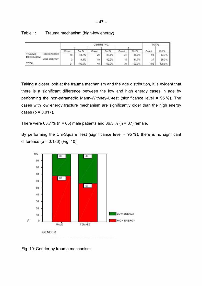
– 47 –
Table 1: Trauma mechanism (high-low energy)
Taking a closer look at the trauma mechanism and the age distribution, it is evident that
there is a significant difference between the low and high energy cases in age by
performing the non-parametric Mann-Withney-U-test (significance level = 95 %). The
cases with low energy fracture mechanism are significantly older than the high energy
cases (p = 0.017).
There were 63.7 % (n = 65) male patients and 36.3 % (n = 37) female.
By performing the Chi-Square Test (significance level = 95 %), there is no significant
difference (p = 0.186) (Fig. 10).
Fig. 10: Gender by trauma mechanism
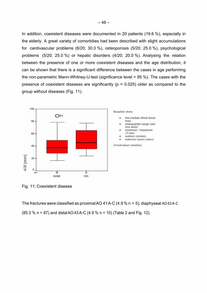
– 48 –
In addition, coexistent diseases were documented in 20 patients (19.6 %), especially in
the elderly. A great variety of comorbities had been described with slight accumulations
for cardivascular problems (6/20; 30.0 %), osteoporosis (5/20; 25.0 %), psychological
problems (5/20; 25.0 %) or hepatic disorders (4/20; 20.0 %). Analysing the relation
between the presence of one or more coexistent diseases and the age distribution, it
can be shown that there is a significant difference between the cases in age performing
the non-parametric Mann-Whitney-U-test (significance level = 95 %). The cases with the
presence of coexistent diseases are significantly (p = 0.025) older as compared to the
group without diseases (Fig. 11).
Fig. 11: Coexistent disease
The fractures were classified as proximal AO 41 A-C (4.9 % n = 5), diaphyseal AO42A-C
(85.3 % n = 87) and distal AO 43 A-C (4.9 % n = 10) (Table 2 and Fig. 12).
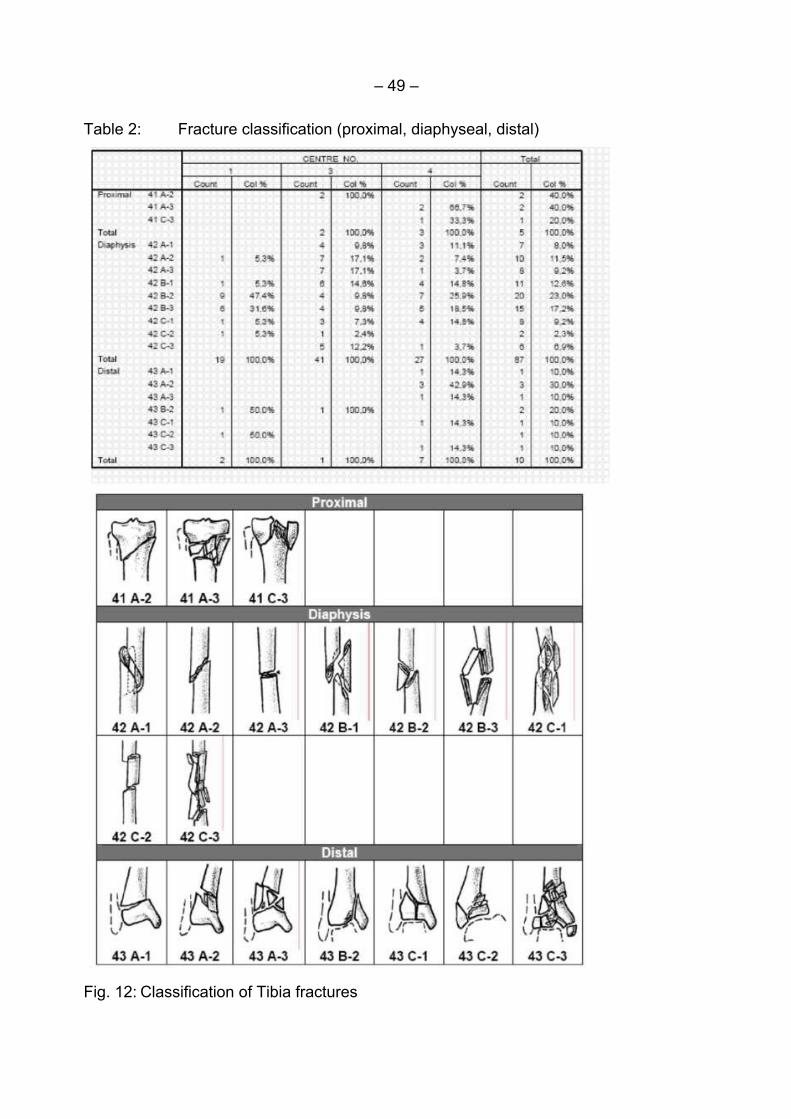
– 49 –
Table 2: Fracture classification (proximal, diaphyseal, distal)
Fig. 12: Classification of Tibia fractures

– 50 –
5.2 Perioperative
The mean intraoperative fluoroscopy time was 369 seconds with a range of 15 to 1314
seconds.
Table 3: Fluoroscopy time
Mean operative time (skin to skin) was 104 minutes and a range from 35 up to 300
minutes (Table 4).
Table 4: Operative time
Analysing the relation between the operation time and the trauma mechanism, it can be
shown that there is a significant difference between the low and high energy cases in
operation time by performing the non-parametric Mann-Whitney-U-test. The cases with
high energy trauma had a significantly longer surgery as compared to the group with low
energy trauma (p = 0.007).

– 51 –
Fig. 13: Relation between trauma mechanism and operative time
The fracture was reduced in 48 % of cases without traction and 18 % of the cases with
traction. In 34 % of the cases this information was unknown. Open reduction was
performed in 12 % as compared to closed reduction in 88 % of the cases. There were 69
% of the patients who were operated on a standard operation table as compared to 31
% who were operated on an orthopaedic table. Mean blood loss was 184 cc with a
range from 10 cc up to 600 cc. Performing the non-parametric Mann-Whitney-U-test
(significance level = 95 %), there is no clear significant difference in blood loss between
the groups with high and low energy trauma (p = 0.069) (Fig. 14).

– 52 –
Fig. 14: Blood loss between high-low energy trauma
A tourniquet was used during surgery in 45 % compared to being not used in 55 % of
the cases. Analysing the relation between the blood loss and the use or not use of a
tourniquet, it can be shown that there is a significant difference between the groups in
blood loss by performing the non parametric Mann-Whitney-U-test. The cases where a
tourniquet was used had significantly less blood loss compared to the group where the
tourniquet was not used (p< 0.001) (Fig.15).

– 53 –
Fig. 15: Blood loss and whether tourniquet was used or not
The following POSITIVE comments were collected about subjective appreciation/
handling and instrument reliability of the T2 Tibial Nailing system (n = 58 patients):
• Good handling/reliable instruments/no problems n = 54
The following NEGATIVE comments were collected about subjective appreciation/
handling and instrument reliability of the T2 tibial nailing system (n = 58 patients):
• Nail length measurement problems n = 2
• Difficulties with screw length measurements n = 3
• Difficulties with locking screw placement n = 2
Among the 100 cases ( 2 patients were lost of follow-up) only 5 % (n = 5) reported a
deviation of the operated leg. No deviation was measured in 64 %. This information was
unknown for 31 % of the cases. In two out of the five cases an axial deviation was
detected. For the remaining three cases the deviation was a rotational failure. The axial

– 54 –
and rotational failures were re-operated. There were 66 % (n = 100) patients who
showed no difference in leg length after surgery. In 2 % (n = 100) the operated leg was
shorter compared to the contralateral side and in 32 % (n = 100) this information was
unknown. Full weight bearing was possible in 48 % (n = 100) of the patients during the
first days after surgery, 18 % (n = 100) were able to do partial weight bearing and 30 %
(n = 100) could not do weight bearing within the first days for several reasons
(polytrauma, general status). In four percent (n = 100) of the patients this information
was unknown. The overall mean stay of the patients in hospital was 14 days (± 10 days)
and a range from 3 days up to 60 days (Table 5).
Table 5: Stay in hospital
In performing the non-parametric Mann-Whitney-U-test (significance level = 95 %), there
is no clear significant difference in hospital stay between the group’s high and low
energy trauma. For details, see Fig.16.
Table 5: Stay in hospital

– 55 –
Fig. 16: Stay in hospital (days) by trauma mechanism
(44 %) (n = 94) of the patients’ discharge location was back to their homes, 21 % (n =
94) were discharged to another hospital, 9% (n = 94) went to a rehabilitation unit, and 7
% (n = 94) to a health institution. In 19 % (n = 94) of the patients this information was
unknown. Taking a closer look at the six cases that developed an early compartment
syndrome postoperatively, it can be shown that all six cases were not treated with a
tourniquet intraoperatively (p = 0.024).
5.3 Postoperative assessments: 4-6 weeks, 4 months, 12 months
Details about patient participation in follow-up are shown in Table 6. For interpretation of
percentage rates, please keep in mind that there are not the same total numbers in the
different assessments.
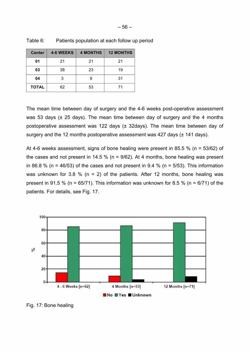
– 56 –
Table 6: Patients population at each follow up period
Center 4-6 WEEKS 4 MONTHS 12 MONTHS
01 21 21 21
03 38 23 19
04 3 9 31
TOTAL 62 53 71
The mean time between day of surgery and the 4-6 weeks post-operative assessment
was 53 days (± 25 days). The mean time between day of surgery and the 4 months
postoperative assessment was 122 days (± 32days). The mean time between day of
surgery and the 12 months postoperative assessment was 427 days (± 141 days).
At 4-6 weeks assessment, signs of bone healing were present in 85.5 % (n = 53/62) of
the cases and not present in 14.5 % (n = 9/62). At 4 months, bone healing was present
in 86.8 % (n = 46/53) of the cases and not present in 9.4 % (n = 5/53). This information
was unknown for 3.8 % (n = 2) of the patients. After 12 months, bone healing was
present in 91.5 % (n = 65/71). This information was unknown for 8.5 % (n = 6/71) of the
patients. For details, see Fig. 17.
Fig. 17: Bone healing

– 57 –
After 4 months 45.3 % of the patients (n = 24/53) were able to return to work or previous
activities. At 12 months this value increases to 76.1 % (n = 54/71). Fig. 18 shows the
number of patients who were able to return to work or do their previous activities
postoperatively.
Fig. 18: Returning to work – previous activities
At 4-6 weeks, 22.6 % (n = 14/62) of the patient population were able to work with full
previous capacity. 9.7 % (n = 6/62) were able to work up to 75 % of their previous
capacity.
There were 17.7 % (n = 11/62) who were able to work up to 50 % of their previous
capacity and 41.9 % (n = 26/62) of the patients were not able to reach more than 25 %
of their previous working capacity (unknown: 8.1 %, n = 5/62).
At 4 months, 20.8 % (n = 11/53) of the patient population were able to work with full
previous capacity postoperatively. There were 47.2 % (n = 25/53) who were able to work
up to 75 % of their previous capacity, 11.3 % (n = 6)/53 were able to work up to 50 % of
their previous capacity and 11.3 % (n = 6/53) of the patients were not able to reach more
than 25 % of their previous working capacity (unknown: 15.1 %, n = 8/53) (Fig.19).

– 58 –
At 12 months, 74.7 % (n = 53/71) of the patient population were able to work with full
previous capacity postoperatively. There were 4.2 % (n = 3/71) who were able to work
up to 75 % of their previous capacity, 4.2 % (n = 3/71) were able to work up to 50 % of
their previous capacity and 4.2 % (n = 3/71) were not able to reach more than 25 % of
their previous working capacity (unknown 12.7 %, n = 9/71). Fig. 19 below illustrates the
working capacity of the patients.
Fig. 19: Working capacity
For 51.6 % (n = 32/62) of the cases, it was not difficult to put on socks and shoes 4-6
weeks postoperatively. At 4 months assessment, this value increases to 67.9 % (n =
36/53). Finally at 12 months assessment, 83.1 % (n = 59/71) were able to put on socks
and shoes without any difficulty (Fig. 20).

– 59 –
Fig. 20: Putting on socks and shoes
There were 43.5 % (n = 27/62) of the cases who could rise from a chair without upper
extremity support 4-6 weeks postoperatively. At 4 months assessment, this value
increased to 64.2 % (n = 34/53) and at 12 months assessment 83.1 % (n = 59/71) of the
patient were able to rise from a chair without upper extremity support (Fig. 21).
Fig. 21: Sitting and standing
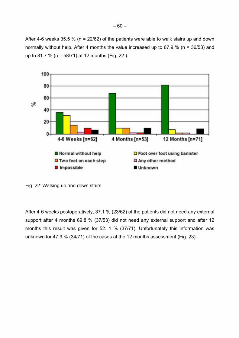
– 60 –
After 4-6 weeks 35.5 % (n = 22/62) of the patients were able to walk stairs up and down
normally without help. After 4 months the value increased up to 67.9 % (n = 36/53) and
up to 81.7 % (n = 58/71) at 12 months (Fig. 22 ).
Fig. 22: Walking up and down stairs
After 4-6 weeks postoperatively, 37.1 % (23/62) of the patients did not need any external
support after 4 months 69.8 % (37/53) did not need any external support and after 12
months this result was given for 52. 1 % (37/71). Unfortunately this information was
unknown for 47.9 % (34/71) of the cases at the 12 months assessment (Fig. 23).

– 61 –
Fig. 23: Walking capacity and external support
Total weight bearing was possible for 30.6 % (n = 19/62) of the cases at the 4-6 weeks
post operation assessment. After 4 months the value increased to 77.4 % (n = 41/53).
After 12 months 53.5 % (n = 38/71) could perform total weight bearing (unknown 46.5
%, n = 33/71) (Fig. 24).
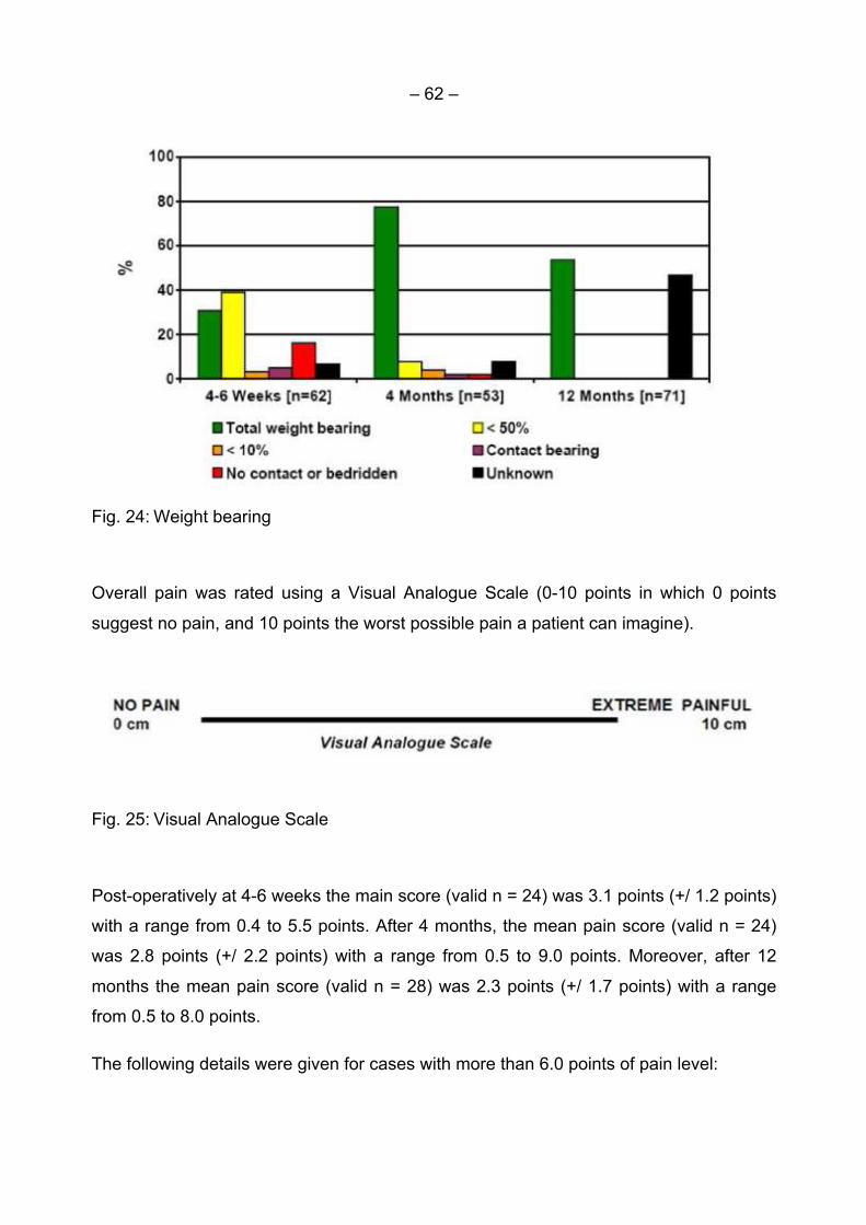
– 62 –
Fig. 24: Weight bearing
Overall pain was rated using a Visual Analogue Scale (0-10 points in which 0 points
suggest no pain, and 10 points the worst possible pain a patient can imagine).
Fig. 25: Visual Analogue Scale
Post-operatively at 4-6 weeks the main score (valid n = 24) was 3.1 points (+/ 1.2 points)
with a range from 0.4 to 5.5 points. After 4 months, the mean pain score (valid n = 24)
was 2.8 points (+/ 2.2 points) with a range from 0.5 to 9.0 points. Moreover, after 12
months the mean pain score (valid n = 28) was 2.3 points (+/ 1.7 points) with a range
from 0.5 to 8.0 points.
The following details were given for cases with more than 6.0 points of pain level:

– 63 –
After 4 months pain level above 9 was in 2 cases. One patient had swelling in the knee
joint and deep infection. The other had delayed healing and distal migration of the nail.
After 12 months, one patient had a pain level of 8. This specific case had a
pseudoarthrosis and needed several re-operations.
Anterior knee pain
At the 4-6 weeks follow-up anterior knee pain was found in 11 patients, at 4 months in
14 patients, and at 12 months in 13 patients. None of the patients included in this study
had any chronic knee pain. There was a gradual increase in total weight bearing
capacity of the patients. The daily activity (walking up and down stairs, putting on shoes
and socks, sitting in a chair or standing from it, walking distance, using one or two
crutches) showed improvement. There was no difference in age between the two groups
(anterior knee pain mean age 43 years, no knee pain mean age 41 years).
At 4-6 weeks, 62 patients were available for assessment. Eleven patients (18 %) had
anterior knee pain (4 paratendinous/7 transtendinous). Patients with a transtendinous
incision had more pain compared to those with paratendinous incision. At the follow-up
after 4 months, 53 patients were assessed and 14 (26 %) of them had anterior knee
pain. The cases were the nail was introduced through the mid-patellar incision (n = 10)
had more pain than those with the para-patellar incision (n = 4). At the 12 months follow-
up 13 patients (n = 71) had anterior knee pain. It was interesting to notice that patients
with a para patellar- incision (n = 7) had more pain compared to those with the mid
patellar incision (n = 6). However, the number of patients was too small to make a
statistical calculation.
The VAS decreased from a mean of 3.1 at 4-6 weeks to a mean of 2.3 at 12 months
follow-up. Despite the fact that there was anterior knee pain, all the patients (n = 13)
were able to perform weight bearing and bony healing was inconspicuous in all the
patients (n = 13) after 12 months. At 4-6 weeks, 2 patients with anterior knee pain were
able to work with full previous capacity and this was increased to 9 patients after 12

– 64 –
months. Two patients (n = 13) were working for 25 % after 12 months. The reason for
this was that they were poly-traumatised patients. After 4-6 weeks postoperatively, only
one patient could walk up and down the stairs without any help. This was increased to 7
patients after 4 months and to 8 patients after 12 months. Total weight bearing was
improved and was possible in 9 patients after 4 months and in 6 patients after 12
months.
Unfortunately at the two months follow-up 7 patients went lost to the study. Putting on
shoes and socks without any difficulty was possible in 9 patients (n = 13) after 12
months. There were no missing patients after 12 months. Further daily activities like
sitting and standing from a chair without arm support gradually improved and was
possible in 8 patients (n = 13) after 12 months. There was only 1 patient missing after 12
months. The time taken until patients could walk without any support was also
examined. It showed improvement, but this information was missing for 8 patients after
12 months (n = 13). The use of external support while walking was decreased after 12
months. Again there was information missing in 8 cases.
5.4 Postoperative complications
After 4-6 weeks out of 62 patients 1 patient had a hematoma, 3 had nerve problems, 1 a
deep and 3 a superficial infection. Out of 53 patients 2 cases had neurodystrophy
(CRPS Type 1) at 4 months assessment, 6 patients had mechanical or implant
associated complications (broken screws, risk of proximal screw perforating the skin).
After 1 year out of 71 patients 9 patients had mechanical problems, 4 patients a nerve
lesion, 3 were reported with superficial infections, 4 with neurodystrophy and 1 with
heterotopic bone at the fracture site.

– 65 –
5.5 Dynamisation of the nail
Dynamisation of the nail was performed in 4/62 patients after 4-6 weeks, in 5/53 patients
after 4 months and in 5/71 patients after 1 year.
Reasons for dynamisation were delayed healing or screw problems(broken/migration of
screws).
5.6 Revision surgery
Revision surgery (malrotation, malpositioning proximal nail migration) was performed in
7/102 patients. Postoperative time between primary surgery and revision had a mean of
134 days (2-362 days).
5.7 Material removal
In 47/102 patients the nail was removed. The mean time between surgery and removal
was 422 days (93-1126 days). Reasons for nail removal were a healed fracture, anterior
knee pain or broken screws.

– 66 –
6 Discussion
The present study had been planned as a prospective international multicenter trial with
a 1-year follow-up. We included 102 patients with all types of tibia fractures who
underwent a reamed intermedullary nailing. The re-examinations were frequented by 62
patients after 4-6 weeks, by 53 patients after 4 months and by 71 patients after 12
months. The reason for this variable attendance may be that probably some
polytraumatised patients were referred to their local hospital after the first treatment in
the trauma centers and did not return for follow-up for medical, practical, or personal
reasons. The patients who had no complaints did possibly not feel the need to visit the
outpatient clinic, which may have resulted in an overestimation of less-than-ideal results
in the present study.
Another limitiation of this study were the differing medical conditions. On the one hand,
the operations were performed by surgeons with different experience, working ethics
and institutional financial frameworks that could possibly have led to deviating treatment
modalities irrespective of the same intramedullary nailing method. On the other hand,
the study population was heterogeneous with proximal, shaft and distal fractures of
different severity in polytraumatised or monotraumatised patients. Because of the broad
range of fracture types, the number of cases in each fracture subtype became too small
for a comparative statistical analysis which could have thrown light on some pros and
cons for the suitability of the T2 nail for different fracture types.
We suggest that the ideal study ought to be a randomised prospective clinical study. The
patients should have only one type of tibial fracture. The T2 nail should be compared
with another nail and both should be reamed. Despite these shortcomings, our study
population allows an overview over the suitablity of the tested nail system in the daily
tasks of a trauma center.The population consisted of 63.7 % male and 36.3 % female
patients with a mean age of 42 ± 16 years. Other studies confirm that tibia fractures
occur more often in male than in female patients (Weiss et al., 2008; Larsen et al.,
2015).

– 67 –
The present study shows that most fractures occurred in traffic accidents (31.4 %) and in
accidents on the road as pedestrian (22.5 %) or in the domestic setting (22.5 %). More
rarely the accident occurred during sports (13.7 %) or at work (5.9%). High energy
traumata predominated low energy traumata (63.7 % vs. 36.3 %). We found some
correlations between trauma, age and gender: In younger patients, especially in men,
tibia fractures often take place in the context of a high energy trauma caused by
traffic accidents while in older patients low energy traumata at home are more
frequent. These findings are supported by other studies (Court-Brown and Caesar,
2006; Weiss et al., 2008; Madadi et al., 2010; Larsen et al., 2015). In contrast to our
study, Court-Brown and Caesar (2006) moreover reported a high incidence of sport
accidents that may be due to regional differences or deviations of age distribution.
In our study, diaphyseal wedge fractures of all severities (AO 42-B, n = 46, 45.1 %) were
most common, followed by simple (AO 42-A, n = 25, 24.5 %) and complex diaphyseal
fractures (AO 42-C, n = 18, 17.6 %). Only very few studies have reported the AO
distribution of tibia fractures, and these are in contrast to our results. Court-Brown and
Caesar (2006) reported a predominance of type A42-A3, while in the study of Larsen
et al. (2015) fractures of type A42-A1 were most common. These differences are
assumedly caused by patient selection: The population of the present study was
recruited in large trauma centers while both cited studies were population-based and
therefore more reliable represent the real life distribution of fracture types.
The treatment in trauma centers may also be the cause for the observed low
frequency of patients with a single tibia fracture (35.2 %) while in the rest of patients the
fibula was fractured, too, (50.0 %) or the patients had been polytraumatized (14.7 %),
because multiple fractures often occur in a population with a high frequency of high
energy traumata (Larsen et al., 2015).
Unsurprisingly, the surgical efforts were correlated with the fracture severity. High
energy trauma causing complex fractures resulted in a significantly longer operation
time as compared to low energy fractures (p = 0.007). Indirect hints on this relation are
also given by high variances of blood loss (184 ± 174 cc) and duration of hospital stay
(14 ± 10 days) which ranged from 3 to 60 days. The high variances are due to the wide
– 68 –
spectrum of fracture types in our study. Therefore, a comparison with published data is
aggravated, and even more so with regard to the complexity of fractures, the classification
of fracture types, the number and age of patients, the observation period, the treatment
modalities, the endpoints, and other parameters.
Full weight bearing was allowed and possible in 48/100 of our patients during the first
days after surgery, while in 18/100 patients a partial and in 30/100 patients no weight
bearing was allowed or possible within the first days for several reasons
(polytrauma, general status). Especially in static nailing a delay in full weight bearing is
recommended until the first signs of bone union are visible (Drosos et al., 2006).
In our patients, bone healing was radiologically confirmed in 85.5 % (53/62) of patients
after 4-6 weeks, in 86.8 % (46/53) after 4 months and in 91.5 % (65/71) after 12 months.
Rates of union of more than 90 % after intramedullary nailing of tibial fractures have
been reported in other studies, too (Klemm and Borner, 1986; Alho et al., 1990; Court-
Brown et al., 1991). In our study the intramedullary canal was reamed in all cases. This
has two advantages: Firstly, the debris formed by reaming is deposited at the fractures
site and acts as an autologous bone graft (Reynders and Broos, 2000). Secondly, a
better cortical contact between nail and bone results, allowing for a larger nail to be
inserted and hence improving stability (Chapman, 1998). Thirdly, adding a compression
screw further enhanced the stability of the nail. All of these factors helped to achieve the
good bone healing in our patients.
In addition to radiological bone healing, the regaining of our patients’ daily activities
was documented as a reference for fracture healing. Unfortunately, for some
parameters the 12-month-data were missing. E.g. we analysed that total (i.e.
monopodial) weight bearing was possible in 77.5 % and walking stairs without
external support (e.g. cane, crutch, walker, heelpiece) in 69.8 % after 4 months.
As expected, there was a gradual increase over time in the number of patients who went
back to work. The percentage of patients who returned to work or to their previous
activities increased continuously from 22.6 % (14/62, 4-6 weeks) to 45.3 % (24/53; 4
months) and to 76.1 % (54/71; 12 months), respectively. There were 3 patients whose
– 69 –
final working capacity did not exceed 25 %: the first patient with polytrauma (upper
extremities, open fractures, nerve problems), the second patient preoperatively
underwent extensive sport training in judo and the third patient had been invalid at
baseline and needed several reoperations. A total of 11 % of patients were not able to
work after one year. It is well known that tibial fractures are severe injuries and may
result in permanent disability with significant socio-economic implications (Bode et al.,
2012). Taking the complexity of fractures in many of our patients into account, the
achieved results are yet very satisfying. In the population of Court-Brown et al. (1990)
the mean time to resumption of full activities except sports in those patients who did
not have multiple injuries was 14.3 (5-30) weeks and 78% returned to full activities.
The results of Alho et al. (1990) were better: They reported their experience with locked
intramedullary reamed nailing for 93 displaced tibial shaft fractures. After 12 months 90 %
of their patients returned to work, 100 % had full weight-bearing, and all fractures showed
a radiographic consolidation. The reason for the discrepancy to our results may be the
greater number of comminuted fractures and our additional inclusion of proximal and
distal tibia fractures.
Interestingly, in the long term the functional results seem to be independent of the
decision for a reamed or unreamed nail, respectively. Alho et al. (1990) found no
significant difference in time of return to work with an average of 10.9 weeks for the
reamed group and 9.3 weeks for the unreamed group, and in another study by Keating et
al. (1997), the functional outcome, in terms of knee pains, return to work and recreational
activity, did not differ significantly between reamed and unreamed nailing.
Naturally, the functional outcome is influenced by postoperative pain. Anterior knee pain
has been identified as the most common and most significant complication of
intramedullary tibia nailing. The incidence of knee pain is identical in patients with
a reamed or unreamed intramedullary nailing (Court-Brown et al., 1996).
It is the most common reason for nail removal. However the outcome of nail
removal is unpredictable. Court-Brown et al. (1990) presented the results of their
prospective study about the use of the Grosse-Kempf tibial nail in the treatment of 125
closed and type 1 open (Gustilo and Andersen 1976) tibial fractures. Knee pain was

– 70 –
present in 51 (40.8%) patients, and in 33 of them the nail had to be removed. The knee
pain usually occured after a few weeks and was associated with kneeling. It was usually
abolished by removing the nail although in some cases the relief took several months to
occur.
Postoperatively at 4-6 weeks, the main score was 3.1 ± 1.2 points (n = 24) with a range
from 0.4 to 5.5 points. After 4 months, the mean pain score decreased to 2.8 ± 2.2
points (n = 24) with a range from 0.5 to 9.0 points. At that time a pain level above 9 was
seen in 2 cases due to complications: One patient had a swollen knee and deep
infection. The other patient showed delayed healing and distal migration of the nail.
Moreover, after 12 months the mean pain score was 2.3 ± 1.7 points (n = 28) with a range
from 0.5 to 8.0 points. There was one patient with a pain level of 8 points as a
symptom of complications: He suffered from pseudarthrosis and needed several re-
operations.
In patients who underwent an intramedullary nailing postoperative pain, especially
anterior knee pain, is associated with the surgical approach. Karladani et al. (2000)
performed a prospective randomised study at 53 patients with unilateral, displaced and
closed or grade 1 open (Gustilo and Anderson 1976) tibial shaft fractures which were
randomised to treatment with an intramedullary nail or a plaster cast. In this study, 12 of
27 patients with an intramedullary nail, but no patient with a plaster cast, suffered from
anterior knee pain (p< 0.001). Because the nails had been inserted paratendinously,
the authors suspected that the dissection in the patellar region, a trauma of the patellar
tendon during nail insertion, and iatrogenic damage of the infrapatellar nerve might
have caused anterior knee pain. Hernigou and Cohen (2000) studied the relationship
between the intra-articular structures of the knee and the entry point used for nailing in 54
tibiae from cadaver specimens. The results showed that in some bones the safe zone
is smaller than the size of standard reamers and the proximal part of some nails. The
structures at risk are the anterior horns of the medial and lateral menisci, the anterior
part of the medial and lateral plateau and the ligamentum transversum. They
concluded that unrecognized intraarticular injury of the knee may be one of the reasons
for anterior knee pain.

– 71 –
In the present study, 11/61 patients (18.0 %) had anterior knee pain at 4-6 weeks. Four
patients had been operated with a paratendinous and 7 with a transtendinous
approach, and the patients with a transtendinous incision reported more pain
compared to those with paratendinous incision. After 4 months 14/53 patients (26.4 %)
suffered from anterior knee pain (paratendinous n = 10; transtendinous n = 4), and after
12 months 13/71 patients (18.3 %) (paratendinous n = 6; transtendinous n = 7). Our
database is too small to generalize the results but the transtendinous approach
seems to cause more frequent pain than the paratendinous approach. Despite the fact
that there was anterior knee pain, all patients concerned were able to perform weight
bearing, and bony healing was inconspicuous in all of them after 12 months.
Orfaly et al. (1995) reviewed their experience with intramedullary nailing to determine
whether the nail entry point has an influence on the development of knee pain. They
operated on 110 fractures in 107 patients with reamed Gross-Kempf nails, and the
site of nail insertion had been recorded for 101 fractures. The nail insertion had been
conducted by paratendinous incision in 65 fractures, and 33 of these cases (51 %) were
associated with subsequent knee pain. When nail insertion was conducted by tendon-
splitting 28 of 36 fractures (78%) developed subsequent knee pain (p< 0.01). Therefore
the authors recommended the paratendinous approach to reduce anterior knee pain.
Toivanen et al. (2002) performed a prospective randomised study comparing two different
nail insertion techniques. Fifty patients with tibial shaft fracture requiring intramedullary
nailing were randomised equally to treatment with paratendinous or transtendinous
nailing.
Compared with a transpatellar tendon approach, a paratendinous approach for nail
insertion did not reduce the prevalence of chronic anterior knee pain. There was no
significant difference between the two groups. The authors’ conclusion was that it is not
possible to reduce anterior knee pain by using a paratendinous approach.
Devitt et al. (1998) measured the patellofemoral joint forces and pressures during
intramedullary nailing of the tibia in 8 cadaveric knees. They noticed that the contact
pressure in the patello-femoral articulation increased after nailing regardless of the

– 72 –
approach that had been used. This may result in chondral injury, which in turn may
cause anterior knee pain.
Karladani and Styf (2001) described an approach in which the skin incision is located on
either side of the patella and the insertion of the nail is performed percutaneously and
used it in 13 patients with tibia fractures. After a follow-up of 22 months, all patients
could kneel and there was no sign of dysfunction of the infrapatellar branch of the
saphenous nerve.
It is obvious that the source of chronic anterior knee pain after intramedullary nailing of
tibial fracture is multifactorial. A prospective randomised controlled trial is required to
confirm the benefit of the percutaneous technique, but this new approach may have
advantages including no dissection around the patellar tendon, a skin incision in an area
that is not involved in kneeling and less risk of damage to the infrapatellar nerve.
However, reaming also damages the blood supply to the inner two thirds of the cortex
(Klein et al., 1990) which may result in impaired fracture healing and increased infection
rates despite the fact that published literature does not reflect these clinical worries
(Anglen and Blue, 1995; Mayr et al., 1995; Ruchholtz et al., 1995; Court-Brown et al.,
1996; Blachut et al., 1997; Keating et al., 1997; Finkemeier et al., 2000). Further
dreaded complications of the reaming procedure are compartment syndrome and the
production of emboli by reaming and subsequent fat embolism or pulmonary embolism
(Pell et al., 1993). Interestingly, while reaming damages the blood flow to the cortex, it
simultaneously induces a six-fold increase of the periosteal blood flow (Reichert et al.,
1995). Perhaps this factor accounts for the good clinical results of reamed nailing, but
this currently remains speculation.
In our population there were no cases of postoperative embolism or compartment
syndrome, but one patient (1/62 (1.6 %) with an open fracture suffered from a deep
infection after 4-6 weeks which could be treated by conservative management. In
reamed intramedullary nailing deep infection may be regarded as a relatively rare
complication which is reported only in individual cases (Finkemeier et al., 2000) while
many authors pointed out that there was no deep infection in their population after
nailing tibia fractures of different types (Court-Brown et al., 1996; Blachut et al., 1997;
– 73 –
Larsen et al., 2004; Sadighi et al., 2011). Other infections occured during the first 4-6
weeks post operationem in three patients (n = 3/62; 4.8%); they were superficial and
healed by antibiotic therapy. The percentage of superficial infections is consistent with
published data that reach from 2.0 to 16.0 % for reamed nailed tibia fractures (Gregory
and Sanders, 1995; Wiss and Stetson, 1995; Chiu et al., 1996a, b).
In the present study complications associated with the nails or screws were seen later in
the course of time: After 4 months there were 4/53 (7.5 %) cases with mechanical
or implant associated complications such as broken screws (n = 3/53; 5.7 %) or risk of
proximal screw perforating the skin (n = 1/53; 1.9%). After 1 year, 9/71 (12.7 %)
patients had mechanical problems: three cases (3/73; 4.1 %) each of screw breakage
and screw loosening/migration or pain from screws, respectively. One patient with a
migration of the proximal screw to the skin showed a pseudarthrosis. A dynamisation
was conducted after 4 months, but in the course of time other (not documented)
reoperations were necessary after transfer to another hospital due to his multitrauma
and bad general condition.
The design of interlocking nails inevitably necessitates screw holes, and this bears
the biomechanical potential for a high local concentration of stress (Bucholz et al.,
1987). Thus, the screws bear the stress and act as a weak link in the interlocking
nail-to-bone-complex when its strength is lower than the strength of the nail alone. There
are reports of interlocking screw breakages occurring in up to 50 % of cases which
where nailed without reaming and of nail failures in up to 10 % (Hutson et al., 1995;
Whittle et al., 1995; Court-Brown et al., 1996). Especially unreamed nailing is prone to
failures of the nails and distal locking screws because the nails do not fit tightly and more
cyclical loading occurs.
The risk is growing with decreasing nail diameter as the smaller the nail the smaller (and
weaker) must be the screws. In reamed nailing the risk is lower. For example Blachut et
al. (1997), Court-Brown et al. (1996), and (Gonschorek et al., 1998) reported of 3/73
(4.1 %), 1/25 (4.0 %), and 13/402 (3.2 %) respectively, of their patients with broken
screws, and Blachut et al. (1997) as well as Court-Brown et al. (1996) reported 4/73
(5.5 %) or 1/25 (4.0 %), respectively, with screw migration. These data are comparable
– 74 –
to our results, where a total of 3 patients (3 %) had a distal broken screw that
needed revision. All affected patients had received a nail with a small diameter, and they
were fairly heavy, young and active as well as fully weight bearing within the first week,
therefore the osteosynthesis had to bear an outstanding stress.
In other patients, no revision was necessary: In a total of 14/102 (13.7 %) patients a
dynamisation of the nail had to be performed due to delayed healing and/or screw
problems. The dynamisation was conducted in 4/62 patients (6.5 %) after 4-6 weeks,
in 5/53 patients (9.4 %) after 4 months and in 5/71 patients (7.0 %) after 1 year. On the
one hand, the rigidity of the osteosyntheses is important to keep the bone fragments in
good collaboration, therefore creating the conditions for rapid recovery of medullary
circulation, which in turn is an essential condition for the formation of endosteal callus.
Static intramedullary nailing unables movements between fragments which directly
stimulates bone formation and formation of angiogenic minimal callus. On the other
hand, the fracture phenomenon "dynamization” allows micro-movements at the point of
fracture, assuming that axially moved fragments simultaneously reduce fracture gaps,
and accelerate callus maturation and bone remodeling. This dynamization should take
place early, when the fibrous callus provides stability (Omerovic et al., 2015). The
transformation of static into dynamic intramedullary osteosynthesis by dynamization is
recommended if the fractures heal inadequately (Wiss and Stetson, 1995; Brumback,
1996), but it is not a mandatory requirement for healing (Brumback et al., 1988). The
possibility that dynamization may cause malunion in spiral, short oblique, or high-
comminuted fractures has even led to a complete refusal of this method in favour of the
static intermedullary nailing (Templeman et al., 1997). In the present study, all cases
with dynamisation, except one, had no signs of pseudarthrosis or nonunion.
Revision surgery had been performed in 7/102 patients (6.9%). In 3 patients the revision
was indicated because of a malrotation due to screw breakage, in 2 patients because of
pain by the proximal screws, and in 2 patients because of screw loosening with the risk
of skin perforation. This low rate of revisions underlines the finding that reamed
intramedullary nailing leads to low nonunion rates and implant failure rates (Kessler et
al., 1986; Bhandari et al., 2000; Forster et al., 2005; Bhandari et al., 2008).

– 75 –
In only 47/102 patients (46.1 %) the nail was known to be removed. The mean time
between surgery and removal in these cases was 422 days (93-1126 days). Reasons for
nail removal were a healed fracture (n = 34/47; 72.3 %), anterior knee pain despite
healed fracture (n = 11/47; 23.4 %), broken/migrating screws (n = 2/47; 4.3 %) or patient
request (n = 1/47; 2.1 %).
Unfortunately, no comparisons can be drawn to published data comparable to our
results, where a total of 3 patients (3 %) had a distal broken screw that needed revision.
All affected patients had received a nail with a small diameter, and they were fairly
heavy, young and active as well as fully weight bearing within the first week, therefore
the osteosynthesis had to bear an outstanding stress.
Altogether the results of our study show that the intramedullary nailing of tibia
fractures by means of the T2 nail led to very satisfying results with excellent healing and
low complication rates underlining the efficacy of reamed nailing in comparison to
other methods of osteosynthesis, especially to non-reamed nailing. In a review of
prospective studies about the management and treatment complications in a total of
895 tibial fractures, Coles and Gross (2000) found out that the reoperation rates of
reamed nailing are comparable to plate fixation (5.7 % vs. 4.7 %) and much lower than in
unreamed nailing (16.7 %). The incidence of malunion is highest in closed treatment with
a plaster cast (31.7 %) but low in operative treatment with best results for plate
osteosyntheses (0 %), followed by reamed nailing (3.2 %) and unreamed nailing
(11.8%). These good results of plates are limited by a high infection rate (9.0 %) as
compared to reamed (2.9%) and unreamed nailing (0.5 %) and plaster cast treatment
(0 %). That means that all treatment methods are associated with complications, but that
the results of reamed nailing are favorable. Up to now great randomized controlled
studies are lacking to get deeper insight in the benefits of reamed nailing and the
best method of treatment, respectively.

– 76 –
7 Comments (criticism) on this study
Prospective follow-up of the (poly) traumatised patient is not an easy task. In our study
population, there were patients missing at every follow-up period. The reasons for this is
that probably polytrauma patients are referred to their local hospital after the first
treatment in the trauma centers and these patients do not come back for the follow-up.
Another reason for this is that patients without any complaints do not see the necessity
for visiting the outpatient clinic. Operations were performed by surgeons with different
experience and working ethics. In this study population we had different types (proximal,
shaft and distal) of tibial fracture. We had also poly traumatised patients that were
compared with patients who had a monotrauma. An international multicenter study is
very difficult to coordinate. The patients have different social backgrounds and social
systems.
We suggest that the ideal study ought to be a randomised prospective clinical study. The
patient should have only one type of tibial fracture. The T2 nail should be compared with
another nail and both should be reamed.

– 77 –
8 Summary
In this prospective multicenter clinical study 102 patients with a tibial fracture were
operated with the reamed T2TM Stryker tibia nail system in 3 European Level 1
Traumacenters during 01-2003 to 12-2004. We documented demographic (e.g. age,
gender), preoperative (e.g. trauma cause, fracture type), general operative (e.g. surgery
time, blood loss), and postoperative data (e.g. radiologic bone healing, weight bearing,
activities of daily living, return to work, anterior knee pain). The follow-up assessments
took place at 4-6 weeks (n = 62), 4 months (n = 53) and 12 months (n = 71) after
suegery.
There were 63.7 % male and 36.3 % female patients with a mean age of 42 ± 16 years.
Most common were AO 42-B1-B3 fractures (45.1 %), followed by simple (AO 42-A1-3,
24.5 %) and complex diaphyseal fractures (AO 42-C1-3, 17.6 %). The rest of 15
fractures consisted of 10 distal (AO 43-A1-3, AO 43-B2, AO 43-C1-3) and 5 proximal
fractures (AO 41 A2-3, AO 41-C3). 65 patients (63.7 %) had been involved in high-, and
37 patients (36.3 %) in low-energy trauma. Mostly, the accidents had occurred in traffic
(31.4 %), on road as pedestrian (22.5 %), or at home (22.5 %). Only 36 patients
(35.2 %) had a single tibia fracture, in 51 patients (50.0 %) the fibula was fractured, too,
and 15 patients (14.7 %) had been polytraumatized.
After 12 months bone healing was radiologically confirmed in 91.5 % (n = 65/71). 76.1 %
(n = 54/71) had returned to work and 74.7 % (n = 53/71) were able to work with full
previous capacity. The mean pain score decreased from 3.1 ± 1.2 (4-6 weeks) to 2.8 ±
2.2 (4 months) to 2.3 ± 1.7 points (12 months). After 12 months, 13 patients suffered
from anterior knee pain, which was mild in 12 patients so that they could work and bear
weight. One polytraumatized patient showed a pain score of 8 points due to
pseudarthrosis and needed several reoperations.
In a total of 14 patients a dynamisation of the nail had to be performed due to delayed
healing and/or screw problems. Revision surgery had been performed in 7/102 patients
(6.9%). In 3 patients the revision was indicated because of a malrotation due to screw

– 78 –
breakage, in 2 patients because of pain by the proximal screws, and in 2 patients
because of screw loosening with the risk of skin perforation.
Our study about the T2 TM nail system showed results comparable with other studies
regarding intramedullary reamed nailing and underlines the hypothesis that this
osteosynthesis method is effective and exhibits relatively few complications,

– 79 –
9 Bibliography
Ahmad M, Nanda R, Bajwa AS, Candal-Couto J, Green S, Hui AC. Biomechanical testing of the locking compression plate: when does the distance between bone and implant significantly reduce construct stability? Injury 2007;38: 358-364
Alho A, Ekeland A, Stromsoe K, Folleras G, Thoresen BO. Locked intramedullary nailing for displaced tibial shaft fractures. J Bone Joint Surg Br 1990;72: 805-809
Anglen JO, Blue JM. A comparison of reamed and unreamed nailing of the tibia. J Trauma 1995;39: 351-355
Aro HT, Chao EY. Bone-healing patterns affected by loading, fracture fragment stability, fracture type, and fracture site compression. Clin Orthop Relat Res 1993;8-17
Attal R, Blauth M. Unaufgebohrte Marknagelung. Orthopäde 2010;39: 182-191
Bayne G, Turner RG. Closed fracture manipulation--improving Charnley's three point fixation technique. Ann R Coll Surg Engl 2006;88: 504
Bhandari M, Guyatt G, Tornetta P, 3rd, Schemitsch EH, Swiontkowski M, Sanders D et al. Randomized trial of reamed and unreamed intramedullary nailing of tibial shaft fractures. J Bone Joint Surg Am 2008;90: 2567-2578
Bhandari M, Guyatt GH, Tong D, Adili A, Shaughnessy SG. Reamed versus nonreamed intramedullary nailing of lower extremity long bone fractures: a systematic overview and meta-analysis. J Orthop Trauma 2000;14: 2-9
Bircher H. Eine neue Methode unmittelbarer Retention bei Fracturen der Röhrenknochen.Arch Klin Chir 1886;34: 410-422
Blachut PA, O'Brien PJ, Meek RN, Broekhuyse HM. Interlocking intramedullary nailing with and without reaming for the treatment of closed fractures of the tibial shaft. A prospective, randomized study. J Bone Joint Surg Am 1997;79: 640-646
Bode G, Strohm PC, Sudkamp NP, Hammer TO. Tibial shaft fractures - management and treatment options. A review of the current literature. Acta Chir Orthop Traumatol Cech 2012;79: 499-505
Boenisch UW, de Boer PG, Journeaux SF. Unreamed intramedullary tibial nailing--fatigue of locking bolts. Injury 1996;27: 265-270
Böhler J. Treatment of nonunion of the tibia with closed and semiclosed intramedullary nailing.Clin Orthop Relat Res 1965;43: 93-101
Bong MR, Kummer FJ, Koval KJ, Egol KA. Intramedullary nailing of the lower extremity: biomechanics and biology. J Am Acad Orthop Surg 2007;15: 97-106

– 80 –
Brumback RJ. The rationales of interlocking nailing of the femur, tibia, and humerus. Clin Orthop Relat Res 1996;292-320
Brumback RJ, Uwagie-Ero S, Lakatos RP, Poka A, Bathon GH, Burgess AR. Intramedullary nailing of femoral shaft fractures. Part II: Fracture-healing with static interlocking fixation. J Bone Joint Surg Am 1988;70: 1453-1462
Brunner CF, Weber BG. Besondere Osteosynthesetechniken. Berlin-Heidelberg-New York: Springer, 1981
Bucholz RW, Ross SE, Lawrence KL. Fatigue fracture of the interlocking nail in the treatment of fractures of the distal part of the femoral shaft. J Bone Joint Surg Am 1987;69: 1391-1399
Cantu RV, Koval KJ. The use of locking plates in fracture care. J Am Acad Orthop Surg 2006;14: 183-190
Carter DR, Beaupre GS, Giori NJ, Helms JA. Mechanobiology of skeletal regeneration. Clin Orthop Relat Res 1998;S41-55
Chapman MW. The effect of reamed and nonreamed intramedullary nailing on fracture healing. Clin Orthop Relat Res 1998;S230-238
Chiu FY, Lo WH, Chen CM, Chen TH, Huang CK. Treatment of unstable tibial fractures with interlocking nail versus Ender nail: a prospective evaluation. Zhonghua Yi Xue Za Zhi (Taipei) 1996a;57: 124-133
Chiu FY, Lo WH, Chen CM, Chen TH, Huang CK. Unstable closed tibial shaft fractures: a prospective evaluation of surgical treatment. J Trauma 1996b;40: 987-991
Clatworthy MG, Clark DI, Gray DH, Hardy AE. Reamed versus unreamed femoral nails. A randomised, prospective trial. J Bone Joint Surg Br 1998;80: 485-489
Coles CP, Gross M. Closed tibial shaft fractures: management and treatment complications. A review of the prospective literature. Can J Surg 2000;43: 256-262
Collinge C, Sanders R, DiPasquale T. Treatment of complex tibial periarticular fractures using percutaneous techniques. Clin Orthop Relat Res 2000;69-77Cornell CN, Lane JM. Newest factors in fracture healing. Clin Orthop Relat Res 1992;297-311 Court-Brown CM, Caesar B. Epidemiology of adult fractures: A review. Injury 2006;37:691-697
Court-Brown CM, Christie J, McQueen MM. Closed intramedullary tibial nailing. Its use in closed and type I open fractures. J Bone Joint Surg Br 1990;72: 605-611
Court-Brown CM, McQueen MM, Quaba AA, Christie J. Locked intramedullary nailing of open tibial fractures. J Bone Joint Surg Br 1991;73: 959-964

– 81 –
Court-Brown CM, Will E, Christie J, McQueen MM. Reamed or unreamed nailing for closed tibial fractures. A prospective study in Tscherne C1 fractures. J Bone Joint Surg Br 1996;78: 580-583
Danckwardt-Lilliestrom G. Reaming of the medullary cavity and its effect on diaphyseal bone. A fluorochromic, microangiographic and histologic study on the rabbit tibia and dog femur. Acta Orthop Scand Suppl 1969;128: 1-153
Danckwardt-Lilliestrom G, Lorenzi L, Olerud S. Intracortical circulation after intramedullary reaming with reduction of pressure in the medullary cavity. J Bone Joint Surg Am 1970;52: 1390-1394
Dehne E, Metz CW, Deffer PA, Hall RM. Nonoperative treatment of the fractured tibia by immediate weight bearing. J Trauma 1961;1: 514-535
den Outer AJ, Meeuwis JD, Hermans J, Zwaveling A. Conservative versus operative treatment of displaced noncomminuted tibial shaft fractures. A retrospective comparative study. Clin Orthop Relat Res 1990;231-237
Devitt AT, Coughlan KA, Ward T, McCormack D, Mulcahy D, Felle P et al. Patellofemoral contact forces and pressures during intramedullary tibial nailing. Int Orthop 1998;22: 92-96
Downie WW, Leatham PA, Rhind VM, Wright V, Branco JA, Anderson JA. Studies with pain rating scales. Ann Rheum Dis 1978;37: 378-381
Drosos GI, Bishay M, Karnezis IA, Alegakis AK. Factors affecting fracture healing after intramedullary nailing of the tibial diaphysis for closed and grade I open fractures. J Bone Joint Surg Br 2006;88: 227-231
Eriksson AR, Albrektsson T. Temperature threshold levels for heat-induced bone tissue injury: a vital-microscopic study in the rabbit. J Prosthet Dent 1983;50: 101-107
Finkemeier CG, Schmidt AH, Kyle RF, Templeman DC, Varecka TF. A prospective, randomized study of intramedullary nails inserted with and without reaming for the treatment of open and closed fractures of the tibial shaft. J Orthop Trauma 2000;14: 187-193
Fisher WD, Hamblen OI. Problems and pitfalls of compression fixation of long bone fractures: a review of results and complications. Injury 1978;10:99-107
Forster MC, Aster AS, Ahmed S. Reaming during anterograde femoral nailing: is it worth it? Injury 2005;36: 445-449
Francois J, Vandeputte G, Verheyden F, Nelen G. Percutaneous plate fixation of fractures of the distal tibia. Acta Orthop Belg 2004;70: 148-154

– 82 –
Frigg R. Locking Compression Plate (LCP). An osteosynthesis plate based on the Dynamic Compression Plate and the Point Contact Fixator (PC-Fix). Injury 2001;32 Suppl 2: 63-66
Frigg R. Development of the Locking Compression Plate. Injury 2003;34 Suppl 2: B6-10
Gautier E, Ganz R. Die biologische Plattenosteosynthese. Zentralbl Chir 1994;119: 564-572
Gomez-Benito MJ, Gonzalez-Torres LA, Reina-Romo E, Grasa J, Seral B, Garcia-Aznar JM. Influence of high-frequency cyclical stimulation on the bone fracture-healing process: mathematical and experimental models. Philos Trans A Math Phys Eng Sci 2011;369: 4278-4294
Gonschorek O, Hofmann GO, Buhren V. Interlocking compression nailing: a report on 402 applications. Arch Orthop Trauma Surg 1998;117: 430-437
Gonzalez-Torres LA, Gomez-Benito MJ, Doblare M, Garcia-Aznar JM. Influence of the frequency of the external mechanical stimulus on bone healing: a computational study. Med Eng Phys 2010;32: 363-371
Greenwood DC, Muir KR, Doherty M, Milner SA, Stevens M, Davis TR. Conservatively managed tibial shaft fractures in Nottingham, UK: are pain, osteoarthritis, and disability long-term complications? J Epidemiol Community Health 1997;51: 701-704
Gregory P, Sanders R. The treatment of closed, unstable tibial shaft fractures with unreamed interlocking nails. Clin Orthop Relat Res 1995;48-55
Greiwe RM, Archdeacon MT. Locking plate technology: current concepts. J Knee Surg 2007;20: 50-55
Gustilo RB, Merkow RL, Templeman D. The management of open fractures. J Bone Joint Surg Am 1990;72: 299-304
Habermeyer P, Wolf K, Schweiberer L. Konservative Behandlung diaphysärer Frakturen des Unterschenkels beim Erwachsenen. Chirurg 1990;61: 772-776
Heitemeyer U, Hierholzer G. [Bridging osteosynthesis in closed compound fractures of the femur shaft]. Aktuelle Traumatol 1985;15: 205-209
Helfet DL, Shonnard PY, Levine D, Borrelli J, Jr. Minimally invasive plate osteosynthesis of distal fractures of the tibia. Injury 1997;28 Suppl 1: A42-47; discussion A47-48
Henry SL, Adcock RA, Von Fraunhofer JA, Seligson D. Heat of intramedullary reaming. South Med J 1987;80: 173-176
Hente R, Fuchtmeier B, Schlegel U, Ernstberger A, Perren SM. The influence of cyclic compression and distraction on the healing of experimental tibial fractures. J Orthop Res 2004;22: 709-715

– 83 –
Hernigou P, Cohen D. Proximal entry for intramedullary nailing of the tibia. The risk of unrecognised articular damage. J Bone Joint Surg Br 2000;82: 33-41
Herzig N, Müller CA, Eckhardt C, Schlegel U, Pfister U, Suedkamp NP. Temperaturentwicklung bei der Markraumbohrung enger Markhöhlen. Unfallchirurg 2001;283: 467-468
Herzog K. Die Technik der geschlossenen Marknagelung frischer Tibiafrakturen mit dem Rohrschlitznagel. Chirurg 1958;29: 501-506
Horn J, Linke B, Hontzsch D, Gueorguiev B, Schwieger K. Angle stable interlocking screws improve construct stability of intramedullary nailing of distal tibia fractures: a biomechanical study. Injury 2009;40: 767-771
Hulth A. Current concepts of fracture healing. Clin Orthop Relat Res 1989;265-284
Hupel TM, Aksenov SA, Schemitsch EH. Effect of limited and standard reaming on cortical bone blood flow and early strength of union following segmental fracture. J Orthop Trauma 1998;12: 400-406
Husebye EE, Lyberg T, Madsen JE, Eriksen M, Roise O. The influence of a one-step reamer-irrigator-aspirator technique on the intramedullary pressure in the pig femur. Injury 2006;37: 935-940
Hutson JJ, Zych GA, Cole JD, Johnson KD, Ostermann P, Milne EL et al. Mechanical failures of intramedullary tibial nails applied without reaming. Clin Orthop Relat Res 1995;129-137
Hutter CG, Oden R, Kirk R. The intramedullary compression rod. Clin Orthop Relat Res 1977;165-173
Isaksson H, Wilson W, van Donkelaar CC, Huiskes R, Ito K. Comparison of biophysical stimuli for mechano-regulation of tissue differentiation during fracture healing. J Biomech 2006;39: 1507-1516
Jagodzinski M, Krettek C. Effect of mechanical stability on fracture healing--an update. Injury 2007;38 Suppl 1: S3-10
Jahna H, Wittich H. Konservative Methoden in der Frakturbehandlung. Wien, München, Baltimore: Urban und Schwarzenberg, 1985
Joist A, Schult M, Ortmann C, Frerichmann U, Frebel T, Spiegel HU et al. Rinsing-suction reamer attenuates intramedullary pressure increase and fat intravasation in a sheep model. J Trauma 2004;57: 146-151
Karladani AH, Granhed H, Edshage B, Jerre R, Styf J. Displaced tibial shaft fractures: a prospective randomized study of closed intramedullary nailing versus cast treatment in 53 patients. Acta Orthop Scand 2000;71: 160-167

– 84 –
Karladani AH, Styf J. Percutaneous intramedullary nailing of tibial shaft fractures: a new approach for prevention of anterior knee pain. Injury 2001;32: 736-739
Keating JF, O'Brien PJ, Blachut PA, Meek RN, Broekhuyse HM. Locking intramedullary nailing with and without reaming for open fractures of the tibial shaft. A prospective, randomized study. J Bone Joint Surg Am 1997;79: 334-341
Kempf I, Jaeger JH, Weigel A. Biomechanische Untersuchungen zur Verriegelungsnagelung. Symposium Wien. Wien, München, Bern: Maudrich, 1978
Kessler SB, Hallfeldt KK, Perren SM, Schweiberer L. The effects of reaming and intramedullary nailing on fracture healing. Clin Orthop Relat Res 1986;18-25
Klein MP, Rahn BA, Frigg R, Kessler S, Perren SM. Reaming versus non-reaming in medullary nailing: interference with cortical circulation of the canine tibia. Arch Orthop Trauma Surg 1990;109: 314-316
Klemm K, Schellmann WD. Dynamische und statische Verriegelung des Marknagels. Monatsschr Unfallheilkd Versicher Versorg Verkehrsmed 1972;75: 568-575
Klemm KW, Borner M. Interlocking nailing of complex fractures of the femur and tibia. Clin Orthop Relat Res 1986;89-100
König F. Über die Implantation von Elfenbein zum Ersatz von Knochen und Gelenkenden. Beitr Klin Chir 1913;91-114
Krause WR, Bradbury DW, Kelly JE, Lunceford EM. Temperature elevations in orthopaedic cutting operations. J Biomech 1982;15: 267-275
Krettek C. Prinzipien der intramedullären Knochenbruchstabilisierung. Teil 1. Unfallchirurg 2001;104: 639-651; quiz 652
Krettek C, Schandelmaier P, Tscherne H. Nonreamed interlocking nailing of closed tibial fractures with severe soft tissue injury. Clin Orthop Relat Res 1995;34-47
Küntscher G. Die Technik des Aufweitens der Markhöhle. Chirurg 1959;30: 28-35 Küntscher G. Praxis der Marknagelung. Stuttgart: Schattauer, 1962
Lacroix D, Prendergast PJ. A mechano-regulation model for tissue differentiation during fracture healing: analysis of gap size and loading. J Biomech 2002;35: 1163-1171
Lambotte A. Chirurgie opératoire des fractures. Paris: Masson & Cie, 1913
Larsen LB, Madsen JE, Hoiness PR, Ovre S. Should insertion of intramedullary nails for tibial fractures be with or without reaming? A prospective, randomized study with 3.8 years' follow-up. J Orthop Trauma 2004;18: 144-149
Larsen P, Elsoe R, Hansen SH, Graven-Nielsen T, Laessoe U, Rasmussen S. Incidence and epidemiology of tibial shaft fractures. Injury 2015;46: 746-750

– 85 –
Leach RE. Fractures of the tibia and fibula. In: Rockwood C.A., Green DP (Hrsg.) Fractures in adults. Philadelphia: Lippincott, 1984: S. 1593-1664
Leunig M, Hertel R, Siebenrock K, Balmer F., Mast J, Ganz R. The evaluation of indirect reduction techniques for the treatment of fractures. Clin Orthop 2001;357: 307-314
Li M, Zhang X, Liu X, Jing Y. The recent development of MIPO in long bone fractures. Open Journal of Orthopedics 2012;2: 159-165
Madadi F, Vahid Farahmandi M, Eajazi A, Daftari Besheli L, Madadi F, Nasri Lari M. Epidemiology of adult tibial shaft fractures: a 7-year study in a major referral orthopedic center in Iran. Med Sci Monit 2010;16: CR217-221
Maffulli N, Toms AD, McMurtie A, Oliva F. Percutaneous plating of distal tibial fractures. Int Orthop 2004;28: 159-162
Mayr E, Barnikel C, Braun W, Rüter A. Die geschlossene Unterschenkelfraktur - aufgebohrte oder unaufgebohrte Marknagelung? Eine klinische Studie. Zentralbl Chir 1995;120: 24-30; discussion 30-21
Müller CA. Einfluss der Marknagelosteosynthese auf die lokale Perfusion, Geschwindigkeit und Qualität der Frakturheilung. Vergleich eines experimentellen Bohrsystems mit dem konventionellen AO-Bohrsystem der ungebohrten Nagelung. Habilitationsschrift. Albert-Ludwigs-Universität Freiburg, 2003
Müller CA, Frigg R, Pfister U. Can modifications to reamer and flexible shaft design decrease intramedullary pressure during reaming? An experimental investigation. Techn Orthop 1996;11: 18-27
Müller CA, McIff T, Rahn BA, Pfister U, Perren SM, Weller S. Influence of the compression force on the intramedullary pressure development in reaming of the femoral medullary cavity. Injury 1993;24 Suppl 3: S36-39
Müller CA, Schlegel V, Hoegel F, Eckhardt C, Schlegel U, Rahn BA et al. Cortical perfusion and local fat occlusion after intramedullary nailing of the ovine tibia--comparison of different surgical procedures. Injury 2009;40: 760-766
Nicoll EA. Fractures of the tibial shaft. A survey of 705 cases. . J Bone Joint Surg Br 1964;46: 373-387
Ochsner PE, Baumgart F, Kohler G. Heat-induced segmental necrosis after reaming of one humeral and two tibial fractures with a narrow medullary canal. Injury 1998;29 Suppl 2: B1-10
Olerud S, Danckwardt-Lilliestrom G. Fracture healing in compression osteosynthesis. An experimental study in dogs with an avascular, diaphyseal, intermediate fragment. Acta Orthop Scand Suppl 1971;137: 1-44

– 86 –
Olerud S, Karlström G. Secondary intramedullary nailing of tibial fractures. J Bone Joint Surg Am 1972;54: 1419-1424
Olerud S, Karlström G. Tibial fractures treated by AO compression osteosynthesis. Experiences from a five year material. Acta Orthop Scand Suppl 1992; 140: 100-104
Omerovic D, Lazovic F, Hadzimehmedagic A. Static or dynamic intramedullary nailing of femur and tibia. Med Arch 2015;69: 110-113
Oni OO, Hui A, Gregg PJ. The healing of closed tibial shaft fractures. The natural history of union with closed treatment. J Bone Joint Surg Br 1988;70: 787-790
Orfaly R, Keating JE, O'Brien PJ. Knee pain after tibial nailing: does the entry point matter? J Bone Joint Surg Br 1995;77: 976-977
Pape HC, Dwenger A, Regel G, Schweitzer G, Jonas M, Remmers D et al. Pulmonary damage after intramedullary femoral nailing in traumatized sheep--is there an effect from different nailing methods? J Trauma 1992;33: 574-581
Pape HC, Remmers D, Regel G, Tscherne H. Pulmonale Komplikationen nach intramedullärer Stabilisierung langer Röhrenknochen. Einfluss von Operationsverfahren, -Zeitpunkt und Verletzungsmuster. Orthopäde 1995;24: 164-172
Pell AC, Christie J, Keating JF, Sutherland GR. The detection of fat embolism by transoesophageal echocardiography during reamed intramedullary nailing. A study of 24 patients with femoral and tibial fractures. J Bone Joint Surg Br 1993;75: 921-925
Peltier LF. Fractures: A history and iconography of their treatment. San Francisco: Norman Publishing, 1990
Perren SM. Point contact fixator: part I. Scientific background, design and application. Injury 1995;22: 1-10
Perren SM. Evolution and rational of locked internal fixator technology. Introductory remarks. Injury 2001;32: B3-B9
Perren SM. Evolution of the internal fixation of long bone fractures. The scientific basis of biological internal fixation: choosing a new balance between stability and biology. J Bone Joint Surg Br 2002;84: 1093-1110
Pfister U. Marknagelung nach Aufbohrung. Orthopäde 2010;39: 171-181
Pfister U, Frigg R. Die Verklemmung des Marknagels in der Markhöhle der Tibia. In vitro-Messung der Längs- und Querdeformation des Marknagels mit Hilfe von Dehnungsmessstreifen. Aktuelle Traumatol 1980;10: 117-121
Pfister U, Rahn BA, Perren SM, Weller S. Vaskularität und Knochenumbau nach Marknagelung langer Röhrenknochen. Aktuel Traumatol 1979;9: 191-195

– 87 –
Poacz F. Verbrennungsschaden an der Tibiadiaphyse nach Marknagelung mit Aufbohrung. Unfallheilkunde 1979;82: 126-128
Puno RM, Teynor JT, Nagano J, Gustilo RB. Critical analysis of results of treatment of 201 tibial shaft fractures. Clin Orthop Relat Res 1986;113-121
Rahn BA. Knochenheilung unter den Bedingungen der Marknagelung. Osteo Int 1995;4: 240-245
Rammelt S, Endres T, Grass R, Zwipp H. The role of external fixation in acute ankle trauma. Foot Ankle Clin 2004;9: 455-474, vii-viii
Redfern DJ, Syed SU, Davies SJ. Fractures of the distal tibia: minimally invasive plate osteosynthesis. Injury 2004;35: 615-620
Rehm J, Übing D. Die Behandlung von Frakturen langer Röhrenknochen mit dem Marknagel nach Küntscher. Arch Orthop Unfallchir 1963;55: 82-109
Reichert IL, McCarthy ID, Hughes SP. The acute vascular response to intramedullary reaming. Microsphere estimation of blood flow in the intact ovine tibia. J Bone Joint Surg Br 1995;77: 490-493
Reynders PA, Broos PL. Healing of closed femoral shaft fractures treated with the AO unreamed femoral nail. A comparative study with the AO reamed femoral nail. Injury 2000;31: 367-371
Rhinelander FW. The normal microcirculation of diaphyseal cortex and its response to fracture. J Bone Joint Surg Am 1968;50: 784-800
Rhinelander FW. Tibial blood supply in relation to fracture healing. Clin Orthop Relat Res 1974;34-81
Richardson TE, Seligson D, Voor M. Fracture site compression and motion with three types of intramedullary fixation. Osteo Int 1998;6: 261-264
Riemer BL, DiChristina DG, Cooper A, Sagiv S, Butterfield SL, Burke CJ, 3rd et al. Nonreamed nailing of tibial diaphyseal fractures in blunt polytrauma patients. J Orthop Trauma 1995;9: 66-75
Ruchholtz S, Nast-Kolb D, Betz A, Schweiberer L. Frakturheilung nach Marknagelung einfacher Tibiaschaftfrakturen. Ein klinischer Vergleich gebohrter und ungebohrter Verfahren. Unfallchirurg 1995;98: 369-375
Rüedi T, Murphy W. AO principles of fracture management. Stuttgart, New York: Thieme, 2000
Runkel M, Wenda K, Degreif J, Blum J. Ergebnisse nach primärer ungebohrter Tibianagelung von Unterschenkelfrakturen mit schwerem offenem oder geschlossenem Weichteilschaden. Unfallchirurg 1996;99: 771-777
– 88 –
Sadighi A, Elmi A, Jafari MA, Sadeghifard V, Goldust M. Comparison study of therapeutic results of closed tibial shaft fracture with intramedullary nails inserted with and without reaming. Pak J Biol Sci 2011;14: 950-953
Sarmiento A, Gersten LM, Sobol PA, Shankwiler JA, Vangsness CT. Tibial shaft fractures treated with functional braces. Experience with 780 fractures. J Bone Joint Surg Br 1989;71: 602-609
Schandelmaier P, Krettek C, Rudolf J, Tscherne H. Outcome of tibial shaft fractures with severe soft tissue injury treated by unreamed nailing versus external fixation. J Trauma 1995;39: 707-711
Schandelmaier P, Stephan C, Reimers N, Krettek C. LISS-osteosynthese von distalen Femurfrakturen. Trauma Berufskrankh 1999;1: 392-397
Schemitsch EH, Turchin DC, Kowalski MJ, Swiontkowski MF. Quantitative assessment of bone injury and repair after reamed and unreamed locked intramedullary nailing. J Trauma 1998;45: 250-255
Schneider R. Die Marknagelung der Tibia. Helv Chir Acta 1961;28: 207-213
Strecker W, Gonschorek O, Fleischmann W, Bruckner U, Beyer M, Kinzl L. Thromboxane--co-factor of pulmonary disturbances in intramedullary nailing. Injury 1993;24 Suppl 3: S68-72
Stürmer KM, Schuchardt W. Neue Aspekte der gedeckten Marknagelung und des Aufbohrens der Markhöhle im Tierexperiment. III. Knochenheilung, Gefäßversorgung und Knochenumbau. Unfallheilkunde 1980;83: 433-435
Stürmer KM, Tammen E. Verminderung der corticalen Gefäßschädigung durch kontinuierliches Spülen und Absaugen während des Aufbohrens der Markhöhle. Unfallheilkunde 1986;181: 236-240
Templeman D, Larson C, Varecka T, Kyle RF. Decision making errors in the use of interlocking tibial nails. Clin Orthop Relat Res 1997;65-70
Tepic S, Perren SM. The biomechanics of the PC-Fix internal fixator. Injury 1995;26 (Suppl 1): 5-10
Toivanen JA, Vaisto O, Kannus P, Latvala K, Honkonen SE, Jarvinen MJ. Anterior knee pain after intramedullary nailing of fractures of the tibial shaft. A prospective, randomized study comparing two different nail-insertion techniques. J Bone Joint Surg Am 2002;84-A: 580-585
Tornetta P, 3rd, Tiburzi D. The treatment of femoral shaft fractures using intramedullary interlocked nails with and without intramedullary reaming: a preliminary report. J Orthop Trauma 1997;11: 89-92
– 89 –
Trojan E. Die konservative Behandlung des frischen geschlossenen Unterschenkelschaftbruches nach Lorenz Böhler. Orthopäde 1984;13: 256-261
Trueta J, Cavadias AX. Vascular changes caused by the Kuntscher type of nailing; an experimental study in the rabbit. J Bone Joint Surg Br 1955;37-B: 492-505
Tull F, Borrelli J, Jr. Soft-tissue injury associated with closed fractures: evaluation and management. J Am Acad Orthop Surg 2003;11: 431-438
Vallier HA, Le TT, Bedi A. Radiographic and clinical comparisons of distal tibia shaft fractures (4 to 11 cm proximal to the plafond): plating versus intramedullary nailing. J Orthop Trauma 2008;22: 307-311
Wagner M. General principles for the clinical use of the LCP. Injury 2003;34 Suppl 2: B31-42 Wagner M, Frigg R. Locking Compression Plate (LCP): Ein neuer AO-Standard. OP-Journal 2000;16: 238-243
Wehner W, Morgenstern C, Zeumer G. Das Verhalten des intramedullären Drucks bei Markbohrung und -nagelung. Zentralbl Chir 1966;91: 209-215
eiss RJ, Montgomery SM, Ehlin A, Al Dabbagh Z, Stark A, Jansson KA. Decreasing incidence of tibial shaft fractures between 1998 and 2004: information based on 10,627 Swedish inpatients. Acta Orthop 2008;79: 526-533
Weller S, Knapp U. Die Marknagelung. Gute und relative Indikationen. Chirurg 1975;46: 152-154
Wenda K, Ritter G, Ahlers J, Issendorff WD. Nachweis und Effekte von Knochenmarkeinschwemmungen bei Operationen im Bereich der Markhöhle des Oberschenkels. Unfallchirurg 1990;93: 56-61
Wenda K, Runkel M, Rudig L, Degreif J. Einfluss der Knochenmarkembolisation auf die Verfahrenswahl bei der Stabilisierung von Femurfrakturen. Orthopäde 1995;24: 151-163
Whittle AP, Wester W, Russell TA. Fatigue failure in small diameter tibial nails. Clin Orthop Relat Res 1995;119-128
Wieling R, Hagen R, Green J, Bresina S. Erste Resultate des Reamer Irrigator Aspirator in Vergleich mit der unaufgebohrten Nagelung: Eine in vivo Untersuchung am intakten Schafsfemurschaft. Hefte Unfallchir 1999;275: 469-470
Wiss DA, Stetson WB. Unstable fractures of the tibia treated with a reamed intramedullary interlocking nail. Clin Orthop Relat Res 1995;56-63

– 90 –
10 List of figures
Fig. 1: Fracture reduction technique (Habermayer et al , 1990) ...................................... 19
Fig. 2: Technique of wedging open the plaster (Habermeyer et al., 1990) ...................... 20
Fig. 3: Traction treatment technique (Habermeyer et al., 1990) ...................................... 21
Fig. 4: Correction of axis positions during traction treatment (Habermeyer et al., 1990) . 21
Fig. 5: Avoidance of the talipes equinus position (Habermeyer et al., 1990) ................... 22
Fig. 6: Angular Stable Locking System (ASLS) (Attal et al., 2010) .................................. 32
Fig. 7: Intramedullary nailing of the tibia (Pfister, 2010) ................................................... 37
Fig. 8: Description of the device: T2 TM Tibial Nailing System ....................................... 44
Fig. 9: Age distribution by trauma circumstances (Stryker Trauma R & D) ..................... 46
Fig. 10: Gender by trauma mechanism ............................................................................ 47
Fig. 11: Coexistent disease .............................................................................................. 48
Fig. 12: Classification of Tibia fractures ............................................................................ 49
Fig. 13: Relation between trauma mechanism and operative time ................................... 51
Fig. 14: Blood loss between high-low energy trauma ....................................................... 52
Fig. 15: Blood loss and whether tourniquet was used or not ............................................ 53
Fig. 16: Stay in hospital (days) by trauma mechanism ..................................................... 55
Fig. 17: Bone healing ........................................................................................................ 56
Fig. 18: Returning to work – previous activities ................................................................ 57
Fig. 19: Working capacity ................................................................................................. 58
Fig. 20: Putting on socks and shoes ................................................................................. 59
Fig. 21: Sitting and standing ............................................................................................. 59
Fig. 22: Walking up and down stairs ................................................................................. 60
Fig. 23: Walking capacity and external support ................................................................ 61
Fig. 24: Weight bearing .................................................................................................... 62
Fig. 25: Visual Analogue Scale ......................................................................................... 62

– 91 –
11 List of tables
Table 1: Trauma mechanism (high-low energy) ................................................................ 47
Table 2: Fracture classification (proximal, diaphyseal, distal) ........................................... 49
Table 3: Fluoroscopy time ................................................................................................. 50
Table 4: Operative time ..................................................................................................... 50
Table 5: Stay in hospital .................................................................................................... 54
Table 6: Patients population at each follow up period ....................................................... 56

– 92 –
12 Appendix - Clinical Review Form (CRF) Demographic data Age Gender Side: left/right Height in cm Weight in kg Body mass index (BMI) Trauma circumstances: At home At work On road (pedestrian) Traffic accident Sport Suicide intention Other (specify) Trauma mechanism: High energy/Low energy Preoperative data Surgical tactics: Initial treatment (0-36 h) Initial treatment (< 7 days) Delayed treatment (> 7 days) Revision /reoperation Fixateur externe Pseudoarthrosis Mono or multi fractures: Only one fracture Multi fractures (specify) Patient situation: Poly-trauma patient Coma > 7 days Transferred patient Other (please specify) Activity level prior to accident: Very active/sport Home activity Invalid Coexistent disease: None Yes (please specify) General diseases: Respiratory Cardiovascular Renal Hepatic Neurological Gastrointestinal Malignancy Other (please specify) Skeletal diseases: Osteoporosis Skin lesion: No Yes Missing Gustilo-Anderson classification: Grade 1 Grade 11 Grade 111 A Grade 111 B Grade 111 C Missing Tscherne classification: Grade 0 Grade 1 Grade 11 Grade 111 Missing Classification of tibial fractures: Proximal ( 41 A-C) Diaphysis (42 A-C) Distal (43 A-C) Preoperative complications: No/Yes General operative data Time between injury and surgery(days) Compression done (advanced compression screw): Yes No Missing Fluoroscopy time (seconds)
Fluoroscopy number of shots (shots) Operative time skin to skin (minutes) Blood loss (cc) Tourniquet used: Yes No Missing Nail parameters: Length Diameter Missing Skin incision proximal: Middle of patella tendon Tendon borderline Missing Stay in hospital (days) Postoperative assessments: 4-6-weeks, 4 months, 12 months. Bone healing: 4-6 weeks, 4 months, 12 months Yes No Missing Leg length (mm): Shortened No difference Longer Missing Leg Deviation: Axial Rotational No difference Missing Returning to work/ previous activities: 4-6 weeks, 4 months, 12 months No Yes Missing Working capacity: 4-6 weeks, 4 months, 12 months 0-25 % Up to 50 % Up to 75 % 100 % or close to Missing Putting on socks and shoes: 4-6 weeks, 4 months, 12 months No difficulty Some difficulty Very difficult Impossible Missing Sitting and standing: 4-6 weeks, 4 months, 12 months Can raise from chair without support Can raise from chair with support Cannot raise from chair independently Missing Walking up and down stairs: 4-6 weeks, 4 months, 12 months Normal without help Two feet on each step Impossible Foot over foot using banister Any other method Missing Walking capacity and external support: 4-6-weeks, 4 months, 12 months No need One cane or crutch Walker Heelpieces Two canes or crutches Unable or bedridden Missing Weight bearing: 4-6 weeks, 4 months, 12 months Total weight bearing < 50 % <10 % Contact bearing No contact or bedridden Missing VAS: 4-6 weeks, 4 months, 12 months Anterior knee pain: 4-6 weeks, 4 months, 12 months Postoperative complications: 4-6 weeks, 4 months, 12 months Dynamisation of the nail: 4-6 weeks, 4 months, 12 months.

– 93 –
13 Acknowledgement
One of the joys of completion is to look over the past journey and remember all the
friends and family who helped and supported me along this long but fulfilling road. It is a
pleasure to thank the many people who made this thesis possible.
Foremost, I would like to express my sincere gratitude to my advisor (Doktorvater) Prof.
Dr. Clayton Kraft for his continuous support, patience, motivation, enthusiasm and
immense knowledge. I could not have imagined having a better advisor and mentor for
my thesis. I would have been lost without him.
Besides my advisor I would like to thank Prof. Dr. Henk Haarman, head of the
Department of General Surgery and Trauma surgery from the Vrije Universiteit medisch
centrum (VUmc), Amsterdam, my teacher in trauma surgery and who gave me the
opportunity to coordinate this research. Whilst taking part in the weekly lunch research
meetings with the trauma surgery fellows and trauma surgeons with their
encouragement, insightful comments and hard questioning contributed greatly in
developing my interest in research.
My sincere thanks also go to Prof. Dr. Peter Patka from Erasmus MC, Rotterdam and
Dr. Fred Bakker from VUmc, Amsterdam both were my two other teachers in
traumasurgery.
I thank Dr. Kord Westermann from the Klinik für Unfall-, Hand-, und
Wiederherstellungschirurgie Klinikum Hannover Nordstadt Germany and Dr. Pablo de
Lucas Hospital Ramon y Cajal, Madrid, Spain for allowing me to use their data.
I am indebted to Claudia Beimel from Stryker Trauma R&D, Kiel, Germany for collecting,
processing and providing me with the data.
I am grateful to Marie-Luise Stein for her reading and correcting the English. She was
involved from the beginning of this thesis, encouraged me, gave sound advice and was
a good company.

– 94 –
I would like to thank my entire family for providing a caring environment for me. Special
thanks goes to my lovely wife, Joan Indrawatie Vishnudatt, who raised our children, and
who has always supported me during my career. I would never have been able to
achieve this without you, standing by me. My two children, Anuradha and Ranjana, both
medical doctors as well: I am a proud father.
Last but not least I would like to thank my mother (Gangadei Ramnarain) and father
(Ramadhar Jairam ✝). They bore me, raised me, supported me, taught me, and loved
me.
To them I dedicate this thesis.
![MiPo'11: Innovationsbezogene Kompetenzentwicklung in „Open Innovation“-Netzwerken der IT-Branche [Slides] (Sabrina Ziebarth et. al.)](https://img.pdfslide.org/doc/110x75/54c31f824a7959397c8b45b4/mipo11-innovationsbezogene-kompetenzentwicklung-in-open-innovation-netzwerken-der-it-branche-slides-sabrina-ziebarth-et-al.jpg)