Embed Size (px)
Citation preview
1
Title: Germline Variants in Asporin Vary by Race, Modulate the Tumor
Microenvironment and are Differentially Associated with Metastatic Prostate Cancer
Authors: Paula J. Hurley1,2,3, Debasish Sundi1, Brian Shinder1, Brian W. Simons1,4, Robert M. Hughes1,
Rebecca M. Miller1, Benjamin Benzon1, Sheila F. Faraj5, George J. Netto5, Ismael A. Vergara6, Nicholas Erho6,
Elai Davicioni6, R. Jeffrey Karnes7, Guifang Yan1, Charles Ewing1, Sarah D. Isaacs1, David M. Berman8,
Jennifer R. Rider9, Kristina M. Jordahl9, Lorelei A. Mucci9, Jessie Huang10, Steven S. An10,11,12, Ben H. Park2,3,
William B. Isaacs1, Luigi Marchionni2, Ashley E. Ross1,2,3,5, and Edward M. Schaeffer1,2,3
Authors’ Affiliations:
1Brady Urological Institute, Department of Urology, Johns Hopkins University, Baltimore, MD
2Department of Oncology, Johns Hopkins University, Baltimore, MD
3Sidney Kimmel Comprehensive Cancer Institute, Johns Hopkins University, Baltimore, MD
4Department of Molecular and Comparative Pathobiology, Johns Hopkins University, Baltimore, MD
5Department of Pathology, Johns Hopkins University, Baltimore, MD
6Genome Dx Biosciences Inc., Vancouver, BC, Canada
7Department of Urology, Mayo Clinic, Rochester, MN
8Department of Pathology and Molecular Medicine and Cancer Research Institute, Queens University,
Kingston, ON Canada
9Department of Epidemiology, Harvard University, Boston, MA
10The Department of Environmental Health Sciences, Johns Hopkins Bloomberg School of Public Health,
Baltimore, MD.
11The Department of Chemical and Biomolecular Engineering, The Johns Hopkins University, Baltimore, MD
12Physical Sciences-Oncology Center, Johns Hopkins University, Baltimore, MD
Research. on March 13, 2020. © 2015 American Association for Cancerclincancerres.aacrjournals.org Downloaded from
Author manuscripts have been peer reviewed and accepted for publication but have not yet been edited. Author Manuscript Published OnlineFirst on October 7, 2015; DOI: 10.1158/1078-0432.CCR-15-0256
2
Running Title: Germline ASPN Variants and Metastatic Progression
Keywords: Asporin, Germline Variants, Metastasis, Tumor Microenvironment, Prostate
Grant Support: This work was supported by The Department of Defense Prostate Cancer Research Program
Idea Award, DOD Award No PC110902 (PJH, LM, EMS); Flight Attendant Medical Research Institute Young
Clinical Scientist Award (PJH); The Patrick C. Walsh Prostate Cancer Fund Beth W. and A. Ross Myer Scholar
(PJH); The Prostate Cancer Foundation Hagen Challenge Award (EMS, PJH); National Institutes of Health
Grant T32DK007552 (DS); and National Institutes of Health Grant P30CA006973 (LM); The Johns Hopkins
Clinician Scientist Award (AER).
Corresponding Author: Paula J. Hurley, PhD, Brady Urological Institute, Department of Urology, Johns
Hopkins University, CRBII 154, 1550 Orleans Avenue, Baltimore, MD 21231. Phone: 410-614-9453; Fax:
410-502-5742; E-mail: [email protected]
Disclosure of Potential Conflicts of Interest The authors do not have financial interests that are a conflict of interest. We have the following disclosures.
ED, IAV, and NE are employees of GenomeDx and have stock in the company. BHP is a consultant for LOXO
Oncology and Horizon Discovery. AER and EMS are consultants for GenomeDx. PJH and EMS have
submitted a provisional patent through the Johns Hopkins Technology Transfer office.
Abstract word count: 238 Statement of Translational Relevance word count: 135 Research Article word count: 5062 Figures: 3 Tables: 3 Supplements: 8
References: 50
Research. on March 13, 2020. © 2015 American Association for Cancerclincancerres.aacrjournals.org Downloaded from
Author manuscripts have been peer reviewed and accepted for publication but have not yet been edited. Author Manuscript Published OnlineFirst on October 7, 2015; DOI: 10.1158/1078-0432.CCR-15-0256
3
Statement of Translational Relevance:
In this study, we report that germline ASPN D-repeat-length variants expressed in the tumor microenvironment
are differentially associated with progression to metastatic prostate cancer. To the best of our knowledge, this is
the first report of an association of a germline variant expressed by non-tumor cells in the tumor
microenvironment with metastatic prostate cancer progression. We additionally show that ASPN expression is
associated with local prostate cancer aggressiveness as measured by Gleason grade and poorer oncologic
outcomes including metastasis. We begin to establish biologic roles for ASPN D-repeat-length variants in
progression to metastatic prostate cancer. This study highlights the potential clinical utility of using germline
ASPN D-repeat-length to improve early therapeutic decisions for men who have localized cancer with lethal
potential, as well as providing novel heritable and mechanistic insights into progression to lethal disease.
Research. on March 13, 2020. © 2015 American Association for Cancerclincancerres.aacrjournals.org Downloaded from
Author manuscripts have been peer reviewed and accepted for publication but have not yet been edited. Author Manuscript Published OnlineFirst on October 7, 2015; DOI: 10.1158/1078-0432.CCR-15-0256
4
Abstract:
Purpose: Prostate cancers incite tremendous morbidity upon metastatic growth. We previously identified
Asporin (ASPN) as a potential mediator of metastatic progression found within the tumor microenvironment.
ASPN contains an aspartic acid (D)-repeat domain and germline polymorphisms in D-repeat-length have been
associated with degenerative diseases. Associations of germline ASPN D polymorphisms with risk of prostate
cancer progression to metastatic disease have not been assessed.
Experimental Design: Germline ASPN D-repeat-length was retrospectively analyzed in 1600 men who
underwent radical prostatectomy for clinically localized prostate cancer and in 548 non-cancer controls.
Multivariable Cox proportional hazards models were used to test the associations of ASPN variations with risk
of subsequent oncologic outcomes including metastasis. Orthotopic xenografts were used to establish allele- and
stroma-specific roles for ASPN D variants in metastatic prostate cancer.
Results: Variation at the ASPN D locus was differentially associated with poorer oncologic outcomes. ASPN
D14 (HR=1.72, 95%CI=1.05-2.81, P=0.032) and heterozygosity for ASPN D13/14 (HR=1.86, 95%CI=1.03-
3.35, P=0.040) were significantly associated with metastatic recurrence, while homozygosity for the ASPN D13
variant was significantly associated with a reduced risk of metastatic recurrence (HR=0.44, 95%CI=0.21-0.94,
P=0.035) in multivariable analyses. Orthotopic xenografts established biologic roles for ASPN D14 and ASPN
D13 variants in metastatic prostate cancer progression that were consistent with patient based data.
Conclusions: We observed associations between ASPN D variants and oncologic outcomes including
metastasis. Our data suggest that ASPN expressed in the tumor microenvironment is a heritable modulator of
metastatic progression.
Research. on March 13, 2020. © 2015 American Association for Cancerclincancerres.aacrjournals.org Downloaded from
Author manuscripts have been peer reviewed and accepted for publication but have not yet been edited. Author Manuscript Published OnlineFirst on October 7, 2015; DOI: 10.1158/1078-0432.CCR-15-0256
5
Introduction:
The burden of prostate cancer-related mortality remains significant due to the subset of prostate cancers that
progress to metastatic disease (1). Identification and differentiation of patients with aggressive cancer from
those with indolent disease remains a key issue in the management of clinically localized prostate cancer. To
date most biomarker studies have focused on carcinoma specific features predictive of aggressive disease. As
the tumor microenvironment influences prostate cancer progression (2), we hypothesized that molecules in the
primary tumor microenvironment secreted by cancer associated fibroblasts could modulate tumor progression
and predict metastatic potential. We previously reported that Asporin (ASPN), a member of a small leucine-rich
proteoglycan (SLRP) family of extracellular proteins, was one of the most highly enriched androgen-induced
mesenchymal genes during both physiologic prostate development and pathologic growth in the prostate (3).
ASPN expression has been shown to be elevated in prostate cancer associated fibroblasts (PCAFs) (4), in blood
from men with advanced prostate cancer (5), and in scirrhous gastric cancer associated stroma (6). A recent
study supports that ASPN induces coordinated fibroblast and cancer cell invasion through CD44 mediated
RAC1 activation (6); however, a biologic role for ASPN in the prostate cancer microenvironment is not well
understood.
In addition to expression, a genetic association exists between variation at the ASPN locus and multiple
disease states (7-9). ASPN, located at 9q22.31, contains an aspartic acid (D)-repeat domain and over 10
polymorphisms in the length of the D-repeat have been identified with the most common variants ranging from
12-16 aspartate residues (7). Polymorphisms in the length of this allelic repeat have been associated with
susceptibility to and severity of several bone and joint diseases including osteoarthritis (7-9). While functional
differences among these polymorphisms are not fully known, polymorphisms in the ASPN D-repeat-length
have been shown to differentially inhibit Bone Morphogenetic Protein 2 (BMP-2) signaling (10) and
Transforming Growth Factor Beta 1 (TGFβ1)-induced gene expression (7). Intriguingly, the loss of
TGFβ responsiveness in prostate stromal cells has been shown to promote prostate cancer initiation (11) and the
development of bone lesions (12). Due to its stromal specific expression and the differential role of ASPN D
Research. on March 13, 2020. © 2015 American Association for Cancerclincancerres.aacrjournals.org Downloaded from
Author manuscripts have been peer reviewed and accepted for publication but have not yet been edited. Author Manuscript Published OnlineFirst on October 7, 2015; DOI: 10.1158/1078-0432.CCR-15-0256
6
variants in TGFβ signaling, we hypothesized that ASPN D-repeat-length variants may differentially modulate
metastatic progression in the prostate.
Herein, we genotyped germline ASPN D-repeat-lengths in men with clinically localized prostate cancer
and in non-cancer controls to determine if germline ASPN D variants were differentially associated with risk of
prostate cancer incidence or progression to metastatic disease. We utilized an orthotopic xenograft model to
establish allele- and stroma-specific roles for these variants in facilitating prostate cancer metastasis.
Materials and Methods:
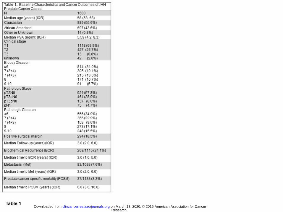
JHH Germline Study Population.
We retrospectively analyzed a database of 19,142 men who had undergone radical prostatectomy (RP) and
pelvic lymphadenectomy at Johns Hopkins Hospital (JHH) since 1992 (PSA era). Germline DNA was available
for approximately 10,000 patients. We selected 1672 cases for genomic analysis that were weighted for
approximately equal Gleason grades and to generate approximately equal white and non-white race groups, of
which 1600 had adequate DNA for germline genotyping. 55.6% men self-reported Caucasian, 43.6% self-
reported African American, and 0.8% self-reported other ethnicity or were of unknown ethnicity. We
retrospectively analyzed 192 self-reported Caucasian and 370 self-reported African American male controls
with no cancer, of which 179 and 369, respectively, had adequate germline DNA for genotyping. All
participants provided written informed consent. The protocol and consent documents were approved by the
Johns Hopkins University (JHU) Institutional Review Board (IRB).
RP specimens were processed as previously described (13). Each tumor was graded using the Gleason
scoring system and staged using the TNM (tumor-node-metastasis) system. Clinical outcome data included
biochemical recurrence (BCR) (defined as a post-operative Prostate Specific Antigen [PSA] ≥0.2 ng/ml), distant
metastasis (defined as post-operative clinical or radiographic spread of disease to extra-pelvic lymph nodes,
bones, or viscera), and prostate-cancer specific mortality (PCSM). Outcome or time to outcome data was not
Research. on March 13, 2020. © 2015 American Association for Cancerclincancerres.aacrjournals.org Downloaded from
Author manuscripts have been peer reviewed and accepted for publication but have not yet been edited. Author Manuscript Published OnlineFirst on October 7, 2015; DOI: 10.1158/1078-0432.CCR-15-0256
7
available for all men in the cohort; any resulting differences in sample size number were recorded in the results
section.
Genomic DNA isolation. Tissue for genomic DNA isolation was taken from seminal vesicles (SV) at the time
of RP (cases) or from blood of men without prostate cancer diagnoses (controls). SV tissue was suspended in
12ml Suspension buffer (20mM Tris; 25mM EDTA; 100mM NaCl) + 1ml 10% SDS + 60µl Proteinase K
solution (20mg/mL), inverted twice, and then incubated overnight at 50°C. The next day, RNA was digested by
adding 60µl RNase A Solution (Qiagen) and incubating at 37°C for 1 hour. Proteins were precipitated in 4ml
Protein Precipitation Solution (Promega) on ice for 20 minutes and then centrifuged at 2,000g for 10 minutes.
DNA was extracted from the supernatant with 30ml 100% Isopropanol, centrifuged 2,000g for 5 minutes, and
then washed with 70% ethanol followed by centrifugation.
Genotyping of the D-repeat polymorphism. The D-repeat polymorphism located in the N-terminal region of the
ASPN gene was PCR amplified using 5’ primer 6-FAM-ATTCCTGGCTTTGTGCTCTG and 3’ primer
TGGCTTCTTGGCTCTCTTGT. Primers were designed using Oligo software. Reactions were carried out in
10µL consisting of 30ng DNA, 0.125µM primers, 0.6mM dNTPs (Continental Lab Products), 10mM Tris-HCl
pH8.3, 50mM KCL, 1.5mM MgCl, and 0.6units of Taq Gold DNA polymerase (Perkin Elmer). Amplification
was performed in a Veriti Thermal Cycler (Applied Biosystems Inc.) for an initial denaturation of 12 minutes at
94ºC followed by 40 cycles of 94°C for 20 seconds, 58°C for 20 seconds, 72°C for 30 seconds, and a final 10
minute elongation at 72°C. The PCR products were electrophoresed on an ABI 3730xl DNA Analyzer
(Applied Biosystems Inc.). Data was collected and analyzed with GeneMapper software (Applied Biosystems
Inc.) that calculates fragment length in reference to an internal lane standard (LIZ500). Three homozygous
samples of different repeat-length were confirmed by Sanger sequencing. PCR products were sequenced using
fluorescent dideoxy terminator method of cycle sequencing. Reactions were run on a 3730xl DNA Analyzer
(Applied Biosystems Division) following Applied Biosystems protocols. Sequence data was analyzed using
Sequencher Software (Gene Codes).
Research. on March 13, 2020. © 2015 American Association for Cancerclincancerres.aacrjournals.org Downloaded from
Author manuscripts have been peer reviewed and accepted for publication but have not yet been edited. Author Manuscript Published OnlineFirst on October 7, 2015; DOI: 10.1158/1078-0432.CCR-15-0256
8
Statistical analysis. Characteristics of patient groups defined by distinct ASPN variations were compared.
Exploratory data visualization was used to identify departure from normality. Means of continuous variables
were compared by t-tests. Non-normally distributed variables were compared by Wilcoxon-Mann-Whitney
rank-sum tests. Chi-squared tests were used to examine the association of ASPN variants with biopsy and
pathologic Gleason sum. In lieu of univariate models to test strengths of association, bivariate adjusting for
race were performed due to clinical and genomic race based differences in the analyzed cohort. Bivariate and
multivariable logistic regression analysis was performed in order to test strengths of association with ASPN
variants and pathologic outcomes: extraprostatic extension (EPE), seminal vesicle invasion (SVI), and lymph
node involvement (LNI). Similarly, bivariate and multivariable Cox proportional hazards models were used to
test the associations of ASPN variations with risk of subsequent oncologic outcomes: BCR and metastasis.
This study was under-powered to examine PCSM. For multivariable analyses Gleason sum was categorized as
≤7 or ≥8 and clinical stage was categorized as non-palpable (T1a, T1b, T1c) vs. palpable (T2a, T2b, T2c, T3).
Metastasis free survival estimates were derived from Kaplan-Meier life tables with surgery at the zero starting
point. Statistics were computed with Stata 11.0 (StataCorp).
Immunohistochemistry, Immunofluorescence and Immunoblotting.
For immunohistochemistry (IHC) and immunofluorescence (IF), formalin fixed paraffin embedded (FFPE)
tissues were sectioned, deparaffinized, steamed in Target Retrieval Solution Ready to Use (Dako) for 40
minutes, blocked with Protein Block Serum-Free (Dako), incubated with antibodies against ASPN (Sigma
Prestige HPA008425; 1:400) and pancytokeratin (Sigma C2562; 1:400) in Antibody Diluent (Invitrogen).
Sigma Prestige HPA008425 ASPN antibody was validated for immunohistochemistry and immunoblotting in
accordance with JHU pathology TMA criteria (Supplementary Fig. S1A-E). For IHC, primary antibodies were
followed by secondary antibodies (Vector Laboratories) and then detected with 3,3’-Diaminobenzidine kit
(Vector Laboratories). For IF, primary antibodies were followed by Alexa Fluor Dye secondary antibodies
(Invitrogen) and mounted with Vectashield hard-set mounting medium with DAPI counterstain (Vector
Research. on March 13, 2020. © 2015 American Association for Cancerclincancerres.aacrjournals.org Downloaded from
Author manuscripts have been peer reviewed and accepted for publication but have not yet been edited. Author Manuscript Published OnlineFirst on October 7, 2015; DOI: 10.1158/1078-0432.CCR-15-0256
9
Laboratories). Images were captured at room temperature on a Nikon E800 fluorescence microscope with 40x
Plan Apo objective and a Nikon DS-QiMc camera with Nikon Elements imaging software (vAR 3.0).
For immunoblotting, lysates were fractionated on NuPAGE gels (Invitrogen). Proteins were transferred
to poly-vinylidene difluoride membranes, blocked, and then incubated with primary antibodies. Antibodies
used included: ASPN (Sigma Prestige HPA008425; 1:1000) and GAPDH (Santa Cruz sc-32233; 1:10,000).
Blots were developed using enhanced chemiluminescence (Thermo Fisher Scientific).
Quantitative Real Time PCR.
Total RNA was purified using RNeasy Minikit Plus (Qiagen). First-strand cDNA was synthesized using
random hexamer primers (Applied Biosystems) and Ready-To-Go You-Prime First-Strand Beads (GE
Healthcare), according to the manufacturers’ instructions. Quantitative PCR was performed using TaqMan
Universal PCR Master Mix (Applied Biosystems) with TaqMan primers specific to human or to mouse ASPN
(Applied Biosystems). Applied Biosystems software was used to calculate threshold cycle values for ASPN and
the reference genes HPRT (Applied Biosystems) and GAPDH (Applied Biosystems).
JHU and Harvard University Tissue Microarrays.
JHU Gleason grade tissue microarrays (TMAs) were constructed from archival tissue from RP performed at
JHU between 2000 and 2001. Cases for the TMAs were reviewed and selected by a genitourinary pathologist.
The index lesion was defined as the largest tumor of the highest grade. In each case, both benign adjacent
glands and the index tumors of Gleason sum 5, 6, 7, 8, and 9 were spotted in triplicate.
The Harvard Health Professional Follow-Up Study TMAs have been described previously (14). TMAs
were constructed from archival FFPE prostate tissue from men treated with RP for prostate cancer who were
participants in the HPFS, and included 715 evaluable prostate cancer cases, diagnosed between 1983 and 2004.
The mean follow-up time from diagnosis was 13 years. Tumor tissue from RP was reviewed by multiple
pathologists to provide uniform evaluation of Gleason score and to identify areas of high-density tumor for
Research. on March 13, 2020. © 2015 American Association for Cancerclincancerres.aacrjournals.org Downloaded from
Author manuscripts have been peer reviewed and accepted for publication but have not yet been edited. Author Manuscript Published OnlineFirst on October 7, 2015; DOI: 10.1158/1078-0432.CCR-15-0256
10
construction of TMAs. At least three tumor cores were sampled from each case from the index lesion, which
was defined as the largest tumor of the highest grade.
Four µm sections were stained for ASPN by IHC. Cases were scored by urologic pathologists for ASPN
expression. Using established scoring schemes, ASPN intensity was evaluated and assigned an incremental
score of 0 (negative), 1 (weak), 2 (moderate), or 3 (strong) (15). For the JHU Gleason grade TMAs, the extent
of staining was assigned a score for 0-25% (score 0), 26-50% (score 1), 51-75% (score 2) and 76-100% (score
3). For each sample, an ASPN score was calculated by adding the intensity score and the extent score (H-
score). For the Harvard HPFS TMAs, the extent of staining was assigned a percentage from 0-100%. For each
sample, an ASPN score was calculated by multiplying the intensity score and the extent score (H-score). H-
scores were compared using one-way ANOVA with pairwise comparisons. Statistical tests performed using
GraphPad Software were two sided and P values less than 0.05 were considered statistically significant.
Mayo Clinic Progression Analyses.
Methods including tissue preparation, RNA extraction, microarray hybridization and microarray expression
analysis have been previously described (15, 16).
Study Design. Patients were selected from a cohort of high-risk RP patients from the Mayo Clinic with a
median follow-up of 8.1 years. The cohort was comprised of 1010 high-risk men that underwent RP between
2000-2006, of which 73 patients developed metastatic disease as evidenced by positive bone or CT scan. High-
risk was defined as pre-operative PSA >20ng/ml, pathological Gleason score 8-10, SVI, or GPSM score ≥10.
The sub-cohort incorporated all 73 metastatic patients and a 20% random sampling of the entire cohort.
Statistical Analysis. The summarized expression of core transcript cluster, 3214845, was used to represent the
expression of ASPN. PAM (Partition Around Medoids) unsupervised clustering method was used on the
expression values of all clinical samples to define two groups of high and low expression of ASPN. Statistical
analysis on the association of ASPN with clinical outcomes was done using two endpoints: BCR, defined as two
Research. on March 13, 2020. © 2015 American Association for Cancerclincancerres.aacrjournals.org Downloaded from
Author manuscripts have been peer reviewed and accepted for publication but have not yet been edited. Author Manuscript Published OnlineFirst on October 7, 2015; DOI: 10.1158/1078-0432.CCR-15-0256
11
consecutive PSA values ≥0.2ng/ml after RP and Metastasis (MET), as defined by a positive bone scan and/or
CT/MRI evidence of metastatic disease.
Stable Cell Lines.
Quick Change Site directed mutagenesis kit (Agilent Technologies) was used to generate ASPN D13 and ASPN
D14 cDNAs from parent human ASPN D17 cDNA (Origene). ASPN D13 and ASPN D14 were subcloned into
pIRESNeo3 (Clontech). Stable cell lines were generated through transfection with Fugene (Roche) of cDNAs
into WPMY1 (ATCC low passage) according to the manufacturer’s instruction, plating at limiting dilutions, and
isolating single clones. WPMY1 cells were grown in DMEM (Invitrogen) supplemented with 10% FBS
(Invitrogen) and PS (Invitrogen).
Orthotopic Xenografts.
All animal experiments were approved by the Institutional Animal Care and Use Committee at Johns Hopkins
University. 500,000 WPMY1, WPMY1 ASPN D13 (A), and WPMY1 ASPN D14 (A) cells were mixed with
500,000 PC3 cells (authenticated at JHU in September, 2014) in Matrigel (BD Biosciences) at a 1:1 ratio. Cells
were grafted into the prostate of male NOD scid gamma (NSG) mice (JHU) that were 10 weeks in age. 7 mice
were used for each group. An isoflurane gas anesthetic system (Caliper Life Sciences) was used for surgical
anesthesia. Mice were induced by inhalation of 2% isoflurane (Oncology Animal Resources) in oxygen and
then maintained on 1.5% isoflurane with adjustment to maintain a deep plane of anesthesia. A midline incision
was made in the lower abdomen to externalize the seminal vesicles, bladder, and prostate. Following injection
of 20µl of cells 1:1 in Matrigel into the anterior lobe of the prostate, the wound was closed with surgical sutures
and surgical metal clips. Postoperatively, mice were administered 5mg/kg Ketoprofen subcutaneously and then
monitored daily for distress. One mouse with a WPMY1 ASPN D13/PC3 orthotopic xenograft was euthanized
at day 19 due to an unrelated condition, and was excluded from the final analyses. The experiment was
terminated on day 59 due to jaundice in three of the mice with WPMY1 ASPN D14/PC3 orthotopic xenografts.
Research. on March 13, 2020. © 2015 American Association for Cancerclincancerres.aacrjournals.org Downloaded from
Author manuscripts have been peer reviewed and accepted for publication but have not yet been edited. Author Manuscript Published OnlineFirst on October 7, 2015; DOI: 10.1158/1078-0432.CCR-15-0256
12
Jaundice was not detected in the other mice. Mice were euthanized, necropsied, and the following organs
including any associated mesentery and lymph nodes were removed from each mouse and inspected for gross
evidence of metastatic disease in a blinded fashion: salivary gland, thymus, heart, lung, liver, gall bladder,
stomach, pancreas, intestine, spleen, kidneys, prostate, seminal vesicles, testes, and bladder. Metastases were
quantified by counting the number of visible metastases for each mouse in the above organs and associated
distant lymph nodes. The prostate regional lymph nodes were not included in the analyses examining distant
metastases (distant lymph nodes and other organs). Following visual quantification of evidence of metastatic
disease, organs were formalin-fixed, sectioned, paraffin embedded, and then sectioned using standard methods.
One H&E stained slide of every dissected organ was examined for confirmation of metastatic disease.
Micrometastatic disease observed on H&E was not include in the analyses. Orthotopic xenografts were
repeated with independent WPMY1 ASPN D13 (B) and WPMY1 ASPN D14 (B) clones. The experiment was
terminated on day 61 due to jaundice in one mouse with a WPMY1 ASPN D14/PC3 orthotopic xenograft. One
mouse with a WPMY1/PC3 orthotopic xenograft from experiment B was excluded from final analyses due to
cell leakage into the body cavity as evidenced by over 30 metastatic lesions on the body cavity wall.
Results:
Germline ASPN D-Repeat-Length Variants and Adverse Prostate Cancer Clinical Outcomes
To determine if ASPN D-repeat-length correlates with prostate cancer progression to metastasis, ASPN D-
repeat-length was examined in 1600 men who underwent RP for clinically localized prostate cancer at JHH
(Table 1). Men in this study had ASPN D-repeat-lengths ranging from 10 to 19 aspartate residues with most
(approximately 90%) of men having alleles containing 13, 14, 15, or 16 residues (Supplementary Fig. S2). The
most common ASPN D-length genotypes in JHH men with prostate cancer were (where the integer corresponds
to the number of aspartate-coding repeats in a single allele of ASPN): 13/15, 13/13, 15/15, 14/15 and 13/14
with the remaining genotypes between 0.1% and 5.8% of the study population (Supplementary Table S1). To
allow for adequately-powered comparative tests, genotypes present in greater than 10% of the population were
Research. on March 13, 2020. © 2015 American Association for Cancerclincancerres.aacrjournals.org Downloaded from
Author manuscripts have been peer reviewed and accepted for publication but have not yet been edited. Author Manuscript Published OnlineFirst on October 7, 2015; DOI: 10.1158/1078-0432.CCR-15-0256
13
selected for comparison to all other genotypes in the study population. Furthermore, single ASPN D-repeat-
length alleles (either homozygous or heterozygous) present in greater than 10% of the population were also
analyzed: Any 13, Any 14, and Any 15. Similar to prior reports (7, 9, 17, 18), the distribution of ASPN D-
repeat-length varied with ethnicity (Supplementary Fig. S2 and Supplementary Table S1), and thus all analyses
were either matched or controlled for race.
Germline ASPN D-repeat-length variants and adverse prostate cancer clinical outcomes
We examined the relationship between ASPN-D-repeat length and metastatic disease progression following
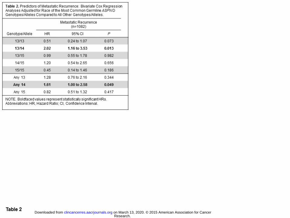
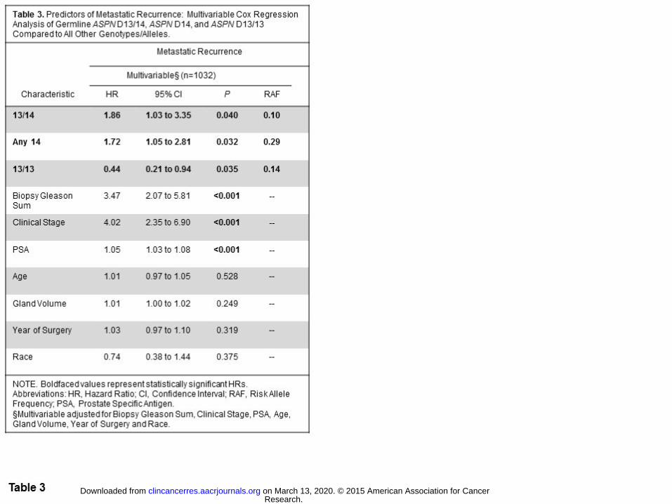
local therapy for prostate cancer. Through Cox regression analyses, we found that germline ASPN D13/14 was
significantly associated with metastatic recurrence following surgery (HR=2.02, 95%CI=1.16-3.53, P=0.013)
even when adjusted for pre-operative variables and race (HR=1.86, 95%CI=1.03-3.35, P=0.040) (Tables 2 and
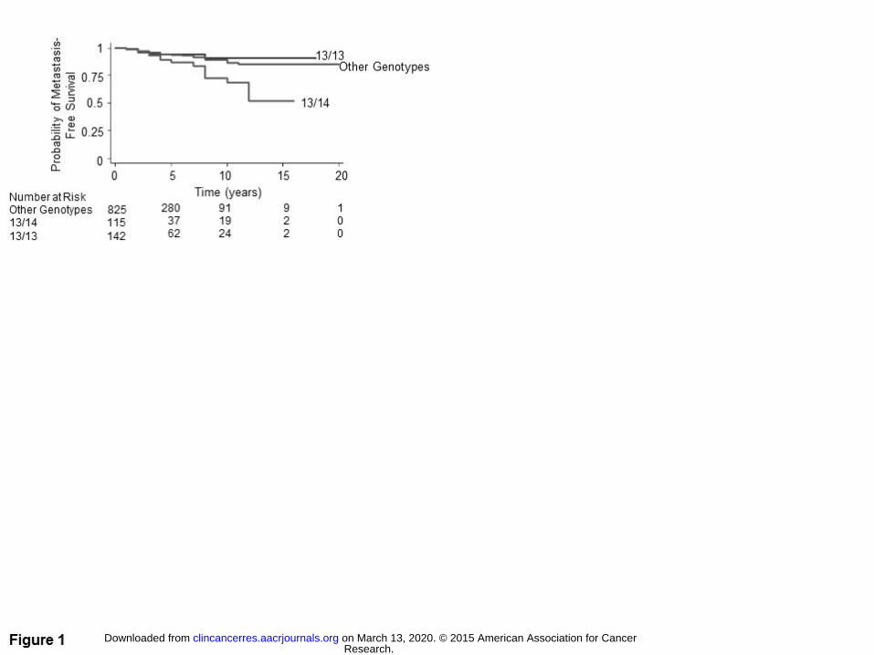
3). Consistent with this, Kaplan-Meier survival estimates demonstrated that heterozygous ASPN D13/14 was a
significant germline prognostic marker for worse metastasis free survival (Fig. 1). In addition, germline carriers
of the ASPN D14 allele were significantly more likely to progress to metastatic disease by multivariable
analyses compared to all other alleles (HR=1.72, 95%CI=1.05-2.81, P=0.032; Tables 2 and 3). These data
suggest that germline ASPN D13/14 or any ASPN D14 is a risk factor for metastatic recurrence following
surgery.
In contrast to germline carriers of ASPN D13/14 or any ASPN D14, multivariable Cox regression
analyses demonstrated that homozygous ASPN D13 (ASPN D13/13) was significantly associated with a reduced
risk of metastatic progression following surgery (HR=0.44, 95%CI=0.21-0.94, P=0.035; Table 3). Kaplan-
Meier survival estimates demonstrated that germline carriers of ASPN D13/13 had better metastasis-free
survival than men with germline ASPN D13/14 (Fig. 1). When compared to germline ASPN D13/13, men with
germline ASPN D13/14 had over a four-fold higher hazard of metastatic progression (HR=4.53, 95%CI=1.65-
12.42, P=0.003; Supplementary Table S2). While ASPN D13/13 remained significantly protective when
Research. on March 13, 2020. © 2015 American Association for Cancerclincancerres.aacrjournals.org Downloaded from
Author manuscripts have been peer reviewed and accepted for publication but have not yet been edited. Author Manuscript Published OnlineFirst on October 7, 2015; DOI: 10.1158/1078-0432.CCR-15-0256
14
controlled for race, Chi-Square analysis demonstrated that significantly fewer African American men carried
the ASPN D13/13 genotype (P<0.0001; Supplementary Table S1).
Given the association of BCR with metastatic progression (19), we evaluated the relationship between
ASPN D-repeat-length and BCR. When compared to all other genotypes, bivariate analyses controlled for race
demonstrated that ASPN D13/14 was significantly prognostic of BCR following surgery (HR=1.43,
95%CI=1.01-2.03, P=0.042; Supplementary Table S3); however when controlled for pre-surgical variables in
multivariable Cox regression analyses, the hazard ratio associated with ASPN D13/14 only approached
significance (Supplementary Table S4).
Germline ASPN D-repeat-length variants and adverse prostate cancer pathology
Both pre- and post-treatment clinical findings are used by the NCCN to stratify risk of disease recurrence and to
guide treatment recommendations (20). Logistic regression analyses demonstrated that none of the genotypes
examined were significantly associated with biopsy or pathologic Gleason grade, PSA, local invasion beyond
the prostate (EPE) or invasion into the seminal vesicles (SVI) (Supplementary Table S5). However, logistic
regression analyses demonstrated that ASPN D13/14 was significantly associated with prostate cancer involving
the lymph nodes at surgery (OR=2.52, 95%CI=1.44-4.42, P=0.001; Supplementary Table S6). Multivariable
logistic regression analyses adjusted for pre-operative variables and race demonstrated that men with ASPN
D13/14 (OR=2.42, 95%CI=1.29-4.56, P=0.006) or carriers of any ASPN D14 (homozygous or heterozygous)
(OR=1.74, 95%CI=1.02-2.96, P=0.041) were significantly more likely to have lymph node involvement at
surgery than men with other ASPN D genotypes or alleles (Supplementary Table S7).
Germline ASPN D-repeat-length variants in men with prostate cancer and non-cancer diagnoses
Polymorphisms in ASPN D-repeat-length have reported associations with susceptibility to diseases such as
osteoarthritis and lumbar-disc degeneration (7, 9). To determine if ASPN D-repeat length was associated with
prostate cancer incidence, we compared ASPN D-repeat-length in prostate cancer cases and non-cancer male
Research. on March 13, 2020. © 2015 American Association for Cancerclincancerres.aacrjournals.org Downloaded from
Author manuscripts have been peer reviewed and accepted for publication but have not yet been edited. Author Manuscript Published OnlineFirst on October 7, 2015; DOI: 10.1158/1078-0432.CCR-15-0256
15
controls matched for race. The distribution of ASPN D-repeat-length in JHH race-matched prostate cancer
cases and controls was nearly identical with none of the alleles or genotypes being statistically different by Chi-
square analyses (Supplementary Fig. S2 and Supplementary Table S1) suggesting that ASPN D allelic repeat-
lengths are not associated with prostate cancer incidence.
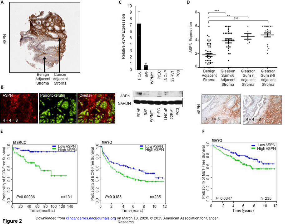
ASPN Expression in Cancer Associated Fibroblasts
Our data suggest that germline ASPN-repeat-length variants are differentially associated with metastatic
progression; however, little is known about the biologic role of ASPN in cancer progression. To better
understand this, we first analyzed human TMAs and demonstrated that ASPN expression was not elevated in
tumor cells but rather was significantly elevated in PCAFs (Fig. 2A, B, D). Both mRNA and protein expression
of ASPN were elevated in primary PCAFs isolated from human prostates compared to primary benign
associated fibroblasts (BAF) and an immortalized benign associated fibroblast cell line (WPMY1) (Fig. 2C and
Supplementary Fig. S1E). ASPN expression was not detected in primary human prostate epithelial cells (PrEC)
or in prostate cancer cell lines (Fig. 1C and Supplementary Fig. S1E). Interestingly, elevated levels of ASPN
mRNA were also detected in multiple prostate cancer cohorts (Supplementary Fig. S3A, B); however, ASPN
induction was not noted in benign prostatic hyperplasia (BPH) or in prostatic intraepithelial neoplasia (PIN)
(Supplementary Fig. S3B). Similar to prostate cancer, ASPN expression was also significantly elevated in
multiple other cancers including breast, colon, and pancreatic compared to normal tissue (Supplementary Fig.
S3C), suggesting that ASPN may have a conserved role in cancer development.
ASPN Expression and Prostate Cancer Gleason Grade
Gleason grade of the primary tumor is the strongest single prognostic marker of prostate cancer aggressiveness
(21). While low Gleason grade tumors (sum ≤6) rarely progress to metastasis, high Gleason grade tumors (sum
8-10) can have poor oncologic outcomes, even after RP (21). IHC analysis of ASPN protein expression in
TMAs from JHU (15) and Harvard (22) demonstrated that ASPN was associated with increasing Gleason grade
Research. on March 13, 2020. © 2015 American Association for Cancerclincancerres.aacrjournals.org Downloaded from
Author manuscripts have been peer reviewed and accepted for publication but have not yet been edited. Author Manuscript Published OnlineFirst on October 7, 2015; DOI: 10.1158/1078-0432.CCR-15-0256
16
with the highest expression seen in PCAFs surrounding Gleason sum 8-9 tumors (Fig. 2D; Supplementary Fig.
S3D). Consistent with protein expression, analysis of ASPN mRNA expression in a cohort from Memorial
Sloan-Kettering Cancer Center (MSKCC) (23) demonstrated that ASPN was significantly elevated in samples
from Gleason sum 8-9 tumors (Supplementary Fig. S3E).
ASPN Expression and Prostate Cancer Outcomes
To determine if ASPN expression in the primary tumor microenvironment was associated with disease
progression, we examined ASPN mRNA expression in prostate cancer cohorts from MSKCC (23) and the Mayo
Clinic (15, 24) and ASPN protein expression in a cohort from Harvard (22). ASPN expression was
significantly associated with BCR in all three cohorts as demonstrated by Kaplan-Meir survival curves and/or
Cox regression analyses (Fig. 2E and Supplementary Fig. S4A, B). While Kaplan-Meir analysis was not
available for the MSKCC cohort due to metastatic event time being unavailable, Kaplan-Meir survival curves
demonstrated that ASPN expression was significantly associated with metastatic recurrence in the Mayo Clinic
cohort (Fig. 2F). Univariate analyses also demonstrated a significant association between ASPN expression and
metastatic recurrence in both the MSKCC and Mayo Clinic cohorts; however, this was not significant in the
Harvard cohort (Supplementary Fig. 4C, D).
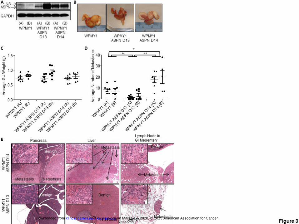
ASPN D-Repeat Domain Variants in Animal Models of Metastatic Progression
Germline ASPN D14 was associated with metastatic recurrence, while homozygous germline ASPN D13 was
associated with a reduced risk of metastases; yet, the functional significance of these variants in metastatic
progression is not known. To begin to investigate this, we examined tumor progression in an in vivo model
incorporating tumor/fibroblast interactions. We stably overexpressed ASPN D14 and ASPN D13 in an
immortalized human prostate fibroblast cell line derived from stroma adjacent to benign glands (WPMY1) (Fig.
3A). While WPMY1 are germline homozygous for ASPN D13, they did not express ASPN in vitro (Fig. 3A).
Two independent stable clones of both WPMY1 over-expressing ASPN D13 [WPMY1 ASPN D13 (A) and (B)]
Research. on March 13, 2020. © 2015 American Association for Cancerclincancerres.aacrjournals.org Downloaded from
Author manuscripts have been peer reviewed and accepted for publication but have not yet been edited. Author Manuscript Published OnlineFirst on October 7, 2015; DOI: 10.1158/1078-0432.CCR-15-0256
17
and WPMY1 over-expressing ASPN D14 [WPMY1 ASPN D14 (A) and (B)] were generated. Parental
WPMY1, WPMY1 ASPN D13, and WPMY1 ASPN D14 were mixed with PC3 cells and then grafted into
murine prostates. Expression of ASPN polymorphisms did not affect average orthotopic xenograft size as
measured by GU weight (Fig. 3B, C); however, ASPN polymorphisms significantly affected the number of
metastases (Fig. 3D, E). Due to differences in 5-year survival rates between regional and distant prostate cancer
in humans (1), we quantified the number of metastases to distant lymph nodes and to other organs in the
orthotopic xenografts. WPMY1 ASPN D13 significantly decreased the number of metastases compared to
parental WPMY1, while conversely WPMY1 ASPN D14 significantly increased the number of metastases to
distant lymph nodes and other organs including lung, liver, and pancreas (Fig. 3D, E). Both gross inspection
and pathological analyses supported that metastatic obstruction of the bile duct led to jaundice in approximately
33% of the WPMY1 ASPN D14 mice, while, conversely, jaundice was not detected in WPMY1 or WPMY1
ASPN D13 mice. Histological examination demonstrated micro-metastatic disease in WPMY1 ASPN D13
animals compared to the much larger visual metastases in WPMY1 ASPN D14 animals (Fig. 3E). Collectively,
these studies suggest that ASPN polymorphisms may have both prognostic and biologic roles in disease
progression.
Discussion:
Significant progress has been made in the past several years to identify genetic risk factors for prostate cancer.
Genome wide association studies (GWAS) have identified many single nucleotide polymorphisms associated
with prostate cancer incidence (25, 26); yet few inherited determinants of aggressive prostate cancer have been
identified (27, 28). Furthermore, associations of germline variants expressed by non-tumor cells in the tumor
microenvironment with metastatic prostate cancer have not been described. Based upon its association with
other diseases, we report a hypothesis-driven study to determine if germline variants in ASPN D-repeat-length
were differentially associated with metastatic prostate cancer. The distribution of ASPN D-repeat-length
variants was similar between prostate cancer cases and race matched controls, supporting the absence of an
Research. on March 13, 2020. © 2015 American Association for Cancerclincancerres.aacrjournals.org Downloaded from
Author manuscripts have been peer reviewed and accepted for publication but have not yet been edited. Author Manuscript Published OnlineFirst on October 7, 2015; DOI: 10.1158/1078-0432.CCR-15-0256
18
association between ASPN D variants and prostate cancer incidence. This is consistent with prior GWAS or
family-based linkage studies that have not found a strong association between this genomic region and prostate
cancer risk (29-31). However, two common variants in ASPN D-repeat-length affected the risk of having
metastatic disease. Multivariable analyses demonstrated that homozygous ASPN D13/13 was significantly
protective of metastatic recurrence following surgery. Conversely, germline genotype ASPN D13/14 and the
ASPN D14 allele were significantly prognostic of lymph node involvement and metastatic recurrence following
surgery. While previous studies have not reported an association of this genomic region with metastatic
prostate cancer, this may be due to limitations of prior studies that examined candidate genes (27) or genes
associated with incidence (32). Consistent with our patient-based findings, an animal model of prostate cancer
metastasis supports that ASPN D14 specifically expressed in PCAFs promotes metastatic prostate cancer
progression while ASPN D13 hinders metastatic progression.
Development of a minimally invasive and cost-effective genomic test for the early identification of
aggressive prostate cancer has the potential to impact therapeutic decisions in clinical settings. For instance,
controversy exists over the optimal treatment volumes and the use of hormonal suppression for men with
NCCN intermediate risk prostate cancer receiving radiation therapy (20, 33-36). Additionally there is an
increasing emphasis on the use of surveillance for men with low-grade cancers. Incorporation of germline
markers of aggressive prostate cancer at cancer diagnosis may better identify patients in need of more
aggressive therapies while also preventing the overtreatment of patients with indolent tumors. Additional
studies including prospective trials are needed; however, this initial large study suggests that germline ASPN D-
repeat-length is associated with clinical outcome.
Prostate cancer incidence and mortality have been shown to differ by race (1). While the etiology
driving racial disparities is multi-factorial, differences in biologic and genetic factors most likely play
contributing roles (37-40). The observation that African American men were approximately one eighth as
likely to be germline for ASPN D13/13 is intriguing and the first time that germline factors in the tumor
microenvironment have been described to vary by race.
Research. on March 13, 2020. © 2015 American Association for Cancerclincancerres.aacrjournals.org Downloaded from
Author manuscripts have been peer reviewed and accepted for publication but have not yet been edited. Author Manuscript Published OnlineFirst on October 7, 2015; DOI: 10.1158/1078-0432.CCR-15-0256
19
The molecular roles of ASPN D variants in metastatic prostate cancer progression are not well
understood. It has been postulated that ASPN and in particular ASPN D14 inhibits TGFβ family members
including TGFβ1 (7) and BMP2 (10, 41). Two recent reports support that ASPN induces CD44 (7) and EGFR
(42) mediated signaling pathways in gastric cancer cells. It is plausible that ASPN variants differentially
regulate multiple signaling pathways; however, a role for ASPN variants in CD44 or EGFR signaling in the
prostate has not been reported.
The mesenchymal/fibroblast specific expression of ASPN in prostate development and cancer and its
extracellular localization suggest that ASPN variants may have differential roles in modifying the extracellular
matrix (ECM). Dynamic regulation of the ECM has been shown to influence disease progression (43, 44).
ASPN may modify the ECM through direct collagen interactions. ASPN has recently been shown to regulate
osteoblast-driven collagen mineralization and to induce osteogenic factors (45). While the ASPN D-repeat
domain has been shown to bind calcium (45), the C-terminal domain of ASPN containing 10 Leucine Rich
Repeats has been shown to bind to Type I (45) and to Type II (46) collagens. ASPN competes with Decorin, an
inhibitor of prostate tumor growth (47), for collagen binding (45, 48, 49). In contrast to ASPN expression,
which is increased (4), Decorin expression is decreased in PCAFs (50). ASPN and Decorin may work
antagonistically to regulate the ECM environment. The D-repeat domain has been shown to directly regulate
collagen mineralization; however, the differential roles ASPN D-repeat-length variants play in these processes
have not been examined.
While the function of the ASPN D motif is not known, it is possible that it regulates protein aggregation
or functions as a scaffold. Interestingly, heterozygous carriers of ASPN D13 (D13/14 or D13/15) were not
protected from prostate cancer metastases suggesting that potential protective functions of ASPN D13 are
recessive to other alleles. As ASPN D13/14 and the D14 allele were associated with metastatic recurrence, the
function of ASPN D14 may be dominant. Unfortunately, the number of germline carriers of homozygous ASPN
D14 were not powered for adequate comparisons. It is possible that ASPN D13 and ASPN D14 monomers or
Research. on March 13, 2020. © 2015 American Association for Cancerclincancerres.aacrjournals.org Downloaded from
Author manuscripts have been peer reviewed and accepted for publication but have not yet been edited. Author Manuscript Published OnlineFirst on October 7, 2015; DOI: 10.1158/1078-0432.CCR-15-0256
20
ASPN D14 monomers have a better binding affinity than two ASPN D13 monomers. In support of this, ASPN
D14 has been shown to have better binding affinity to BMP2 than ASPN D13 (10).
In sum, this study suggests that ASPN is a key regulator in the microenvironment and may promote
metastatic progression of prostate cancer. Men with the ASPN D13/14 genotype or carriers of the ASPN D14
allele may be at higher risk of disease progression and thus genotyping ASPN has the potential of improving
risk stratification and early therapeutic decision making in localized prostate cancer. Further understanding of
the biologic role of ASPN in the stroma has the potential to impact both prognostic and therapeutic
development.
Acknowledgments
We thank Phuoc Tran, Ballentine Carter, Ken Pienta, Patrick Walsh, John Isaacs and Mike Brown for helpful
discussions and/or critical reading of this manuscript. We thank John Isaacs for reagents. We thank Laura
Kasch-Semenza at the Johns Hopkins GRCF Fragment Analysis Facility and the Prostate Cancer Biorepository
Network (PCBN), supported by the Department of Defense Prostate Cancer Research Program, DOD Award
No W81XWH-10-2-0056 and W81XWH-10-2-0046.
References
1. National Cancer Institute Surveillance E, and End Results Program:
seer.cancer.gov/statfacts/html/prost.html.
2. Barron DA, Rowley DR. The reactive stroma microenvironment and prostate cancer progression.
Endocrine-related cancer. 2012;19:R187-204.
3. Schaeffer EM, Marchionni L, Huang Z, Simons B, Blackman A, Yu W, et al. Androgen-induced
programs for prostate epithelial growth and invasion arise in embryogenesis and are reactivated in cancer.
Oncogene. 2008;27:7180-91.
Research. on March 13, 2020. © 2015 American Association for Cancerclincancerres.aacrjournals.org Downloaded from
Author manuscripts have been peer reviewed and accepted for publication but have not yet been edited. Author Manuscript Published OnlineFirst on October 7, 2015; DOI: 10.1158/1078-0432.CCR-15-0256
21
4. Orr B, Riddick AC, Stewart GD, Anderson RA, Franco OE, Hayward SW, et al. Identification of
stromally expressed molecules in the prostate by tag-profiling of cancer-associated fibroblasts, normal
fibroblasts and fetal prostate. Oncogene. 2012;31:1130-42.
5. Klee EW, Bondar OP, Goodmanson MK, Dyer RB, Erdogan S, Bergstralh EJ, et al. Candidate serum
biomarkers for prostate adenocarcinoma identified by mRNA differences in prostate tissue and verified with
protein measurements in tissue and blood. Clin Chem. 2012;58:599-609.
6. Satoyoshi R, Kuriyama S, Aiba N, Yashiro M, Tanaka M. Asporin activates coordinated invasion of
scirrhous gastric cancer and cancer-associated fibroblasts. Oncogene. 2015; 34:650-60.
7. Kizawa H, Kou I, Iida A, Sudo A, Miyamoto Y, Fukuda A, et al. An aspartic acid repeat polymorphism
in asporin inhibits chondrogenesis and increases susceptibility to osteoarthritis. Nat Genet. 2005;37:138-44.
8. Liu D, Yang Q, Li M, Mu K, Zhang Y. Association of an asporin repeat polymorphism with ankylosing
spondylitis in Han Chinese population: a case-control study. Clin Invest Med. 2010;33:E63-8.
9. Song YQ, Cheung KM, Ho DW, Poon SC, Chiba K, Kawaguchi Y, et al. Association of the asporin D14
allele with lumbar-disc degeneration in Asians. Am J Hum Genet. 2008;82:744-7.
10. Kajikawa T, Yamada S, Tauchi T, Awata T, Yamaba S, Fujihara C, et al. Inhibitory effects of PLAP-
1/asporin on periodontal ligament cells. Journal of dental research. 2014;93:400-5.
11. Bhowmick NA, Chytil A, Plieth D, Gorska AE, Dumont N, Shappell S, et al. TGF-beta signaling in
fibroblasts modulates the oncogenic potential of adjacent epithelia. Science. 2004;303:848-51.
12. Li X, Sterling JA, Fan KH, Vessella RL, Shyr Y, Hayward SW, et al. Loss of TGF-beta responsiveness
in prostate stromal cells alters chemokine levels and facilitates the development of mixed osteoblastic/osteolytic
bone lesions. Molecular cancer research: MCR. 2012;10:494-503.
13. Epstein JI, Pizov G, Walsh PC. Correlation of pathologic findings with progression after radical
retropubic prostatectomy. Cancer. 1993;71:3582-93.
14. Flavin R, Pettersson A, Hendrickson WK, Fiorentino M, Finn S, Kunz L, et al. SPINK1 protein
expression and prostate cancer progression. Clin Cancer Res. 2014;20:4904-11.
Research. on March 13, 2020. © 2015 American Association for Cancerclincancerres.aacrjournals.org Downloaded from
Author manuscripts have been peer reviewed and accepted for publication but have not yet been edited. Author Manuscript Published OnlineFirst on October 7, 2015; DOI: 10.1158/1078-0432.CCR-15-0256
22
15. Hurley PJ, Marchionni L, Simons BW, Ross AE, Peskoe SB, Miller RM, et al. Secreted protein, acidic
and rich in cysteine-like 1 (SPARCL1) is down regulated in aggressive prostate cancers and is prognostic for
poor clinical outcome. Proc Natl Acad Sci U S A. 2012;109:14977-82.
16. Karnes RJ, Bergstralh EJ, Davicioni E, Ghadessi M, Buerki C, Mitra AP, et al. Validation of a genomic
classifier that predicts metastasis following radical prostatectomy in an at risk patient population. J Urol.
2013;190:2047-53.
17. Mustafa Z, Dowling B, Chapman K, Sinsheimer JS, Carr A, Loughlin J. Investigating the aspartic acid
(D) repeat of asporin as a risk factor for osteoarthritis in a UK Caucasian population. Arthritis Rheum.
2005;52:3502-6.
18. Atif U, Philip A, Aponte J, Woldu EM, Brady S, Kraus VB, et al. Absence of association of asporin
polymorphisms and osteoarthritis susceptibility in US Caucasians. Osteoarthritis Cartilage. 2008;16:1174-7.
19. Pound CR, Partin AW, Eisenberger MA, Chan DW, Pearson JD, Walsh PC. Natural history of
progression after PSA elevation following radical prostatectomy. Jama. 1999;281:1591-7.
20. Mohler JL, Armstrong AJ, Bahnson RR, Boston B, Busby JE, D'Amico AV, et al. Prostate cancer,
Version 3.2012: featured updates to the NCCN guidelines. Journal of the National Comprehensive Cancer
Network : JNCCN. 2012;10:1081-7.
21. Eggener SE, Scardino PT, Walsh PC, Han M, Partin AW, Trock BJ, et al. Predicting 15-year prostate
cancer specific mortality after radical prostatectomy. J Urol. 2011;185:869-75.
22. Flavin RJ, Pettersson A, Hendrickson WK, Fiorentino M, Finn SP, Kunz L, et al. SPINK1 Protein
Expression and Prostate Cancer Progression. Clin Cancer Res. 2014;20:4904-11.
23. Taylor BS, Schultz N, Hieronymus H, Gopalan A, Xiao Y, Carver BS, et al. Integrative genomic
profiling of human prostate cancer. Cancer cell. 2010;18:11-22.
24. Erho N, Crisan A, Vergara IA, Mitra AP, Ghadessi M, Buerki C, et al. Discovery and validation of a
prostate cancer genomic classifier that predicts early metastasis following radical prostatectomy. PloS one.
2013;8:e66855.
Research. on March 13, 2020. © 2015 American Association for Cancerclincancerres.aacrjournals.org Downloaded from
Author manuscripts have been peer reviewed and accepted for publication but have not yet been edited. Author Manuscript Published OnlineFirst on October 7, 2015; DOI: 10.1158/1078-0432.CCR-15-0256
23
25. Eeles RA, Kote-Jarai Z, Giles GG, Olama AA, Guy M, Jugurnauth SK, et al. Multiple newly identified
loci associated with prostate cancer susceptibility. Nat Genet. 2008;40:316-21.
26. Thomas G, Jacobs KB, Yeager M, Kraft P, Wacholder S, Orr N, et al. Multiple loci identified in a
genome-wide association study of prostate cancer. Nat Genet. 2008;40:310-5.
27. Lin DW, FitzGerald LM, Fu R, Kwon EM, Zheng SL, Kolb S, et al. Genetic variants in the LEPR,
CRY1, RNASEL, IL4, and ARVCF genes are prognostic markers of prostate cancer-specific mortality. Cancer
Epidemiol Biomarkers Prev. 2011;20:1928-36.
28. Stark JR, Wiklund F, Gronberg H, Schumacher F, Sinnott JA, Stampfer MJ, et al. Toll-like receptor
signaling pathway variants and prostate cancer mortality. Cancer Epidemiol Biomarkers Prev. 2009;18:1859-63.
29. Al Olama AA, Kote-Jarai Z, Berndt SI, Conti DV, Schumacher F, Han Y, et al. A meta-analysis of
87,040 individuals identifies 23 new susceptibility loci for prostate cancer. Nat Genet. 2014;46:1103-9.
30. Lu L, Cancel-Tassin G, Valeri A, Cussenot O, Lange EM, Cooney KA, et al. Chromosomes 4 and 8
implicated in a genome wide SNP linkage scan of 762 prostate cancer families collected by the ICPCG.
Prostate. 2012;72:410-26.
31. Xu J, Dimitrov L, Chang BL, Adams TS, Turner AR, Meyers DA, et al. A combined genomewide
linkage scan of 1,233 families for prostate cancer-susceptibility genes conducted by the international
consortium for prostate cancer genetics. Am J Hum Genet. 2005;77:219-29.
32. Helfand BT, Roehl KA, Cooper PR, McGuire BB, Fitzgerald LM, Cancel-Tassin G, et al. Associations
of prostate cancer risk variants with disease aggressiveness: results of the NCI-SPORE Genetics Working
Group analysis of 18,343 cases. Hum Genet. 2015;134:439-50.
33. Tran PT, Hales RK, Zeng J, Aziz K, Salih T, Gajula RP, et al. Tissue biomarkers for prostate cancer
radiation therapy. Current molecular medicine. 2012;12:772-87.
34. Nguyen PL, D'Amico AV. Targeting pelvic lymph nodes in men with intermediate- and high-risk
prostate cancer despite two negative randomized trials. J Clin Oncol. 2008;26:2055-6; author reply 6-7.
Research. on March 13, 2020. © 2015 American Association for Cancerclincancerres.aacrjournals.org Downloaded from
Author manuscripts have been peer reviewed and accepted for publication but have not yet been edited. Author Manuscript Published OnlineFirst on October 7, 2015; DOI: 10.1158/1078-0432.CCR-15-0256
24
35. Morikawa LK, Roach M, 3rd. Pelvic nodal radiotherapy in patients with unfavorable intermediate and
high-risk prostate cancer: evidence, rationale, and future directions. International journal of radiation oncology,
biology, physics. 2011;80:6-16.
36. Bastian PJ, Boorjian SA, Bossi A, Briganti A, Heidenreich A, Freedland SJ, et al. High-risk prostate
cancer: from definition to contemporary management. Eur Urol. 2012;61:1096-106.
37. Powell IJ, Bock CH, Ruterbusch JJ, Sakr W. Evidence supports a faster growth rate and/or earlier
transformation to clinically significant prostate cancer in black than in white American men, and influences
racial progression and mortality disparity. J Urol. 2010;183:1792-6.
38. Khani F, Mosquera JM, Park K, Blattner M, O'Reilly C, MacDonald TY, et al. Evidence for Molecular
Differences in Prostate Cancer between African American and Caucasian Men. Clin Cancer Res. 2014;20:4925-
34.
39. Sundi D, Schaeffer EM. Active surveillance for African-American men with prostate cancer: proceed
with caution. Con. Oncology (Williston Park). 2014;28:83-5.
40. Freedland SJ, Isaacs WB. Explaining racial differences in prostate cancer in the United States: sociology
or biology? Prostate. 2005;62:243-52.
41. Tomoeda M, Yamada S, Shirai H, Ozawa Y, Yanagita M, Murakami S. PLAP-1/asporin inhibits
activation of BMP receptor via its leucine-rich repeat motif. Biochem Biophys Res Commun. 2008;371:191-6.
42. Ding Q, Zhang M, Liu C. Asporin participates in gastric cancer cell growth and migration by
influencing EGF receptor signaling. Oncology reports. 2015;33:1783-90.
43. Lu P, Weaver VM, Werb Z. The extracellular matrix: a dynamic niche in cancer progression. The
Journal of cell biology. 2012;196:395-406.
44. Lu P, Takai K, Weaver VM, Werb Z. Extracellular matrix degradation and remodeling in development
and disease. Cold Spring Harbor perspectives in biology. 2011;3:1-24.
45. Kalamajski S, Aspberg A, Lindblom K, Heinegard D, Oldberg A. Asporin competes with decorin for
collagen binding, binds calcium and promotes osteoblast collagen mineralization. Biochem J. 2009;423:53-9.
Research. on March 13, 2020. © 2015 American Association for Cancerclincancerres.aacrjournals.org Downloaded from
Author manuscripts have been peer reviewed and accepted for publication but have not yet been edited. Author Manuscript Published OnlineFirst on October 7, 2015; DOI: 10.1158/1078-0432.CCR-15-0256
25
46. Kou I, Nakajima M, Ikegawa S. Binding characteristics of the osteoarthritis-associated protein asporin. J
Bone Miner Metab. 2010;28:395-402.
47. Hu Y, Sun H, Owens RT, Wu J, Chen YQ, Berquin IM, et al. Decorin suppresses prostate tumor growth
through inhibition of epidermal growth factor and androgen receptor pathways. Neoplasia. 2009;11:1042-53.
48. Lorenzo P, Aspberg A, Onnerfjord P, Bayliss MT, Neame PJ, Heinegard D. Identification and
characterization of asporin. a novel member of the leucine-rich repeat protein family closely related to decorin
and biglycan. J Biol Chem. 2001;276:12201-11.
49. Henry SP, Takanosu M, Boyd TC, Mayne PM, Eberspaecher H, Zhou W, et al. Expression pattern and
gene characterization of asporin. a newly discovered member of the leucine-rich repeat protein family. J Biol
Chem. 2001;276:12212-21.
50. Henke A, Grace OC, Ashley GR, Stewart GD, Riddick AC, Yeun H, et al. Stromal expression of
decorin, Semaphorin6D, SPARC, Sprouty1 and Tsukushi in developing prostate and decreased levels of decorin
in prostate cancer. PLoS One.7:e42516.
Authors’ Contributions
Conception and design: PJH and EMS.
Acquisition of data: PJH, BWS, RMH, BS, RMM, BB, IAV, NE, ED, RJK, GY, CE, SDI, KMJ, JRR, LAM,
WBI, AER, and EMS.
Analysis and interpretation of the data: PJH, DS, BS, BWS, SFF, GN, IAV, NE, ED, RJK, DMB, JRR,
LAM, KMJ, SSA, JH, WBI, LM, BHP, AER, and EMS.
Writing, review and/or revision of the manuscript: PJH, DS, BHP, DMB, ED, AER, SSA, JRR, WBI,
LAM, LM, and EMS.
Research. on March 13, 2020. © 2015 American Association for Cancerclincancerres.aacrjournals.org Downloaded from
Author manuscripts have been peer reviewed and accepted for publication but have not yet been edited. Author Manuscript Published OnlineFirst on October 7, 2015; DOI: 10.1158/1078-0432.CCR-15-0256

Figure Legends:
Figure 1. Kaplan-Meier metastasis-free survival curves for germline ASPN D 13/14 compared to ASPN D13/13
(P=0.012) or to all other germline ASPN D genotypes (P=0.003).
Figure 2. ASPN expression in prostate cancer associated fibroblasts is associated with Gleason grade and
oncologic outcomes. A, ASPN expression as determined by IHC in prostate tissue following radical
prostatectomy. B, ASPN expression in prostate cancer associated fibroblasts in prostate tissue following radical
prostatectomy as determined by IF of ASPN and pancytokeratin at 400x magnification. C, ASPN expression in
human primary prostate cancer associated fibroblasts (PCAF), human primary benign prostate associated
fibroblasts (BAF), an immortalized human benign prostate fibroblast cell line (WPMY1), primary human
benign prostate epithelial cells (PreC), and human prostate cancer cell lines (LNCaP, 22RV1, PC3) as
determined by qRT-PCR (top) and immunoblotting (bottom). D, ASPN expression as determined by IHC in
JHU TMAs containing stroma adjacent to benign (n=48), Gleason sum ≤6 (n=31), Gleason sum 7 (n=6), and
Gleason sum 8-9 (n=27) prostate tissue. Statistical analysis performed by one-way ANOVA with Tukey’s
multiple comparison test (mean ± SEM; **P≤ 0.001, ***P ≤ 0.0001; upper). ASPN staining in human prostate
cancer sections at 200x magnification (lower). E, ASPN expression is associated with biochemical recurrence
(BCR). Kaplan–Meier survival curves of ASPN in MSKCC and in Mayo High Risk Cohort for BCR-free
survival. F, ASPN expression is associated with metastatic recurrence. Kaplan–Meier survival curves of ASPN
in Mayo High Risk Cohort for metastatic recurrence.
Figure 3. ASPN D13 is protective of metastatic progression while ASPN D14 promotes metastatic progression.
A, ASPN expression in WPMY1 (A and B), WPMY1 ASPN D13 (A and B), and ASPN D14 (A and B) as
measured by immunoblotting. B and C, Photomicrographs and GU weight of WPMY1 (A) + PC3 (n=7),
WPMY1 (B) + PC3 (n=5), WPMY1 ASPN D13 (A) + PC3 (n=6), WPMY1 ASPN D13 (B) + PC3 (n=7),
WPMY1 ASPN D14 (A) + PC3 (n=7), and WPMY1 ASPN D14 (B) + PC3 (n=5) prostate orthotopic xenografts
Research. on March 13, 2020. © 2015 American Association for Cancerclincancerres.aacrjournals.org Downloaded from
Author manuscripts have been peer reviewed and accepted for publication but have not yet been edited. Author Manuscript Published OnlineFirst on October 7, 2015; DOI: 10.1158/1078-0432.CCR-15-0256
(mean ± SEM). D, Average number of visible metastases to other organs and distant lymph nodes in WPMY1
(A) + PC3 (n=7), WPMY1 (B) + PC3 (n=5), WPMY1 ASPN D13 (A) + PC3 (n=6), WPMY1 ASPN D13 (B) +
PC3 (n=7), WPMY1 ASPN D14 (A) + PC3 (n=7), and WPMY1 ASPN D14 (B) + PC3 (n=5) prostate orthotopic
xenografts (mean ± SEM). E, Representative images of metastases taken at 40x magnification from WPMY1
ASPN D13 + PC3 and WPMY1 ASPN D14 + PC3 prostate orthotopic xenografts. Inset is a 400x magnification
of the tumor region. Statistical analyses performed by One-way ANOVA with Tukey’s multiple comparison
test to compare WPMY1 (A+B), WPMY1 ASPN D13 (A+B), and WPMY1 ASPN D14 (A+B) (mean ± SEM;
*P≤0.004 and **P≤0.0003).
Research. on March 13, 2020. © 2015 American Association for Cancerclincancerres.aacrjournals.org Downloaded from
Author manuscripts have been peer reviewed and accepted for publication but have not yet been edited. Author Manuscript Published OnlineFirst on October 7, 2015; DOI: 10.1158/1078-0432.CCR-15-0256

Published OnlineFirst October 7, 2015.Clin Cancer Res Paula J Hurley, Debasish Sundi, Brian Shinder, et al. Metastatic Prostate CancerMicroenvironment and are Differentially Associated with Germline Variants in Asporin Vary by Race, Modulate the Tumor
Updated version
10.1158/1078-0432.CCR-15-0256doi:
Access the most recent version of this article at:
Material
Supplementary
http://clincancerres.aacrjournals.org/content/suppl/2015/10/07/1078-0432.CCR-15-0256.DC1
Access the most recent supplemental material at:
Manuscript
Authoredited. Author manuscripts have been peer reviewed and accepted for publication but have not yet been
E-mail alerts related to this article or journal.Sign up to receive free email-alerts
Subscriptions
Reprints and
To order reprints of this article or to subscribe to the journal, contact the AACR Publications
Permissions
Rightslink site. Click on "Request Permissions" which will take you to the Copyright Clearance Center's (CCC)
.http://clincancerres.aacrjournals.org/content/early/2015/10/07/1078-0432.CCR-15-0256To request permission to re-use all or part of this article, use this link
Research. on March 13, 2020. © 2015 American Association for Cancerclincancerres.aacrjournals.org Downloaded from
Author manuscripts have been peer reviewed and accepted for publication but have not yet been edited. Author Manuscript Published OnlineFirst on October 7, 2015; DOI: 10.1158/1078-0432.CCR-15-0256


![01 01 0 01 01 0 01 0 01 01 0 01 1 01 01 0 10 01 0 10 0 01 ...b[parent::TRF:a]/TRF-ANYSTEP::TRF:c XPathOptimizer XPath-Variants ranked XPath-Variants Timo Böhme 10 01 01 0 01 01 0](https://img.pdfslide.org/doc/110x75/5b37888f7f8b9a5f288e3298/01-01-0-01-01-0-01-0-01-01-0-01-1-01-01-0-10-01-0-10-0-01-bparenttrfatrf-anysteptrfc.jpg)