Embed Size (px)
Citation preview
Zeitschrift für Kraniomandibuläre Funktion 2011;3(4):?–? 1
SCIENCE wISSENSChaft
Zusammenfassung
hintergrund und Zielsetzung: Das Ziel dieser Querschnittsstudie war es, sonografisch gestützte Diagnosen mit Ergebnissen der Magnetresonanztomografie (MRT) bei Diskusverlagerungen und degenerativen Veränderungen im temporomandibulären Gelenk zu vergleichen.Material und Methoden: 22 Probanden (44 Kiefergelenke; 16 Frauen, 6 Männer, Alter zwischen 20 und 70 Jahren) wurden mit Ultraschall und MRT untersucht und die Befunde verglichen.Ergebnisse: Die Übereinstimmungen bezüglich der Diskusposition sind mit κ = 0,62 als gut einzuschätzen (Sensitivität: p = 1,0; Spezifität: p = 0,63). Die Übereinstimmungen bezüglich der degenerativen Veränderungen sind mit κ = 0,24 als schwach bis unzureichend zu sehen (Sensitivität: p = 0,83; Spezifität: p = 0,25). Bei allen Untersuchungen fällt besonders die hohe Rate an falsch positiven Befunden auf. Schlussfolgerung: Die Sonografie am Kiefergelenk war im Vergleich zur MRT bezüglich der Diagnose einer Diskusverlagerung brauchbar. Sie ist geeignet, um intraartikuläre
1 Wiebke Starke, B. Sc. Phys. Th., Hamburg2 Prof. Dr. Harry von Piekartz, Professur für Physio
therapie, Fakultät Wirtschafts und Sozialwissenschaften, Osnabrück
1 Wiebke Starke, B. Sc. Phys. Th., Hamburg/Germany2 Prof. Dr. Harry von Piekartz, Professor for Physio
therapy, Faculty of Economic and Social Sciences, Osnabrück/Germany
W. Starke1, H. von Piekartz2
Comparison of sonography and MRI in the case of intra-articular temporomandibular (TMJ) dysfunctions: a cross-sectional study
Sonografie vs. MRT bei intraartikulären temporomandibulären Dysfunktionen: eine Querschnittsstudie
abstract
Background information and objective: The objective of this crosssectional study was to compare diagnoses obtained by sonographic findings, with the results of magnetic resonance imaging (MRI), in the case of displacement of the articular disc and degenerative alterations of the temporomandibular joint. Material and methods: A total of 22 test subjects (44 temporomandibular joints; 16 women and 6 men aged between 20 and 70 years) were subjected to sonographic and MRI examination and the results were then compared.Results: Agreement with regard to the position of the articular disc can be considered as good, with a value of κ = 0.62 (sensitivity: P = 1.0; specificity: P = 0.63). Agreement with the degenerative alterations can be regarded as poor, due to inadequacies, with κ = 0.24 (sensitivity: P = 0.83; specificity: P = 0.25). It was significant that all examinations were characterized by a high rate of false positive findings.Conclusion: The sonographic findings of the TMJ with regard to the diagnosis of articular disc displacement were viable in comparison to the MRI findings. Sonography is
SCIENCE Starke/ von Piekartz Intraarticular temporomandibular dysfunctions: sonography vs MRI
Journal of Craniomandibular Function 2011;3(4):?–?2
suitable for the rough depiction of intraarticular structural changes. The greatest disadvantage lay in the fact that transversal and rotatory discrepancies of the articular disc can only be insufficiently depicted. A detailed medical case history and functional examination still remain indispensable.
Keywords: craniomandibular dysfunction, magnetic reso-nance imaging (MRI), sonography, temporomandibular joint
Introduction
Craniomandibular dysfunctions (CMD) are the most frequent form of maxillofacial pain. Over 10% of the population aged over 18 years suffer from CMD and report that this has a severe impact on their daily life1. These findings
Strukturveränderungen grob abzubilden. Der größte Nachteil lag darin, dass transversale und rotatorische Abweichungen des Diskus nur unzureichend darzustellen sind. Eine ausführliche Anamnese und Funktionsuntersuchung bleiben unerlässlich.
Indizes: kraniomandibuläre Dysfunktion, Kiefergelenk, Sonografie, Magnetresonanztomografie
Einleitung
Kraniomandibuläre Dysfunktionen (CMD) sind die häufigste Form des Gesichtsschmerzes. Über 10 % der über 18Jährigen in der Bevölkerung haben eine CMD und geben an, dadurch im Alltag stark eingeschränkt zu sein.1 Diese Angaben konnten auch durch eine epidemiologische Studie von John et al.2 bestätigt werden. Die Probanden geben einerseits große Einschränkungen im Kausystem selbst an, andererseits auch Beeinträchtigungen bei der Arbeit, sozialen Kontakte und der Erholung. Bleiben Funktionsstörungen längere Zeit bestehen, können sie angrenzende Systeme (zum Beispiel die Halswirbelsäule und Kopfgelenke) beeinflussen. Zum Teil kommt es auch zur Ausbreitung pathologischer Informationen aus dem Kiefergelenk über die gesamte Wirbelsäule nach kaudal, mit Einfluss auf die Extremitäten.3
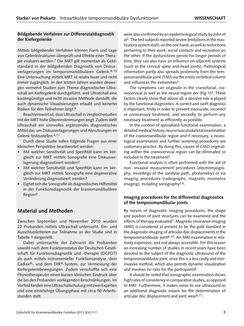
Quellen der Symptome können dabei im kraniomandibulären System, in der kraniofazialen, in der kranio zervikalen und auch in der neuralen Region liegen (Abb. 1).4 Anhand dieses Sachverhalts wird deutlich, dass vor allem die Funktionsdiagnostik eine entscheidende Rolle spielt. Eine korrekte und schnelle Diagnose ist wichtig, um ungenaue, fehlerhafte oder unnötige Behandlungen zu verhindern und im Gegensatz dazu die Behandlungen mit der größten Effizienz voranzubringen.
In der spezialisierten Funktionsuntersuchung sind eine ausführliche Anamnese, die neuromuskuloskelet tale Untersuchung der kraniomandibulären Region, gegebenenfalls eine neurologische Untersuchung und weitere Screeningverfahren üblich. So können zum Beispiel Ursachen in der kraniozervikalen Region ausgeschlossen oder ergänzend behandelt werden.5
Die Funktionsuntersuchung wird häufig durch nichtinvasive Messverfahren (Elektromyografie, Gelenkbahnaufzeichnungen, Fotometrie) oder durch bildgebende Verfahren (Röntgenbild, Magnetresonanztomografie) unterstützt, wozu auch die Sonografie zählt.3,6
fig 1 Regions from which the symptoms can originate (taken from: von Piekartz, 2005).
abb. 1 Regionen aus denen die Symptome stammen können (aus von Piekartz 2005).
craniofacial regionkaniofaziale Region
craniocervical regionkaniozervikaleRegion
craniomandibular regionkaniomandibuläre Region
craniofacial regionkaniofaziale Region
craniomandibular regionkaniomandibuläre Region
Zeitschrift für Kraniomandibuläre Funktion 2011;3(4):?–? 3
Starke/ von Piekartz Intraartikuläre temporomandibuläre Dysfunktionen wISSENSChaft
Bildgebende Verfahren zur Differenzialdiagnostik der Kiefergelenke
Mittels bildgebender Verfahren können Form und Lage von Gelenkstrukturen überprüft und Effekte einer Therapie evaluiert werden.7 Die MRT gilt momentan als Goldstandard in der bildgebenden Diagnostik von Diskusverlagerungen im temporomandibulären Gelenk.810 Eine Untersuchung mittels MRT ist relativ teuer und nicht immer zugänglich. In den letzten Jahren wurden deswegen vermehrt Studien zum Thema diagnostischer Ultraschall am Kiefergelenk durchgeführt, weil Ultraschall eine kostengünstige und nichtinvasive Methode darstellt, die auch dynamische Visualisierungen erlaubt und keinerlei Risiken für den Teilnehmer birgt.8
Beachtenswert ist, dass Ultraschall in Vergleichs studien mit der MRT hohe Übereinstimmungen zeigt. Zudem stellt Ultraschall ein sinnvolles ergänzendes diagnostisches Mittel dar, um Diskusverlagerungen und Abnutzungen im Gelenk festzustellen.6,11
Durch diese Studie sollen folgende Fragen aus einer klinischen Perspektive beantwortet werden: • Mit welcher Sensitivität und Spezifität kann im Ver
gleich zur MRT mittels Sonografie eine Diskusverlagerung diagnostiziert werden?
• Mit welcher Sensitivität und Spezifität kann im Vergleich zur MRT mittels Sonografie eine degenerative Veränderung diagnostiziert werden?
• Eignet sich die Sonografie als diagnostisches Hilfsmittel in der Funktionsdiagnostik der kraniomandibulären Region?
Material und Methoden
Zwischen September und November 2010 wurden 22 Probanden mittels Ultraschall untersucht. Ein und Ausschlusskriterien zur Teilnahme an der Studie sind in Tabelle 1 dargestellt.
Dabei untersuchte der Zahnarzt die Probanden sowohl nach dem Funktionsstatus der Deutschen Gesellschaft für Funktionsdiagnostik und therapie (DGFDT) als auch mittels instrumenteller Funktionsanalyse, dem Cadiax® und dem DIR®System, zur Vermessung der Kiefer gelenkbewegungen. Zudem verschaffte sich eine Physiotherapeutin einen kurzen klinischen Eindruck über die bei den Probanden vorliegenden Einschränkungen. Im Vorfeld fanden eine Ultraschallschulung mit zwei Experten und eine einwöchige Übungsphase mit circa 50 Arbeitsstunden statt.
were also confirmed by an epidemiological study by John et al2. The test subjects reported severe limitations in the masticatory system itself, on the one hand, as well as restrictions pertaining to their work, social contacts and recreation on the other. If the dysfunctions persist for longer periods of time, they can also have an influence on adjacent systems (such as the cervical spine and head joints). Pathological information partly also spreads posteriorly from the temporomandibular joint (TMJ) via the entire vertebral column and influences the extremities3.
The symptoms can originate in the craniofacial, craniocervical as well as the neural region der (Fig 1)4. These factors clearly show that above all, a decisive role is played by the functional diagnostics. A correct and swift diagnosis is important, firstly in order to prevent inaccurate, incorrect or unnecessary treatment, and secondly, to perform any necessary treatment as efficiently as possible.
In the context of specialized functional examination, a detailed medical history, neuromusculoskeletal examination of the craniomandibular region and if necessary, a neurological examination and further screening procedures are customary practice. By doing this, causes of CMD originating within the craniocervical region can be eliminated or included in the treatment5.
Functional analysis is often performed with the aid of non invasive measurement procedures (electromyography, recordings of the condylar path, photometry) or via imaging procedures (radiographs, magnetic resonance imaging), including sonography3,6.
Imaging procedures for the differential diagnostics of the temporomandibular joints
By means of diagnostic imaging procedures, the shape and position of joint structures can be examined and the effects of therapy evaluated7. Magnetic resonance imaging (MRI) is considered at present to be the gold standard in the diagnostic imaging of articular disc displacements in the temporomandibular joint810. An MRI examination is relatively expensive, and not always accessible. For this reason an increasing number of studies in recent years have been devoted to the subject of the diagnostic ultrasound of the temporomandibular joint, since this is a less costly and noninvasive method, which also permits dynamic visualization and involves no risks for the participants8.
It should be noted that sonographic examination shows high rates of consistency in comparative studies, as opposed to MRI. Furthermore, it makes sense to use ultrasound as an additional diagnostic means for the determination of articular disc displacement and joint wear6,11.
SCIENCE Starke/ von Piekartz Intraarticular temporomandibular dysfunctions: sonography vs MRI
Journal of Craniomandibular Function 2011;3(4):?–?4
Die Untersuchungen wurden den ethischen Kriterien der Declaration of Helsinki der World Medical Association gerecht (wma.net, 2008).
Die Durchführung der Studie erfolgte in einem physiotherapeutischen Behandlungsraum der Hochschule Osnabrück und in einem Raum einer lokal ansässigen Zahnarztpraxis. Der Ablauf während der Untersuchungen war immer der gleiche und war wie folgt standardisiert.
anamnese und klinische Untersuchung
Zur Erfassung des Hautproblems, der Nebendiagnosen sowie anderen soziodemografischen Daten und eventuellen Kontraindikationen zur Teilnahme an der Studie, erfolgte eine kurze klinische Untersuchung: • Beurteilung des Mundöffnungsmusters (Gerade, Devi
ation, Deflektion, andere) • Messung der maximalen aktiven Mundöffnung mit
einem Lineal • Messung der Laterotrusion mit einem Lineal • Beurteilung des Sensitivität des M. masseter pars
superficialis und M. temporalis pars anterior mit einem Algometer (Wagner Instruments, Model FDI 2004)
• Beurteilung der Kiefergelenkgeräusche (keine, Knacken, Reiben – bei Mundöffnung und/ oder Mund schluss)1215
• Erfassung des subjektiven Schmerzempfindens durch die visuelle Analogskala (VAS)16
Ultraschalluntersuchung und Interpretation
In dieser Studie wurde das Ultraschallgerät Venue 40 der Firma GE Healthcare, Wauwatosa/ Wi, USA, verwendet. Die Aufnahmen des Kiefergelenks wurden in dem Modus
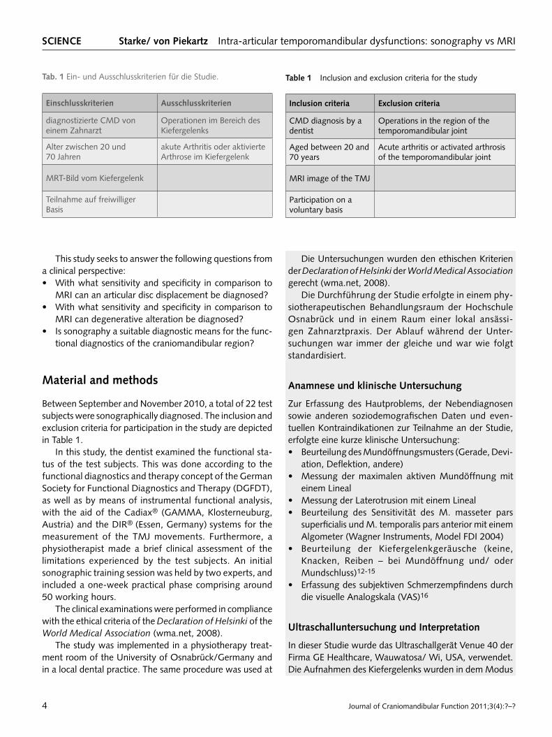
tab. 1 Ein und Ausschlusskriterien für die Studie.
Einschlusskriterien ausschlusskriterien
diagnostizierte CMD von einem Zahnarzt
Operationen im Bereich des Kiefergelenks
Alter zwischen 20 und 70 Jahren
akute Arthritis oder aktivierte Arthrose im Kiefergelenk
MRTBild vom Kiefergelenk
Teilnahme auf freiwilliger Basis
This study seeks to answer the following questions from a clinical perspective: • With what sensitivity and specificity in comparison to
MRI can an articular disc displacement be diagnosed? • With what sensitivity and specificity in comparison to
MRI can degenerative alteration be diagnosed? • Is sonography a suitable diagnostic means for the func
tional diagnostics of the craniomandibular region?
Material and methods
Between September and November 2010, a total of 22 test subjects were sonographically diagnosed. The inclusion and exclusion criteria for participation in the study are depicted in Table 1.
In this study, the dentist examined the functional status of the test subjects. This was done according to the functional diagnostics and therapy concept of the German Society for Functional Diagnostics and Therapy (DGFDT), as well as by means of instrumental functional analysis, with the aid of the Cadiax® (GAMMA, Klosterneuburg, Austria) and the DIR® (Essen, Germany) systems for the measurement of the TMJ movements. Furthermore, a physiotherapist made a brief clinical assessment of the limitations experienced by the test subjects. An initial sonographic training session was held by two experts, and included a oneweek practical phase comprising around 50 working hours.
The clinical examinations were performed in compliance with the ethical criteria of the Declaration of Helsinki of the World Medical Association (wma.net, 2008).
The study was implemented in a physiotherapy treatment room of the University of Osnabrück/Germany and in a local dental practice. The same procedure was used at
table 1 Inclusion and exclusion criteria for the study
Inclusion criteria Exclusion criteria
CMD diagnosis by a dentist
Operations in the region of the temporomandibular joint
Aged between 20 and 70 years
Acute arthritis or activated arthrosis of the temporomandibular joint
MRI image of the TMJ
Participation on a voluntary basis
Zeitschrift für Kraniomandibuläre Funktion 2011;3(4):?–? 5
Starke/ von Piekartz Intraartikuläre temporomandibuläre Dysfunktionen wISSENSChaft
Muskuloskeletal durchgeführt. Der lineare Schallkopf hat eine Frequenz von 4 bis 13 MHz.
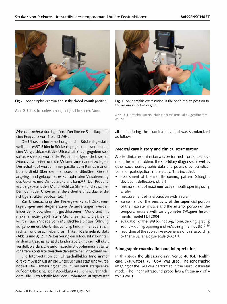
Die Ultraschalluntersuchung fand in Rückenlage statt, weil auch MRTBilder in Rückenlage gemacht werden und eine Vergleichbarkeit der UltraschallBilder gegeben sein sollte. Als erstes wurde der Proband aufgefordert, seinen Mund zu schließen und die Molaren aufeinander zu legen. Der Schallkopf wurde immer parallel zum Ramus mandibularis direkt über dem temporomandibulären Gelenk angelegt und gekippt bis es zur optimalen Visualisierung des Gelenks und Diskus artikularis kam.8,17 Der Proband wurde gebeten, den Mund leicht zu öffnen und zu schließen, damit der Untersucher die Sicherheit hat, dass er die richtige Struktur beobachtet.18
Zur Untersuchung des Kiefergelenks auf Diskusverlagerungen und degenerative Veränderungen wurden Bilder der Probanden mit geschlossenem Mund und mit maximal aktiv geöffnetem Mund gemacht. Ergänzend wurden auch Videos vom Mundschluss bis zur Öffnung aufgenommen. Die Untersuchung fand immer zuerst am rechten und anschließend am linken Kiefergelenk statt (Abb. 2 und 3). Zur Verbesserung der Bildqualität konnten an dem Ultraschallgerät die Eindringtiefe und die Helligkeit verstellt werden. Die automatische Bildoptimierung stellte schärfere Kontraste zwischen den einzelnen Strukturen her.
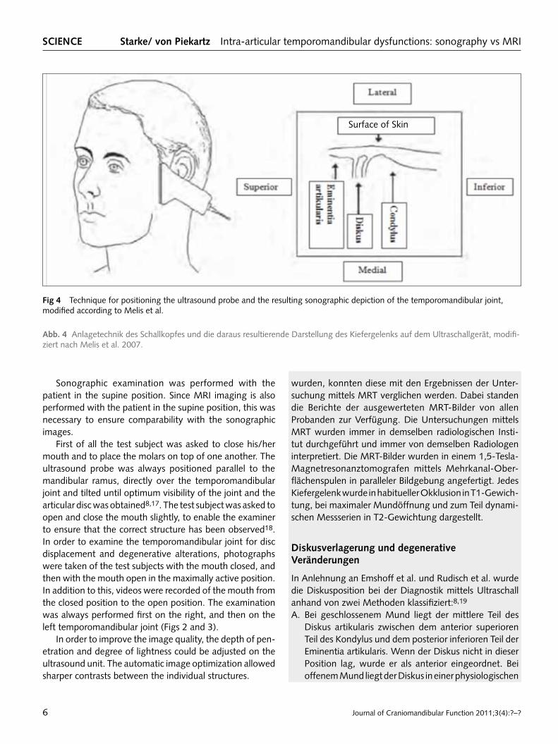
Die Interpretation der Ultraschallbilder fand immer direkt im Anschluss an die Untersuchung statt und wurde notiert. Die Darstellung der Strukturen der Kiefergelenke auf dem Ultraschall ist in Abbildung 4 zu sehen. Erst nachdem alle Ultraschallbilder der Probanden ausgewertet
fig 2 Sonographic examination in the closedmouth position.
abb. 2 Ultraschalluntersuchung bei geschlossenem Mund.
fig 3 Sonographic examination in the openmouth position to the maximum active degree.
abb. 3 Ultraschalluntersuchung bei maximal aktiv geöffnetem Mund.
all times during the examinations, and was standardized as follows.
Medical case history and clinical examination
A brief clinical examination was performed in order to document the main problem, the subsidiary diagnoses as well as other sociodemographic data and possible contraindications for participation in the study. This included: • assessment of the mouthopening pattern (straight,
deviation, deflection, other) • measurement of maximum active mouth opening using
a ruler • measurement of laterotrusion with a ruler • assessment of the sensitivity of the superficial portion
of the masseter muscle and the anterior portion of the temporal muscle with an algometer (Wagner Instruments, model FDI 2004)
• evaluation of the TMJ sounds (eg, none, clicking, grating sound – during opening and or/closing the mouth)1215
• recording of the subjective experience of pain according to the visual analogue scale (VAS)16.
Sonographic examination and interpretation
In this study the ultrasound unit Venue 40 (GE Healthcare, Wauwatosa, WI, USA) was used. The sonographic imaging of the TMJ was performed in the musculoskeletal mode. The linear ultrasound probe has a frequency of 4 to 13 MHz.
SCIENCE Starke/ von Piekartz Intraarticular temporomandibular dysfunctions: sonography vs MRI
Journal of Craniomandibular Function 2011;3(4):?–?6
wurden, konnten diese mit den Ergebnissen der Untersuchung mittels MRT verglichen werden. Dabei standen die Berichte der ausgewerteten MRTBilder von allen Probanden zur Verfügung. Die Untersuchungen mittels MRT wurden immer in demselben radiologischen Institut durch geführt und immer von demselben Radiologen interpretiert. Die MRTBilder wurden in einem 1,5TeslaMagnetresonanztomografen mittels MehrkanalOberflächenspulen in paralleler Bildgebung angefertigt. Jedes Kiefergelenk wurde in habitueller Okklusion in T1Gewichtung, bei maximaler Mundöffnung und zum Teil dynamischen Messserien in T2Gewichtung dargestellt.
Diskusverlagerung und degenerative Veränderungen
In Anlehnung an Emshoff et al. und Rudisch et al. wurde die Diskusposition bei der Diagnostik mittels Ultraschall anhand von zwei Methoden klassifiziert:8,19
A. Bei geschlossenem Mund liegt der mittlere Teil des Diskus artikularis zwischen dem anterior superioren Teil des Kondylus und dem posterior inferioren Teil der Eminentia artikularis. Wenn der Diskus nicht in dieser Position lag, wurde er als anterior eingeordnet. Bei offenem Mund liegt der Diskus in einer physiologischen
fig 4 Technique for positioning the ultrasound probe and the resulting sonographic depiction of the temporomandibular joint, modified according to Melis et al.
abb. 4 Anlagetechnik des Schallkopfes und die daraus resultierende Darstellung des Kiefergelenks auf dem Ultraschallgerät, modifiziert nach Melis et al. 2007.
Sonographic examination was performed with the patient in the supine position. Since MRI imaging is also performed with the patient in the supine position, this was necessary to ensure comparability with the sonographic images.
First of all the test subject was asked to close his/her mouth and to place the molars on top of one another. The ultrasound probe was always positioned parallel to the mandibular ramus, directly over the temporomandibular joint and tilted until optimum visibility of the joint and the articular disc was obtained8,17. The test subject was asked to open and close the mouth slightly, to enable the examiner to ensure that the correct structure has been observed18. In order to examine the temporomandibular joint for disc displacement and degenerative alterations, photographs were taken of the test subjects with the mouth closed, and then with the mouth open in the maximally active position. In addition to this, videos were recorded of the mouth from the closed position to the open position. The examination was always performed first on the right, and then on the left temporomandibular joint (Figs 2 and 3).
In order to improve the image quality, the depth of penetration and degree of lightness could be adjusted on the ultrasound unit. The automatic image optimization allowed sharper contrasts between the individual structures.
Surface of Skin
Zeitschrift für Kraniomandibuläre Funktion 2011;3(4):?–? 7
Starke/ von Piekartz Intraartikuläre temporomandibuläre Dysfunktionen wISSENSChaft
Position, wenn er sich noch immer zwischen dem Kondylus und der Eminentia befindet.8,20
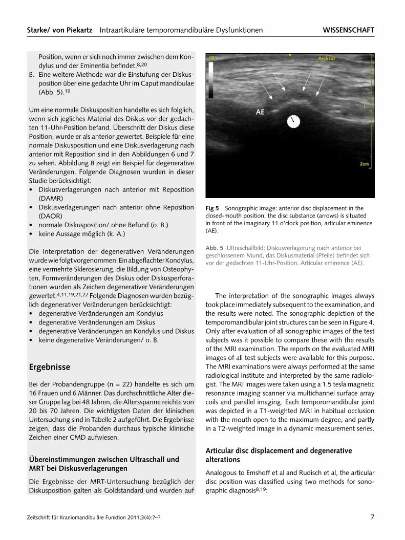
B. Eine weitere Methode war die Einstufung der Diskusposition über eine gedachte Uhr im Caput mandibulae (Abb. 5).19
Um eine normale Diskusposition handelte es sich folglich, wenn sich jegliches Material des Diskus vor der gedachten 11UhrPosition befand. Überschritt der Diskus diese Position, wurde er als anterior gewertet. Beispiele für eine normale Diskusposition und eine Diskusverlagerung nach anterior mit Reposition sind in den Abbildungen 6 und 7 zu sehen. Abbildung 8 zeigt ein Beispiel für degenerative Veränderungen. Folgende Diagnosen wurden in dieser Studie berücksichtigt: • Diskusverlagerungen nach anterior mit Reposition
(DAMR) • Diskusverlagerungen nach anterior ohne Reposition
(DAOR) • normale Diskusposition/ ohne Befund (o. B.) • keine Aussage möglich (k. A.)
Die Interpretation der degenerativen Veränderungen wurde wie folgt vorgenommen: Ein abgeflachter Kondylus, eine vermehrte Sklerosierung, die Bildung von Osteophyten, Formveränderungen des Diskus oder Diskusperforationen wurden als Zeichen degenerativer Veränderungen gewertet.4,11,19,21,22 Folgende Diagnosen wurden bezüglich degenerativer Veränderungen berücksichtigt: • degenerative Veränderungen am Kondylus • degenerative Veränderungen am Diskus • degenerative Veränderungen an Kondylus und Diskus • keine degenerative Veränderungen/ o. B.
Ergebnisse
Bei der Probandengruppe (n = 22) handelte es sich um 16 Frauen und 6 Männer. Das durchschnittliche Alter dieser Gruppe lag bei 48 Jahren, die Altersspanne reichte von 20 bis 70 Jahren. Die wichtigsten Daten der klinischen Untersuchung sind in Tabelle 2 aufgeführt. Die Ergebnisse zeigen, dass die Probanden durchaus typische klinische Zeichen einer CMD aufwiesen.
Übereinstimmungen zwischen Ultraschall und MRt bei Diskusverlagerungen
Die Ergebnisse der MRTUntersuchung bezüglich der Diskusposition galten als Goldstandard und wurden auf
fig 5 Sonographic image: anterior disc displacement in the closedmouth position, the disc substance (arrows) is situated in front of the imaginary 11 o’clock position, articular eminence (AE).
abb. 5 Ultraschallbild: Diskusverlagerung nach anterior bei geschlossenem Mund, das Diskusmaterial (Pfeile) befindet sich vor der gedachten 11UhrPosition, Articular eminence (AE).
aE
The interpretation of the sonographic images always took place immediately subsequent to the examination, and the results were noted. The sonographic depiction of the temporomandibular joint structures can be seen in Figure 4. Only after evaluation of all sonographic images of the test subjects was it possible to compare these with the results of the MRI examination. The reports on the evaluated MRI images of all test subjects were available for this purpose. The MRI examinations were always performed at the same radiological institute and interpreted by the same radiologist. The MRI images were taken using a 1.5 tesla magnetic resonance imaging scanner via multichannel surface array coils and parallel imaging. Each temporomandibular joint was depicted in a T1weighted MRI in habitual occlusion with the mouth open to the maximum degree, and partly in a T2weighted image in a dynamic measurement series.
articular disc displacement and degenerative alterations
Analogous to Emshoff et al and Rudisch et al, the articular disc position was classified using two methods for sonographic diagnosis8,19:
SCIENCE Starke/ von Piekartz Intraarticular temporomandibular dysfunctions: sonography vs MRI
Journal of Craniomandibular Function 2011;3(4):?–?8
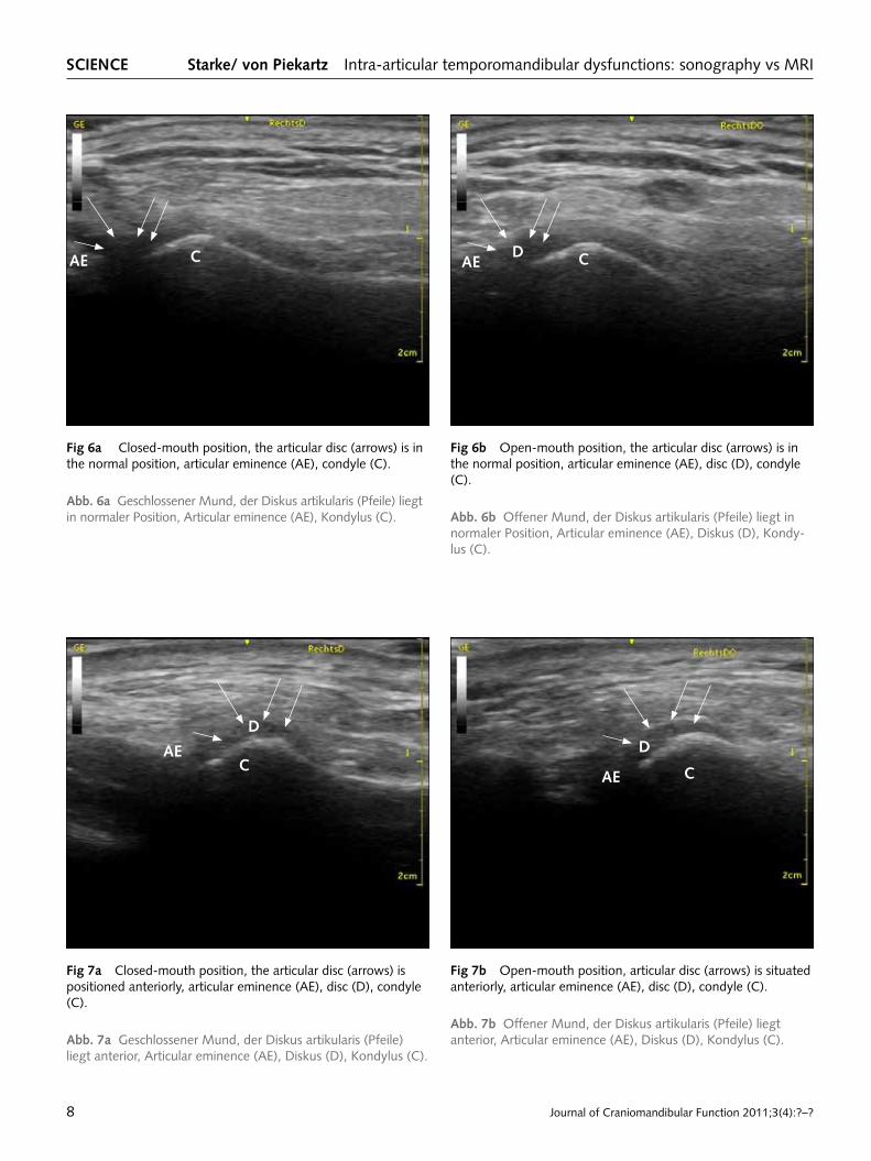
fig 6a Closedmouth position, the articular disc (arrows) is in the normal position, articular eminence (AE), condyle (C).
abb. 6a Geschlossener Mund, der Diskus artikularis (Pfeile) liegt in normaler Position, Articular eminence (AE), Kondylus (C).
fig 6b Openmouth position, the articular disc (arrows) is in the normal position, articular eminence (AE), disc (D), condyle (C).
abb. 6b Offener Mund, der Diskus artikularis (Pfeile) liegt in normaler Position, Articular eminence (AE), Diskus (D), Kondylus (C).
fig 7a Closedmouth position, the articular disc (arrows) is positioned anteriorly, articular eminence (AE), disc (D), condyle (C).
abb. 7a Geschlossener Mund, der Diskus artikularis (Pfeile) liegt anterior, Articular eminence (AE), Diskus (D), Kondylus (C).
fig 7b Openmouth position, articular disc (arrows) is situated anteriorly, articular eminence (AE), disc (D), condyle (C).
abb. 7b Offener Mund, der Diskus artikularis (Pfeile) liegt anterior, Articular eminence (AE), Diskus (D), Kondylus (C).
aE C aE CD
aEC
D
aE C
D
Zeitschrift für Kraniomandibuläre Funktion 2011;3(4):?–? 9
Starke/ von Piekartz Intraartikuläre temporomandibuläre Dysfunktionen wISSENSChaft
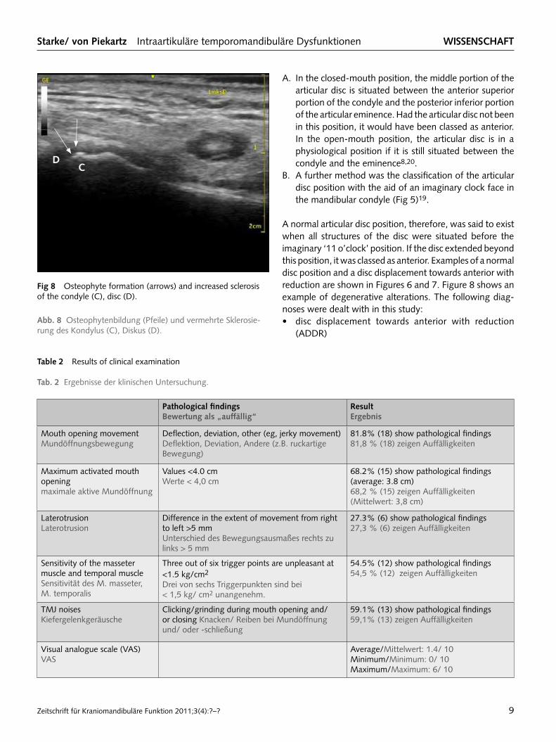
fig 8 Osteophyte formation (arrows) and increased sclerosis of the condyle (C), disc (D).
abb. 8 Osteophytenbildung (Pfeile) und vermehrte Sklerosierung des Kondylus (C), Diskus (D).
CD
table 2 Results of clinical examination
tab. 2 Ergebnisse der klinischen Untersuchung.
Pathological findingsBewertung als „auffällig“
ResultErgebnis
Mouth opening movementMundöffnungsbewegung
Deflection, deviation, other (eg, jerky movement) Deflektion, Deviation, Andere (z.B. ruckartige Bewegung)
81.8% (18) show pathological findings 81,8 % (18) zeigen Auffälligkeiten
Maximum activated mouth openingmaximale aktive Mundöffnung
Values <4.0 cmWerte < 4,0 cm
68.2% (15) show pathological findings (average: 3.8 cm)68,2 % (15) zeigen Auffälligkeiten (Mittelwert: 3,8 cm)
LaterotrusionLaterotrusion
Difference in the extent of movement from right to left >5 mm Unterschied des Bewegungsausmaßes rechts zu links > 5 mm
27.3% (6) show pathological findings 27,3 % (6) zeigen Auffälligkeiten
Sensitivity of the masseter muscle and temporal muscleSensitivität des M. masseter, M. temporalis
Three out of six trigger points are unpleasant at <1.5 kg/cm2
Drei von sechs Triggerpunkten sind bei < 1,5 kg/ cm2 unangenehm.
54.5% (12) show pathological findings 54,5 % (12) zeigen Auffälligkeiten
TMJ noisesKiefergelenkgeräusche
Clicking/grinding during mouth opening and/or closing Knacken/ Reiben bei Mundöffnung und/ oder schließung
59.1% (13) show pathological findings59,1% (13) zeigen Auffälligkeiten
Visual analogue scale (VAS)VAS
Average/Mittelwert: 1.4/ 10Minimum/Minimum: 0/ 10Maximum/Maximum: 6/ 10
A. In the closedmouth position, the middle portion of the articular disc is situated between the anterior superior portion of the condyle and the posterior inferior portion of the articular eminence. Had the articular disc not been in this position, it would have been classed as anterior. In the openmouth position, the articular disc is in a physiological position if it is still situated between the condyle and the eminence8,20.
B. A further method was the classification of the articular disc position with the aid of an imaginary clock face in the mandibular condyle (Fig 5)19.
A normal articular disc position, therefore, was said to exist when all structures of the disc were situated before the imaginary ‘11 o’clock’ position. If the disc extended beyond this position, it was classed as anterior. Examples of a normal disc position and a disc displacement towards anterior with reduction are shown in Figures 6 and 7. Figure 8 shows an example of degenerative alterations. The following diagnoses were dealt with in this study: • disc displacement towards anterior with reduction
(ADDR)
SCIENCE Starke/ von Piekartz Intraarticular temporomandibular dysfunctions: sonography vs MRI
Journal of Craniomandibular Function 2011;3(4):?–?10
Übereinstimmungen der Diagnosen mittels Ultraschall überprüft. Da es sich bei den festgestellten Diagnosen (Diskusposition und degenerative Veränderungen) um nominalskalierte Daten handelt, wurde das Übereinstimmungsmaß Kappa, entwickelt von Cohen (1960), zur Auswertung gewählt. Cohens Kappa (κ) wurde zudem gewählt, weil es das geeignetste standardisierte Übereinstimmungsmaß darstellt, welches als zufallsbereinigt gelten kann.23,24
Mit einem KappaWert von κ = 0,62 kann die Übereinstimmung als stark eingeschätzt werden.25,26 In Tabelle 3 sind die Übereinstimmungen der Diagnosen bezüglich der Diskusposition zu sehen, die durch den Ultraschall ermittelt wurden. Die Fälle, in denen keine Angabe über die Diskusposition gemacht werden konnten (8), wurden nicht gewertet. Folglich konnte bei 36 der 44 Kiefer gelenke eine Aussage zur Diagnose getroffen werden.
Auffällig ist, dass kein falsch negativer Befund vorkam. Der größte Fehler liegt darin, dass der Ultraschall eine Diskusverlagerung anzeigte, die MRT hingegen keinen Befund angab. Hierbei handelte es sich also um einen falsch positiven Befund. Alle sieben falsch positiven Klassifikationen waren Diskusverlagerungen nach anterior mit Reposition (DAMR), die laut MRT in vier Fällen ohne Befund sein müssten und in drei Fällen eine Diskus verlagerung nach anterior ohne Reposition (DAOR).
Um einen Teil der Validität des Ultraschalls bezüglich der Identifikation der Diskusposition zu bestimmen, wurden zusätzlich die Sensitivität und die Spezifität berechnet. Die Sensitivität, also die Fähigkeit des Ultraschalls Diskusverlagerungen als solche darzustellen, ist mit einem Wert von p = 1 als sehr gut zu beschreiben. Die Spezifität – Fähigkeit des Ultraschalls richtig Gesunde als solche zu erkennen – ist mit einem Wert von p = 0,63 als moderat einzuordnen.
Übereinstimmungen zwischen Ultraschall und MRt bei degenerativen Veränderungen
Die Ergebnisse der MRTUntersuchung bezüglich der degenerativen Veränderungen wurden als Goldstandard gesehen und auf Übereinstimmungen der Diagnosen mittels Ultraschall überprüft. Der KappaWert beträgt κ = 0,24 und kann somit als schwache Übereinstimmung gewertet werden.25,26 In Tabelle 4 sind die Übereinstimmungen der Diagnosen bezüglich der degenerativen Veränderungen zu sehen, die durch den Ultraschall ermittelt wurden. Bei allen 44 Kiefergelenken konnte bezüglich der
• disc displacement towards anterior without reduction (ADDWR)
• normal disc position/normal findings • no diagnosis/no statement possible.
The interpretation of the degenerative alterations was performed as follows: a flattened condyle; increased sclerosis; formation of osteophytes; alterations of the shape of the disc or disc perforation were seen as signs of degenerative alterations4,11,19,21,22. The following diagnoses were taken into account with regard to degenerative alterations: • degenerative alterations of the condyle • degenerative alterations of the disc • degenerative alterations of the condyle and the disc • no degenerative alterations (normal findings).
Results
The group of test subjects (n = 22) consisted of 16 women and 6 men. The average age of this group was 48 years with the ages of the participants ranging from 20 to 70 years.
The most important data of the clinical study are listed in Table 2. The results show that the test subjects definitely demonstrate characteristic clinical signs of CMD.
agreement between sonographic and MRI results in the case of articular disc displacement
The results of the MRI examination with regard to the disc position were considered to be of ‘gold standard’, and were checked for consistency between the MRI and sonographic diagnoses. Since the findings (with regard to disc position and degenerative alterations) were nominally scaled data, the extent of agreement kappa developed by Cohen was selected for evaluation. Cohen’s kappa (κ) was selected also because it is the most suitable standardized measure of agreement, which can be considered to be coincidence adjusted23,24.
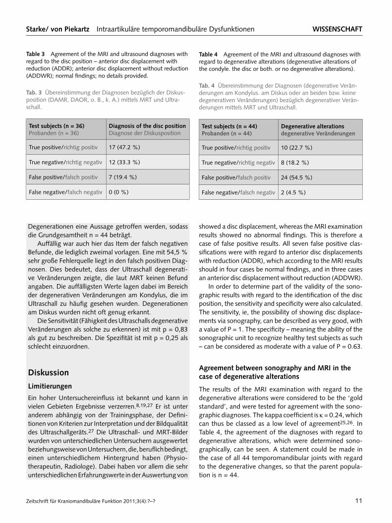
With a Kappa coefficient of κ = 0.62, the level of agreement can be considered as high25,26. Table 3 depicts the agreements of the sonographically determined diagnoses with regard to the disc position. The cases in which no information was obtained on the disc position (8) were not evaluated. Consequently, a statement could be made with regard to the diagnosis for 36 of the 44 temporomandibular joints examined.
Strikingly, there were no false negative results. The greatest error lay in the fact that the sonographic results
Zeitschrift für Kraniomandibuläre Funktion 2011;3(4):?–? 11
Starke/ von Piekartz Intraartikuläre temporomandibuläre Dysfunktionen wISSENSChaft
Degenerationen eine Aussage getroffen werden, sodass die Grundgesamtheit n = 44 beträgt.
Auffällig war auch hier das Item der falsch negativen Befunde, die lediglich zweimal vorlagen. Eine mit 54,5 % sehr große Fehlerquelle liegt in den falsch positiven Diagnosen. Dies bedeutet, dass der Ultraschall degenerative Veränderungen zeigte, die laut MRT keinen Befund angaben. Die auffälligsten Werte lagen dabei im Bereich der degenerativen Veränderungen am Kondylus, die im Ultraschall zu häufig gesehen wurden. Degenerationen am Diskus wurden nicht oft genug erkannt.
Die Sensitivität (Fähigkeit des Ultraschalls degenerative Veränderungen als solche zu erkennen) ist mit p = 0,83 als gut zu beschreiben. Die Spezifität ist mit p = 0,25 als schlecht einzuordnen.
Diskussion
Limitierungen
Ein hoher Untersuchereinfluss ist bekannt und kann in vielen Gebieten Ergebnisse verzerren.8,19,27 Er ist unter anderem abhängig von der Trainingsphase, der Definitionen von Kriterien zur Interpretation und der Bildqualität des Ultraschallgeräts.27 Die Ultraschall und MRTBilder wurden von unterschiedlichen Untersuchern ausgewertet beziehungsweise von Untersuchern, die, beruflich bedingt, einen unterschiedlichem Hintergrund haben (Physiotherapeutin, Radiologe). Dabei haben vor allem die sehr unterschiedlichen Erfahrungswerte in der Auswertung von
table 4 Agreement of the MRI and ultrasound diagnoses with regard to degenerative alterations (degenerative alterations of the condyle. the disc or both. or no degenerative alterations).
tab. 4 Übereinstimmung der Diagnosen (degenerative Veränderungen am Kondylus. am Diskus oder an beiden bzw. keine degenerativen Veränderungen) bezüglich degenerativer Veränderungen mittels MRT und Ultraschall.
test subjects (n = 44) Probanden (n = 44)
Degenerative alterations degenerative Veränderungen
True positive/richtig positiv 10 (22.7 %)
True negative/richtig negativ 8 (18.2 %)
False positive/falsch positiv 24 (54.5 %)
False negative/falsch negativ 2 (4.5 %)
table 3 Agreement of the MRI and ultrasound diagnoses with regard to the disc position – anterior disc displacement with reduction (ADDR); anterior disc displacement without reduction (ADDWR); normal findings; no details provided.
tab. 3 Übereinstimmung der Diagnosen bezüglich der Diskusposition (DAMR. DAOR, o. B., k. A.) mittels MRT und Ultraschall.
test subjects (n = 36) Probanden (n = 36)
Diagnosis of the disc position Diagnose der Diskusposition
True positive/richtig positiv 17 (47.2 %)
True negative/richtig negativ 12 (33.3 %)
False positive/falsch positiv 7 (19.4 %)
False negative/falsch negativ 0 (0 %)
showed a disc displacement, whereas the MRI examination results showed no abnormal findings. This is therefore a case of false positive results. All seven false positive classifications were with regard to anterior disc displacements with reduction (ADDR), which according to the MRI results should in four cases be normal findings, and in three cases an anterior disc displacement without reduction (ADDWR).
In order to determine part of the validity of the sonographic results with regard to the identification of the disc position, the sensitivity and specificity were also calculated. The sensitivity, ie, the possibility of showing disc displacements via sonography, can be described as very good, with a value of P = 1. The specificity – meaning the ability of the sonographic unit to recognize healthy test subjects as such – can be considered as moderate with a value of P = 0.63.
agreement between sonography and MRI in the case of degenerative alterations
The results of the MRI examination with regard to the degenerative alterations were considered to be the ‘gold standard’, and were tested for agreement with the sonographic diagnoses. The kappa coefficient is κ = 0.24, which can thus be classed as a low level of agreement25,26. In Table 4, the agreement of the diagnoses with regard to degenerative alterations, which were determined sonographically, can be seen. A statement could be made in the case of all 44 temporomandibular joints with regard to the degenerative changes, so that the parent population is n = 44.
SCIENCE Starke/ von Piekartz Intraarticular temporomandibular dysfunctions: sonography vs MRI
Journal of Craniomandibular Function 2011;3(4):?–?12
bildgebenden Verfahren einen Einfluss. Eine Zusammenarbeit vor der eigentlichen Datenerhebung bezüglich der Interpretation der MRT und Ultraschallbilder wäre wünschenswert gewesen. Es hätten Testbeurteilungen und Ergebnisvergleiche zwischen den Untersuchern (Ratern) stattfinden können, sodass die Bilder nach einem einheitlichen Schema interpretiert worden wären. Interessant wäre es außerdem gewesen, die InterraterReliabilität mit anderen Untersuchern mit physiotherapeutischem oder radiologischem Hintergrund zu bestimmen. Sowohl die Übereinstimmungen bezüglich der Interpretation der Ultraschallbilder als auch die der MRTBilder wären aufschlussreich. Für noch aussagekräftigere Ergebnisse wären zudem eine Kontrollgruppe und eine verblindete Durchführung sinnvoll gewesen.
Ein weiterer diskussionswürdiger Punkt ist die Definition einer Diskusverlagerung beziehungsweise einer normalen Diskusposition. In dieser Arbeit wurden die Ultraschall bilder nach Emshoff et al. und Rudisch et al. ausgewertet.8,19 Andere Autoren verwenden auch abweichende Klassifikationen.9,10,28 Diese lassen noch einen Spielraum, ab wann der Diskus als anterior gewertet wird. Durch diese verschiedenen Ansätze zur Klassifikation kann es zu unterschiedlichen Interpretationen kommen und daraus resultierend auch zu anderen Ergebnissen. Die Interpretation der degenerativen Veränderungen folgte keiner genauen Beschreibung. Ein abgeflachter Kondylus, eine vermehrte Sklerosierung, die Bildung von Osteophyten, Formveränderungen des Diskus oder Diskusperforationen wurden als Zeichen degenerativer Veränderungen gewertet. Auch hier ist eventuell eine genauere Unterteilung in Stadien sinnvoll.
Zudem kann das Datum der MRTBefunde die Ergebnisse beeinflussen. Der größte Teil der Befunde stammt aus dem Jahr 2010. Drei Befunde sind aus dem Jahr 2007, einer sogar aus dem Jahr 2001, wodurch bei diesen Probanden die Vergleichbarkeit fraglich erscheint. Die Tatsache, dass eine Diskusverlagerung, wenn sie länger als sechs Monate besteht, als chronisch beziehungsweise gefestigt gilt, kommt hier zur Geltung.29 Die untersuchten Probanden haben seit mindestens neun Monaten Beschwerden und fallen in diese Kategorie. 72,7 % dieser Teilnehmer erhielten sowohl eine physiotherapeutische Verordnung als auch eine Schienentherapie, die die Probleme zusätzlich verbessert haben könnten. Eine optimale Vergleichbarkeit wäre daher nur gegeben gewesen, wenn die Untersuchungen mittels MRT und Ultraschall direkt nacheinander durchgeführt worden wären. Dieses Vorgehen war im Rahmen dieser Studie vor allem aus Kostengründen nicht zu realisieren. Bei genauerer Betrachtung fällt auf, dass sowohl bei der
The striking aspect here was the false negative findings, which occurred only in two cases. The very high error rate of 54.5% lies in the false positive diagnoses. This means that the sonographic examination results showed degenerative alterations, which were normal findings according to the MRI examination. The most conspicuous values were found to be in the area of the degenerative alterations of the condyle, which were observed too frequently in the sonographic examinations. Degenerative changes to the disc were not recognized often enough.
The sensitivity (the ability of the ultrasound to recognize degenerative alterations as such) can be described as good with a value of P = 0.83. The specificity can be considered poor at P = 0.25.
Discussion
Limitations
A high examiner influence is known, and can in many areas distort the results8,19,27. It is, among other things, dependent on the training phase, the definitions of criteria for interpretation and the image quality of the ultrasound unit27. The ultrasound and MRI images were evaluated by different examiners and/or examiners with different professional backgrounds (physiotherapist, radiologist). Above all, the wide difference in experience with regard to the evaluation of imaging procedures has an impact. Collaboration prior to the phase of the actual data capture with regard to the interpretation of the MRI and sonographic images would have been desirable. Trial assessments and comparisons of results could have been made, with the result that the images would have been interpreted according to a unified system. It would also have been interesting to determine the interrater reliability with other examiners with a physiotherapy or radiology background. The agreement of the interpretation of the sonographic and the MRI images would have also been of value. For even more meaningful results it would also have made sense to have a control group and to blind the study.
A further point worthy of discussion is the definition of a disc displacement and a normal disc position. In this study, the sonographic images were evaluated according to Emshoff et al and Rudisch et al8,19. Other authors also use deviant classifications9,10,28. These still leave room for interpretation as to when the disc is classified as anterior. These different approaches to classification can result in different interpretations, which in turn can result in different results. The interpretation of the degenerative alterations does not follow any precise description. A flattened condyle, increased sclerosis, osteophyte formation, changes in the
Zeitschrift für Kraniomandibuläre Funktion 2011;3(4):?–? 13
Starke/ von Piekartz Intraartikuläre temporomandibuläre Dysfunktionen wISSENSChaft
Interpretation der Bilder aus dem Jahr 2007 als auch bei den aktuellen Befunden aus dem Jahr 2010 gleichermaßen falsche Ergebnisse vorkamen.
Die letztendlich untersuchte Probandenanzahl belief sich auf 22. Dies ist eine kleine Stichprobengröße, was bedeutet, dass die Ergebnisse nicht zu verallgemeinern sind. Zudem stammen die Probanden größtenteils aus dem Raum Osnabrück und wurden alle von dem gleichen Zahnarzt rekrutiert, der auf die Funktionsdiagnostik von CMD spezialisiert ist. Diese Faktoren können zusätzlich Einfluss nehmen. Die Probanden, die an dieser Studie teilnahmen, spiegeln folglich nicht die Allgemeinheit der Patienten mit einer CMD wider.
Studienvergleich
Die Hauptfrage dieser Studie bezieht sich auf die Vergleichbarkeit von Ultraschall und MRT bezüglich der Feststellung der Diskusposition. Die Ergebnisse, die berechnet werden konnten, zeigen eine gute bis deutliche Übereinstimmung. Zudem ist es durch Ultraschall gut möglich Diskusverlagerungen als solche zu erkennen. Diese Ergebnisse stimmen zum Großteil mit vorangegangenen Studien überein. Die Sensitivitäten anderer Studien liegen zwischen p = 0,73 und p = 1,0. Angegebene Werte für die Spezifitäten liegen zwischen p = 0,75 und p = 1,0.8–10,19,28 Auch andere Autoren sehen die größten Probleme in den falsch positiven Befunden. Emshoff et al. halten fest, dass es bei lang bestehenden Diskusverlagerungen im Kiefergelenk zu Flüssigkeitsansammlungen oder fibrösen Strukturen kommen kann, die die Interpretation der Bilder zusätzlich erschweren.8 Fast alle falsch positiven Befunde in ihrer Studie resultieren aus falschen Interpretationen aufgrund von synovialen Ergüssen, die sich auf dem Ultraschallbild nahezu genauso darstellen wie eine Diskusverlagerung. Zudem können stärkere degenerative Veränderungen oder leichte Rupturen des Diskus das Bild verzerren.19,30 Weitere Faktoren können laut Jank et al. auch anatomische Variationen sein, die vom Untersucher nicht erkannt werden.9
Ein diskussionswürdiger Punkt ist die schlechte Visualisierung einer rotatorischen Komponente oder einer Seitverschiebung des Diskus mittels Ultraschall. Auf dieses Problem wurde bereits in anderen Studien hingewiesen.8,28 Rudisch et al. versuchten dieses durch folgende Kategorisierung zu verbessern: „Die Abwesenheit von Diskusmaterial im Gelenkspalt ist ein indirektes Zeichen für eine Diskusverlagerung nach medial, weil die medialen Teile des Articulatio temporomandibularis nicht direkt dargestellt werden können.“ Sie schränken aber selbst
shape of the disc or disc perforations were considered as signs of degenerative alterations. Here too, a more precise classification into stages could make sense.
Furthermore, the date of the MRI findings can influence the results. Most of the results are from 2010. Three findings are from the year 2007, and one even from 2001, which makes the comparability seem questionable in the case of these test subjects. The fact that a disc displacement, if it lasts more than six months, is considered as chronic or at least stable, is now borne out29. The test subjects examined had been experiencing symptoms for at least nine months and fell into this category. An amount of 72.7% of these participants received both physiotherapy and splint therapy, which could have considerably improved the symptoms. Optimum comparability, however, would therefore be given if the MRI and sonographic examinations were performed in direct succession. Within the context of this study such a procedure was not feasible, mainly due to reasons of expense. A closer look at the situation reveals that during the interpretation of both the images from 2007 and of the current findings from 2010, the results were equally false in both cases.
A total of 22 test subjects were ultimately examined. This is a small sample size, which means that the results cannot be generalized. Furthermore, the test subjects are mainly from the Osnabrück region in Germany, and were all recruited by the same dentist who specializes in the functional diagnostics of CMD. These factors can have an additional influence. The test subjects who participated in this study therefore do not reflect the general population of CMD patients.
Study comparison
The main question of this study refers to the comparability of ultrasound and MRI, with regard to the determination of the disc position. The results calculated show a good to clear agreement. Furthermore, disc displacements can be recognized by means of sonography. These results largely correspond to those of previous studies. The sensitivity values of other studies lie between P = 0.73 and P = 1.0. The given specificity values are situated between P = 0.75 and P = 1.0810,19,28. Also other authors consider that the greatest problems lie in the false negative findings. Emshoff et al find that longlasting disc displacements in the TMJ can give rise to fluid accumulations or fibrous structures, which additionally complicate the interpretation of the images8. Almost all false positive findings in their study are the result of incorrect interpretations due to synovial effusions, which look virtually the same as a disc displacement in the sonographic depiction. Additional degenerative alterations or slight ruptures of the disc can also distort the image19,30.
SCIENCE Starke/ von Piekartz Intraarticular temporomandibular dysfunctions: sonography vs MRI
Journal of Craniomandibular Function 2011;3(4):?–?14
ein, dass diese Ansicht vor allem durch degenerative oder fibröse Veränderungen verzerrt werden kann. Trotzdem ist es wichtig, dass auch die mediale und laterale Komponente Berücksichtigung findet.19
Die Übereinstimmungen von Ultraschall und MRT bei der Feststellung von degenerativen Veränderungen ist in der vorliegenden Arbeit als schlecht einzustufen. Bisher existiert zu dem Thema Visualisierung degenerativer Veränderungen des Kiefergelenks nicht so viel Literatur wie zum Thema Diskusverlagerungen. Emshoff et al. untersuchten ebenfalls Diskusverlagerungen und degenerative Veränderungen, geben aber nur Werte für die Probanden mit Diskusverlagerungen und zusätzlicher Degeneration am Kondylus an. Die Sensitivität betrug dafür p = 0,80 und die Spezifität p = 0,79. Beide Werte können somit als gut eingestuft werden.18 Rudisch et al. haben ebenfalls Degenerationen am Kondylus untersucht und bestätigen mit einer Sensitivität von p = 0,95 und einer Spezifität von p = 0,90 die guten Ergebnisse von Emshoff et al.19
Bezüglich der degenerativen Veränderungen sollten in Zukunft einheitliche Klassifikationen beschrieben und verwendet werden. Eine genaue Einteilung ab wann eine Veränderung wirklich auffällig ist und für bestimmte Symptome verantwortlich sein kann und welche Veränderungen noch als dem Alter entsprechende Degeneration angesehen werden können, ist notwendig. Zudem ist es laut Emshoff et al. von Bedeutung, die verschiedenen Stadien der Degenerationen herauszufinden, weil sie eine unterschiedliche Therapie benötigen.18 Sie sollten daher ebenfalls in der Klassifikation Berücksichtigung finden.
Verbessert Ultraschall die funktionsdiagnostik der kraniomandibulären Region?
Die sonografische Untersuchung am Kiefergelenk gibt einen groben Überblick über die Situation. Die Technik wird stetig weiter entwickelt, sodass verwendbare Bilder vom Kiefergelenk gemacht werden können. Zudem erlaubt Ultraschall dynamische Visualisierungen und könnte somit auch zur funktionellen Beurteilung eingesetzt werden, natürlich mit den oben genannten Limitationen. Der Diskus könnte während aktiver Bewegungen der Mandibula dargestellt werden, was mit zunehmenden technischen Kenntnissen in der sonografischen Untersuchung eine Herausforderung für die Zukunft ist. Diese würde vor allem in der physiotherapeutischen gegebenenfalls auch zahnärztlichen Praxis einen Nutzen bieten.
Als weiterer Vorteil kann die Untersuchung mittels Ultraschall schnell in die Diagnostik integriert werden
According to Jank et al, further factors can also be anatomical variations, which are not recognized by the examiner9.
A point worthy of discussion is the poor visualization of a rotatory component, or a lateral displacement of the disc via ultrasound. This problem has already been referred to in other studies8,28. Rudisch et al tried to improve this by the following categorization: “The absence of disc substance in the joint gap is an indirect sign of a mesial shift of the disc, since the mesial components of the TMJ cannot be depicted directly.” They do, however, admit that this view can be distorted, particularly by degenerative or fibrous alterations. It is nevertheless important that also the mesial and lateral components are taken into account19.
The agreement between sonography and MRI in the determination of degenerative changes can be seen in the following study as being poor.
Considerably less literature is available to date on the visualization of degenerative alterations of the TMJ, as on the subject of disc displacement. Emshoff et al likewise investigated disc displacements and degenerative alterations, but only indicate values for the test subjects with disc displacement and additional degeneration of the condyle. The values for sensitivity and specificity were P = 0.80 and P = 0.79 respectively. Both values can therefore be classed as good18. Rudisch et al also studied condylar degeneration, and confirm the good results of Emshoff et al with a sensitivity of P = 0.95 and a specificity of P = 0.9019.
With regard to degenerative alterations, a unified classification system should be defined and used in future. A precise classification is necessary as to when an alteration is truly noticeable and can be responsible for specific symptoms, and which alterations can be classed as agerelated degenerations. Furthermore it is important, according to Emshoff et al, to identify the different stages of degeneration since these require a different therapy18. For this reason, these should likewise be taken into account in the classification.
Does sonography improve the functional diagnostics of the craniomandibular region?
The sonographic examination of the temporomandibular joint gives a rough overview of the clinical situation. The ongoing further development of the technology enables usable images of the TMJ. Furthermore, ultrasound allows dynamic visualization, and could therefore also be used for the purpose of functional analysis, naturally with the limitations mentioned above. The articular disc could be depicted during active movements of the mandible, which presents a challenge for the future, in view of increasing technical knowledge in the field of sonographic examination. This
Zeitschrift für Kraniomandibuläre Funktion 2011;3(4):?–? 15
Starke/ von Piekartz Intraartikuläre temporomandibuläre Dysfunktionen wISSENSChaft
und den Aufwand für den Patienten im Gegensatz zur MRT reduzieren. Durch den Ultraschall können qualitative Beobachtungen gemacht werden, die die quantitativen Messungen und die Ergebnisse der Anamnese und Funktionsuntersuchung komplettieren und so zu einer höheren Wertigkeit führen.
In Deutschland müssten die Physiotherapeuten einheitlicher im Umgang mit diagnostischem Ultraschall geschult werden, um falsche Befunde oder Fehlinterpretationen zu vermeiden und den Umgang und die Möglichkeiten eines Ultraschallgeräts zu erlernen.
fazit für die Praxis
Der diagnostische Ultraschall am Kiefergelenk ist bezüglich der Diagnose einer Diskusverlagerung nach anterior mit und ohne Reposition im Vergleich zur MRT anwendbar. Er stellt ein geeignetes Mittel dar, um einen Überblick über intraartikuläre kraniomandibuläre Dysfunktionen zu bekommen. Als größter Nachteil der Methode erwies sich die Tatsache, dass transversale und rotatorische Abweichungen des Diskus nur unzureichend darzustellen sind. Eine detaillierte Anamnese und Funktionsuntersuchung bleiben zudem unerlässlich.
Danksagung
Ein herzlicher Dank gilt Herrn Taubmann und der gesamten Zahnarztpraxis für die tatkräftige Unterstützung. Ebenso bedanken wir uns bei Dr. Frank und dem RöntgenNuclearInstitut Drewes + Partner für die zur Verfügung gestellten Befunde. Für die Leihgabe des in dieser Studie verwendeten Ultraschallgerätes gilt der Dank GE Healthcare.
would be of use above all in physiotherapy, and possibly also in the dental practice.
A further advantage is that ultrasonic examination can be quickly integrated into the diagnostic procedure, and reduces the time required by the patient, in comparison to MRI. By using ultrasound, qualitative observations can be made which complement the quantitative measurements and the results of anamnesis and functional analysis. This improves the overall quality of diagnosis.
In Germany, there is more unified training of physiotherapists with regard to the operation of diagnostic ultrasound in order to avoid incorrect diagnoses or wrong interpretations, and to learn the operation of a sonographic unit and the possibilities it offers.
Conclusion for clinical practice
The diagnostic ultrasound of the temporomandibular joint is viable in comparison to MRI, with regard to the diagnosis of an anterior disc displacement with and without reduction. It represents a suitable means of gaining a clinical overview of intraarticular craniomandibular dysfunctions. The greatest disadvantage of the method was found to lie in the fact that transverse and rotatory disc deviations can only be inadequately depicted. A detailed medical case history and functional analysis therefore remain indispensable.
acknowledgements
The authors would especially like to thank Mr. Taubmann as well as the entire dental practice for their dedicated support. Thanks also go to Dr. Frank and the Radiological Nuclear Institute, and Drewes + Partner for providing the diagnoses, and also to GE Healthcare for giving permission for the sonographic unit to be used in this study.
References
1. LeResche L. Epidemiology of temporomandibular disorders: implications for the investigation of etiologic factors. Crit Rev Oral Biol Med 1997;8:291–305.
2. John M, Hirsch C, Reiber T. Häufigkeit, Bedeutung und Be handlungsbedarf kraniomandibulärer Dysfunktionen. J Public Health 2001;9:136–155.
3. Lund JP, Widmer CG, Feine JS. Validity of diagnostic and monitoring tests used for temporomandibular disorders. J Dent Res 1995;74:1133–1143.
4. von Piekartz H. Kiefer, Gesichts und ZervikalregionNeuromuskuloskeletale Untersuchung. Therapie und Management. Stuttgart, Germany: Thieme Verlag, 2005:15.
5. Westerhuis P, Wiesner R. Kapitel 3: Praxisrealisation: Anwendung des Konzepts. In: BucherDollenz G, Wiesner R, Blake R, et al. Therapiekonzepte in der Physiotherapie – Maitland. Stuttgart, Germany: Thieme Verlag, 2008:56–71.
6. Melis M, Secci S, Ceneviz C. Use of ultrasonography for the diagnosis of the temporomandibular joint disorders: a review. Am J Dent 2007;2:73–78.
SCIENCE Starke/ von Piekartz Intraarticular temporomandibular dysfunctions: sonography vs MRI
Journal of Craniomandibular Function 2011;3(4):?–?16
7. Brooks SL, Brand JW, Gibbs SJ, et al. Imaging of the temporomandibular joint. Oral Surg Oral Med Oral Pathol Oral Radiol Endod 1997;83:609–618.
8. Emshoff R, Jank S, Bertram S, Rudisch A, Bodner G. Disk Displacement of the Temporomandibular Joint: Sonography Versus MR imaging. AJR Am J Roentgenol 2002;178:1557–1562.
9. Jank S, Rudisch A, Bodner G, Brandlmaier I, Gerhard S, Emshoff R. Highresolution ultrasonography of the TMJ: helpful diagnostic approach for patients with TMJ disorders? J Craniomaxillofac Surg 2001;29:366–371.
10. Uysal S, Kansu H, Akhan O, Kansu Ö. Comparison of ultrasonography with magnetic resonance imaging in the diagnosis of temporomandibular joint internal derangements: a preliminary investigation. Oral Surg Oral Med Oral Pathol Oral Radiol Endod 2002;94:115–121.
11. Hussain AM, Packota G, Major PW, Flores Mir C. Role of different imaging modalities in assessment of temporomandibular joint erosions and osteophytes: a systematic review. Dentomaxillofac Radiol 2008;37:63–71.
12. John M, Hirsch C, Reiber T, Dworkin S. Translating the research diagnostic criteria for temporomandibular disorders into German: evaluation of content and process. J Orofac Pain 2006;20:43–52.
13. Schomacher J. Diagnostik und Therapie des Bewegungsapparates in der Physiotherapie. Stuttgart, Germany: ThiemeVerlag, 2001;567–577.
14. Pöntinen P, Gleditsch J, Pothmann R. Triggerpunkte und Triggermechanismen, ed 4. Stuttgart, Germany: HippokratesVerlag, 2007.
15. Huddleston Slater JJR, Lobbezzoo F, Chen YJ, Naeije M. A comparative study between clinical and instrumental methods for the recognition of internal derangements with a clicking sound on condylar movement. J Orofac Pain 2004;18: 138–147.
16. Oesch P, Hilfiker R, Keller S, Kool J, Schädler S, TalAkabi A, Verra M, Widmer Leu C. Assessments in der muskuloskelettalen Rehabilitation, ed 1. Mannheim, Germany: HuberVerlag, 2007.
17. Landes C, Walendzik H, Klein C. Sonography of the temporomandibular joint from 60 examinations and comparison with MRI and axiography. J Craniomaxillofac Surg 2000;28:352–361.
18. Emshoff R, Brandlmaier I, Bodner G, Rudisch A. Condylar erosion and disc displacement: detection with highresolution ultrasonography. J Oral Maxillofac Surg 2003;61:877–881.
19. Rudisch A, Emshoff R, Maurer H, Kovacs P, Bodner G. Pathologic sonographic correlation in temporomandibular joint pathology. Eur Radiol 2006;16:1750–1756.
20. Emshoff R, Brandlmaier I, Bertram S, Rudisch A. Comparing methods for diagnosing temporomandibular joint disk displacement without reduction. J Am Dent Assoc 2002;133:442–451.
21. Streeck U, Focke J, Klimpel L, Noack D. Manuelle Therapie und komplexe Rehabilitation, Band 1: Grundlagen, obere Körperregion. Berlin, Germany: Springer Verlag, 2006;433–441.
22. TaskayaYylmaz N, ÖgütcenToller M. Clinical correlation of MRI findings of internal derangements of the temporomandibular joint. J Oral Maxillofac Surg 2002;40:317–321.
23. Kool J, Bie R. Der Weg zum wissenschaftlichen Arbeiten – Ein Einstieg für Physiotherapeuten. Stuttgart, Germany: Thieme Verlag, 2001;92–107.
24. Wirtz M, Caspar F. Beurteilungsübereinstimmung und Beurteilerreliabilität. Göttingen, Germany: HogrefeVerlag, 2002.
25. Sachs L. Angewandte Statistik, ed 11. Berlin, Germany: SpringerVerlag, 2004;450–481.
26. Bortz J. Statistik für Human und Sozialwissenschaftler, ed 6. Berlin, Germany: SpringerVerlag, 2005:136–236.
27. Visscher C, De Boer W, Lobbezoo F, Habets L, Naeije M. Is there a relationship between head posture and craniomandibular pain? J Oral Rehabil 2002;29:1030–1036.
28. Hayashi T, Ito J, Koyama J, Yamada K. The accuracy of sonography for evaluation of internal derangement of the temporomandibular joint in asymptomatic elementary school children: comparison with MR and CT. AJNR Am J Neuroradiol 2001;22:728–734.
29. Schupp W. Schmerz und Kieferorthopädie Eine interdisziplinäre Betrachtung kybernetischer Zusammenhänge. In: Manuelle Medizin. Berlin, Germany: Springer Verlag 2000;38:322–328.
30. Emshoff R, Jank S, Rudisch A, Walch C, Bodner G. Error patterns and observer variations in the highresolution ultrasonography imaging evaluation of the disk position of the temporomandibular joint. Oral Surg Oral Med Oral Pathol Oral Radiol Endod 2002;93:369–375.
address/adresse
Wiebke Starke, B. Sc. Phys. Th.Langenfelder Str. 5322769 Hamburg, GermanyEMail: [email protected]