Embed Size (px)
Citation preview
GLAUCOMA
This disease, a major cause of blindness, mainly afflicts people
over 40. It occurs when fluid is secreted into the eye faster than
it can drain out, thus increasing the pressure inside the eyeball
Glaucoma is probably the major cause of blindness in this country. The eye is a precision instru
ment; its function requires stability of structure. The light-bending organs in the front of the eye must be fixed precisely and steadily to bring the light into focus on the light-sensing organs at the back of the eye. But the tissues of the eye are inherently flexible and elastic. To give it stability the eyeball is inflated with fluid under pressure. Normally a balanced inflow and outflow of fluid maintains the pressure constant. In certain eyes, however, anatomical or functional defect blocks the outflow of fluid. With inflow undiminished, the pressure rises, sometimes steeply to three or four times normal. In such cases the onset of glaucoma is acute; there is intense pain, vision is fogged and lights are ringed with haloes. But often the disease is insidious; pressure in the eye rises slowly over long periods of time, and the symptoms are transitory and mild. In either circumstance prolonged elevation of pressure destroys the retina and the optic nerve, and blindness follows.
Glaucoma principally affects the aged, a cruel outcome of normal physiological decline. Anatomy, however, also plays a part; the markedly farsighted eye may be more prone to the disease than the normal or the nearsighted. Glaucoma can also be incidental to other conditions (for example, a tumor) that raise pressure in the eyeball. Occasionally it appears in infants, associated with congenital abnormality in the structure of the eye.
Progress in the diagnosis and treatment of glaucoma achieved during the last two decades permits the prediction that the next two decades will see a marked reduction in the numbers of the blind. Fulfillment of this hope requires
110
by Sidney Lerman
a wider public understanding of the disease. Laymen and physicians must be alert to its symptoms and ready to take countermeasures before it has done irreversible harm.
It is in the forward part of the eyeball, containing the optical mechanism of the eye, that the trouble occurs. The eye has a double lens: the cornea (which possesses the major light-bending power) and the ocular lens proper (which is endowed with capacity to change its refractive power by the flexion of its front surface) . Drawn snugly around the outer margin of the front surface of the lens is the colored iris, which by dilating or constricting controls the volume of light passing through the pupil to the retina [see illustration on page 112]. These are the working parts of the eye concerned with the physics of vision. Behind the lens the light enters the large rear chamber of the eye, which is sealed off from the optical mechanism in front. This chamber is filled with the vitreous humor: a perfectly transparent, gel-like tissue whose major functions are to maintain the shape of the eye and to transmit light without any further refraction. At the back of the eye the points of light focused on the retina are translated into nerve impulses and relayed by the optic nerve to the brain.
T he hydrostatic pressure needed to stabilize the whole system is sup
plied by the aqueous humor, a thin, watery fluid that fills the forward part of the eye. It is secreted behind the iris by the ciliary body at the rate of approximately three cubic millimeters per minute. The fluid first fills the posterior chamber, an annular space between the iris and the lens. From here, the inflow pressure forces the fluid out through the pupillary aperture of the iris into the
anterior chamber between the iris and the cornea. Since the two chambers together have a capacity of only 125 to 150 cubic millimeters, it is clear that the fluid must turn over every 40 to 50 minutes. The functioning of the outflow system is therefore of critical importance.
A simple but effective structure provides for drainage of the aqueous humor from the anterior chamber. It surrounds the periphery of the chamber just in the angle between the cornea and the iris. A spongy meshwork of fibers, lined by a single layer of cells, the so-called trabecular meshwork, affords a series of tiny canals, with small openings about one half the diameter of a red blood cell, to receive the fluid from the chamber. These canals in turn drain into a larger collecting vessel called Schlemm's canal, which finally returns the fluid to the veins in the "white" or scleral tissue of the eye. Strips of fine muscle-tissue, intertwined with the fibers of the trabecular meshwork and surrounding Schlemm's canal, promote the percolation of the fluid through this system by rhythmic contractions that exert a "milking" action.
The operation of the outflow system is so delicately adjusted to the inflow of fluid that the pressure in the normal eyeball varies little throughout a lifetime. In the population as a whole the pressure may range between 14 and 25 millimeters of mercury in the eyes of healthy individuals. But in any one individual it does not vary more than one or two millimeters. Just how the balance of inflow and outflow is maintained no one knows. According to one theory the pressure regulates itself via a neural and chemical feedback linkage.
A small but statistically significant percentage of our adult population un-
© 1959 SCIENTIFIC AMERICAN, INC
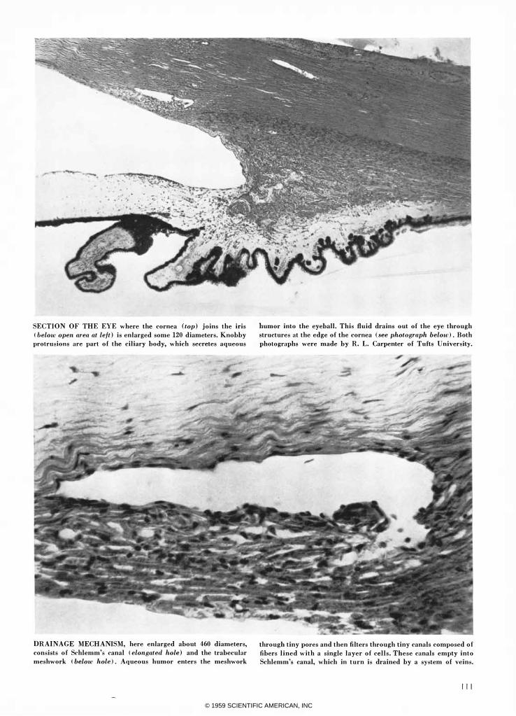
SECTION OF THE EYE where the cornea (top) joins the iris
(below open area at left) is enlarged some 120 diameters. Knobby
protrusions are part of the ciliary body, which secretes aqueous
DRAINAGE MECHANISM, here enlarged about 460 diameters,
consists of Schlemm's canal (elongated hole) and the trabecular
meshwork (below hole). Aqueous humor enters the meshwork
humor into the eyeball. This fluid drains out of the eye through
structures at the edge of the cornea (see photograph below). Both
photographs were made by R. L. Carpenter of Tufts University.
through tiny pores and then filters through tiny canals composed of
fibers lined with a single layer of cells. These canals empty into
Schlemm's canal, which in turn is drained by a system of veins.
III
© 1959 SCIENTIFIC AMERICAN, INC
CONJUNCTIVA
OPTIC NERVE
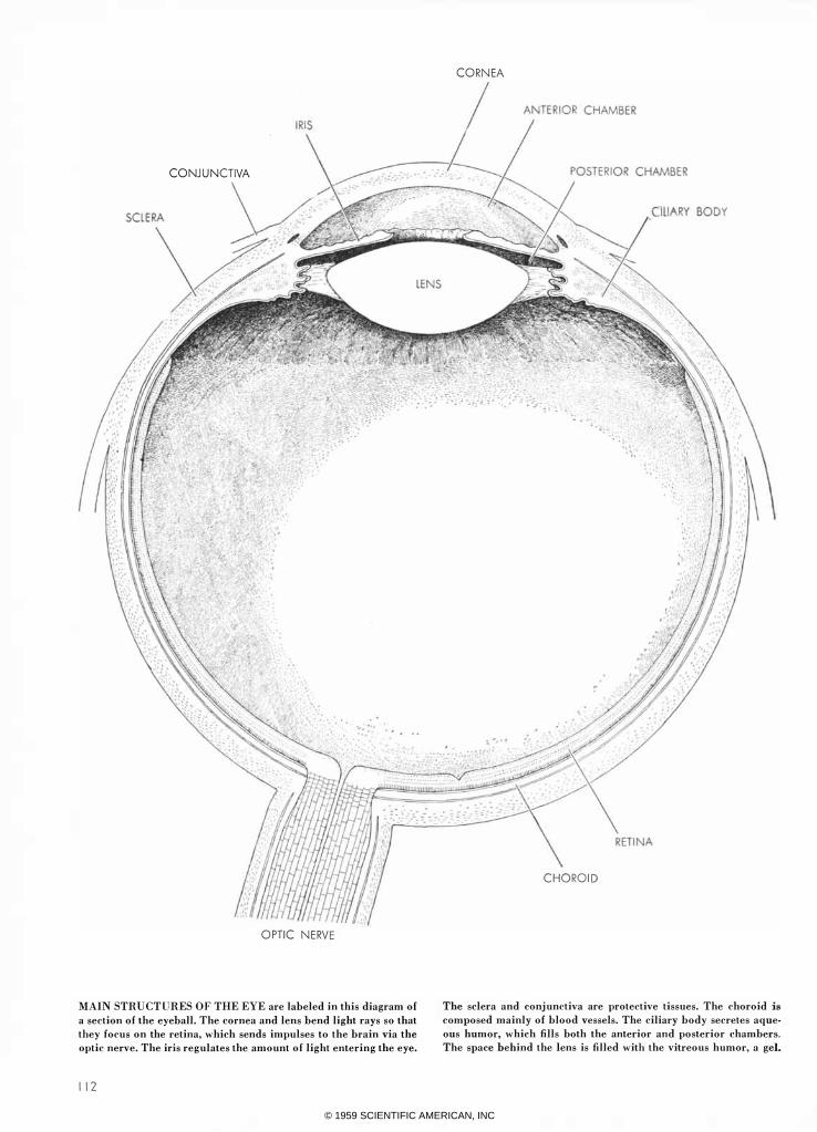
MAIN STRUCTURES OF THE EYE are labeled in this diagram of
a section of the eyeball. The cornea and lens bend light rays so that
they focus on the retina, which sends impulses to the brain via the
optic nerve. The iris regulates the amount of light entering the eye.
112
CORNEA
CHOROID
The sclera and conjunctiva are protective tissues. The choroid is composed mainly of blood vessels. The ciliary body secretes aque
ous humor, which fills both the anterior and posterior chambers.
The space behind the lens is filled with the vitreous humor, a gel.
© 1959 SCIENTIFIC AMERICAN, INC
knowingly harbors a tendency toward developing an acute type of glaucoma: the "angle-closure" glaucoma. These people have eyes with shallow anterior chambers-that is, the distance between the cornea and iris is significantly smaller than in the average individual-and the angle where the two come together is abnormally acute. Their eyes are sometimes actually smaller in diameter from front to back than the average. In such an individual the space between the iris and cornea becomes smaller with advancing age, decreasing the volume of the anterior chamber still further and making the angle between the comea and iris even more acute. Since the trabecular meshwork is located in this angle, its tiny intake pores begin to be dangerously confined. Now when the pupil of the eye opens to its fullest, the iris bunches up on itself. If the angle between the iris and cornea is very narrow, the thickened iris may push against the trabecular meshwork, plug its pores and stop the outRow of fluid. The pressure in the eye rises rapidly, and the victim experiences a sudden attack of glaucoma. Since the pupil opens to its fullest in prolonged darkness, it is not surprising that many persons suffer their first attacks of this type of glaucoma at the theater. Emotional disturbances or pain may also initiate an attack by causing the pupils to dilate or by producing swelling (edema) in the iris and other eye tissues.
W ithin an hour of the blockage the pressure in the eye may reach three
or four times the normal value. Fortunately the elevated pressure eventually inhibits the secretion of aqueous humor, otherwise the eye would rupture. As it is, the eye feels "hard as a marble" to the touch and appears grossly red and inflamed. If the pressure remains elevated for a while, fluid is taken up by the cornea, causing it to become waterlogged; it loses its transparency and appears steamy both to the observer and to the patient, who may complain of seeing rainbows and haloes around lights, due to a prism effect caused by droplets of water accumulating between the corneal fibers. The pain in and about the eye and the severe headache that attend these attacks may cause further and more prolonged dilation of the pupil, thereby aggravating the condition; frequently the patient may also suffer from nausea and vomiting.
Although the immediate consequences of this sudden rise in intraocular pressure are dramatic and distressing, permanent damage can be averted by
prompt diagnosis and treatment. Should the high pressure persist for more than 36 to 48 hours, however, the eye may be severely damaged and even blinded. An attack of this kind should therefore be handled as a true medical emergency. If there is any doubt, the diagnosis can be substantiated by simply pressing gently on the eye and comparing its consistency with that of the unaffected eye. For a more precise estimate of the intraocular pressure the ophthalmologist uses an instrument called the tonometer, which measures the degree to which the cornea can be indented by the pressure of a known weight on its surface.
Treatment to bring down the pressure should begin at once. Since iris tissue has been pushed into the outflow pores by dilation of the pupil, the obvious thing to do is to make the pupil constrict. Fortunately there are several drugs (such as eserine and pilocarpine) that rapidly induce this effect when they are instilled into the eye in the form of drops or ointment. These drugs act directly on the sphincter muscle within the iris, and pull the iris tissue away from the trabecular area. The fluid once again drains out of the eye, and the intraocular pres-
ON PERIODICAL
STORAGE SPACE
SCIENTIFIC AMERICAN and
over seven hundred and fifty
other leading periodicals are
now available on microfilm-
cost about equal to library bind-
ing. Microfilms of issues from
sure falls. May, 1948 [first issue of new As is apparent, it should also be pos- ,
sible to relieve the pressure by reducing SCIENTIFIC AMERICAN ] may be the secretion of fluid. A new drug called Diamox has this effect, cutting down the inflow of aqueous humor from about three cubic millimeters per minute to one cubic millimeter. This drug can be administered orally, but it is more effective when it is given intravenously. When the reduction of inflow is combined with the freeing of the outflow channel, the pressure drops within minutes, and the eye resumes its normal state, appearance and function. But the degree of recovery depends to a great extent on the length of time the eye has had to endure high pressure.
If the iris tissue remains too long in contact with the trabecular meshwork, fibrous tissue eventually forms between them. These "adhesions," composed of a dense material, close off the trabecular pores permanently and render the drainage mechanism functionless. Continued high pressure leads to the atrophy and degeneration of the nerve fibers within the retina and in the optic-nerve head. Since nerve tissue emanating directly from the brain does not regenerate, the eye loses its sight.
Though serious consequences may be averted in the initial attack, the pa
tient stands in grave peril of a recurrence. With each succeeding attack the
secured.
Addressing inquiries to
us on your organization's letter-
head will help us be of better
service to you. Write for details
to Department SA.
UNIVERSITY MICROFILMS
ANN ARBOR
MICHIGAN
II � ##"""",#,##,#####,####,,##,#,###
1 13
© 1959 SCIENTIFIC AMERICAN, INC
c o R N E
ANTERIOR
- . -.... 0 .. .. .. " : .: . .. .. co " .. .. : ... .. : .. : .... :- : .. ..
• D ... ... .. -"" ........ .. - .. 0 '" 0 .. .. ..
E N s
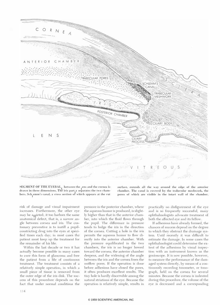
SEGMENT OF THE EYEBALL between the lens and the cornea is
d.-awn in three dimensions. The iris partly separates the two chaIn
bers. Schlemm's canal, a cross section of which appears at the cut
surface, extends all the way around the edge of the anterior
chamber. The canal is covered by the trabecular meshwork, the
pores of which are visible in the intact wall of the chamber.
risk of damage and visual impairment increases. Furthermore, the other eye may be afflicted; it too harbors the same anatomical defect, that is, a narrow angle between cornea and iris. The customary preventive is to instill a pupilconstricting drug into the eyes at specified times each day; in most cases the patient must keep up the treatment for the remainder of his life.
Within the last decade or two it has actually become possible in many cases to cure this form of glaucoma and free the patient from a life of continuous treatment. The treatment consists of a relatively simple operation, in which a small piece of tissue is removed from the outer edge of the iris disk. The success of this procedure depends on the fact that under normal conditions the
114
pressure in the posterior chamber, where the aqueous humor is produced, is slightly higher than that in the anterior chamber, into which the fluid flows through the pupil. The difference in pressure tends to bulge the iris in the direction of the cornea. Cutting a hole in the iris permits the aqueous humor to flow directly into the anterior chamber. With the pressure equilibrated in the two chambers, the iris is no longer bowed toward the cornea; the anterior chamber deepens, and the widening of the angle between the iris and the cornea frees the outflow pores. If the operation is done before adhesions have closed the pores, it often produces excellent results. The tiny hole is hardly discernible among the natural striations of the eye. Because the operation is relatively simple, results in
practically no disfigurement of the eye and is so frequently successful, many ophthalmologists advocate treatment of both the affected eye and its fellow.
If adhesions have already formed, the chances of success depend on the degree to which they obstruct the drainage system. Until recently it was difficult to estimate the damage. In some cases the ophthalmologist could determine the extent of the adhesions by visual inspection with an instrument known as the gonioscope. It is now possible, however, to measure the performance of the damaged system directly, by means of a continuously recording tonometer, or tonograph, held on the cornea for several minutes. Because the cornea is indented during this procedure, the volume of the eye is decreased and a corresponding
© 1959 SCIENTIFIC AMERICAN, INC
amount of aqueous humor is squeezed out of the eye. In the case of a healthy eye the readings will show a decline in intraocular pressure since aqueous humor is flowing out at an increased rate. In the glaucomatous eye, however, the pressure decreases much more slowly. From the readings of this test a series of computations yields a measure of the functional ability of the drainage mechanism. The ophthalmologist can then decide on the course of therapy.
If the test indicates that the drainage mechanism is no longer capable of functioning, surgery of the iris obviously offers no relief. In such a case surgery must be aimed at building a new drainage mechanism for the eye. By a variety of procedures the surgeon undertakes to open a new channel between the anterior chamber and the tissues containing the collecting veins. Unfortunately the results of these operations are uncertain, and much research is now directed toward their improvement.
A less dramatic but far more widespread disease is the chronic form of glaucoma. It is known as open-angle glaucoma, and, as the name implies, the drainage angle between the iris and the cornea offers no obstruction to the flow of fluid. The trouble lies within the drainage system itself. Just how openangle glaucoma starts is not clear. It probably results from a disturbance in the mechanism that controls intraocular pressure. The disturbance brings a diminution in the size of the trabecular pores and thus slows the outflow of aqueous humor. Another cause of open-angle glaucoma may be the gradual thickening and sclerosis of the trabecular fibers with increasing age. These are normal processes which usually proceed so slowly that they do not interfere perceptibly with aqueous drainage during the average lifetime. In some persons, however, they develop early enough in life to bring trouble.
\Vhatever the cause, the clogging of the drainage mechanism proceeds
at a gradual pace. Since intraocular pressure builds up over a long period of time, the eye has time to adapt itself, remaining white, giving no pain and otherwise showing no warning symptom. Many victims therefore remain unaware that the disease is in progress until it actually impairs their vision by destruction of the optic-nerve fibers. The occasional headache and the sensation of rainbows and haloes around lights, arising from intermittent corneal edema, do not cause sufficient distress to suggest the need for medical attention.
Once the condition is discovered, the intraocular pressure can often be adequately controlled by medication alone. Drugs like Diamox which decrease the input of fluid obviously are of help. The pupil-constricting drugs such as eserine may also be beneficial in this condition because they stimulate the muscles of the ciliary body to open up the narrowed pores and in general improve the drainage. Ophthalmologists usually do not
advise surgery so long as the intraocular pressure responds to the drugs and there is no further visual loss. 'When necessary, surgery may be attempted to develop an artificial drainage route.
In the rare appearance of glaucoma in infants the symptoms may be somewhat different from those in adults. The young growing eye, unlike the adult eye, is capable of being stretched and enlarged by an abnormally high internal
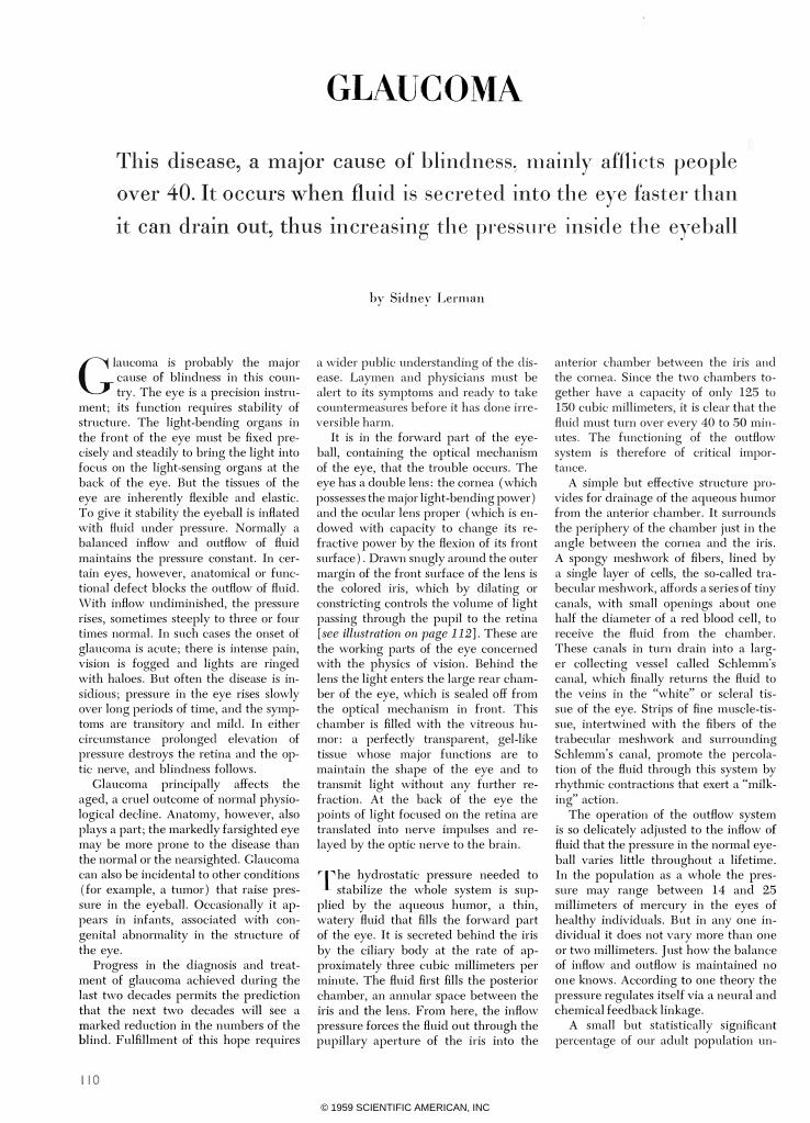
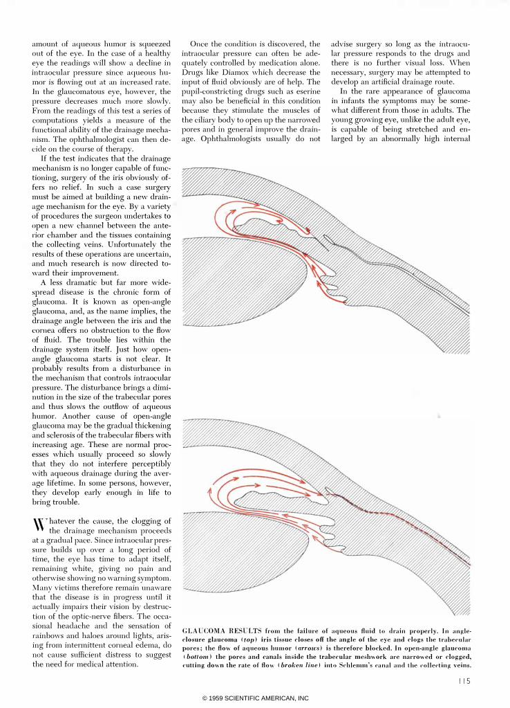
GLAUCOMA RESULTS from the failure of aqueous fluid to drain properly. In angle·
closure glaucoma (top) ids tissue closes off the angle of the eye and clogs the trabecular
pores; the flow of aqueous humor (arrows) is therefore blocked. In open·angle glaucoma
tbottom) the pores and canals inside the trabecular meshwork are narrowed or clogged,
cutting down the rate of flow (broken lin.e) into Sthlemm's (',mal and the colle('ting veins.
115
© 1959 SCIENTIFIC AMERICAN, INC

CEC Electro Mechanical Instrument Division
1 16
© 1959 SCIENTIFIC AMERICAN, INC
ELECTRO MECHANICAL INSTRUMENT DIVISION
Direct-writing oscillographs, data amplifiers, vibration meters and power supplies ... for dynamic tests in missile, industrial, and , flight-test environments .
• Write for Bulletin CEC 131 O-S.
DATATAPE DIVISION
Magnetic tape recording and reproduciflg·equipment for groundstation, airborne and mobile applications. Bulletin CEC 1312-S.
ANALYTICAL & CONTROL INSTRUMENT DIVISION
Mass spectrometers, leak detectors, chromatographs, moisture monitors, ... lab or process models. Bulletin 1313-S.
CONSOLIDATED SYSTEMS CORPORATION
Subsidiary of CEC. Systems for industrial process control, digital data handling, chemical analysis, missile checkout. Bulletin 1304-S.
DATALAB DIVISION
CEC's Advanced Electronic Data Laboratory ... conceives and develops new instrumentation under R&D contracts. Bulletin 1314-S.
TRANSDUCER DIVISION
Sensing devices for industrial and military applications, pressure and vibration instrumentation, galvanometers, and telemetry equipment. Bulletin 1308-S.
Consolidated Electrodynamics Corporation 360 Sierra Madre Villa Pasadena, California
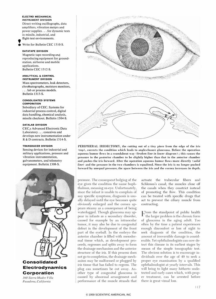
PERIPHERAL IRIDECTOMY, the cutting out of a tiny piece from the edge of the iris
(top), corrects the condition which leads to angle.closure glaucoma. Before the operation
aqueous humor flows in a roundabout way (broken line in lower diagram) ; this causes the
pressure in the posterior chamber to be slightly higher than that in the anterior chamber
and pushes the iris forward. After the operation aqueous humor flows more directly (solid
line) and the pressure in the two chambers is equalized. Since the iris is no longer pushed
forward by unequal pressure, the space between the iris and the cornea increases in depth.
pressure. The consequent bulging of the eye gives the condition the name buphthalmos, meaning ox-eye. Unfortunately, since the infant is unable to complain of any specific symptoms, diagnosis is usually delayed until the eye becomes quite obviously enlarged and the cornea appears steamy as a consequence of being waterlogged. Though glaucoma may appear in infants as a secondary disorder, caused for example by an intraocular tumor, it may also be due to congenital defect in the development of the front part of the eyeball. In the embryo the anterior chamber is filled with mesodermal tissue which, as development proceeds, regresses and splits away to form the drainage mechanism and the anterior structure of the iris. If this process does not go to completion, the drainage mechanism may be malformed or plugged by iris tissue that has failed to regress. The plug can sometimes be cut away. Another type of congenital glaucoma is caused by abnormal arrangement and performance of the muscle strands that
actuate the trabecular fibers and Schlemm's canal; the muscles close off the canals when they constrict instead of promoting the flow. This condition can be treated with specific drugs that act to prevent the ciliary muscle from contracting.
From the standpoint of public health the larger problem is the chronic form
of glaucoma in the aging adult. Too often by the time a patient experiences enough discomfort or loss of sight to seek diagnosis of the condition, the amount of irreversible damage is considerable. Yet ophthalmologists can now detect this disease in its earliest stages by means of the simple tonographic test. The obvious solution is to encourage individuals over the age of 40 to seek a proper eye examination by a qualified ophthalmologist at yearly intervals. This will bring to light many hitherto undetected and early cases which, with proper treatment, can be arrested before there is great visual loss.
1 17
© 1959 SCIENTIFIC AMERICAN, INC

118
From resins and rubbers to foodstuffs and soaps, very small percentages of organotins and inorganic tin compounds have proved their ability to protect products against degradation through exposure to air, heat and light-or from the harmful catalytic effects of impurities or contaminants. The results have been entirely new life-expectancies or new application possibilities for a wide range of products.
Our "summary of reported stabilization applications" will be helpful to those concerned with the problems of product degradation. Request a copy as a "starter" and you can count on further help from M&T based on the detailed information contained in our extensive application files .
•
I I METAL & THERMIT Corporation,
Sn Sb P inorganics and Si Ti Zr organometallics
Rahway, N. J.
© 1959 SCIENTIFIC AMERICAN, INC