Embed Size (px)
Citation preview
Synthesis of acene based organic gelators. Examination of the spectroscopical, photochemical and
rheological properties
Vom Fachbereich für Chemie und Pharmazie
der Technischen Universität Carolo-Wilhelmina
zu Braunschweig
zur Erlangung des Grades eines
Doktors der Naturwissenschaften
(Dr. rer. nat.)
genehmigte
D i s s e r t a t i o n
von Jens Reichwagen
aus Hamburg
1. Referent: Professor Dr. H. Hopf
2. Referentin: Professor Dr. M. Mazik
eingereicht am: 03.02.2005
mündliche Prüfung (Disputation) am: 18.04.2005
Vorveröffentlichungen der Dissertation
Teilergebnisse aus dieser Arbeit wurden mit Genehmigung der Gemeinsamen
Naturwissenschaftlichen Fakultät, vertreten durch den Betreuer der Arbeit, in folgenden
Beiträgen vorab veröffentlicht:
Publikationen
Jens Reichwagen, Henning Hopf, André Del Guerzo, Jean-Pierre Desvergne, and Henri
Bouas-Laurent: Photodimers of a Soluble Tetracene Derivative. Excimer Fluorescence from
the Head-to-Head Isomer, Org.Lett., 2004, 6, 12, 1899-1902.
André Del Guerzo, Colette Belin, Henri Bouas-Laurent, Jean-Pierre Desvergne, Jens
Reichwagen, Henning Hopf: Photochromism ans Self-Assembly of Soluble Tetracenes, Mol.
Cryst. Liq. Cryst., in press.
Jens Reichwagen, Henning Hopf, André Del Guerzo, Colette Belin, Henri Bouas-Laurent,
Jean-Pierre Desvergne: Synthesis of 2,3-substituted tetracenes and evaluation of their self-
assembling properties in organic solvents, Org. Lett., 2005, 7, 6, 971-974.
Tagungsbeiträge
Reichwagen, J., Hopf H., Desvergne J.-P., Bouas-Laurent H.: Synthese und Eigenschaften
organischer Gelbildner; 2,3 Didecyloxytetracen (DDOA) (Poster) 13. Vortragstagung der
LIEBIG-Vereinigung für Organische Chemie der Gesellschaft Deutscher Chemiker
(ORCHEM 2002), Bad Nauheim (2002).
Reichwagen, J., Hopf, H., Del Guerzo, A. Desvergne, J.-P., Bouas-Laurent, H.: Alkoxiacene:
Spektroskopie, Gel-Bildung und Photochemie einer variablen Substanzklasse (Poster)
Jahrestagung der GDCh 2003, München (2003).
André Del Guerzo, Jens Reichwagen, Henning Hopf, Colette Belin, Henri Bouas-Laurent,
Jean-Pierre Desvergne: Self-Assembled Fibers with Tunable Optical Properties: Organogels
of Anthracene and Tetracene Derivatives (Poster), HRSMC & EPA Summer School "New
Perspectives in Photochemistry" Egmond aan Zee (2003).

André Del Guerzo, Colette Belin, Henri Bouas-Laurent, Jean-Pierre Desvergne, Jens
Reichwagen, Henning Hopf: Self-Assembling and Photochemical Properties of soluble
Tetracenes, XXth IUPAC Symposium on Photochemistry, Granada (2004).
Del Guerzo, A., Belin, C., Bouas-Laurent, H., Desvergne, J.-P., Reichwagen, J., Hopf, H.:
Photochromism and Selfassembling of Soluble Tetracene Derivatives (Poster), ISOP'04,
Arcachon (2004)
Die vorliegende Arbeit wurde in der Zeit von Oktober 2001 bis November 2004 am Institut
für Organische Chemie der Technischen Universität Braunschweig unter der Leitung von
Prof. Dr. H. Hopf angefertigt.
This study could not have been carried out and completed without the help of numerous
persons.
I would like to express my sincere gratitude to Prof. Dr. Henning Hopf for the possibility to
perform this study, for his encouragement, advice and his interest.
Further, I am very grateful to Prof. Dr. Monika Mazik for agreeing to be the co-referee of my
thesis.
For the opportunity to study and do research at the Université Bordeaux in 2002 and 2003, I
would like to thank Prof. Dr. Henri Bouas-Laurent and Prof. Dr. Jean-Pierre Desvergne.
I am very grateful to Ms. Petra Holba-Schulz for the measurement of high resolution and 2D
NMR spectra; Ms. Karin Kadhim for measuring UV and IR spectra; Dr. Ulrich Papke and Ms.
Doris Döring for the measurement of high and low resolution mass spectra.
For the measurement of AFM and fluorescence spectra, as well as valuable discussions about
photophysical techniques, I am thankful to Mme. Colette Belin and Dr. André Del Guerzo.
I would like to express further my sincere gratitude to Dr. Kerstin Ibrom for many valuable
discussions about my work in general and about NMR spectroscopy.
For financial support of this study, I would like to thank the "Fonds der Chemischen
Industrie".
Last, but nor least, I thank all my colleagues for the incountable valuable discussions, the
support and the enjoyable working atmosphere I experienced throughout these years.
Table of contents
Table of contents ........................................................................................................................ 5 1 Introduction ........................................................................................................................ 1 2 Polyacenes as low molecular mass gelators ....................................................................... 3
2.1 Introduction ................................................................................................................ 3 2.2 Classes of organogelators........................................................................................... 4
2.2.1 Fatty acid derivatives ......................................................................................... 4 2.2.2 Steroid derivatives.............................................................................................. 5 2.2.3 Anthryl derivatives ............................................................................................. 6 2.2.4 Gelators containing steroidal and condensed aromatic rings ............................. 7 2.2.5 Amino acid type organogelators ........................................................................ 9 2.2.6 Organometallic compounds.............................................................................. 11 2.2.7 Miscellaneous types of gelators ....................................................................... 12 2.2.8 Two component systems .................................................................................. 14
2.3 Synthesis of gel forming tetracenes ......................................................................... 15 2.3.1 Synthetic strategies........................................................................................... 15
2.3.1.1 Strategy A..................................................................................................... 16 2.3.1.2 Strategy B..................................................................................................... 19 2.3.1.3 Strategy C..................................................................................................... 20
2.4 Retrosynthesis of 2,3-dialkoxy-tetracenes (2) via aldol condensation, second approach ............................................................................................................................... 22 2.5 Synthesis of 2,3-dialkoxy-tetracenes (2) .................................................................. 24
2.5.1 Explanation of the individual synthetic steps................................................... 25 2.5.1.1 2,3-Dimethoxy-buta-1,3-diene (37) ............................................................. 25 2.5.1.2 6,7-Dimethoxy-1,4-naphthoquinone (58). ................................................... 25 2.5.1.3 Reduction of 6,7-dimethoxy-1,4-naphthoquinone (58)................................ 27 2.5.1.4 Condensation of 1,4-dihydroxy-6,7-dimethoxy-naphthalene (57) with phthalic dialdehyde (46)............................................................................................... 28 2.5.1.5 Demethylation of 2,3-dimethoxy-tetracene-5,12-quinone (42). .................. 33 2.5.1.6 Alkylation of 2,3-dihydroxy-tetracene-5,12-quinone (35)........................... 38 2.5.1.7 Reduction of 2,3-dialkoxy-tetracene-5,12-quinones (61) ............................ 43
2.5.2 Variations of the synthesis ............................................................................... 49 2.6 Synthesis of gel forming pentacenes ........................................................................ 50
2.6.1 Introduction ...................................................................................................... 50 2.7 Retrosynthesis of 2,3-dialkoxypentacenes (74) ....................................................... 51 2.8 Synthesis of 2,3-dialkoxy-pentacenes (74) .............................................................. 52
2.8.1 Explanation of the individual synthetic steps................................................... 52 2.8.1.1 Synthesis of naphthalene-2,3-dialdehyde (52) ............................................. 53 2.8.1.2 Synthesis of 2,3-dimethoxy-5,14-pentacene-quinone (77) .......................... 55 2.8.1.3 Demethylation of 2,3-dimethoxy-pentacene-5,14-quinone (77).................. 57 2.8.1.4 Alkylation of 2,3-dihydroxy-pentacene-5,14-quinone (76) ......................... 57 2.8.1.5 Reduction of 2,3-dialkoxy-pentacene-5,14-quinones (75)........................... 60
2.9 Synthesis of gel forming anthracenes....................................................................... 64 2.9.1 Introduction ...................................................................................................... 64
2.10 Retrosynthesis .......................................................................................................... 64 2.11 Synthesis of 2,3-didecyloxy-anthracene (1)............................................................. 65
2.11.1 Explanation of the individual synthetic steps................................................... 65
2.11.1.1 Synthesis of 2,3-dimethoxy-anthra-9,10-quinone (38) ............................ 65 2.11.1.2 Demethylation of 2,3-dimethoxy-9,10-anthraquinone (38) ..................... 66 2.11.1.3 Alkylation of 2,3-dihydroxy-anthracene-9,10-quinone (28).................... 66 2.11.1.4 Reduction of 2,3-didecyloxy-anthracene-9,10-quinone (8) ..................... 67
3 Examination of the gel forming abilities of 2,3-dialkoxy-acenes .................................... 68 3.1 2,3-Dialkoxy-tetracenes 2 ........................................................................................ 68
3.1.1 Inverted test tube examinations........................................................................ 68 3.1.1.1 Influence of the chain length of different 2,3-dialkoxy-tetracenes 2 on gel formation ...................................................................................................................... 70
3.1.2 Spectroscopic examination of the gel formation.............................................. 72 3.1.3 Thermodynamic data of the gel formation ....................................................... 74 3.1.4 AFM-measurements on 2,3-dialkoxy-tetracene 2 gels..................................... 79
3.1.4.1 General introduction to AFM measurements ............................................... 79 3.1.4.2 Sample preparation and measurements ........................................................ 80
3.2 2,3-Dialkoxy-pentacenes.......................................................................................... 83 3.2.1 Inverted tube tests............................................................................................. 84 3.2.2 UV/Vis measurements...................................................................................... 84 3.2.3 AFM-measurements ......................................................................................... 85
3.3 Comparison of gel forming acenes (2,3-DDOA, 2,3-DDOT/ 2,3-DHdOT, 2,3-DHdOP)................................................................................................................................ 86 3.4 Mixed gels between 2,3-DDOA (1) and 2,3-DDOT (86) ........................................ 87
3.4.1 Spectroscopic examination of the mixed-gel-formation .................................. 87 4 Photochemistry................................................................................................................. 90
4.1 Introduction .............................................................................................................. 90 4.2 General technique..................................................................................................... 92 4.3 Tetracene (82) .......................................................................................................... 92
4.3.1 Possible photoproducts of tetracene (82) ......................................................... 92 4.3.2 Irradiation of tetracene (82).............................................................................. 94
4.4 2,3-Disubstituted tetracenes ..................................................................................... 98 4.4.1 Possible photoproducts..................................................................................... 98 4.4.2 Irradiation of 2,3-DDOT (86)......................................................................... 100
4.4.2.1 Structure elucidation of the photoproducts ................................................ 110 4.5 5,12-Disubstituted tetracenes ................................................................................. 112
4.5.1 Retrosynthesis of 5,12-didecyloxytetracene (85)........................................... 112 4.5.1.1 Synthesis of 5,12-didecyloxy-tetracene (85).............................................. 114 4.5.1.2 Explanation of the individual synthetic steps............................................. 115
4.5.2 Possible photoproducts................................................................................... 121 4.5.3 Irradiation of 5,12-DDOT (85)....................................................................... 122
4.5.3.1 Differentiation of the photodimers 96 and 97 ............................................ 127 4.5.4 Further investigation of 96 and 97 ................................................................. 128
4.5.4.1 Reaction-quantum-yields of the 5,12-DDOT-dimerization ....................... 128 4.5.4.2 Thermal and photochemical decomposition of 96 and 97 ......................... 130
4.6 Crossed photodimers of 5,12-DDOT (85) and tetracene (82)................................ 133 4.6.1 Possible photoproducts................................................................................... 133 4.6.2 Irradiation of a mixture of 5,12-DDOT (85) and tetracene (82) .................... 134
4.6.2.1 Mixed dimers.............................................................................................. 135 4.6.2.2 Structural elucidation ................................................................................. 141
4.7 Summary of the structural elucidation of tetracene dimers.................................... 142 5 Photophysical examination of various tetracenes .......................................................... 145
5.1 Measurement principles ......................................................................................... 146 5.1.1 Fluorescence quantum yield........................................................................... 146
5.1.2 Fluorescence life time .................................................................................... 147 5.2 Results of the fluorescence quantum yield measurements..................................... 149
5.2.1 Fluorescence-quantum-yield of 5,12-DDOT (85).......................................... 149 5.2.2 Fluorescence-quantum-yield of 2,3-DDOT (86)............................................ 150 5.2.3 Fluorescence-quantum-yield of 2,3-DHdOT (111)........................................ 151 5.2.4 Discussion of the results................................................................................. 151
5.3 Results of the fluorescence lifetime measurements ............................................... 152 5.3.1 Discussion of the results................................................................................. 152
5.4 Radiative decay rate constant ................................................................................. 153 6 Summary ........................................................................................................................ 154 7 Experimental part ........................................................................................................... 158
7.1 Techniques and equipment..................................................................................... 158 7.2 General working procedures (GWP)...................................................................... 159
7.2.1 GWP 1 (alkylation of dihydroxy-acene-quinones) ........................................ 159 7.2.2 GWP 2 (reduction of alkoxy-acene-quinones)............................................... 160 7.2.3 GWP 3 (photodimerization) ........................................................................... 160
7.3 Synthetic procedures .............................................................................................. 161 7.3.1 2,3-Dialkoxy-tetracene synthesis ................................................................... 161
7.3.1.1 2,3-Dimethoxy-buta-1,3-diene (37) ........................................................... 161 7.3.1.2 6,7-Dimethoxy-1,4-naphthoquinone (58) .................................................. 162 7.3.1.3 1,4-Dihydroxy-6,7-dimethoxy-naphthalene (57) ....................................... 163 7.3.1.4 2,3-Dimethoxy-tetracene-5,12-quinone (42).............................................. 164 7.3.1.5 2,3-Dihydroxy-tetracene-5,12-quinone (35) .............................................. 165 7.3.1.6 2,3-Dibutoxy-tetracene-5,12-quinone (106) .............................................. 166 7.3.1.7 2,3-Dihexyloxy-tetracene-5,12-quinone (107)........................................... 167 7.3.1.8 2,3-Didecyloxy-tetracene-5,12-quinone (56) ............................................. 168 7.3.1.9 2,3-Dihexadecyloxy-tetracene-5,12-quinone (108) ................................... 169 7.3.1.10 2,3-Dimethoxy-tetracene (73) ................................................................ 170 7.3.1.11 2,3-Dibutoxy-tetracene (109) ................................................................. 171 7.3.1.12 2,3-Dihexyloxy-tetracene (110) ............................................................. 172 7.3.1.13 2,3-Didecyloxy-tetracene (86) ............................................................... 173 7.3.1.14 2,3-Dihexadecyloxy-tetracene (111) ...................................................... 174
7.3.2 2,3-Didecyloxy-anthracene (1) synthesis ....................................................... 176 7.3.2.1 2,3-Dimethoxy-9,10-anthraquinone (38) ................................................... 176 7.3.2.2 2,3-Dihydroxy-9,10-anthraquinone (28) .................................................... 177 7.3.2.3 2,3-Didecyloxy-9,10-anthraquinone (8)..................................................... 178 7.3.2.4 2,3-Didecyloxy-anthracene (1)................................................................... 179
7.3.3 2,3-Dialkoxy-pentacene synthesis.................................................................. 180 7.3.3.1 Naphthalene-2,3-dicarboxaldehyde............................................................ 180 7.3.3.2 2,3-Dimethoxy-pentacene-5,14-quinone.................................................... 181 7.3.3.3 2,3-Dihydroxy-pentacene-5,14-quinone .................................................... 182 7.3.3.4 2,3-Didecyloxy-pentacene-5,14-quinone (112) ......................................... 182 7.3.3.5 2,3-Dihexadecyloxy-pentacene-5,14-quinone (113).................................. 183 7.3.3.6 2,3-Didecyloxy-pentacene (114) ................................................................ 184 7.3.3.7 2,3-Dihexadecyloxy-pentacene (115) ........................................................ 185
7.3.4 5,12-Dialkoxy-tetracene synthesis ................................................................. 186 7.3.4.1 1,4-Dihydroxy-naphthalene (95) ................................................................ 186 7.3.4.2 Tetracene-5,12-quinone (94) ...................................................................... 187 7.3.4.3 5,12-Didecyloxy-tetracene (85) ................................................................. 188
7.3.5 Photoreactions ................................................................................................ 189 7.3.5.1 Photodimerization of 2,3-DDOT (86) ........................................................ 189
7.3.5.2 Photodimerization of 5,12-didecyloxy-tetracene (85)................................ 194 7.3.5.3 Crossed photodimerization of 5,12-didecyloxy-tetracene (85) ....................... with tetracene (82)...................................................................................... 196
Appendix I: Gel formation measurements ............................................................................. 201 A) Inverted tube tests, solvent screening..................................................................... 201 B) Thermodynamic data of the gel formation using UV/Vis-spectroscopy................ 204 C) Cooperative behavior of 2,3-DDOA (1) and 2,3-DDOT (86)................................ 218 D) Thermal dissociation of 96 and 97. ........................................................................ 220
D.1) Dissociation of 96, hexane, 308 K ......................................................................... 220 D.2) Dissociation of 96, MCH, 342 K........................................................................ 220 D.3) Dissociation of 97, hexane, 308 K ..................................................................... 221 D.4) Dissociation of 97, MCH, 342 K........................................................................ 221
Appendix II: Commonly used abbreviations ......................................................................... 222 Appendix III: Literature ......................................................................................................... 223
1
1 Introduction
Low molecular mass gelators (LMMOGs) have in recent years been the target of growing
scientific interest. Even though they played a minor role in the organic literature during the
1970s, new developments and new structures of LMMOGs have been reported over the past
20 years. The focus on this class of gel forming substances has two reasons. For the organic
chemist LMMOGs play an important role (in comparison to polymeric gelators,
systematization vide infra) because they can be examined by structure effect relationships and
are subject of rational synthetic approaches.
The second reason is the widespread use of gels in industrial applications, pharmaceutical
applications and "everyday life".
This range of applications can be explained by the great diversity of structures that are
displayed on the microscopic and mesoscopic scale. Gels often show thermoreversibility,
exhibit a great chemical sensitivity and, as will be shown later in this work, a large diversity
of nanostructures.
A short summary of applications for gels may illustrate the importance of gel-like
materials.[1a] Reverse micellar systems (microemulsions) involving gels are used in
biocatalysis, biomembrane mimetics, extraction processes and the preparation of
microparticles. In waste disposal, gels are frequently involved in the recovery of spilled crude
oil or the disposal of used cooking oil. The application of organogels in drug delivery can be
illustrated by the example of active enzymes and bacteria entrapped in apolar gels of gelatin.
Many aqueous gels or complex mixtures of surface-active components in water and oil are
utilized for ointments or macromolecular separations, protein crystallizations, etc. One final
example is the use of gels containing ester groups for the gelification of electrolyte solutions
for lithium battery applications.
Like with many new gelators, the first discovery that simple acene structures can form gels in
low concentrations with organic solvents, was made accidentally.
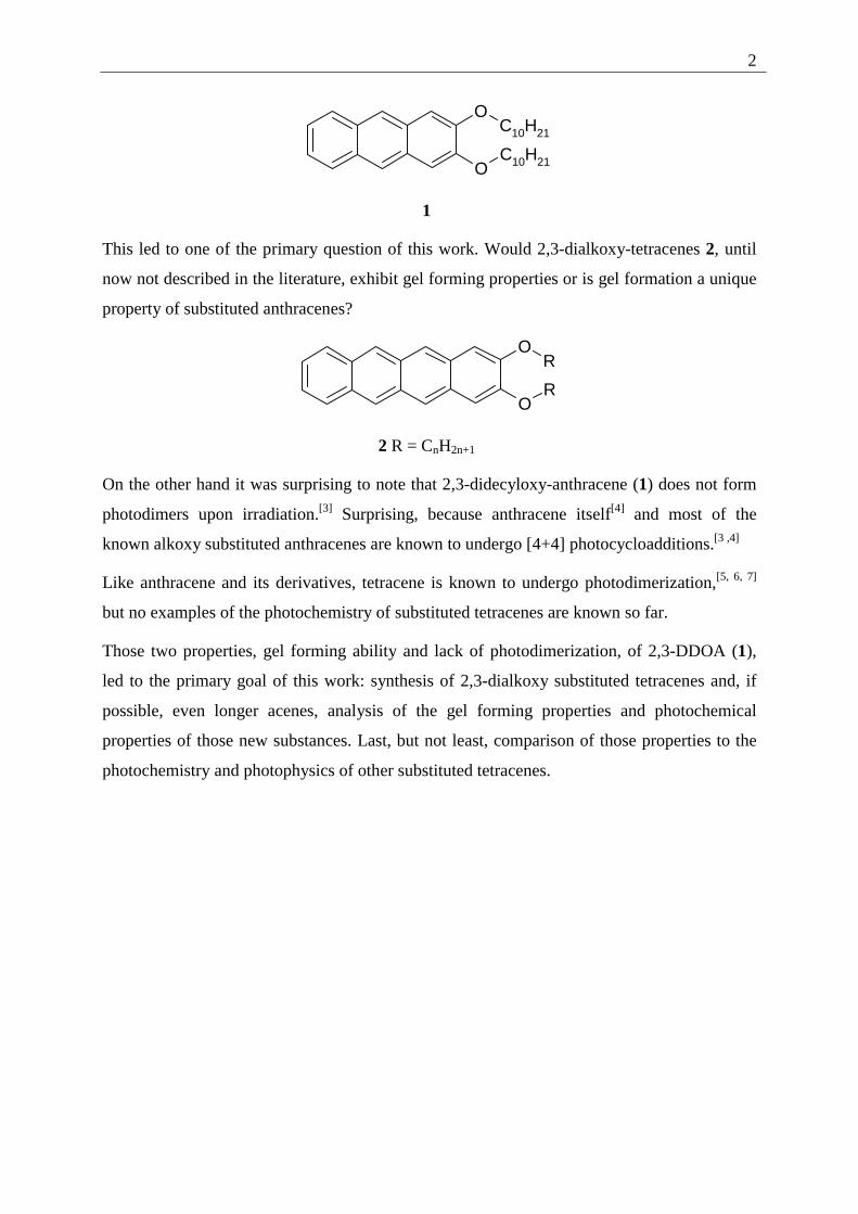
In 1991 Bouas-Laurent et al. discovered the gel forming abilities of 2,3-didecyloxy-
anthracene (1) (2,3-DDOA) in the course of photochemical investigations on substituted
anthracenes.[2] Similarly substituted naphthalenes or benzenes do not form gels under similar
conditions. Angular substitution (like in the case of dialkoxy-phenanthrenes) does not lead to
gel forming substances, too.[2]
2
O
O
C10H21
C10H21
1
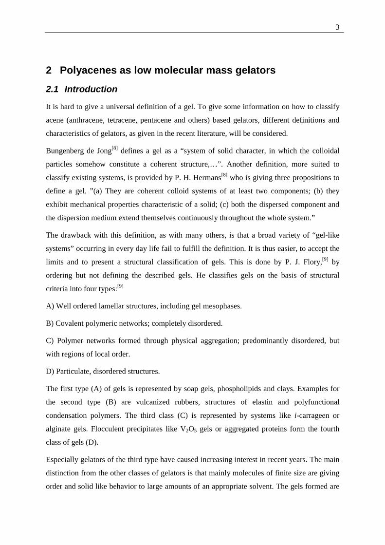
This led to one of the primary question of this work. Would 2,3-dialkoxy-tetracenes 2, until
now not described in the literature, exhibit gel forming properties or is gel formation a unique
property of substituted anthracenes?
O
O
R
R
2 R = CnH2n+1
On the other hand it was surprising to note that 2,3-didecyloxy-anthracene (1) does not form
photodimers upon irradiation.[3] Surprising, because anthracene itself[4] and most of the
known alkoxy substituted anthracenes are known to undergo [4+4] photocycloadditions.[3 ,4]
Like anthracene and its derivatives, tetracene is known to undergo photodimerization,[5, 6, 7]
but no examples of the photochemistry of substituted tetracenes are known so far.
Those two properties, gel forming ability and lack of photodimerization, of 2,3-DDOA (1),
led to the primary goal of this work: synthesis of 2,3-dialkoxy substituted tetracenes and, if
possible, even longer acenes, analysis of the gel forming properties and photochemical
properties of those new substances. Last, but not least, comparison of those properties to the
photochemistry and photophysics of other substituted tetracenes.
3
2 Polyacenes as low molecular mass gelators
2.1 Introduction
It is hard to give a universal definition of a gel. To give some information on how to classify
acene (anthracene, tetracene, pentacene and others) based gelators, different definitions and
characteristics of gelators, as given in the recent literature, will be considered.
Bungenberg de Jong[8] defines a gel as a “system of solid character, in which the colloidal
particles somehow constitute a coherent structure,…”. Another definition, more suited to
classify existing systems, is provided by P. H. Hermans[8] who is giving three propositions to
define a gel. ”(a) They are coherent colloid systems of at least two components; (b) they
exhibit mechanical properties characteristic of a solid; (c) both the dispersed component and
the dispersion medium extend themselves continuously throughout the whole system.”
The drawback with this definition, as with many others, is that a broad variety of “gel-like
systems” occurring in every day life fail to fulfill the definition. It is thus easier, to accept the
limits and to present a structural classification of gels. This is done by P. J. Flory,[9] by
ordering but not defining the described gels. He classifies gels on the basis of structural
criteria into four types:[9]
A) Well ordered lamellar structures, including gel mesophases.
B) Covalent polymeric networks; completely disordered.
C) Polymer networks formed through physical aggregation; predominantly disordered, but
with regions of local order.
D) Particulate, disordered structures.
The first type (A) of gels is represented by soap gels, phospholipids and clays. Examples for
the second type (B) are vulcanized rubbers, structures of elastin and polyfunctional
condensation polymers. The third class (C) is represented by systems like i-carrageen or
alginate gels. Flocculent precipitates like V2O5 gels or aggregated proteins form the fourth
class of gels (D).
Especially gelators of the third type have caused increasing interest in recent years. The main
distinction from the other classes of gelators is that mainly molecules of finite size are giving
order and solid like behavior to large amounts of an appropriate solvent. The gels formed are
4
often thermoreversible and in many cases only small amounts of the gelator (typically
< 2 wt%)[1a] are needed to form the viscoelastic liquidlike or solidlike material.
To show the structural versatility of organic gelators, some examples are described in the next
chapter.
2.2 Classes of organogelators
Terech and Weiss, who have reviewed the field of organic gelators extensively[1a] describe
eight major classes of organic gelators and their applications.
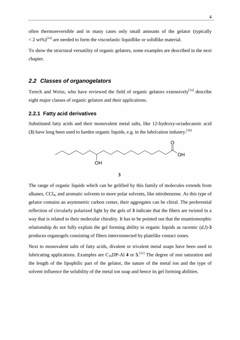
2.2.1 Fatty acid derivatives
Substituted fatty acids and their monovalent metal salts, like 12-hydroxy-octadecanoic acid
(3) have long been used to harden organic liquids, e.g. in the lubrication industry.[1b]
O
OH
OH
3
The range of organic liquids which can be gelified by this family of molecules extends from
alkanes, CCl4, and aromatic solvents to more polar solvents, like nitrobenzene. As this type of
gelator contains an asymmetric carbon center, their aggregates can be chiral. The preferential
reflection of circularly polarized light by the gels of 3 indicate that the fibers are twisted in a
way that is related to their molecular chirality. It has to be pointed out that the enantiomorphic
relationship do not fully explain the gel forming ability in organic liquids as racemic (d,l)-3
produces organogels consisting of fibers interconnected by platelike contact zones.
Next to monovalent salts of fatty acids, divalent or trivalent metal soaps have been used in
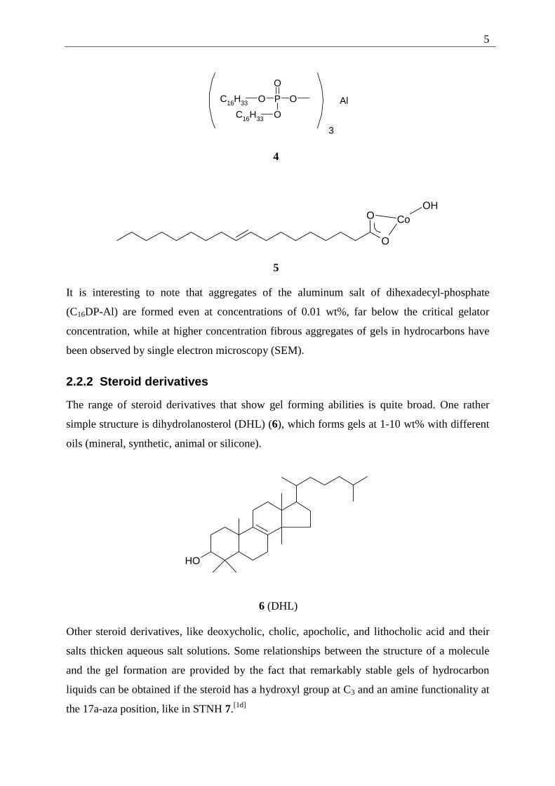
lubricating applications. Examples are C16DP-Al 4 or 5.[1c] The degree of non saturation and
the length of the lipophilic part of the gelator, the nature of the metal ion and the type of
solvent influence the solubility of the metal ion soap and hence its gel forming abilities.
5
P
O
O O
O
C16H33
C16H33 3
Al
4
O
O CoOH
5
It is interesting to note that aggregates of the aluminum salt of dihexadecyl-phosphate
(C16DP-Al) are formed even at concentrations of 0.01 wt%, far below the critical gelator
concentration, while at higher concentration fibrous aggregates of gels in hydrocarbons have
been observed by single electron microscopy (SEM).
2.2.2 Steroid derivatives
The range of steroid derivatives that show gel forming abilities is quite broad. One rather
simple structure is dihydrolanosterol (DHL) (6), which forms gels at 1-10 wt% with different
oils (mineral, synthetic, animal or silicone).
OH
6 (DHL)
Other steroid derivatives, like deoxycholic, cholic, apocholic, and lithocholic acid and their
salts thicken aqueous salt solutions. Some relationships between the structure of a molecule
and the gel formation are provided by the fact that remarkably stable gels of hydrocarbon
liquids can be obtained if the steroid has a hydroxyl group at C3 and an amine functionality at
the 17a-aza position, like in STNH 7.[1d]
6
N
H
OH
7 (STNH)
Other facts which determine the gel forming abilities of steroids are the position of
unsaturated functionalities. Two allyl groups at C17 inhibit gelation, but a double bond at C5,6
does not.
The steroid gel structures could be derived by small angle neutron scattering (SANS). These
data show that the aggregates are very long, rigid fibers, possibly symmetrical double helices
with a diameter of 99 Å. The structure contains further "contact points" that are formed by
specific overlap through fusion or coiling of two or more individual fibers over distances
much longer than their diameters. As for other gels, the TEM measurements of STNH
indicate that fiber interactions are (pseudo) crystalline zones in a heterogeneous network, with
a solidlike behavior.
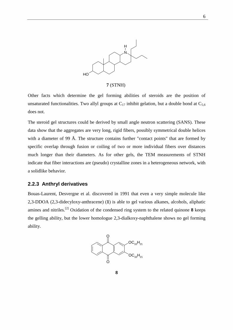
2.2.3 Anthryl derivatives
Bouas-Laurent, Desvergne et al. discovered in 1991 that even a very simple molecule like
2,3-DDOA (2,3-didecyloxy-anthracene) (1) is able to gel various alkanes, alcohols, aliphatic
amines and nitriles.[2] Oxidation of the condensed ring system to the related quinone 8 keeps
the gelling ability, but the lower homologue 2,3-dialkoxy-naphthalene shows no gel forming
ability.
OC10H21
OC10H21
O
O
8
7
The solution to gel phase transition of 2,3-DDOA (1) varies over a wide temperature range,
depending on the solvent and the gelator concentration. In comparison to other gelators, the
2,3-DDOA (1) gels show a magnitude of elasticity, which suggests that the interaction zones
are permanent crystalline microdomains at a given temperature.
2.2.4 Gelators containing steroidal and condensed aromatic rings
A combination of the gelators described in Chapter 2.1.2 and 2.1.3 can be seen in the class of
gelators containing steroidal and condensed aromatic rings. In contrast to STNH, the gelation
ability of which relies upon the presence of a hydroxyl group at C3, this class of molecules are
functionalized at C3 and cannot be H-bond donors. This type of gelators (ALS) consist of an
aromatic group (A) connected to a steroidal moiety (S) via a linking group (L). This
versatility makes the analysis of gel interactions easier, as any of the three building units can
be changed and some of the molecular attributes, like luminescence or chirality can be used to
investigate changes in the path from isotropic phase to the gel phase.
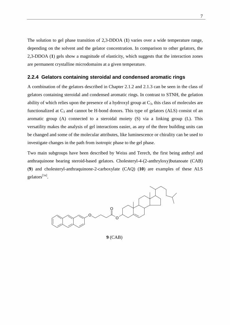
Two main subgroups have been described by Weiss and Terech, the first being anthryl and
anthraquinone bearing steroid-based gelators. Cholesteryl-4-(2-anthryloxy)butanoate (CAB)
(9) and cholesteryl-anthraquinone-2-carboxylate (CAQ) (10) are examples of these ALS
gelators[1e].
OO
O
9 (CAB)
8
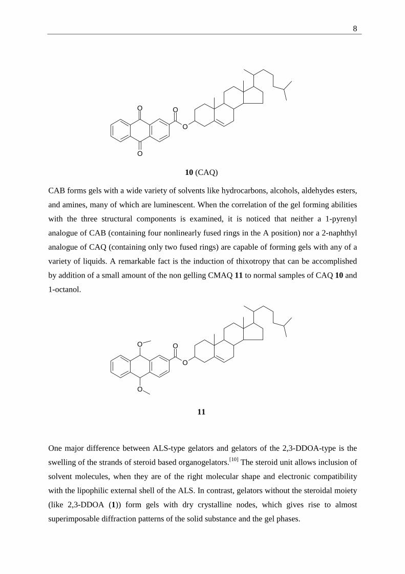
O
OO
O
10 (CAQ)
CAB forms gels with a wide variety of solvents like hydrocarbons, alcohols, aldehydes esters,
and amines, many of which are luminescent. When the correlation of the gel forming abilities
with the three structural components is examined, it is noticed that neither a 1-pyrenyl
analogue of CAB (containing four nonlinearly fused rings in the A position) nor a 2-naphthyl
analogue of CAQ (containing only two fused rings) are capable of forming gels with any of a
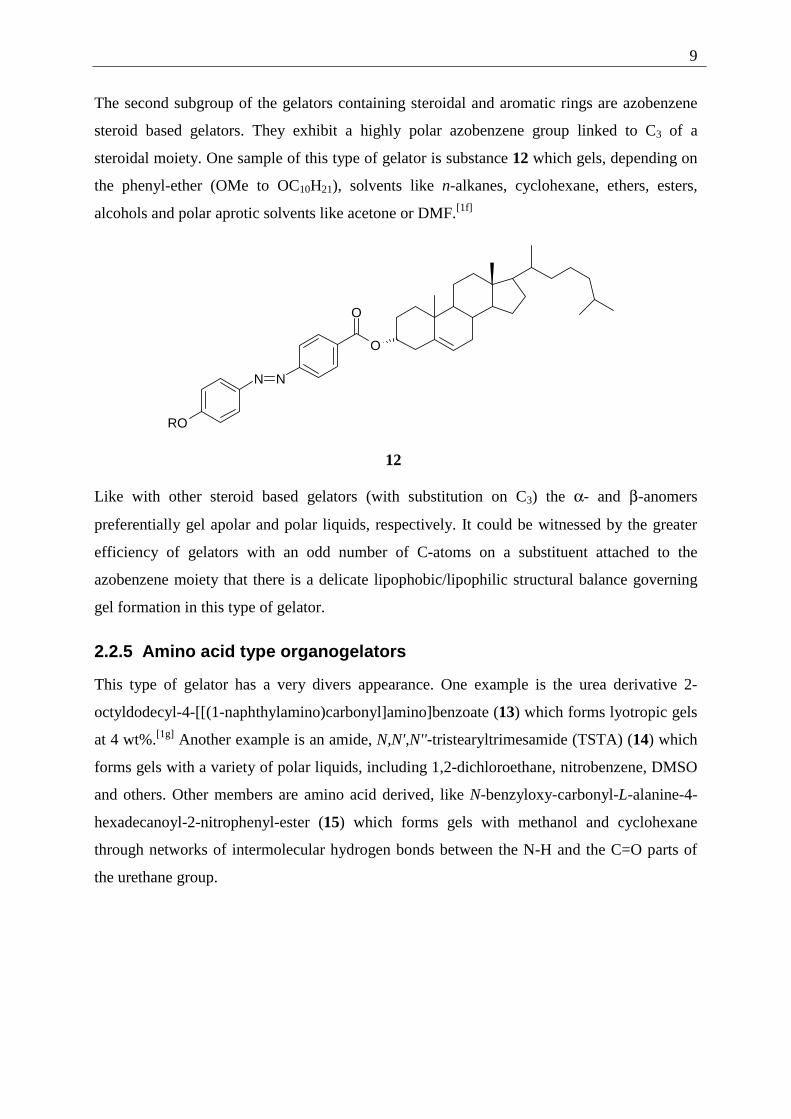
variety of liquids. A remarkable fact is the induction of thixotropy that can be accomplished
by addition of a small amount of the non gelling CMAQ 11 to normal samples of CAQ 10 and
1-octanol.
O
OO
O
11
One major difference between ALS-type gelators and gelators of the 2,3-DDOA-type is the
swelling of the strands of steroid based organogelators.[10] The steroid unit allows inclusion of
solvent molecules, when they are of the right molecular shape and electronic compatibility
with the lipophilic external shell of the ALS. In contrast, gelators without the steroidal moiety
(like 2,3-DDOA (1)) form gels with dry crystalline nodes, which gives rise to almost
superimposable diffraction patterns of the solid substance and the gel phases.
9
The second subgroup of the gelators containing steroidal and aromatic rings are azobenzene
steroid based gelators. They exhibit a highly polar azobenzene group linked to C3 of a
steroidal moiety. One sample of this type of gelator is substance 12 which gels, depending on
the phenyl-ether (OMe to OC10H21), solvents like n-alkanes, cyclohexane, ethers, esters,
alcohols and polar aprotic solvents like acetone or DMF.[1f]
RO
N N
O
O
12
Like with other steroid based gelators (with substitution on C3) the α- and β-anomers
preferentially gel apolar and polar liquids, respectively. It could be witnessed by the greater
efficiency of gelators with an odd number of C-atoms on a substituent attached to the
azobenzene moiety that there is a delicate lipophobic/lipophilic structural balance governing
gel formation in this type of gelator.
2.2.5 Amino acid type organogelators
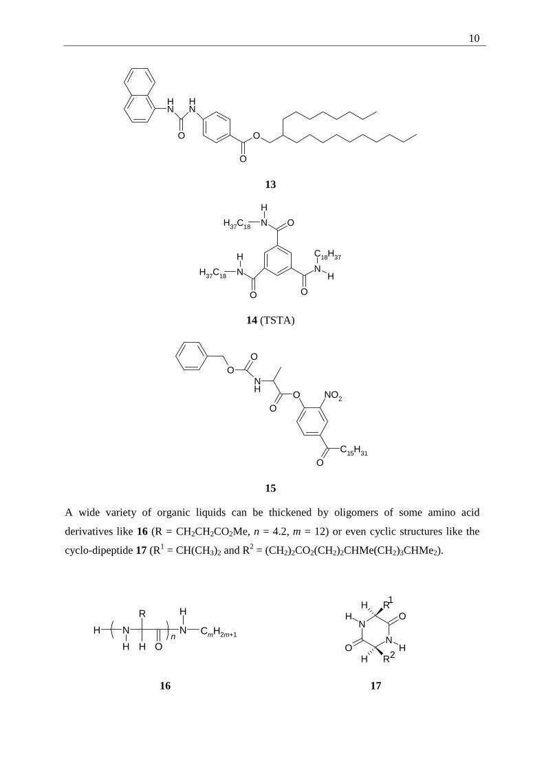
This type of gelator has a very divers appearance. One example is the urea derivative 2-
octyldodecyl-4-[[(1-naphthylamino)carbonyl]amino]benzoate (13) which forms lyotropic gels
at 4 wt%.[1g] Another example is an amide, N,N',N''-tristearyltrimesamide (TSTA) (14) which
forms gels with a variety of polar liquids, including 1,2-dichloroethane, nitrobenzene, DMSO
and others. Other members are amino acid derived, like N-benzyloxy-carbonyl-L-alanine-4-
hexadecanoyl-2-nitrophenyl-ester (15) which forms gels with methanol and cyclohexane
through networks of intermolecular hydrogen bonds between the N-H and the C=O parts of
the urethane group.
10
NH
O
NH
O
O
13
N
N N
O
O O
H37C18
H37C18
C18H37
H
H
H
14 (TSTA)
OO
NH
OO
OC15H31
NO2
15
A wide variety of organic liquids can be thickened by oligomers of some amino acid
derivatives like 16 (R = CH2CH2CO2Me, n = 4.2, m = 12) or even cyclic structures like the
cyclo-dipeptide 17 (R1 = CH(CH3)2 and R2 = (CH2)2CO2(CH2)2CHMe(CH2)3CHMe2).
H N
R
OHn N
H
H
CmH2m+1
N
NO
OR
R
H
H
H
H
1
2
16 17
11
Even some structurally simple tertiary amines and related tertiary and quaternary ammonium
halide salts form viscoelastic, thermally reversible gels with organic liquids ranging from
DMF to dodecane. Like with other gelators, the mode of gelation involves a network of
interconnected strands.
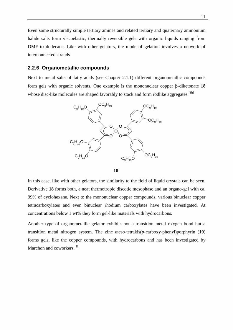
2.2.6 Organometallic compounds
Next to metal salts of fatty acids (see Chapter 2.1.1) different organometallic compounds
form gels with organic solvents. One example is the mononuclear copper β-diketonate 18
whose disc-like molecules are shaped favorably to stack and form rodlike aggregates.[1h]
CuO O
O O
OC9H19C9H19O
OC9H19
OC9H19
OC9H19
C9H19O
C9H19O C9H19O
18
In this case, like with other gelators, the similarity to the field of liquid crystals can be seen.
Derivative 18 forms both, a neat thermotropic discotic mesophase and an organo-gel with ca.
99% of cyclohexane. Next to the mononuclear copper compounds, various binuclear copper
tetracarboxylates and even binuclear rhodium carboxylates have been investigated. At
concentrations below 1 wt% they form gel-like materials with hydrocarbons.
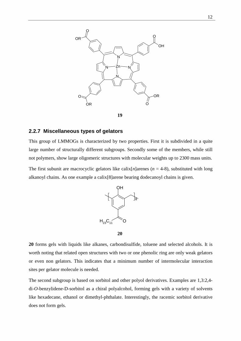
Another type of organometallic gelator exhibits not a transition metal oxygen bond but a
transition metal nitrogen system. The zinc meso-tetrakis(p-carboxy-phenyl)porphyrin (19)
forms gels, like the copper compounds, with hydrocarbons and has been investigated by
Marchon and coworkers.[1i]
12
N
N
N
N
Zn
OH
OO
OR
O
OR O
OR
19
2.2.7 Miscellaneous types of gelators
This group of LMMOGs is characterized by two properties. First it is subdivided in a quite
large number of structurally different subgroups. Secondly some of the members, while still
not polymers, show large oligomeric structures with molecular weights up to 2300 mass units.
The first subunit are macrocyclic gelators like calix[n]arenes (n = 4-8), substituted with long
alkanoyl chains. As one example a calix[8]arene bearing dodecanoyl chains is given.
OH
8
H23C11 O
20
20 forms gels with liquids like alkanes, carbondisulfide, toluene and selected alcohols. It is
worth noting that related open structures with two or one phenolic ring are only weak gelators
or even non gelators. This indicates that a minimum number of intermolecular interaction
sites per gelator molecule is needed.
The second subgroup is based on sorbitol and other polyol derivatives. Examples are 1,3:2,4-
di-O-benzylidene-D-sorbitol as a chiral polyalcohol, forming gels with a variety of solvents
like hexadecane, ethanol or dimethyl-phthalate. Interestingly, the racemic sorbitol derivative
does not form gels.
13
Another sugar based gelator is cellobiose-octa(decanoate) 21, another example of a structure
at the border between liquid crystalline and gel forming behavior (depending upon the
concentration and temperature in hexadecane).[1j]
ORCOO
RCOO
RCOO
OCOR
O
O
RCOO
OCOR
RCOO
OCORα
β
R=C10H21
21
A structurally rather simple gelator for n-alkanes are partially fluorinated n-alkanes (FnHm).
They form birefringent gels when n = 12 and 8 < m < 20. With this gelator, in contrast to
many other alkane gelling substances, cyclic alkanes (like cyclohexane) are not gelled.
Another interesting molecule in this group, being a LMMOG with a rather high molecular
mass, is β-cyclodextrin (β-CD), which is a cyclic heptamer of α-1,4-linked glucosidic units.
β-CD forms gels with pyridine (when both, gelator and solvent are rigorously dried) or with
toluene and chloroform (in the presence of small amounts of water), showing the strong
dependency of gel formation from the measurement conditions.
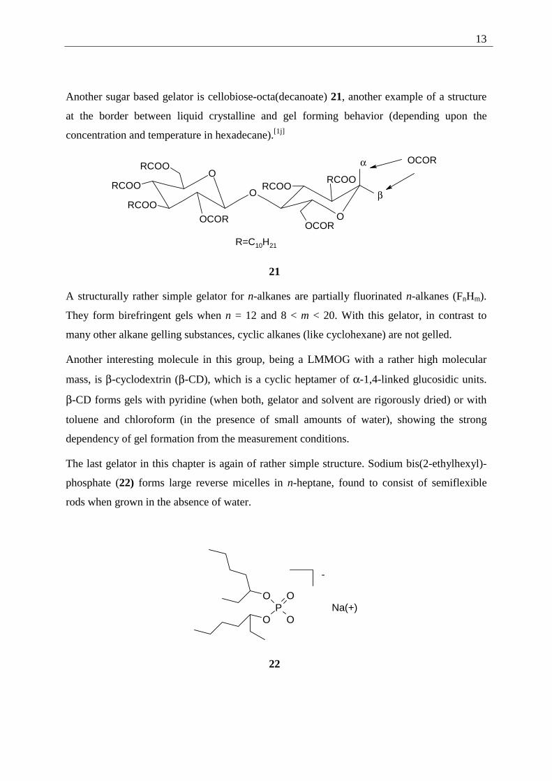
The last gelator in this chapter is again of rather simple structure. Sodium bis(2-ethylhexyl)-
phosphate (22) forms large reverse micelles in n-heptane, found to consist of semiflexible
rods when grown in the absence of water.
OP
O
O
O
-
Na(+)
22
14
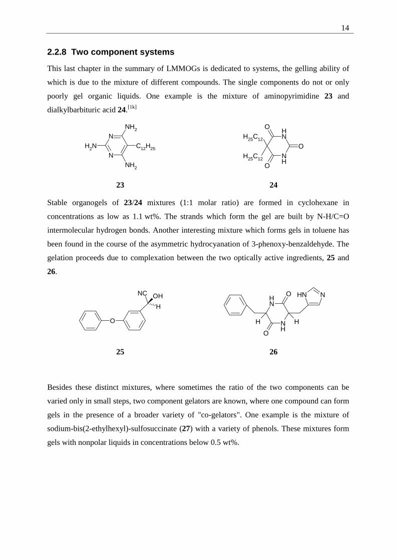
2.2.8 Two component systems
This last chapter in the summary of LMMOGs is dedicated to systems, the gelling ability of
which is due to the mixture of different compounds. The single components do not or only
poorly gel organic liquids. One example is the mixture of aminopyrimidine 23 and
dialkylbarbituric acid 24.[1k]
N
N
NH2
NH2 C12H25
NH2
NH
NH
O
O
O
H25C12
H25C12
23 24
Stable organogels of 23/24 mixtures (1:1 molar ratio) are formed in cyclohexane in
concentrations as low as 1.1 wt%. The strands which form the gel are built by N-H/C=O
intermolecular hydrogen bonds. Another interesting mixture which forms gels in toluene has
been found in the course of the asymmetric hydrocyanation of 3-phenoxy-benzaldehyde. The
gelation proceeds due to complexation between the two optically active ingredients, 25 and
26.
O
H
OHNC
NNH
NH
NH
O
O
H H
25 26
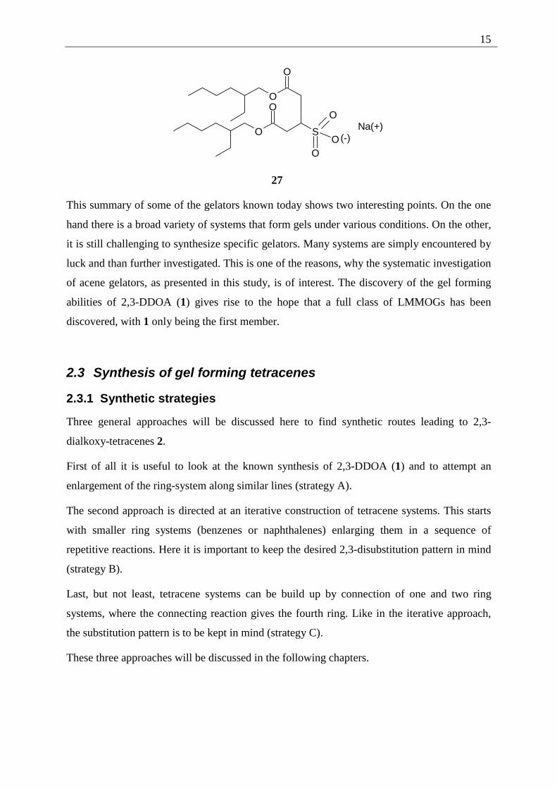
Besides these distinct mixtures, where sometimes the ratio of the two components can be
varied only in small steps, two component gelators are known, where one compound can form
gels in the presence of a broader variety of "co-gelators". One example is the mixture of
sodium-bis(2-ethylhexyl)-sulfosuccinate (27) with a variety of phenols. These mixtures form
gels with nonpolar liquids in concentrations below 0.5 wt%.
15
Na(+)
O
O
SO
OO
OO
(-)
27
This summary of some of the gelators known today shows two interesting points. On the one
hand there is a broad variety of systems that form gels under various conditions. On the other,
it is still challenging to synthesize specific gelators. Many systems are simply encountered by
luck and than further investigated. This is one of the reasons, why the systematic investigation
of acene gelators, as presented in this study, is of interest. The discovery of the gel forming
abilities of 2,3-DDOA (1) gives rise to the hope that a full class of LMMOGs has been
discovered, with 1 only being the first member.
2.3 Synthesis of gel forming tetracenes
2.3.1 Synthetic strategies
Three general approaches will be discussed here to find synthetic routes leading to 2,3-
dialkoxy-tetracenes 2.
First of all it is useful to look at the known synthesis of 2,3-DDOA (1) and to attempt an
enlargement of the ring-system along similar lines (strategy A).
The second approach is directed at an iterative construction of tetracene systems. This starts
with smaller ring systems (benzenes or naphthalenes) enlarging them in a sequence of
repetitive reactions. Here it is important to keep the desired 2,3-disubstitution pattern in mind
(strategy B).
Last, but not least, tetracene systems can be build up by connection of one and two ring
systems, where the connecting reaction gives the fourth ring. Like in the iterative approach,
the substitution pattern is to be kept in mind (strategy C).
These three approaches will be discussed in the following chapters.
16
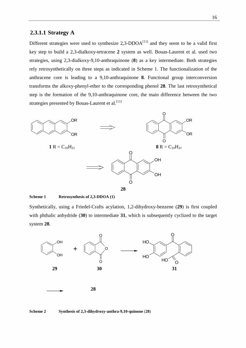
2.3.1.1 Strategy A
Different strategies were used to synthesize 2,3-DDOA[11] and they seem to be a valid first
key step to build a 2,3-dialkoxy-tetracene 2 system as well. Bouas-Laurent et al. used two
strategies, using 2,3-dialkoxy-9,10-anthraquinone (8) as a key intermediate. Both strategies
rely retrosynthetically on three steps as indicated in Scheme 1. The functionalization of the
anthracene core is leading to a 9,10-anthraquinone 8. Functional group interconversion
transforms the alkoxy-phenyl-ether to the corresponding phenol 28. The last retrosynthetical
step is the formation of the 9,10-anthraquinone core, the main difference between the two
strategies presented by Bouas-Laurent et al.[11]
OR
OR
O
O
OR
OR
1 R = C10H21 8 R = C10H21
O
O
OH
OH
28
Scheme 1 Retrosynthesis of 2,3-DDOA (1)
Synthetically, using a Friedel-Crafts acylation, 1,2-dihydroxy-benzene (29) is first coupled
with phthalic anhydride (30) to intermediate 31, which is subsequently cyclized to the target
system 28.
OH
OH +
O
O
O
O
OH
OHOOH
29 30 31
28
Scheme 2 Synthesis of 2,3-dihydroxy-anthra-9,10-quinone (28)
17
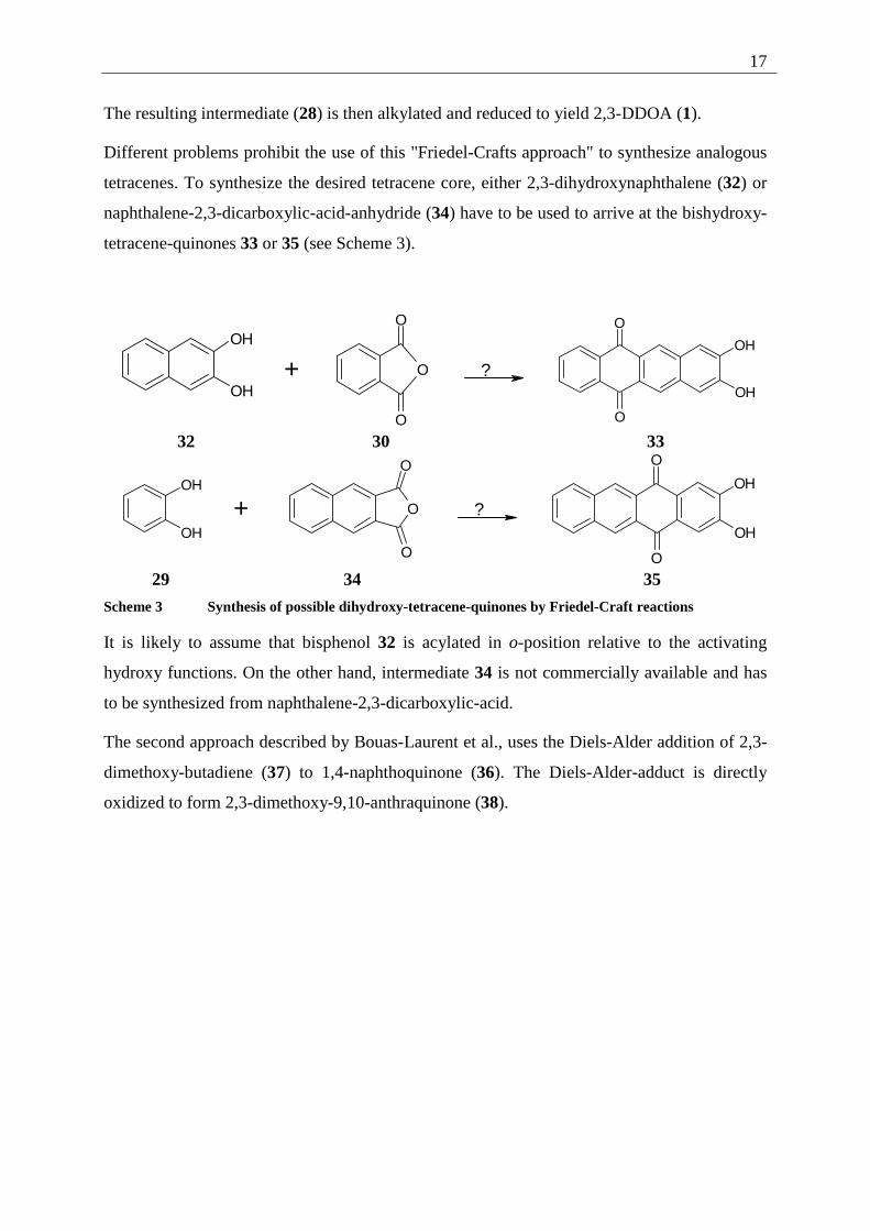
The resulting intermediate (28) is then alkylated and reduced to yield 2,3-DDOA (1).
Different problems prohibit the use of this "Friedel-Crafts approach" to synthesize analogous
tetracenes. To synthesize the desired tetracene core, either 2,3-dihydroxynaphthalene (32) or
naphthalene-2,3-dicarboxylic-acid-anhydride (34) have to be used to arrive at the bishydroxy-
tetracene-quinones 33 or 35 (see Scheme 3).
OH
OH
+
O
O
O
?
OH
OH
O
O 32 30 33
OH
OH
+
O
O
O
?
OH
OH
O
O 29 34 35
Scheme 3 Synthesis of possible dihydroxy-tetracene-quinones by Friedel-Craft reactions
It is likely to assume that bisphenol 32 is acylated in o-position relative to the activating
hydroxy functions. On the other hand, intermediate 34 is not commercially available and has
to be synthesized from naphthalene-2,3-dicarboxylic-acid.
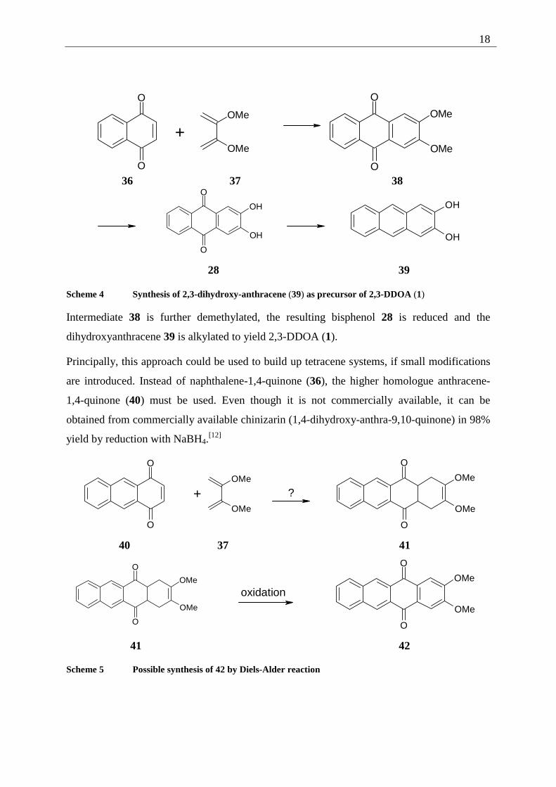
The second approach described by Bouas-Laurent et al., uses the Diels-Alder addition of 2,3-
dimethoxy-butadiene (37) to 1,4-naphthoquinone (36). The Diels-Alder-adduct is directly
oxidized to form 2,3-dimethoxy-9,10-anthraquinone (38).
18
O
O
+
OMe
OMe
O
O
OMe
OMe
36 37 38
O
O
OH
OH
OH
OH
28 39
Scheme 4 Synthesis of 2,3-dihydroxy-anthracene (39) as precursor of 2,3-DDOA (1)
Intermediate 38 is further demethylated, the resulting bisphenol 28 is reduced and the
dihydroxyanthracene 39 is alkylated to yield 2,3-DDOA (1).
Principally, this approach could be used to build up tetracene systems, if small modifications
are introduced. Instead of naphthalene-1,4-quinone (36), the higher homologue anthracene-
1,4-quinone (40) must be used. Even though it is not commercially available, it can be
obtained from commercially available chinizarin (1,4-dihydroxy-anthra-9,10-quinone) in 98%
yield by reduction with NaBH4.[12]
O
O
OMe
OMe
+
?
O
O
OMe
OMe
40 37 41
O
O
OMe
OMe
oxidation
O
O
OMe
OMe
41 42
Scheme 5 Possible synthesis of 42 by Diels-Alder reaction
19
Only the poor yield of the Diels-Alder reaction between the smaller naphthoquinone 36 and
diene 37 (61% dropping to 19% with larger amounts of reagent)[11] made it a good idea to
look for different alternatives first.
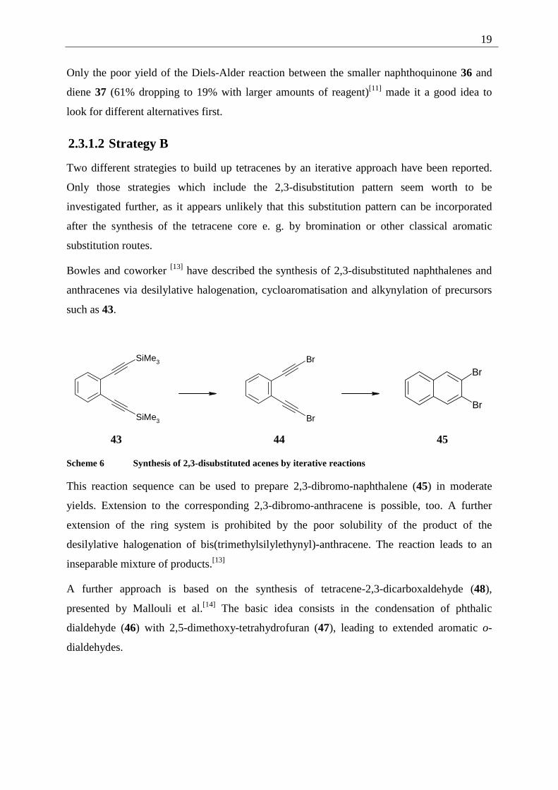
2.3.1.2 Strategy B
Two different strategies to build up tetracenes by an iterative approach have been reported.
Only those strategies which include the 2,3-disubstitution pattern seem worth to be
investigated further, as it appears unlikely that this substitution pattern can be incorporated
after the synthesis of the tetracene core e. g. by bromination or other classical aromatic
substitution routes.
Bowles and coworker [13] have described the synthesis of 2,3-disubstituted naphthalenes and
anthracenes via desilylative halogenation, cycloaromatisation and alkynylation of precursors
such as 43.
SiMe3
SiMe3
Br
Br
Br
Br
43 44 45
Scheme 6 Synthesis of 2,3-disubstituted acenes by iterative reactions
This reaction sequence can be used to prepare 2,3-dibromo-naphthalene (45) in moderate
yields. Extension to the corresponding 2,3-dibromo-anthracene is possible, too. A further
extension of the ring system is prohibited by the poor solubility of the product of the
desilylative halogenation of bis(trimethylsilylethynyl)-anthracene. The reaction leads to an
inseparable mixture of products.[13]
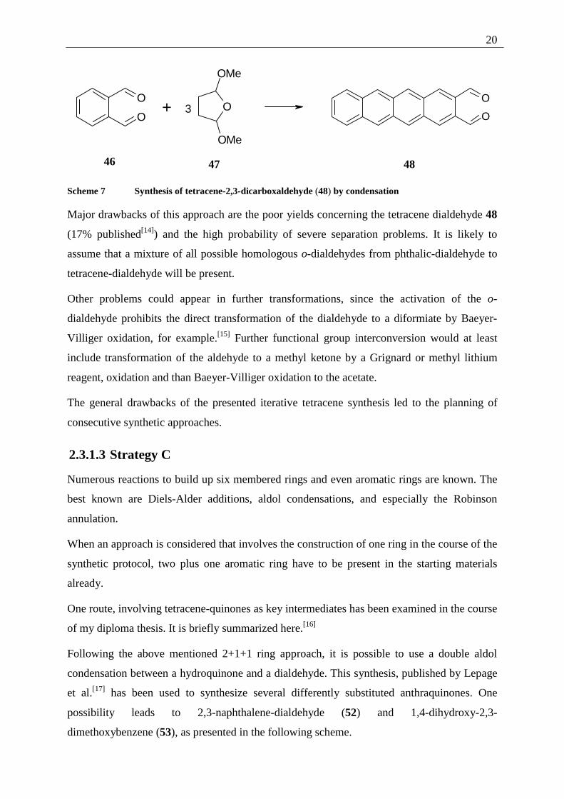
A further approach is based on the synthesis of tetracene-2,3-dicarboxaldehyde (48),
presented by Mallouli et al.[14] The basic idea consists in the condensation of phthalic
dialdehyde (46) with 2,5-dimethoxy-tetrahydrofuran (47), leading to extended aromatic o-
dialdehydes.
20
O
O
+ O
OMe
OMe
3
O
O
46 47 48
Scheme 7 Synthesis of tetracene-2,3-dicarboxaldehyde (48) by condensation
Major drawbacks of this approach are the poor yields concerning the tetracene dialdehyde 48
(17% published[14]) and the high probability of severe separation problems. It is likely to
assume that a mixture of all possible homologous o-dialdehydes from phthalic-dialdehyde to
tetracene-dialdehyde will be present.
Other problems could appear in further transformations, since the activation of the o-
dialdehyde prohibits the direct transformation of the dialdehyde to a diformiate by Baeyer-
Villiger oxidation, for example.[15] Further functional group interconversion would at least
include transformation of the aldehyde to a methyl ketone by a Grignard or methyl lithium
reagent, oxidation and than Baeyer-Villiger oxidation to the acetate.
The general drawbacks of the presented iterative tetracene synthesis led to the planning of
consecutive synthetic approaches.
2.3.1.3 Strategy C
Numerous reactions to build up six membered rings and even aromatic rings are known. The
best known are Diels-Alder additions, aldol condensations, and especially the Robinson
annulation.
When an approach is considered that involves the construction of one ring in the course of the
synthetic protocol, two plus one aromatic ring have to be present in the starting materials
already.
One route, involving tetracene-quinones as key intermediates has been examined in the course
of my diploma thesis. It is briefly summarized here.[16]
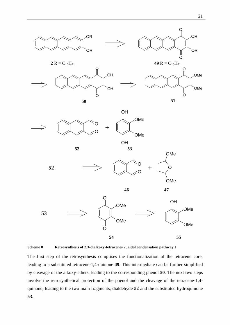
Following the above mentioned 2+1+1 ring approach, it is possible to use a double aldol
condensation between a hydroquinone and a dialdehyde. This synthesis, published by Lepage
et al.[17] has been used to synthesize several differently substituted anthraquinones. One
possibility leads to 2,3-naphthalene-dialdehyde (52) and 1,4-dihydroxy-2,3-
dimethoxybenzene (53), as presented in the following scheme.
21
OR
OR
OR
OR
O
O 2 R = C10H21 49 R = C10H21
O
O
OH
OH
50
O
O
OMe
OMe
51
O
O
+
OH
OH
OMe
OMe
52 53
52
O
O
+ O
OMe
OMe
46 47
53
O
O
OMe
OMe
OH
OMe
OMe
54 55
Scheme 8 Retrosynthesis of 2,3-dialkoxy-tetracenes 2, aldol condensation pathway I
The first step of the retrosynthesis comprises the functionalization of the tetracene core,
leading to a substituted tetracene-1,4-quinone 49. This intermediate can be further simplified
by cleavage of the alkoxy-ethers, leading to the corresponding phenol 50. The next two steps
involve the retrosynthetical protection of the phenol and the cleavage of the tetracene-1,4-
quinone, leading to the two main fragments, dialdehyde 52 and the substituted hydroquinone
53.
22
Although it is possible to follow this pathway up to the 2,3-dialkoxy-tetracene-1,4-quinone 49
(yield 2% over 6 steps), the reduction of the quinone to the corresponding hydrocarbon failed,
possibly due to the activation of the quinone system by the directly connected alkoxy
functions.[16]
This set back led to the idea of investigating the "inverted approach", using phthalic-
dialdehyde 46 and 1,4-dihydroxy-6,7-dimethoxy-naphthalene (57) as reactants for the aldol
condensation.
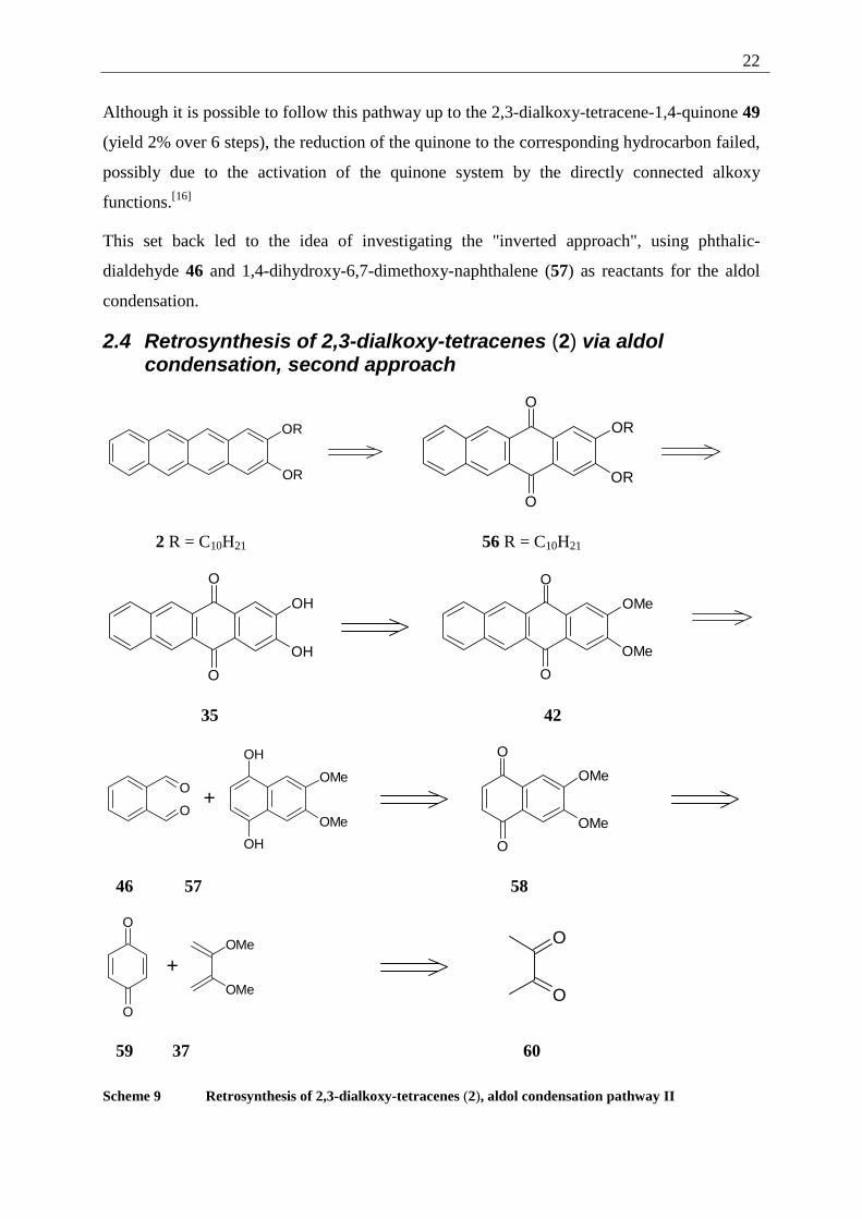
2.4 Retrosynthesis of 2,3-dialkoxy-tetracenes (2) via aldol condensation, second approach
OR
OR
O
O
OR
OR
2 R = C10H21 56 R = C10H21
O
O
OH
OH
O
O
OMe
OMe
35 42
O
O+
OH
OH
OMe
OMe
O
O
OMe
OMe
46 57 58
O
O
+OMe
OMe
O
O
59 37 60
Scheme 9 Retrosynthesis of 2,3-dialkoxy-tetracenes (2), aldol condensation pathway II
23
The first step of the retrosynthesis is again the functionalization of the tetracene core, leading
to the dialkoxy-tetracene-quinone 61. Subsequently, the alkoxy chains are retrosynthetically
converted to phenol-functions and those are protected as methoxy ethers.
The key step is the retrosynthesis of the tetracene ring system, now leading to a benzene
fragment and a naphthalene fragment. Phthalic-dialdehyde (46) is commercially available.
The naphthalene fragment, 1,4-dihydroxy-6,7-dimethoxy-naphthalene (57) is not, it is not
even known in the literature. It can be derived from 6,7-dimethoxy-naphthoquinone (58),
which can be transformed back to benzoquinone (59) and 2,3-dimethoxy-buta-1,3-diene (37),
a commercially available and easy to synthesize compound. Following these ideas, the
synthesis of differently substituted 2,3-dialkoxytetracenes (2) was possible and will be
presented in detail in the following chapter.
24
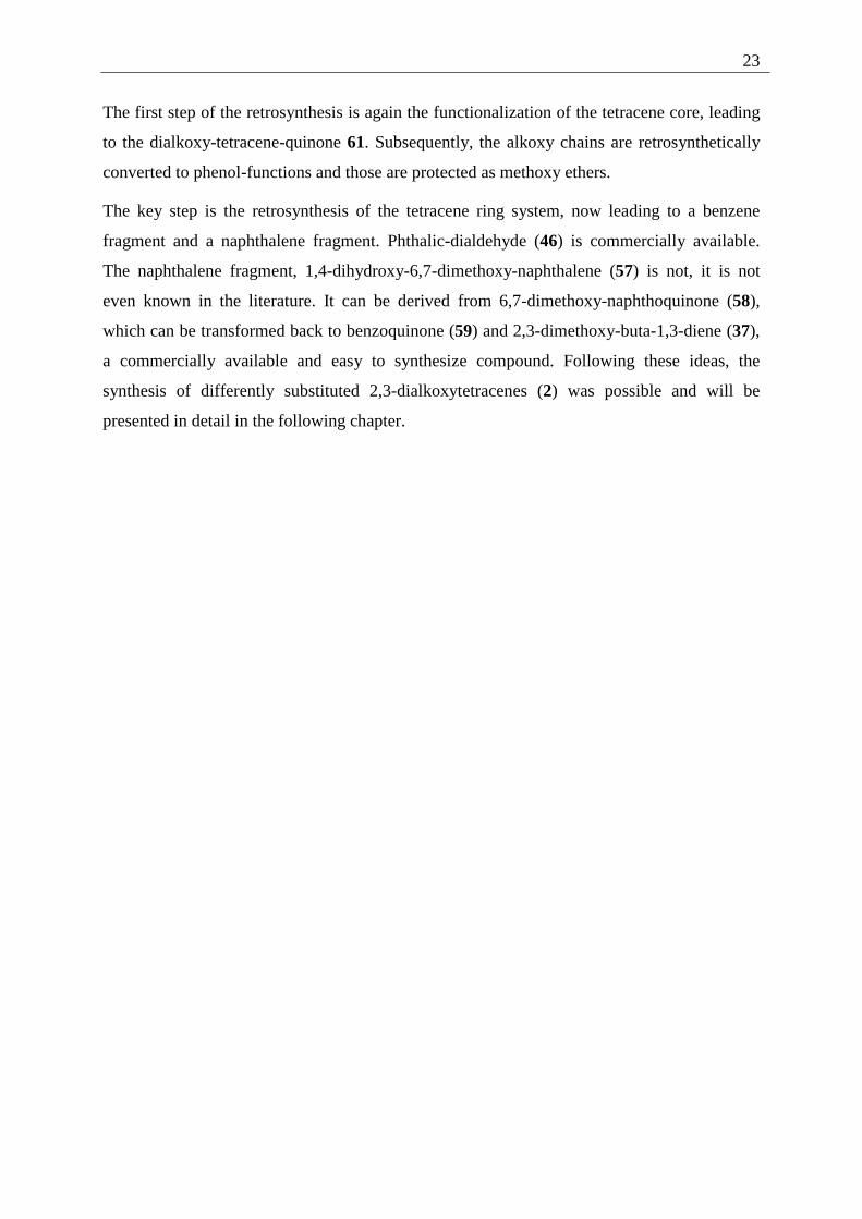
2.5 Synthesis of 2,3-dialkoxy-tetracenes (2)
O
O
HC(OMe)3 /H2SO4 /MeOH
29%
OMe
OMe
+
O
O
60 37 59
toluene
58 %
O
O
OMe
OMe
Zn / HOAc / ultrasound
95 %
OH
OH
OMe
OMe
58 57
+ O
O
Na2CO3 / CF3CH2OH
83 %
O
O
OMe
OMe
46 42
HBr / HOAc90 %
O
O
OH
OH
K2CO3 / CnH2n+1Br / DMF
67-76%
35
O
O
OR
OR
Al(C6H11O)3
C6H11OH
55-70%
OR
OR
61 2
Scheme 10 Synthesis of 2,3-dialkoxy-tetracenes (2) by aldol condensation
25
2.5.1 Explanation of the individual synthetic steps
The different steps of the above shown synthesis will be presented in the order in which they
were carried out. The spectroscopic data, especially of substances not described in the
chemical literature, will be discussed.
2.5.1.1 2,3-Dimethoxy-buta-1,3-diene (37)
Even though 2,3-dimethoxy-1,3-butadiene (37) is a commercially available substance, it is
easy to synthesize following the method described by Bouas-Laurent et al.[11] The only
drawback is the problem of slow polymerization of the diene 37 during purification by
distillation. Therefore a 90% purity (determined by gas chromatography) was considered to
be acceptable and the compound was stored under an atmosphere of N2 and at low
temperatures prior to use (see Chapter 7.3.1.1).
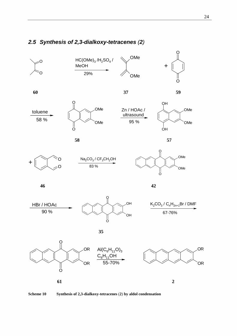
2.5.1.2 6,7-Dimethoxy-1,4-naphthoquinone (58).
The synthesis of 6,7-dimethoxy-1,4-naphthoquinone (58) has first been published by Adams
et al.[18] in 1941: 6,7-dimethoxy-naphthalene-1-ol was oxidized with K2Cr2O7. One more
recent synthetic approach is the TEMPO oxidation of 1,2-diethynyl-4,5-dimethoxybenzene,
published by Grissom et al. in 1995.[19] In the course of this study, the Diels-Alder addition of
2,3-dimethoxy-buta-1,3-diene (37) to benzoquinone (59), published in 1996 by Zhang, Fox
and Hadfield,[20] was chosen. Two problems, though, have to be faced. First of all, the
dienophile is symmetrical, offering the possibility of double addition, which would lead to an
anthraquinone system. Secondly, the primary adduct 62 would have to be oxidized to yield
58.
O
O
+
OMe
OMe
O
O
OMe
OMe
O
O
OMe
OMe
[Ox] 1
4 6
7
59 37 62 58
Scheme 11 Diels Alder addition/oxidation to 6,7-dimethoxy-1,4-naphthoquinone (58)
26
Both problems can be solved by using a threefold excess of 59. This leads to the formation of
the mono adduct and oxidation of the intermediate 62 to the desired product in situ. With this
excess of benzoquinone, the yield of the reaction improves in comparison to the procedure
given in the literature (33% [20]) to 58% (see Chapter 7.3.1.2).
In contrast to the given procedure, the addition was carried out in toluene and for the work-up
recrystallization, rather than column chromatography, was used.
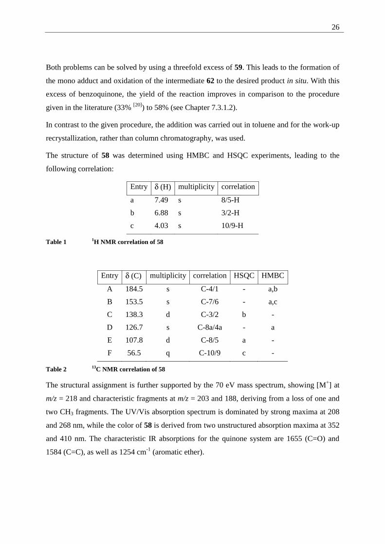
The structure of 58 was determined using HMBC and HSQC experiments, leading to the
following correlation:
Entry δ (H) multiplicity correlation
a 7.49 s 8/5-H
b 6.88 s 3/2-H
c 4.03 s 10/9-H
Table 1 1H NMR correlation of 58
Entry δ (C) multiplicity correlation HSQC HMBC
A 184.5 s C-4/1 - a,b
B 153.5 s C-7/6 - a,c
C 138.3 d C-3/2 b -
D 126.7 s C-8a/4a - a
E 107.8 d C-8/5 a -
F 56.5 q C-10/9 c -
Table 2 13C NMR correlation of 58
The structural assignment is further supported by the 70 eV mass spectrum, showing [M+] at
m/z = 218 and characteristic fragments at m/z = 203 and 188, deriving from a loss of one and
two CH3 fragments. The UV/Vis absorption spectrum is dominated by strong maxima at 208
and 268 nm, while the color of 58 is derived from two unstructured absorption maxima at 352
and 410 nm. The characteristic IR absorptions for the quinone system are 1655 (C=O) and
1584 (C=C), as well as 1254 cm-1 (aromatic ether).
27
2.5.1.3 Reduction of 6,7-dimethoxy-1,4-naphthoquinone (58)
As 1,4-dihydroxy-6,7-dimethoxy-naphthalene (57) has, until now, not been described in the
literature, procedures for the reduction of other known naphthoquinones had to be used as
guidelines. Classical conditions to reduce quinones to the corresponding hydroquinones have
been known for a long time. Plimpton used tin and hydrochloric acid as early as 1880 to
perform this reaction.[21] Other commonly used systems are hydrogen with Pd on charcoal
(Laatsch et al.[22]) or sodium azide with hydrochloric acid (Papageorgiou et al.[23]). When the
original work of Adams et al. is considered[18] 6,7-dimethoxy-naphtho-1,4-quinone (58) was
in fact reduced, using Zn dust in acetic anhydride with acetic acid and sodium acetate. This
procedure led to the formation of 1,4-diacetoxy-6,7-dimethoxy-naphthalene, an acylated form
of the desired hydroquinone.
In the course of this work, the reduction of the naphthoquinone 58 to the corresponding
hydroquinone was carried out, using Zn in acetic acid (without acetic anhydride) as reductant
and ultrasound conditions to shorten reaction times. This procedure is adapted from the
reduction of the unsubstituted 1,4-naphthoquinone (36) (see Chapter 7.3.4.1) published by
Marchand et al. in 1991.[24]
The given procedure was chosen for the short reaction times and excellent yields that were
reported (< 5 min and 100% for naphthoquinone (36)).[24]
While the reaction is generally fast (around 1.5 h, depending on the reaction scale) special
care has to be taken during work-up to prevent reoxidation (see Chapter 7.3.1.3).
Furthermore, it is worth to note that the reaction times differ largely with the quality of the Zn
surface. The activation of the metal, using diluted hydrochloric acid and afterwards methanol
and ether to dry the activated metal has advantages. For general synthetic procedures it is
sufficient to remove the solvent after the completion of the reaction (detected by TLC) and
use the crude product directly in the aldol condensation without further purification.
The structure of the product was assigned by comparison of the 1H- and the 13C NMR data
with the one taken for the unsubstituted 1,4-dihydroxy-naphthalene (95) (see Chapter 7.3.4.1).
This indirect approach had to be taken due to the reoxidation of 57 that complicates the
measurement of multidimensional NMR spectra.
28
Further proof of the assigned structure derives from the 70 eV mass spectrum, showing [M+]
at m/z = 220 as well as from the IR spectrum, lacking the characteristic intense quinone-bands
at 1655 (C=O) and 1584 cm-1 (C=C).
2.5.1.4 Condensation of 1,4-dihydroxy-6,7-dimethoxy-naphthalene (57)
with phthalic dialdehyde (46).
The key step of the synthesis of 2,3-dialkoxy-tetracenes (2) is the aldol condensation of
hydroquinone 57 with phthalic-dialdehyde 46 under basic conditions in trifluorethanol.
Reactions of this type have been published by Serpaud and Lepage in 1977. They synthesized
differently substituted polycyclic quinones and heterocyclic quinones.[17] The facile synthesis
of tetracene-5,12-quinone (94) (see Chapter 7.3.4.2), used later in this work to synthesize
5,12-DDOT (85), has already been described. More recently, Martín et al. used the same
approach to prepare N,N'-dicyanoquinonediimine for studies on electrically conducting charge
transfer complexes.[25]
The reaction proceeds smoothly by refluxing the two starting compounds in the presence of
sodium carbonate (see Chapter 7.3.1.4). The low solubility of the product drives the reaction
and makes work-up easier. After the end of the reaction the mixture can be diluted with water
to dissolve the sodium carbonate. The reaction leads to the key intermediate 2,3-dimethoxy-
tetracene-5,12-quinone (42) in 83% yield.
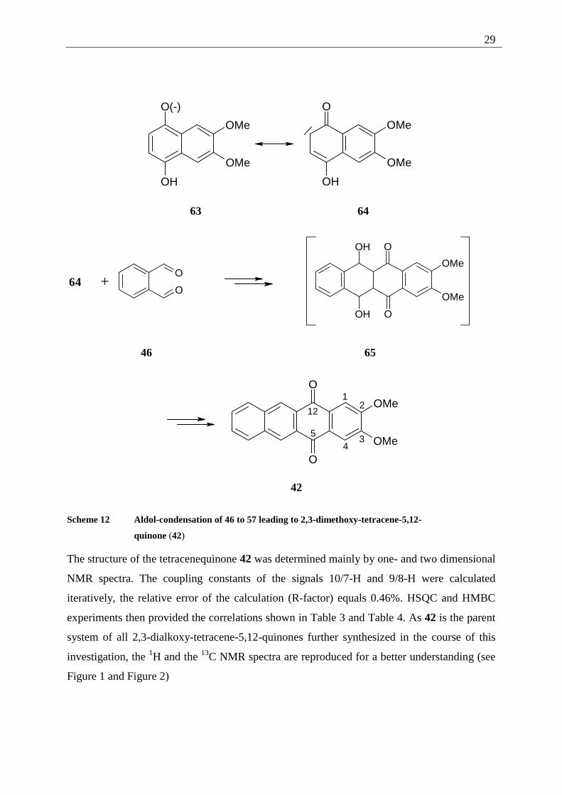
The mechanism of the reaction can be assumed as follows: the hydroquinone 57 is
deprotonated to the hydroquinonate 63. This intermediate, from its carbanion resonance
structure, attacks one of the aldehyde functions of 46 as a nucleophile. The resulting
alcoholate is protonated to form the aldol adduct. This intermediate is not stable under the
reaction conditions but eliminates directly (no intermediate can be detected by TLC
monitoring of the reaction) to the aromatic quinone. Repetition of this sequence leads to the
2,3-dimethoxy-tetracene-5,12-quinone (42).
29
O(-)
OH
OMe
OMe
O
OH
OMe
OMe
63 64
O
O64 +
OMe
OMe
O
O
OH
OH
46 65
O
O
OMe
OMe
12
34
5
12
42
Scheme 12 Aldol-condensation of 46 to 57 leading to 2,3-dimethoxy-tetracene-5,12-
quinone (42)
The structure of the tetracenequinone 42 was determined mainly by one- and two dimensional
NMR spectra. The coupling constants of the signals 10/7-H and 9/8-H were calculated
iteratively, the relative error of the calculation (R-factor) equals 0.46%. HSQC and HMBC
experiments then provided the correlations shown in Table 3 and Table 4. As 42 is the parent
system of all 2,3-dialkoxy-tetracene-5,12-quinones further synthesized in the course of this
investigation, the 1H and the 13C NMR spectra are reproduced for a better understanding (see
Figure 1 and Figure 2)
30
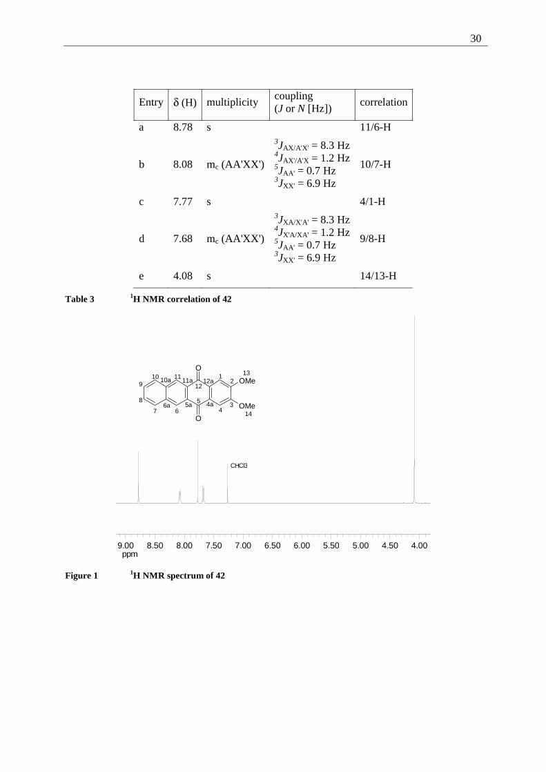
Entry δ (H) multiplicity coupling (J or N [Hz])
correlation
a 8.78 s 11/6-H
b 8.08 mc (AA'XX')
3JAX/A'X' = 8.3 Hz 4JAX'/A'X = 1.2 Hz 5JAA' = 0.7 Hz 3JXX' = 6.9 Hz
10/7-H
c 7.77 s 4/1-H
d 7.68 mc (AA'XX')
3JXA/X'A' = 8.3 Hz 4JX'A/XA' = 1.2 Hz 5JAA' = 0.7 Hz 3JXX' = 6.9 Hz
9/8-H
e 4.08 s 14/13-H
Table 3 1H NMR correlation of 42
ppm4.004.505.005.506.006.507.007.508.008.509.00
CHCl3
12
34
5
67
8
910 11
12
13
14
O
O
OMe
OMe4a5a6a
10a 11a 12a
Figure 1 1H NMR spectrum of 42
31
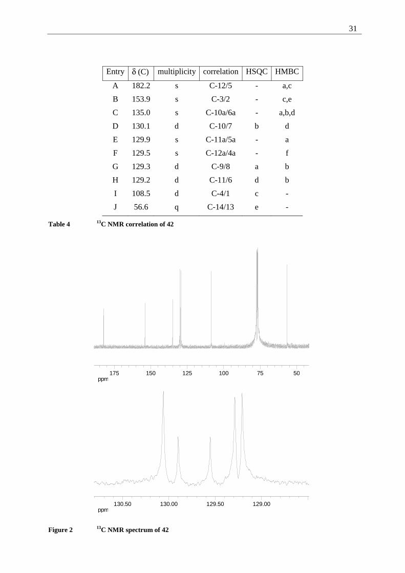
Entry δ (C) multiplicity correlation HSQC HMBC
A 182.2 s C-12/5 - a,c
B 153.9 s C-3/2 - c,e
C 135.0 s C-10a/6a - a,b,d
D 130.1 d C-10/7 b d
E 129.9 s C-11a/5a - a
F 129.5 s C-12a/4a - f
G 129.3 d C-9/8 a b
H 129.2 d C-11/6 d b
I 108.5 d C-4/1 c -
J 56.6 q C-14/13 e -
Table 4 13C NMR correlation of 42
ppm5075100125150175
ppm129.00129.50130.00130.50
Figure 2 13C NMR spectrum of 42
32
The 1H NMR spectrum exhibits, next to the OCH3-groups with δ = 4.08 ppm, two singlets (δ
= 8.78 and 7.77) for the 11/6 and 4/1-H adjacent to the quinone system. 10/7 and 9/8-H are
represented by a characteristic AA'XX' system with δ(center) = 8.08 and 7.68 ppm. The lower
field signal corresponds to the 10/7-H. This can be explained by the stronger deshielding of
those protons which are near to the second ring of the naphthalene subsystem. Similar
spectroscopic features can be found in all of the tetracenequinones, pentacene quinones and as
well in the tetracenes and pentacenes (vide infra).
The 13C NMR spectrum shows characteristic low-field signals for the quinone C=O (182.2
ppm) and the C(aryl)-OR (153.9 ppm). The lowest field quaternary carbon is C-6a/10a
junction of the naphthalene subsystem, belonging to two aromatic rings. The signals D to H
show only little difference in chemical shifts but are still resolved well enough to be assigned.
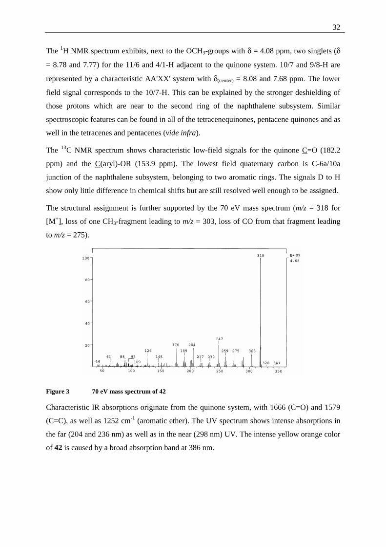
The structural assignment is further supported by the 70 eV mass spectrum (m/z = 318 for
[M+], loss of one CH3-fragment leading to m/z = 303, loss of CO from that fragment leading
to m/z = 275).
Figure 3 70 eV mass spectrum of 42
Characteristic IR absorptions originate from the quinone system, with 1666 (C=O) and 1579
(C=C), as well as 1252 cm-1 (aromatic ether). The UV spectrum shows intense absorptions in
the far (204 and 236 nm) as well as in the near (298 nm) UV. The intense yellow orange color
of 42 is caused by a broad absorption band at 386 nm.
33
2.5.1.5 Demethylation of 2,3-dimethoxy-tetracene-5,12-quinone (42).
The cleavage of aromatic methoxy ethers is a widely used synthetic step. Today the most
commonly used reactant is BBr3 in different solvents (DCM or hexane),[26] a mixture
commercially available. Even though it is possible to demethylate 2,3-dimethoxy-tetracene-
5,12-quinone (42) in this fashion, the reaction proceeds very slowly and leads to a mixture of
unreacted starting material, the singly demethylated compound and only 10% of the desired
product (after direct alkylation of the mixture, see Chapter 7.3.1.5.c). Other reported
procedures rely on metal halides like NaI/SiI4[27] or Lewis acids like AlI3.
[28]
Still, the classical procedure, using strong Broenstedt acids, is widely used. The procedure is
only limited by the stability of the starting material and product under the harsh conditions
(acidity and temperature). Reagents employed are reaching from HI (with various coreactants
like acetic acid anhydride)[29] to mixtures of aqueous HBr with glacial acetic acid.[30] The
demethylation is generally a SN type reaction, where the strong acid activates the ether oxygen
by protonation, and the acid anion attacks as the nucleophile at the methyl group. Another
classical method is the use of the "protected" acid HCl as pyridine hydrochloride.[31] This
offers the advantage of high reaction temperatures (the melting point of pyridine
hydrochloride is around 150 °C) and the absence of free acid in a still strongly nucleophilic
system.
In the course of this study three techniques of demethylation have been examined:
1) Demethylation by BBr3
In the course of this study, the procedure failed, possibly due to the low solubility of the 2,3-
dimethoxy-tetracene-5,12-quinone (42) even in DCM.
Even after several days of stirring a suspension of 2,3-dimethoxytetracene-5,12-quinone (42)
in DCM with a solution of BBr3 in DCM, only a minor part of the starting material was
demethylated, as could be determined after alkylation (see Chapter 7.3.1.5.c).
34
2) Demethylation by HBr/HOAc
Due to the high reaction temperatures (48% HBr refluxing at 128 °C) and the simple reaction
conditions (the starting material is suspended in HBr-solution and refluxed for several days),
this procedure is well suited for reactions on a large scale (see Chapter 7.3.1.5.a).
The major drawback of this procedure is its two phase nature. It is nearly impossible to obtain
fully deprotected 2,3-dihydroxy-tetracene-5,12-quinone (35). As the reaction mixture is only
soluble in polar aprotic solvents like DMF or DMSO, a separation on this stage of the reaction
path is not advisable. Separation and purification is accomplished on the stage of the 2,3-
dialkoxy-tetracene-5,12-quinones (61) after alkylation of the crude mixture.
3) Demethylation by pyridine•HCl
The reaction of aromatic methyl ethers with pyridine•HCl (see Chapter 7.3.1.5.b) proceeds in
principle by the same pathway as the demethylation using strong acids. Advantageous is the
shorter reaction time (3 h with pyridine•HCl versus 48 h with HBr•HOAc). Disadvantageous
is the availability of the pyridinium salt. It has either to be prepared under rigorously dried
conditions or purchased and stored under Ar, as the salt is highly hygroscopic. The reaction
proceeds smoothly under inert conditions leading to the demethylated product 35. Again
traces of the singly demethylated compound can be found due to the reaction in two phases
(the starting material is not fully soluble in the molten mixture).
Thus both procedures, using HBr•HOAc or pyridine•HCl can be used in parallel with similar
results.
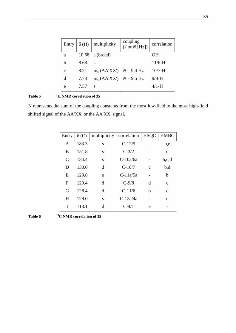
The structure of 2,3-dihydroxy-tetracene-5,12-quinone (35) was determined, using one- and
two dimensional NMR spectroscopy. The solvent chosen for the NMR spectra was DMSO-d6,
since 35 is only poorly soluble in CDCl3.
35
Entry δ (H) multiplicity coupling (J or N [Hz])
correlation
a 10.68 s (broad) OH
b 8.68 s 11/6-H
c 8.21 mc (AA'XX') N = 9.4 Hz 10/7-H
d 7.73 mc (AA'XX') N = 9.5 Hz 9/8-H
e 7.57 s 4/1-H
Table 5 1H NMR correlation of 35
N represents the sum of the coupling constants from the most low-field to the most high-field
shifted signal of the AA'XX' or the AA'XX' signal.
Entry δ (C) multiplicity correlation HSQC HMBC
A 183.3 s C-12/5 - b,e
B 151.8 s C-3/2 - e
C 134.4 s C-10a/6a - b,c,d
D 130.0 d C-10/7 c b,d
E 129.8 s C-11a/5a - b
F 129.4 d C-9/8 d c
G 128.4 d C-11/6 b c
H 128.0 s C-12a/4a - e
I 113.1 d C-4/1 e -
Table 6 13C NMR correlation of 35
36
ppm7.007.508.008.509.009.5010.0010.5011.00
OH
12
34
5
67
8
910 11
12
O
O
OH
OH4a5a6a
10a 11a 12a
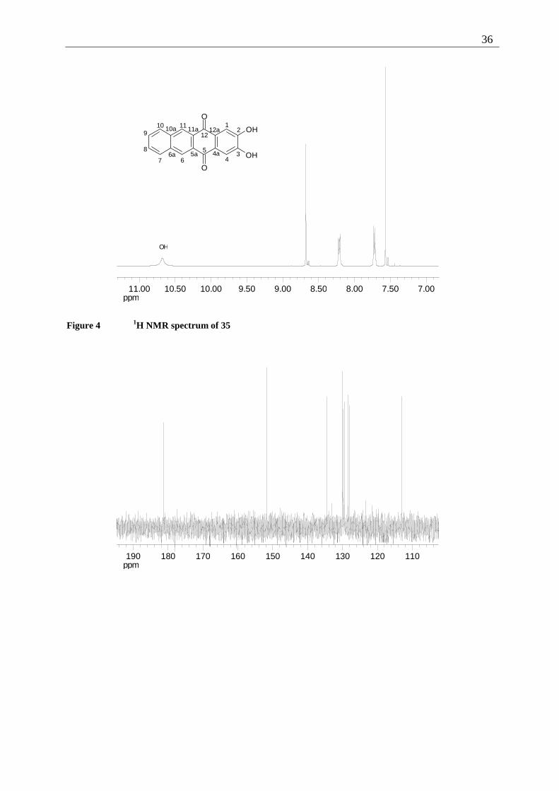
Figure 4 1H NMR spectrum of 35
ppm110120130140150160170180190
37
ppm128.0129.0130.0131.0132.0133.0134.0
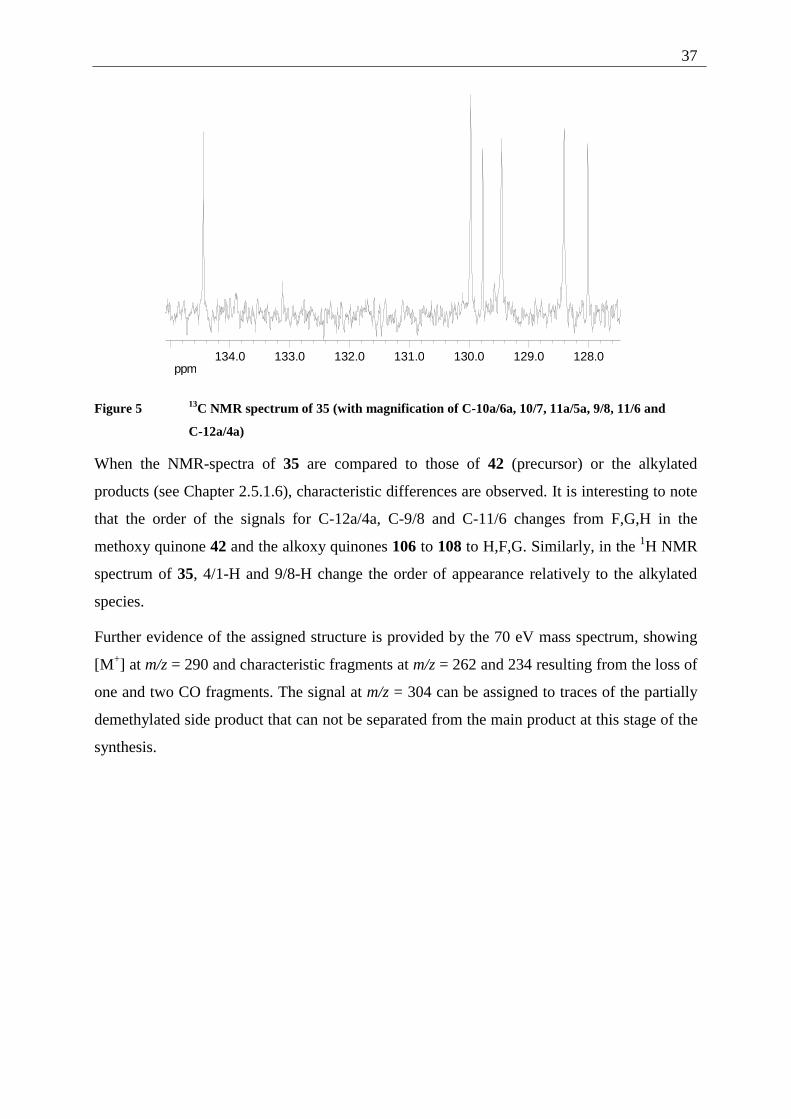
Figure 5 13C NMR spectrum of 35 (with magnification of C-10a/6a, 10/7, 11a/5a, 9/8, 11/6 and
C-12a/4a)
When the NMR-spectra of 35 are compared to those of 42 (precursor) or the alkylated
products (see Chapter 2.5.1.6), characteristic differences are observed. It is interesting to note
that the order of the signals for C-12a/4a, C-9/8 and C-11/6 changes from F,G,H in the
methoxy quinone 42 and the alkoxy quinones 106 to 108 to H,F,G. Similarly, in the 1H NMR
spectrum of 35, 4/1-H and 9/8-H change the order of appearance relatively to the alkylated
species.
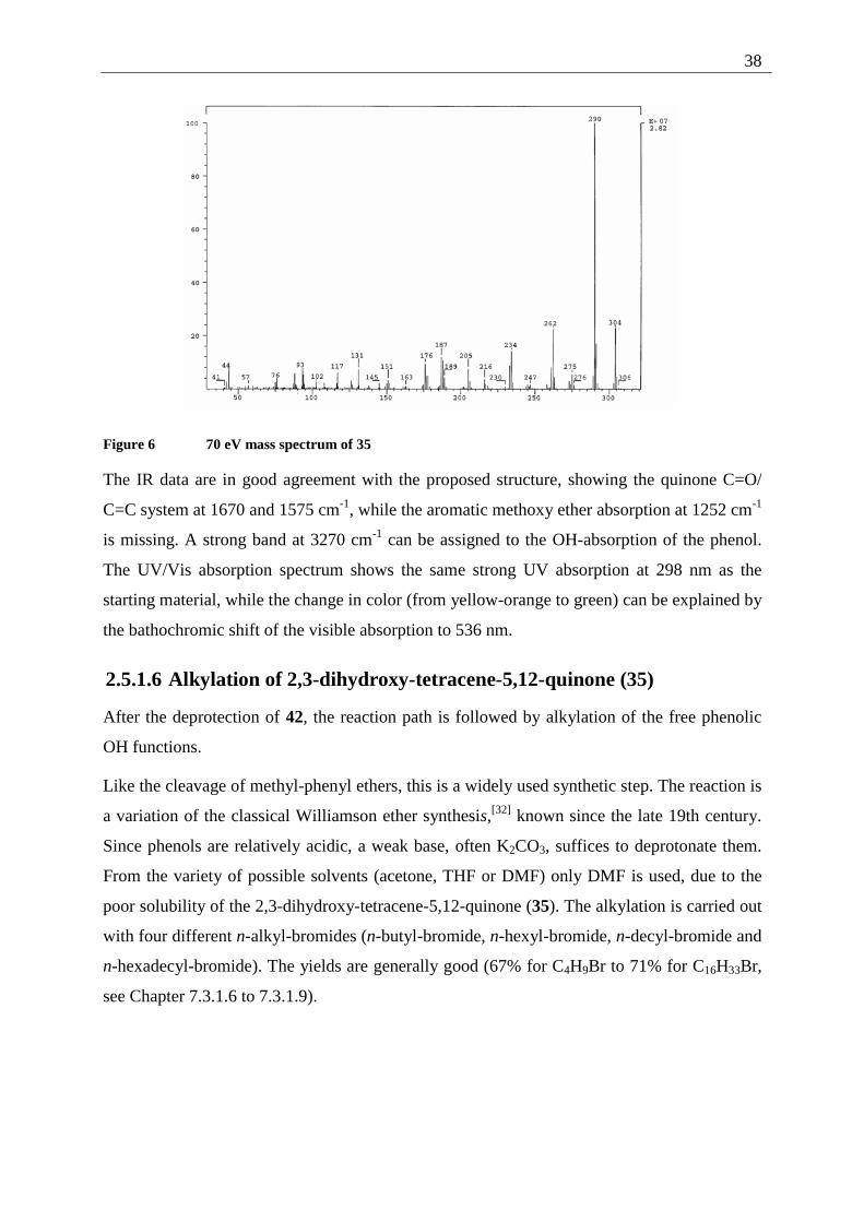
Further evidence of the assigned structure is provided by the 70 eV mass spectrum, showing
[M+] at m/z = 290 and characteristic fragments at m/z = 262 and 234 resulting from the loss of
one and two CO fragments. The signal at m/z = 304 can be assigned to traces of the partially
demethylated side product that can not be separated from the main product at this stage of the
synthesis.
38
Figure 6 70 eV mass spectrum of 35
The IR data are in good agreement with the proposed structure, showing the quinone C=O/
C=C system at 1670 and 1575 cm-1, while the aromatic methoxy ether absorption at 1252 cm-1
is missing. A strong band at 3270 cm-1 can be assigned to the OH-absorption of the phenol.
The UV/Vis absorption spectrum shows the same strong UV absorption at 298 nm as the
starting material, while the change in color (from yellow-orange to green) can be explained by
the bathochromic shift of the visible absorption to 536 nm.
2.5.1.6 Alkylation of 2,3-dihydroxy-tetracene-5,12-quinone (35)
After the deprotection of 42, the reaction path is followed by alkylation of the free phenolic
OH functions.
Like the cleavage of methyl-phenyl ethers, this is a widely used synthetic step. The reaction is
a variation of the classical Williamson ether synthesis,[32] known since the late 19th century.
Since phenols are relatively acidic, a weak base, often K2CO3, suffices to deprotonate them.
From the variety of possible solvents (acetone, THF or DMF) only DMF is used, due to the
poor solubility of the 2,3-dihydroxy-tetracene-5,12-quinone (35). The alkylation is carried out
with four different n-alkyl-bromides (n-butyl-bromide, n-hexyl-bromide, n-decyl-bromide and
n-hexadecyl-bromide). The yields are generally good (67% for C4H9Br to 71% for C16H33Br,
see Chapter 7.3.1.6 to 7.3.1.9).
39
As the demethylation is often not carried out with full conversion of the starting material, it is
possible to separate unreacted 2,3-dimethoxy-tetracene-5,12-quinone (42) and partially
demethylated/realkylated side product from the desired 2,3-dialkoxy-tetracene-5,12-quinones
61 by column chromatography on silica using DCM/pentane as a mobile phase.
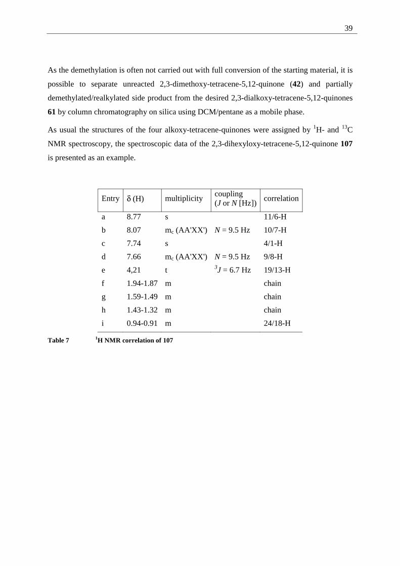
As usual the structures of the four alkoxy-tetracene-quinones were assigned by 1H- and 13C
NMR spectroscopy, the spectroscopic data of the 2,3-dihexyloxy-tetracene-5,12-quinone 107
is presented as an example.
Entry δ (H) multiplicity coupling (J or N [Hz])
correlation
a 8.77 s 11/6-H
b 8.07 mc (AA'XX') N = 9.5 Hz 10/7-H
c 7.74 s 4/1-H
d 7.66 mc (AA'XX') N = 9.5 Hz 9/8-H
e 4,21 t 3J = 6.7 Hz 19/13-H
f 1.94-1.87 m chain
g 1.59-1.49 m chain
h 1.43-1.32 m chain
i 0.94-0.91 m 24/18-H
Table 7 1H NMR correlation of 107
40
ppm1.02.03.04.05.06.07.08.09.0
CHCl3
1920
12
34
5
67
8
910 11
12
O
O
O
O4a5a6a
10a 11a 12a 151316
1714
18
2122
23 24
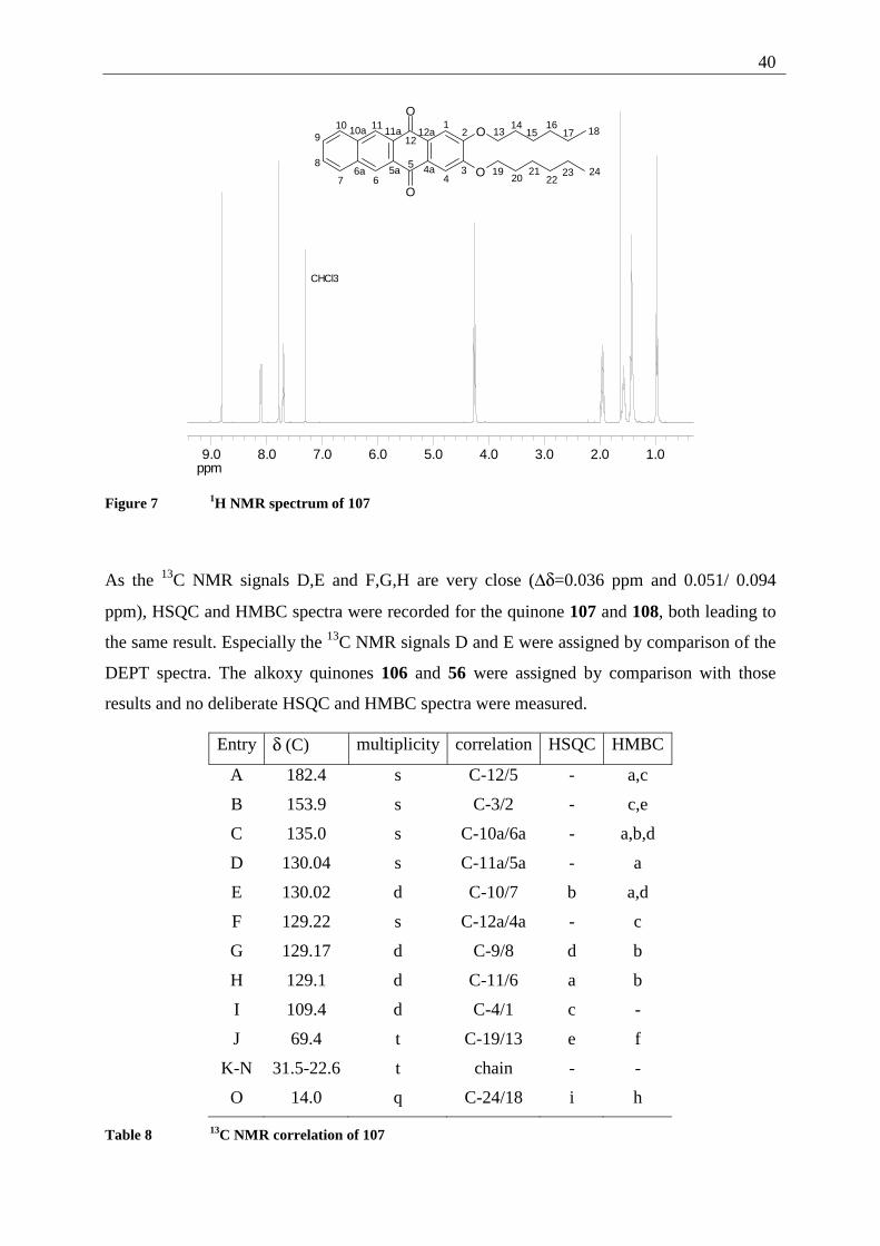
Figure 7 1H NMR spectrum of 107
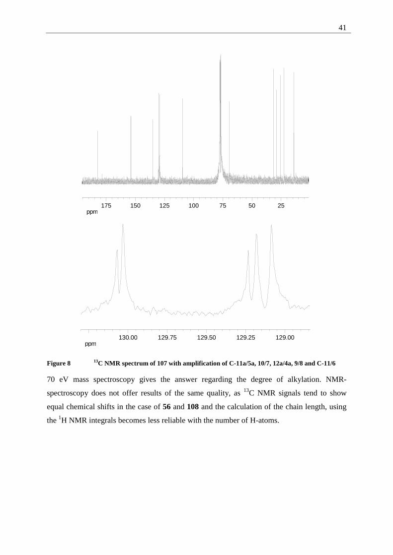
As the 13C NMR signals D,E and F,G,H are very close (∆δ=0.036 ppm and 0.051/ 0.094
ppm), HSQC and HMBC spectra were recorded for the quinone 107 and 108, both leading to
the same result. Especially the 13C NMR signals D and E were assigned by comparison of the
DEPT spectra. The alkoxy quinones 106 and 56 were assigned by comparison with those
results and no deliberate HSQC and HMBC spectra were measured.
Entry δ (C) multiplicity correlation HSQC HMBC
A 182.4 s C-12/5 - a,c
B 153.9 s C-3/2 - c,e
C 135.0 s C-10a/6a - a,b,d
D 130.04 s C-11a/5a - a
E 130.02 d C-10/7 b a,d
F 129.22 s C-12a/4a - c
G 129.17 d C-9/8 d b
H 129.1 d C-11/6 a b
I 109.4 d C-4/1 c -
J 69.4 t C-19/13 e f
K-N 31.5-22.6 t chain - -
O 14.0 q C-24/18 i h
Table 8 13C NMR correlation of 107
41
ppm255075100125150175
ppm129.00129.25129.50129.75130.00
Figure 8 13C NMR spectrum of 107 with amplification of C-11a/5a, 10/7, 12a/4a, 9/8 and C-11/6
70 eV mass spectroscopy gives the answer regarding the degree of alkylation. NMR-
spectroscopy does not offer results of the same quality, as 13C NMR signals tend to show
equal chemical shifts in the case of 56 and 108 and the calculation of the chain length, using
the 1H NMR integrals becomes less reliable with the number of H-atoms.
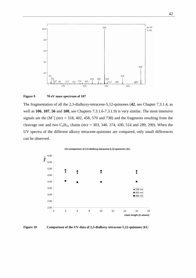
42
Figure 9 70 eV mass spectrum of 107
The fragmentation of all the 2,3-dialkoxy-tetracene-5,12-quinones (42, see Chapter 7.3.1.4, as
well as 106, 107, 56 and 108, see Chapters 7.3.1.6-7.3.1.9) is very similar. The most intensive
signals are the [M+] (m/z = 318, 402, 458, 570 and 738) and the fragments resulting from the
cleavage one and two CnH2n chains (m/z = 303, 346, 374, 430, 514 and 289, 290). When the
UV spectra of the different alkoxy tetracene-quinones are compared, only small differences
can be observed.
UV-comparison of 2,3-dialkoxy-tetracene-5,12-quinones (61)
2,00
2,50
3,00
3,50
4,00
4,50
5,00
5,50
6,00
2 4 6 8 10 12 14 16 18
chain length (C-atoms)
log
ε
238 nm302 nm390 nm
Figure 10 Comparison of the UV-data of 2,3-dialkoxy tetracene-5,12-quinones (61)
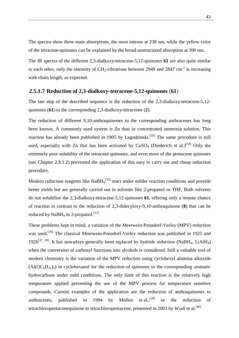
43
The spectra show three main absorptions, the most intense at 238 nm, while the yellow color
of the tetracene-quinones can be explained by the broad unstructured absorption at 390 nm.
The IR spectra of the different 2,3-dialkoxy-tetracene-5,12-quinones 61 are also quite similar
to each other, only the intensity of CH2-vibrations between 2949 and 2847 cm-1 is increasing
with chain length, as expected.
2.5.1.7 Reduction of 2,3-dialkoxy-tetracene-5,12-quinones (61)
The last step of the described sequence is the reduction of the 2,3-dialkoxy-tetracene-5,12-
quinones (61) to the corresponding 2,3-dialkoxy-tetracenes (2).
The reduction of different 9,10-anthraquinones to the corresponding anthracenes has long
been known. A commonly used system is Zn dust in concentrated ammonia solution. This
reaction has already been published in 1905 by Lagodzinski.[33] The same procedure is still
used, especially with Zn that has been activated by CuSO4 (Diederich et al.)[34] Only the
extremely poor solubility of the tetracene quinones, and even more of the pentacene quinones
(see Chapter 2.8.1.2) prevented the application of this easy to carry out and cheap reduction
procedure.
Modern reduction reagents like NaBH4[35] react under milder reaction conditions and provide
better yields but are generally carried out in solvents like 2-propanol or THF. Both solvents
do not solubilize the 2,3-dialkoxy-tetracene-5,12-quinones 61, offering only a remote chance
of reaction in contrast to the reduction of 2,3-didecyloxy-9,10-anthraquinone (8) that can be
reduced by NaBH4 in 2-propanol.[11]
These problems kept in mind, a variation of the Meerwein-Ponndorf-Verley (MPV) reduction
was used.[36] The classical Meerwein-Ponndorf-Verley reduction was published in 1925 and
1926[37, 38]. It has nowadays generally been replaced by hydride reduction (NaBH4, LiAlH4)
when the conversion of carbonyl functions into alcohols is considered. Still a valuable tool of
modern chemistry is the variation of the MPV reduction using cyclohexyl alumina alkoxide
(Al(OC6H11)3) in cyclohexanol for the reduction of quinones to the corresponding aromatic
hydrocarbons under mild conditions. The only limit of this reaction is the relatively high
temperature applied preventing the use of the MPV process for temperature sensitive
compounds. Current examples of the application are the reduction of anthraquinones to
anthracenes, published in 1994 by Müllen et al.,[39] or the reduction of
tetrachloropentacenequinone to tetrachloropentacene, presented in 2003 by Wudl et al.[40]
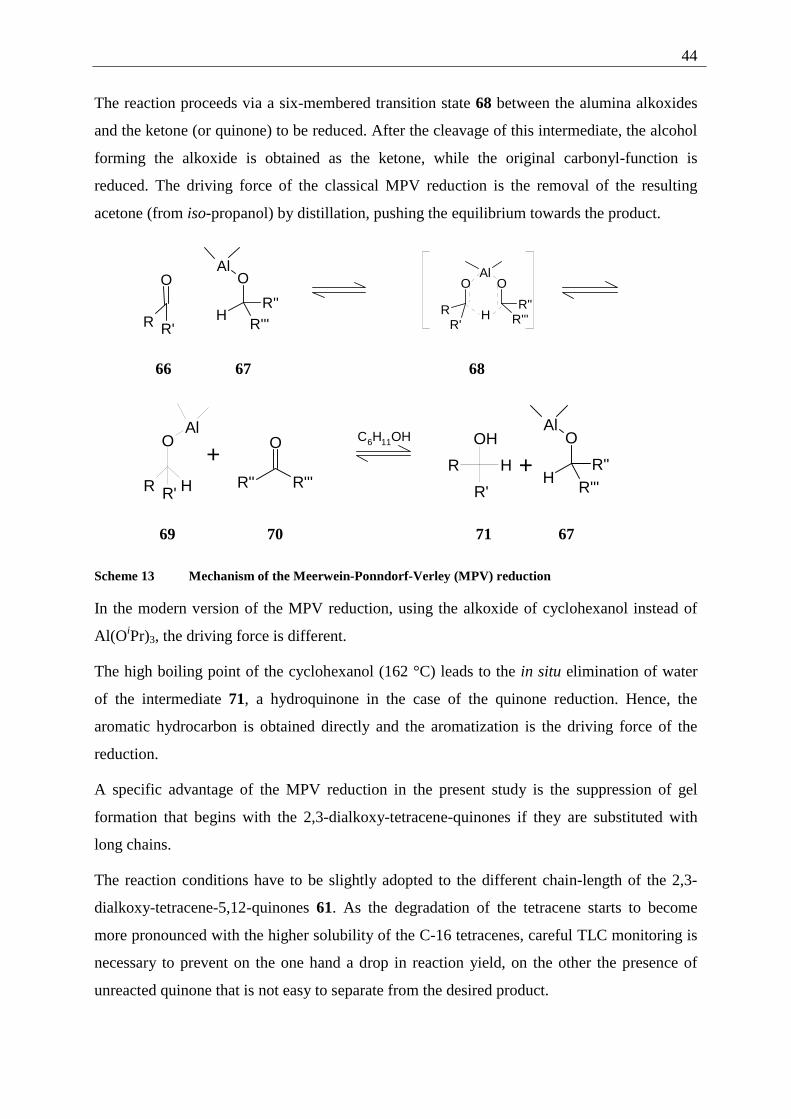
44
The reaction proceeds via a six-membered transition state 68 between the alumina alkoxides
and the ketone (or quinone) to be reduced. After the cleavage of this intermediate, the alcohol
forming the alkoxide is obtained as the ketone, while the original carbonyl-function is
reduced. The driving force of the classical MPV reduction is the removal of the resulting
acetone (from iso-propanol) by distillation, pushing the equilibrium towards the product.
R'
O
R
AlO
R''R'''
H
O
RR'
AlO
R''R'''H
66 67 68
O
R R' H
Al
+R'' R'''
O
C6H11OH
R H
OH
R'
+
AlO
R''R'''
H
69 70 71 67
Scheme 13 Mechanism of the Meerwein-Ponndorf-Verley (MPV) reduction
In the modern version of the MPV reduction, using the alkoxide of cyclohexanol instead of
Al(OiPr)3, the driving force is different.
The high boiling point of the cyclohexanol (162 °C) leads to the in situ elimination of water
of the intermediate 71, a hydroquinone in the case of the quinone reduction. Hence, the
aromatic hydrocarbon is obtained directly and the aromatization is the driving force of the
reduction.
A specific advantage of the MPV reduction in the present study is the suppression of gel
formation that begins with the 2,3-dialkoxy-tetracene-quinones if they are substituted with
long chains.
The reaction conditions have to be slightly adopted to the different chain-length of the 2,3-
dialkoxy-tetracene-5,12-quinones 61. As the degradation of the tetracene starts to become
more pronounced with the higher solubility of the C-16 tetracenes, careful TLC monitoring is
necessary to prevent on the one hand a drop in reaction yield, on the other the presence of
unreacted quinone that is not easy to separate from the desired product.
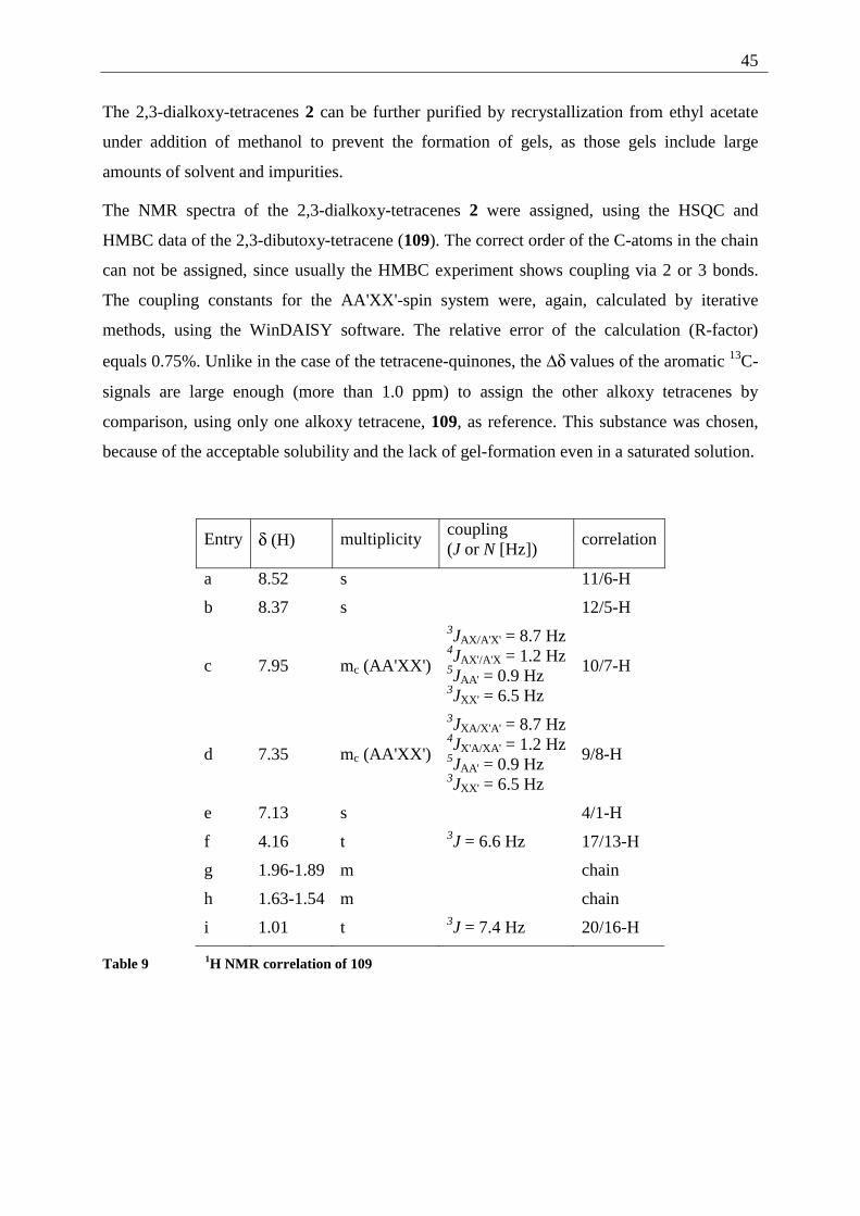
45
The 2,3-dialkoxy-tetracenes 2 can be further purified by recrystallization from ethyl acetate
under addition of methanol to prevent the formation of gels, as those gels include large
amounts of solvent and impurities.
The NMR spectra of the 2,3-dialkoxy-tetracenes 2 were assigned, using the HSQC and
HMBC data of the 2,3-dibutoxy-tetracene (109). The correct order of the C-atoms in the chain
can not be assigned, since usually the HMBC experiment shows coupling via 2 or 3 bonds.
The coupling constants for the AA'XX'-spin system were, again, calculated by iterative
methods, using the WinDAISY software. The relative error of the calculation (R-factor)
equals 0.75%. Unlike in the case of the tetracene-quinones, the ∆δ values of the aromatic 13C-
signals are large enough (more than 1.0 ppm) to assign the other alkoxy tetracenes by
comparison, using only one alkoxy tetracene, 109, as reference. This substance was chosen,
because of the acceptable solubility and the lack of gel-formation even in a saturated solution.
Entry δ (H) multiplicity coupling (J or N [Hz])
correlation
a 8.52 s 11/6-H
b 8.37 s 12/5-H
c 7.95 mc (AA'XX')
3JAX/A'X' = 8.7 Hz 4JAX'/A'X = 1.2 Hz 5JAA' = 0.9 Hz 3JXX' = 6.5 Hz
10/7-H
d 7.35 mc (AA'XX')
3JXA/X'A' = 8.7 Hz 4JX'A/XA' = 1.2 Hz 5JAA' = 0.9 Hz 3JXX' = 6.5 Hz
9/8-H
e 7.13 s 4/1-H
f 4.16 t 3J = 6.6 Hz 17/13-H
g 1.96-1.89 m chain
h 1.63-1.54 m chain
i 1.01 t 3J = 7.4 Hz 20/16-H
Table 9 1H NMR correlation of 109
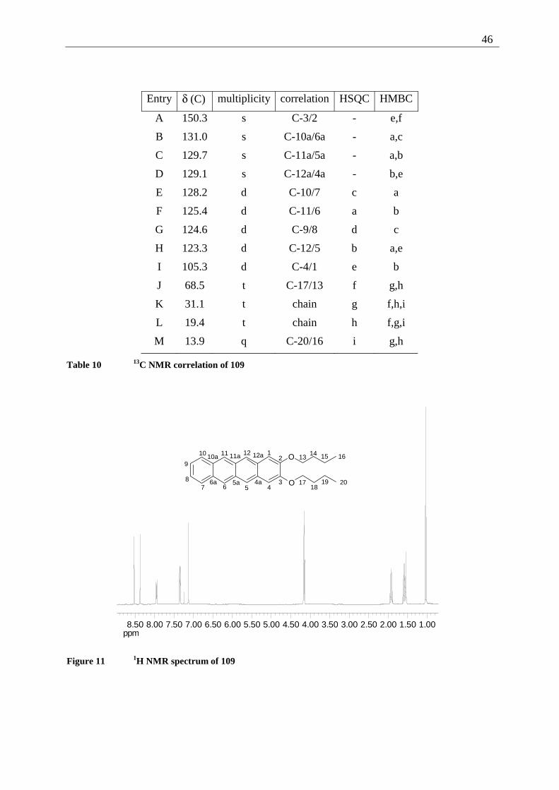
46
Entry δ (C) multiplicity correlation HSQC HMBC
A 150.3 s C-3/2 - e,f
B 131.0 s C-10a/6a - a,c
C 129.7 s C-11a/5a - a,b
D 129.1 s C-12a/4a - b,e
E 128.2 d C-10/7 c a
F 125.4 d C-11/6 a b
G 124.6 d C-9/8 d c
H 123.3 d C-12/5 b a,e
I 105.3 d C-4/1 e b
J 68.5 t C-17/13 f g,h
K 31.1 t chain g f,h,i
L 19.4 t chain h f,g,i
M 13.9 q C-20/16 i g,h
Table 10 13C NMR correlation of 109
ppm1.001.502.002.503.003.504.004.505.005.506.006.507.007.508.008.50
12
34567
8
9
10 11 1213
14O
O4a5a6a
10a 11a 12a 15 16
1718
19 20
Figure 11 1H NMR spectrum of 109
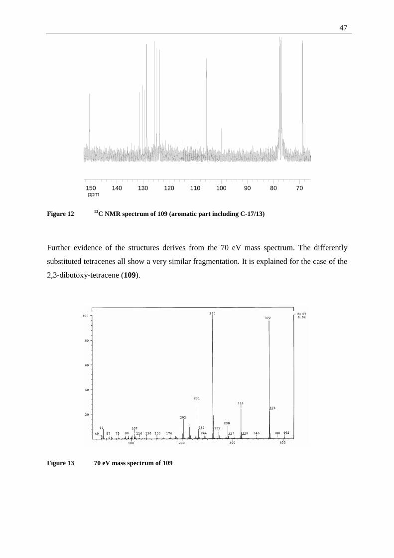
47
ppm708090100110120130140150
Figure 12 13C NMR spectrum of 109 (aromatic part including C-17/13)
Further evidence of the structures derives from the 70 eV mass spectrum. The differently
substituted tetracenes all show a very similar fragmentation. It is explained for the case of the
2,3-dibutoxy-tetracene (109).
Figure 13 70 eV mass spectrum of 109
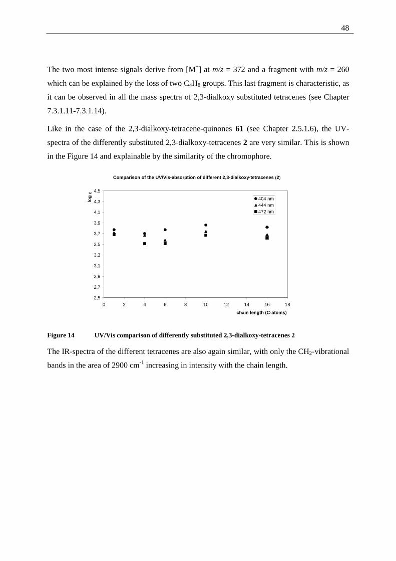
48
The two most intense signals derive from [M+] at m/z = 372 and a fragment with m/z = 260
which can be explained by the loss of two C4H8 groups. This last fragment is characteristic, as
it can be observed in all the mass spectra of 2,3-dialkoxy substituted tetracenes (see Chapter
7.3.1.11-7.3.1.14).
Like in the case of the 2,3-dialkoxy-tetracene-quinones 61 (see Chapter 2.5.1.6), the UV-
spectra of the differently substituted 2,3-dialkoxy-tetracenes 2 are very similar. This is shown
in the Figure 14 and explainable by the similarity of the chromophore.
Comparison of the UV/Vis-absorption of different 2,3-dialkoxy-tetracenes (2)
2,5
2,7
2,9
3,1
3,3
3,5
3,7
3,9
4,1
4,3
4,5
0 2 4 6 8 10 12 14 16 18
chain length (C-atoms)
log
ε
404 nm444 nm472 nm
Figure 14 UV/Vis comparison of differently substituted 2,3-dialkoxy-tetracenes 2
The IR-spectra of the different tetracenes are also again similar, with only the CH2-vibrational
bands in the area of 2900 cm-1 increasing in intensity with the chain length.
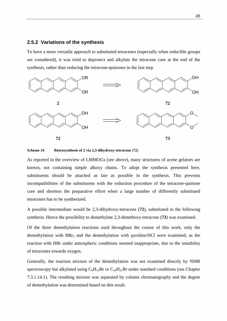
49
2.5.2 Variations of the synthesis
To have a more versatile approach to substituted tetracenes (especially when reducible groups
are considered), it was tried to deprotect and alkylate the tetracene core at the end of the
synthesis, rather than reducing the tetracene-quinones in the last step.
OR
OR
OH
OH
2 72
OH
OH
O
O
72 73
Scheme 14 Retrosynthesis of 2 via 2,3-dihydroxy-tetracene (72)
As reported in the overview of LMMOGs (see above), many structures of acene gelators are
known, not containing simple alkoxy chains. To adopt the synthesis presented here,
substituents should be attached as late as possible in the synthesis. This prevents
incompatibilities of the substituents with the reduction procedure of the tetracene-quinone
core and shortens the preparative effort when a large number of differently substituted
tetracenes has to be synthesized.
A possible intermediate would be 2,3-dihydroxy-tetracene (72), substituted in the following
synthesis. Hence the possibility to demethylate 2,3-dimethoxy-tetracene (73) was examined.
Of the three demethylation reactions used throughout the course of this work, only the
demethylation with BBr3 and the demethylation with pyridine/HCl were examined, as the
reaction with HBr under atmospheric conditions seemed inappropriate, due to the instability
of tetracenes towards oxygen.
Generally, the reaction mixture of the demethylation was not examined directly by NMR
spectroscopy but alkylated using C6H13Br or C10H21Br under standard conditions (see Chapter
7.3.1.14.1). The resulting mixture was separated by column chromatography and the degree
of demethylation was determined based on this result.
50
Even by variation of the reaction conditions (solvent, temperature and inert atmosphere), the
yield of the desired 2,3-dialkoxy-tetracene 2 could not be improved beyond 16% (see Chapter
7.3.1.14.1) by using BBr3, even if Nuckolls et al. used the approach to demethylate 2,3-
dimethoxy-tetracene (73) with BBr3 in investigations of semiconductor effects of 2,3-
dihydroxy-tetracene (72) on aluminum oxide surfaces.[41] When pyridine•HCl was used at
different temperatures, either no reaction took place or the starting material was destroyed
(see Chapter 7.3.1.14.2).
Until now, no competitive strategy to the alkylation of 2,3-dihydroxy-tetracene-quinone (35)
and reduction of the yielding 2,3-dialkoxy-tetracene-quinones 61 could be found. Still, by
demethylation of 2,3-dimethoxy-tetracene (73), 2,3-dihydroxy-tetracene (2) could be
synthesized even if it has not been isolated in substance as published by Nuckolls et al.[41]
2.6 Synthesis of gel forming pentacenes
2.6.1 Introduction
Since 2,3-DDOA 1 is not unique as a gel-forming acene (see Chapter 3) but only the lowest
homologue of a class of gel-forming substances, it is of principal interest to see, how far the
acene system can be extended, while gel formation is still possible. So the attempt to prepare
gel forming pentacenes is a logical step.
But there are further reasons to enlarge the synthetic principle presented in the previous
chapter.
During the last few years, pentacene has attracted much interest as component in organic thin
film transistors (OTFTs).[42, 43] The main problem is the poor solubility that only allows for
vacuum deposition of the acene. Two concepts are used to circumvent this drawback. First the
thermoreversible Diels-Alder addition/ reversion proposed by Dehaen et al. or Afzali et al.[42,
43] The second approach lies in the use of substituted pentacenes as presented by Anthony et
al. or Suzuki et al.[44, 45] to change solubility and electronic properties of the pentacenes.
This dual interest, possible formation of gel forming pentacenes and improvement of the
solubility for the possible application in OTFTs, led to the extension of the already successful
synthesis of 2,3-alkoxy-tetracenes 2 to the corresponding pentacenes 74.
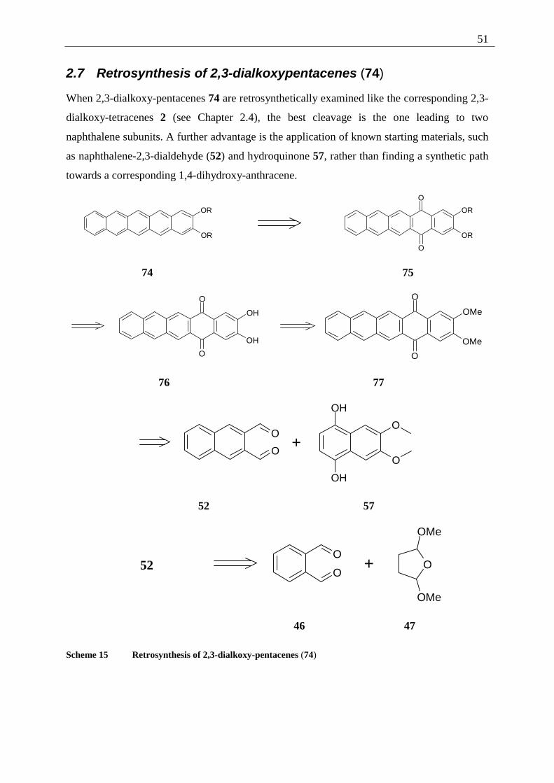
51
2.7 Retrosynthesis of 2,3-dialkoxypentacenes (74)
When 2,3-dialkoxy-pentacenes 74 are retrosynthetically examined like the corresponding 2,3-
dialkoxy-tetracenes 2 (see Chapter 2.4), the best cleavage is the one leading to two
naphthalene subunits. A further advantage is the application of known starting materials, such
as naphthalene-2,3-dialdehyde (52) and hydroquinone 57, rather than finding a synthetic path
towards a corresponding 1,4-dihydroxy-anthracene.
OR
OR
OR
OR
O
O
74 75
O
O
OH
OH
O
O
OMe
OMe
76 77
OH
OH
O
O
O
O+
52 57
52 O
O+ O
OMe
OMe
46 47
Scheme 15 Retrosynthesis of 2,3-dialkoxy-pentacenes (74)
52
Following Scheme 15 the retrosynthetic examination of 74 leads by oxidation of the
pentacene core to the corresponding pentacene-5,14-quinone 75. Cleavage of the alkoxy-
phenyl ether and protection of the resulting phenol 76 leads to 2,3-dimethoxy-pentacene-5,14-
quinone (77). This intermediate can be analyzed back to two naphthalene subunits, the
dialdehyde 52 and the p-dihydroxy-naphthalene 57. The former can be further traced back to
the lower homologue 46, the retrosynthesis of the latter is explained in Scheme 9.
2.8 Synthesis of 2,3-dialkoxy-pentacenes (74)
Some difficulties noted during the synthesis of 2,3-dialkoxy-tetracenes 2 can be expected to
be comparable in the case of the pentacenes series. These are:
Poor solubility of the 2,3-dimethoxy-pentacene-5,14-quinone (77), leading to slow
demethylation.
Even poorer solubility of the 2,3-dihydroxy-pentacene-5,14-quinone (76), leading to a slow
alkylation and severe problems in purifying 76.
High sensitivity of the pentacenes towards oxygen combined with low solubility hampering
purification and limiting gel formation to compounds with at least C10 chains (see Chapter
3.1). The solution to these problems is presented in the following chapters.
2.8.1 Explanation of the individual synthetic steps
The synthesis of 2,3-dialkoxy-pentacenes 74 follows the synthesis of the 2,3-dialkoxy-
tetracenes 2, with only two major differences. First, the dialdehyde to be condensed with 1,4-
dihydroxy-6,7-dimethoxy-naphthalene (57) is the higher homologue of 46, naphthalene-2,3-
dialdehyde (52). The second difference can be found in the poor solubility of all 2,3-dialkoxy-
pentacene-5,14-quinones 75 and the corresponding 2,3-dialkoxy-pentacenes 74, which
complicates the work. The reduction of the 2,3-dialkoxy-pentacene-5,14-quinones 75 to the
2,3-dialkoxy-pentacenes 74 was thus carried out under argon to prevent oxidation of the
substrate.
53
2.8.1.1 Synthesis of naphthalene-2,3-dialdehyde (52)
The synthesis of 52 has first been published by Cook et al. in 1949.[46] The given procedure
though (oxidation of anthracene with OsO4 followed by cleavage with periodic acid) is not
suitable to produce larger amounts of the substance due to the complicated procedure and the
high price of OsO4.
An improvement can be found in the synthesis by Weygand et al.[47] published in 1950. This
reaction sequence is building on naphthalene-2,3-dicarboxylic acid. It is first reduced to the
diol and subsequently partially oxidized. Unfortunately, this synthesis can only be carried out
in small amounts since the oxidant is SeO2.[47]
Another synthesis, using functional group interconversion of a substituted naphthalene, has
been published by Ried et al. in 1956.[48] 2,3-Dimethyl-naphthalene is brominated in two steps
to provide 2,3-bis-dibromomethyl-naphthalene. This intermediate is hydrolyzed by aqueous
potassium oxalate which gives the desired dialdehyde 52 in an overall yield of 34%. It is a
serious drawback though that 2,3-dimethyl-naphthalene is not anymore available
commercially as a side product of tar production as described by Ried.[48]
This makes it necessary to look for synthetic approaches in which the naphthalene nucleus is
build up from cheap and available precursors.
Two strategies to synthesize naphthalene-2,3-dicarboxaldehyde (52) from benzenic subunits
are described in the literature. Givens et al. have published a synthesis in 1986,[49] using
α,α,α´,α´-tetrabromo-o-xylene. This precursor is converted with NaI to the dibromo-o-
quinomethide which is trapped in a Diels-Alder addition with maleic acid anhydride. Work-
up leads to the 2,3-naphtalene-dicaboxylic acid, which is, similar to the approach published
by Weygand, reduced to the diol and then oxidized to the dialdehyde. The overall yield of this
sequence is 32%.
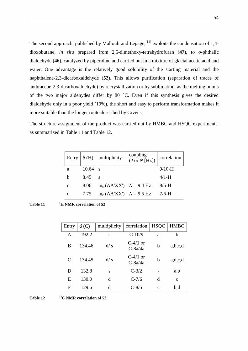
54
The second approach, published by Mallouli and Lepage,[14] exploits the condensation of 1,4-
dioxobutane, in situ prepared from 2,5-dimethoxy-tetrahydrofuran (47), to o-phthalic
dialdehyde (46), catalyzed by piperidine and carried out in a mixture of glacial acetic acid and
water. One advantage is the relatively good solubility of the starting material and the
naphthalene-2,3-dicarboxaldehyde (52). This allows purification (separation of traces of
anthracene-2,3-dicarboxaldehyde) by recrystallization or by sublimation, as the melting points
of the two major aldehydes differ by 80 °C. Even if this synthesis gives the desired
dialdehyde only in a poor yield (19%), the short and easy to perform transformation makes it
more suitable than the longer route described by Givens.
The structure assignment of the product was carried out by HMBC and HSQC experiments.
as summarized in Table 11 and Table 12.
Entry δ (H) multiplicity coupling (J or N [Hz])
correlation
a 10.64 s 9/10-H
b 8.45 s 4/1-H
c 8.06 mc (AA'XX') N = 9.4 Hz 8/5-H
d 7.75 mc (AA'XX') N = 9.5 Hz 7/6-H
Table 11 1H NMR correlation of 52
Entry δ (C) multiplicity correlation HSQC HMBC
A 192.2 s C-10/9 a b
B 134.46 d/ s C-4/1 or C-8a/4a
b a,b,c,d
C 134.45 d/ s C-4/1 or C-8a/4a
b a,d,c,d
D 132.8 s C-3/2 - a,b
E 130.0 d C-7/6 d c
F 129.6 d C-8/5 c b,d
Table 12 13C NMR correlation of 52
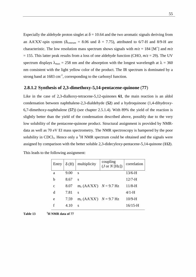
55
Especially the aldehyde proton singlet at δ = 10.64 and the two aromatic signals deriving from
an AA'XX'-spin system (δcentrum = 8.06 und δ = 7.75), attributed to 6/7-H and 8/9-H are
characteristic. The low resolution mass spectrum shows signals with m/z = 184 [M+] and m/z
= 155. This latter peak results from a loss of one aldehyde function (CHO, m/z = 29). The UV
spectrum displays λmax = 258 nm and the absorption with the longest wavelength at λ = 360
nm consistent with the light yellow color of the product. The IR spectrum is dominated by a
strong band at 1683 cm-1, corresponding to the carbonyl function.
2.8.1.2 Synthesis of 2,3-dimethoxy-5,14-pentacene-quinone (77)
Like in the case of 2,3-dialkoxy-tetracene-5,12-quinones 61, the main reaction is an aldol
condensation between naphthalene-2,3-dialdehyde (52) and a hydroquinone (1,4-dihydroxy-
6,7-dimethoxy-naphthalene (57)) (see chapter 2.5.1.4). With 89% the yield of the reaction is
slightly better than the yield of the condensation described above, possibly due to the very
low solubility of the pentacene-quinone product. Structural assignment is provided by NMR-
data as well as 70 eV EI mass spectrometry. The NMR spectroscopy is hampered by the poor
solubility in CDCl3. Hence only a 1H NMR spectrum could be obtained and the signals were
assigned by comparison with the better soluble 2,3-didecyloxy-pentacene-5,14-quinone (112).
This leads to the following assignment:
Entry δ (H) multiplicity coupling (J or N [Hz])
correlation
a 9.00 s 13/6-H
b 8.67 s 12/7-H
c 8.07 mc (AA'XX') N = 9.7 Hz 11/8-H
d 7.81 s 4/1-H
e 7.59 mc (AA'XX') N = 9.7 Hz 10/9-H
f 4.10 s 16/15-H
Table 13 1H NMR data of 77
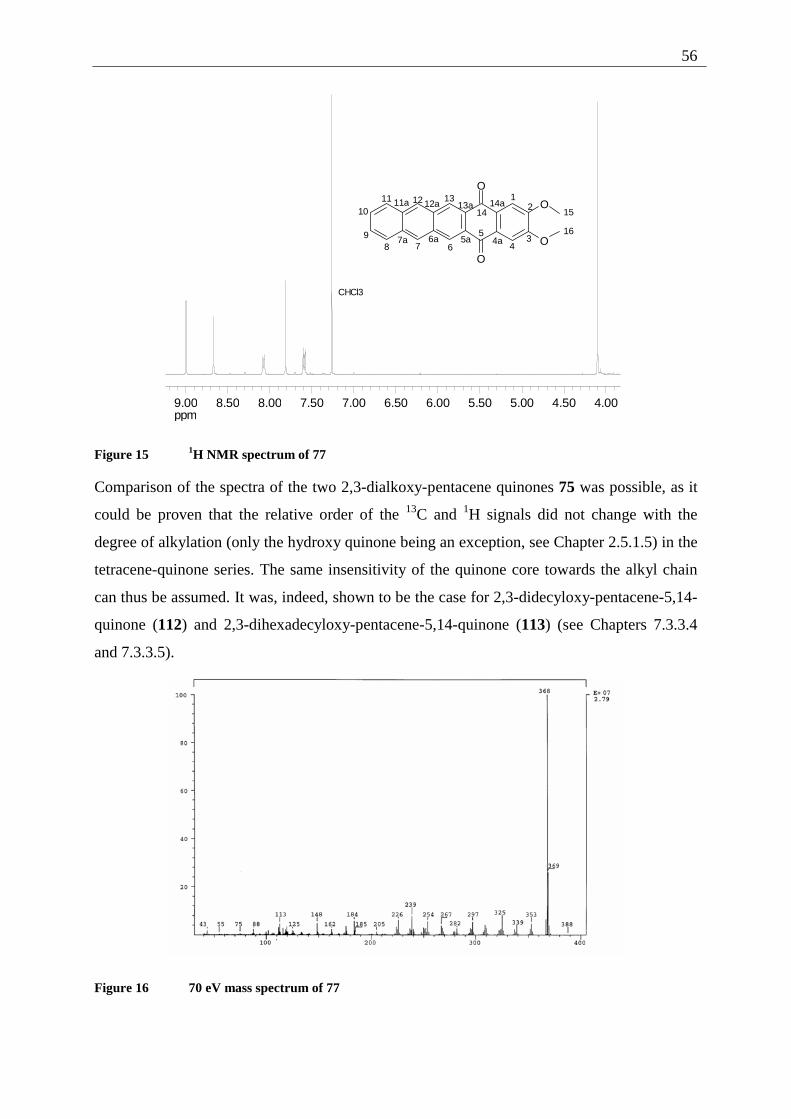
56
ppm4.004.505.005.506.006.507.007.508.008.509.00
CHCl3
O
O
O
O
12
34
5
6789
1011 12 13
14 15
164a5a6a7a
11a 12a 13a 14a
Figure 15 1H NMR spectrum of 77
Comparison of the spectra of the two 2,3-dialkoxy-pentacene quinones 75 was possible, as it
could be proven that the relative order of the 13C and 1H signals did not change with the
degree of alkylation (only the hydroxy quinone being an exception, see Chapter 2.5.1.5) in the
tetracene-quinone series. The same insensitivity of the quinone core towards the alkyl chain
can thus be assumed. It was, indeed, shown to be the case for 2,3-didecyloxy-pentacene-5,14-
quinone (112) and 2,3-dihexadecyloxy-pentacene-5,14-quinone (113) (see Chapters 7.3.3.4
and 7.3.3.5).
Figure 16 70 eV mass spectrum of 77
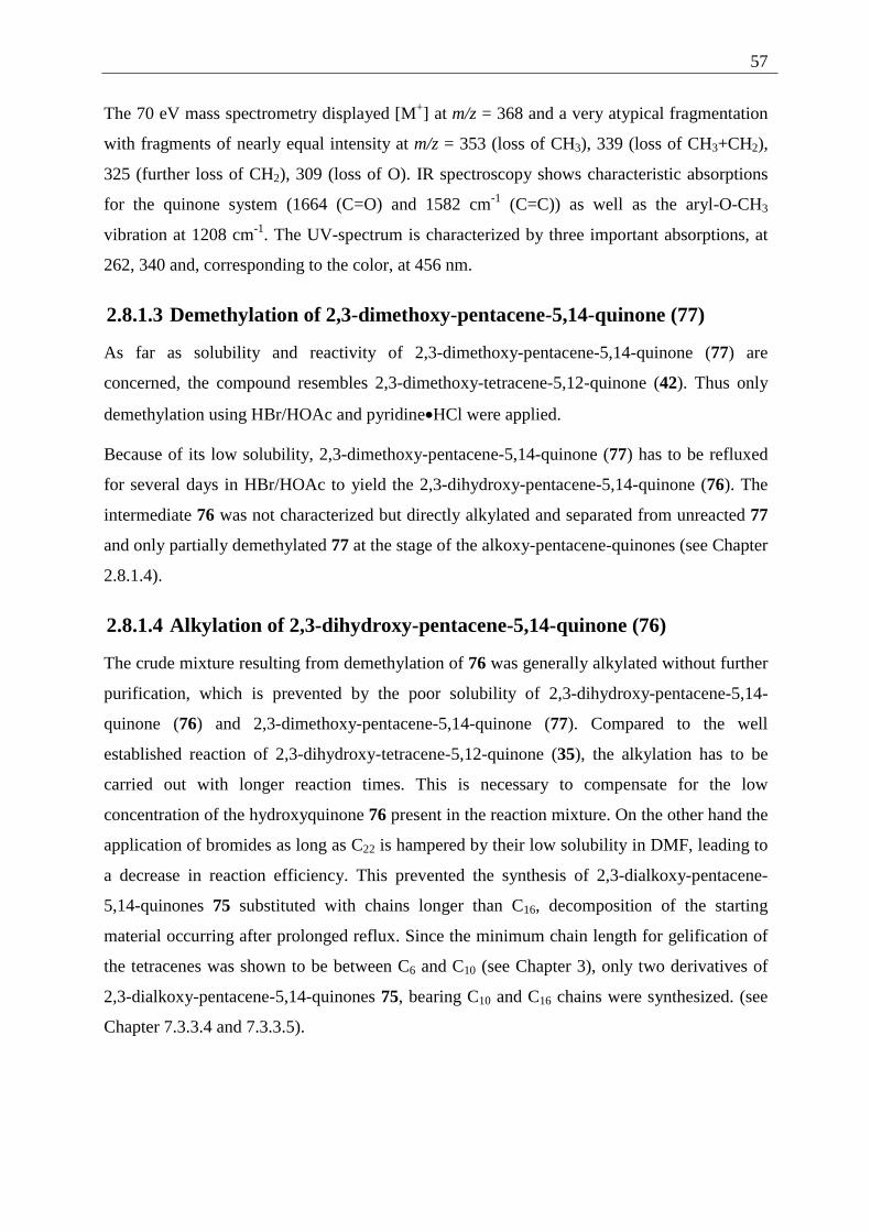
57
The 70 eV mass spectrometry displayed [M+] at m/z = 368 and a very atypical fragmentation
with fragments of nearly equal intensity at m/z = 353 (loss of CH3), 339 (loss of CH3+CH2),
325 (further loss of CH2), 309 (loss of O). IR spectroscopy shows characteristic absorptions
for the quinone system (1664 (C=O) and 1582 cm-1 (C=C)) as well as the aryl-O-CH3
vibration at 1208 cm-1. The UV-spectrum is characterized by three important absorptions, at
262, 340 and, corresponding to the color, at 456 nm.
2.8.1.3 Demethylation of 2,3-dimethoxy-pentacene-5,14-quinone (77)
As far as solubility and reactivity of 2,3-dimethoxy-pentacene-5,14-quinone (77) are
concerned, the compound resembles 2,3-dimethoxy-tetracene-5,12-quinone (42). Thus only
demethylation using HBr/HOAc and pyridine•HCl were applied.
Because of its low solubility, 2,3-dimethoxy-pentacene-5,14-quinone (77) has to be refluxed
for several days in HBr/HOAc to yield the 2,3-dihydroxy-pentacene-5,14-quinone (76). The
intermediate 76 was not characterized but directly alkylated and separated from unreacted 77
and only partially demethylated 77 at the stage of the alkoxy-pentacene-quinones (see Chapter
2.8.1.4).
2.8.1.4 Alkylation of 2,3-dihydroxy-pentacene-5,14-quinone (76)
The crude mixture resulting from demethylation of 76 was generally alkylated without further
purification, which is prevented by the poor solubility of 2,3-dihydroxy-pentacene-5,14-
quinone (76) and 2,3-dimethoxy-pentacene-5,14-quinone (77). Compared to the well
established reaction of 2,3-dihydroxy-tetracene-5,12-quinone (35), the alkylation has to be
carried out with longer reaction times. This is necessary to compensate for the low
concentration of the hydroxyquinone 76 present in the reaction mixture. On the other hand the
application of bromides as long as C22 is hampered by their low solubility in DMF, leading to
a decrease in reaction efficiency. This prevented the synthesis of 2,3-dialkoxy-pentacene-
5,14-quinones 75 substituted with chains longer than C16, decomposition of the starting
material occurring after prolonged reflux. Since the minimum chain length for gelification of
the tetracenes was shown to be between C6 and C10 (see Chapter 3), only two derivatives of
2,3-dialkoxy-pentacene-5,14-quinones 75, bearing C10 and C16 chains were synthesized. (see
Chapter 7.3.3.4 and 7.3.3.5).
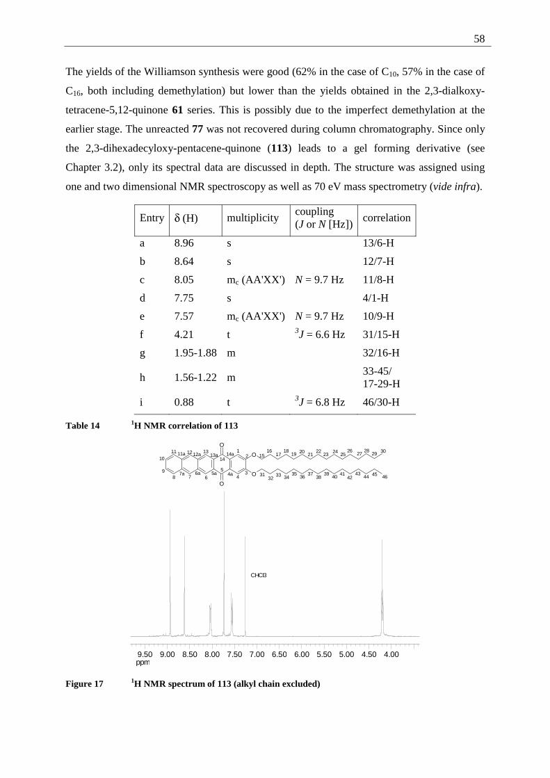
58
The yields of the Williamson synthesis were good (62% in the case of C10, 57% in the case of
C16, both including demethylation) but lower than the yields obtained in the 2,3-dialkoxy-
tetracene-5,12-quinone 61 series. This is possibly due to the imperfect demethylation at the
earlier stage. The unreacted 77 was not recovered during column chromatography. Since only
the 2,3-dihexadecyloxy-pentacene-quinone (113) leads to a gel forming derivative (see
Chapter 3.2), only its spectral data are discussed in depth. The structure was assigned using
one and two dimensional NMR spectroscopy as well as 70 eV mass spectrometry (vide infra).
Entry δ (H) multiplicity coupling (J or N [Hz])
correlation
a 8.96 s 13/6-H
b 8.64 s 12/7-H
c 8.05 mc (AA'XX') N = 9.7 Hz 11/8-H
d 7.75 s 4/1-H
e 7.57 mc (AA'XX') N = 9.7 Hz 10/9-H
f 4.21 t 3J = 6.6 Hz 31/15-H
g 1.95-1.88 m 32/16-H
h 1.56-1.22 m 33-45/
17-29-H
i 0.88 t 3J = 6.8 Hz 46/30-H
Table 14 1H NMR correlation of 113
ppm4.004.505.005.506.006.507.007.508.008.509.009.50
CHCl3
O
O
O
O
12
34
5
6789
1011 12 13
1415
16
4a5a6a7a
11a 12a 13a 14a 1718
19 2021
2223
2425
2627
2829 30
3132
33 3435
3637
3839
4041
4243
44 45 46
Figure 17 1H NMR spectrum of 113 (alkyl chain excluded)
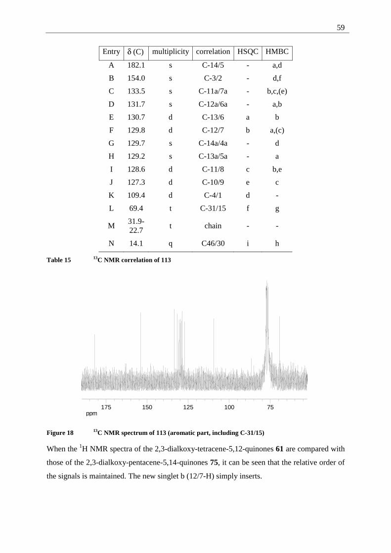
59
Entry δ (C) multiplicity correlation HSQC HMBC
A 182.1 s C-14/5 - a,d
B 154.0 s C-3/2 - d,f
C 133.5 s C-11a/7a - b,c,(e)
D 131.7 s C-12a/6a - a,b
E 130.7 d C-13/6 a b
F 129.8 d C-12/7 b a,(c)
G 129.7 s C-14a/4a - d
H 129.2 s C-13a/5a - a
I 128.6 d C-11/8 c b,e
J 127.3 d C-10/9 e c
K 109.4 d C-4/1 d -
L 69.4 t C-31/15 f g
M 31.9- 22.7
t chain - -
N 14.1 q C46/30 i h
Table 15 13C NMR correlation of 113
ppm75100125150175
Figure 18 13C NMR spectrum of 113 (aromatic part, including C-31/15)
When the 1H NMR spectra of the 2,3-dialkoxy-tetracene-5,12-quinones 61 are compared with
those of the 2,3-dialkoxy-pentacene-5,14-quinones 75, it can be seen that the relative order of
the signals is maintained. The new singlet b (12/7-H) simply inserts.
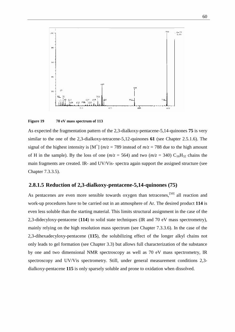
60
Figure 19 70 eV mass spectrum of 113
As expected the fragmentation pattern of the 2,3-dialkoxy-pentacene-5,14-quinones 75 is very
similar to the one of the 2,3-dialkoxy-tetracene-5,12-quinones 61 (see Chapter 2.5.1.6). The
signal of the highest intensity is [M+] (m/z = 789 instead of m/z = 788 due to the high amount
of H in the sample). By the loss of one (m/z = 564) and two (m/z = 340) C16H32 chains the
main fragments are created. IR- and UV/Vis- spectra again support the assigned structure (see
Chapter 7.3.3.5).
2.8.1.5 Reduction of 2,3-dialkoxy-pentacene-5,14-quinones (75)
As pentacenes are even more sensible towards oxygen than tetracenes,[50] all reaction and
work-up procedures have to be carried out in an atmosphere of Ar. The desired product 114 is
even less soluble than the starting material. This limits structural assignment in the case of the
2,3-didecyloxy-pentacene (114) to solid state techniques (IR and 70 eV mass spectrometry),
mainly relying on the high resolution mass spectrum (see Chapter 7.3.3.6). In the case of the
2,3-dihexadecyloxy-pentacene (115), the solubilizing effect of the longer alkyl chains not
only leads to gel formation (see Chapter 3.3) but allows full characterization of the substance
by one and two dimensional NMR spectroscopy as well as 70 eV mass spectrometry, IR
spectroscopy and UV/Vis spectrometry. Still, under general measurement conditions 2,3-
dialkoxy-pentacene 115 is only sparsely soluble and prone to oxidation when dissolved.
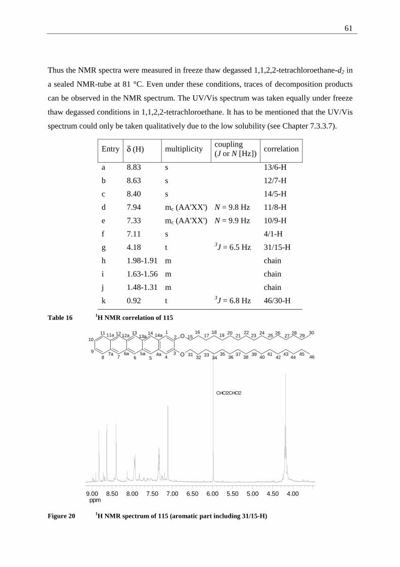
61
Thus the NMR spectra were measured in freeze thaw degassed 1,1,2,2-tetrachloroethane-d2 in
a sealed NMR-tube at 81 °C. Even under these conditions, traces of decomposition products
can be observed in the NMR spectrum. The UV/Vis spectrum was taken equally under freeze
thaw degassed conditions in 1,1,2,2-tetrachloroethane. It has to be mentioned that the UV/Vis
spectrum could only be taken qualitatively due to the low solubility (see Chapter 7.3.3.7).
Entry δ (H) multiplicity coupling (J or N [Hz])
correlation
a 8.83 s 13/6-H
b 8.63 s 12/7-H
c 8.40 s 14/5-H
d 7.94 mc (AA'XX') N = 9.8 Hz 11/8-H
e 7.33 mc (AA'XX') N = 9.9 Hz 10/9-H
f 7.11 s 4/1-H
g 4.18 t 3J = 6.5 Hz 31/15-H
h 1.98-1.91 m chain
i 1.63-1.56 m chain
j 1.48-1.31 m chain
k 0.92 t 3J = 6.8 Hz 46/30-H
Table 16 1H NMR correlation of 115
ppm4.004.505.005.506.006.507.007.508.008.509.00
CHCl2CHCl2
O
O
12
345678
9
1011 12 13 14
1516
4a5a6a7a
11a 12a 13a 14a 1718
19 2021
2223
24 25 2627
28 2930
3132 33
3435
3637
3839
4041
4243
4445
46
Figure 20 1H NMR spectrum of 115 (aromatic part including 31/15-H)
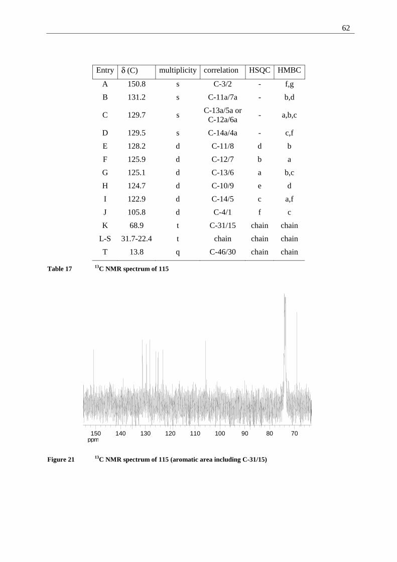
62
Entry δ (C) multiplicity correlation HSQC HMBC
A 150.8 s C-3/2 - f,g
B 131.2 s C-11a/7a - b,d
C 129.7 s C-13a/5a or
C-12a/6a - a,b,c
D 129.5 s C-14a/4a - c,f
E 128.2 d C-11/8 d b
F 125.9 d C-12/7 b a
G 125.1 d C-13/6 a b,c
H 124.7 d C-10/9 e d
I 122.9 d C-14/5 c a,f
J 105.8 d C-4/1 f c
K 68.9 t C-31/15 chain chain
L-S 31.7-22.4 t chain chain chain
T 13.8 q C-46/30 chain chain
Table 17 13C NMR spectrum of 115
ppm708090100110120130140150
Figure 21 13C NMR spectrum of 115 (aromatic area including C-31/15)
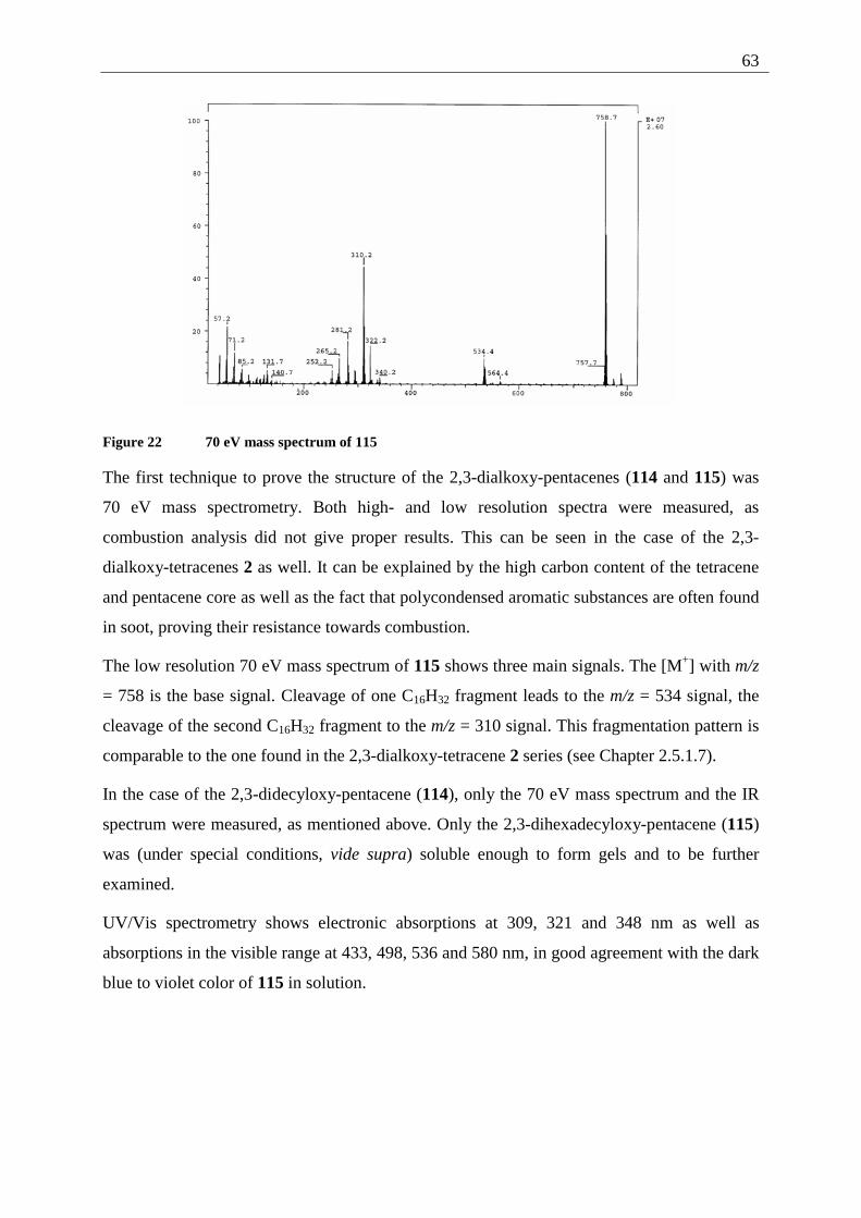
63
Figure 22 70 eV mass spectrum of 115
The first technique to prove the structure of the 2,3-dialkoxy-pentacenes (114 and 115) was
70 eV mass spectrometry. Both high- and low resolution spectra were measured, as
combustion analysis did not give proper results. This can be seen in the case of the 2,3-
dialkoxy-tetracenes 2 as well. It can be explained by the high carbon content of the tetracene
and pentacene core as well as the fact that polycondensed aromatic substances are often found
in soot, proving their resistance towards combustion.
The low resolution 70 eV mass spectrum of 115 shows three main signals. The [M+] with m/z
= 758 is the base signal. Cleavage of one C16H32 fragment leads to the m/z = 534 signal, the
cleavage of the second C16H32 fragment to the m/z = 310 signal. This fragmentation pattern is
comparable to the one found in the 2,3-dialkoxy-tetracene 2 series (see Chapter 2.5.1.7).
In the case of the 2,3-didecyloxy-pentacene (114), only the 70 eV mass spectrum and the IR
spectrum were measured, as mentioned above. Only the 2,3-dihexadecyloxy-pentacene (115)
was (under special conditions, vide supra) soluble enough to form gels and to be further
examined.
UV/Vis spectrometry shows electronic absorptions at 309, 321 and 348 nm as well as
absorptions in the visible range at 433, 498, 536 and 580 nm, in good agreement with the dark
blue to violet color of 115 in solution.
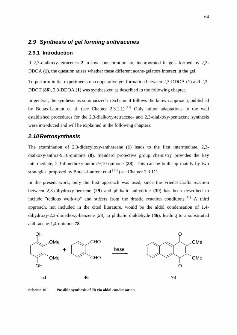
64
2.9 Synthesis of gel forming anthracenes
2.9.1 Introduction
If 2,3-dialkoxy-tetracenes 2 in low concentration are incorporated in gels formed by 2,3-
DDOA (1), the question arises whether these different acene-gelators interact in the gel.
To perform initial experiments on cooperative gel formation between 2,3-DDOA (1) and 2,3-
DDOT (86), 2,3-DDOA (1) was synthesized as described in the following chapter.
In general, the synthesis as summarized in Scheme 4 follows the known approach, published
by Bouas-Laurent et al. (see Chapter 2.3.1.1).[11] Only minor adaptations to the well
established procedures for the 2,3-dialkoxy-tetracene- and 2,3-dialkoxy-pentacene synthesis
were introduced and will be explained in the following chapters.
2.10 Retrosynthesis
The examination of 2,3-didecyloxy-anthracene (1) leads to the first intermediate, 2,3-
dialkoxy-anthra-9,10-quinone (8). Standard protective group chemistry provides the key
intermediate, 2,3-dimethoxy-anthra-9,10-quinone (38). This can be build up mainly by two
strategies, proposed by Bouas-Laurent et al.[11] (see Chapter 2.3.11).
In the present work, only the first approach was used, since the Friedel-Crafts reaction
between 2,3-dihydroxy-benzene (29) and phthalic anhydride (30) has been described to
include "tedious work-up" and suffers from the drastic reaction conditions.[11] A third
approach, not included in the cited literature, would be the aldol condensation of 1,4-
dihydroxy-2,3-dimethoxy-benzene (53) to phthalic dialdehyde (46), leading to a substituted
anthracene-1,4-quinone 78.
OH
OH
OMe
OMe
+CHO
CHO
base
O
O
OMe
OMe
53 46 78
Scheme 16 Possible synthesis of 78 via aldol condensation
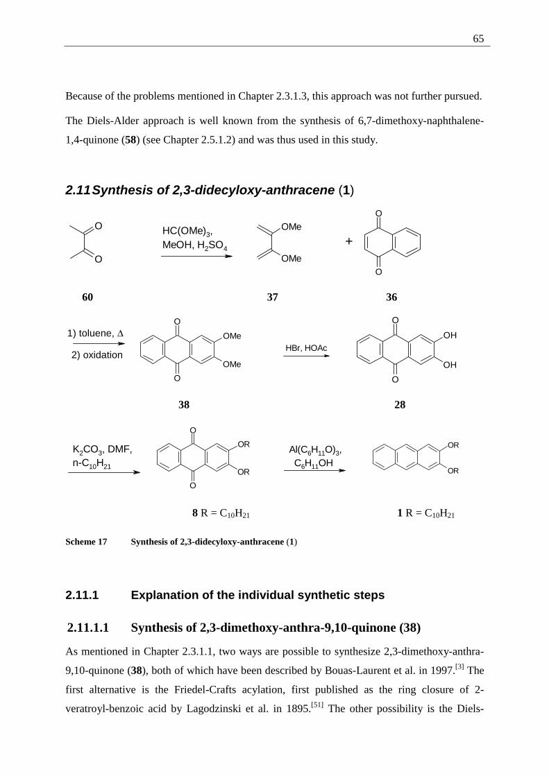
65
Because of the problems mentioned in Chapter 2.3.1.3, this approach was not further pursued.
The Diels-Alder approach is well known from the synthesis of 6,7-dimethoxy-naphthalene-
1,4-quinone (58) (see Chapter 2.5.1.2) and was thus used in this study.
2.11 Synthesis of 2,3-didecyloxy-anthracene (1)
O
O
HC(OMe)3, MeOH, H2SO4
OMe
OMe
+
O
O
60 37 36
1) toluene, ∆
2) oxidation
O
O
OMe
OMe
HBr, HOAc
O
O
OH
OH
38 28
K2CO3, DMF,n-C10H21
O
O
OR
OR
Al(C6H11O)3, C6H11OH
OR
OR
8 R = C10H21 1 R = C10H21
Scheme 17 Synthesis of 2,3-didecyloxy-anthracene (1)
2.11.1 Explanation of the individual synthetic steps
2.11.1.1 Synthesis of 2,3-dimethoxy-anthra-9,10-quinone (38)
As mentioned in Chapter 2.3.1.1, two ways are possible to synthesize 2,3-dimethoxy-anthra-
9,10-quinone (38), both of which have been described by Bouas-Laurent et al. in 1997.[3] The
first alternative is the Friedel-Crafts acylation, first published as the ring closure of 2-
veratroyl-benzoic acid by Lagodzinski et al. in 1895.[51] The other possibility is the Diels-
66
Alder addition of 2,3-dimethoxy-buta-1,3-diene (37) to naphthalene-1,4-quinone (36). This
synthesis was first published by Johnson et al. in 1941[52] and was used in the present
investigation because of the simpler work-up.[11] When compared to the addition of 37 to
benzoquinone (59) two major differences have to be mentioned (see Chapter 7.3.2.1). First,
the addition can only take place once. Therefore the diene 37 is used in excess. On the other
hand, naphthoquinone is, unlike benzoquinone, not able to oxidize the primary adduct. Hence
the Diels-Alder addition is followed by an oxidation step with air in basic solution. To make
work-up easier, the oxidation was carried out without the separation and purification of the
primary adduct. The spectroscopic data (1H NMR and 13C NMR) agree well with the data
provided in the literature.[11] Further proof was drawn from the 70 eV mass spectrum with
[M+] at m/z = 268 and characteristic fragments at m/z = 253 and 238, resulting from the loss
of one and two CH3-groups. The IR-spectrum exhibits characteristic bands at 1655 and 1572
cm-1 for the quinone system and 1224 cm-1 for the aromatic methoxy ether.
2.11.1.2 Demethylation of 2,3-dimethoxy-9,10-anthraquinone (38)
The higher solubility of the anthraquinone 38 in comparison to 2,3-dimethoxy-tetracene-5,12-
quinone (42) makes it possible to use modern reagents and mild conditions (BBr3 in DCM) as
published by Bouas-Laurent et al.[11] Still, the convenient and cheap demethylation using
strong acids that was used with good success in the course of this work is applicable to the
lower homologue of the tetracenequinone. The reaction time is comparatively short (24 h, see
Chapter 7.3.2.2) and the relatively high solubility allows good conversions.
The spectroscopic data provided in Chapter 7.3.2.2 is in good agreement with the 1H NMR, 13C NMR and IR data provided by Bouas-Laurent et al.[11]
2.11.1.3 Alkylation of 2,3-dihydroxy-anthracene-9,10-quinone (28)
The classical Williamson conditions, using DMF as solvent and potassium carbonate as base
were used successfully as described in the literature.[11] The spectral data of the resulting 2,3-
didecyloxy-anthracene-9,10-quinone (8) agree with the published data[11] (see Chapter
7.3.2.3).
67
2.11.1.4 Reduction of 2,3-didecyloxy-anthracene-9,10-quinone (8)
The procedure to reduce 2,3-didecyloxy-anthra-9,10-quinone (8) to the corresponding
dialkoxy-anthracene 1, published by Bouas-Laurent at al.[11] is a two step reduction using
NaBH4 in iso-propanol. The higher solubility of anthraquinones in contrast to alkoxy-
tetracene-quinones 61 makes this low temperature procedure possible. Still, the gel formation
of the product 2,3-DDOA (1), a good gelator for alcohols, causes problems. Bouas-Laurent et
al. circumvented these by changing the order of reduction/substitution.[11] In this study, a
different approach was chosen to prevent gel formation, the proven high temperature
reduction using Al(OC6H11). The relatively high solubility of 2,3-DDOA (1) in DCM led to a
different work-up, the extraction after decomposition of the alumina species in contrast to
filtration and recrystallization (see Chapter 7.3.2.4).
The spectroscopic data (1H NMR, 13C NMR, IR-spectroscopy and 70 eV mass spectrum) are
in good agreement with the data published by Bouas-Laurent et al.[11]
68
3 Examination of the gel forming abilities of 2,3-dialkoxy-acenes
The techniques to examine gel formation can be subdivided into three main groups. The first
and most obvious group contains rheological tests like the "inverted tube test", following the
"definition" given by Lloyd, quoted by Terech and Weiss[1a] who stated that "if it looks like
"Jello", it must be a gel". The second possibility is to observe spectroscopic changes, when a
solution of a possible gelator is cooled and by this transferred into the gel-state. UV/Vis- and
fluorescence spectroscopy can provide these "indirect" evidences for gel formation. The last,
and most interesting group of techniques contains direct structural determinations of the gel.
Measurement of the microscopic structures involved can be accomplished by electron
microscopy, neutron scattering or scanning probe microscopy techniques.
The use of the three types of gel determination will be presented for the acene gelators in the
following chapters.
3.1 2,3-Dialkoxy-tetracenes 2
3.1.1 Inverted test tube examinations
A first test to evaluate the gel forming properties of different solvents and gelators is the
“inverted tube” method. A precisely weighed sample of the finely powdered gelator is
transferred into a sample tube (length 6 cm, diameter 0.7 cm). A known volume of the solvent
to be tested is added. The tube is capped and the content homogenized in an ultrasound bath.
The suspension is warmed until dissolution of the gelator. Upon cooling to room temperature,
a solidification of the whole liquid indicates gel formation. Other possible results are the
precipitation of the gelator, the lack of solubility or the gelator staying dissolved. In this last
case, the sample can be cooled to -24 °C if the solvent allows such a treatment.
This test was used to gain a first impression of the gelification properties and to screen a
broad variety of solvents.
The full data of the inverted tube tests is given in Appendix I A. To summarize those results,
the following table is given:
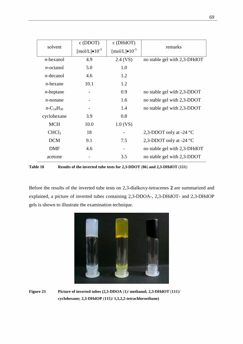
69
solvent c (DDOT)
[mol/L]•10-3
c (DHdOT)
[mol/L]•10-3 remarks
n-hexanol 4.9 2.4 (VS) no stable gel with 2,3-DHdOT
n-octanol 5.0 1.0
n-decanol 4.6 1.2
n-hexane 10.1 1.2
n-heptane - 0.9 no stable gel with 2,3-DDOT
n-nonane - 1.6 no stable gel with 2,3-DDOT
n-C14H30 - 1.4 no stable gel with 2,3-DDOT
cyclohexane 3.9 0.8
MCH 10.0 1.0 (VS)
CHCl3 18 - 2,3-DDOT only at -24 °C
DCM 9.1 7.5 2,3-DDOT only at -24 °C
DMF 4.6 - no stable gel with 2,3-DHdOT
acetone - 3.5 no stable gel with 2,3-DDOT
Table 18 Results of the inverted tube tests for 2,3-DDOT (86) and 2,3-DHdOT (111)
Before the results of the inverted tube tests on 2,3-dialkoxy-tetracenes 2 are summarized and
explained, a picture of inverted tubes containing 2,3-DDOA-, 2,3-DHdOT- and 2,3-DHdOP
gels is shown to illustrate the examination technique.
Figure 23 Picture of inverted tubes (2,3-DDOA (1)/ methanol; 2,3-DHdOT (111)/
cyclohexane; 2,3-DHdOP (115)/ 1,1,2,2-tetrachloroethane)
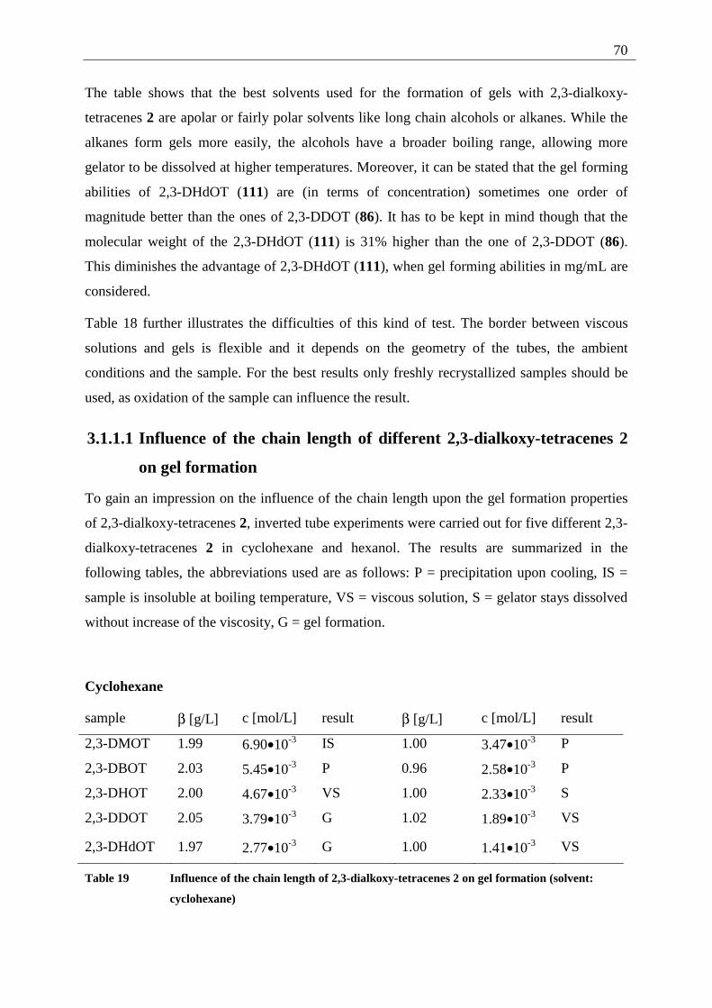
70
The table shows that the best solvents used for the formation of gels with 2,3-dialkoxy-
tetracenes 2 are apolar or fairly polar solvents like long chain alcohols or alkanes. While the
alkanes form gels more easily, the alcohols have a broader boiling range, allowing more
gelator to be dissolved at higher temperatures. Moreover, it can be stated that the gel forming
abilities of 2,3-DHdOT (111) are (in terms of concentration) sometimes one order of
magnitude better than the ones of 2,3-DDOT (86). It has to be kept in mind though that the
molecular weight of the 2,3-DHdOT (111) is 31% higher than the one of 2,3-DDOT (86).
This diminishes the advantage of 2,3-DHdOT (111), when gel forming abilities in mg/mL are
considered.
Table 18 further illustrates the difficulties of this kind of test. The border between viscous
solutions and gels is flexible and it depends on the geometry of the tubes, the ambient
conditions and the sample. For the best results only freshly recrystallized samples should be
used, as oxidation of the sample can influence the result.
3.1.1.1 Influence of the chain length of different 2,3-dialkoxy-tetracenes 2
on gel formation
To gain an impression on the influence of the chain length upon the gel formation properties
of 2,3-dialkoxy-tetracenes 2, inverted tube experiments were carried out for five different 2,3-
dialkoxy-tetracenes 2 in cyclohexane and hexanol. The results are summarized in the
following tables, the abbreviations used are as follows: P = precipitation upon cooling, IS =
sample is insoluble at boiling temperature, VS = viscous solution, S = gelator stays dissolved
without increase of the viscosity, G = gel formation.
Cyclohexane
sample β [g/L] c [mol/L] result β [g/L] c [mol/L] result
2,3-DMOT 1.99 6.90•10-3 IS 1.00 3.47•10-3 P
2,3-DBOT 2.03 5.45•10-3 P 0.96 2.58•10-3 P
2,3-DHOT 2.00 4.67•10-3 VS 1.00 2.33•10-3 S
2,3-DDOT 2.05 3.79•10-3 G 1.02 1.89•10-3 VS
2,3-DHdOT 1.97 2.77•10-3 G 1.00 1.41•10-3 VS
Table 19 Influence of the chain length of 2,3-dialkoxy-tetracenes 2 on gel formation (solvent:
cyclohexane)
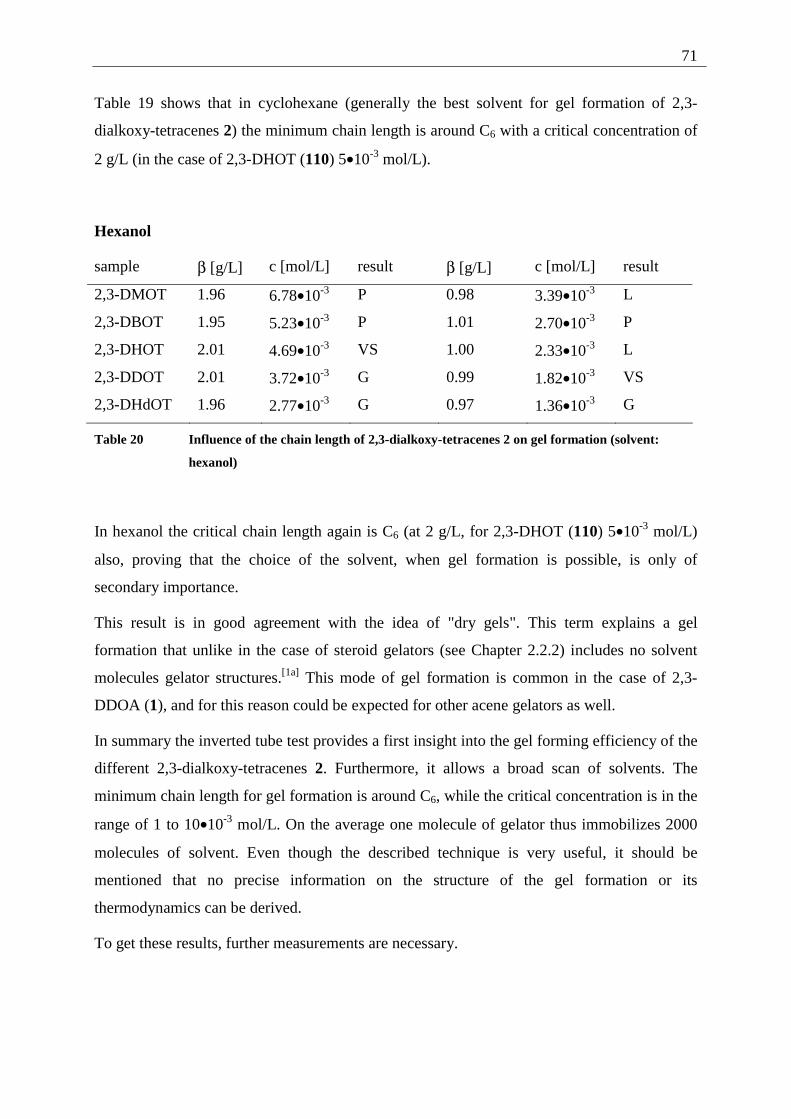
71
Table 19 shows that in cyclohexane (generally the best solvent for gel formation of 2,3-
dialkoxy-tetracenes 2) the minimum chain length is around C6 with a critical concentration of
2 g/L (in the case of 2,3-DHOT (110) 5•10-3 mol/L).
Hexanol
sample β [g/L] c [mol/L] result β [g/L] c [mol/L] result
2,3-DMOT 1.96 6.78•10-3 P 0.98 3.39•10-3 L
2,3-DBOT 1.95 5.23•10-3 P 1.01 2.70•10-3 P
2,3-DHOT 2.01 4.69•10-3 VS 1.00 2.33•10-3 L
2,3-DDOT 2.01 3.72•10-3 G 0.99 1.82•10-3 VS
2,3-DHdOT 1.96 2.77•10-3 G 0.97 1.36•10-3 G
Table 20 Influence of the chain length of 2,3-dialkoxy-tetracenes 2 on gel formation (solvent:
hexanol)
In hexanol the critical chain length again is C6 (at 2 g/L, for 2,3-DHOT (110) 5•10-3 mol/L)
also, proving that the choice of the solvent, when gel formation is possible, is only of
secondary importance.
This result is in good agreement with the idea of "dry gels". This term explains a gel
formation that unlike in the case of steroid gelators (see Chapter 2.2.2) includes no solvent
molecules gelator structures.[1a] This mode of gel formation is common in the case of 2,3-
DDOA (1), and for this reason could be expected for other acene gelators as well.
In summary the inverted tube test provides a first insight into the gel forming efficiency of the
different 2,3-dialkoxy-tetracenes 2. Furthermore, it allows a broad scan of solvents. The
minimum chain length for gel formation is around C6, while the critical concentration is in the
range of 1 to 10•10-3 mol/L. On the average one molecule of gelator thus immobilizes 2000
molecules of solvent. Even though the described technique is very useful, it should be
mentioned that no precise information on the structure of the gel formation or its
thermodynamics can be derived.
To get these results, further measurements are necessary.
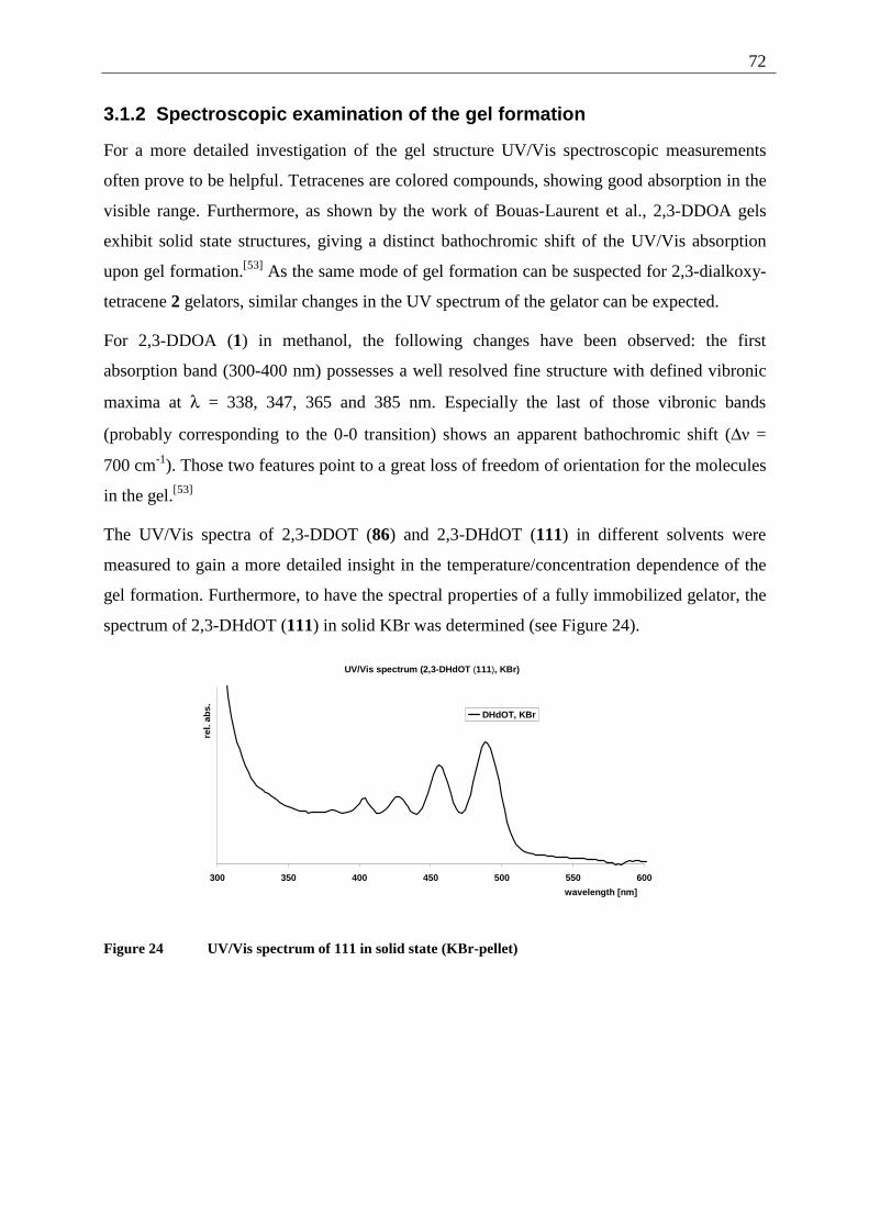
72
3.1.2 Spectroscopic examination of the gel formation
For a more detailed investigation of the gel structure UV/Vis spectroscopic measurements
often prove to be helpful. Tetracenes are colored compounds, showing good absorption in the
visible range. Furthermore, as shown by the work of Bouas-Laurent et al., 2,3-DDOA gels
exhibit solid state structures, giving a distinct bathochromic shift of the UV/Vis absorption
upon gel formation.[53] As the same mode of gel formation can be suspected for 2,3-dialkoxy-
tetracene 2 gelators, similar changes in the UV spectrum of the gelator can be expected.
For 2,3-DDOA (1) in methanol, the following changes have been observed: the first
absorption band (300-400 nm) possesses a well resolved fine structure with defined vibronic
maxima at λ = 338, 347, 365 and 385 nm. Especially the last of those vibronic bands
(probably corresponding to the 0-0 transition) shows an apparent bathochromic shift (∆ν =
700 cm-1). Those two features point to a great loss of freedom of orientation for the molecules
in the gel.[53]
The UV/Vis spectra of 2,3-DDOT (86) and 2,3-DHdOT (111) in different solvents were
measured to gain a more detailed insight in the temperature/concentration dependence of the
gel formation. Furthermore, to have the spectral properties of a fully immobilized gelator, the
spectrum of 2,3-DHdOT (111) in solid KBr was determined (see Figure 24).
UV/Vis spectrum (2,3-DHdOT (111), KBr)
300 350 400 450 500 550 600
wavelength [nm]
rel.
abs.
DHdOT, KBr
Figure 24 UV/Vis spectrum of 111 in solid state (KBr-pellet)
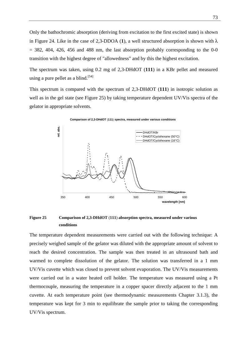
73
Only the bathochromic absorption (deriving from excitation to the first excited state) is shown
in Figure 24. Like in the case of 2,3-DDOA (1), a well structured absorption is shown with λ
= 382, 404, 426, 456 and 488 nm, the last absorption probably corresponding to the 0-0
transition with the highest degree of "allowedness" and by this the highest excitation.
The spectrum was taken, using 0.2 mg of 2,3-DHdOT (111) in a KBr pellet and measured
using a pure pellet as a blind.[54]
This spectrum is compared with the spectrum of 2,3-DHdOT (111) in isotropic solution as
well as in the gel state (see Figure 25) by taking temperature dependent UV/Vis spectra of the
gelator in appropriate solvents.
Comparison of 2,3-DHdOT (111) spectra, measured under various conditions
350 400 450 500 550 600
wavelength [nm]
rel.
abs.
DHdOT/KBrDHdOT/Cyclohexane (50°C)DHdOT/Cyclohexane (16°C)
Figure 25 Comparison of 2,3-DHdOT (111) absorption spectra, measured under various
conditions
The temperature dependent measurements were carried out with the following technique: A
precisely weighed sample of the gelator was diluted with the appropriate amount of solvent to
reach the desired concentration. The sample was then treated in an ultrasound bath and
warmed to complete dissolution of the gelator. The solution was transferred in a 1 mm
UV/Vis cuvette which was closed to prevent solvent evaporation. The UV/Vis measurements
were carried out in a water heated cell holder. The temperature was measured using a Pt
thermocouple, measuring the temperature in a copper spacer directly adjacent to the 1 mm
cuvette. At each temperature point (see thermodynamic measurements Chapter 3.1.3), the
temperature was kept for 3 min to equilibrate the sample prior to taking the corresponding
UV/Vis spectrum.
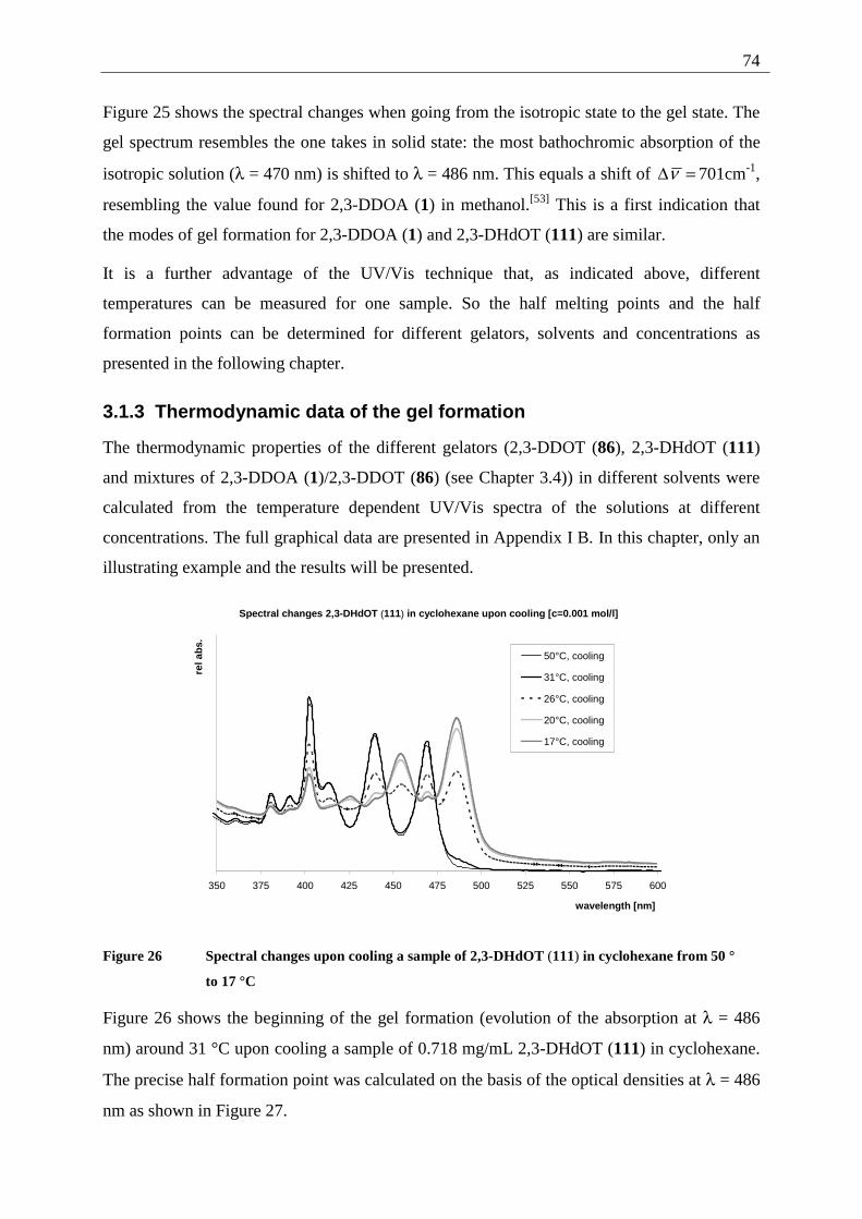
74
Figure 25 shows the spectral changes when going from the isotropic state to the gel state. The
gel spectrum resembles the one takes in solid state: the most bathochromic absorption of the
isotropic solution (λ = 470 nm) is shifted to λ = 486 nm. This equals a shift of =∆ν 701cm-1,
resembling the value found for 2,3-DDOA (1) in methanol.[53] This is a first indication that
the modes of gel formation for 2,3-DDOA (1) and 2,3-DHdOT (111) are similar.
It is a further advantage of the UV/Vis technique that, as indicated above, different
temperatures can be measured for one sample. So the half melting points and the half
formation points can be determined for different gelators, solvents and concentrations as
presented in the following chapter.
3.1.3 Thermodynamic data of the gel formation
The thermodynamic properties of the different gelators (2,3-DDOT (86), 2,3-DHdOT (111)
and mixtures of 2,3-DDOA (1)/2,3-DDOT (86) (see Chapter 3.4)) in different solvents were
calculated from the temperature dependent UV/Vis spectra of the solutions at different
concentrations. The full graphical data are presented in Appendix I B. In this chapter, only an
illustrating example and the results will be presented.
Spectral changes 2,3-DHdOT (111) in cyclohexane upon cooling [c=0.001 mol/l]
350 375 400 425 450 475 500 525 550 575 600
wavelength [nm]
rel a
bs.
50°C, cooling
31°C, cooling
26°C, cooling
20°C, cooling
17°C, cooling
Figure 26 Spectral changes upon cooling a sample of 2,3-DHdOT (111) in cyclohexane from 50 °
to 17 °C
Figure 26 shows the beginning of the gel formation (evolution of the absorption at λ = 486
nm) around 31 °C upon cooling a sample of 0.718 mg/mL 2,3-DHdOT (111) in cyclohexane.
The precise half formation point was calculated on the basis of the optical densities at λ = 486
nm as shown in Figure 27.
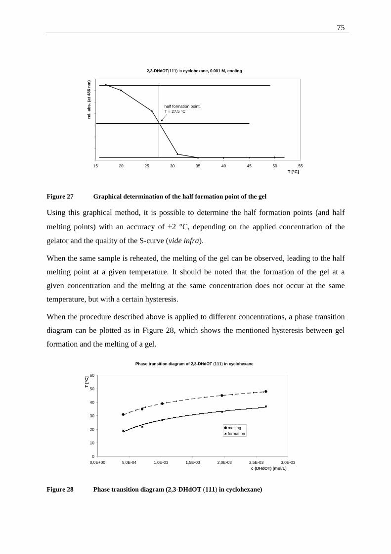
75
2,3-DHdOT(111) in cyclohexane, 0.001 M, cooling
15 20 25 30 35 40 45 50 55T [°C]
rel.
abs.
(at
486
nm
)
half formation point,T = 27.5 °C
Figure 27 Graphical determination of the half formation point of the gel
Using this graphical method, it is possible to determine the half formation points (and half
melting points) with an accuracy of ±2 °C, depending on the applied concentration of the
gelator and the quality of the S-curve (vide infra).
When the same sample is reheated, the melting of the gel can be observed, leading to the half
melting point at a given temperature. It should be noted that the formation of the gel at a
given concentration and the melting at the same concentration does not occur at the same
temperature, but with a certain hysteresis.
When the procedure described above is applied to different concentrations, a phase transition
diagram can be plotted as in Figure 28, which shows the mentioned hysteresis between gel
formation and the melting of a gel.
Phase transition diagram of 2,3-DHdOT (111) in cyclohexane
0
10
20
30
40
50
60
0,0E+00 5,0E-04 1,0E-03 1,5E-03 2,0E-03 2,5E-03 3,0E-03c (DHdOT) [mol/L]
T [
°C]
meltingformation
Figure 28 Phase transition diagram (2,3-DHdOT (111) in cyclohexane)
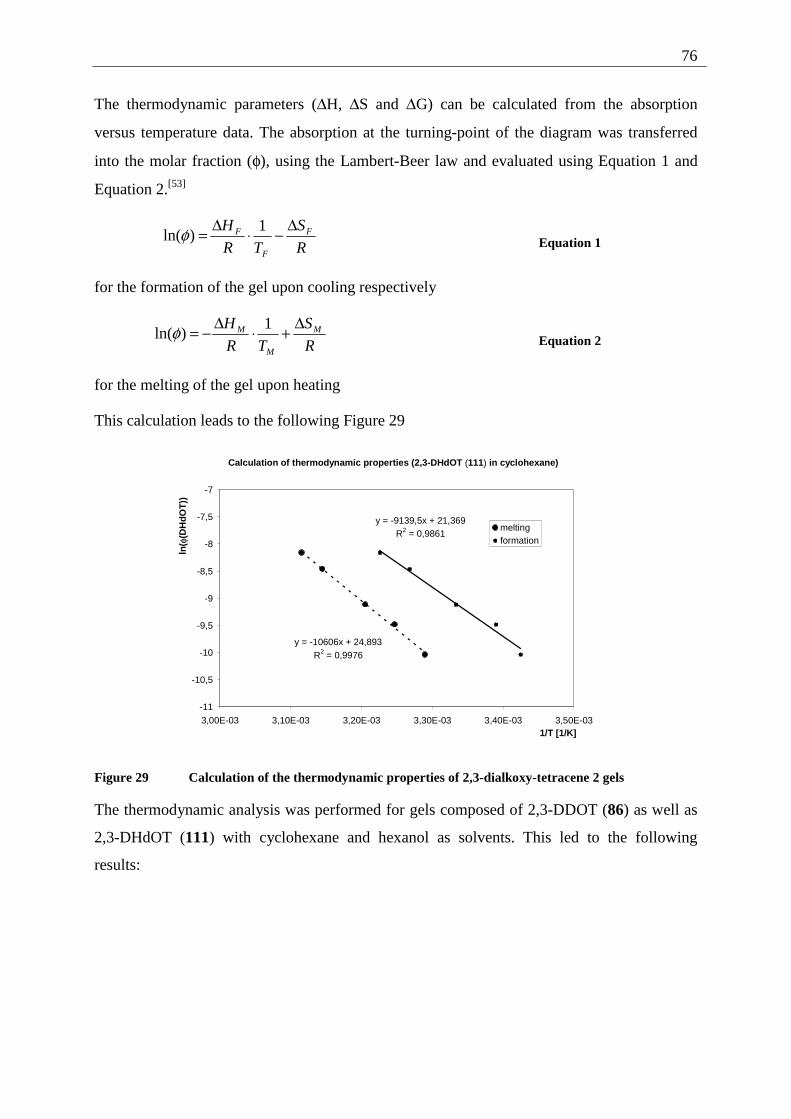
76
The thermodynamic parameters (∆H, ∆S and ∆G) can be calculated from the absorption
versus temperature data. The absorption at the turning-point of the diagram was transferred
into the molar fraction (φ), using the Lambert-Beer law and evaluated using Equation 1 and
Equation 2.[53]
R
S
TR
H F
F
F ∆−⋅∆= 1)ln(φ Equation 1
for the formation of the gel upon cooling respectively
R
S
TR
H M
M
M ∆+⋅∆−= 1)ln(φ Equation 2
for the melting of the gel upon heating
This calculation leads to the following Figure 29
Calculation of thermodynamic properties (2,3-DHdOT (111) in cyclohexane)
y = -10606x + 24,893R2 = 0,9976
y = -9139,5x + 21,369R2 = 0,9861
-11
-10,5
-10
-9,5
-9
-8,5
-8
-7,5
-7
3,00E-03 3,10E-03 3,20E-03 3,30E-03 3,40E-03 3,50E-031/T [1/K]
ln( φ
(DH
dO
T))
meltingformation
Figure 29 Calculation of the thermodynamic properties of 2,3-dialkoxy-tetracene 2 gels
The thermodynamic analysis was performed for gels composed of 2,3-DDOT (86) as well as
2,3-DHdOT (111) with cyclohexane and hexanol as solvents. This led to the following
results:
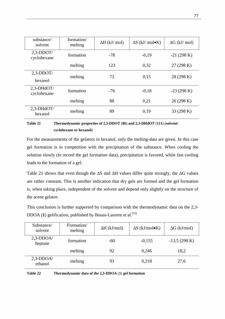
77
substance/ solvent
formation/ melting ∆H (kJ/ mol) ∆S (kJ/ mol•K) ∆G (kJ/ mol)
2,3-DDOT/ cyclohexane
formation -78 -0,19 -21 (298 K)
melting 123 0,32 27 (298 K)
2,3-DDOT/
hexanol melting 73 0,15 28 (298 K)
2,3-DHdOT/ cyclohexane
formation -76 -0,18 -23 (298 K)
melting 88 0,21 26 (298 K)
2,3-DHdOT/ hexanol
melting 89 0,19 33 (298 K)
Table 21 Thermodynamic properties of 2,3-DDOT (86) and 2,3-DHdOT (111) (solvent:
cyclohexane or hexanol)
For the measurements of the gelators in hexanol, only the melting-data are given. In this case
gel formation is in competition with the precipitation of the substance. When cooling the
solution slowly (to record the gel formation data), precipitation is favored, while fast cooling
leads to the formation of a gel.
Table 21 shows that even though the ∆S and ∆H values differ quite strongly, the ∆G values
are rather constant. This is another indication that dry gels are formed and the gel formation
is, when taking place, independent of the solvent and depend only slightly on the structure of
the acene gelator.
This conclusion is further supported by comparison with the thermodynamic data on the 2,3-
DDOA (1) gelification, published by Bouas-Laurent et al.[55]
Substance/ solvent
Formation/ melting ∆H (kJ/mol) ∆S (kJ/mol•K) ∆G (kJ/mol)
2,3-DDOA/ heptane
formation -60 -0,155 -13,5 (298 K)
melting 92 0,246 18,2
2,3-DDOA/ ethanol
melting 93 0,218 27,6
Table 22 Thermodynamic data of the 2,3-DDOA (1) gel formation
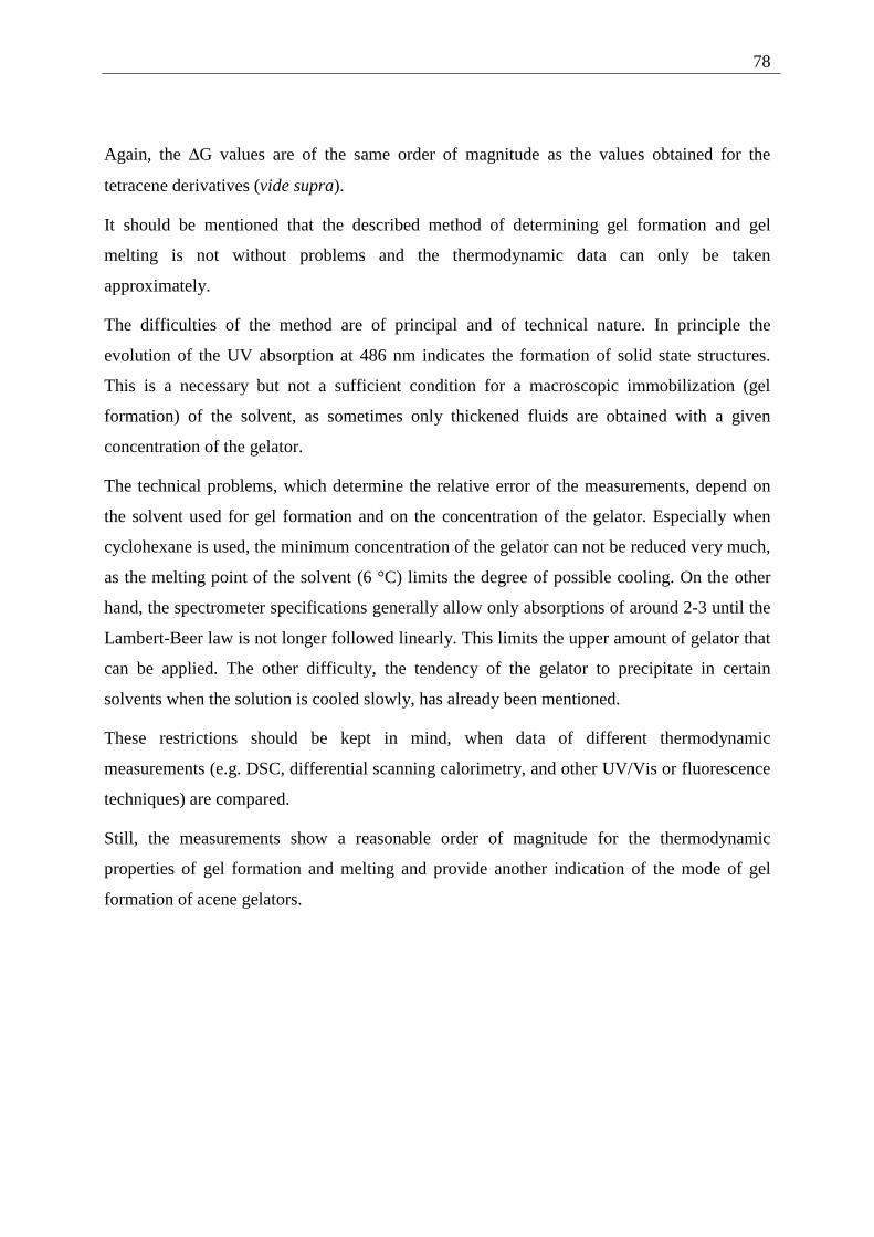
78
Again, the ∆G values are of the same order of magnitude as the values obtained for the
tetracene derivatives (vide supra).
It should be mentioned that the described method of determining gel formation and gel
melting is not without problems and the thermodynamic data can only be taken
approximately.
The difficulties of the method are of principal and of technical nature. In principle the
evolution of the UV absorption at 486 nm indicates the formation of solid state structures.
This is a necessary but not a sufficient condition for a macroscopic immobilization (gel
formation) of the solvent, as sometimes only thickened fluids are obtained with a given
concentration of the gelator.
The technical problems, which determine the relative error of the measurements, depend on
the solvent used for gel formation and on the concentration of the gelator. Especially when
cyclohexane is used, the minimum concentration of the gelator can not be reduced very much,
as the melting point of the solvent (6 °C) limits the degree of possible cooling. On the other
hand, the spectrometer specifications generally allow only absorptions of around 2-3 until the
Lambert-Beer law is not longer followed linearly. This limits the upper amount of gelator that
can be applied. The other difficulty, the tendency of the gelator to precipitate in certain
solvents when the solution is cooled slowly, has already been mentioned.
These restrictions should be kept in mind, when data of different thermodynamic
measurements (e.g. DSC, differential scanning calorimetry, and other UV/Vis or fluorescence
techniques) are compared.
Still, the measurements show a reasonable order of magnitude for the thermodynamic
properties of gel formation and melting and provide another indication of the mode of gel
formation of acene gelators.
79
3.1.4 AFM-measurements on 2,3-dialkoxy-tetracene 2 gels
3.1.4.1 General introduction to AFM measurements[56]
Atomic force microscopy is one technique from the group of scanning probe microscopy
techniques. In this group of techniques, a fine probe (usually a sharp tip) is scanned across a
sample surface by means of piezoelectric translators. The signal measured is recorded as a
function of the lateral position. Scanning tunneling microscopy (STM) and atomic force
microscopy (AFM) have gained the highest importance among the different probe techniques
so far.
The AFM can further be defined as scanning force microscopy, as different forces (attractive
or repulsive) are the basis of the measurement.
In the AFM, a sharp tip is mounted on a soft lever and it is scanned across the sample surface,
while generally the tip is in contact with the surface. The force acting on the tip changes
according to the sample topography, resulting in a varying deflection of the lever. The
deflection is measured by means of laser beam deflection and detected by a double segment
photodiode.
As cantilevers silicon nitride with integrated pyramidal tips (base 4µm×4µm, height 4µm) is
commonly used, displaying a nominal radius of curvature of the tip apex of 20 to 50 nm.
Ideally, the tip is terminated by a single atom.
Two measurement modes can be applied, the contact mode and the tapping mode (explained
later). In the contact mode, the AFM tip always touches the sample surface. This leads to an
interatomic repulsive force at the contact area due to penetration of the electronic shells of the
tip and substrate atoms. In addition, long-range forces (Coulomb, dipole-dipole etc.), which
can be attractive or repulsive, can occur but only the varying repulsive interatomic force
allows high-resolution imaging of surfaces. The measurements can be operated in two
different force modes, the constant force mode and the constant height mode, depending on
the sample roughness and sample size. In the constant height mode, a higher scan rate can be
achieved, but the danger of destruction of the tip on the surface ("tip crash") is higher.
In the tapping mode, the cantilever is vibrated above the sample surface in such a way that the
tip is just slightly tapping the surface while scanning across it. In this way the force load can
be reduced significantly and shear forces that could scratch off material can practically be
80
eliminated. In the tapping mode AFM, the surface topography is accessible by monitoring the
oscillation amplitude of the cantilever, which is influenced by the damping due to tip-sample
interaction.
The analytical potential of the AFM technique is based on the following possibilities:
1) On the one hand, atomic resolutions can be achieved, on the other hand imaging on areas
larger than 100µm square is possible. This can be used to zoom in on details with high
resolution without changing the sample.
2) AFM images contain direct depth information which is important for roughness
determinations and can be displayed in topography images.
3) As no conducting surface is necessary, further sample preparation can be avoided (which
could lead to artifacts introduced by the coating process).
4) In situ measurements (even under liquids and in air) and short measurement times allow
direct observation of surface processes with high resolution.
All those advantages can be used to get a further insight in the structures that form the acene
gels.
3.1.4.2 Sample preparation and measurements
All AFM measurements were carried out at the Laboratoire de Physico-Chimie Moléculaire,
(CNRS, UMR 5803) Université Bordeaux 1, by Mme. Colette Belin.
Direct measurement of the surface of a freshly prepared gel (2,3-DHdOT (111) or 2,3-DDOT
(86) in alkanes or long chain alcohols) was not possible due to shrinking of the gel after
formation on a mica-surface. This shrinking leads to exclusion of solvent on the surface, thus
crashing the tip of the AFM cantilever on the solvent surface in both, contact or tapping
mode.
To get insight of the structures formed, measurements on xero gels were carried out. Those
xero gels were produced by heating a solution of the gelator (between 0.4 and 1.0 mg per mL)
in the chosen solvent until complete dissolution. This solution was applied on a freshly
cleaved mica surface and the solvent was slowly allowed to evaporate while cooling to room
temperature. After this procedure the samples were generally measured in the tapping mode
(low or high resolution) to prevent scratching of the sample surface.
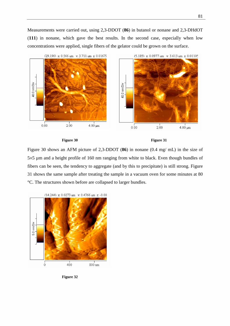
81
Measurements were carried out, using 2,3-DDOT (86) in butanol or nonane and 2,3-DHdOT
(111) in nonane, which gave the best results. In the second case, especially when low
concentrations were applied, single fibers of the gelator could be grown on the surface.
Figure 30 Figure 31
Figure 30 shows an AFM picture of 2,3-DDOT (86) in nonane (0.4 mg/ mL) in the size of
5×5 µm and a height profile of 160 nm ranging from white to black. Even though bundles of
fibers can be seen, the tendency to aggregate (and by this to precipitate) is still strong. Figure
31 shows the same sample after treating the sample in a vacuum oven for some minutes at 80
°C. The structures shown before are collapsed to larger bundles.
Figure 32
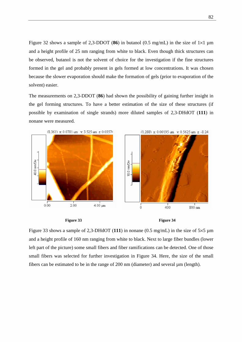
82
Figure 32 shows a sample of 2,3-DDOT (86) in butanol (0.5 mg/mL) in the size of 1×1 µm
and a height profile of 25 nm ranging from white to black. Even though thick structures can
be observed, butanol is not the solvent of choice for the investigation if the fine structures
formed in the gel and probably present in gels formed at low concentrations. It was chosen
because the slower evaporation should make the formation of gels (prior to evaporation of the
solvent) easier.
The measurements on 2,3-DDOT (86) had shown the possibility of gaining further insight in
the gel forming structures. To have a better estimation of the size of these structures (if
possible by examination of single strands) more diluted samples of 2,3-DHdOT (111) in
nonane were measured.
Figure 33 Figure 34
Figure 33 shows a sample of 2,3-DHdOT (111) in nonane (0.5 mg/mL) in the size of 5×5 µm
and a height profile of 160 nm ranging from white to black. Next to large fiber bundles (lower
left part of the picture) some small fibers and fiber ramifications can be detected. One of those
small fibers was selected for further investigation in Figure 34. Here, the size of the small
fibers can be estimated to be in the range of 200 nm (diameter) and several µm (length).
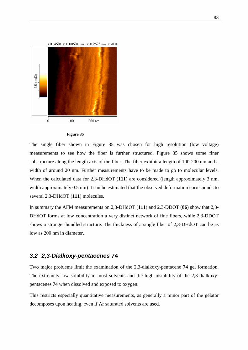
83
Figure 35
The single fiber shown in Figure 35 was chosen for high resolution (low voltage)
measurements to see how the fiber is further structured. Figure 35 shows some finer
substructure along the length axis of the fiber. The fiber exhibit a length of 100-200 nm and a
width of around 20 nm. Further measurements have to be made to go to molecular levels.
When the calculated data for 2,3-DHdOT (111) are considered (length approximately 3 nm,
width approximately 0.5 nm) it can be estimated that the observed deformation corresponds to
several 2,3-DHdOT (111) molecules.
In summary the AFM measurements on 2,3-DHdOT (111) and 2,3-DDOT (86) show that 2,3-
DHdOT forms at low concentration a very distinct network of fine fibers, while 2,3-DDOT
shows a stronger bundled structure. The thickness of a single fiber of 2,3-DHdOT can be as
low as 200 nm in diameter.
3.2 2,3-Dialkoxy-pentacenes 74
Two major problems limit the examination of the 2,3-dialkoxy-pentacene 74 gel formation.
The extremely low solubility in most solvents and the high instability of the 2,3-dialkoxy-
pentacenes 74 when dissolved and exposed to oxygen.
This restricts especially quantitative measurements, as generally a minor part of the gelator
decomposes upon heating, even if Ar saturated solvents are used.
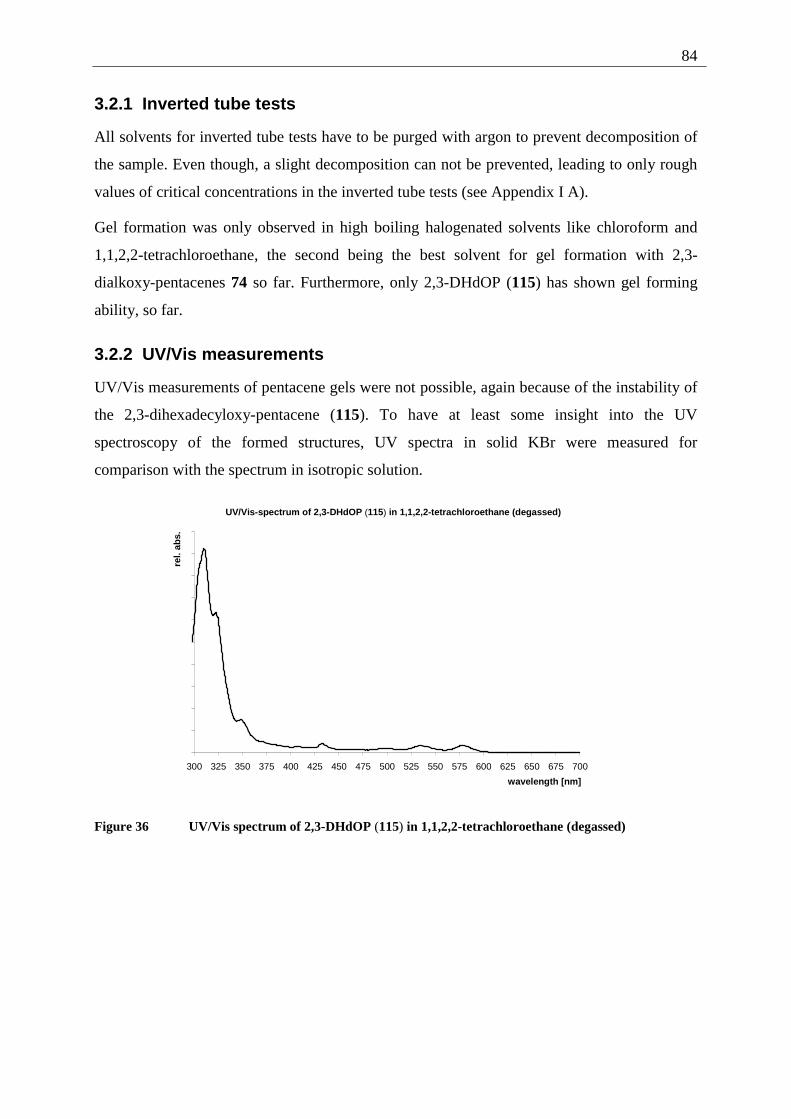
84
3.2.1 Inverted tube tests
All solvents for inverted tube tests have to be purged with argon to prevent decomposition of
the sample. Even though, a slight decomposition can not be prevented, leading to only rough
values of critical concentrations in the inverted tube tests (see Appendix I A).
Gel formation was only observed in high boiling halogenated solvents like chloroform and
1,1,2,2-tetrachloroethane, the second being the best solvent for gel formation with 2,3-
dialkoxy-pentacenes 74 so far. Furthermore, only 2,3-DHdOP (115) has shown gel forming
ability, so far.
3.2.2 UV/Vis measurements
UV/Vis measurements of pentacene gels were not possible, again because of the instability of
the 2,3-dihexadecyloxy-pentacene (115). To have at least some insight into the UV
spectroscopy of the formed structures, UV spectra in solid KBr were measured for
comparison with the spectrum in isotropic solution.
UV/Vis-spectrum of 2,3-DHdOP (115) in 1,1,2,2-tetrachloroethane (degassed)
300 325 350 375 400 425 450 475 500 525 550 575 600 625 650 675 700
wavelength [nm]
rel.
abs.
Figure 36 UV/Vis spectrum of 2,3-DHdOP (115) in 1,1,2,2-tetrachloroethane (degassed)
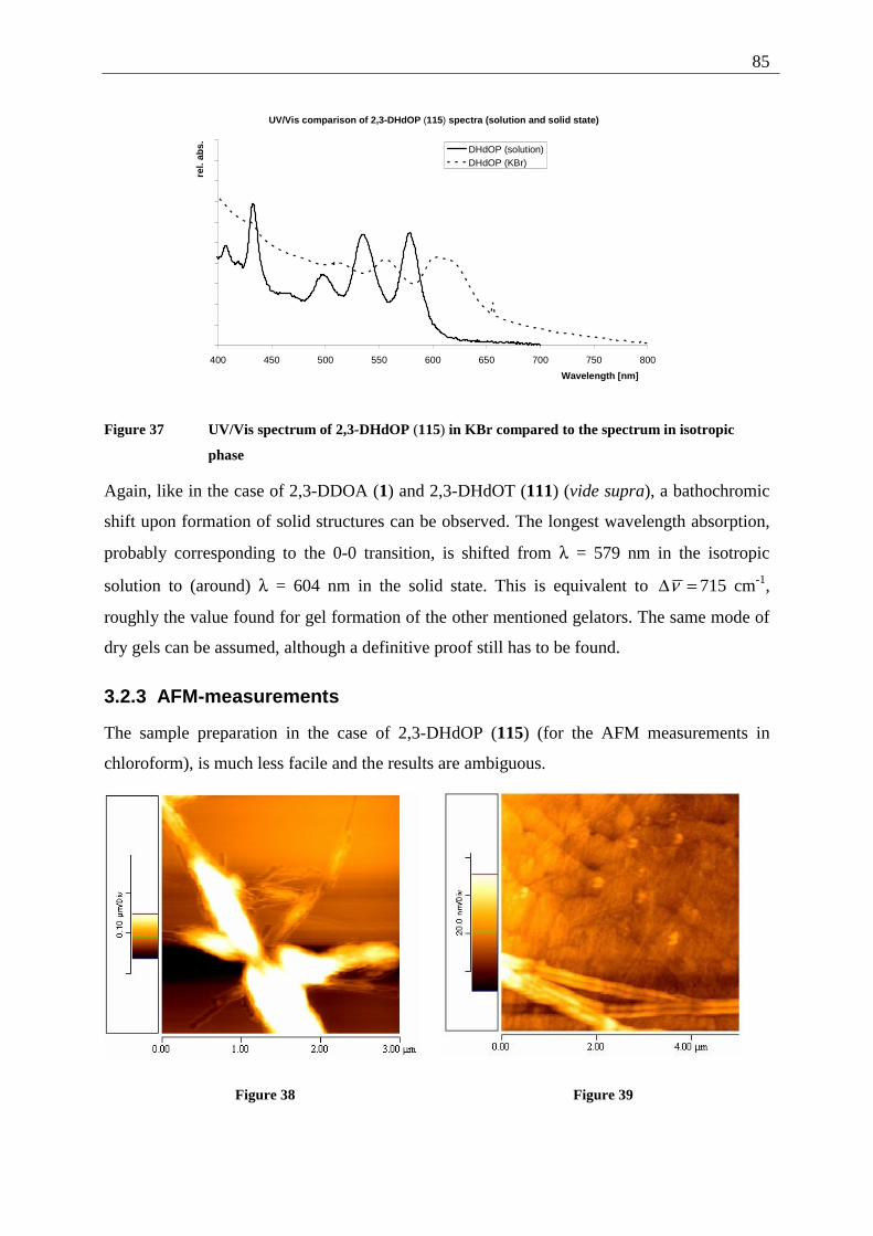
85
UV/Vis comparison of 2,3-DHdOP (115) spectra (solution and solid state)
400 450 500 550 600 650 700 750 800
Wavelength [nm]
rel.
abs. DHdOP (solution)
DHdOP (KBr)
Figure 37 UV/Vis spectrum of 2,3-DHdOP (115) in KBr compared to the spectrum in isotropic
phase
Again, like in the case of 2,3-DDOA (1) and 2,3-DHdOT (111) (vide supra), a bathochromic
shift upon formation of solid structures can be observed. The longest wavelength absorption,
probably corresponding to the 0-0 transition, is shifted from λ = 579 nm in the isotropic
solution to (around) λ = 604 nm in the solid state. This is equivalent to =∆ν 715 cm-1,
roughly the value found for gel formation of the other mentioned gelators. The same mode of
dry gels can be assumed, although a definitive proof still has to be found.
3.2.3 AFM-measurements
The sample preparation in the case of 2,3-DHdOP (115) (for the AFM measurements in
chloroform), is much less facile and the results are ambiguous.
Figure 38 Figure 39
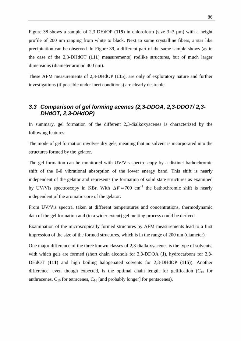
86
Figure 38 shows a sample of 2,3-DHdOP (115) in chloroform (size 3×3 µm) with a height
profile of 200 nm ranging from white to black. Next to some crystalline fibers, a star like
precipitation can be observed. In Figure 39, a different part of the same sample shows (as in
the case of the 2,3-DHdOT (111) measurements) rodlike structures, but of much larger
dimensions (diameter around 400 nm).
These AFM measurements of 2,3-DHdOP (115), are only of exploratory nature and further
investigations (if possible under inert conditions) are clearly desirable.
3.3 Comparison of gel forming acenes (2,3-DDOA, 2,3-DDOT/ 2,3-DHdOT, 2,3-DHdOP)
In summary, gel formation of the different 2,3-dialkoxyacenes is characterized by the
following features:
The mode of gel formation involves dry gels, meaning that no solvent is incorporated into the
structures formed by the gelator.
The gel formation can be monitored with UV/Vis spectroscopy by a distinct bathochromic
shift of the 0-0 vibrational absorption of the lower energy band. This shift is nearly
independent of the gelator and represents the formation of solid state structures as examined
by UV/Vis spectroscopy in KBr. With ≈∆ν 700 cm-1 the bathochromic shift is nearly
independent of the aromatic core of the gelator.
From UV/Vis spectra, taken at different temperatures and concentrations, thermodynamic
data of the gel formation and (to a wider extent) gel melting process could be derived.
Examination of the microscopically formed structures by AFM measurements lead to a first
impression of the size of the formed structures, which is in the range of 200 nm (diameter).
One major difference of the three known classes of 2,3-dialkoxyacenes is the type of solvents,
with which gels are formed (short chain alcohols for 2,3-DDOA (1), hydrocarbons for 2,3-
DHdOT (111) and high boiling halogenated solvents for 2,3-DHdOP (115)). Another
difference, even though expected, is the optimal chain length for gelification (C10 for
anthracenes, C16 for tetracenes, C16 [and probably longer] for pentacenes).
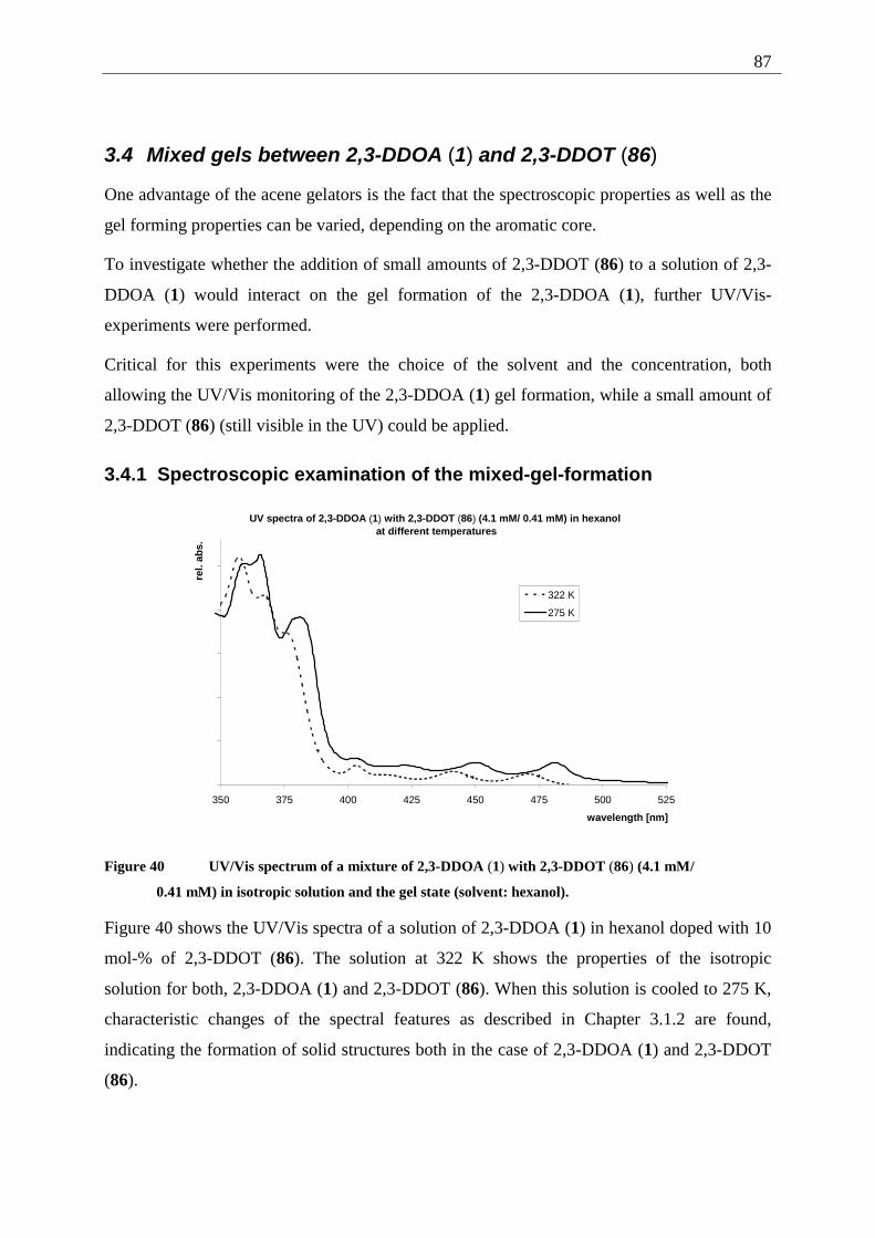
87
3.4 Mixed gels between 2,3-DDOA (1) and 2,3-DDOT (86)
One advantage of the acene gelators is the fact that the spectroscopic properties as well as the
gel forming properties can be varied, depending on the aromatic core.
To investigate whether the addition of small amounts of 2,3-DDOT (86) to a solution of 2,3-
DDOA (1) would interact on the gel formation of the 2,3-DDOA (1), further UV/Vis-
experiments were performed.
Critical for this experiments were the choice of the solvent and the concentration, both
allowing the UV/Vis monitoring of the 2,3-DDOA (1) gel formation, while a small amount of
2,3-DDOT (86) (still visible in the UV) could be applied.
3.4.1 Spectroscopic examination of the mixed-gel-formation
UV spectra of 2,3-DDOA (1) with 2,3-DDOT (86) (4.1 mM/ 0.41 mM) in hexanol at different temperatures
350 375 400 425 450 475 500 525
wavelength [nm]
rel.
abs.
322 K
275 K
Figure 40 UV/Vis spectrum of a mixture of 2,3-DDOA (1) with 2,3-DDOT (86) (4.1 mM/
0.41 mM) in isotropic solution and the gel state (solvent: hexanol).
Figure 40 shows the UV/Vis spectra of a solution of 2,3-DDOA (1) in hexanol doped with 10
mol-% of 2,3-DDOT (86). The solution at 322 K shows the properties of the isotropic
solution for both, 2,3-DDOA (1) and 2,3-DDOT (86). When this solution is cooled to 275 K,
characteristic changes of the spectral features as described in Chapter 3.1.2 are found,
indicating the formation of solid structures both in the case of 2,3-DDOA (1) and 2,3-DDOT
(86).
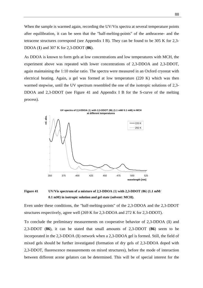
88
When the sample is warmed again, recording the UV/Vis spectra at several temperature points
after equilibration, it can be seen that the "half-melting-points" of the anthracene- and the
tetracene structures correspond (see Appendix I B). They can be found to be 305 K for 2,3-
DDOA (1) and 307 K for 2,3-DDOT (86).
As DDOA is known to form gels at low concentrations and low temperatures with MCH, the
experiment above was repeated with lower concentrations of 2,3-DDOA and 2,3-DDOT,
again maintaining the 1:10 molar ratio. The spectra were measured in an Oxford cryostat with
electrical heating. Again, a gel was formed at low temperature (220 K) which was then
warmed stepwise, until the UV spectrum resembled the one of the isotropic solutions of 2,3-
DDOA and 2,3-DDOT (see Figure 41 and Appendix I B for the S-curve of the melting
process).
UV spectra of 2,3-DDOA (1) with 2,3-DDOT (86) (1.1 mM/ 0.1 mM) in MCH at different temperatures
350 375 400 425 450 475 500 525
wavelength [nm]
rel.
abs.
220 K
292 K
Figure 41 UV/Vis spectrum of a mixture of 2,3-DDOA (1) with 2,3-DDOT (86) (1.1 mM/
0.1 mM) in isotropic solution and gel state (solvent: MCH).
Even under these conditions, the "half-melting-points" of the 2,3-DDOA and the 2,3-DDOT
structures respectively, agree well (269 K for 2,3-DDOA and 272 K for 2,3-DDOT).
To conclude the preliminary measurements on cooperative behavior of 2,3-DDOA (1) and
2,3-DDOT (86), it can be stated that small amounts of 2,3-DDOT (86) seem to be
incorporated in the 2,3-DDOA (1) network when a 2,3-DDOA gel is formed. Still, the field of
mixed gels should be further investigated (formation of dry gels of 2,3-DDOA doped with
2,3-DDOT, fluorescence measurements on mixed structures), before the mode of interaction
between different acene gelators can be determined. This will be of special interest for the
89
investigation of energy transfer between the different aromatic cores, one possible application
of mixed gels.
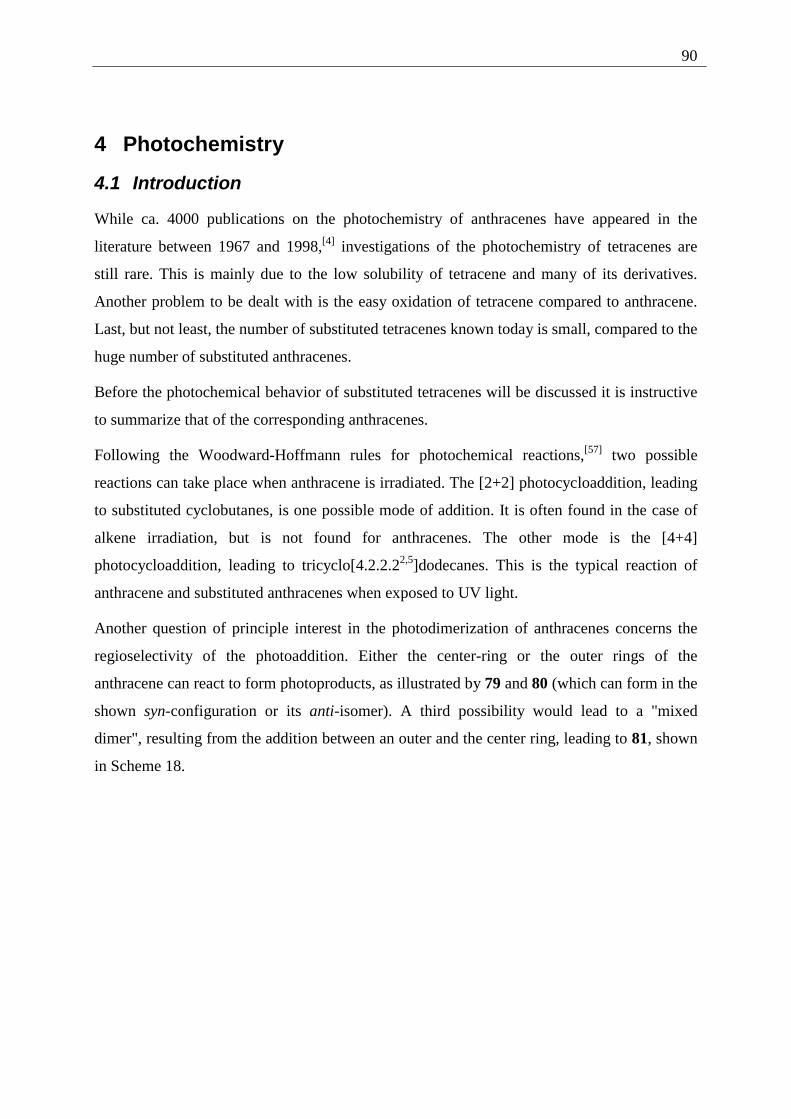
90
4 Photochemistry
4.1 Introduction
While ca. 4000 publications on the photochemistry of anthracenes have appeared in the
literature between 1967 and 1998,[4] investigations of the photochemistry of tetracenes are
still rare. This is mainly due to the low solubility of tetracene and many of its derivatives.
Another problem to be dealt with is the easy oxidation of tetracene compared to anthracene.
Last, but not least, the number of substituted tetracenes known today is small, compared to the
huge number of substituted anthracenes.
Before the photochemical behavior of substituted tetracenes will be discussed it is instructive
to summarize that of the corresponding anthracenes.
Following the Woodward-Hoffmann rules for photochemical reactions,[57] two possible
reactions can take place when anthracene is irradiated. The [2+2] photocycloaddition, leading
to substituted cyclobutanes, is one possible mode of addition. It is often found in the case of
alkene irradiation, but is not found for anthracenes. The other mode is the [4+4]
photocycloaddition, leading to tricyclo[4.2.2.22,5]dodecanes. This is the typical reaction of
anthracene and substituted anthracenes when exposed to UV light.
Another question of principle interest in the photodimerization of anthracenes concerns the
regioselectivity of the photoaddition. Either the center-ring or the outer rings of the
anthracene can react to form photoproducts, as illustrated by 79 and 80 (which can form in the
shown syn-configuration or its anti-isomer). A third possibility would lead to a "mixed
dimer", resulting from the addition between an outer and the center ring, leading to 81, shown
in Scheme 18.
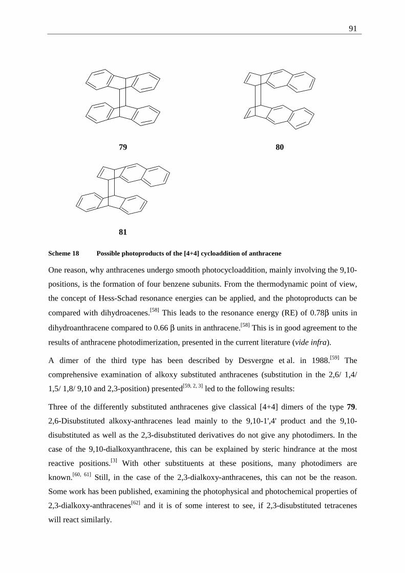
91
79 80
81
Scheme 18 Possible photoproducts of the [4+4] cycloaddition of anthracene
One reason, why anthracenes undergo smooth photocycloaddition, mainly involving the 9,10-
positions, is the formation of four benzene subunits. From the thermodynamic point of view,
the concept of Hess-Schad resonance energies can be applied, and the photoproducts can be
compared with dihydroacenes.[58] This leads to the resonance energy (RE) of 0.78β units in
dihydroanthracene compared to 0.66 β units in anthracene.[58] This is in good agreement to the
results of anthracene photodimerization, presented in the current literature (vide infra).
A dimer of the third type has been described by Desvergne et al. in 1988.[59] The
comprehensive examination of alkoxy substituted anthracenes (substitution in the 2,6/ 1,4/
1,5/ 1,8/ 9,10 and 2,3-position) presented[59, 2, 3] led to the following results:
Three of the differently substituted anthracenes give classical [4+4] dimers of the type 79.
2,6-Disubstituted alkoxy-anthracenes lead mainly to the 9,10-1',4' product and the 9,10-
disubstituted as well as the 2,3-disubstituted derivatives do not give any photodimers. In the
case of the 9,10-dialkoxyanthracene, this can be explained by steric hindrance at the most
reactive positions.[3] With other substituents at these positions, many photodimers are
known.[60, 61] Still, in the case of the 2,3-dialkoxy-anthracenes, this can not be the reason.
Some work has been published, examining the photophysical and photochemical properties of
2,3-dialkoxy-anthracenes[62] and it is of some interest to see, if 2,3-disubstituted tetracenes
will react similarly.
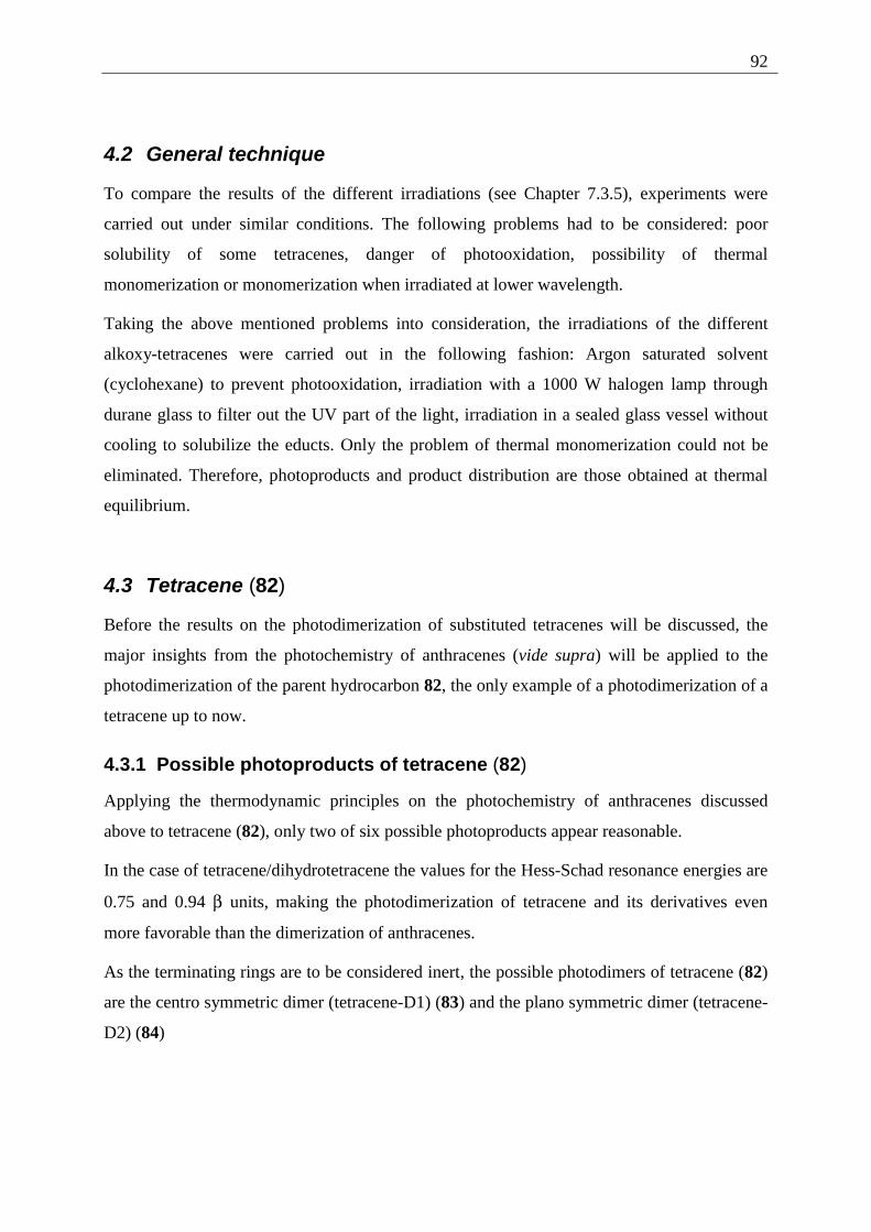
92
4.2 General technique
To compare the results of the different irradiations (see Chapter 7.3.5), experiments were
carried out under similar conditions. The following problems had to be considered: poor
solubility of some tetracenes, danger of photooxidation, possibility of thermal
monomerization or monomerization when irradiated at lower wavelength.
Taking the above mentioned problems into consideration, the irradiations of the different
alkoxy-tetracenes were carried out in the following fashion: Argon saturated solvent
(cyclohexane) to prevent photooxidation, irradiation with a 1000 W halogen lamp through
durane glass to filter out the UV part of the light, irradiation in a sealed glass vessel without
cooling to solubilize the educts. Only the problem of thermal monomerization could not be
eliminated. Therefore, photoproducts and product distribution are those obtained at thermal
equilibrium.
4.3 Tetracene (82)
Before the results on the photodimerization of substituted tetracenes will be discussed, the
major insights from the photochemistry of anthracenes (vide supra) will be applied to the
photodimerization of the parent hydrocarbon 82, the only example of a photodimerization of a
tetracene up to now.
4.3.1 Possible photoproducts of tetracene (82)
Applying the thermodynamic principles on the photochemistry of anthracenes discussed
above to tetracene (82), only two of six possible photoproducts appear reasonable.
In the case of tetracene/dihydrotetracene the values for the Hess-Schad resonance energies are
0.75 and 0.94 β units, making the photodimerization of tetracene and its derivatives even
more favorable than the dimerization of anthracenes.
As the terminating rings are to be considered inert, the possible photodimers of tetracene (82)
are the centro symmetric dimer (tetracene-D1) (83) and the plano symmetric dimer (tetracene-
D2) (84)
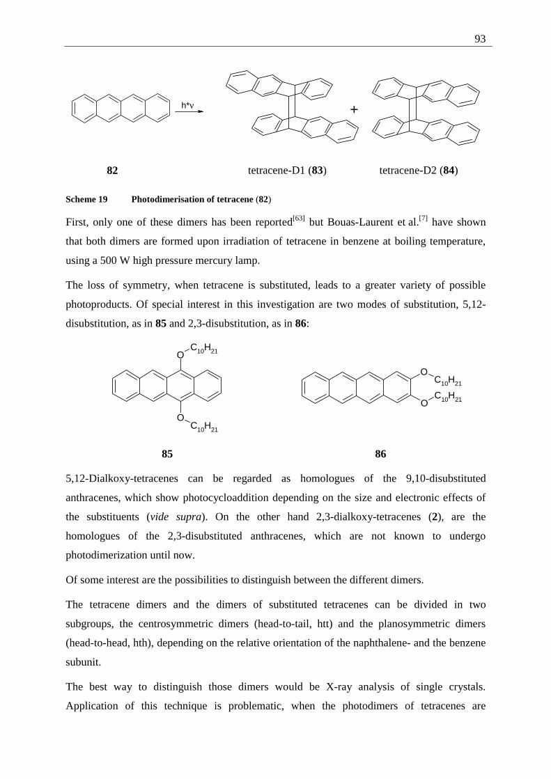
93
h*ν
+
82 tetracene-D1 (83) tetracene-D2 (84)
Scheme 19 Photodimerisation of tetracene (82)
First, only one of these dimers has been reported[63] but Bouas-Laurent et al.[7] have shown
that both dimers are formed upon irradiation of tetracene in benzene at boiling temperature,
using a 500 W high pressure mercury lamp.
The loss of symmetry, when tetracene is substituted, leads to a greater variety of possible
photoproducts. Of special interest in this investigation are two modes of substitution, 5,12-
disubstitution, as in 85 and 2,3-disubstitution, as in 86:
O
O
C10H21
C10H21
O
O
C10H21
C10H21
85 86
5,12-Dialkoxy-tetracenes can be regarded as homologues of the 9,10-disubstituted
anthracenes, which show photocycloaddition depending on the size and electronic effects of
the substituents (vide supra). On the other hand 2,3-dialkoxy-tetracenes (2), are the
homologues of the 2,3-disubstituted anthracenes, which are not known to undergo
photodimerization until now.
Of some interest are the possibilities to distinguish between the different dimers.
The tetracene dimers and the dimers of substituted tetracenes can be divided in two
subgroups, the centrosymmetric dimers (head-to-tail, htt) and the planosymmetric dimers
(head-to-head, hth), depending on the relative orientation of the naphthalene- and the benzene
subunit.
The best way to distinguish those dimers would be X-ray analysis of single crystals.
Application of this technique is problematic, when the photodimers of tetracenes are
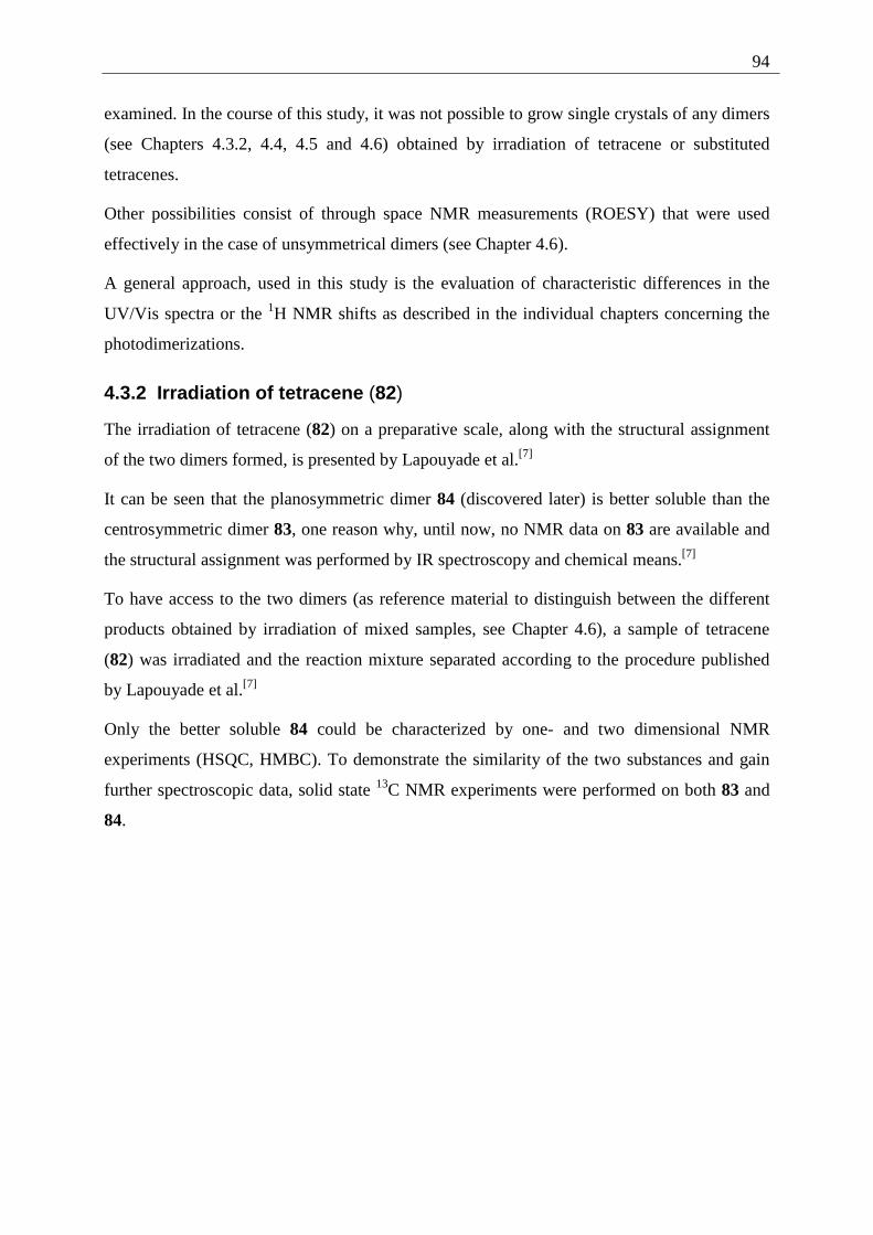
94
examined. In the course of this study, it was not possible to grow single crystals of any dimers
(see Chapters 4.3.2, 4.4, 4.5 and 4.6) obtained by irradiation of tetracene or substituted
tetracenes.
Other possibilities consist of through space NMR measurements (ROESY) that were used
effectively in the case of unsymmetrical dimers (see Chapter 4.6).
A general approach, used in this study is the evaluation of characteristic differences in the
UV/Vis spectra or the 1H NMR shifts as described in the individual chapters concerning the
photodimerizations.
4.3.2 Irradiation of tetracene (82)
The irradiation of tetracene (82) on a preparative scale, along with the structural assignment
of the two dimers formed, is presented by Lapouyade et al.[7]
It can be seen that the planosymmetric dimer 84 (discovered later) is better soluble than the
centrosymmetric dimer 83, one reason why, until now, no NMR data on 83 are available and
the structural assignment was performed by IR spectroscopy and chemical means.[7]
To have access to the two dimers (as reference material to distinguish between the different
products obtained by irradiation of mixed samples, see Chapter 4.6), a sample of tetracene
(82) was irradiated and the reaction mixture separated according to the procedure published
by Lapouyade et al.[7]
Only the better soluble 84 could be characterized by one- and two dimensional NMR
experiments (HSQC, HMBC). To demonstrate the similarity of the two substances and gain
further spectroscopic data, solid state 13C NMR experiments were performed on both 83 and
84.
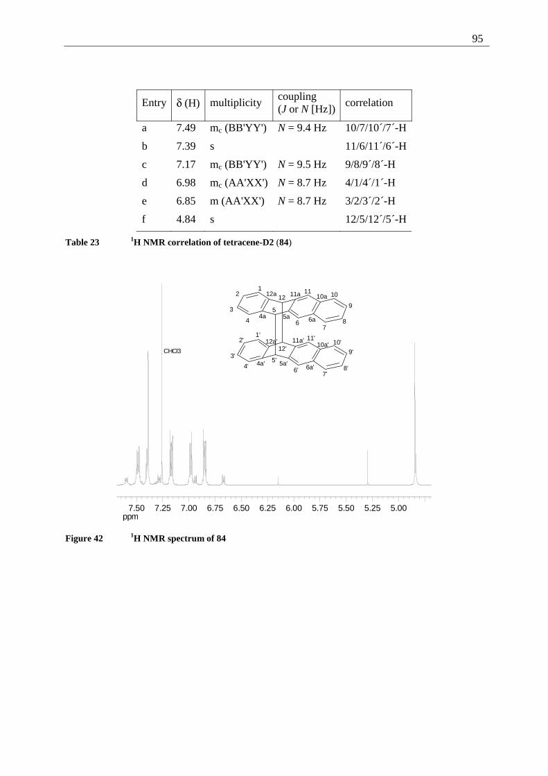
95
Entry δ (H) multiplicity coupling (J or N [Hz])
correlation
a 7.49 mc (BB'YY') N = 9.4 Hz 10/7/10´/7´-H
b 7.39 s 11/6/11´/6´-H
c 7.17 mc (BB'YY') N = 9.5 Hz 9/8/9´/8´-H
d 6.98 mc (AA'XX') N = 8.7 Hz 4/1/4´/1´-H
e 6.85 m (AA'XX') N = 8.7 Hz 3/2/3´/2´-H
f 4.84 s 12/5/12´/5´-H
Table 23 1H NMR correlation of tetracene-D2 (84)
ppm5.005.255.505.756.006.256.506.757.007.257.50
CHCl3
12
3
4
5
67
8
9
101112
4a 5a 6a
10a11a12a
1'2'
3'
4'5'
6'7'
8'
9'
10'11'
12'
4a' 5a'6a'
10a'11a'12a'
Figure 42 1H NMR spectrum of 84
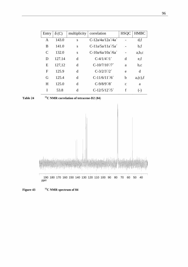
96
Entry δ (C) multiplicity correlation HSQC HMBC
A 143.0 s C-12a/4a/12a´/4a´ - d,f
B 141.0 s C-11a/5a/11a´/5a´ - b,f
C 132.0 s C-10a/6a/10a´/6a` - a,b,c
D 127.14 d C-4/1/4´/1´ d e,f
E 127,12 d C-10/7/10´/7´ a b,c
F 125.9 d C-3/2/3´/2´ e d
G 125.4 d C-11/6/11´/6´ b a,(c),f
H 125.0 d C-9/8/9´/8´ c a
I 53.8 d C-12/5/12´/5´ f (-)
Table 24 13C NMR correlation of tetracene-D2 (84)
ppm405060708090100110120130140150160170180190
Figure 43 13C NMR spectrum of 84
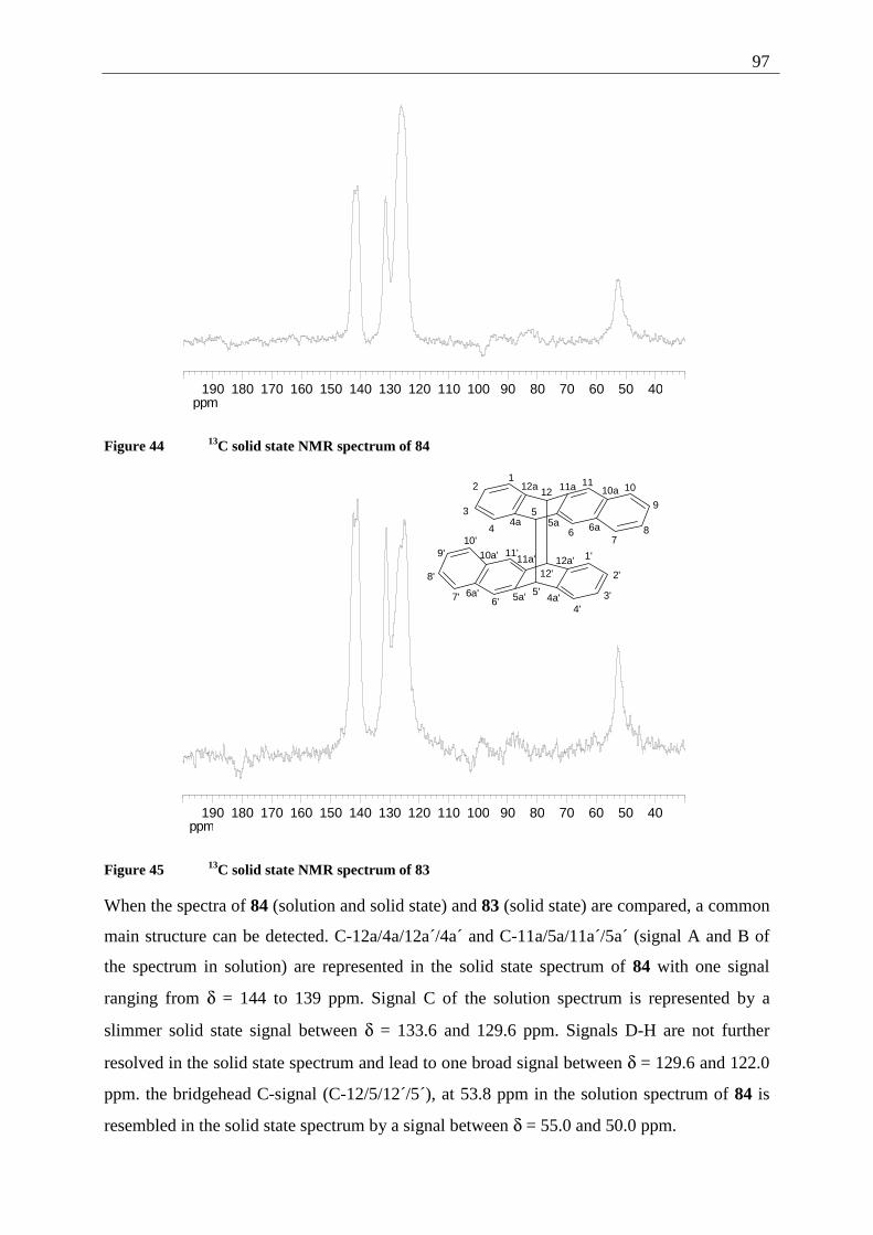
97
ppm405060708090100110120130140150160170180190
Figure 44 13C solid state NMR spectrum of 84
ppm405060708090100110120130140150160170180190
12
3
4
5
67
8
9
101112
4a 5a 6a
10a11a12a
1'
2'
3'4'
5'6'7'
8'
9'10'
11'
12'
4a'5a'6a'
10a' 11a' 12a'
Figure 45 13C solid state NMR spectrum of 83
When the spectra of 84 (solution and solid state) and 83 (solid state) are compared, a common
main structure can be detected. C-12a/4a/12a´/4a´ and C-11a/5a/11a´/5a´ (signal A and B of
the spectrum in solution) are represented in the solid state spectrum of 84 with one signal
ranging from δ = 144 to 139 ppm. Signal C of the solution spectrum is represented by a
slimmer solid state signal between δ = 133.6 and 129.6 ppm. Signals D-H are not further
resolved in the solid state spectrum and lead to one broad signal between δ = 129.6 and 122.0
ppm. the bridgehead C-signal (C-12/5/12´/5´), at 53.8 ppm in the solution spectrum of 84 is
resembled in the solid state spectrum by a signal between δ = 55.0 and 50.0 ppm.
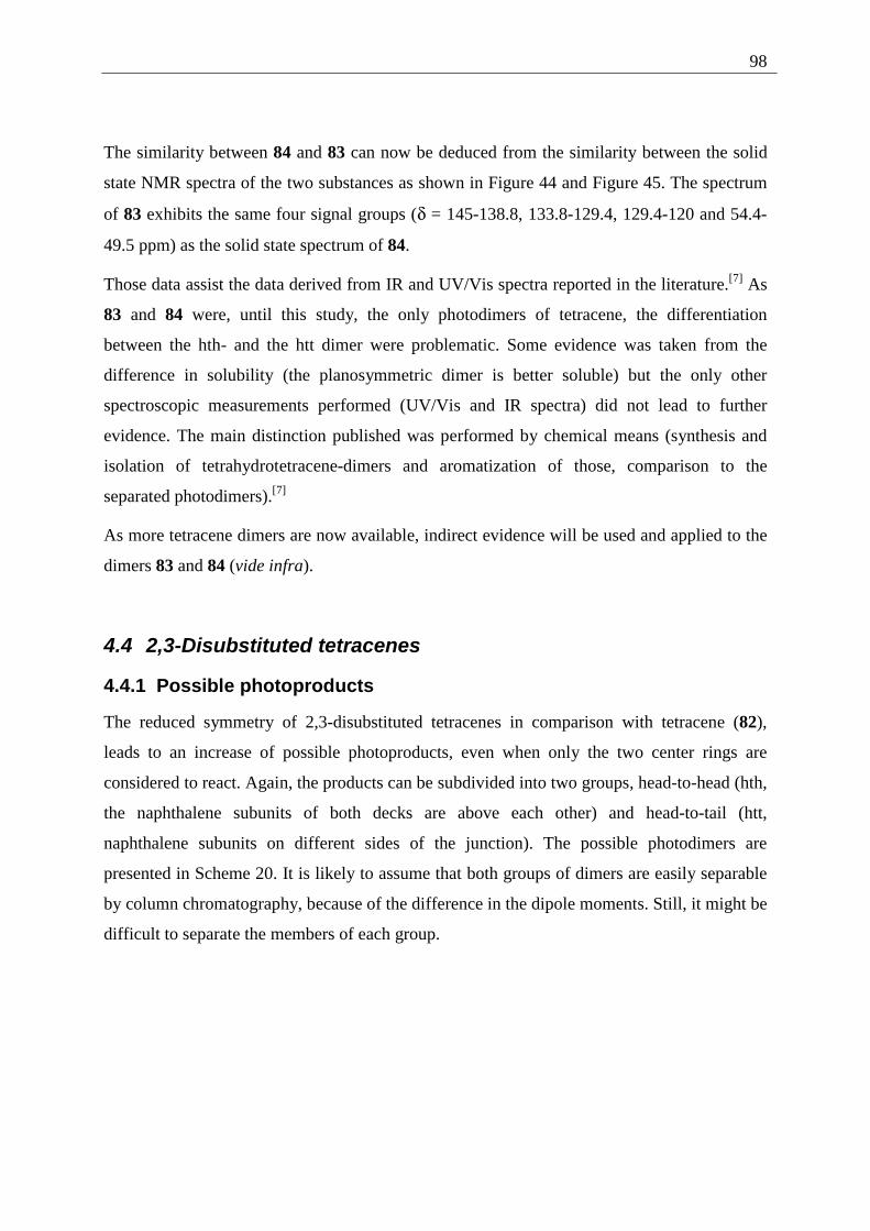
98
The similarity between 84 and 83 can now be deduced from the similarity between the solid
state NMR spectra of the two substances as shown in Figure 44 and Figure 45. The spectrum
of 83 exhibits the same four signal groups (δ = 145-138.8, 133.8-129.4, 129.4-120 and 54.4-
49.5 ppm) as the solid state spectrum of 84.
Those data assist the data derived from IR and UV/Vis spectra reported in the literature.[7] As
83 and 84 were, until this study, the only photodimers of tetracene, the differentiation
between the hth- and the htt dimer were problematic. Some evidence was taken from the
difference in solubility (the planosymmetric dimer is better soluble) but the only other
spectroscopic measurements performed (UV/Vis and IR spectra) did not lead to further
evidence. The main distinction published was performed by chemical means (synthesis and
isolation of tetrahydrotetracene-dimers and aromatization of those, comparison to the
separated photodimers).[7]
As more tetracene dimers are now available, indirect evidence will be used and applied to the
dimers 83 and 84 (vide infra).
4.4 2,3-Disubstituted tetracenes
4.4.1 Possible photoproducts
The reduced symmetry of 2,3-disubstituted tetracenes in comparison with tetracene (82),
leads to an increase of possible photoproducts, even when only the two center rings are
considered to react. Again, the products can be subdivided into two groups, head-to-head (hth,
the naphthalene subunits of both decks are above each other) and head-to-tail (htt,
naphthalene subunits on different sides of the junction). The possible photodimers are
presented in Scheme 20. It is likely to assume that both groups of dimers are easily separable
by column chromatography, because of the difference in the dipole moments. Still, it might be
difficult to separate the members of each group.
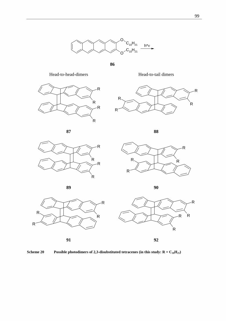
99
h*νO
O
C10H21
C10H21
86
Head-to-head-dimers Head-to-tail dimers
R
RR
R
R
RR
R
87 88
R
RR
R
R
R
R
R
89 90
R
R
R
R
R
RR
R
91 92
Scheme 20 Possible photodimers of 2,3-disubstituted tetracenes (in this study: R = C10H21)
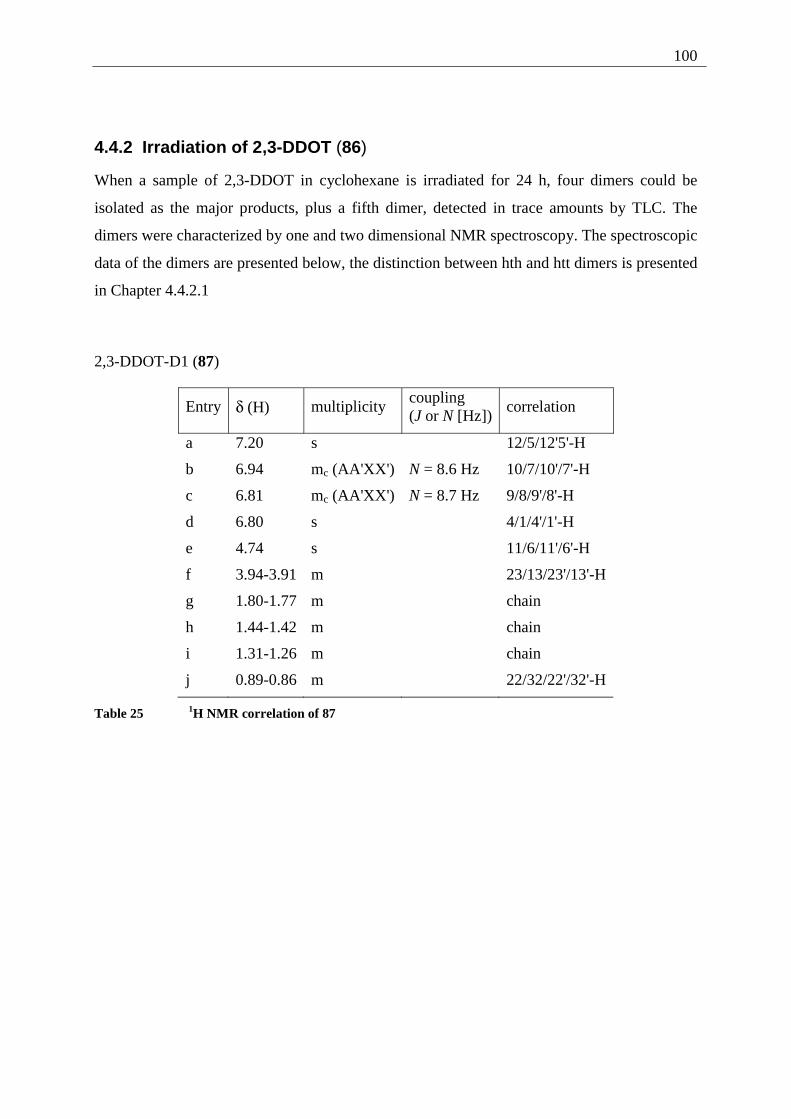
100
4.4.2 Irradiation of 2,3-DDOT (86)
When a sample of 2,3-DDOT in cyclohexane is irradiated for 24 h, four dimers could be
isolated as the major products, plus a fifth dimer, detected in trace amounts by TLC. The
dimers were characterized by one and two dimensional NMR spectroscopy. The spectroscopic
data of the dimers are presented below, the distinction between hth and htt dimers is presented
in Chapter 4.4.2.1
2,3-DDOT-D1 (87)
Entry δ (H) multiplicity coupling (J or N [Hz])
correlation
a 7.20 s 12/5/12'5'-H
b 6.94 mc (AA'XX') N = 8.6 Hz 10/7/10'/7'-H
c 6.81 mc (AA'XX') N = 8.7 Hz 9/8/9'/8'-H
d 6.80 s 4/1/4'/1'-H
e 4.74 s 11/6/11'/6'-H
f 3.94-3.91 m 23/13/23'/13'-H
g 1.80-1.77 m chain
h 1.44-1.42 m chain
i 1.31-1.26 m chain
j 0.89-0.86 m 22/32/22'/32'-H
Table 25 1H NMR correlation of 87
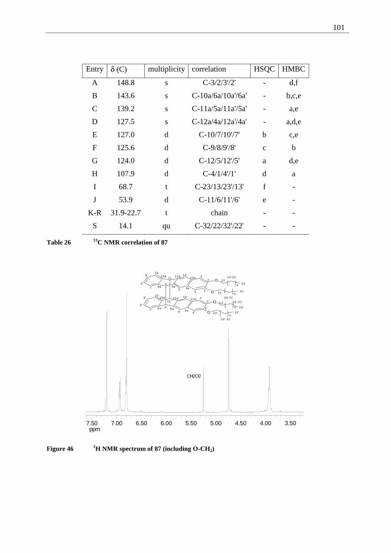
101
Entry δ (C) multiplicity correlation HSQC HMBC
A 148.8 s C-3/2/3'/2' - d,f
B 143.6 s C-10a/6a/10a'/6a' - b,c,e
C 139.2 s C-11a/5a/11a'/5a' - a,e
D 127.5 s C-12a/4a/12a'/4a' - a,d,e
E 127.0 d C-10/7/10'/7' b c,e
F 125.6 d C-9/8/9'/8' c b
G 124.0 d C-12/5/12'/5' a d,e
H 107.9 d C-4/1/4'/1' d a
I 68.7 t C-23/13/23'/13' f -
J 53.9 d C-11/6/11'/6' e -
K-R 31.9-22.7 t chain - -
S 14.1 qu C-32/22/32'/22' - -
Table 26 13C NMR correlation of 87
ppm3.504.004.505.005.506.006.507.007.50
CH2Cl2
O
O
8
8
O
O8
8
12
345
67
8
910
1112
1314-21
22
23
24-31
324a5a6a
10a 11a 12a
1'2 '
3 '4 '5 '
6 '7'
8 '
9 '10'
11 '
12 '
13 ' 14 '-21 '
22 '
23 '
24 '-31'
32 '4a '
5a '6a '
10a' 11a' 12a'
Figure 46 1H NMR spectrum of 87 (including O-CH2)
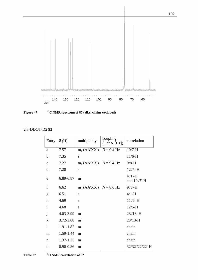
102
ppm60708090100110120130140
Figure 47 13C NMR spectrum of 87 (alkyl chains excluded)
2,3-DDOT-D2 92
Entry δ (H) multiplicity coupling (J or N [Hz])
correlation
a 7.57 mc (AA'XX') N = 9.4 Hz 10/7-H
b 7.35 s 11/6-H
c 7.27 mc (AA'XX') N = 9.4 Hz 9/8-H
d 7.20 s 12'/5'-H
e 6.89-6.87 m 4'/1'-H
and 10'/7'-H
f 6.62 mc (AA'XX') N = 8.6 Hz 9'/8'-H
g 6.51 s 4/1-H
h 4.69 s 11'/6'-H
i 4.68 s 12/5-H
j 4.03-3.99 m 23'/13'-H
k 3.72-3.68 m 23/13-H
l 1.91-1.82 m chain
m 1.59-1.44 m chain
n 1.37-1.25 m chain
o 0.90-0.86 m 32/32'/22/22'-H
Table 27 1H NMR correlation of 92
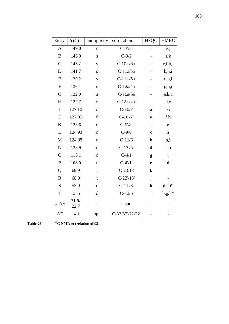
103
Entry δ (C) multiplicity correlation HSQC HMBC
A 149.0 s C-3'/2' - e,j
B 146.9 s C-3/2 - g,k
C 143.2 s C-10a'/6a' - e,f,h,i
D 141.7 s C-11a/5a - b,h,i
E 139.2 s C-11a'/5a' - d,h,i
F 136.1 s C-12a/4a - g,h,i
G 132.0 s C-10a/6a - a,b,c
H 127.7 s C-12a'/4a' - d,e
I 127.10 d C-10/7 a b,c
J 127.05 d C-10'/7' e f,h
K 125.6 d C-9'/8' f e
L 124.93 d C-9/8 c a
M 124.88 d C-11/6 b a,i
N 123.9 d C-12'/5' d e,h
O 115.1 d C-4/1 g i
P 108.0 d C-4'/1' e d
Q 69.9 t C-23/13 k -
R 68.9 t C-23'/13' j -
S 53.9 d C-11'/6' h d,e,i*
T 53.5 d C-12/5 i b,g,h*
U-AE 31.9- 22.7
t chain - -
AF 14.1 qu C-32/32'/22/22' - -
Table 28 13C NMR correlation of 92
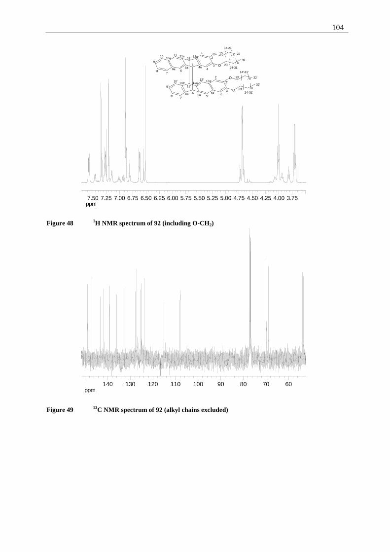
104
ppm3.754.004.254.504.755.005.255.505.756.006.256.506.757.007.257.50
O
O
O
O
8
8
8
81
2
34
5
67
8
9
10 1112
13
14-21
22
2324-31
32
4a5a6a
10a11a 12a
1'
2'
3'4'5'
6'
7'8'
9'
10'
11'
12'13'
14'-21'
22'
23'24'-31'
32'
4a'5a'6a'
10a' 11a'12a'
Figure 48 1H NMR spectrum of 92 (including O-CH2)
ppm60708090100110120130140
Figure 49 13C NMR spectrum of 92 (alkyl chains excluded)
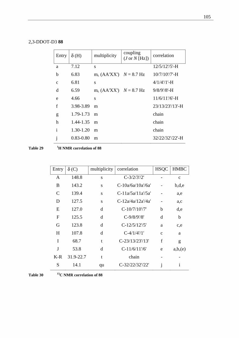
105
2,3-DDOT-D3 88
Entry δ (H) multiplicity coupling (J or N [Hz])
correlation
a 7.12 s 12/5/12'/5'-H
b 6.83 mc (AA'XX') N = 8.7 Hz 10/7/10'/7'-H
c 6.81 s 4/1/4'/1'-H
d 6.59 mc (AA'XX') N = 8.7 Hz 9/8/9'/8'-H
e 4.66 s 11/6/11'/6'-H
f 3.98-3.89 m 23/13/23'/13'-H
g 1.79-1.73 m chain
h 1.44-1.35 m chain
i 1.30-1.20 m chain
j 0.83-0.80 m 32/22/32'/22'-H
Table 29 1H NMR correlation of 88
Entry δ (C) multiplicity correlation HSQC HMBC
A 148.8 s C-3/2/3'/2' - c
B 143.2 s C-10a/6a/10a'/6a' - b,d,e
C 139.4 s C-11a/5a/11a'/5a' - a,e
D 127.5 s C-12a/4a/12a'/4a' - a,c
E 127.0 d C-10/7/10'/7' b d,e
F 125.5 d C-9/8/9'/8' d b
G 123.8 d C-12/5/12'/5' a c,e
H 107.8 d C-4/1/4'/1' c a
I 68.7 t C-23/13/23'/13' f g
J 53.8 d C-11/6/11'/6' e a,b,(e)
K-R 31.9-22.7 t chain - -
S 14.1 qu C-32/22/32'/22' j i
Table 30 13C NMR correlation of 88
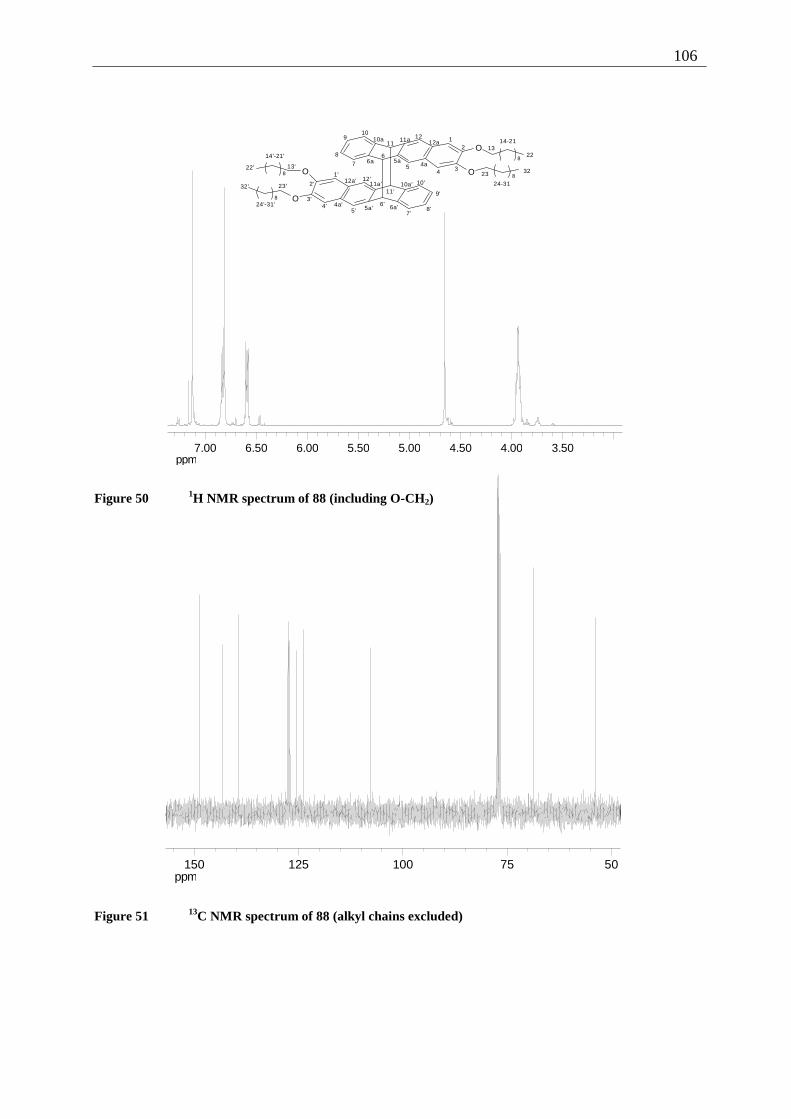
106
ppm3.504.004.505.005.506.006.507.00
O
OO
O
8
88
8
12
345
67
8
910
1112
1314-21
22
23
24-31
324a
5a6a
10a 11a 12a
1'
2'
3'4'
5'6'
7'8'
9'
10'
11'
12'
13'
14'-21'
22'
23'
24'-31'
32'
4a'5a' 6a'
10a'11a'12a'
Figure 50 1H NMR spectrum of 88 (including O-CH2)
ppm5075100125150
Figure 51 13C NMR spectrum of 88 (alkyl chains excluded)
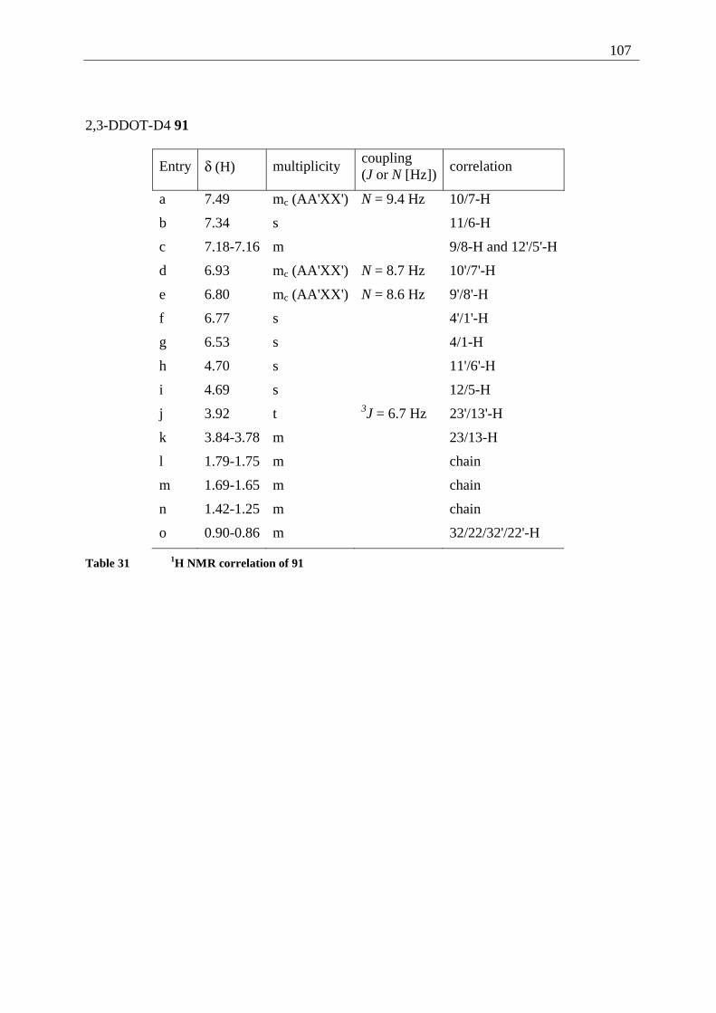
107
2,3-DDOT-D4 91
Entry δ (H) multiplicity coupling (J or N [Hz])
correlation
a 7.49 mc (AA'XX') N = 9.4 Hz 10/7-H
b 7.34 s 11/6-H
c 7.18-7.16 m 9/8-H and 12'/5'-H
d 6.93 mc (AA'XX') N = 8.7 Hz 10'/7'-H
e 6.80 mc (AA'XX') N = 8.6 Hz 9'/8'-H
f 6.77 s 4'/1'-H
g 6.53 s 4/1-H
h 4.70 s 11'/6'-H
i 4.69 s 12/5-H
j 3.92 t 3J = 6.7 Hz 23'/13'-H
k 3.84-3.78 m 23/13-H
l 1.79-1.75 m chain
m 1.69-1.65 m chain
n 1.42-1.25 m chain
o 0.90-0.86 m 32/22/32'/22'-H
Table 31 1H NMR correlation of 91
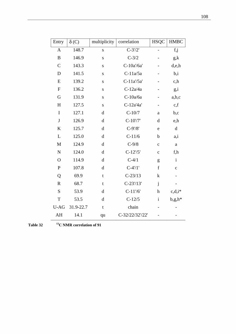
108
Entry δ (C) multiplicity correlation HSQC HMBC
A 148.7 s C-3'/2' - f,j
B 146.9 s C-3/2 - g,k
C 143.3 s C-10a'/6a' - d,e,h
D 141.5 s C-11a/5a - b,i
E 139.2 s C-11a'/5a' - c,h
F 136.2 s C-12a/4a - g,i
G 131.9 s C-10a/6a - a,b,c
H 127.5 s C-12a'4a' - c,f
I 127.1 d C-10/7 a b,c
J 126.9 d C-10'/7' d e,h
K 125.7 d C-9'/8' e d
L 125.0 d C-11/6 b a,i
M 124.9 d C-9/8 c a
N 124.0 d C-12'/5' c f,h
O 114.9 d C-4/1 g i
P 107.8 d C-4'/1' f c
Q 69.9 t C-23/13 k -
R 68.7 t C-23'/13' j -
S 53.9 d C-11'/6' h c,d,i*
T 53.5 d C-12/5 i b,g,h*
U-AG 31.9-22.7 t chain - -
AH 14.1 qu C-32/22/32'/22' - -
Table 32 13C NMR correlation of 91
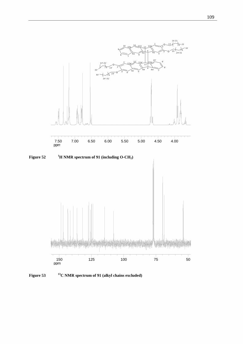
109
ppm4.004.505.005.506.006.507.007.50
O
O
8
8
O
O
8
8
1
2
34
5
67
8
9
10 1112
13
14-21
22
2324 -31
32
4a5a6a
10a11a 12a
1'
2 '
3 ' 4 '5 '
6 '7 '
8 '
9 '10 '
11 '
12 '
13 '
14 '-21 '
22 '
23 '
24 '-31 '
32 '
4a '5a ' 6a '
10a '11a '12a '
Figure 52 1H NMR spectrum of 91 (including O-CH2)
ppm5075100125150
Figure 53 13C NMR spectrum of 91 (alkyl chains excluded)
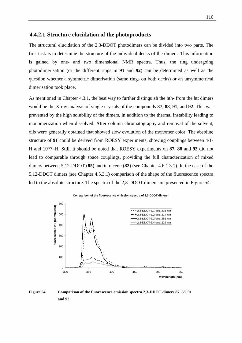
110
4.4.2.1 Structure elucidation of the photoproducts
The structural elucidation of the 2,3-DDOT photodimers can be divided into two parts. The
first task is to determine the structure of the individual decks of the dimers. This information
is gained by one- and two dimensional NMR spectra. Thus, the ring undergoing
photodimerisation (or the different rings in 91 and 92) can be determined as well as the
question whether a symmetric dimerisation (same rings on both decks) or an unsymmetrical
dimerisation took place.
As mentioned in Chapter 4.3.1, the best way to further distinguish the hth- from the htt dimers
would be the X-ray analysis of single crystals of the compounds 87, 88, 91, and 92. This was
prevented by the high solubility of the dimers, in addition to the thermal instability leading to
monomerization when dissolved. After column chromatography and removal of the solvent,
oils were generally obtained that showed slow evolution of the monomer color. The absolute
structure of 91 could be derived from ROESY experiments, showing couplings between 4/1-
H and 10'/7'-H. Still, it should be noted that ROESY experiments on 87, 88 and 92 did not
lead to comparable through space couplings, providing the full characterization of mixed
dimers between 5,12-DDOT (85) and tetracene (82) (see Chapter 4.6.1.3.1). In the case of the
5,12-DDOT dimers (see Chapter 4.5.3.1) comparison of the shape of the fluorescence spectra
led to the absolute structure. The spectra of the 2,3-DDOT dimers are presented in Figure 54.
Comparison of the fluorescence emission spectra of 2,3-DDOT dimers
0
100
200
300
400
500
600
300 350 400 450 500 550
wavelength [nm]
flu
ore
scen
ce in
t. (
no
rmal
ized
)
2,3-DDOT-D1 exc.:236 nm2,3-DDOT-D2 exc.:234 nm2,3-DDOT-D3 exc.:255 nm2,3-DDOT-D4 exc.:232 nm
Figure 54 Comparison of the fluorescence emission spectra 2,3-DDOT dimers 87, 88, 91
and 92
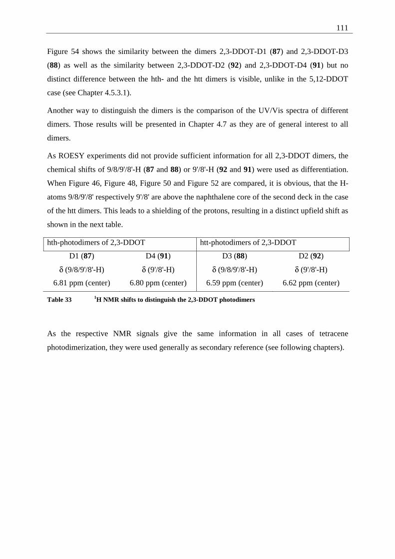
111
Figure 54 shows the similarity between the dimers 2,3-DDOT-D1 (87) and 2,3-DDOT-D3
(88) as well as the similarity between 2,3-DDOT-D2 (92) and 2,3-DDOT-D4 (91) but no
distinct difference between the hth- and the htt dimers is visible, unlike in the 5,12-DDOT
case (see Chapter 4.5.3.1).
Another way to distinguish the dimers is the comparison of the UV/Vis spectra of different
dimers. Those results will be presented in Chapter 4.7 as they are of general interest to all
dimers.
As ROESY experiments did not provide sufficient information for all 2,3-DDOT dimers, the
chemical shifts of 9/8/9'/8'-H (87 and 88) or 9'/8'-H (92 and 91) were used as differentiation.
When Figure 46, Figure 48, Figure 50 and Figure 52 are compared, it is obvious, that the H-
atoms 9/8/9'/8' respectively 9'/8' are above the naphthalene core of the second deck in the case
of the htt dimers. This leads to a shielding of the protons, resulting in a distinct upfield shift as
shown in the next table.
hth-photodimers of 2,3-DDOT htt-photodimers of 2,3-DDOT
D1 (87) D4 (91) D3 (88) D2 (92)
δ (9/8/9'/8'-H) δ (9'/8'-H) δ (9/8/9'/8'-H) δ (9'/8'-H)
6.81 ppm (center) 6.80 ppm (center) 6.59 ppm (center) 6.62 ppm (center)
Table 33 1H NMR shifts to distinguish the 2,3-DDOT photodimers
As the respective NMR signals give the same information in all cases of tetracene
photodimerization, they were used generally as secondary reference (see following chapters).
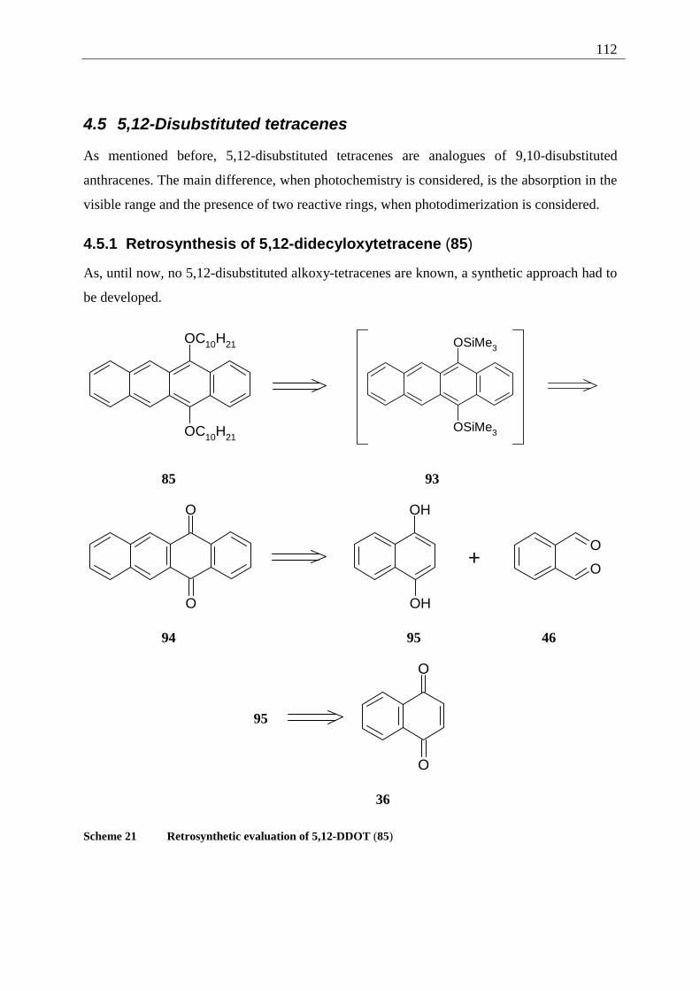
112
4.5 5,12-Disubstituted tetracenes
As mentioned before, 5,12-disubstituted tetracenes are analogues of 9,10-disubstituted
anthracenes. The main difference, when photochemistry is considered, is the absorption in the
visible range and the presence of two reactive rings, when photodimerization is considered.
4.5.1 Retrosynthesis of 5,12-didecyloxytetracene (85)
As, until now, no 5,12-disubstituted alkoxy-tetracenes are known, a synthetic approach had to
be developed.
OC10H21
OC10H21
OSiMe3
OSiMe3
85 93
O
O
OH
OH
+O
O
94 95 46
95
O
O
36
Scheme 21 Retrosynthetic evaluation of 5,12-DDOT (85)
113
Using the synthesis of 2,3-dialkoxy-tetracenes (2) (see Chapter 2), the alkylation of 5,12-
dihydroxytetracene would be the synthesis of choice. This intermediate has been published in
1931 by Fieser et al.[64] along with numerous other 5,12-disubstituted tetracenes. The main
problem is already presented in this early literature: the low stability towards oxidation. As in
the comparison between dihydroacenes and the corresponding acenes,[65] the disruption of the
π-conjugation stabilizes the molecule. Therefore acene-quinones are generally more stable
than the corresponding p-dihydroxy acenes.[58] In order to prevent the reoxidation of 5,12-
dihydroxy-tetracene to 5,12-tetracene-quinone (94), protection of the free OH function was
mandatory. This was accomplished by silylation following the 5,12-bis(trimethylsilyloxy)-
tetracene (93) synthesis presented by Bouas-Laurent et al. in 1970.[66] The further synthesis
contains the preparation of the 5,12-tetracene-quinone (94) and the alkylation of the 5,12-
bis(trimethylsilyloxy)-tetracene (93), as presented in the following chapters.
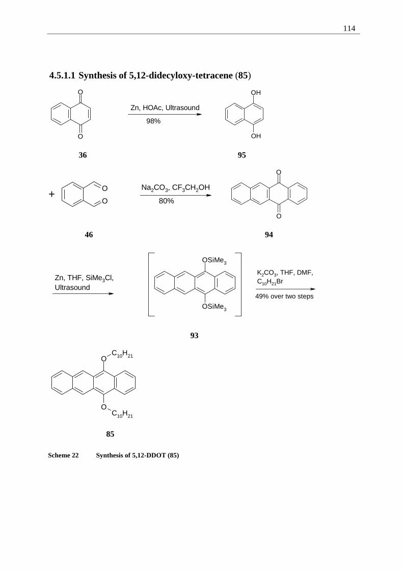
114
4.5.1.1 Synthesis of 5,12-didecyloxy-tetracene (85)
O
O
Zn, HOAc, Ultrasound
98%
OH
OH
36 95
+O
O
Na2CO3, CF3CH2OH
80%
O
O
46 94
Zn, THF, SiMe3Cl,Ultrasound
OSiMe3
OSiMe3
K2CO3, THF, DMF,C10H21Br
49% over two steps
93
O
O
C10H21
C10H21
85
Scheme 22 Synthesis of 5,12-DDOT (85)
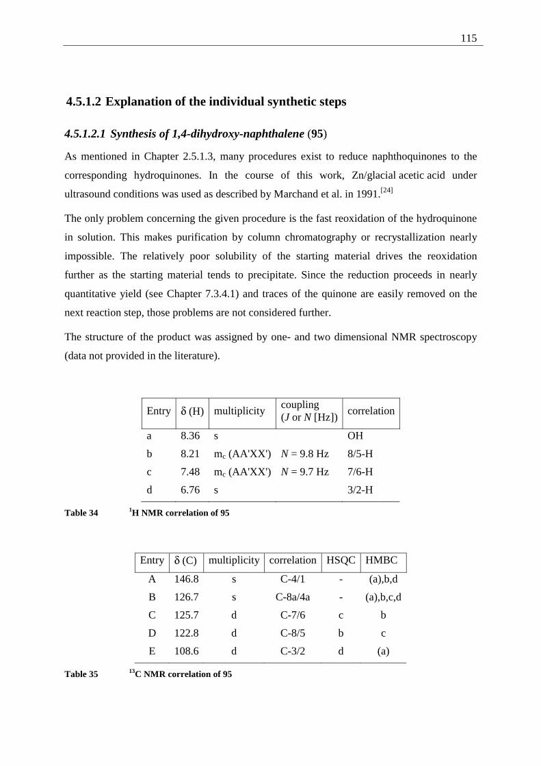
115
4.5.1.2 Explanation of the individual synthetic steps
4.5.1.2.1 Synthesis of 1,4-dihydroxy-naphthalene (95)
As mentioned in Chapter 2.5.1.3, many procedures exist to reduce naphthoquinones to the
corresponding hydroquinones. In the course of this work, Zn/glacial acetic acid under
ultrasound conditions was used as described by Marchand et al. in 1991.[24]
The only problem concerning the given procedure is the fast reoxidation of the hydroquinone
in solution. This makes purification by column chromatography or recrystallization nearly
impossible. The relatively poor solubility of the starting material drives the reoxidation
further as the starting material tends to precipitate. Since the reduction proceeds in nearly
quantitative yield (see Chapter 7.3.4.1) and traces of the quinone are easily removed on the
next reaction step, those problems are not considered further.
The structure of the product was assigned by one- and two dimensional NMR spectroscopy
(data not provided in the literature).
Entry δ (H) multiplicity coupling (J or N [Hz])
correlation
a 8.36 s OH
b 8.21 mc (AA'XX') N = 9.8 Hz 8/5-H
c 7.48 mc (AA'XX') N = 9.7 Hz 7/6-H
d 6.76 s 3/2-H
Table 34 1H NMR correlation of 95
Entry δ (C) multiplicity correlation HSQC HMBC
A 146.8 s C-4/1 - (a),b,d
B 126.7 s C-8a/4a - (a),b,c,d
C 125.7 d C-7/6 c b
D 122.8 d C-8/5 b c
E 108.6 d C-3/2 d (a)
Table 35 13C NMR correlation of 95
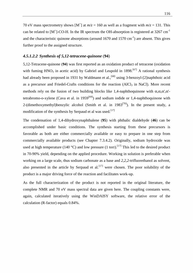
116
70 eV mass spectrometry shows [M+] at m/z = 160 as well as a fragment with m/z = 131. This
can be related to [M+]-CO-H. In the IR spectrum the OH-absorption is registered at 3267 cm-1
and the characteristic quinone absorptions (around 1670 and 1570 cm-1) are absent. This gives
further proof to the assigned structure.
4.5.1.2.2 Synthesis of 5,12-tetracene-quinone (94)
5,12-Tetracene-quinone (94) was first reported as an oxidation product of tetracene (oxidation
with fuming HNO3 in acetic acid) by Gabriel and Leupold in 1898.[67] A rational synthesis
had already been proposed in 1931 by Waldmann et al.,[68] using 3-benzoyl-[2]naphthoic acid
as a precursor and Friedel-Crafts conditions for the reaction (AlCl3 in NaCl). More recent
methods rely on the fusion of two building blocks like 1,4-naphthoquinone with α,α,α',α'-
tetrabromo-o-xylene (Cava et al. in 1959[69]) and sodium iodide or 1,4-naphthoquinone with
2-(dimethoxymethyl)benzylic alcohol (Smith et al. in 1983[70]). In the present study, a
modification of the synthesis by Serpaud et al was used.[17]
The condensation of 1,4-dihydroxynaphthalene (95) with phthalic dialdehyde (46) can be
accomplished under basic conditions. The synthesis starting from these precursors is
favorable as both are either commercially available or easy to prepare in one step from
commercially available products (see Chapter 7.3.4.2). Originally, sodium hydroxide was
used at high temperature (140 °C) and low pressure (1 torr).[17] This led to the desired product
in 70-90% yield, depending on the applied procedure. Working in solution is preferable when
working on a large scale, thus sodium carbonate as a base and 2,2,2-trifluorethanol as solvent,
also presented in the article by Serpaud et al.[17] were chosen. The poor solubility of the
product is a major driving force of the reaction and facilitates work-up.
As the full characterization of the product is not reported in the original literature, the
complete NMR and 70 eV mass spectral data are given here. The coupling constants were,
again, calculated iteratively using the WinDAISY software, the relative error of the
calculation (R-factor) equals 0.84%.
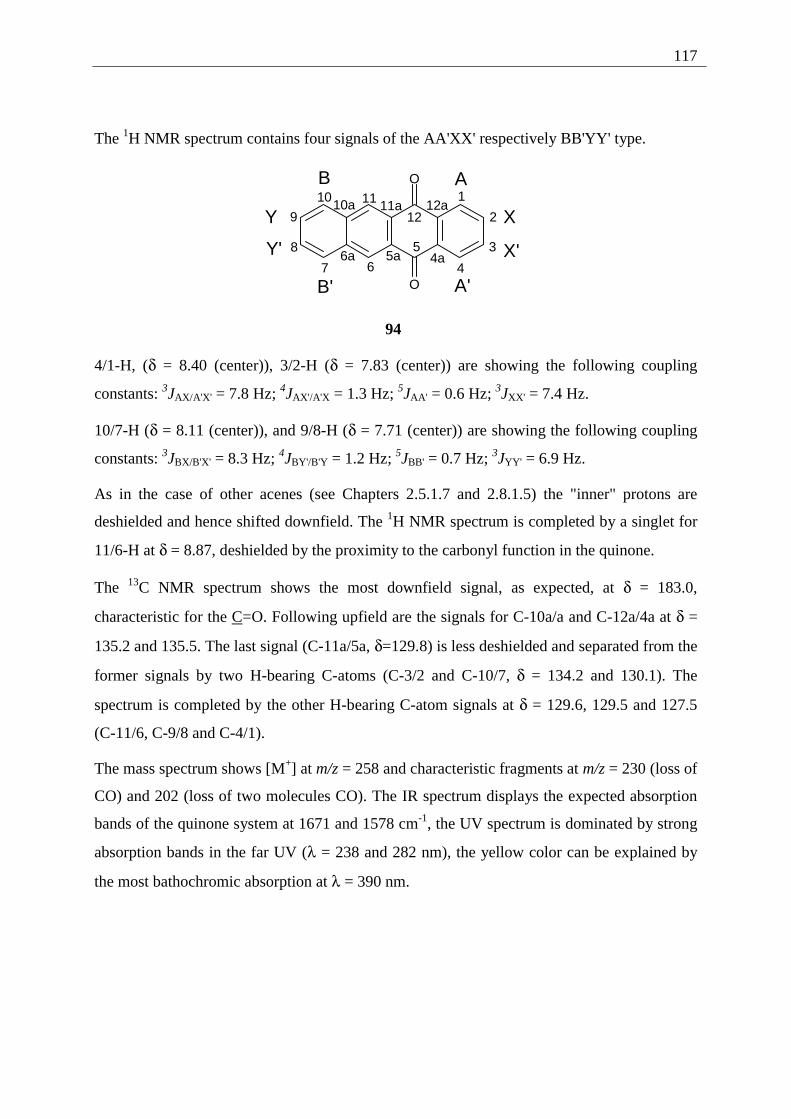
117
The 1H NMR spectrum contains four signals of the AA'XX' respectively BB'YY' type.
O
O
1
2
3
4
5
67
8
9
10 1112
4a5a6a
10a 11a 12a
A
A'
B
B'
X
X'
Y
Y'
94
4/1-H, (δ = 8.40 (center)), 3/2-H (δ = 7.83 (center)) are showing the following coupling
constants: 3JAX/A'X' = 7.8 Hz; 4JAX'/A'X = 1.3 Hz; 5JAA' = 0.6 Hz; 3JXX' = 7.4 Hz.
10/7-H (δ = 8.11 (center)), and 9/8-H (δ = 7.71 (center)) are showing the following coupling
constants: 3JBX/B'X' = 8.3 Hz; 4JBY'/B'Y = 1.2 Hz; 5JBB' = 0.7 Hz; 3JYY' = 6.9 Hz.
As in the case of other acenes (see Chapters 2.5.1.7 and 2.8.1.5) the "inner" protons are
deshielded and hence shifted downfield. The 1H NMR spectrum is completed by a singlet for
11/6-H at δ = 8.87, deshielded by the proximity to the carbonyl function in the quinone.
The 13C NMR spectrum shows the most downfield signal, as expected, at δ = 183.0,
characteristic for the C=O. Following upfield are the signals for C-10a/a and C-12a/4a at δ =
135.2 and 135.5. The last signal (C-11a/5a, δ=129.8) is less deshielded and separated from the
former signals by two H-bearing C-atoms (C-3/2 and C-10/7, δ = 134.2 and 130.1). The
spectrum is completed by the other H-bearing C-atom signals at δ = 129.6, 129.5 and 127.5
(C-11/6, C-9/8 and C-4/1).
The mass spectrum shows [M+] at m/z = 258 and characteristic fragments at m/z = 230 (loss of
CO) and 202 (loss of two molecules CO). The IR spectrum displays the expected absorption
bands of the quinone system at 1671 and 1578 cm-1, the UV spectrum is dominated by strong
absorption bands in the far UV (λ = 238 and 282 nm), the yellow color can be explained by
the most bathochromic absorption at λ = 390 nm.
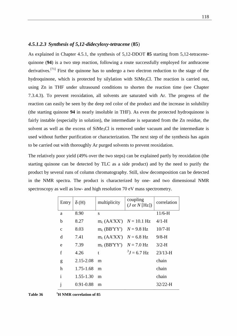
118
4.5.1.2.3 Synthesis of 5,12-didecyloxy-tetracene (85)
As explained in Chapter 4.5.1, the synthesis of 5,12-DDOT 85 starting from 5,12-tetracene-
quinone (94) is a two step reaction, following a route successfully employed for anthracene
derivatives.[71] First the quinone has to undergo a two electron reduction to the stage of the
hydroquinone, which is protected by silylation with SiMe3Cl. The reaction is carried out,
using Zn in THF under ultrasound conditions to shorten the reaction time (see Chapter
7.3.4.3). To prevent reoxidation, all solvents are saturated with Ar. The progress of the
reaction can easily be seen by the deep red color of the product and the increase in solubility
(the starting quinone 94 in nearly insoluble in THF). As even the protected hydroquinone is
fairly instable (especially in solution), the intermediate is separated from the Zn residue, the
solvent as well as the excess of SiMe3Cl is removed under vacuum and the intermediate is
used without further purification or characterization. The next step of the synthesis has again
to be carried out with thoroughly Ar purged solvents to prevent reoxidation.
The relatively poor yield (49% over the two steps) can be explained partly by reoxidation (the
starting quinone can be detected by TLC as a side product) and by the need to purify the
product by several runs of column chromatography. Still, slow decomposition can be detected
in the NMR spectra. The product is characterized by one- and two dimensional NMR
spectroscopy as well as low- and high resolution 70 eV mass spectrometry.
Entry δ (H) multiplicity coupling (J or N [Hz])
correlation
a 8.90 s 11/6-H
b 8.27 mc (AA'XX') N = 10.1 Hz 4/1-H
c 8.03 mc (BB'YY') N = 9.8 Hz 10/7-H
d 7.41 mc (AA'XX') N = 6.8 Hz 9/8-H
e 7.39 mc (BB'YY') N = 7.0 Hz 3/2-H
f 4.26 t 3J = 6.7 Hz 23/13-H
g 2.15-2.08 m chain
h 1.75-1.68 m chain
i 1.55-1.30 m chain
j 0.91-0.88 m 32/22-H
Table 36 1H NMR correlation of 85
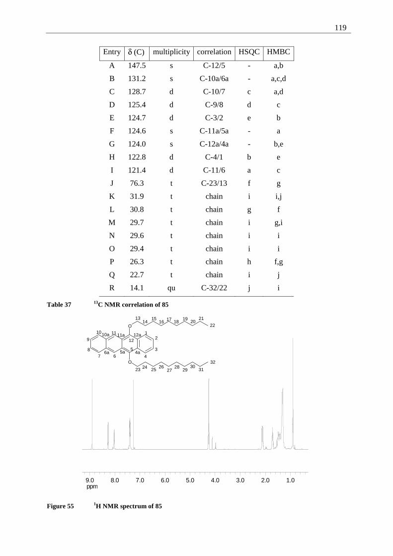
119
Entry δ (C) multiplicity correlation HSQC HMBC
A 147.5 s C-12/5 - a,b
B 131.2 s C-10a/6a - a,c,d
C 128.7 d C-10/7 c a,d
D 125.4 d C-9/8 d c
E 124.7 d C-3/2 e b
F 124.6 s C-11a/5a - a
G 124.0 s C-12a/4a - b,e
H 122.8 d C-4/1 b e
I 121.4 d C-11/6 a c
J 76.3 t C-23/13 f g
K 31.9 t chain i i,j
L 30.8 t chain g f
M 29.7 t chain i g,i
N 29.6 t chain i i
O 29.4 t chain i i
P 26.3 t chain h f,g
Q 22.7 t chain i j
R 14.1 qu C-32/22 j i
Table 37 13C NMR correlation of 85
ppm1.02.03.04.05.06.07.08.09.0
O
O
12
3
4
5
678
9
10 11
12
1314
1516 17 18
1920
21
22
23 24 2526
2728
2930
31
32
4a5a6a
10a 11a 12a
Figure 55 1H NMR spectrum of 85
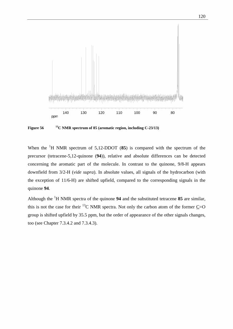
120
ppm8090100110120130140
Figure 56 13C NMR spectrum of 85 (aromatic region, including C-23/13)
When the 1H NMR spectrum of 5,12-DDOT (85) is compared with the spectrum of the
precursor (tetracene-5,12-quinone (94)), relative and absolute differences can be detected
concerning the aromatic part of the molecule. In contrast to the quinone, 9/8-H appears
downfield from 3/2-H (vide supra). In absolute values, all signals of the hydrocarbon (with
the exception of 11/6-H) are shifted upfield, compared to the corresponding signals in the
quinone 94.
Although the 1H NMR spectra of the quinone 94 and the substituted tetracene 85 are similar,
this is not the case for their 13C NMR spectra. Not only the carbon atom of the former C=O
group is shifted upfield by 35.5 ppm, but the order of appearance of the other signals changes,
too (see Chapter 7.3.4.2 and 7.3.4.3).
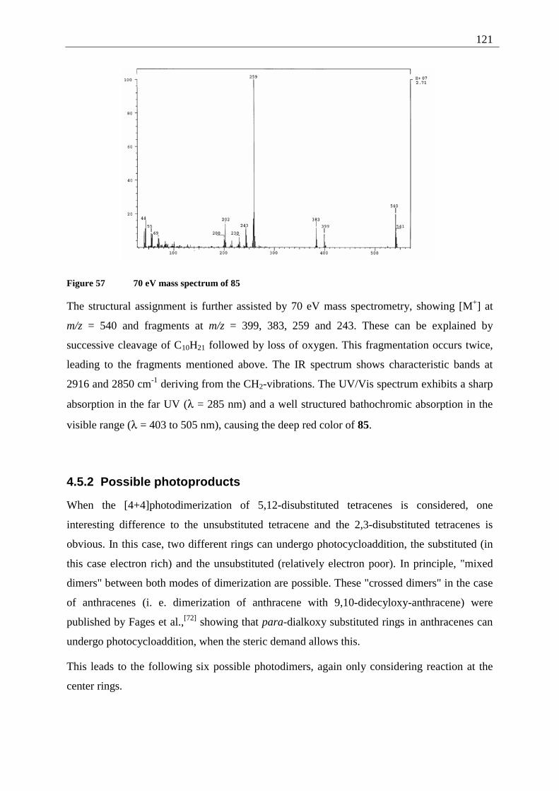
121
Figure 57 70 eV mass spectrum of 85
The structural assignment is further assisted by 70 eV mass spectrometry, showing [M+] at
m/z = 540 and fragments at m/z = 399, 383, 259 and 243. These can be explained by
successive cleavage of C10H21 followed by loss of oxygen. This fragmentation occurs twice,
leading to the fragments mentioned above. The IR spectrum shows characteristic bands at
2916 and 2850 cm-1 deriving from the CH2-vibrations. The UV/Vis spectrum exhibits a sharp
absorption in the far UV (λ = 285 nm) and a well structured bathochromic absorption in the
visible range (λ = 403 to 505 nm), causing the deep red color of 85.
4.5.2 Possible photoproducts
When the [4+4]photodimerization of 5,12-disubstituted tetracenes is considered, one
interesting difference to the unsubstituted tetracene and the 2,3-disubstituted tetracenes is
obvious. In this case, two different rings can undergo photocycloaddition, the substituted (in
this case electron rich) and the unsubstituted (relatively electron poor). In principle, "mixed
dimers" between both modes of dimerization are possible. These "crossed dimers" in the case
of anthracenes (i. e. dimerization of anthracene with 9,10-didecyloxy-anthracene) were
published by Fages et al.,[72] showing that para-dialkoxy substituted rings in anthracenes can
undergo photocycloaddition, when the steric demand allows this.
This leads to the following six possible photodimers, again only considering reaction at the
center rings.
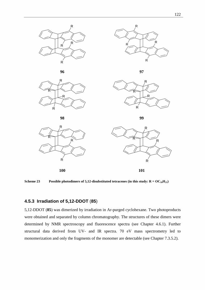
122
R
R
R
R
R
RR
R
96 97
R
R
R
R
R
R
R
R
98 99
R
R
R
R
R
R
R
R
100 101
Scheme 23 Possible photodimers of 5,12-disubstituted tetracenes (in this study: R = OC10H21)
4.5.3 Irradiation of 5,12-DDOT (85)
5,12-DDOT (85) was dimerized by irradiation in Ar-purged cyclohexane. Two photoproducts
were obtained and separated by column chromatography. The structures of these dimers were
determined by NMR spectroscopy and fluorescence spectra (see Chapter 4.6.1). Further
structural data derived from UV- and IR spectra. 70 eV mass spectrometry led to
monomerization and only the fragments of the monomer are detectable (see Chapter 7.3.5.2).
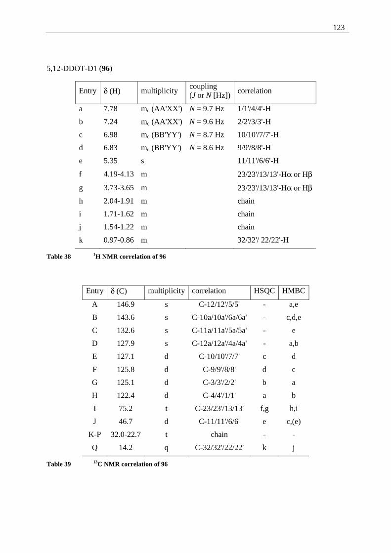
123
5,12-DDOT-D1 (96)
Entry δ (H) multiplicity coupling (J or N [Hz])
correlation
a 7.78 mc (AA'XX') N = 9.7 Hz 1/1'/4/4'-H
b 7.24 mc (AA'XX') N = 9.6 Hz 2/2'/3/3'-H
c 6.98 mc (BB'YY') N = 8.7 Hz 10/10'/7/7'-H
d 6.83 mc (BB'YY') N = 8.6 Hz 9/9'/8/8'-H
e 5.35 s 11/11'/6/6'-H
f 4.19-4.13 m 23/23'/13/13'-Hα or Hβ
g 3.73-3.65 m 23/23'/13/13'-Hα or Hβ
h 2.04-1.91 m chain
i 1.71-1.62 m chain
j 1.54-1.22 m chain
k 0.97-0.86 m 32/32'/ 22/22'-H
Table 38 1H NMR correlation of 96
Entry δ (C) multiplicity correlation HSQC HMBC
A 146.9 s C-12/12'/5/5' - a,e
B 143.6 s C-10a/10a'/6a/6a' - c,d,e
C 132.6 s C-11a/11a'/5a/5a' - e
D 127.9 s C-12a/12a'/4a/4a' - a,b
E 127.1 d C-10/10'/7/7' c d
F 125.8 d C-9/9'/8/8' d c
G 125.1 d C-3/3'/2/2' b a
H 122.4 d C-4/4'/1/1' a b
I 75.2 t C-23/23'/13/13' f,g h,i
J 46.7 d C-11/11'/6/6' e c,(e)
K-P 32.0-22.7 t chain - -
Q 14.2 q C-32/32'/22/22' k j
Table 39 13C NMR correlation of 96
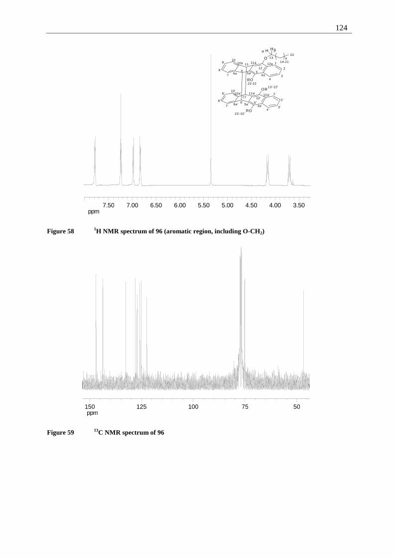
124
ppm3.504.004.505.005.506.006.507.007.50
O
H H
OR
RO
8
RO
1
2
34
567
8
910
1112
4a5a6a
10a 11a 12a
1'
2'
3'4'
5'6'7'
8'
9'10'
11' 12'
4a'5a'6a'
10a' 11a' 12a'
1314-21
22
23-3213'-22'
23'-32'
α β
Figure 58 1H NMR spectrum of 96 (aromatic region, including O-CH2)
ppm5075100125150
Figure 59 13C NMR spectrum of 96
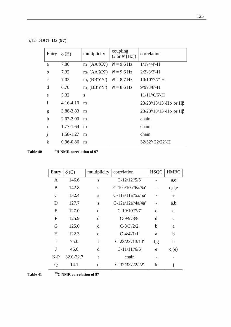
125
5,12-DDOT-D2 (97)
Entry δ (H) multiplicity coupling (J or N [Hz])
correlation
a 7.86 mc (AA'XX') N = 9.6 Hz 1/1'/4/4'-H
b 7.32 mc (AA'XX') N = 9.6 Hz 2/2'/3/3'-H
c 7.02 mc (BB'YY') N = 8.7 Hz 10/10'/7/7'-H
d 6.70 mc (BB'YY') N = 8.6 Hz 9/9'/8/8'-H
e 5.32 s 11/11'/6/6'-H
f 4.16-4.10 m 23/23'/13/13'-Hα or Hβ
g 3.88-3.83 m 23/23'/13/13'-Hα or Hβ
h 2.07-2.00 m chain
i 1.77-1.64 m chain
j 1.58-1.27 m chain
k 0.96-0.86 m 32/32'/ 22/22'-H
Table 40 1H NMR correlation of 97
Entry δ (C) multiplicity correlation HSQC HMBC
A 146.6 s C-12/12'/5/5' - a,e
B 142.8 s C-10a/10a'/6a/6a' - c,d,e
C 132.4 s C-11a/11a'/5a/5a' - e
D 127.7 s C-12a/12a'/4a/4a' - a,b
E 127.0 d C-10/10'/7/7' c d
F 125.9 d C-9/9'/8/8' d c
G 125.0 d C-3/3'/2/2' b a
H 122.3 d C-4/4'/1/1' a b
I 75.0 t C-23/23'/13/13' f,g h
J 46.6 d C-11/11'/6/6' e c,(e)
K-P 32.0-22.7 t chain - -
Q 14.1 q C-32/32'/22/22' k j
Table 41 13C NMR correlation of 97
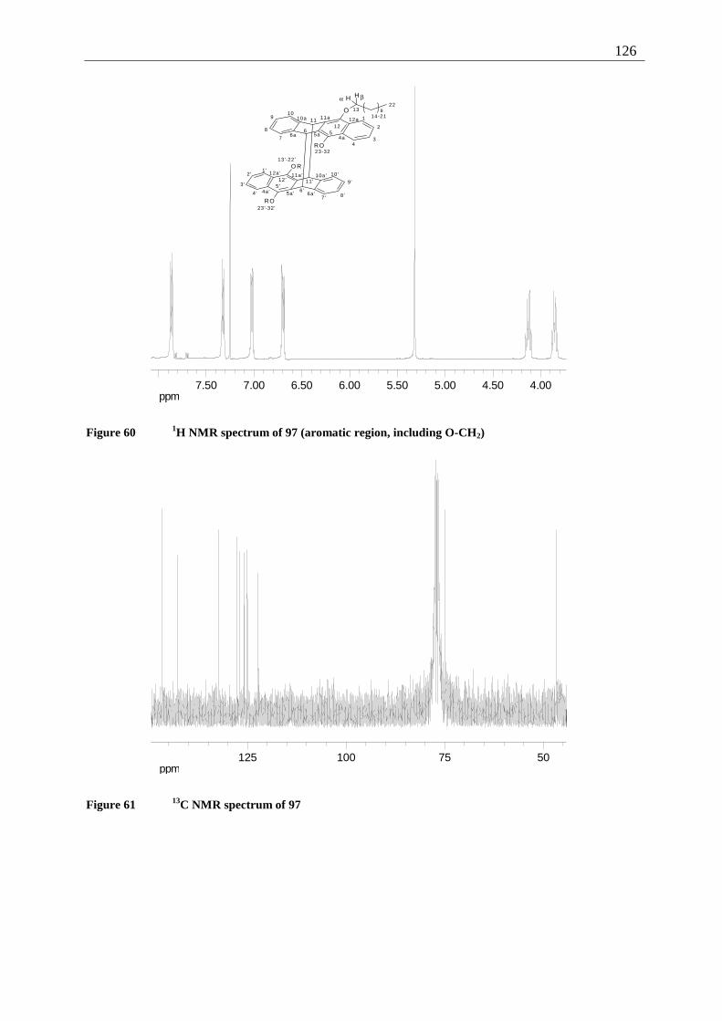
126
ppm4.004.505.005.506.006.507.007.50
O
H H
8
RO
O R
RO
1
2
34
567
8
910
1112
4a5a6a
10a 11a 12a
1'2 '
3 '
4 '5 '
6 '7 ' 8 '
9 '
10 '11'12'
4a' 5a' 6a'
10a'11a'12a'
1314-21
22
23-32
13'-22'
23'-32'
α β
Figure 60 1H NMR spectrum of 97 (aromatic region, including O-CH2)
ppm5075100125
Figure 61 13C NMR spectrum of 97
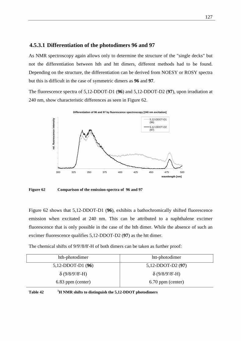
127
4.5.3.1 Differentiation of the photodimers 96 and 97
As NMR spectroscopy again allows only to determine the structure of the "single decks" but
not the differentiation between hth and htt dimers, different methods had to be found.
Depending on the structure, the differentiation can be derived from NOESY or ROSY spectra
but this is difficult in the case of symmetric dimers as 96 and 97.
The fluorescence spectra of 5,12-DDOT-D1 (96) and 5,12-DDOT-D2 (97), upon irradiation at
240 nm, show characteristic differences as seen in Figure 62.
Differentiation of 96 and 97 by fluorescence spectroscopy [240 nm excitation]
300 325 350 375 400 425 450 475 500
wavelength [nm]
rel.
fluo
resc
ence
inte
nsit
y 5,12-DDOT-D1(96)
5,12-DDOT-D2(97)
Figure 62 Comparison of the emission-spectra of 96 and 97
Figure 62 shows that 5,12-DDOT-D1 (96), exhibits a bathochromically shifted fluorescence
emission when excitated at 240 nm. This can be attributed to a naphthalene excimer
fluorescence that is only possible in the case of the hth dimer. While the absence of such an
excimer fluorescence qualifies 5,12-DDOT-D2 (97) as the htt dimer.
The chemical shifts of 9/9'/8/8'-H of both dimers can be taken as further proof:
hth-photodimer htt-photodimer
5,12-DDOT-D1 (96) 5,12-DDOT-D2 (97)
δ (9/8/9'/8'-H) δ (9/8/9'/8'-H)
6.83 ppm (center) 6.70 ppm (center)
Table 42 1H NMR shifts to distinguish the 5,12-DDOT photodimers
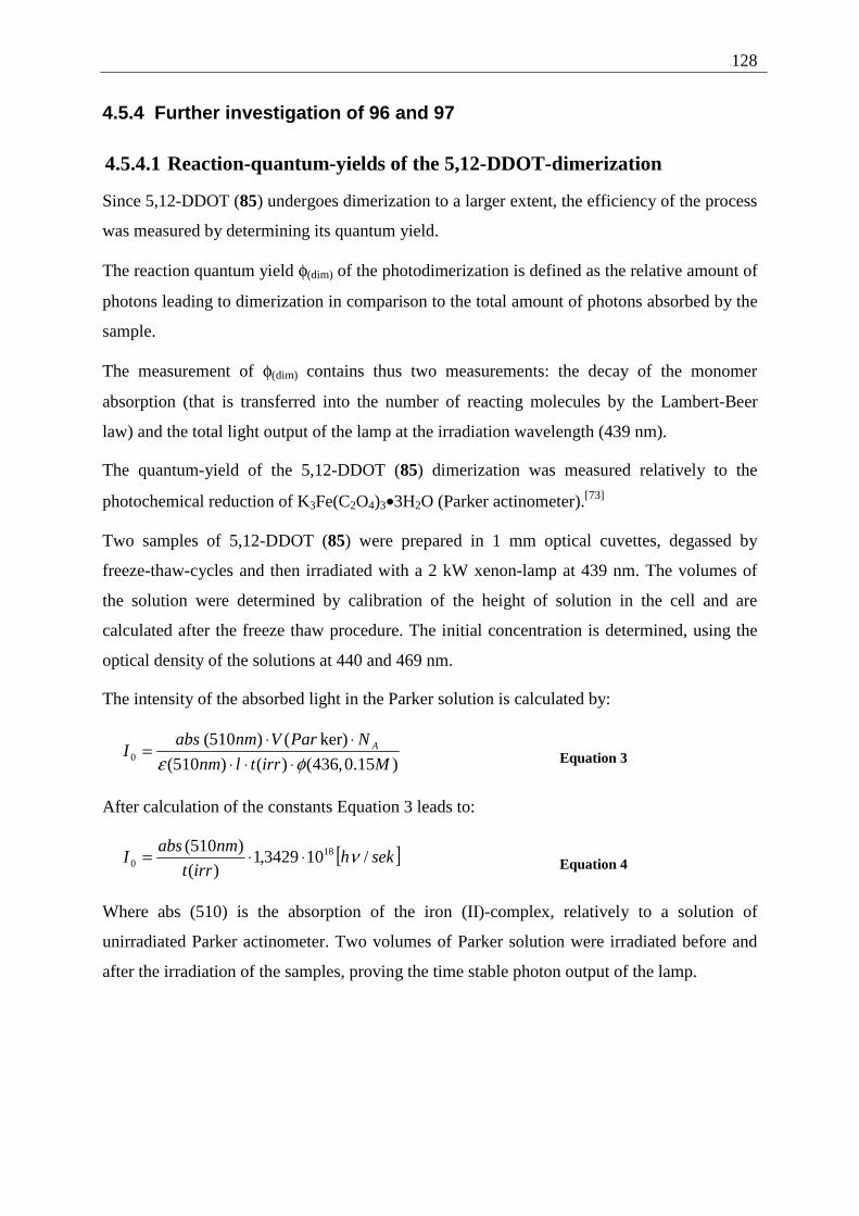
128
4.5.4 Further investigation of 96 and 97
4.5.4.1 Reaction-quantum-yields of the 5,12-DDOT-dimerization
Since 5,12-DDOT (85) undergoes dimerization to a larger extent, the efficiency of the process
was measured by determining its quantum yield.
The reaction quantum yield φ(dim) of the photodimerization is defined as the relative amount of
photons leading to dimerization in comparison to the total amount of photons absorbed by the
sample.
The measurement of φ(dim) contains thus two measurements: the decay of the monomer
absorption (that is transferred into the number of reacting molecules by the Lambert-Beer
law) and the total light output of the lamp at the irradiation wavelength (439 nm).
The quantum-yield of the 5,12-DDOT (85) dimerization was measured relatively to the
photochemical reduction of K3Fe(C2O4)3•3H2O (Parker actinometer).[73]
Two samples of 5,12-DDOT (85) were prepared in 1 mm optical cuvettes, degassed by
freeze-thaw-cycles and then irradiated with a 2 kW xenon-lamp at 439 nm. The volumes of
the solution were determined by calibration of the height of solution in the cell and are
calculated after the freeze thaw procedure. The initial concentration is determined, using the
optical density of the solutions at 440 and 469 nm.
The intensity of the absorbed light in the Parker solution is calculated by:
)15.0,436()()510(
ker)()510(0 Mirrtlnm
NParVnmabsI A
φε ⋅⋅⋅⋅⋅
= Equation 3
After calculation of the constants Equation 3 leads to:
[ ]sekhirrt
nmabsI /103429,1
)(
)510( 180 ν⋅⋅= Equation 4
Where abs (510) is the absorption of the iron (II)-complex, relatively to a solution of
unirradiated Parker actinometer. Two volumes of Parker solution were irradiated before and
after the irradiation of the samples, proving the time stable photon output of the lamp.
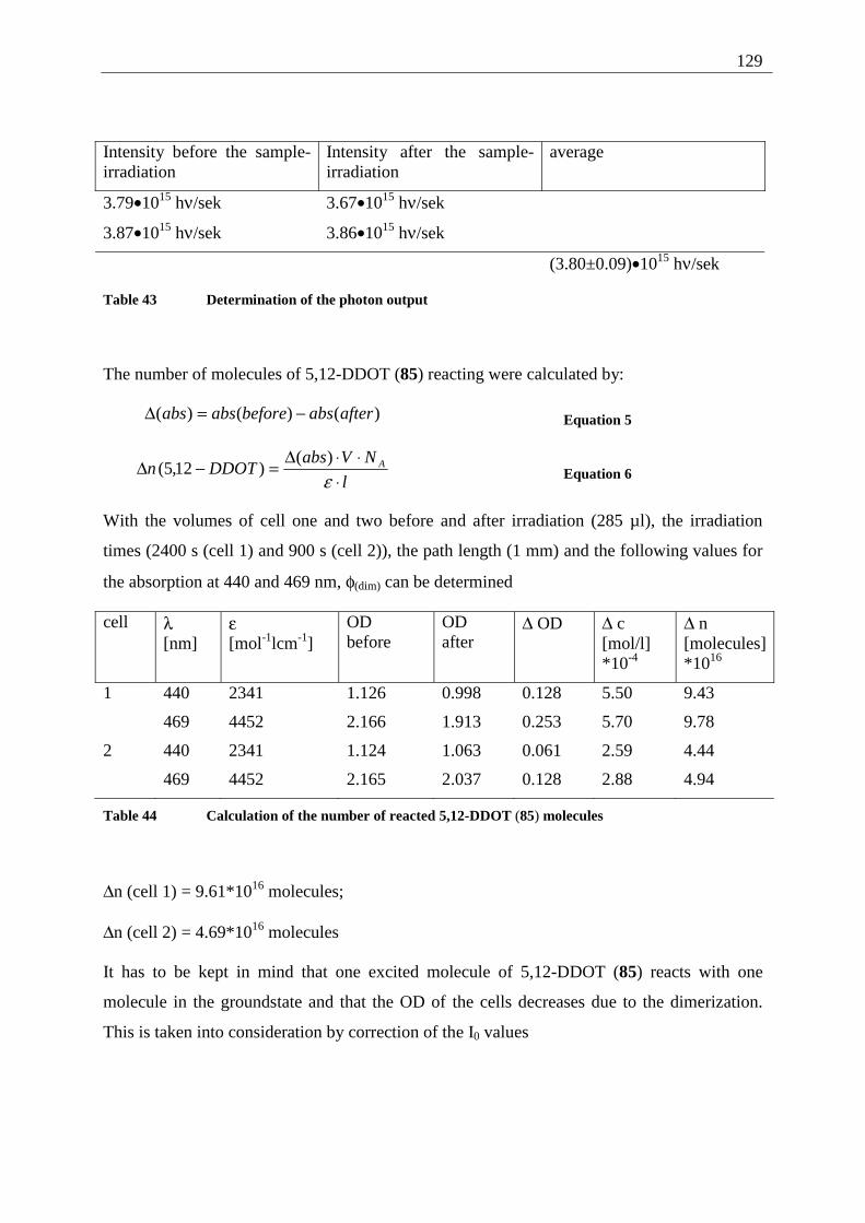
129
Intensity before the sample- irradiation
Intensity after the sample- irradiation
average
3.79•1015 hν/sek 3.67•1015 hν/sek
3.87•1015 hν/sek 3.86•1015 hν/sek
(3.80±0.09)•1015 hν/sek
Table 43 Determination of the photon output
The number of molecules of 5,12-DDOT (85) reacting were calculated by:
)()()( afterabsbeforeabsabs −=∆ Equation 5
l
NVabsDDOTn A
⋅⋅⋅∆
=−∆ε)(
)12,5( Equation 6
With the volumes of cell one and two before and after irradiation (285 µl), the irradiation
times (2400 s (cell 1) and 900 s (cell 2)), the path length (1 mm) and the following values for
the absorption at 440 and 469 nm, φ(dim) can be determined
cell λ [nm]
ε [mol-1lcm-1]
OD before
OD after
∆ OD ∆ c [mol/l] *10-4
∆ n [molecules] *1016
1 440 2341 1.126 0.998 0.128 5.50 9.43
469 4452 2.166 1.913 0.253 5.70 9.78
2 440 2341 1.124 1.063 0.061 2.59 4.44
469 4452 2.165 2.037 0.128 2.88 4.94
Table 44 Calculation of the number of reacted 5,12-DDOT (85) molecules
∆n (cell 1) = 9.61*1016 molecules;
∆n (cell 2) = 4.69*1016 molecules
It has to be kept in mind that one excited molecule of 5,12-DDOT (85) reacts with one
molecule in the groundstate and that the OD of the cells decreases due to the dimerization.
This is taken into consideration by correction of the I0 values
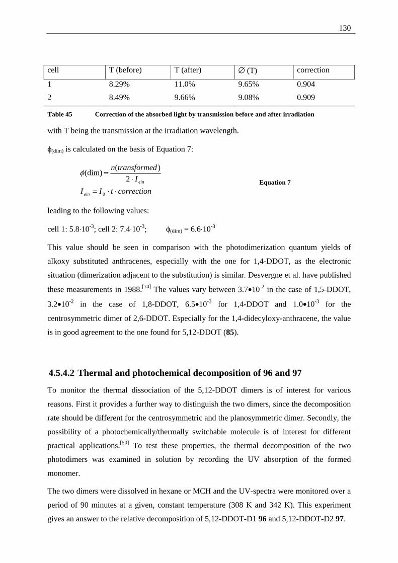
130
cell T (before) T (after) ∅ (T) correction
1 8.29% 11.0% 9.65% 0.904
2 8.49% 9.66% 9.08% 0.909
Table 45 Correction of the absorbed light by transmission before and after irradiation
with T being the transmission at the irradiation wavelength.
φ(dim) is calculated on the basis of Equation 7:
correctiontII
I
dtransformen
ein
ein
⋅⋅=⋅
=
0
2
)((dim)φ
Equation 7
leading to the following values:
cell 1: 5.8⋅10-3; cell 2: 7.4⋅10-3; φ(dim) = 6.6⋅10-3
This value should be seen in comparison with the photodimerization quantum yields of
alkoxy substituted anthracenes, especially with the one for 1,4-DDOT, as the electronic
situation (dimerization adjacent to the substitution) is similar. Desvergne et al. have published
these measurements in 1988.[74] The values vary between 3.7•10-2 in the case of 1,5-DDOT,
3.2•10-2 in the case of 1,8-DDOT, 6.5•10-3 for 1,4-DDOT and 1.0•10-3 for the
centrosymmetric dimer of 2,6-DDOT. Especially for the 1,4-didecyloxy-anthracene, the value
is in good agreement to the one found for 5,12-DDOT (85).
4.5.4.2 Thermal and photochemical decomposition of 96 and 97
To monitor the thermal dissociation of the 5,12-DDOT dimers is of interest for various
reasons. First it provides a further way to distinguish the two dimers, since the decomposition
rate should be different for the centrosymmetric and the planosymmetric dimer. Secondly, the
possibility of a photochemically/thermally switchable molecule is of interest for different
practical applications.[50] To test these properties, the thermal decomposition of the two
photodimers was examined in solution by recording the UV absorption of the formed
monomer.
The two dimers were dissolved in hexane or MCH and the UV-spectra were monitored over a
period of 90 minutes at a given, constant temperature (308 K and 342 K). This experiment
gives an answer to the relative decomposition of 5,12-DDOT-D1 96 and 5,12-DDOT-D2 97.
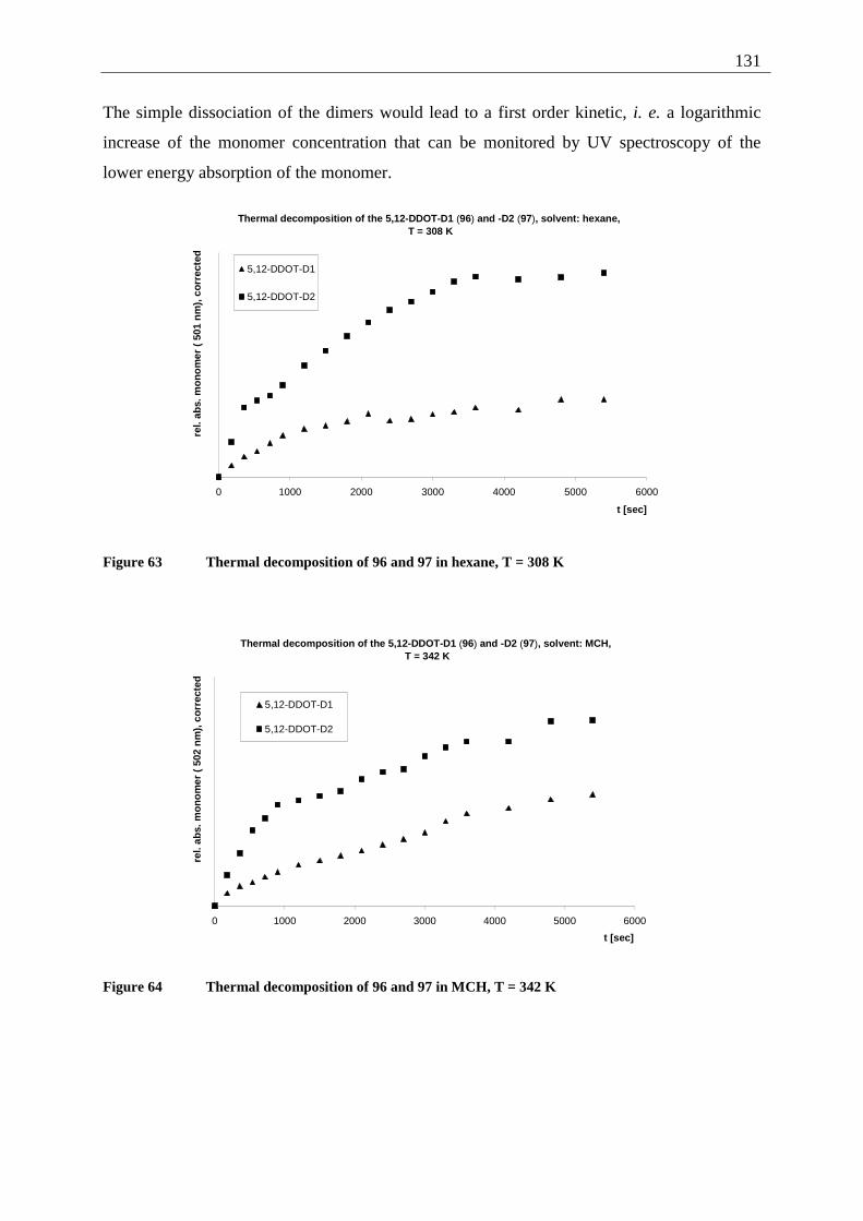
131
The simple dissociation of the dimers would lead to a first order kinetic, i. e. a logarithmic
increase of the monomer concentration that can be monitored by UV spectroscopy of the
lower energy absorption of the monomer.
Thermal decomposition of the 5,12-DDOT-D1 (96) and -D2 (97), solvent: hexane, T = 308 K
0 1000 2000 3000 4000 5000 6000
t [sec]
rel.
abs.
mo
no
mer
( 5
01 n
m),
co
rrec
ted
5,12-DDOT-D1
5,12-DDOT-D2
Figure 63 Thermal decomposition of 96 and 97 in hexane, T = 308 K
Thermal decomposition of the 5,12-DDOT-D1 (96) and -D2 (97), solvent: MCH, T = 342 K
0 1000 2000 3000 4000 5000 6000
t [sec]
rel.
abs.
mo
no
mer
( 5
02 n
m),
co
rrec
ted
5,12-DDOT-D1
5,12-DDOT-D2
Figure 64 Thermal decomposition of 96 and 97 in MCH, T = 342 K
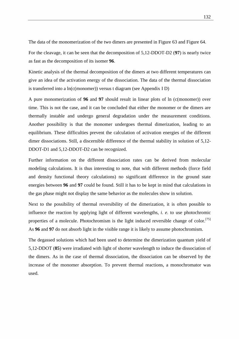
132
The data of the monomerization of the two dimers are presented in Figure 63 and Figure 64.
For the cleavage, it can be seen that the decomposition of 5,12-DDOT-D2 (97) is nearly twice
as fast as the decomposition of its isomer 96.
Kinetic analysis of the thermal decomposition of the dimers at two different temperatures can
give an idea of the activation energy of the dissociation. The data of the thermal dissociation
is transferred into a ln(c(monomer)) versus t diagram (see Appendix I D)
A pure monomerization of 96 and 97 should result in linear plots of ln (c(monomer)) over
time. This is not the case, and it can be concluded that either the monomer or the dimers are
thermally instable and undergo general degradation under the measurement conditions.
Another possibility is that the monomer undergoes thermal dimerization, leading to an
equilibrium. These difficulties prevent the calculation of activation energies of the different
dimer dissociations. Still, a discernible difference of the thermal stability in solution of 5,12-
DDOT-D1 and 5,12-DDOT-D2 can be recognized.
Further information on the different dissociation rates can be derived from molecular
modeling calculations. It is thus interesting to note, that with different methods (force field
and density functional theory calculations) no significant difference in the ground state
energies between 96 and 97 could be found. Still it has to be kept in mind that calculations in
the gas phase might not display the same behavior as the molecules show in solution.
Next to the possibility of thermal reversibility of the dimerization, it is often possible to
influence the reaction by applying light of different wavelengths, i. e. to use photochromic
properties of a molecule. Photochromism is the light induced reversible change of color.[75]
As 96 and 97 do not absorb light in the visible range it is likely to assume photochromism.
The degassed solutions which had been used to determine the dimerization quantum yield of
5,12-DDOT (85) were irradiated with light of shorter wavelength to induce the dissociation of
the dimers. As in the case of thermal dissociation, the dissociation can be observed by the
increase of the monomer absorption. To prevent thermal reactions, a monochromator was
used.
133
Examination of the photochromic properties of 5,12-DDOT (85)
0 50 100 150 200 250
t [min]
rel.
abs.
(5,
12-D
DO
T)
A B
A = irradiation at 322 nmB = irradiation at 469 nm
A
B A
Figure 65 Examination of the photochromic properties of 5,12-DDOT (85) and its dimers 96 and 97
Figure 65 shows that the mixture of the dimers 5,12-DDOT-D1 96 and 5,12-DDOT-D2 97
photodissociats on irradiation at 322 nm (increase of the monomer absorption in the parts A).
When the mixture is again irradiated at 469 nm, the amount of monomer decreases (parts B).
Still, 5,12-DDOT (85) is not a photochromic substance in the pure sense of the definition.[75]
As the monomer concentration does not regain the value it had at the beginning of the
experiment, no pure dimerization/monomerization takes place, but a decomposition of the
monomer parallel to the photoreaction. This can be seen in detail in the part of Figure 65, time
110 to 210 minutes, where the monomer absorption decreases after a long irradiation at 322
nm.
4.6 Crossed photodimers of 5,12-DDOT (85) and tetracene (82)
4.6.1 Possible photoproducts
That 9,10-DDOA (1) does not give any photoproducts upon irradiation,[3] can be attributed to
steric overload of the most reactive (9,10/9',10') positions. One convincing proof of this
rationalization is provided by the formation of mixed photodimers between 9,10-DDOA (1)
and anthracene.[72]
When a 1:1 mixture of tetracene (82) and 5,12-DDOT (85) is irradiated together, three groups
of dimers can be formed: The two groups of "pure dimers" and the mixed dimers. The pure
dimers of tetracene (82) and 5,12-DDOT (85) are discussed in the Chapters 4.3.2 and 4.5.3,
the mixed dimers are described in the following Scheme 24.
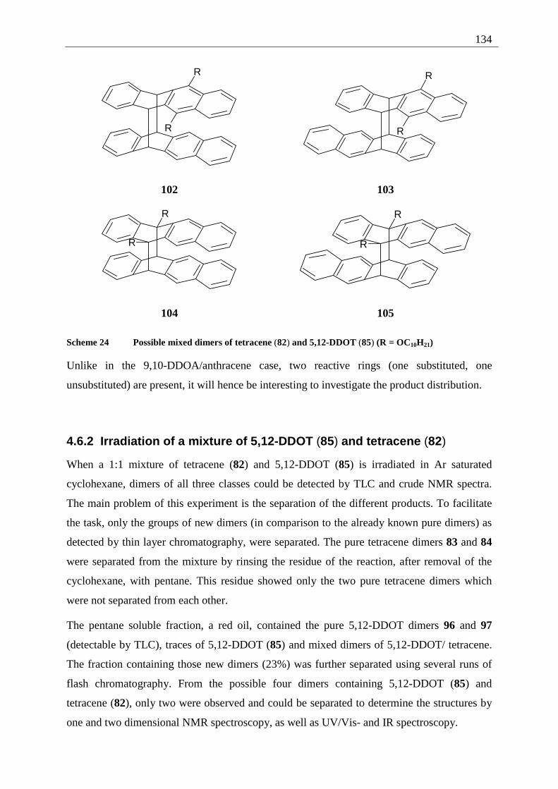
134
R
R
R
R
102 103
R
R
R
R
104 105
Scheme 24 Possible mixed dimers of tetracene (82) and 5,12-DDOT (85) (R = OC10H21)
Unlike in the 9,10-DDOA/anthracene case, two reactive rings (one substituted, one
unsubstituted) are present, it will hence be interesting to investigate the product distribution.
4.6.2 Irradiation of a mixture of 5,12-DDOT (85) and tetracene (82)
When a 1:1 mixture of tetracene (82) and 5,12-DDOT (85) is irradiated in Ar saturated
cyclohexane, dimers of all three classes could be detected by TLC and crude NMR spectra.
The main problem of this experiment is the separation of the different products. To facilitate
the task, only the groups of new dimers (in comparison to the already known pure dimers) as
detected by thin layer chromatography, were separated. The pure tetracene dimers 83 and 84
were separated from the mixture by rinsing the residue of the reaction, after removal of the
cyclohexane, with pentane. This residue showed only the two pure tetracene dimers which
were not separated from each other.
The pentane soluble fraction, a red oil, contained the pure 5,12-DDOT dimers 96 and 97
(detectable by TLC), traces of 5,12-DDOT (85) and mixed dimers of 5,12-DDOT/ tetracene.
The fraction containing those new dimers (23%) was further separated using several runs of
flash chromatography. From the possible four dimers containing 5,12-DDOT (85) and
tetracene (82), only two were observed and could be separated to determine the structures by
one and two dimensional NMR spectroscopy, as well as UV/Vis- and IR spectroscopy.
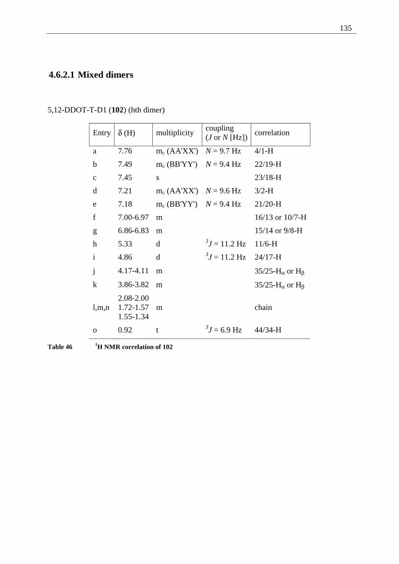
135
4.6.2.1 Mixed dimers
5,12-DDOT-T-D1 (102) (hth dimer)
Entry δ (H) multiplicity coupling (J or N [Hz])
correlation
a 7.76 mc (AA'XX') N = 9.7 Hz 4/1-H
b 7.49 mc (BB'YY') N = 9.4 Hz 22/19-H
c 7.45 s 23/18-H
d 7.21 mc (AA'XX') N = 9.6 Hz 3/2-H
e 7.18 mc (BB'YY') N = 9.4 Hz 21/20-H
f 7.00-6.97 m 16/13 or 10/7-H
g 6.86-6.83 m 15/14 or 9/8-H
h 5.33 d 3J = 11.2 Hz 11/6-H
i 4.86 d 3J = 11.2 Hz 24/17-H
j 4.17-4.11 m 35/25-Hα or Hβ
k 3.86-3.82 m 35/25-Hα or Hβ
l,m,n 2.08-2.00 1.72-1.57 1.55-1.34
m
chain
o 0.92 t 3J = 6.9 Hz 44/34-H
Table 46 1H NMR correlation of 102
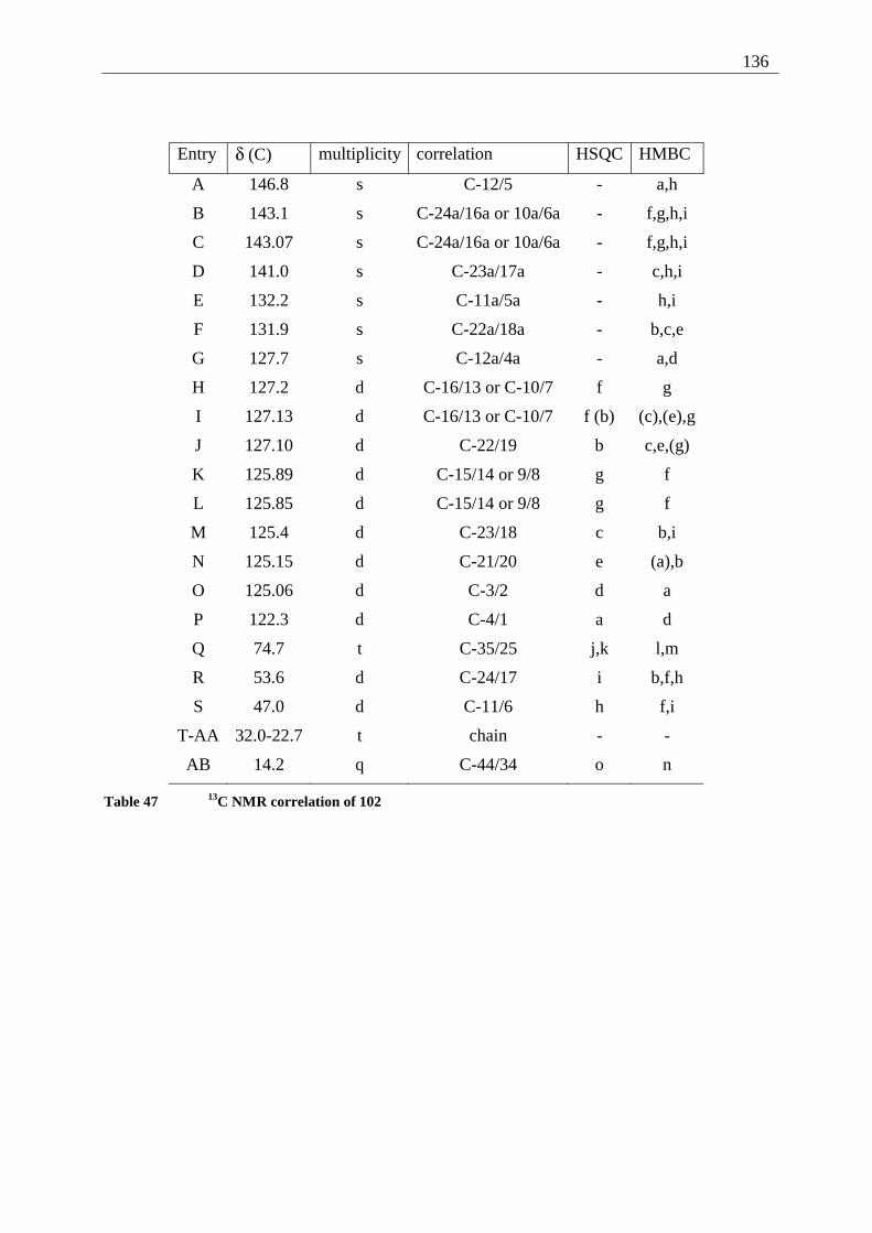
136
Entry δ (C) multiplicity correlation HSQC HMBC
A 146.8 s C-12/5 - a,h
B 143.1 s C-24a/16a or 10a/6a - f,g,h,i
C 143.07 s C-24a/16a or 10a/6a - f,g,h,i
D 141.0 s C-23a/17a - c,h,i
E 132.2 s C-11a/5a - h,i
F 131.9 s C-22a/18a - b,c,e
G 127.7 s C-12a/4a - a,d
H 127.2 d C-16/13 or C-10/7 f g
I 127.13 d C-16/13 or C-10/7 f (b) (c),(e),g
J 127.10 d C-22/19 b c,e,(g)
K 125.89 d C-15/14 or 9/8 g f
L 125.85 d C-15/14 or 9/8 g f
M 125.4 d C-23/18 c b,i
N 125.15 d C-21/20 e (a),b
O 125.06 d C-3/2 d a
P 122.3 d C-4/1 a d
Q 74.7 t C-35/25 j,k l,m
R 53.6 d C-24/17 i b,f,h
S 47.0 d C-11/6 h f,i
T-AA 32.0-22.7 t chain - -
AB 14.2 q C-44/34 o n
Table 47 13C NMR correlation of 102
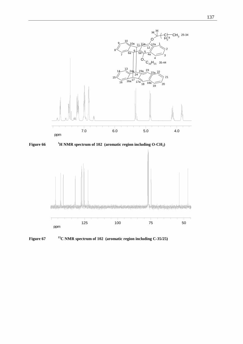
137
ppm4.05.06.07.0
O
O
H HCH2
8CH3
C10H21
1
2
34
567
8
910
1112
23
4a5a6a
10a 11a 12a
16a 17a 18a
1322a23a24a14
15
1617
1819
20
21
2224
25-34
35-44
Figure 66 1H NMR spectrum of 102 (aromatic region including O-CH2)
ppm5075100125
Figure 67 13C NMR spectrum of 102 (aromatic region including C-35/25)
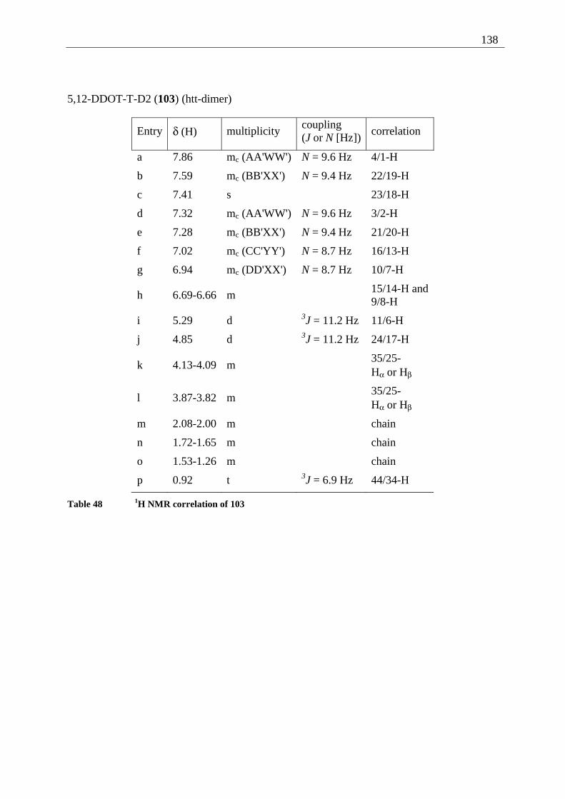
138
5,12-DDOT-T-D2 (103) (htt-dimer)
Entry δ (H) multiplicity coupling (J or N [Hz])
correlation
a 7.86 mc (AA'WW') N = 9.6 Hz 4/1-H
b 7.59 mc (BB'XX') N = 9.4 Hz 22/19-H
c 7.41 s 23/18-H
d 7.32 mc (AA'WW') N = 9.6 Hz 3/2-H
e 7.28 mc (BB'XX') N = 9.4 Hz 21/20-H
f 7.02 mc (CC'YY') N = 8.7 Hz 16/13-H
g 6.94 mc (DD'XX') N = 8.7 Hz 10/7-H
h 6.69-6.66 m 15/14-H and
9/8-H
i 5.29 d 3J = 11.2 Hz 11/6-H
j 4.85 d 3J = 11.2 Hz 24/17-H
k 4.13-4.09 m 35/25-
Hα or Hβ
l 3.87-3.82 m 35/25-
Hα or Hβ
m 2.08-2.00 m chain
n 1.72-1.65 m chain
o 1.53-1.26 m chain
p 0.92 t 3J = 6.9 Hz 44/34-H
Table 48 1H NMR correlation of 103
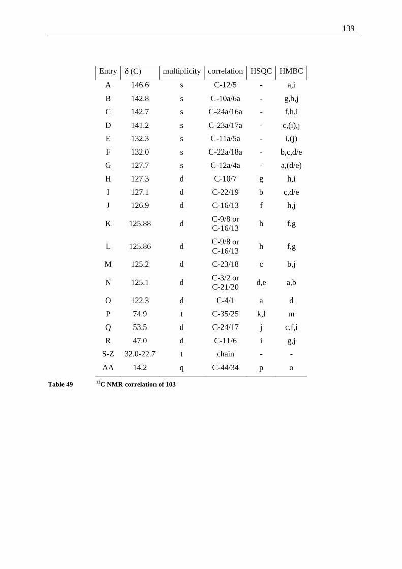
139
Entry δ (C) multiplicity correlation HSQC HMBC
A 146.6 s C-12/5 - a,i
B 142.8 s C-10a/6a - g,h,j
C 142.7 s C-24a/16a - f,h,i
D 141.2 s C-23a/17a - c,(i),j
E 132.3 s C-11a/5a - i,(j)
F 132.0 s C-22a/18a - b,c,d/e
G 127.7 s C-12a/4a - a,(d/e)
H 127.3 d C-10/7 g h,i
I 127.1 d C-22/19 b c,d/e
J 126.9 d C-16/13 f h,j
K 125.88 d C-9/8 or C-16/13
h f,g
L 125.86 d C-9/8 or C-16/13
h f,g
M 125.2 d C-23/18 c b,j
N 125.1 d C-3/2 or C-21/20
d,e a,b
O 122.3 d C-4/1 a d
P 74.9 t C-35/25 k,l m
Q 53.5 d C-24/17 j c,f,i
R 47.0 d C-11/6 i g,j
S-Z 32.0-22.7 t chain - -
AA 14.2 q C-44/34 p o
Table 49 13C NMR correlation of 103
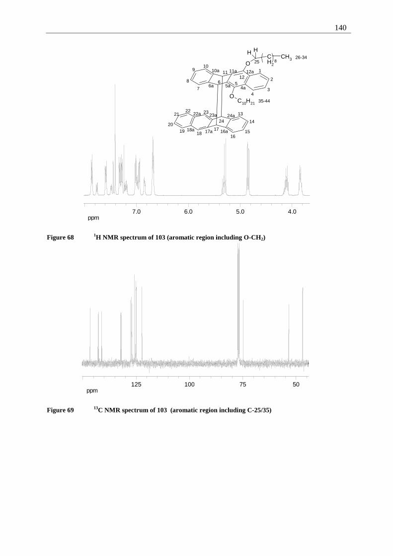
140
ppm4.05.06.07.0
O
O
H HCH2
8CH3
C10H21
1
2
34
567
8
910
1112
23
4a5a6a
10a 11a 12a
16a17a18a
1322a 23a 24a14
1516
171819
20
2122
24
2526-34
35-44
Figure 68 1H NMR spectrum of 103 (aromatic region including O-CH2)
ppm5075100125
Figure 69 13C NMR spectrum of 103 (aromatic region including C-25/35)
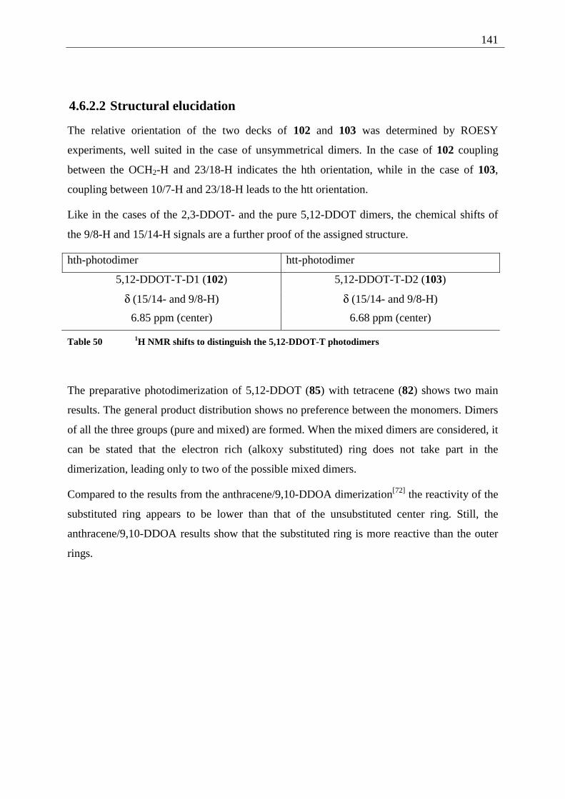
141
4.6.2.2 Structural elucidation
The relative orientation of the two decks of 102 and 103 was determined by ROESY
experiments, well suited in the case of unsymmetrical dimers. In the case of 102 coupling
between the OCH2-H and 23/18-H indicates the hth orientation, while in the case of 103,
coupling between 10/7-H and 23/18-H leads to the htt orientation.
Like in the cases of the 2,3-DDOT- and the pure 5,12-DDOT dimers, the chemical shifts of
the 9/8-H and 15/14-H signals are a further proof of the assigned structure.
hth-photodimer htt-photodimer
5,12-DDOT-T-D1 (102) 5,12-DDOT-T-D2 (103)
δ (15/14- and 9/8-H) δ (15/14- and 9/8-H)
6.85 ppm (center) 6.68 ppm (center)
Table 50 1H NMR shifts to distinguish the 5,12-DDOT-T photodimers
The preparative photodimerization of 5,12-DDOT (85) with tetracene (82) shows two main
results. The general product distribution shows no preference between the monomers. Dimers
of all the three groups (pure and mixed) are formed. When the mixed dimers are considered, it
can be stated that the electron rich (alkoxy substituted) ring does not take part in the
dimerization, leading only to two of the possible mixed dimers.
Compared to the results from the anthracene/9,10-DDOA dimerization[72] the reactivity of the
substituted ring appears to be lower than that of the unsubstituted center ring. Still, the
anthracene/9,10-DDOA results show that the substituted ring is more reactive than the outer
rings.
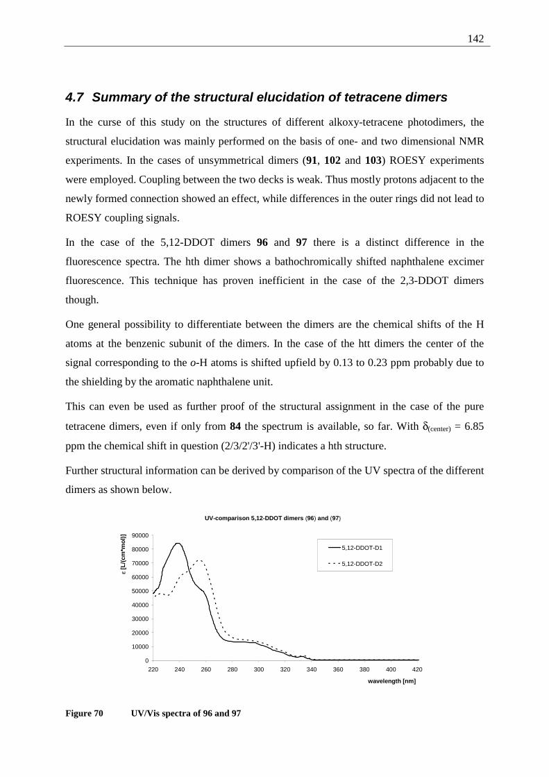
142
4.7 Summary of the structural elucidation of tetracene dimers
In the curse of this study on the structures of different alkoxy-tetracene photodimers, the
structural elucidation was mainly performed on the basis of one- and two dimensional NMR
experiments. In the cases of unsymmetrical dimers (91, 102 and 103) ROESY experiments
were employed. Coupling between the two decks is weak. Thus mostly protons adjacent to the
newly formed connection showed an effect, while differences in the outer rings did not lead to
ROESY coupling signals.
In the case of the 5,12-DDOT dimers 96 and 97 there is a distinct difference in the
fluorescence spectra. The hth dimer shows a bathochromically shifted naphthalene excimer
fluorescence. This technique has proven inefficient in the case of the 2,3-DDOT dimers
though.
One general possibility to differentiate between the dimers are the chemical shifts of the H
atoms at the benzenic subunit of the dimers. In the case of the htt dimers the center of the
signal corresponding to the o-H atoms is shifted upfield by 0.13 to 0.23 ppm probably due to
the shielding by the aromatic naphthalene unit.
This can even be used as further proof of the structural assignment in the case of the pure
tetracene dimers, even if only from 84 the spectrum is available, so far. With δ(center) = 6.85
ppm the chemical shift in question (2/3/2'/3'-H) indicates a hth structure.
Further structural information can be derived by comparison of the UV spectra of the different
dimers as shown below.
UV-comparison 5,12-DDOT dimers (96) and (97)
0
10000
20000
30000
40000
50000
60000
70000
80000
90000
220 240 260 280 300 320 340 360 380 400 420
wavelength [nm]
ε [L
/(cm
*mo
l)]
5,12-DDOT-D1
5,12-DDOT-D2
Figure 70 UV/Vis spectra of 96 and 97
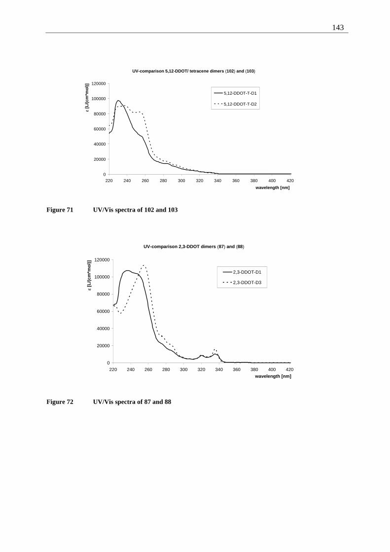
143
UV-comparison 5,12-DDOT/ tetracene dimers (102) and (103)
0
20000
40000
60000
80000
100000
120000
220 240 260 280 300 320 340 360 380 400 420
wavelength [nm]
ε [L
/(cm
*mo
l)]
5,12-DDOT-T-D1
5,12-DDOT-T-D2
Figure 71 UV/Vis spectra of 102 and 103
UV-comparison 2,3-DDOT dimers (87) and (88)
0
20000
40000
60000
80000
100000
120000
220 240 260 280 300 320 340 360 380 400 420
wavelength [nm]
ε [L
/(cm
*mo
l)]
2,3-DDOT-D1
2,3-DDOT-D3
Figure 72 UV/Vis spectra of 87 and 88
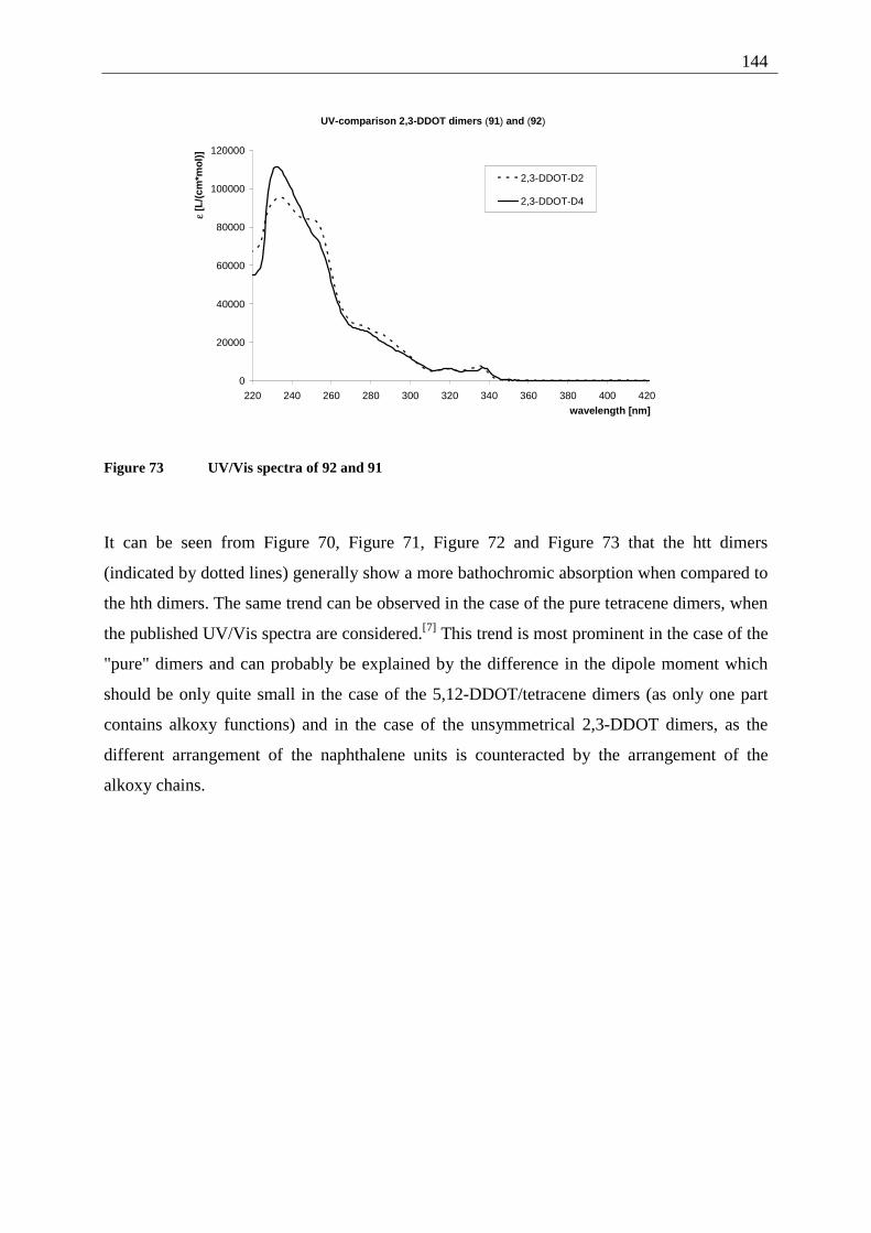
144
UV-comparison 2,3-DDOT dimers (91) and (92)
0
20000
40000
60000
80000
100000
120000
220 240 260 280 300 320 340 360 380 400 420
wavelength [nm]
ε [L
/(cm
*mo
l)]
2,3-DDOT-D2
2,3-DDOT-D4
Figure 73 UV/Vis spectra of 92 and 91
It can be seen from Figure 70, Figure 71, Figure 72 and Figure 73 that the htt dimers
(indicated by dotted lines) generally show a more bathochromic absorption when compared to
the hth dimers. The same trend can be observed in the case of the pure tetracene dimers, when
the published UV/Vis spectra are considered.[7] This trend is most prominent in the case of the
"pure" dimers and can probably be explained by the difference in the dipole moment which
should be only quite small in the case of the 5,12-DDOT/tetracene dimers (as only one part
contains alkoxy functions) and in the case of the unsymmetrical 2,3-DDOT dimers, as the
different arrangement of the naphthalene units is counteracted by the arrangement of the
alkoxy chains.
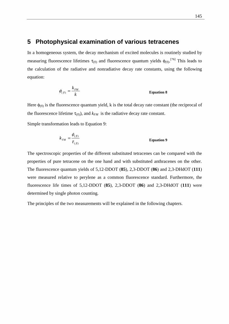
145
5 Photophysical examination of various tetracenes
In a homogeneous system, the decay mechanism of excited molecules is routinely studied by
measuring fluorescence lifetimes τ(fl) and fluorescence quantum yields φ(fl).[76] This leads to
the calculation of the radiative and nonradiative decay rate constants, using the following
equation:
k
kFMfl =)(φ Equation 8
Here φ(fl) is the fluorescence quantum yield, k is the total decay rate constant (the reciprocal of
the fluorescence lifetime τ(fl)), and kFM is the radiative decay rate constant.
Simple transformation leads to Equation 9:
)(
)(
fl
flFMk
τφ
= Equation 9
The spectroscopic properties of the different substituted tetracenes can be compared with the
properties of pure tetracene on the one hand and with substituted anthracenes on the other.
The fluorescence quantum yields of 5,12-DDOT (85), 2,3-DDOT (86) and 2,3-DHdOT (111)
were measured relative to perylene as a common fluorescence standard. Furthermore, the
fluorescence life times of 5,12-DDOT (85), 2,3-DDOT (86) and 2,3-DHdOT (111) were
determined by single photon counting.
The principles of the two measurements will be explained in the following chapters.
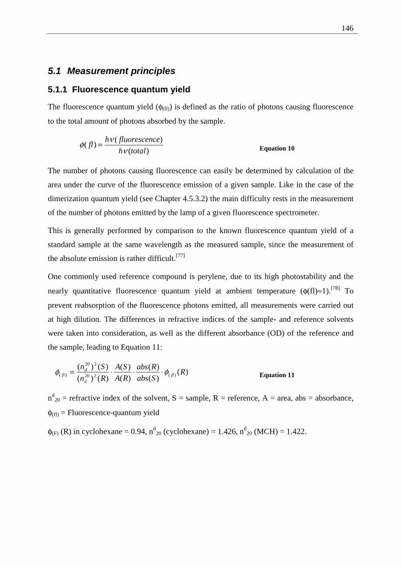
146
5.1 Measurement principles
5.1.1 Fluorescence quantum yield
The fluorescence quantum yield (φ(fl)) is defined as the ratio of photons causing fluorescence
to the total amount of photons absorbed by the sample.
)(
)()(
totalh
cefluorescenhfl
ννφ = Equation 10
The number of photons causing fluorescence can easily be determined by calculation of the
area under the curve of the fluorescence emission of a given sample. Like in the case of the
dimerization quantum yield (see Chapter 4.5.3.2) the main difficulty rests in the measurement
of the number of photons emitted by the lamp of a given fluorescence spectrometer.
This is generally performed by comparison to the known fluorescence quantum yield of a
standard sample at the same wavelength as the measured sample, since the measurement of
the absolute emission is rather difficult.[77]
One commonly used reference compound is perylene, due to its high photostability and the
nearly quantitative fluorescence quantum yield at ambient temperature (φ(fl)≈1).[78] To
prevent reabsorption of the fluorescence photons emitted, all measurements were carried out
at high dilution. The differences in refractive indices of the sample- and reference solvents
were taken into consideration, as well as the different absorbance (OD) of the reference and
the sample, leading to Equation 11:
)()(
)(
)(
)(
)()(
)()()(220
220
)( RSabs
Rabs
RA
SA
Rn
Snfl
d
dfl φφ ⋅⋅⋅= Equation 11
nd20 = refractive index of the solvent, S = sample, R = reference, A = area, abs = absorbance,
φ(fl) = Fluorescence-quantum yield
φ(F) (R) in cyclohexane = 0.94, nd20 (cyclohexane) = 1.426, nd
20 (MCH) = 1.422.
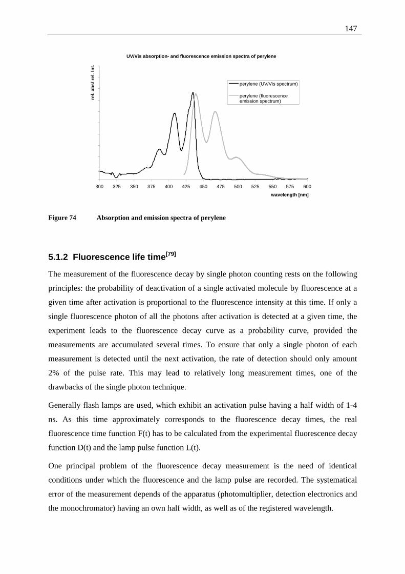
147
UV/Vis absorption- and fluorescence emission spectra of perylene
300 325 350 375 400 425 450 475 500 525 550 575 600
wavelength [nm]
rel.
abs/
rel
. In
t.
perylene (UV/Vis spectrum)
perylene (fluorescenceemission spectrum)
Figure 74 Absorption and emission spectra of perylene
5.1.2 Fluorescence life time[79]
The measurement of the fluorescence decay by single photon counting rests on the following
principles: the probability of deactivation of a single activated molecule by fluorescence at a
given time after activation is proportional to the fluorescence intensity at this time. If only a
single fluorescence photon of all the photons after activation is detected at a given time, the
experiment leads to the fluorescence decay curve as a probability curve, provided the
measurements are accumulated several times. To ensure that only a single photon of each
measurement is detected until the next activation, the rate of detection should only amount
2% of the pulse rate. This may lead to relatively long measurement times, one of the
drawbacks of the single photon technique.
Generally flash lamps are used, which exhibit an activation pulse having a half width of 1-4
ns. As this time approximately corresponds to the fluorescence decay times, the real
fluorescence time function F(t) has to be calculated from the experimental fluorescence decay
function D(t) and the lamp pulse function L(t).
One principal problem of the fluorescence decay measurement is the need of identical
conditions under which the fluorescence and the lamp pulse are recorded. The systematical
error of the measurement depends of the apparatus (photomultiplier, detection electronics and
the monochromator) having an own half width, as well as of the registered wavelength.
148
Another important influence is the color effect of the photomultiplier. This can be explained
by the velocity dependence of the photoelectrons, detected by the photomultiplier, from the
wavelength of the photons that led to emission of the photoelectrons. The dependency leads to
a time shift between lamp pulse and fluorescence, described by the shift parameter δ. The
experimental signal (from which the fluorescence signal is calculated) can be described by:
∫ +−=t
dssFstLtD0
)()()( δ Equation 12
Without providing a full theoretical analysis of the underlying mathematical procedures, it
can be stated that the fluorescence decay F(t) is determined from Equation 12 by iterative
reconvolution. A theoretical function D'(t) is fitted by variation of the parameters to the
experimental curve D(t) by non-linear regression.
Two parameters are generally used to show the quality of this regression. The square error χ2
and the Durbin-Watson factor DW. They are defined by the following equations and provided
directly by the measurement software:
νχ
∑==
)(
)(
2
2
en
aniiR
Equation 13
with )(
)(')(
i
iii
tD
tDtDR
−= Equation 14
where ν is the number of degrees of freedom (ν = ne-na+1-p), p is the number of fit parameters
and ne-na+1 equals the number of channels used to fit the curve.
2
)(
1)(
21 )(
νχ
∑+=
−−=
en
aniii RR
DW Equation 15
In Chapter 5.3 the fluorescence decay times as well as the χ2 and DW values are presented for
the different tetracenes.
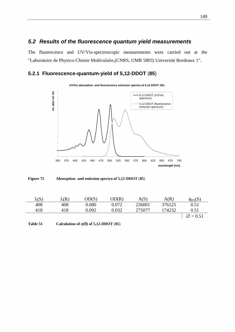
149
5.2 Results of the fluorescence quantum yield measurements
The fluorescence and UV/Vis-spectroscopic measurements were carried out at the
"Laboratoire de Physico-Chimie Moléculaire,(CNRS, UMR 5803) Université Bordeaux 1".
5.2.1 Fluorescence-quantum-yield of 5,12-DDOT (85)
UV/Vis absorption- and fluorescence emission spectra of 5,12-DDOT (85)
350 375 400 425 450 475 500 525 550 575 600 625 650 675 700
wavelength [nm]
rel.
abs/
rel
. In
t.
5,12-DDOT (UV/Visspectrum)
5,12-DDOT (fluorescenceemission spectrum)
Figure 75 Absorption- and emission spectra of 5,12-DDOT (85)
λ(S) λ(R) OD(S) OD(R) A(S) A(R) φ(F)(S) 408 408 0.080 0.072 226001 376125 0.51 418 418 0.092 0.032 275077 174232 0.51
∅ = 0.51
Table 51 Calculation of φ(fl) of 5,12-DDOT (85)
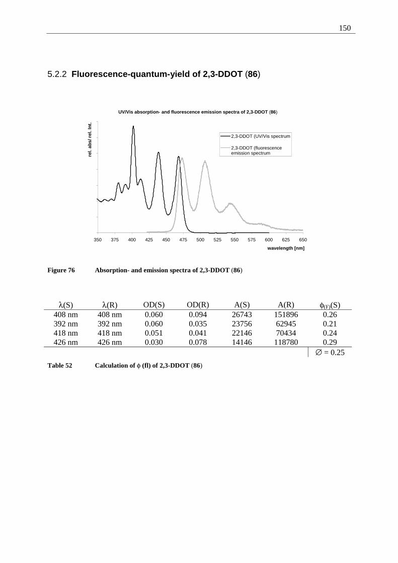
150
5.2.2 Fluorescence-quantum-yield of 2,3-DDOT (86)
UV/Vis absorption- and fluorescence emission spectra of 2,3-DDOT (86)
350 375 400 425 450 475 500 525 550 575 600 625 650
wavelength [nm]
rel.
abs/
rel
. In
t.
2,3-DDOT (UV/Vis spectrum
2,3-DDOT (fluorescenceemission spectrum
Figure 76 Absorption- and emission spectra of 2,3-DDOT (86)
λ(S) λ(R) OD(S) OD(R) A(S) A(R) φ(F)(S) 408 nm 408 nm 0.060 0.094 26743 151896 0.26 392 nm 392 nm 0.060 0.035 23756 62945 0.21 418 nm 418 nm 0.051 0.041 22146 70434 0.24 426 nm 426 nm 0.030 0.078 14146 118780 0.29
∅ = 0.25
Table 52 Calculation of φ (fl) of 2,3-DDOT (86)
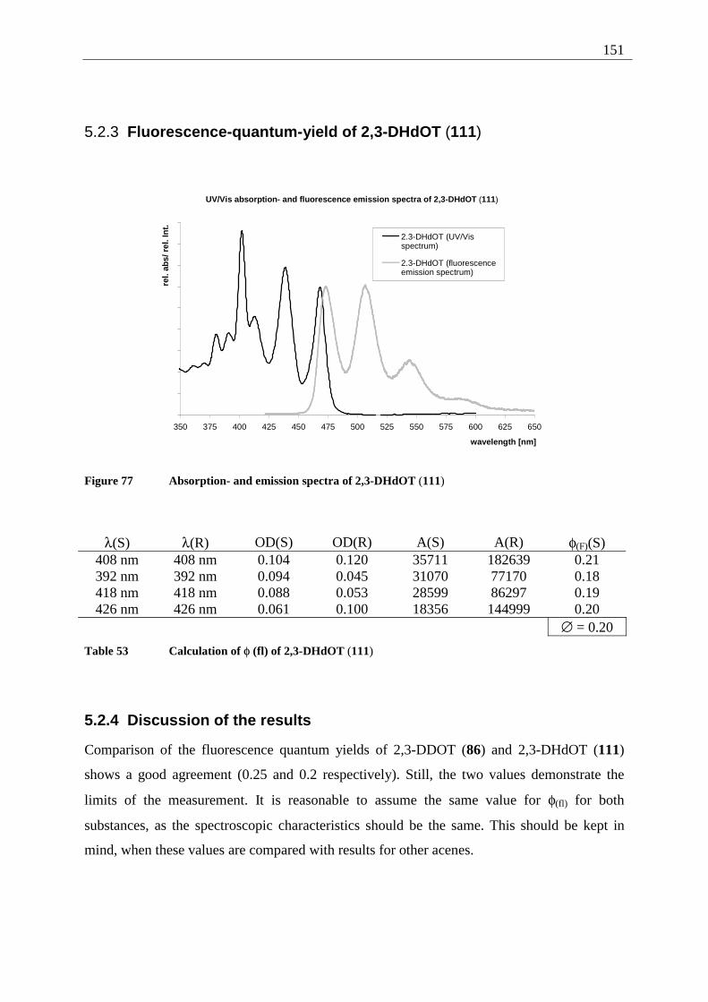
151
5.2.3 Fluorescence-quantum-yield of 2,3-DHdOT (111)
UV/Vis absorption- and fluorescence emission spectra of 2,3-DHdOT (111)
350 375 400 425 450 475 500 525 550 575 600 625 650
wavelength [nm]
rel.
abs/
rel
. In
t.
2.3-DHdOT (UV/Visspectrum)
2.3-DHdOT (fluorescenceemission spectrum)
Figure 77 Absorption- and emission spectra of 2,3-DHdOT (111)
λ(S) λ(R) OD(S) OD(R) A(S) A(R) φ(F)(S) 408 nm 408 nm 0.104 0.120 35711 182639 0.21 392 nm 392 nm 0.094 0.045 31070 77170 0.18 418 nm 418 nm 0.088 0.053 28599 86297 0.19 426 nm 426 nm 0.061 0.100 18356 144999 0.20
∅ = 0.20
Table 53 Calculation of φ (fl) of 2,3-DHdOT (111)
5.2.4 Discussion of the results
Comparison of the fluorescence quantum yields of 2,3-DDOT (86) and 2,3-DHdOT (111)
shows a good agreement (0.25 and 0.2 respectively). Still, the two values demonstrate the
limits of the measurement. It is reasonable to assume the same value for φ(fl) for both
substances, as the spectroscopic characteristics should be the same. This should be kept in
mind, when these values are compared with results for other acenes.
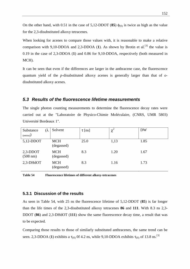
152
On the other hand, with 0.51 in the case of 5,12-DDOT (85) φ(fl) is twice as high as the value
for the 2,3-disubstituted alkoxy tetracenes.
When looking for acenes to compare those values with, it is reasonable to make a relative
comparison with 9,10-DDOA and 2,3-DDOA (1). As shown by Brotin et al.[3] the value is
0.19 in the case of 2,3-DDOA (1) and 0.86 for 9,10-DDOA, respectively (both measured in
MCH).
It can be seen that even if the differences are larger in the anthracene case, the fluorescence
quantum yield of the p-disubstituted alkoxy acenes is generally larger than that of o-
disubstituted alkoxy acenes.
5.3 Results of the fluorescence lifetime measurements
The single photon counting measurements to determine the fluorescence decay rates were
carried out at the "Laboratoire de Physico-Chimie Moléculaire, (CNRS, UMR 5803)
Université Bordeaux 1".
Substance (λ (emiss))
Solvent τ [ns] χ2 DW
5,12-DDOT MCH (degassed)
25.0 1,13 1.85
2,3-DDOT (508 nm)
MCH (degassed)
8.3 1.20 1.67
2,3-DHdOT MCH (degassed)
8.3 1.16 1.73
Table 54 Fluorescence lifetimes of different alkoxy-tetracenes
5.3.1 Discussion of the results
As seen in Table 54, with 25 ns the fluorescence lifetime of 5,12-DDOT (85) is far longer
than the life times of the 2,3-disubstituted alkoxy tetracenes 86 and 111. With 8.3 ns 2,3-
DDOT (86) and 2,3-DHdOT (111) show the same fluorescence decay time, a result that was
to be expected.
Comparing those results to those of similarly substituted anthracenes, the same trend can be
seen. 2,3-DDOA (1) exhibits a τ(fl) 0f 4.2 ns, while 9,10-DDOA exhibits τ(fl) of 13.8 ns.[3]
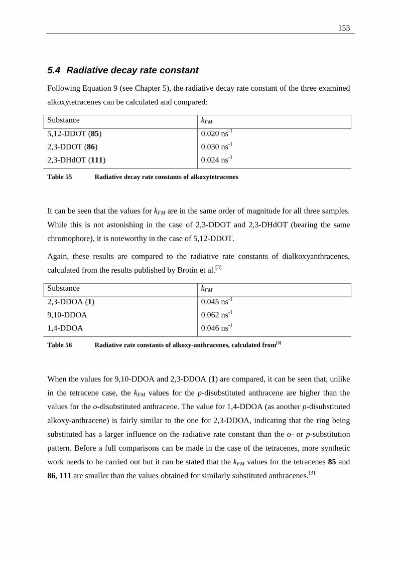
153
5.4 Radiative decay rate constant
Following Equation 9 (see Chapter 5), the radiative decay rate constant of the three examined
alkoxytetracenes can be calculated and compared:
Substance kFM
5,12-DDOT (85) 0.020 ns-1
2,3-DDOT (86) 0.030 ns-1
2,3-DHdOT (111) 0.024 ns-1
Table 55 Radiative decay rate constants of alkoxytetracenes
It can be seen that the values for kFM are in the same order of magnitude for all three samples.
While this is not astonishing in the case of 2,3-DDOT and 2,3-DHdOT (bearing the same
chromophore), it is noteworthy in the case of 5,12-DDOT.
Again, these results are compared to the radiative rate constants of dialkoxyanthracenes,
calculated from the results published by Brotin et al.[3]
Substance kFM
2,3-DDOA (1) 0.045 ns-1
9,10-DDOA 0.062 ns-1
1,4-DDOA 0.046 ns-1
Table 56 Radiative rate constants of alkoxy-anthracenes, calculated from[3]
When the values for 9,10-DDOA and 2,3-DDOA (1) are compared, it can be seen that, unlike
in the tetracene case, the kFM values for the p-disubstituted anthracene are higher than the
values for the o-disubstituted anthracene. The value for 1,4-DDOA (as another p-disubstituted
alkoxy-anthracene) is fairly similar to the one for 2,3-DDOA, indicating that the ring being
substituted has a larger influence on the radiative rate constant than the o- or p-substitution
pattern. Before a full comparisons can be made in the case of the tetracenes, more synthetic
work needs to be carried out but it can be stated that the kFM values for the tetracenes 85 and
86, 111 are smaller than the values obtained for similarly substituted anthracenes.[3]
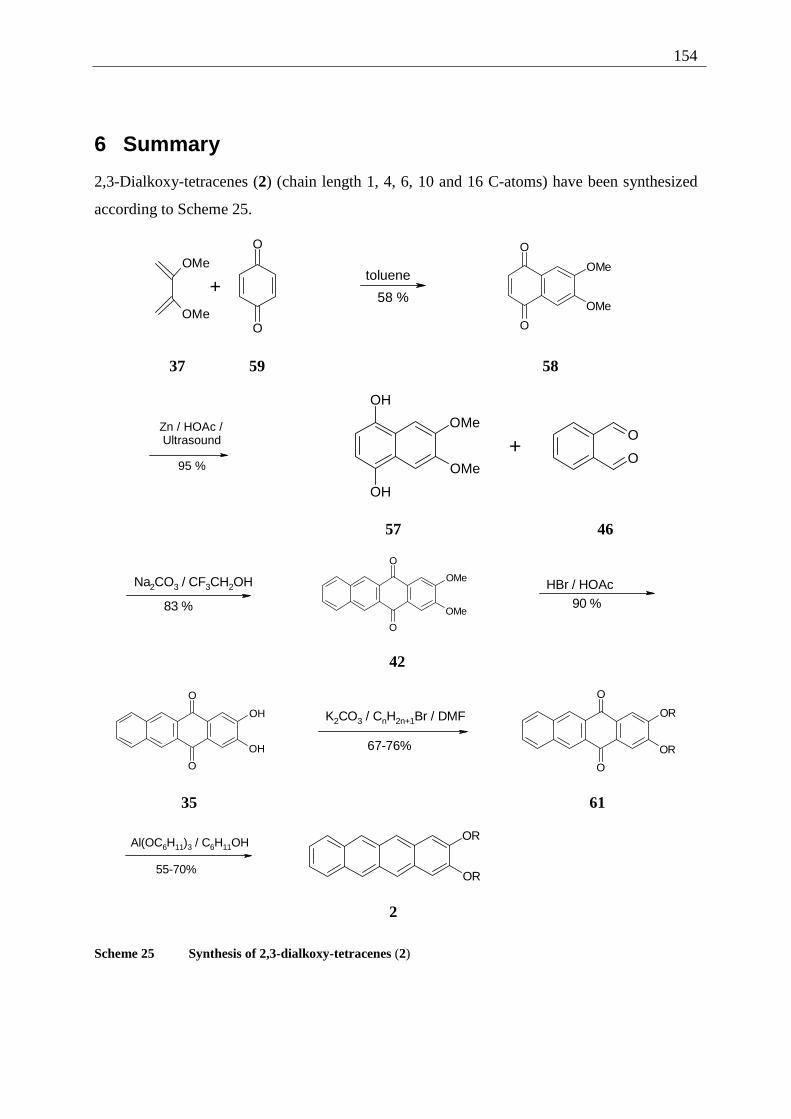
154
6 Summary
2,3-Dialkoxy-tetracenes (2) (chain length 1, 4, 6, 10 and 16 C-atoms) have been synthesized
according to Scheme 25.
+
O
O
OMe
OMe
toluene
58 %
O
O
OMe
OMe
37 59 58
Zn / HOAc / Ultrasound
95 %
OH
OH
OMe
OMe
+O
O
57 46
Na2CO3 / CF3CH2OH
83 %
O
O
OMe
OMe
HBr / HOAc90 %
42
O
O
OH
OH
K2CO3 / CnH2n+1Br / DMF
67-76%
O
O
OR
OR
35 61
Al(OC6H11)3 / C6H11OH
55-70%
OR
OR
2
Scheme 25 Synthesis of 2,3-dialkoxy-tetracenes (2)
155
Overall this sequence furnished the 2,3-dialkoxy-tetracenes (2) in acceptable yields (21% 6
steps).
Gel formation of the 2,3-dialkoxy-tetracenes 2 with various organic solvents was examined
under aspects of chain length and temperature dependency. Three kinds of experiments were
performed: inverted tube tests to study macroscopic gel formation, temperature dependent
UV/Vis spectroscopy to gain insight into the thermodynamics of the gel formation and AFM
(atomic force microscopy) measurements to study the microscopic structures of the formed
gels.
The inverted tube tests have shown that the minimum chain length for 2,3-dialkoxy-tetracenes
(2) to form gels is around C-6. The best results (stable gels with the minimum of gelator) were
obtained with 2,3-dihexadecyloxy-tetracene (111). This shows that the larger aromatic core,
in comparison to 2,3-didecyloxy-anthracene (1), has to be compensated by longer chains to
increase the solubility. Another difference to the parent molecule is a shift in polarity, when
the solvents that form gels are considered. 2,3-DHdOT (111) forms gels mainly in apolar
solvents, the best results (0.55 mg/mL at ambient temperature) were obtained with
cyclohexane, while the best solvent in the case of 2,3-DDOA (1) is methanol.
UV/Vis spectroscopic experiments provided a first insight into the thermodynamics of gel
formation and the melting of gels. It was shown that the phase transitions are subject to a
hysteresis. Furthermore, the thermodynamic values (∆G) for the formation (∆GF) and the
melting (∆GM) of the gels of 2,3-DDOT (86) and 2,3-DHdOT (111) in two solvents
(cyclohexane and n-hexanol) are fairly similar. In the case of the gel melting the values spread
over a wider range. This similarity, along with the comparison of the UV/Vis-spectra of gel
forming 2,3-dialkoxy-tetracenes in the gel state with spectra measured in solid state (KBr
pellets) gives a first idea of the mode of gel formation. It involves dry gels, i. e. no solvent
molecules are incorporated in the gelator structures.
Those structures were more closely examined using AFM measurements leading to an
estimation of the size of the fibers formed. The smallest structures measured so far exhibit a
size of 200 nm (diameter) and several µm (length) and the measurements show a bridged, net
like structure of the gelator (e. g. 2,3-DHdOT (111) in nonane, 0.5 mg/mL) that immobilizes
the solvent, thus forming the gel.
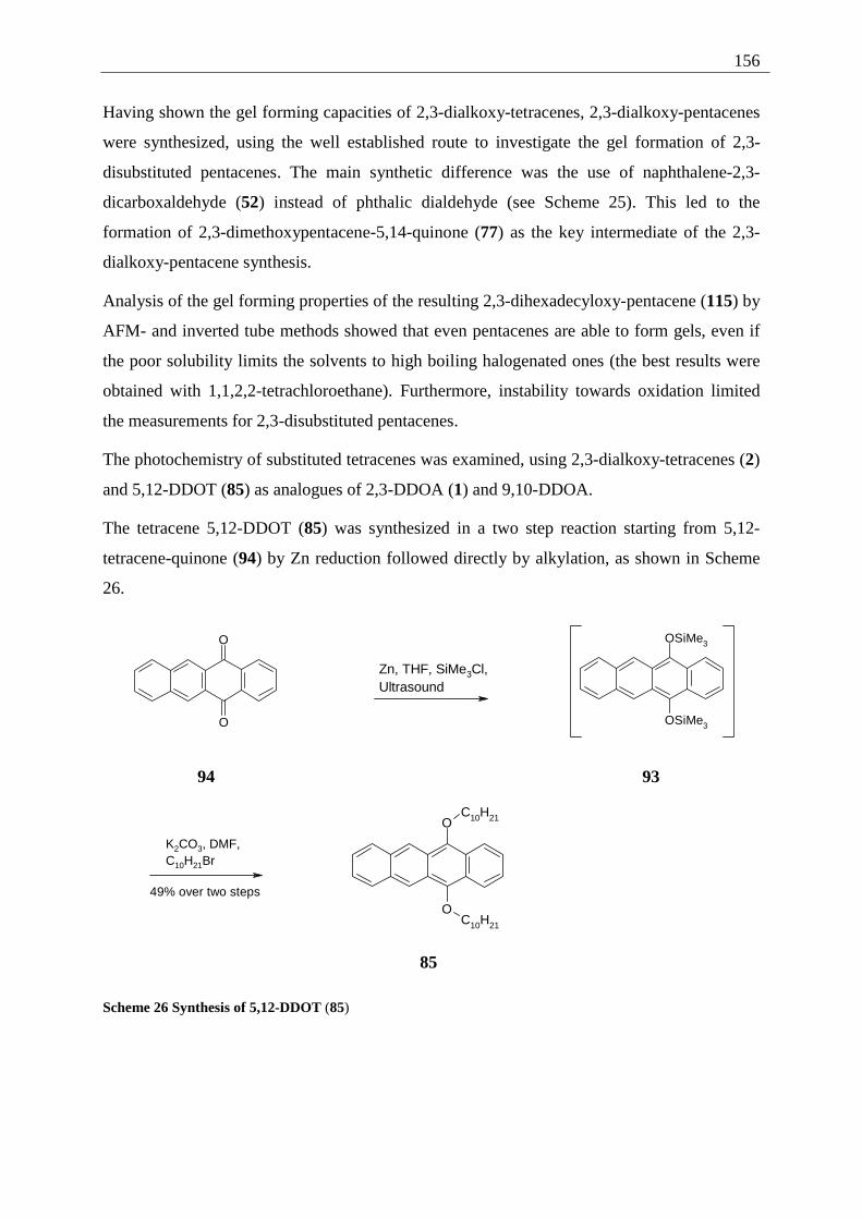
156
Having shown the gel forming capacities of 2,3-dialkoxy-tetracenes, 2,3-dialkoxy-pentacenes
were synthesized, using the well established route to investigate the gel formation of 2,3-
disubstituted pentacenes. The main synthetic difference was the use of naphthalene-2,3-
dicarboxaldehyde (52) instead of phthalic dialdehyde (see Scheme 25). This led to the
formation of 2,3-dimethoxypentacene-5,14-quinone (77) as the key intermediate of the 2,3-
dialkoxy-pentacene synthesis.
Analysis of the gel forming properties of the resulting 2,3-dihexadecyloxy-pentacene (115) by
AFM- and inverted tube methods showed that even pentacenes are able to form gels, even if
the poor solubility limits the solvents to high boiling halogenated ones (the best results were
obtained with 1,1,2,2-tetrachloroethane). Furthermore, instability towards oxidation limited
the measurements for 2,3-disubstituted pentacenes.
The photochemistry of substituted tetracenes was examined, using 2,3-dialkoxy-tetracenes (2)
and 5,12-DDOT (85) as analogues of 2,3-DDOA (1) and 9,10-DDOA.
The tetracene 5,12-DDOT (85) was synthesized in a two step reaction starting from 5,12-
tetracene-quinone (94) by Zn reduction followed directly by alkylation, as shown in Scheme
26.
O
O
Zn, THF, SiMe3Cl,Ultrasound
OSiMe3
OSiMe3
94 93
K2CO3, DMF,C10H21Br
49% over two steps
O
O
C10H21
C10H21
85
Scheme 26 Synthesis of 5,12-DDOT (85)
157
The irradiation of 2,3-DDOT (86) led to several photodimers, in contrast to the 2,3-DDOA
irradiation, that does not provide photodimers. Still, the dimers that can be seen as analogues
of 2,3-DDOA (1) were not formed.
In the case of the 5,12-DDOT (85) neither the pure 5,12-DDOT-dimerization, nor the crossed
dimerization of 5,12-DDOT (85) with tetracene led to dimers under participation of the
alkoxy substituted ring. This is in contrast to the results of the dimerization of 9,10-DDOA
with anthracene that leads to the 9,10-photodimers.
The structures of the different photodimers were investigated by NMR spectroscopy as well
as fluorescence- and UV/Vis-spectroscopy to distinguish the head-to-head from the head-to
tail dimers.
To supplement the photochemical results, measurements of the fluorescence quantum yields
of 5,12-DDOT (85), 2,3-DDOT (86) and 2,3-DHdOT (111) were measured along with
measurements of the fluorescence life times of the compounds to determine the radiative
decay rate constants.
5,12-DDOT dimerisation was shown to be thermally and photochemically reversible, even if
the degradation of the substrate under thermal or UV photochemical conditions was observed.
158
7 Experimental part
7.1 Techniques and equipment
Thin layer chromatography (TLC): TLC- plates „Polygram Sil G/UV254“, Macherey & Nagel
Co. (Düren).
Column chromatography: Flash chromatography was carried out with “Kieselgel 60” (70-230
mesh), Merck KgaA (Darmstadt) under N2 pressure.
Melting points: “Meltemp” electrical melting point apparatus, uncorrected.
Irradiations: The preparative irradiations were carried out in Ar purged solvents with a 1000
W halogen lamp in a glass vessel (Duran: transmittance 5% at 285 nm, 10% at 290 nm).
NMR-spectroscopy: Unless otherwise mentioned, 1H and 13C NMR spectra were measured in
deuterated chloroform using tetramethylsilane (TMS) as internal standard. The multiplicities
are abbreviated as follows: s = singlet, d = doublet, t = triplet, q = quartet, m = multiplet, mc =
multiplet center (used for higher order spin systems).
The coupling constants for the AA'XX'-spin system of compounds 42, 94 and 109 were
analyzed by iterative calculation, using the WinDAISY® 4.1 software (Bruker Rheinstetten).
The calculations were performed by Professor Dr. L. Ernst, TU Braunschweig. R-Factors are
generally better than 0.85%.
In the cases where the spin system was not calculated, the distance between the outer signals
is given in Hz (N-values).
The solid state TOS NMR spectra of 83 and 84 were measured at 4 kHz, using the CH2 signal
of adamanthane (δ = 28.8) as indirect standard.
The following spectrometers were used: Bruker AC 200: 1H NMR (200.2 MHz), 13C (50.3
MHz)
Bruker DRX 400: 1H NMR (400.1 MHz), 13C (100.6 MHz)
Mass spectrometry: Finnigan MAT 8430 Spectrometer. Unless otherwise noted 70 eV
electron impact (EI) ionization was used. High resolution spectra were taken by “peak
matching” with a precision of ± 2 ppm.
159
IR spectroscopy: As KBr pellets on a Nicolet 320 FT-IR-spectrometer or on a diamond ATR
spectrometer Bruker Tensor 27. The intensity of the bands is abbreviated with w (weak, up to
33% of the most intense band), m (medium, 34 - 67% of the most intense band), s (strong,
more than 68% of the most intense band).
UV/Vis absorption spectroscopy: Beckman UV 5230 spectrometer. As solvent DCM of
spectroscopic pure quality was used, unless otherwise stated. The temperature dependent
spectra of the gelators, as well as the absorption spectra to calculate the fluorescence quantum
yields were measured at the Laboratoire de Chimie Organique et Organometallique at the
Université Bordeaux I on a Hitachi U-3300 spectrometer.
Fluorescence spectroscopy: 1680 Spex double-spectrometer at the Laboratoire de Chimie
Organique et Organometallique, Université Bordeaux I. The fluorescence spectra to
distinguish the 2,3-DDOT-photodimers were measured at the TU Braunschweig.
Elementary analysis (EA): The elementary analysis of the compounds were measured at the
Institut für Pharmazeutische Chemie and the Institut für Anorganische und Analytische
Chemie, Technischen Universität Braunschweig.
Fluorescence life times: Single photon timing technique (Applied Photophysics), decay
parameters determined by nonlinear least squares deconvolution method, using the DECAN
(1.0) program, provided by F. De Schryver. Excitation has been performed by a pulsed H2
arc-lamp combined to a band-pass filter, detection has been performed after a monochromator
by photomultiplier. Measured at the Laboratoire de Chimie Organique et Organometallique,
Université Bordeaux I.
7.2 General working procedures (GWP)
7.2.1 GWP 1 (alkylation of dihydroxy-acene-quinones)
The hydroxyacenequinone is dissolved in dry DMF (17 mL/mmol). It is deprotonated over
1 h with K2CO3 (1.25 equivalents per OH group) under N2. After this time the alkylbromide is
added (1.1 equivalents per OH group) over a period of 1 h. The mixture is refluxed for further
3 h. After completion of the reaction the mixture is allowed to cool to room temp. The solvent
is evaporated under vacuum. Column chromatography (silica; DCM/ pentane) gave the
product.
160
7.2.2 GWP 2 (reduction of alkoxy-acene-quinones)
Al powder (6-12 equivalents calculated on the basis of the alkoxy-acene-quinone), HgCl2
(0.2 mg/mmol Al), CCl4 (2 µL/mmol Al) and dry cyclohexanol (0.5-1.0 mL/mmol Al) were
refluxed under N2 or Ar for 3 h (formation of the alkoxide can be observed by formation of a
dark grey solution). The reaction vessel is protected against direct light and the alkoxy-acene-
quinone is added to the cooled down solution. The mixture is refluxed for 1.5 to 3 h. Work up
is carried out as described for the individual experiments.
7.2.3 GWP 3 (photodimerization)
The different tetracenes are dissolved/suspended in cyclohexane in a Teflon® capped glass
reactor equipped with a magnetic stirrer. The concentration is chosen to be formally 0.05 M.
The solution is saturated with Ar (1 min per mL of solution), the reactor is closed and
irradiated for 24-48 h with a 1000 W halogen lamp. After the reaction time has passed, the
reaction is allowed to cool to room temp., the solvent is evaporated under vacuum and the
resulting mixture is separated, using column chromatography under the conditions mentioned
in the individual experimental chapters.
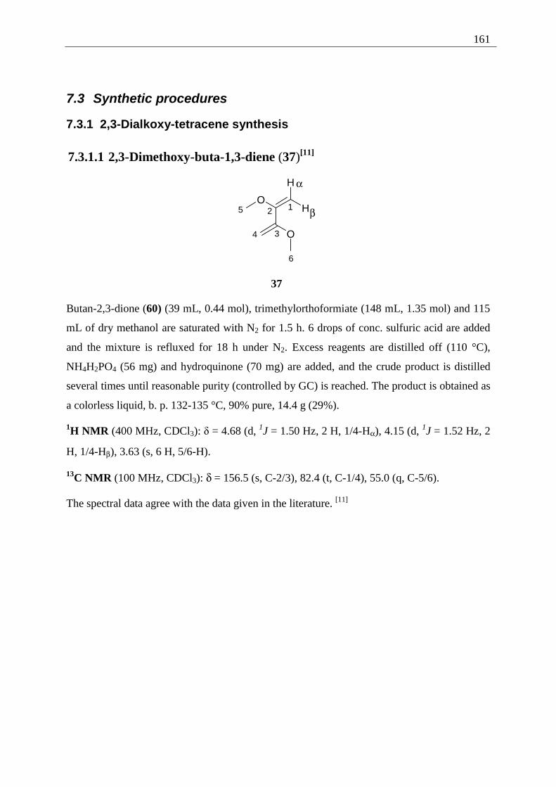
161
7.3 Synthetic procedures
7.3.1 2,3-Dialkoxy-tetracene synthesis
7.3.1.1 2,3-Dimethoxy-buta-1,3-diene (37)[11]
α
β12
34
5
6
O
O
H
H
37
Butan-2,3-dione (60) (39 mL, 0.44 mol), trimethylorthoformiate (148 mL, 1.35 mol) and 115
mL of dry methanol are saturated with N2 for 1.5 h. 6 drops of conc. sulfuric acid are added
and the mixture is refluxed for 18 h under N2. Excess reagents are distilled off (110 °C),
NH4H2PO4 (56 mg) and hydroquinone (70 mg) are added, and the crude product is distilled
several times until reasonable purity (controlled by GC) is reached. The product is obtained as
a colorless liquid, b. p. 132-135 °C, 90% pure, 14.4 g (29%).
1H NMR (400 MHz, CDCl3): δ = 4.68 (d, 1J = 1.50 Hz, 2 H, 1/4-Hα), 4.15 (d, 1J = 1.52 Hz, 2
H, 1/4-Hβ), 3.63 (s, 6 H, 5/6-H).
13C NMR (100 MHz, CDCl3): δ = 156.5 (s, C-2/3), 82.4 (t, C-1/4), 55.0 (q, C-5/6).
The spectral data agree with the data given in the literature. [11]
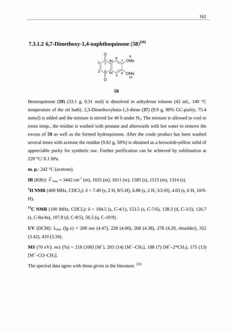
162
7.3.1.2 6,7-Dimethoxy-1,4-naphthoquinone (58)[20]
10
987
65
12
3 4
O
O
OMe
OMe4a
8a
58
Benzoquinone (59) (33.1 g, 0.31 mol) is dissolved in anhydrous toluene (42 mL, 140 °C
temperature of the oil bath). 2,3-Dimethoxybuta-1,3-diene (37) (9.9 g, 90% GC-purity, 75.4
mmol) is added and the mixture is stirred for 40 h under N2. The mixture is allowed to cool to
room temp., the residue is washed with pentane and afterwards with hot water to remove the
excess of 59 as well as the formed hydroquinone. After the crude product has been washed
several times with acetone the residue (9.62 g, 58%) is obtained as a brownish-yellow solid of
appreciable purity for synthetic use. Further purification can be achieved by sublimation at
220 °C/ 0.1 hPa.
m. p.: 242 °C (acetone).
IR (KBr): ν~ max = 3442 cm-1 (m), 1655 (m), 1611 (w), 1585 (s), 1513 (m), 1314 (s).
1H NMR (400 MHz, CDCl3): δ = 7.49 (s, 2 H, 8/5-H), 6.88 (s, 2 H, 3/2-H), 4.03 (s, 6 H, 10/9-
H).
13C NMR (100 MHz, CDCl3): δ = 184.5 (s, C-4/1), 153.5 (s, C-7/6), 138.3 (d, C-3/2), 126.7
(s, C-8a/4a), 107.8 (d, C-8/5), 56.5 (q, C-10/9).
UV (DCM): λmax (lg ε) = 208 nm (4.47), 228 (4.00), 268 (4.38), 278 (4.29, shoulder), 352
(3.42), 410 (3.34).
MS (70 eV): m/z (%) = 218 (100) [M+], 203 (14) [M+–CH3], 188 (7) [M+–2*CH3], 175 (13)
[M+–CO–CH3].
The spectral data agree with those given in the literature. [20]
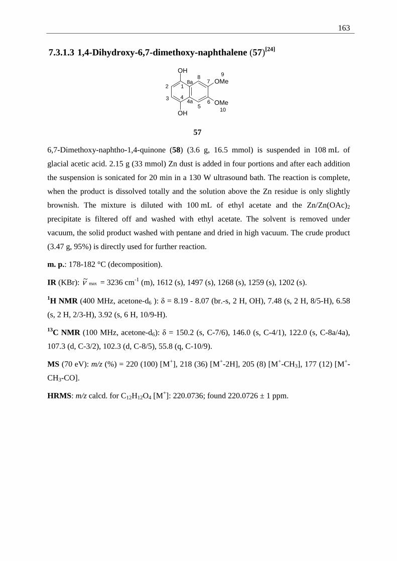
163
7.3.1.3 1,4-Dihydroxy-6,7-dimethoxy-naphthalene (57)[24]
12
3 4
56
78 9
10
OH
OH
OMe
OMe4a
8a
57
6,7-Dimethoxy-naphtho-1,4-quinone (58) (3.6 g, 16.5 mmol) is suspended in 108 mL of
glacial acetic acid. 2.15 g (33 mmol) Zn dust is added in four portions and after each addition
the suspension is sonicated for 20 min in a 130 W ultrasound bath. The reaction is complete,
when the product is dissolved totally and the solution above the Zn residue is only slightly
brownish. The mixture is diluted with 100 mL of ethyl acetate and the Zn/Zn(OAc)2
precipitate is filtered off and washed with ethyl acetate. The solvent is removed under
vacuum, the solid product washed with pentane and dried in high vacuum. The crude product
(3.47 g, 95%) is directly used for further reaction.
m. p.: 178-182 °C (decomposition).
IR (KBr): max~ν = 3236 cm-1 (m), 1612 (s), 1497 (s), 1268 (s), 1259 (s), 1202 (s).
1H NMR (400 MHz, acetone-d6 ): δ = 8.19 - 8.07 (br.-s, 2 H, OH), 7.48 (s, 2 H, 8/5-H), 6.58
(s, 2 H, 2/3-H), 3.92 (s, 6 H, 10/9-H).
13C NMR (100 MHz, acetone-d6): δ = 150.2 (s, C-7/6), 146.0 (s, C-4/1), 122.0 (s, C-8a/4a),
107.3 (d, C-3/2), 102.3 (d, C-8/5), 55.8 (q, C-10/9).
MS (70 eV): m/z (%) = 220 (100) [M+], 218 (36) [M+-2H], 205 (8) [M+-CH3], 177 (12) [M+-
CH3-CO].
HRMS: m/z calcd. for C12H12O4 [M+]: 220.0736; found 220.0726 ± 1 ppm.
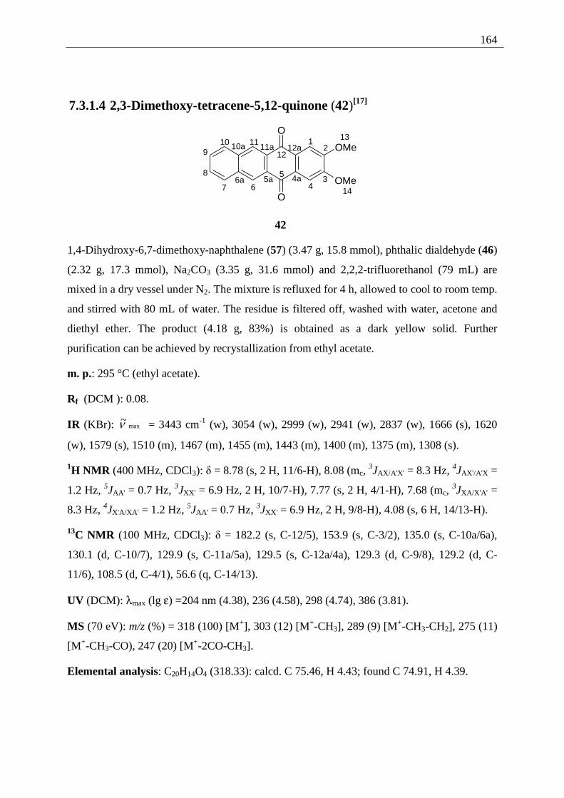
164
7.3.1.4 2,3-Dimethoxy-tetracene-5,12-quinone (42)[17]
12
34
5
67
8
910 11
12
13
14
O
O
OMe
OMe4a5a6a
10a 11a 12a
42
1,4-Dihydroxy-6,7-dimethoxy-naphthalene (57) (3.47 g, 15.8 mmol), phthalic dialdehyde (46)
(2.32 g, 17.3 mmol), Na2CO3 (3.35 g, 31.6 mmol) and 2,2,2-trifluorethanol (79 mL) are
mixed in a dry vessel under N2. The mixture is refluxed for 4 h, allowed to cool to room temp.
and stirred with 80 mL of water. The residue is filtered off, washed with water, acetone and
diethyl ether. The product (4.18 g, 83%) is obtained as a dark yellow solid. Further
purification can be achieved by recrystallization from ethyl acetate.
m. p.: 295 °C (ethyl acetate).
Rf (DCM ): 0.08.
IR (KBr): max~ν = 3443 cm-1 (w), 3054 (w), 2999 (w), 2941 (w), 2837 (w), 1666 (s), 1620
(w), 1579 (s), 1510 (m), 1467 (m), 1455 (m), 1443 (m), 1400 (m), 1375 (m), 1308 (s).
1H NMR (400 MHz, CDCl3): δ = 8.78 (s, 2 H, 11/6-H), 8.08 (mc, 3JAX/A'X' = 8.3 Hz, 4JAX'/A'X =
1.2 Hz, 5JAA' = 0.7 Hz, 3JXX' = 6.9 Hz, 2 H, 10/7-H), 7.77 (s, 2 H, 4/1-H), 7.68 (mc, 3JXA/X'A' =
8.3 Hz, 4JX'A/XA' = 1.2 Hz, 5JAA' = 0.7 Hz, 3JXX' = 6.9 Hz, 2 H, 9/8-H), 4.08 (s, 6 H, 14/13-H).
13C NMR (100 MHz, CDCl3): δ = 182.2 (s, C-12/5), 153.9 (s, C-3/2), 135.0 (s, C-10a/6a),
130.1 (d, C-10/7), 129.9 (s, C-11a/5a), 129.5 (s, C-12a/4a), 129.3 (d, C-9/8), 129.2 (d, C-
11/6), 108.5 (d, C-4/1), 56.6 (q, C-14/13).
UV (DCM): λmax (lg ε) =204 nm (4.38), 236 (4.58), 298 (4.74), 386 (3.81).
MS (70 eV): m/z (%) = 318 (100) [M+], 303 (12) [M+-CH3], 289 (9) [M+-CH3-CH2], 275 (11)
[M+-CH3-CO), 247 (20) [M+-2CO-CH3].
Elemental analysis: C20H14O4 (318.33): calcd. C 75.46, H 4.43; found C 74.91, H 4.39.
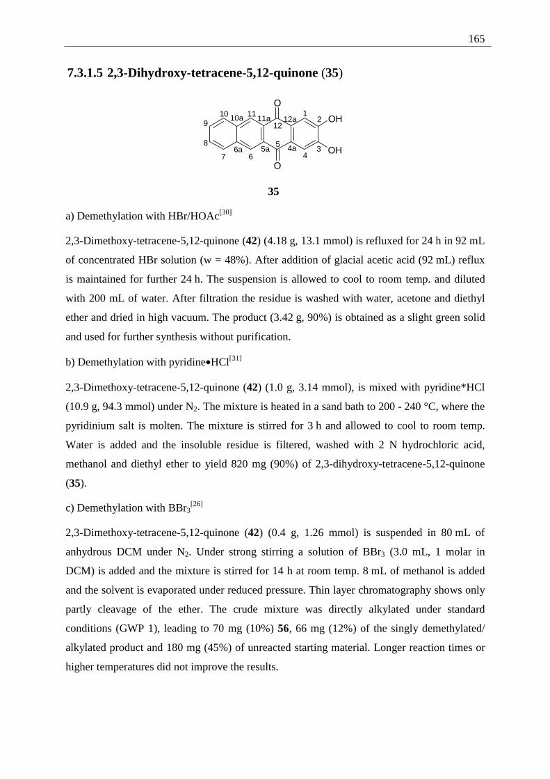
165
7.3.1.5 2,3-Dihydroxy-tetracene-5,12-quinone (35)
12
34
5
67
8
910 11
12
O
O
OH
OH4a5a6a
10a 11a 12a
35
a) Demethylation with HBr/HOAc[30]
2,3-Dimethoxy-tetracene-5,12-quinone (42) (4.18 g, 13.1 mmol) is refluxed for 24 h in 92 mL
of concentrated HBr solution (w = 48%). After addition of glacial acetic acid (92 mL) reflux
is maintained for further 24 h. The suspension is allowed to cool to room temp. and diluted
with 200 mL of water. After filtration the residue is washed with water, acetone and diethyl
ether and dried in high vacuum. The product (3.42 g, 90%) is obtained as a slight green solid
and used for further synthesis without purification.
b) Demethylation with pyridine•HCl[31]
2,3-Dimethoxy-tetracene-5,12-quinone (42) (1.0 g, 3.14 mmol), is mixed with pyridine*HCl
(10.9 g, 94.3 mmol) under N2. The mixture is heated in a sand bath to 200 - 240 °C, where the
pyridinium salt is molten. The mixture is stirred for 3 h and allowed to cool to room temp.
Water is added and the insoluble residue is filtered, washed with 2 N hydrochloric acid,
methanol and diethyl ether to yield 820 mg (90%) of 2,3-dihydroxy-tetracene-5,12-quinone
(35).
c) Demethylation with BBr3[26]
2,3-Dimethoxy-tetracene-5,12-quinone (42) (0.4 g, 1.26 mmol) is suspended in 80 mL of
anhydrous DCM under N2. Under strong stirring a solution of BBr3 (3.0 mL, 1 molar in
DCM) is added and the mixture is stirred for 14 h at room temp. 8 mL of methanol is added
and the solvent is evaporated under reduced pressure. Thin layer chromatography shows only
partly cleavage of the ether. The crude mixture was directly alkylated under standard
conditions (GWP 1), leading to 70 mg (10%) 56, 66 mg (12%) of the singly demethylated/
alkylated product and 180 mg (45%) of unreacted starting material. Longer reaction times or
higher temperatures did not improve the results.
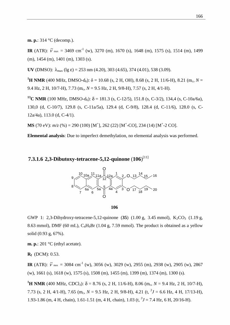
166
m. p.: 314 °C (decomp.).
IR (ATR): max~ν = 3469 cm-1 (w), 3270 (m), 1670 (s), 1648 (m), 1575 (s), 1514 (m), 1499
(m), 1454 (m), 1401 (m), 1303 (s).
UV (DMSO): λmax (lg ε) = 253 nm (4.20), 303 (4.65), 374 (4.01), 538 (3.09).
1H NMR (400 MHz, DMSO-d6): δ = 10.68 (s, 2 H, OH), 8.68 (s, 2 H, 11/6-H), 8.21 (mc, N =
9.4 Hz, 2 H, 10/7-H), 7.73 (mc, N = 9.5 Hz, 2 H, 9/8-H), 7.57 (s, 2 H, 4/1-H).
13C NMR (100 MHz, DMSO-d6): δ = 181.3 (s, C-12/5), 151.8 (s, C-3/2), 134,4 (s, C-10a/6a),
130,0 (d, C-10/7), 129.8 (s, C-11a/5a), 129.4 (d, C-9/8), 128.4 (d, C-11/6), 128.0 (s, C-
12a/4a), 113.0 (d, C-4/1).
MS (70 eV): m/z (%) = 290 (100) [M+], 262 (22) [M+-CO], 234 (14) [M+-2 CO].
Elemental analysis: Due to imperfect demethylation, no elemental analysis was performed.
7.3.1.6 2,3-Dibutoxy-tetracene-5,12-quinone (106)[11]
12
34
5
67
8
910 11
12
O
O
O
O4a5a6a
10a 11a 12a 1513 16
17
14
1819 20
106
GWP 1: 2,3-Dihydroxy-tetracene-5,12-quinone (35) (1.00 g, 3.45 mmol), K2CO3 (1.19 g,
8.63 mmol), DMF (60 mL), C4H9Br (1.04 g, 7.59 mmol). The product is obtained as a yellow
solid (0.93 g, 67%).
m. p.: 201 °C (ethyl acetate).
Rf (DCM): 0.53.
IR (ATR): max~ν = 3084 cm-1 (w), 3056 (w), 3029 (w), 2955 (m), 2938 (w), 2905 (w), 2867
(w), 1661 (s), 1618 (w), 1575 (s), 1508 (m), 1455 (m), 1399 (m), 1374 (m), 1300 (s).
1H NMR (400 MHz, CDCl3): δ = 8.76 (s, 2 H, 11/6-H), 8.06 (mc, N = 9.4 Hz, 2 H, 10/7-H),
7.73 (s, 2 H, 4/1-H), 7.65 (mc, N = 9.5 Hz, 2 H, 9/8-H), 4.21 (t, 3J = 6.6 Hz, 4 H, 17/13-H),
1.93-1.86 (m, 4 H, chain), 1.61-1.51 (m, 4 H, chain), 1.03 (t, 3J = 7.4 Hz, 6 H, 20/16-H).
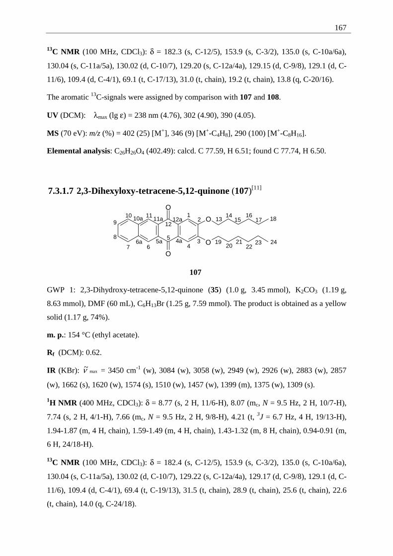
167
13C NMR (100 MHz, CDCl3): δ = 182.3 (s, C-12/5), 153.9 (s, C-3/2), 135.0 (s, C-10a/6a),
130.04 (s, C-11a/5a), 130.02 (d, C-10/7), 129.20 (s, C-12a/4a), 129.15 (d, C-9/8), 129.1 (d, C-
11/6), 109.4 (d, C-4/1), 69.1 (t, C-17/13), 31.0 (t, chain), 19.2 (t, chain), 13.8 (q, C-20/16).
The aromatic 13C-signals were assigned by comparison with 107 and 108.
UV (DCM): λmax (lg ε) = 238 nm (4.76), 302 (4.90), 390 (4.05).
MS (70 eV): m/z (%) = 402 (25) [M+], 346 (9) [M+-C4H8], 290 (100) [M+-C8H16].
Elemental analysis: C26H26O4 (402.49): calcd. C 77.59, H 6.51; found C 77.74, H 6.50.
7.3.1.7 2,3-Dihexyloxy-tetracene-5,12-quinone (107)[11]
1920
12
34
5
67
8
910 11
12
O
O
O
O4a5a6a
10a 11a 12a 151316
1714
18
2122
23 24
107
GWP 1: 2,3-Dihydroxy-tetracene-5,12-quinone (35) (1.0 g, 3.45 mmol), K2CO3 (1.19 g,
8.63 mmol), DMF (60 mL), C6H13Br (1.25 g, 7.59 mmol). The product is obtained as a yellow
solid (1.17 g, 74%).
m. p.: 154 °C (ethyl acetate).
Rf (DCM): 0.62.
IR (KBr): max~ν = 3450 cm-1 (w), 3084 (w), 3058 (w), 2949 (w), 2926 (w), 2883 (w), 2857
(w), 1662 (s), 1620 (w), 1574 (s), 1510 (w), 1457 (w), 1399 (m), 1375 (w), 1309 (s).
1H NMR (400 MHz, CDCl3): δ = 8.77 (s, 2 H, 11/6-H), 8.07 (mc, N = 9.5 Hz, 2 H, 10/7-H),
7.74 (s, 2 H, 4/1-H), 7.66 (mc, N = 9.5 Hz, 2 H, 9/8-H), 4.21 (t, 3J = 6.7 Hz, 4 H, 19/13-H),
1.94-1.87 (m, 4 H, chain), 1.59-1.49 (m, 4 H, chain), 1.43-1.32 (m, 8 H, chain), 0.94-0.91 (m,
6 H, 24/18-H).
13C NMR (100 MHz, CDCl3): δ = 182.4 (s, C-12/5), 153.9 (s, C-3/2), 135.0 (s, C-10a/6a),
130.04 (s, C-11a/5a), 130.02 (d, C-10/7), 129.22 (s, C-12a/4a), 129.17 (d, C-9/8), 129.1 (d, C-
11/6), 109.4 (d, C-4/1), 69.4 (t, C-19/13), 31.5 (t, chain), 28.9 (t, chain), 25.6 (t, chain), 22.6
(t, chain), 14.0 (q, C-24/18).
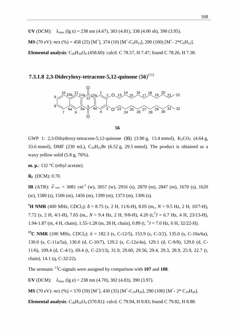
168
UV (DCM): λmax (lg ε) = 238 nm (4.67), 303 (4.81), 338 (4.00 sh), 390 (3.95).
MS (70 eV): m/z (%) = 458 (25) [M+], 374 (10) [M+-C6H12], 290 (100) [M+- 2*C6H12].
Elemental analysis: C30H34O4 (458.60): calcd. C 78.57, H 7.47; found C 78.26, H 7.38.
7.3.1.8 2,3-Didecyloxy-tetracene-5,12-quinone (56)[11]
19201
2
34
5
67
8
910 11
12
O
O
O
O4a5a6a
10a 11a 12a 151316
1714 18
21 22
2324
2526
2728
2930
31 32
56
GWP 1: 2,3-Dihydroxy-tetracene-5,12-quinone (35) (3.90 g, 13.4 mmol), K2CO3 (4.64 g,
33.6 mmol), DMF (230 mL), C10H21Br (6.52 g, 29.5 mmol). The product is obtained as a
waxy yellow solid (5.8 g, 76%).
m. p.: 132 °C (ethyl acetate).
Rf (DCM): 0.70.
IR (ATR): max~ν = 3081 cm-1 (w), 3057 (w), 2916 (s), 2870 (m), 2847 (m), 1670 (s), 1620
(w), 1580 (s), 1506 (m), 1456 (m), 1399 (m), 1373 (m), 1306 (s).
1H NMR (400 MHz, CDCl3): δ = 8.75 (s, 2 H, 11/6-H), 8.05 (mc, N = 9.5 Hz, 2 H, 10/7-H),
7.72 (s, 2 H, 4/1-H), 7.65 (mc, N = 9.4 Hz, 2 H, 9/8-H), 4.20 (t,3J = 6.7 Hz, 4 H, 23/13-H),
1.94-1.87 (m, 4 H, chain), 1.55-1.28 (m, 28 H, chain), 0.89 (t, 3J = 7.0 Hz, 6 H, 32/22-H).
13C NMR (100 MHz, CDCl3): δ = 182.3 (s, C-12/5), 153.9 (s, C-3/2), 135.0 (s, C-10a/6a),
130.0 (s, C-11a/5a), 130.0 (d, C-10/7), 129.2 (s, C-12a/4a), 129.1 (d, C-9/8), 129.0 (d, C-
11/6), 109.4 (d, C-4/1), 69.4 (t, C-23/13), 31.9, 29.60, 29.56, 29.4, 29.3, 28.9, 25.9, 22.7 (t,
chain), 14.1 (q, C-32/22).
The aromatic 13C-signals were assigned by comparison with 107 and 108.
UV (DCM): λmax (lg ε) = 238 nm (4.70), 302 (4.83), 390 (3.97).
MS (70 eV): m/z (%) = 570 (59) [M+], 430 (35) [M+-C10H20], 290 (100) [M+- 2* C10H20].
Elemental analysis: C38H50O4 (570.81): calcd. C 79.94, H 8.83; found C 79.82, H 8.88.
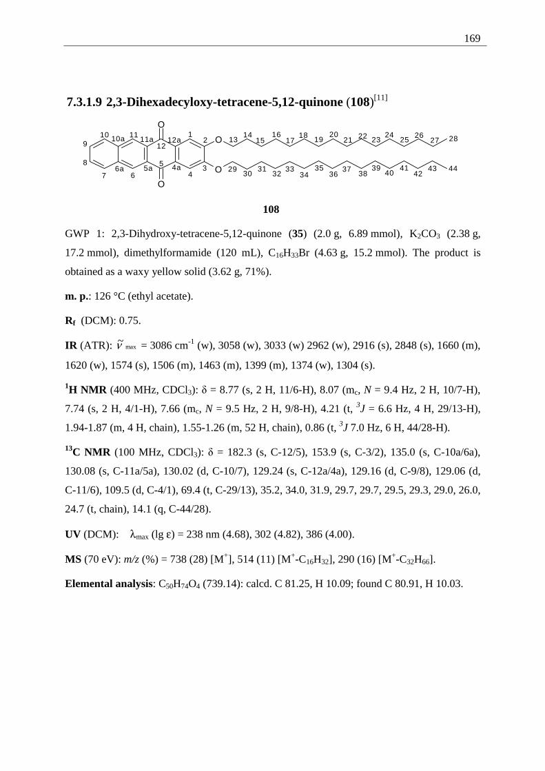
169
7.3.1.9 2,3-Dihexadecyloxy-tetracene-5,12-quinone (108)[11]
19201
2
34
5
67
8
910 11
12
O
O
O
O4a5a6a
10a 11a 12a 151316
1714 18
21 22 2324
2526
27 28
2930
3132
3334
3536
3738
3940
4142
43 44
108
GWP 1: 2,3-Dihydroxy-tetracene-5,12-quinone (35) (2.0 g, 6.89 mmol), K2CO3 (2.38 g,
17.2 mmol), dimethylformamide (120 mL), C16H33Br (4.63 g, 15.2 mmol). The product is
obtained as a waxy yellow solid (3.62 g, 71%).
m. p.: 126 °C (ethyl acetate).
Rf (DCM): 0.75.
IR (ATR): max~ν = 3086 cm-1 (w), 3058 (w), 3033 (w) 2962 (w), 2916 (s), 2848 (s), 1660 (m),
1620 (w), 1574 (s), 1506 (m), 1463 (m), 1399 (m), 1374 (w), 1304 (s).
1H NMR (400 MHz, CDCl3): δ = 8.77 (s, 2 H, 11/6-H), 8.07 (mc, N = 9.4 Hz, 2 H, 10/7-H),
7.74 (s, 2 H, 4/1-H), 7.66 (mc, N = 9.5 Hz, 2 H, 9/8-H), 4.21 (t, 3J = 6.6 Hz, 4 H, 29/13-H),
1.94-1.87 (m, 4 H, chain), 1.55-1.26 (m, 52 H, chain), 0.86 (t, 3J 7.0 Hz, 6 H, 44/28-H).
13C NMR (100 MHz, CDCl3): δ = 182.3 (s, C-12/5), 153.9 (s, C-3/2), 135.0 (s, C-10a/6a),
130.08 (s, C-11a/5a), 130.02 (d, C-10/7), 129.24 (s, C-12a/4a), 129.16 (d, C-9/8), 129.06 (d,
C-11/6), 109.5 (d, C-4/1), 69.4 (t, C-29/13), 35.2, 34.0, 31.9, 29.7, 29.7, 29.5, 29.3, 29.0, 26.0,
24.7 (t, chain), 14.1 (q, C-44/28).
UV (DCM): λmax (lg ε) = 238 nm (4.68), 302 (4.82), 386 (4.00).
MS (70 eV): m/z (%) = 738 (28) [M+], 514 (11) [M+-C16H32], 290 (16) [M+-C32H66].
Elemental analysis: C50H74O4 (739.14): calcd. C 81.25, H 10.09; found C 80.91, H 10.03.
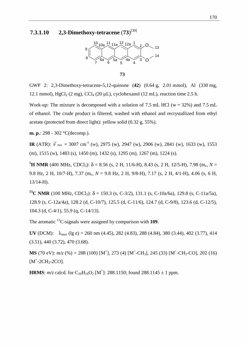
170
7.3.1.10 2,3-Dimethoxy-tetracene (73)[39]
12
34567
8
9
10 11 1213
14
O
O4a5a6a
10a 11a 12a
73
GWP 2: 2,3-Dimethoxy-tetracene-5,12-quinone (42) (0.64 g, 2.01 mmol), Al (330 mg,
12.1 mmol), HgCl2 (2 mg), CCl4 (20 µL), cyclohexanol (12 mL), reaction time 2.5 h.
Work-up: The mixture is decomposed with a solution of 7.5 mL HCl (w = 32%) and 7.5 mL
of ethanol. The crude product is filtered, washed with ethanol and recrystallized from ethyl
acetate (protected from direct light): yellow solid (0.32 g, 55%).
m. p.: 298 - 302 °C(decomp.).
IR (ATR): max~ν = 3007 cm-1 (w), 2975 (w), 2947 (w), 2906 (w), 2841 (w), 1633 (w), 1553
(m), 1515 (w), 1483 (s), 1450 (m), 1432 (s), 1295 (m), 1267 (m), 1224 (s).
1H NMR (400 MHz, CDCl3): δ = 8.56 (s, 2 H, 11/6-H), 8.43 (s, 2 H, 12/5-H), 7.98 (mc, N =
9.8 Hz, 2 H, 10/7-H), 7.37 (mc, N = 9.8 Hz, 2 H, 9/8-H), 7.17 (s, 2 H, 4/1-H), 4.06 (s, 6 H,
13/14-H).
13C NMR (100 MHz, CDCl3): δ = 150.3 (s, C-3/2), 131.1 (s, C-10a/6a), 129.8 (s, C-11a/5a),
128.9 (s, C-12a/4a), 128.2 (d, C-10/7), 125.5 (d, C-11/6), 124.7 (d, C-9/8), 123.6 (d, C-12/5),
104.3 (d, C-4/1), 55.9 (q, C-14/13).
The aromatic 13C-signals were assigned by comparison with 109.
UV (DCM): λmax (lg ε) = 260 nm (4.45), 282 (4.83), 288 (4.84), 380 (3.44), 402 (3.77), 414
(3.51), 440 (3.72), 470 (3.68).
MS (70 eV): m/z (%) = 288 (100) [M+], 273 (4) [M+-CH3], 245 (33) [M+-CH3-CO], 202 (16)
[M+-2CH3-2CO].
HRMS: m/z calcd. for C20H16O2 [M+]: 288.1150; found 288.1145 ± 1 ppm.
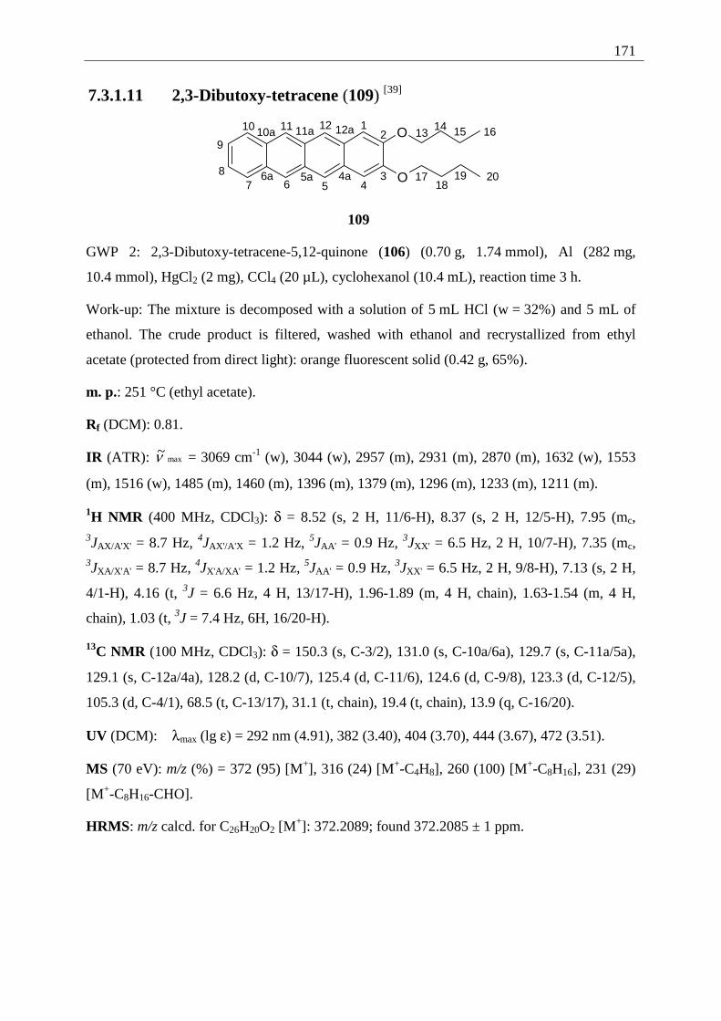
171
7.3.1.11 2,3-Dibutoxy-tetracene (109) [39]
12
34567
8
9
10 11 1213
14O
O4a5a6a
10a 11a 12a 15 16
1718
19 20
109
GWP 2: 2,3-Dibutoxy-tetracene-5,12-quinone (106) (0.70 g, 1.74 mmol), Al (282 mg,
10.4 mmol), HgCl2 (2 mg), CCl4 (20 µL), cyclohexanol (10.4 mL), reaction time 3 h.
Work-up: The mixture is decomposed with a solution of 5 mL HCl (w = 32%) and 5 mL of
ethanol. The crude product is filtered, washed with ethanol and recrystallized from ethyl
acetate (protected from direct light): orange fluorescent solid (0.42 g, 65%).
m. p.: 251 °C (ethyl acetate).
Rf (DCM): 0.81.
IR (ATR): max~ν = 3069 cm-1 (w), 3044 (w), 2957 (m), 2931 (m), 2870 (m), 1632 (w), 1553
(m), 1516 (w), 1485 (m), 1460 (m), 1396 (m), 1379 (m), 1296 (m), 1233 (m), 1211 (m).
1H NMR (400 MHz, CDCl3): δ = 8.52 (s, 2 H, 11/6-H), 8.37 (s, 2 H, 12/5-H), 7.95 (mc, 3JAX/A'X' = 8.7 Hz, 4JAX'/A'X = 1.2 Hz, 5JAA' = 0.9 Hz, 3JXX' = 6.5 Hz, 2 H, 10/7-H), 7.35 (mc, 3JXA/X'A' = 8.7 Hz, 4JX'A/XA' = 1.2 Hz, 5JAA' = 0.9 Hz, 3JXX' = 6.5 Hz, 2 H, 9/8-H), 7.13 (s, 2 H,
4/1-H), 4.16 (t, 3J = 6.6 Hz, 4 H, 13/17-H), 1.96-1.89 (m, 4 H, chain), 1.63-1.54 (m, 4 H,
chain), 1.03 (t, 3J = 7.4 Hz, 6H, 16/20-H).
13C NMR (100 MHz, CDCl3): δ = 150.3 (s, C-3/2), 131.0 (s, C-10a/6a), 129.7 (s, C-11a/5a),
129.1 (s, C-12a/4a), 128.2 (d, C-10/7), 125.4 (d, C-11/6), 124.6 (d, C-9/8), 123.3 (d, C-12/5),
105.3 (d, C-4/1), 68.5 (t, C-13/17), 31.1 (t, chain), 19.4 (t, chain), 13.9 (q, C-16/20).
UV (DCM): λmax (lg ε) = 292 nm (4.91), 382 (3.40), 404 (3.70), 444 (3.67), 472 (3.51).
MS (70 eV): m/z (%) = 372 (95) [M+], 316 (24) [M+-C4H8], 260 (100) [M+-C8H16], 231 (29)
[M+-C8H16-CHO].
HRMS: m/z calcd. for C26H20O2 [M+]: 372.2089; found 372.2085 ± 1 ppm.
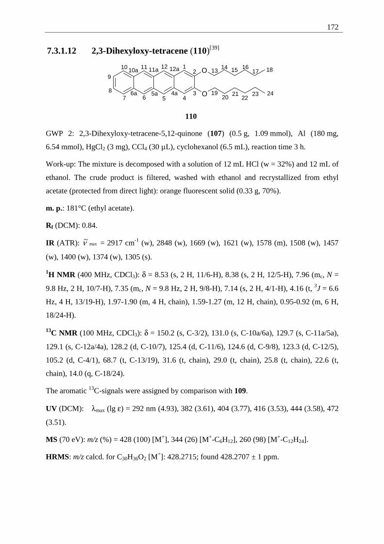
172
7.3.1.12 2,3-Dihexyloxy-tetracene (110)[39]
12
34567
8
9
10 11 1213
14O
O4a5a6a
10a 11a 12a 15 1617 18
1920
2122
23 24
110
GWP 2: 2,3-Dihexyloxy-tetracene-5,12-quinone (107) (0.5 g, 1.09 mmol), Al (180 mg,
6.54 mmol), HgCl2 (3 mg), CCl4 (30 µL), cyclohexanol (6.5 mL), reaction time 3 h.
Work-up: The mixture is decomposed with a solution of 12 mL HCl (w = 32%) and 12 mL of
ethanol. The crude product is filtered, washed with ethanol and recrystallized from ethyl
acetate (protected from direct light): orange fluorescent solid (0.33 g, 70%).
m. p.: 181°C (ethyl acetate).
Rf (DCM): 0.84.
IR (ATR): max~ν = 2917 cm-1 (w), 2848 (w), 1669 (w), 1621 (w), 1578 (m), 1508 (w), 1457
(w), 1400 (w), 1374 (w), 1305 (s).
1H NMR (400 MHz, CDCl3): δ = 8.53 (s, 2 H, 11/6-H), 8.38 (s, 2 H, 12/5-H), 7.96 (mc, N =
9.8 Hz, 2 H, 10/7-H), 7.35 (mc, N = 9.8 Hz, 2 H, 9/8-H), 7.14 (s, 2 H, 4/1-H), 4.16 (t, 3J = 6.6
Hz, 4 H, 13/19-H), 1.97-1.90 (m, 4 H, chain), 1.59-1.27 (m, 12 H, chain), 0.95-0.92 (m, 6 H,
18/24-H).
13C NMR (100 MHz, CDCl3): δ = 150.2 (s, C-3/2), 131.0 (s, C-10a/6a), 129.7 (s, C-11a/5a),
129.1 (s, C-12a/4a), 128.2 (d, C-10/7), 125.4 (d, C-11/6), 124.6 (d, C-9/8), 123.3 (d, C-12/5),
105.2 (d, C-4/1), 68.7 (t, C-13/19), 31.6 (t, chain), 29.0 (t, chain), 25.8 (t, chain), 22.6 (t,
chain), 14.0 (q, C-18/24).
The aromatic 13C-signals were assigned by comparison with 109.
UV (DCM): λmax (lg ε) = 292 nm (4.93), 382 (3.61), 404 (3.77), 416 (3.53), 444 (3.58), 472
(3.51).
MS (70 eV): m/z (%) = 428 (100) [M+], 344 (26) [M+-C6H12], 260 (98) [M+-C12H24].
HRMS: m/z calcd. for C30H36O2 [M+]: 428.2715; found 428.2707 ± 1 ppm.
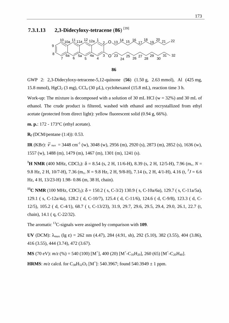
173
7.3.1.13 2,3-Didecyloxy-tetracene (86) [39]
12
34567
8
9
10 11 1213
14O
O4a5a6a
10a 11a 12a 15 1617
1819
20 21 22
2324
2526
2728
2930
31 32
86
GWP 2: 2,3-Didecyloxy-tetracene-5,12-quinone (56) (1.50 g, 2.63 mmol), Al (425 mg,
15.8 mmol), HgCl2 (3 mg), CCl4 (30 µL), cyclohexanol (15.8 mL), reaction time 3 h.
Work-up: The mixture is decomposed with a solution of 30 mL HCl (w = 32%) and 30 mL of
ethanol. The crude product is filtered, washed with ethanol and recrystallized from ethyl
acetate (protected from direct light): yellow fluorescent solid (0.94 g, 66%).
m. p.: 172 - 173°C (ethyl acetate).
Rf (DCM/pentane (1:4)): 0.53.
IR (KBr): max~ν = 3448 cm-1 (w), 3048 (w), 2956 (m), 2920 (s), 2873 (m), 2852 (s), 1636 (w),
1557 (w), 1488 (m), 1479 (m), 1467 (m), 1301 (m), 1241 (s).
1H NMR (400 MHz, CDCl3): δ = 8.54 (s, 2 H, 11/6-H), 8.39 (s, 2 H, 12/5-H), 7.96 (mc, N =
9.8 Hz, 2 H, 10/7-H), 7.36 (mc, N = 9.8 Hz, 2 H, 9/8-H), 7.14 (s, 2 H, 4/1-H), 4.16 (t, 3J = 6.6
Hz, 4 H, 13/23-H) 1.98- 0.86 (m, 38 H, chain).
13C NMR (100 MHz, CDCl3): δ = 150.2 ( s, C-3/2) 130.9 ( s, C-10a/6a), 129.7 ( s, C-11a/5a),
129.1 ( s, C-12a/4a), 128.2 ( d, C-10/7), 125.4 ( d, C-11/6), 124.6 ( d, C-9/8), 123.3 ( d, C-
12/5), 105.2 ( d, C-4/1), 68.7 ( t, C-13/23), 31.9, 29.7, 29.6, 29.5, 29.4, 29.0, 26.1, 22.7 (t,
chain), 14.1 ( q, C-22/32).
The aromatic 13C-signals were assigned by comparison with 109.
UV (DCM): λmax (lg ε) = 262 nm (4.47), 284 (4.91, sh), 292 (5.10), 382 (3.55), 404 (3.86),
416 (3.55), 444 (3.74), 472 (3.67).
MS (70 eV): m/z (%) = 540 (100) [M+], 400 (20) [M+-C10H20], 260 (65) [M+-C20H40].
HRMS: m/z calcd. for C38H52O2 [M+]: 540.3967; found 540.3949 ± 1 ppm.
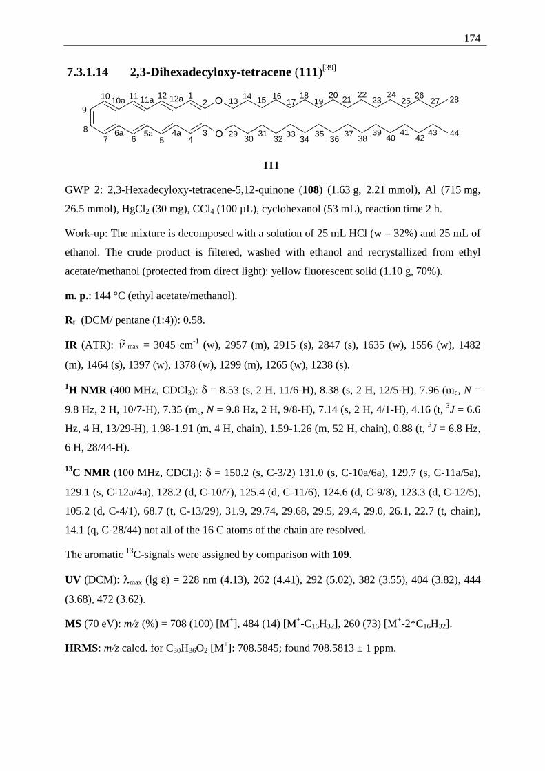
174
7.3.1.14 2,3-Dihexadecyloxy-tetracene (111)[39]
12
34567
8
9
10 11 1213
14O
O4a5a6a
10a 11a 12a 15 1617
1819
20 2122
2324
2526
27 28
2930
3132
3334
3536
3738
3940
4142
43 44
111
GWP 2: 2,3-Hexadecyloxy-tetracene-5,12-quinone (108) (1.63 g, 2.21 mmol), Al (715 mg,
26.5 mmol), HgCl2 (30 mg), CCl4 (100 µL), cyclohexanol (53 mL), reaction time 2 h.
Work-up: The mixture is decomposed with a solution of 25 mL HCl (w = 32%) and 25 mL of
ethanol. The crude product is filtered, washed with ethanol and recrystallized from ethyl
acetate/methanol (protected from direct light): yellow fluorescent solid (1.10 g, 70%).
m. p.: 144 °C (ethyl acetate/methanol).
Rf (DCM/ pentane (1:4)): 0.58.
IR (ATR): max~ν = 3045 cm-1 (w), 2957 (m), 2915 (s), 2847 (s), 1635 (w), 1556 (w), 1482
(m), 1464 (s), 1397 (w), 1378 (w), 1299 (m), 1265 (w), 1238 (s).
1H NMR (400 MHz, CDCl3): δ = 8.53 (s, 2 H, 11/6-H), 8.38 (s, 2 H, 12/5-H), 7.96 (mc, N =
9.8 Hz, 2 H, 10/7-H), 7.35 (mc, N = 9.8 Hz, 2 H, 9/8-H), 7.14 (s, 2 H, 4/1-H), 4.16 (t, 3J = 6.6
Hz, 4 H, 13/29-H), 1.98-1.91 (m, 4 H, chain), 1.59-1.26 (m, 52 H, chain), 0.88 (t, 3J = 6.8 Hz,
6 H, 28/44-H).
13C NMR (100 MHz, CDCl3): δ = 150.2 (s, C-3/2) 131.0 (s, C-10a/6a), 129.7 (s, C-11a/5a),
129.1 (s, C-12a/4a), 128.2 (d, C-10/7), 125.4 (d, C-11/6), 124.6 (d, C-9/8), 123.3 (d, C-12/5),
105.2 (d, C-4/1), 68.7 (t, C-13/29), 31.9, 29.74, 29.68, 29.5, 29.4, 29.0, 26.1, 22.7 (t, chain),
14.1 (q, C-28/44) not all of the 16 C atoms of the chain are resolved.
The aromatic 13C-signals were assigned by comparison with 109.
UV (DCM): λmax (lg ε) = 228 nm (4.13), 262 (4.41), 292 (5.02), 382 (3.55), 404 (3.82), 444
(3.68), 472 (3.62).
MS (70 eV): m/z (%) = 708 (100) [M+], 484 (14) [M+-C16H32], 260 (73) [M+-2*C16H32].
HRMS: m/z calcd. for C30H36O2 [M+]: 708.5845; found 708.5813 ± 1 ppm.
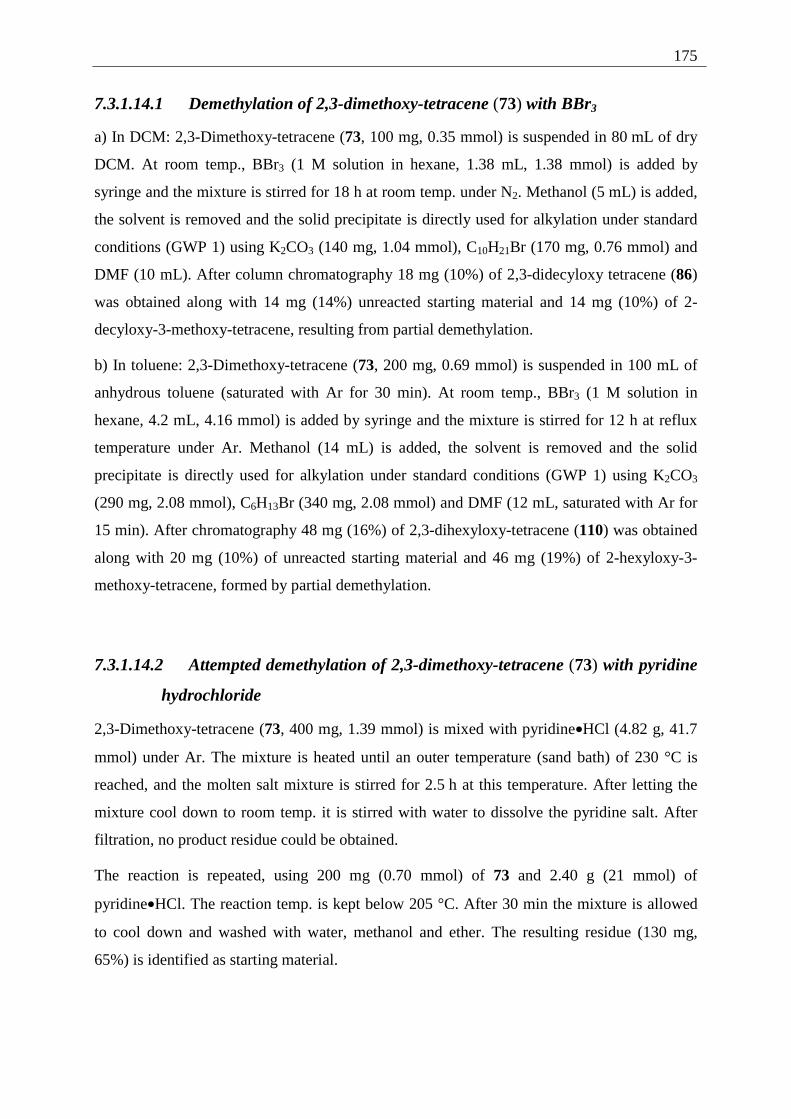
175
7.3.1.14.1 Demethylation of 2,3-dimethoxy-tetracene (73) with BBr3
a) In DCM: 2,3-Dimethoxy-tetracene (73, 100 mg, 0.35 mmol) is suspended in 80 mL of dry
DCM. At room temp., BBr3 (1 M solution in hexane, 1.38 mL, 1.38 mmol) is added by
syringe and the mixture is stirred for 18 h at room temp. under N2. Methanol (5 mL) is added,
the solvent is removed and the solid precipitate is directly used for alkylation under standard
conditions (GWP 1) using K2CO3 (140 mg, 1.04 mmol), C10H21Br (170 mg, 0.76 mmol) and
DMF (10 mL). After column chromatography 18 mg (10%) of 2,3-didecyloxy tetracene (86)
was obtained along with 14 mg (14%) unreacted starting material and 14 mg (10%) of 2-
decyloxy-3-methoxy-tetracene, resulting from partial demethylation.
b) In toluene: 2,3-Dimethoxy-tetracene (73, 200 mg, 0.69 mmol) is suspended in 100 mL of
anhydrous toluene (saturated with Ar for 30 min). At room temp., BBr3 (1 M solution in
hexane, 4.2 mL, 4.16 mmol) is added by syringe and the mixture is stirred for 12 h at reflux
temperature under Ar. Methanol (14 mL) is added, the solvent is removed and the solid
precipitate is directly used for alkylation under standard conditions (GWP 1) using K2CO3
(290 mg, 2.08 mmol), C6H13Br (340 mg, 2.08 mmol) and DMF (12 mL, saturated with Ar for
15 min). After chromatography 48 mg (16%) of 2,3-dihexyloxy-tetracene (110) was obtained
along with 20 mg (10%) of unreacted starting material and 46 mg (19%) of 2-hexyloxy-3-
methoxy-tetracene, formed by partial demethylation.
7.3.1.14.2 Attempted demethylation of 2,3-dimethoxy-tetracene (73) with pyridine
hydrochloride
2,3-Dimethoxy-tetracene (73, 400 mg, 1.39 mmol) is mixed with pyridine•HCl (4.82 g, 41.7
mmol) under Ar. The mixture is heated until an outer temperature (sand bath) of 230 °C is
reached, and the molten salt mixture is stirred for 2.5 h at this temperature. After letting the
mixture cool down to room temp. it is stirred with water to dissolve the pyridine salt. After
filtration, no product residue could be obtained.
The reaction is repeated, using 200 mg (0.70 mmol) of 73 and 2.40 g (21 mmol) of
pyridine•HCl. The reaction temp. is kept below 205 °C. After 30 min the mixture is allowed
to cool down and washed with water, methanol and ether. The resulting residue (130 mg,
65%) is identified as starting material.
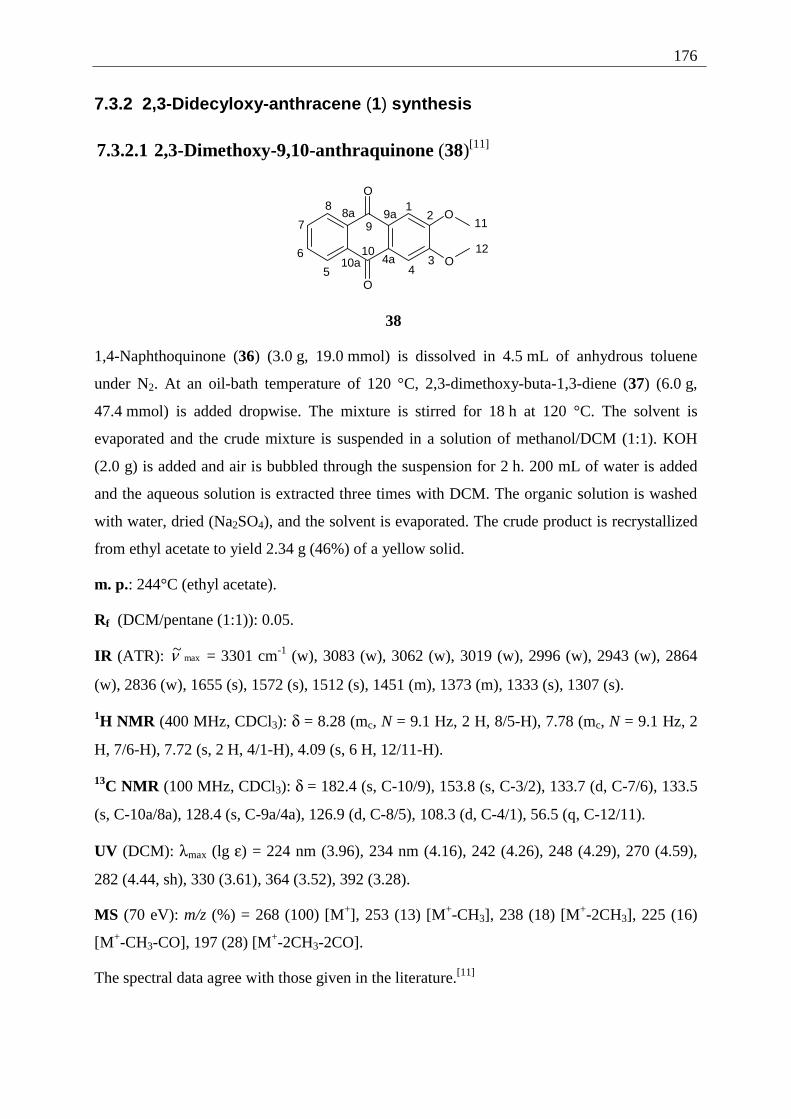
176
7.3.2 2,3-Didecyloxy-anthracene (1) synthesis
7.3.2.1 2,3-Dimethoxy-9,10-anthraquinone (38)[11]
O
O
O
O
12
345
6
7
8
9
10
11
124a10a
8a 9a
38
1,4-Naphthoquinone (36) (3.0 g, 19.0 mmol) is dissolved in 4.5 mL of anhydrous toluene
under N2. At an oil-bath temperature of 120 °C, 2,3-dimethoxy-buta-1,3-diene (37) (6.0 g,
47.4 mmol) is added dropwise. The mixture is stirred for 18 h at 120 °C. The solvent is
evaporated and the crude mixture is suspended in a solution of methanol/DCM (1:1). KOH
(2.0 g) is added and air is bubbled through the suspension for 2 h. 200 mL of water is added
and the aqueous solution is extracted three times with DCM. The organic solution is washed
with water, dried (Na2SO4), and the solvent is evaporated. The crude product is recrystallized
from ethyl acetate to yield 2.34 g (46%) of a yellow solid.
m. p.: 244°C (ethyl acetate).
Rf (DCM/pentane (1:1)): 0.05.
IR (ATR): max~ν = 3301 cm-1 (w), 3083 (w), 3062 (w), 3019 (w), 2996 (w), 2943 (w), 2864
(w), 2836 (w), 1655 (s), 1572 (s), 1512 (s), 1451 (m), 1373 (m), 1333 (s), 1307 (s).
1H NMR (400 MHz, CDCl3): δ = 8.28 (mc, N = 9.1 Hz, 2 H, 8/5-H), 7.78 (mc, N = 9.1 Hz, 2
H, 7/6-H), 7.72 (s, 2 H, 4/1-H), 4.09 (s, 6 H, 12/11-H).
13C NMR (100 MHz, CDCl3): δ = 182.4 (s, C-10/9), 153.8 (s, C-3/2), 133.7 (d, C-7/6), 133.5
(s, C-10a/8a), 128.4 (s, C-9a/4a), 126.9 (d, C-8/5), 108.3 (d, C-4/1), 56.5 (q, C-12/11).
UV (DCM): λmax (lg ε) = 224 nm (3.96), 234 nm (4.16), 242 (4.26), 248 (4.29), 270 (4.59),
282 (4.44, sh), 330 (3.61), 364 (3.52), 392 (3.28).
MS (70 eV): m/z (%) = 268 (100) [M+], 253 (13) [M+-CH3], 238 (18) [M+-2CH3], 225 (16)
[M+-CH3-CO], 197 (28) [M+-2CH3-2CO].
The spectral data agree with those given in the literature.[11]
177
7.3.2.2 2,3-Dihydroxy-9,10-anthraquinone (28)
O
O
OH
OH
12
34
5
67
8
9
10
4a5a
9a 10a
28
2,3-Dimethoxy-9,10-anthraquinone (38) (1.00 g, 3.73 mmol) is suspended in 26 mL of HBr
(w = 48%) and the suspension heated to reflux for 12 h. After addition of 26 mL of glacial
acetic acid, reflux is maintained for further 12 h. The suspension is diluted with 70 mL of
water, filtered and the residue is washed with water, methanol and diethyl ether. The product
is obtained as a yellow solid (0.8 g, 90%).
m. p.: 320 °C (decomp.).
IR (ATR): max~ν = 3077 cm-1 (w), 2956 (w), 2927 (m), 2857 (w), 1666 (s), 1574 (s), 1512
(m), 1464 (m), 1372 (m), 1304 (s), 1217 (s).
1H NMR (400 MHz, DMSO-d6): δ= 10.65 (s, 2 H, OH) 8.10 (mc, N = 9.1 Hz, 2 H, 9/6-H),
7.84 (mc, N = 9.1 Hz, 2 H, 8/7-H), 7.51 (s, 2 H, 4/1-H).
13C NMR (100 MHz, DMSO-d6): δ= 181.7 (s, C-10/5), 151.6 (s, C-3/2), 134.0 (d, C-8/7),
133.2 (s, C-9a/5a), 126.9 (d, C-9/6), 126.4 (s, C-10a/4a), 113.0 (d, C-4/1).
UV (DMSO): λmax (lg ε) = 288 nm (4.50), 398 (3.21).
MS (70 eV): m/z (%) = 240 (100) [M+], 212 (7) [M+-CO], 184 (7) [M+-2•CO].
The spectral data agree with those given in the literature.[11]
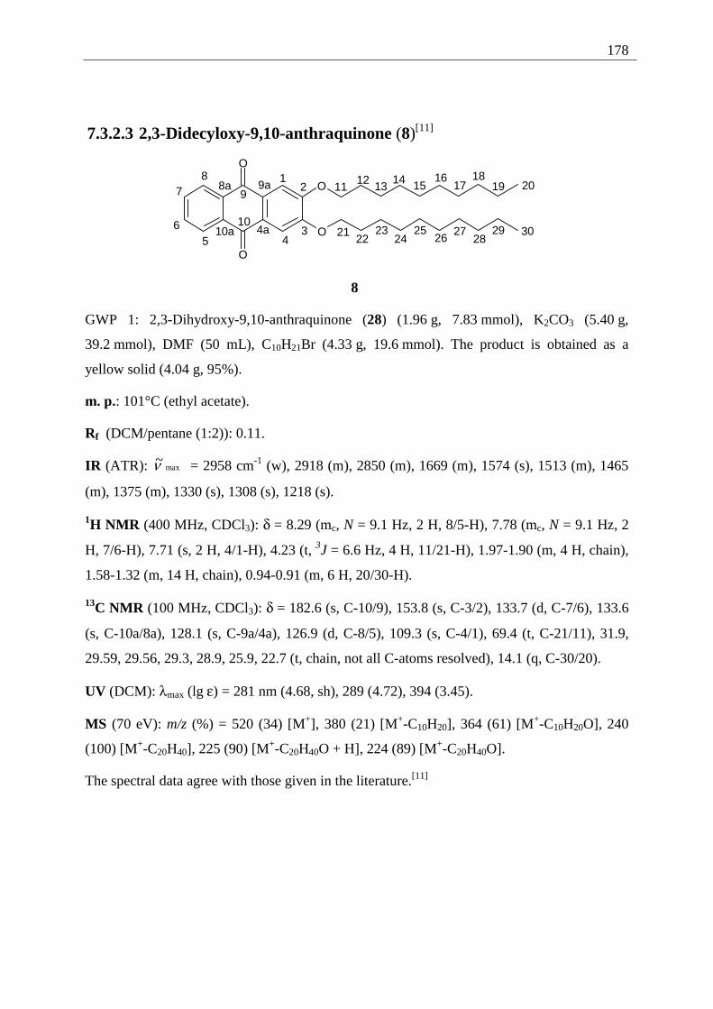
178
7.3.2.3 2,3-Didecyloxy-9,10-anthraquinone (8)[11]
O
O
O
O
12
345
6
78
9
10
1112
4a
9a8a
10a
1314 15
1617
1819 20
2122
2324
2526
2728
29 30
8
GWP 1: 2,3-Dihydroxy-9,10-anthraquinone (28) (1.96 g, 7.83 mmol), K2CO3 (5.40 g,
39.2 mmol), DMF (50 mL), C10H21Br (4.33 g, 19.6 mmol). The product is obtained as a
yellow solid (4.04 g, 95%).
m. p.: 101°C (ethyl acetate).
Rf (DCM/pentane (1:2)): 0.11.
IR (ATR): max~ν = 2958 cm-1 (w), 2918 (m), 2850 (m), 1669 (m), 1574 (s), 1513 (m), 1465
(m), 1375 (m), 1330 (s), 1308 (s), 1218 (s).
1H NMR (400 MHz, CDCl3): δ = 8.29 (mc, N = 9.1 Hz, 2 H, 8/5-H), 7.78 (mc, N = 9.1 Hz, 2
H, 7/6-H), 7.71 (s, 2 H, 4/1-H), 4.23 (t, 3J = 6.6 Hz, 4 H, 11/21-H), 1.97-1.90 (m, 4 H, chain),
1.58-1.32 (m, 14 H, chain), 0.94-0.91 (m, 6 H, 20/30-H).
13C NMR (100 MHz, CDCl3): δ = 182.6 (s, C-10/9), 153.8 (s, C-3/2), 133.7 (d, C-7/6), 133.6
(s, C-10a/8a), 128.1 (s, C-9a/4a), 126.9 (d, C-8/5), 109.3 (s, C-4/1), 69.4 (t, C-21/11), 31.9,
29.59, 29.56, 29.3, 28.9, 25.9, 22.7 (t, chain, not all C-atoms resolved), 14.1 (q, C-30/20).
UV (DCM): λmax (lg ε) = 281 nm (4.68, sh), 289 (4.72), 394 (3.45).
MS (70 eV): m/z (%) = 520 (34) [M+], 380 (21) [M+-C10H20], 364 (61) [M+-C10H20O], 240
(100) [M+-C20H40], 225 (90) [M+-C20H40O + H], 224 (89) [M+-C20H40O].
The spectral data agree with those given in the literature.[11]
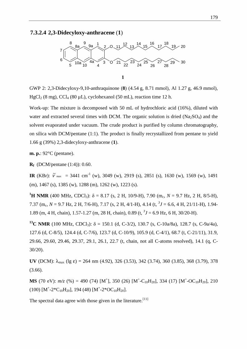
179
7.3.2.4 2,3-Didecyloxy-anthracene (1)
O
O
12
345
6
7
8 9
10
1112
1314
1516
1718
19 20
2122
2324
2526
2728
29 304a
8a 9a
10a
1
GWP 2: 2,3-Didecyloxy-9,10-anthraquinone (8) (4.54 g, 8.71 mmol), Al 1.27 g, 46.9 mmol),
HgCl2 (8 mg), CCl4 (80 µL), cyclohexanol (50 mL), reaction time 12 h.
Work-up: The mixture is decomposed with 50 mL of hydrochloric acid (16%), diluted with
water and extracted several times with DCM. The organic solution is dried (Na2SO4) and the
solvent evaporated under vacuum. The crude product is purified by column chromatography,
on silica with DCM/pentane (1:1). The product is finally recrystallized from pentane to yield
1.66 g (39%) 2,3-didecyloxy-anthracene (1).
m. p.: 92°C (pentane).
Rf (DCM/pentane (1:4)): 0.60.
IR (KBr): max~ν = 3441 cm-1 (w), 3049 (w), 2919 (s), 2851 (s), 1630 (w), 1569 (w), 1491
(m), 1467 (s), 1385 (w), 1288 (m), 1262 (w), 1223 (s).
1H NMR (400 MHz, CDCl3): δ = 8.17 (s, 2 H, 10/9-H), 7.90 (mc, N = 9.7 Hz, 2 H, 8/5-H),
7.37 (mc, N = 9.7 Hz, 2 H, 7/6-H), 7.17 (s, 2 H, 4/1-H), 4.14 (t, 3J = 6.6, 4 H, 21/11-H), 1.94-
1.89 (m, 4 H, chain), 1.57-1.27 (m, 28 H, chain), 0.89 (t, 3J = 6.9 Hz, 6 H, 30/20-H).
13C NMR (100 MHz, CDCl3): δ = 150.1 (d, C-3/2), 130.7 (s, C-10a/8a), 128.7 (s, C-9a/4a),
127.6 (d, C-8/5), 124.4 (d, C-7/6), 123.7 (d, C-10/9), 105.9 (d, C-4/1), 68.7 (t, C-21/11), 31.9,
29.66, 29.60, 29.46, 29.37, 29.1, 26.1, 22.7 (t, chain, not all C-atoms resolved), 14.1 (q, C-
30/20).
UV (DCM): λmax (lg ε) = 264 nm (4.92), 326 (3.53), 342 (3.74), 360 (3.85), 368 (3.79), 378
(3.66).
MS (70 eV): m/z (%) = 490 (74) [M+], 350 (26) [M+-C10H20], 334 (17) [M+-OC10H20], 210
(100) [M+-2*C10H20], 194 (48) [M+-2*OC10H20].
The spectral data agree with those given in the literature.[11]
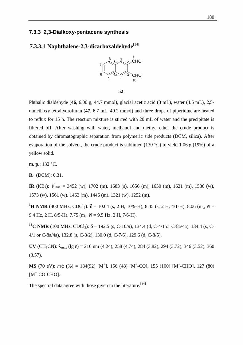
180
7.3.3 2,3-Dialkoxy-pentacene synthesis
7.3.3.1 Naphthalene-2,3-dicarboxaldehyde[14]
CHO
CHO
12
345
6
7
89
10
4a
8a
52
Phthalic dialdehyde (46, 6.00 g, 44.7 mmol), glacial acetic acid (3 mL), water (4.5 mL), 2,5-
dimethoxy-tetrahydrofuran (47, 6.7 mL, 49.2 mmol) and three drops of piperidine are heated
to reflux for 15 h. The reaction mixture is stirred with 20 mL of water and the precipitate is
filtered off. After washing with water, methanol and diethyl ether the crude product is
obtained by chromatographic separation from polymeric side products (DCM, silica). After
evaporation of the solvent, the crude product is sublimed (130 °C) to yield 1.06 g (19%) of a
yellow solid.
m. p.: 132 °C.
Rf (DCM): 0.31.
IR (KBr): max~ν = 3452 (w), 1702 (m), 1683 (s), 1656 (m), 1650 (m), 1621 (m), 1586 (w),
1573 (w), 1561 (w), 1463 (m), 1446 (m), 1321 (w), 1252 (m).
1H NMR (400 MHz, CDCl3): δ = 10.64 (s, 2 H, 10/9-H), 8.45 (s, 2 H, 4/1-H), 8.06 (mc, N =
9.4 Hz, 2 H, 8/5-H), 7.75 (mc, N = 9.5 Hz, 2 H, 7/6-H).
13C NMR (100 MHz, CDCl3): δ = 192.5 (s, C-10/9), 134.4 (d, C-4/1 or C-8a/4a), 134.4 (s, C-
4/1 or C-8a/4a), 132.8 (s, C-3/2), 130.0 (d, C-7/6), 129.6 (d, C-8/5).
UV (CH3CN): λmax (lg ε) = 216 nm (4.24), 258 (4.74), 284 (3.82), 294 (3.72), 346 (3.52), 360
(3.57).
MS (70 eV): m/z (%) = 184(92) [M+], 156 (48) [M+-CO], 155 (100) [M+-CHO], 127 (80)
[M+-CO-CHO].
The spectral data agree with those given in the literature.[14]
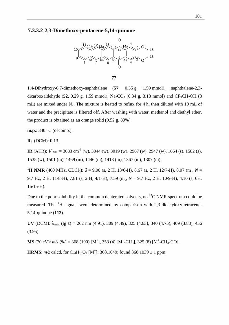
181
7.3.3.2 2,3-Dimethoxy-pentacene-5,14-quinone
O
O
O
O
12
34
5
6789
1011 12 13
14 15
164a5a6a7a
11a 12a 13a 14a
77
1,4-Dihydroxy-6,7-dimethoxy-naphthalene (57, 0.35 g, 1.59 mmol), naphthalene-2,3-
dicarboxaldehyde (52, 0.29 g, 1.59 mmol), Na2CO3 (0.34 g, 3.18 mmol) and CF3CH2OH (8
mL) are mixed under N2. The mixture is heated to reflux for 4 h, then diluted with 10 mL of
water and the precipitate is filtered off. After washing with water, methanol and diethyl ether,
the product is obtained as an orange solid (0.52 g, 89%).
m.p.: 340 °C (decomp.).
Rf (DCM): 0.13.
IR (ATR): max~ν = 3083 cm-1 (w), 3044 (w), 3019 (w), 2967 (w), 2947 (w), 1664 (s), 1582 (s),
1535 (w), 1501 (m), 1469 (m), 1446 (m), 1418 (m), 1367 (m), 1307 (m).
1H NMR (400 MHz, CDCl3): δ = 9.00 (s, 2 H, 13/6-H), 8.67 (s, 2 H, 12/7-H), 8.07 (mc, N =
9.7 Hz, 2 H, 11/8-H), 7.81 (s, 2 H, 4/1-H), 7.59 (mc, N = 9.7 Hz, 2 H, 10/9-H), 4.10 (s, 6H,
16/15-H).
Due to the poor solubility in the common deuterated solvents, no 13C NMR spectrum could be
measured. The 1H signals were determined by comparison with 2,3-didecyloxy-tetracene-
5,14-quinone (112).
UV (DCM): λmax (lg ε) = 262 nm (4.91), 309 (4.49), 325 (4.63), 340 (4.75), 409 (3.88), 456
(3.95).
MS (70 eV): m/z (%) = 368 (100) [M+], 353 (4) [M+-CH3], 325 (8) [M+-CH3-CO].
HRMS: m/z calcd. for C24H16O4 [M+]: 368.1049; found 368.1039 ± 1 ppm.
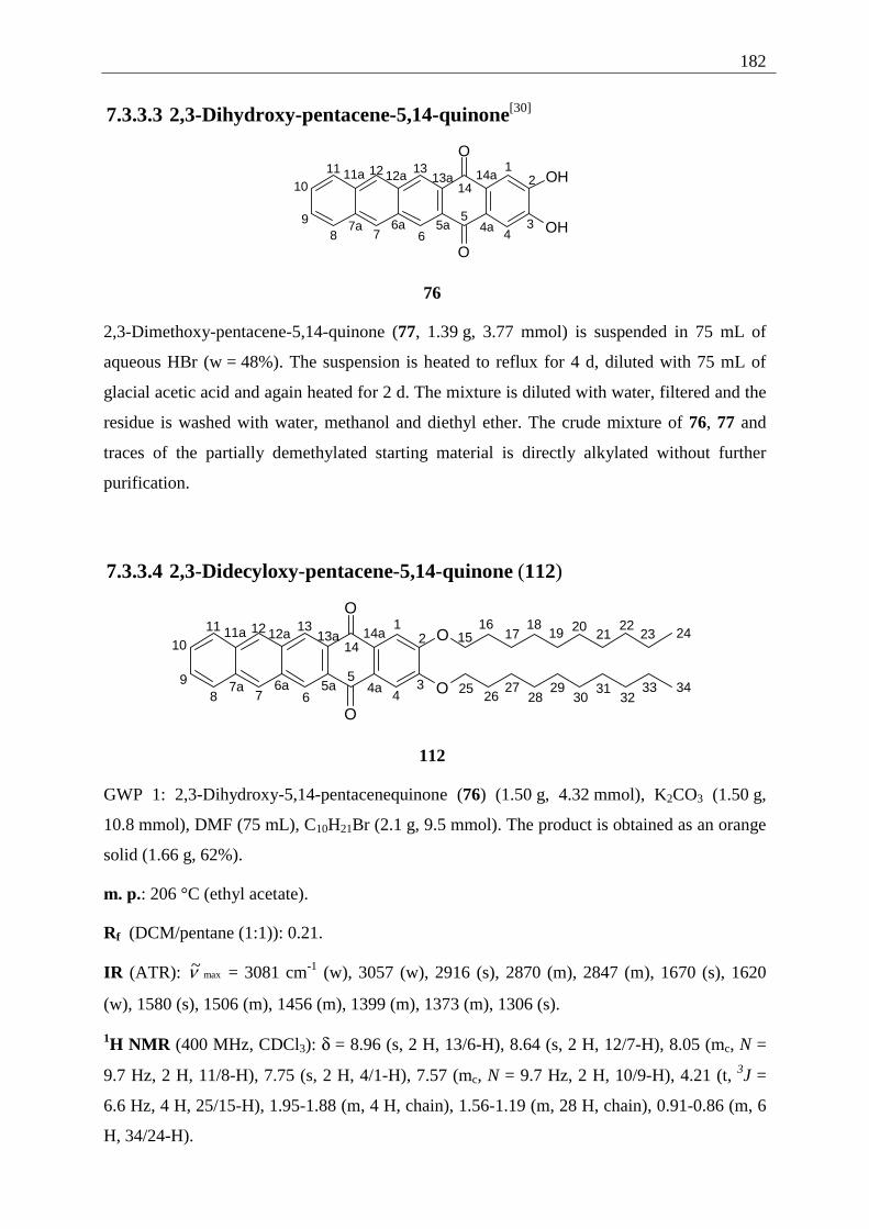
182
7.3.3.3 2,3-Dihydroxy-pentacene-5,14-quinone[30]
O
O
OH
OH
12
34
5
6789
1011 12 13
14
4a5a6a7a
11a 12a 13a 14a
76
2,3-Dimethoxy-pentacene-5,14-quinone (77, 1.39 g, 3.77 mmol) is suspended in 75 mL of
aqueous HBr (w = 48%). The suspension is heated to reflux for 4 d, diluted with 75 mL of
glacial acetic acid and again heated for 2 d. The mixture is diluted with water, filtered and the
residue is washed with water, methanol and diethyl ether. The crude mixture of 76, 77 and
traces of the partially demethylated starting material is directly alkylated without further
purification.
7.3.3.4 2,3-Didecyloxy-pentacene-5,14-quinone (112)
O
O
O
O
12
34
5
6789
1011 12 13
1415
16
4a5a6a7a
11a 12a 13a 14a 1718
19 2021
2223 24
2526
2728
2930
3132
33 34
112
GWP 1: 2,3-Dihydroxy-5,14-pentacenequinone (76) (1.50 g, 4.32 mmol), K2CO3 (1.50 g,
10.8 mmol), DMF (75 mL), C10H21Br (2.1 g, 9.5 mmol). The product is obtained as an orange
solid (1.66 g, 62%).
m. p.: 206 °C (ethyl acetate).
Rf (DCM/pentane (1:1)): 0.21.
IR (ATR): max~ν = 3081 cm-1 (w), 3057 (w), 2916 (s), 2870 (m), 2847 (m), 1670 (s), 1620
(w), 1580 (s), 1506 (m), 1456 (m), 1399 (m), 1373 (m), 1306 (s).
1H NMR (400 MHz, CDCl3): δ = 8.96 (s, 2 H, 13/6-H), 8.64 (s, 2 H, 12/7-H), 8.05 (mc, N =
9.7 Hz, 2 H, 11/8-H), 7.75 (s, 2 H, 4/1-H), 7.57 (mc, N = 9.7 Hz, 2 H, 10/9-H), 4.21 (t, 3J =
6.6 Hz, 4 H, 25/15-H), 1.95-1.88 (m, 4 H, chain), 1.56-1.19 (m, 28 H, chain), 0.91-0.86 (m, 6
H, 34/24-H).
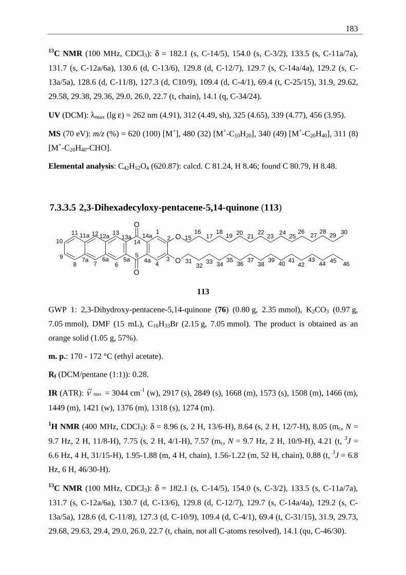
183
13C NMR (100 MHz, CDCl3): δ = 182.1 (s, C-14/5), 154.0 (s, C-3/2), 133.5 (s, C-11a/7a),
131.7 (s, C-12a/6a), 130.6 (d, C-13/6), 129.8 (d, C-12/7), 129.7 (s, C-14a/4a), 129.2 (s, C-
13a/5a), 128.6 (d, C-11/8), 127.3 (d, C10/9), 109.4 (d, C-4/1), 69.4 (t, C-25/15), 31.9, 29.62,
29.58, 29.38, 29.36, 29.0, 26.0, 22.7 (t, chain), 14.1 (q, C-34/24).
UV (DCM): λmax (lg ε) = 262 nm (4.91), 312 (4.49, sh), 325 (4.65), 339 (4.77), 456 (3.95).
MS (70 eV): m/z (%) = 620 (100) [M+], 480 (32) [M+-C10H20], 340 (49) [M+-C20H40], 311 (8)
[M+-C20H40-CHO].
Elemental analysis: C42H52O4 (620.87): calcd. C 81.24, H 8.46; found C 80.79, H 8.48.
7.3.3.5 2,3-Dihexadecyloxy-pentacene-5,14-quinone (113)
O
O
O
O
12
34
5
6789
1011 12 13
1415
16
4a5a6a7a
11a 12a 13a 14a 1718
19 2021
2223
2425
2627
2829 30
3132
33 3435
3637
3839
40 4142
4344 45 46
113
GWP 1: 2,3-Dihydroxy-pentacene-5,14-quinone (76) (0.80 g, 2.35 mmol), K2CO3 (0.97 g,
7.05 mmol), DMF (15 mL), C16H33Br (2.15 g, 7.05 mmol). The product is obtained as an
orange solid (1.05 g, 57%).
m. p.: 170 - 172 °C (ethyl acetate).
Rf (DCM/pentane (1:1)): 0.28.
IR (ATR): max~ν = 3044 cm-1 (w), 2917 (s), 2849 (s), 1668 (m), 1573 (s), 1508 (m), 1466 (m),
1449 (m), 1421 (w), 1376 (m), 1318 (s), 1274 (m).
1H NMR (400 MHz, CDCl3): δ = 8.96 (s, 2 H, 13/6-H), 8.64 (s, 2 H, 12/7-H), 8.05 (mc, N =
9.7 Hz, 2 H, 11/8-H), 7.75 (s, 2 H, 4/1-H), 7.57 (mc, N = 9.7 Hz, 2 H, 10/9-H), 4.21 (t, 3J =
6.6 Hz, 4 H, 31/15-H), 1.95-1.88 (m, 4 H, chain), 1.56-1.22 (m, 52 H, chain), 0.88 (t, 3J = 6.8
Hz, 6 H, 46/30-H).
13C NMR (100 MHz, CDCl3): δ = 182.1 (s, C-14/5), 154.0 (s, C-3/2), 133.5 (s, C-11a/7a),
131.7 (s, C-12a/6a), 130.7 (d, C-13/6), 129.8 (d, C-12/7), 129.7 (s, C-14a/4a), 129.2 (s, C-
13a/5a), 128.6 (d, C-11/8), 127.3 (d, C-10/9), 109.4 (d, C-4/1), 69.4 (t, C-31/15), 31.9, 29.73,
29.68, 29.63, 29.4, 29.0, 26.0, 22.7 (t, chain, not all C-atoms resolved), 14.1 (qu, C-46/30).
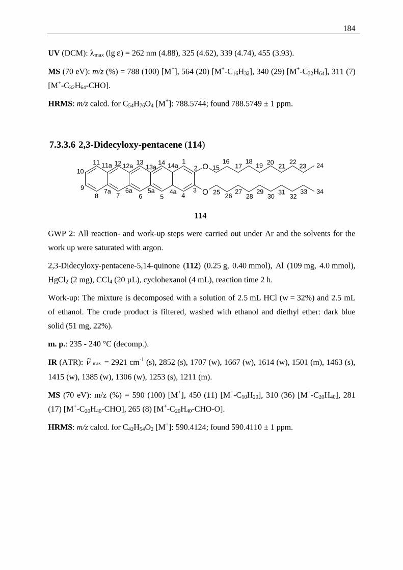
184
UV (DCM): λmax (lg ε) = 262 nm (4.88), 325 (4.62), 339 (4.74), 455 (3.93).
MS (70 eV): m/z (%) = 788 (100) [M+], 564 (20) [M+-C16H32], 340 (29) [M+-C32H64], 311 (7)
[M+-C32H64-CHO].
HRMS: m/z calcd. for C54H76O4 [M+]: 788.5744; found 788.5749 ± 1 ppm.
7.3.3.6 2,3-Didecyloxy-pentacene (114)
O
O
12
345678
9
1011 12 13 14
1516
4a5a6a7a
11a 12a 13a 14a 1718
19 2021
2223 24
2526
2728
2930
3132
33 34
114
GWP 2: All reaction- and work-up steps were carried out under Ar and the solvents for the
work up were saturated with argon.
2,3-Didecyloxy-pentacene-5,14-quinone (112) (0.25 g, 0.40 mmol), Al (109 mg, 4.0 mmol),
HgCl2 (2 mg), CCl4 (20 µL), cyclohexanol (4 mL), reaction time 2 h.
Work-up: The mixture is decomposed with a solution of 2.5 mL HCl (w = 32%) and 2.5 mL
of ethanol. The crude product is filtered, washed with ethanol and diethyl ether: dark blue
solid (51 mg, 22%).
m. p.: 235 - 240 °C (decomp.).
IR (ATR): max~ν = 2921 cm-1 (s), 2852 (s), 1707 (w), 1667 (w), 1614 (w), 1501 (m), 1463 (s),
1415 (w), 1385 (w), 1306 (w), 1253 (s), 1211 (m).
MS (70 eV): m/z (%) = 590 (100) [M+], 450 (11) [M+-C10H20], 310 (36) [M+-C20H40], 281
(17) [M+-C20H40-CHO], 265 (8) [M+-C20H40-CHO-O].
HRMS: m/z calcd. for C42H54O2 [M+]: 590.4124; found 590.4110 ± 1 ppm.
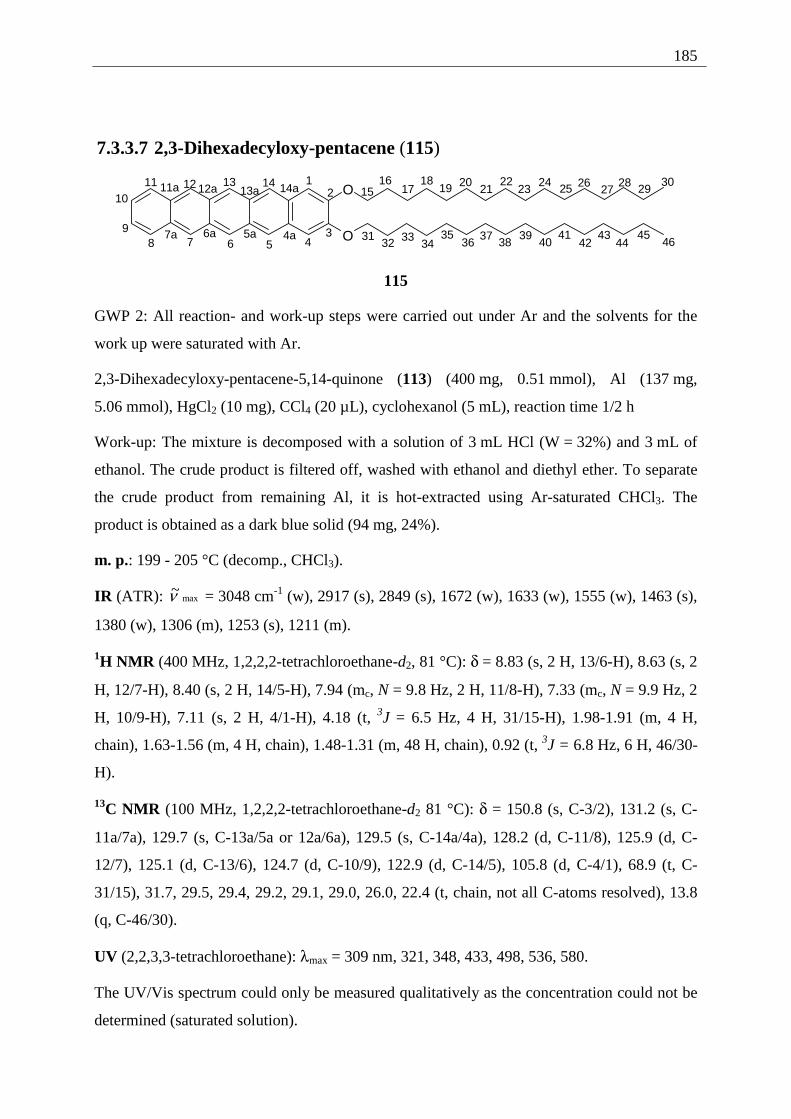
185
7.3.3.7 2,3-Dihexadecyloxy-pentacene (115)
O
O
12
345678
9
1011 12 13 14
1516
4a5a6a7a
11a 12a 13a 14a 1718
19 2021
2223
24 25 2627
28 2930
3132 33
3435
3637 38
3940
4142
4344
4546
115
GWP 2: All reaction- and work-up steps were carried out under Ar and the solvents for the
work up were saturated with Ar.
2,3-Dihexadecyloxy-pentacene-5,14-quinone (113) (400 mg, 0.51 mmol), Al (137 mg,
5.06 mmol), HgCl2 (10 mg), CCl4 (20 µL), cyclohexanol (5 mL), reaction time 1/2 h
Work-up: The mixture is decomposed with a solution of 3 mL HCl (W = 32%) and 3 mL of
ethanol. The crude product is filtered off, washed with ethanol and diethyl ether. To separate
the crude product from remaining Al, it is hot-extracted using Ar-saturated CHCl3. The
product is obtained as a dark blue solid (94 mg, 24%).
m. p.: 199 - 205 °C (decomp., CHCl3).
IR (ATR): max~ν = 3048 cm-1 (w), 2917 (s), 2849 (s), 1672 (w), 1633 (w), 1555 (w), 1463 (s),
1380 (w), 1306 (m), 1253 (s), 1211 (m).
1H NMR (400 MHz, 1,2,2,2-tetrachloroethane-d2, 81 °C): δ = 8.83 (s, 2 H, 13/6-H), 8.63 (s, 2
H, 12/7-H), 8.40 (s, 2 H, 14/5-H), 7.94 (mc, N = 9.8 Hz, 2 H, 11/8-H), 7.33 (mc, N = 9.9 Hz, 2
H, 10/9-H), 7.11 (s, 2 H, 4/1-H), 4.18 (t, 3J = 6.5 Hz, 4 H, 31/15-H), 1.98-1.91 (m, 4 H,
chain), 1.63-1.56 (m, 4 H, chain), 1.48-1.31 (m, 48 H, chain), 0.92 (t, 3J = 6.8 Hz, 6 H, 46/30-
H).
13C NMR (100 MHz, 1,2,2,2-tetrachloroethane-d2 81 °C): δ = 150.8 (s, C-3/2), 131.2 (s, C-
11a/7a), 129.7 (s, C-13a/5a or 12a/6a), 129.5 (s, C-14a/4a), 128.2 (d, C-11/8), 125.9 (d, C-
12/7), 125.1 (d, C-13/6), 124.7 (d, C-10/9), 122.9 (d, C-14/5), 105.8 (d, C-4/1), 68.9 (t, C-
31/15), 31.7, 29.5, 29.4, 29.2, 29.1, 29.0, 26.0, 22.4 (t, chain, not all C-atoms resolved), 13.8
(q, C-46/30).
UV (2,2,3,3-tetrachloroethane): λmax = 309 nm, 321, 348, 433, 498, 536, 580.
The UV/Vis spectrum could only be measured qualitatively as the concentration could not be
determined (saturated solution).
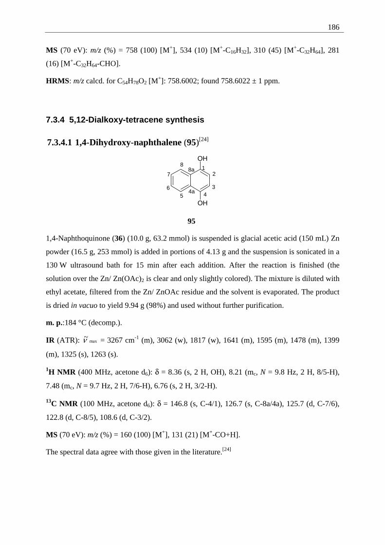
186
MS (70 eV): m/z (%) = 758 (100) [M+], 534 (10) [M+-C16H32], 310 (45) [M+-C32H64], 281
(16) [M+-C32H64-CHO].
HRMS: m/z calcd. for C54H78O2 [M+]: 758.6002; found 758.6022 ± 1 ppm.
7.3.4 5,12-Dialkoxy-tetracene synthesis
7.3.4.1 1,4-Dihydroxy-naphthalene (95)[24]
OH
OH
12
345
6
7
8
4a
8a
95
1,4-Naphthoquinone (36) (10.0 g, 63.2 mmol) is suspended is glacial acetic acid (150 mL) Zn
powder (16.5 g, 253 mmol) is added in portions of 4.13 g and the suspension is sonicated in a
130 W ultrasound bath for 15 min after each addition. After the reaction is finished (the
solution over the Zn/ Zn(OAc)2 is clear and only slightly colored). The mixture is diluted with
ethyl acetate, filtered from the Zn/ ZnOAc residue and the solvent is evaporated. The product
is dried in vacuo to yield 9.94 g (98%) and used without further purification.
m. p.:184 °C (decomp.).
IR (ATR): max~ν = 3267 cm-1 (m), 3062 (w), 1817 (w), 1641 (m), 1595 (m), 1478 (m), 1399
(m), 1325 (s), 1263 (s).
1H NMR (400 MHz, acetone d6): δ = 8.36 (s, 2 H, OH), 8.21 (mc, N = 9.8 Hz, 2 H, 8/5-H),
7.48 (mc, N = 9.7 Hz, 2 H, 7/6-H), 6.76 (s, 2 H, 3/2-H).
13C NMR (100 MHz, acetone d6): δ = 146.8 (s, C-4/1), 126.7 (s, C-8a/4a), 125.7 (d, C-7/6),
122.8 (d, C-8/5), 108.6 (d, C-3/2).
MS (70 eV): m/z (%) = 160 (100) [M+], 131 (21) [M+-CO+H].
The spectral data agree with those given in the literature.[24]
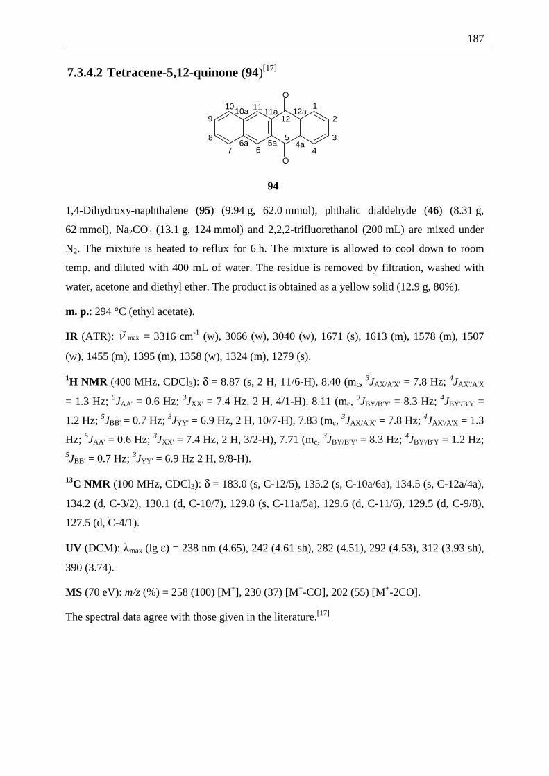
187
7.3.4.2 Tetracene-5,12-quinone (94)[17]
O
O
1
2
3
4
5
67
8
9
10 1112
4a5a6a
10a 11a 12a
94
1,4-Dihydroxy-naphthalene (95) (9.94 g, 62.0 mmol), phthalic dialdehyde (46) (8.31 g,
62 mmol), Na2CO3 (13.1 g, 124 mmol) and 2,2,2-trifluorethanol (200 mL) are mixed under
N2. The mixture is heated to reflux for 6 h. The mixture is allowed to cool down to room
temp. and diluted with 400 mL of water. The residue is removed by filtration, washed with
water, acetone and diethyl ether. The product is obtained as a yellow solid (12.9 g, 80%).
m. p.: 294 °C (ethyl acetate).
IR (ATR): max~ν = 3316 cm-1 (w), 3066 (w), 3040 (w), 1671 (s), 1613 (m), 1578 (m), 1507
(w), 1455 (m), 1395 (m), 1358 (w), 1324 (m), 1279 (s).
1H NMR (400 MHz, CDCl3): δ = 8.87 (s, 2 H, 11/6-H), 8.40 (mc, 3JAX/A'X' = 7.8 Hz; 4JAX'/A'X
= 1.3 Hz; 5JAA' = 0.6 Hz; 3JXX' = 7.4 Hz, 2 H, 4/1-H), 8.11 (mc, 3JBY/B'Y' = 8.3 Hz; 4JBY'/B'Y =
1.2 Hz; 5JBB' = 0.7 Hz; 3JYY' = 6.9 Hz, 2 H, 10/7-H), 7.83 (mc, 3JAX/A'X' = 7.8 Hz; 4JAX'/A'X = 1.3
Hz; 5JAA' = 0.6 Hz; 3JXX' = 7.4 Hz, 2 H, 3/2-H), 7.71 (mc, 3JBY/B'Y' = 8.3 Hz; 4JBY'/B'Y = 1.2 Hz;
5JBB' = 0.7 Hz; 3JYY' = 6.9 Hz 2 H, 9/8-H).
13C NMR (100 MHz, CDCl3): δ = 183.0 (s, C-12/5), 135.2 (s, C-10a/6a), 134.5 (s, C-12a/4a),
134.2 (d, C-3/2), 130.1 (d, C-10/7), 129.8 (s, C-11a/5a), 129.6 (d, C-11/6), 129.5 (d, C-9/8),
127.5 (d, C-4/1).
UV (DCM): λmax (lg ε) = 238 nm (4.65), 242 (4.61 sh), 282 (4.51), 292 (4.53), 312 (3.93 sh),
390 (3.74).
MS (70 eV): m/z (%) = 258 (100) [M+], 230 (37) [M+-CO], 202 (55) [M+-2CO].
The spectral data agree with those given in the literature.[17]
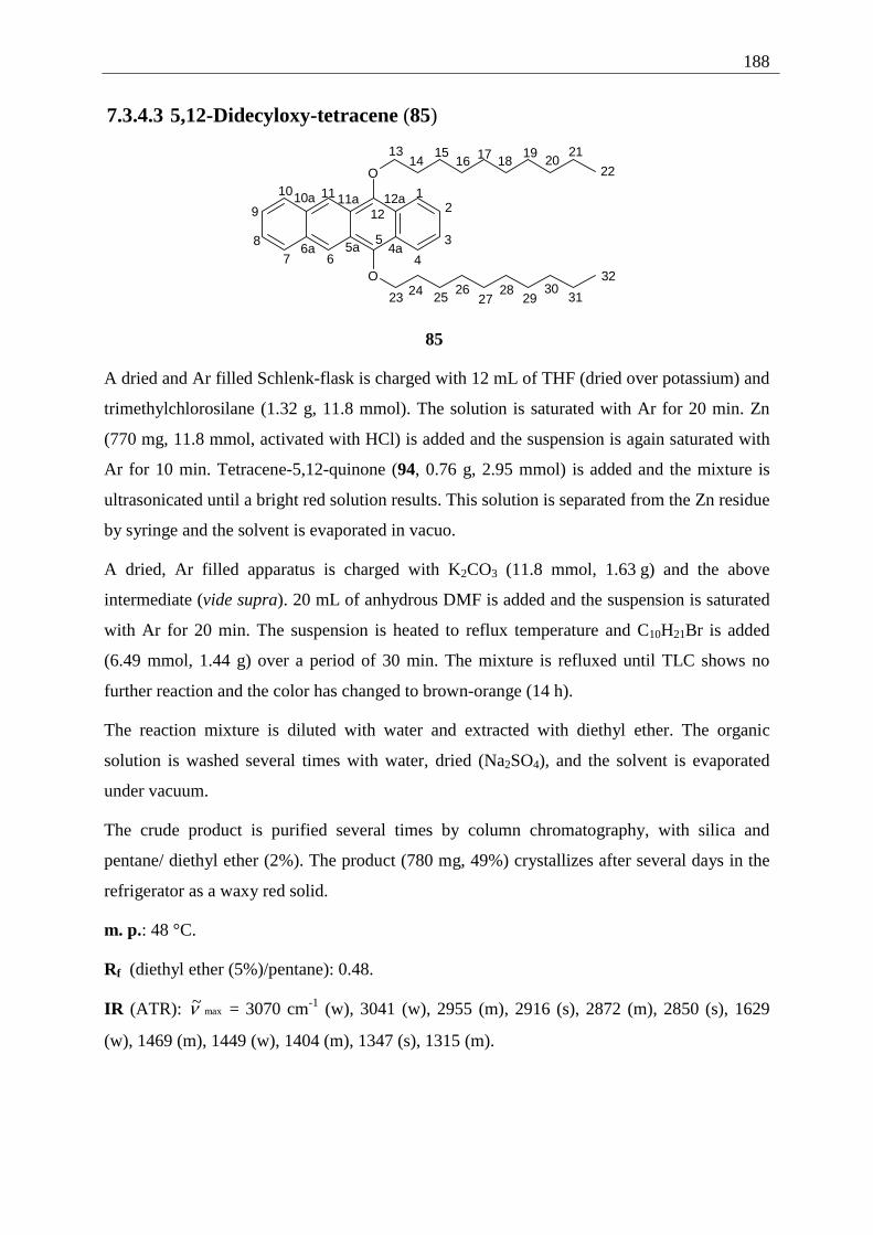
188
7.3.4.3 5,12-Didecyloxy-tetracene (85)
O
O
12
3
4
5
678
9
10 11
12
1314
1516 17 18
1920
21
22
23 24 2526
2728
2930
31
32
4a5a6a
10a 11a 12a
85
A dried and Ar filled Schlenk-flask is charged with 12 mL of THF (dried over potassium) and
trimethylchlorosilane (1.32 g, 11.8 mmol). The solution is saturated with Ar for 20 min. Zn
(770 mg, 11.8 mmol, activated with HCl) is added and the suspension is again saturated with
Ar for 10 min. Tetracene-5,12-quinone (94, 0.76 g, 2.95 mmol) is added and the mixture is
ultrasonicated until a bright red solution results. This solution is separated from the Zn residue
by syringe and the solvent is evaporated in vacuo.
A dried, Ar filled apparatus is charged with K2CO3 (11.8 mmol, 1.63 g) and the above
intermediate (vide supra). 20 mL of anhydrous DMF is added and the suspension is saturated
with Ar for 20 min. The suspension is heated to reflux temperature and C10H21Br is added
(6.49 mmol, 1.44 g) over a period of 30 min. The mixture is refluxed until TLC shows no
further reaction and the color has changed to brown-orange (14 h).
The reaction mixture is diluted with water and extracted with diethyl ether. The organic
solution is washed several times with water, dried (Na2SO4), and the solvent is evaporated
under vacuum.
The crude product is purified several times by column chromatography, with silica and
pentane/ diethyl ether (2%). The product (780 mg, 49%) crystallizes after several days in the
refrigerator as a waxy red solid.
m. p.: 48 °C.
Rf (diethyl ether (5%)/pentane): 0.48.
IR (ATR): max~ν = 3070 cm-1 (w), 3041 (w), 2955 (m), 2916 (s), 2872 (m), 2850 (s), 1629
(w), 1469 (m), 1449 (w), 1404 (m), 1347 (s), 1315 (m).
189
1H NMR (400 MHz, CDCl3): 8.90 (s, 2 H, 11/6-H), 8.27 (mc, N = 10.1 Hz, 2 H, 4/1-H), 8.03
(mc, N = 9.8 Hz 2 H, 10/7-H), 7.41 (mc, N = 6.8 Hz, 2 H, 9/8-H), 7.39 (mc, N = 7.0 Hz, 2 H,
3/2-H), 4.26 (t, 3J = 6.7 Hz, 4 H, 13/23-H), 2.15-2.08 (m, 4 H, chain), 1.75-1.68 (m, 4 H,
chain), 1.55-1.30 (m, 24 H, chain), 0.91-0.88 (m, 6 H, chain).
13C NMR (100 MHz, CDCl3): 147.5 (s, C-12/5), 131.2 (s, C-10a/6a), 128.7 (d, C-10/7), 125.4
(d, C-9/8), 124.7 (d, C-3/2), 124.6 (s, C-11a/5a), 124.0 (s, C-12a/4a), 122.8 (d, C-4/1), 121.4
(d, C-11/6), 76.3 (t, C-13/23), 31.9, 30.8, 29.7, 29.6, 29.4, 26.3, 22.7 (t, chain, not all C-atoms
resolved), 14.1 (qu, C-22/32).
UV (DCM): λmax (lg ε) = 285 nm (5.20), 304 (4.11), 403 (3.13), 3.15), 443 (3.54), 472 (3.81),
505 (3.83).
MS (70 eV): m/z (%) = 540 (20) [M+], 399 (8) [M+-C10H21], 383 (11) [M+-C10H21O], 259
(100) [M+-C20H41].
HRMS: m/z calcd. for C38H52O2 [M+]: 540.3967; found 540.3954 ± 1 ppm.
7.3.5 Photoreactions
7.3.5.1 Photodimerization of 2,3-DDOT (86)
GWP 3: 2,3-Didecyloxy-tetracene (86) (600 mg, 1.11 mmol), cyclohexane (22 mL), solvent
for chromatographic separation: DCM/ pentane (1:3) to pure DCM, further separation of the
dimers by several runs of column chromatography, solvent: 15% DCM/ pentane to 25%
DCM/ pentane.
Besides 123 mg (21%) of 86 the following dimers were obtained as waxy, nearly colorless
solids:
2,3-DDOT-D1 (87): 61 mg (10%)
2,3-DDOT-D2 (92): 67 mg (11%)
2,3-DDOT-D3 (88): 85 mg (15%)
2,3-DDOT-D4 (91): 94 mg (including D5) (16%)
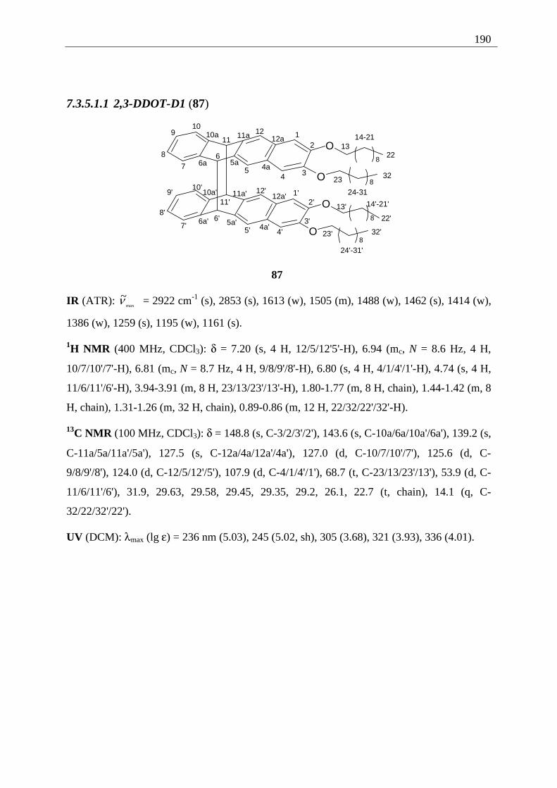
190
7.3.5.1.1 2,3-DDOT-D1 (87)
O
O
8
8
O
O8
8
12
345
67
8
910
1112
1314-21
22
23
24-31
324a
5a6a
10a 11a 12a
1'2'
3'4'5'
6'7'
8'
9'10'
11'
12'
13' 14'-21'
22'
23'
24'-31'
32'4a'
5a'6a'
10a' 11a' 12a'
87
IR (ATR): max
~ν = 2922 cm-1 (s), 2853 (s), 1613 (w), 1505 (m), 1488 (w), 1462 (s), 1414 (w),
1386 (w), 1259 (s), 1195 (w), 1161 (s).
1H NMR (400 MHz, CDCl3): δ = 7.20 (s, 4 H, 12/5/12'5'-H), 6.94 (mc, N = 8.6 Hz, 4 H,
10/7/10'/7'-H), 6.81 (mc, N = 8.7 Hz, 4 H, 9/8/9'/8'-H), 6.80 (s, 4 H, 4/1/4'/1'-H), 4.74 (s, 4 H,
11/6/11'/6'-H), 3.94-3.91 (m, 8 H, 23/13/23'/13'-H), 1.80-1.77 (m, 8 H, chain), 1.44-1.42 (m, 8
H, chain), 1.31-1.26 (m, 32 H, chain), 0.89-0.86 (m, 12 H, 22/32/22'/32'-H).
13C NMR (100 MHz, CDCl3): δ = 148.8 (s, C-3/2/3'/2'), 143.6 (s, C-10a/6a/10a'/6a'), 139.2 (s,
C-11a/5a/11a'/5a'), 127.5 (s, C-12a/4a/12a'/4a'), 127.0 (d, C-10/7/10'/7'), 125.6 (d, C-
9/8/9'/8'), 124.0 (d, C-12/5/12'/5'), 107.9 (d, C-4/1/4'/1'), 68.7 (t, C-23/13/23'/13'), 53.9 (d, C-
11/6/11'/6'), 31.9, 29.63, 29.58, 29.45, 29.35, 29.2, 26.1, 22.7 (t, chain), 14.1 (q, C-
32/22/32'/22').
UV (DCM): λmax (lg ε) = 236 nm (5.03), 245 (5.02, sh), 305 (3.68), 321 (3.93), 336 (4.01).
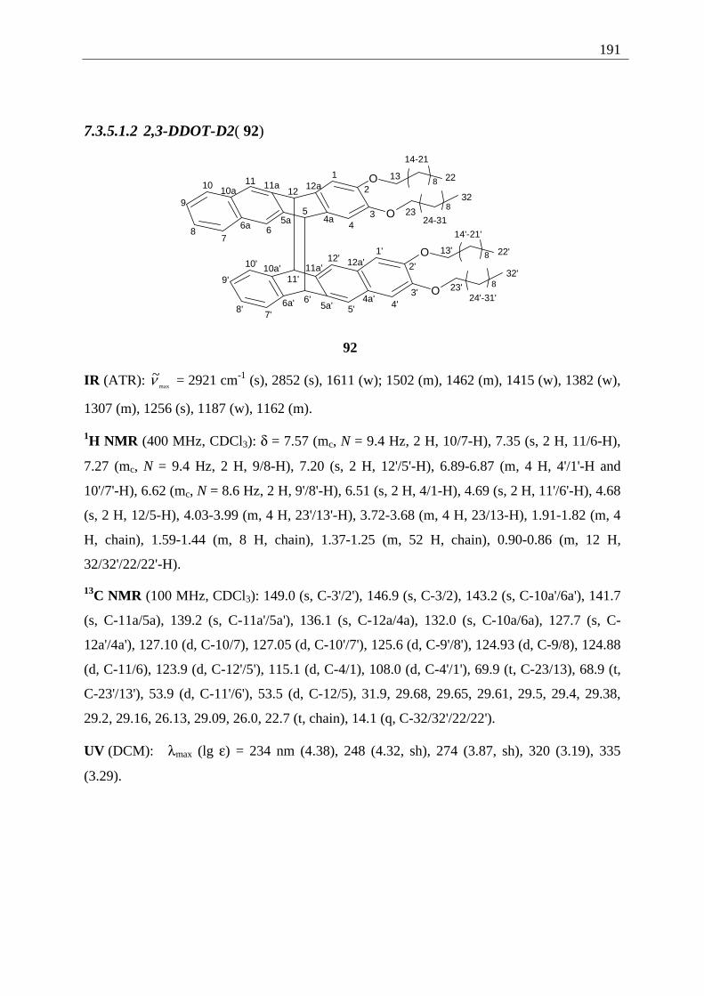
191
7.3.5.1.2 2,3-DDOT-D2( 92)
O
O
O
O
8
8
8
81
2
34
5
67
8
9
10 1112
13
14-21
22
2324-31
32
4a5a6a
10a11a 12a
1'
2'
3'4'5'
6'
7'8'
9'
10'
11'
12'13'
14'-21'
22'
23'24'-31'
32'
4a'5a'6a'
10a' 11a'12a'
92
IR (ATR): max
~ν = 2921 cm-1 (s), 2852 (s), 1611 (w); 1502 (m), 1462 (m), 1415 (w), 1382 (w),
1307 (m), 1256 (s), 1187 (w), 1162 (m).
1H NMR (400 MHz, CDCl3): δ = 7.57 (mc, N = 9.4 Hz, 2 H, 10/7-H), 7.35 (s, 2 H, 11/6-H),
7.27 (mc, N = 9.4 Hz, 2 H, 9/8-H), 7.20 (s, 2 H, 12'/5'-H), 6.89-6.87 (m, 4 H, 4'/1'-H and
10'/7'-H), 6.62 (mc, N = 8.6 Hz, 2 H, 9'/8'-H), 6.51 (s, 2 H, 4/1-H), 4.69 (s, 2 H, 11'/6'-H), 4.68
(s, 2 H, 12/5-H), 4.03-3.99 (m, 4 H, 23'/13'-H), 3.72-3.68 (m, 4 H, 23/13-H), 1.91-1.82 (m, 4
H, chain), 1.59-1.44 (m, 8 H, chain), 1.37-1.25 (m, 52 H, chain), 0.90-0.86 (m, 12 H,
32/32'/22/22'-H).
13C NMR (100 MHz, CDCl3): 149.0 (s, C-3'/2'), 146.9 (s, C-3/2), 143.2 (s, C-10a'/6a'), 141.7
(s, C-11a/5a), 139.2 (s, C-11a'/5a'), 136.1 (s, C-12a/4a), 132.0 (s, C-10a/6a), 127.7 (s, C-
12a'/4a'), 127.10 (d, C-10/7), 127.05 (d, C-10'/7'), 125.6 (d, C-9'/8'), 124.93 (d, C-9/8), 124.88
(d, C-11/6), 123.9 (d, C-12'/5'), 115.1 (d, C-4/1), 108.0 (d, C-4'/1'), 69.9 (t, C-23/13), 68.9 (t,
C-23'/13'), 53.9 (d, C-11'/6'), 53.5 (d, C-12/5), 31.9, 29.68, 29.65, 29.61, 29.5, 29.4, 29.38,
29.2, 29.16, 26.13, 29.09, 26.0, 22.7 (t, chain), 14.1 (q, C-32/32'/22/22').
UV (DCM): λmax (lg ε) = 234 nm (4.38), 248 (4.32, sh), 274 (3.87, sh), 320 (3.19), 335
(3.29).
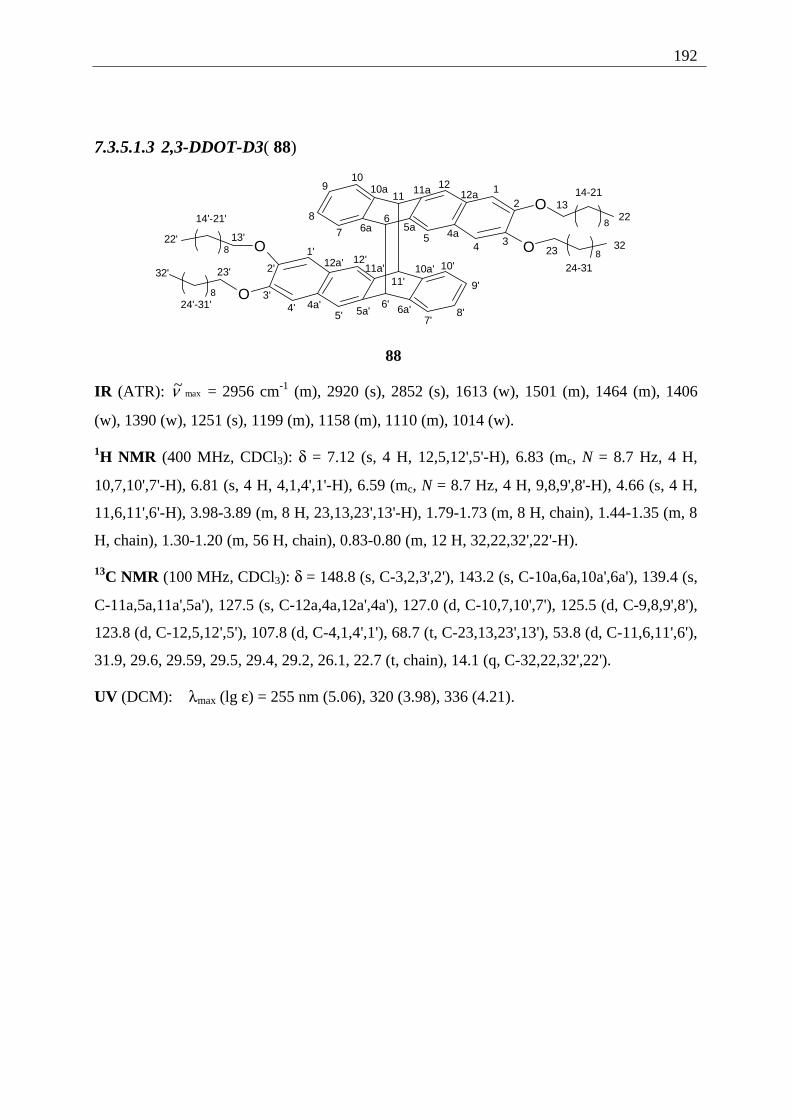
192
7.3.5.1.3 2,3-DDOT-D3( 88)
O
OO
O
8
88
8
12
345
67
8
910
1112
1314-21
22
23
24-31
324a
5a6a
10a 11a 12a
1'
2'
3'4'
5'6'
7'8'
9'
10'
11'
12'
13'
14'-21'
22'
23'
24'-31'
32'
4a'5a' 6a'
10a'11a'12a'
88
IR (ATR): max~ν = 2956 cm-1 (m), 2920 (s), 2852 (s), 1613 (w), 1501 (m), 1464 (m), 1406
(w), 1390 (w), 1251 (s), 1199 (m), 1158 (m), 1110 (m), 1014 (w).
1H NMR (400 MHz, CDCl3): δ = 7.12 (s, 4 H, 12,5,12',5'-H), 6.83 (mc, N = 8.7 Hz, 4 H,
10,7,10',7'-H), 6.81 (s, 4 H, 4,1,4',1'-H), 6.59 (mc, N = 8.7 Hz, 4 H, 9,8,9',8'-H), 4.66 (s, 4 H,
11,6,11',6'-H), 3.98-3.89 (m, 8 H, 23,13,23',13'-H), 1.79-1.73 (m, 8 H, chain), 1.44-1.35 (m, 8
H, chain), 1.30-1.20 (m, 56 H, chain), 0.83-0.80 (m, 12 H, 32,22,32',22'-H).
13C NMR (100 MHz, CDCl3): δ = 148.8 (s, C-3,2,3',2'), 143.2 (s, C-10a,6a,10a',6a'), 139.4 (s,
C-11a,5a,11a',5a'), 127.5 (s, C-12a,4a,12a',4a'), 127.0 (d, C-10,7,10',7'), 125.5 (d, C-9,8,9',8'),
123.8 (d, C-12,5,12',5'), 107.8 (d, C-4,1,4',1'), 68.7 (t, C-23,13,23',13'), 53.8 (d, C-11,6,11',6'),
31.9, 29.6, 29.59, 29.5, 29.4, 29.2, 26.1, 22.7 (t, chain), 14.1 (q, C-32,22,32',22').
UV (DCM): λmax (lg ε) = 255 nm (5.06), 320 (3.98), 336 (4.21).
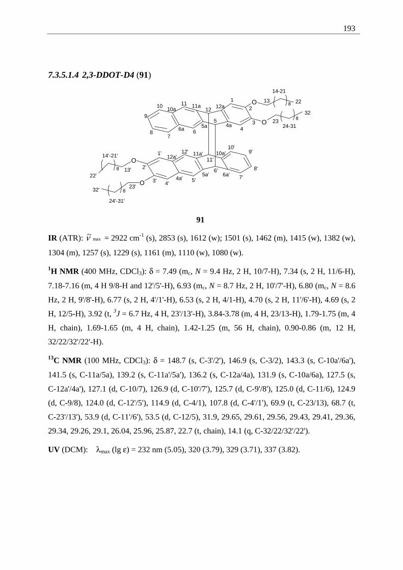
193
7.3.5.1.4 2,3-DDOT-D4 (91)
O
O
8
8
O
O
8
8
1
2
34
5
67
8
9
10 1112
13
14-21
22
2324-31
32
4a5a6a
10a11a 12a
1'
2'
3' 4'5'
6'7'
8'
9'10'
11'
12'
13'
14'-21'
22'
23'
24'-31'
32'
4a'5a' 6a'
10a'11a'12a'
91
IR (ATR): max~ν = 2922 cm-1 (s), 2853 (s), 1612 (w); 1501 (s), 1462 (m), 1415 (w), 1382 (w),
1304 (m), 1257 (s), 1229 (s), 1161 (m), 1110 (w), 1080 (w).
1H NMR (400 MHz, CDCl3): δ = 7.49 (mc, N = 9.4 Hz, 2 H, 10/7-H), 7.34 (s, 2 H, 11/6-H),
7.18-7.16 (m, 4 H 9/8-H and 12'/5'-H), 6.93 (mc, N = 8.7 Hz, 2 H, 10'/7'-H), 6.80 (mc, N = 8.6
Hz, 2 H, 9'/8'-H), 6.77 (s, 2 H, 4'/1'-H), 6.53 (s, 2 H, 4/1-H), 4.70 (s, 2 H, 11'/6'-H), 4.69 (s, 2
H, 12/5-H), 3.92 (t, 3J = 6.7 Hz, 4 H, 23'/13'-H), 3.84-3.78 (m, 4 H, 23/13-H), 1.79-1.75 (m, 4
H, chain), 1.69-1.65 (m, 4 H, chain), 1.42-1.25 (m, 56 H, chain), 0.90-0.86 (m, 12 H,
32/22/32'/22'-H).
13C NMR (100 MHz, CDCl3): δ = 148.7 (s, C-3'/2'), 146.9 (s, C-3/2), 143.3 (s, C-10a'/6a'),
141.5 (s, C-11a/5a), 139.2 (s, C-11a'/5a'), 136.2 (s, C-12a/4a), 131.9 (s, C-10a/6a), 127.5 (s,
C-12a'/4a'), 127.1 (d, C-10/7), 126.9 (d, C-10'/7'), 125.7 (d, C-9'/8'), 125.0 (d, C-11/6), 124.9
(d, C-9/8), 124.0 (d, C-12'/5'), 114.9 (d, C-4/1), 107.8 (d, C-4'/1'), 69.9 (t, C-23/13), 68.7 (t,
C-23'/13'), 53.9 (d, C-11'/6'), 53.5 (d, C-12/5), 31.9, 29.65, 29.61, 29.56, 29.43, 29.41, 29.36,
29.34, 29.26, 29.1, 26.04, 25.96, 25.87, 22.7 (t, chain), 14.1 (q, C-32/22/32'/22').
UV (DCM): λmax (lg ε) = 232 nm (5.05), 320 (3.79), 329 (3.71), 337 (3.82).
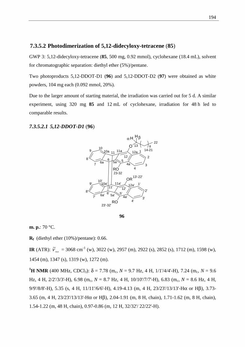
194
7.3.5.2 Photodimerization of 5,12-didecyloxy-tetracene (85)
GWP 3: 5,12-didecyloxy-tetracene (85, 500 mg, 0.92 mmol), cyclohexane (18.4 mL), solvent
for chromatographic separation: diethyl ether (5%)/pentane.
Two photoproducts 5,12-DDOT-D1 (96) and 5,12-DDOT-D2 (97) were obtained as white
powders, 104 mg each (0.092 mmol, 20%).
Due to the larger amount of starting material, the irradiation was carried out for 5 d. A similar
experiment, using 320 mg 85 and 12 mL of cyclohexane, irradiation for 48 h led to
comparable results.
7.3.5.2.1 5,12-DDOT-D1 (96)
O
H H
OR
RO
8
RO
1
2
34
567
8
910
1112
4a5a6a
10a 11a 12a
1'
2'
3'4'
5'6'7'
8'
9'10'
11' 12'
4a'5a'6a'
10a' 11a' 12a'
1314-21
22
23-3213'-22'
23'-32'
α β
96
m. p.: 70 °C.
Rf (diethyl ether (10%)/pentane): 0.66.
IR (ATR): max
~ν = 3068 cm-1 (w), 3022 (w), 2957 (m), 2922 (s), 2852 (s), 1712 (m), 1598 (w),
1454 (m), 1347 (s), 1319 (w), 1272 (m).
1H NMR (400 MHz, CDCl3): δ = 7.78 (mc, N = 9.7 Hz, 4 H, 1/1'/4/4'-H), 7.24 (mc, N = 9.6
Hz, 4 H, 2/2'/3/3'-H), 6.98 (mc, N = 8.7 Hz, 4 H, 10/10'/7/7'-H), 6.83 (mc, N = 8.6 Hz, 4 H,
9/9'/8/8'-H), 5.35 (s, 4 H, 11/11'/6/6'-H), 4.19-4.13 (m, 4 H, 23/23'/13/13'-Hα or Hβ), 3.73-
3.65 (m, 4 H, 23/23'/13/13'-Hα or Hβ), 2.04-1.91 (m, 8 H, chain), 1.71-1.62 (m, 8 H, chain),
1.54-1.22 (m, 48 H, chain), 0.97-0.86 (m, 12 H, 32/32'/ 22/22'-H).
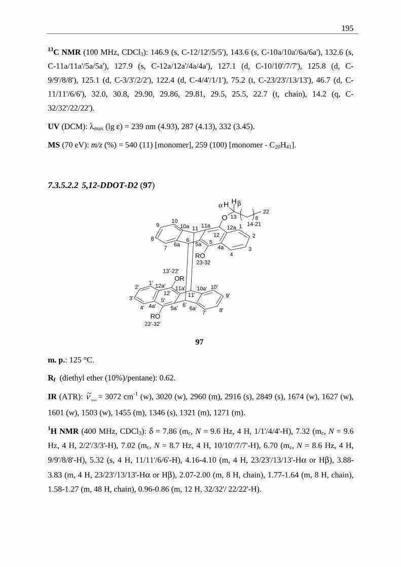
195
13C NMR (100 MHz, CDCl3): 146.9 (s, C-12/12'/5/5'), 143.6 (s, C-10a/10a'/6a/6a'), 132.6 (s,
C-11a/11a'/5a/5a'), 127.9 (s, C-12a/12a'/4a/4a'), 127.1 (d, C-10/10'/7/7'), 125.8 (d, C-
9/9'/8/8'), 125.1 (d, C-3/3'/2/2'), 122.4 (d, C-4/4'/1/1'), 75.2 (t, C-23/23'/13/13'), 46.7 (d, C-
11/11'/6/6'), 32.0, 30.8, 29.90, 29.86, 29.81, 29.5, 25.5, 22.7 (t, chain), 14.2 (q, C-
32/32'/22/22').
UV (DCM): λmax (lg ε) = 239 nm (4.93), 287 (4.13), 332 (3.45).
MS (70 eV): m/z (%) = 540 (11) [monomer], 259 (100) [monomer - C20H41].
7.3.5.2.2 5,12-DDOT-D2 (97)
O
H H
8
RO
OR
RO
1
2
34
567
8
910
1112
4a5a6a
10a 11a 12a
1'2'
3'
4'5'
6'7' 8'
9'
10'11'12'
4a' 5a' 6a'
10a'11a'12a'
1314-21
22
23-32
13'-22'
23'-32'
α β
97
m. p.: 125 °C.
Rf (diethyl ether (10%)/pentane): 0.62.
IR (ATR): max
~ν = 3072 cm-1 (w), 3020 (w), 2960 (m), 2916 (s), 2849 (s), 1674 (w), 1627 (w),
1601 (w), 1503 (w), 1455 (m), 1346 (s), 1321 (m), 1271 (m).
1H NMR (400 MHz, CDCl3): δ = 7.86 (mc, N = 9.6 Hz, 4 H, 1/1'/4/4'-H), 7.32 (mc, N = 9.6
Hz, 4 H, 2/2'/3/3'-H), 7.02 (mc, N = 8.7 Hz, 4 H, 10/10'/7/7'-H), 6.70 (mc, N = 8.6 Hz, 4 H,
9/9'/8/8'-H), 5.32 (s, 4 H, 11/11'/6/6'-H), 4.16-4.10 (m, 4 H, 23/23'/13/13'-Hα or Hβ), 3.88-
3.83 (m, 4 H, 23/23'/13/13'-Hα or Hβ), 2.07-2.00 (m, 8 H, chain), 1.77-1.64 (m, 8 H, chain),
1.58-1.27 (m, 48 H, chain), 0.96-0.86 (m, 12 H, 32/32'/ 22/22'-H).
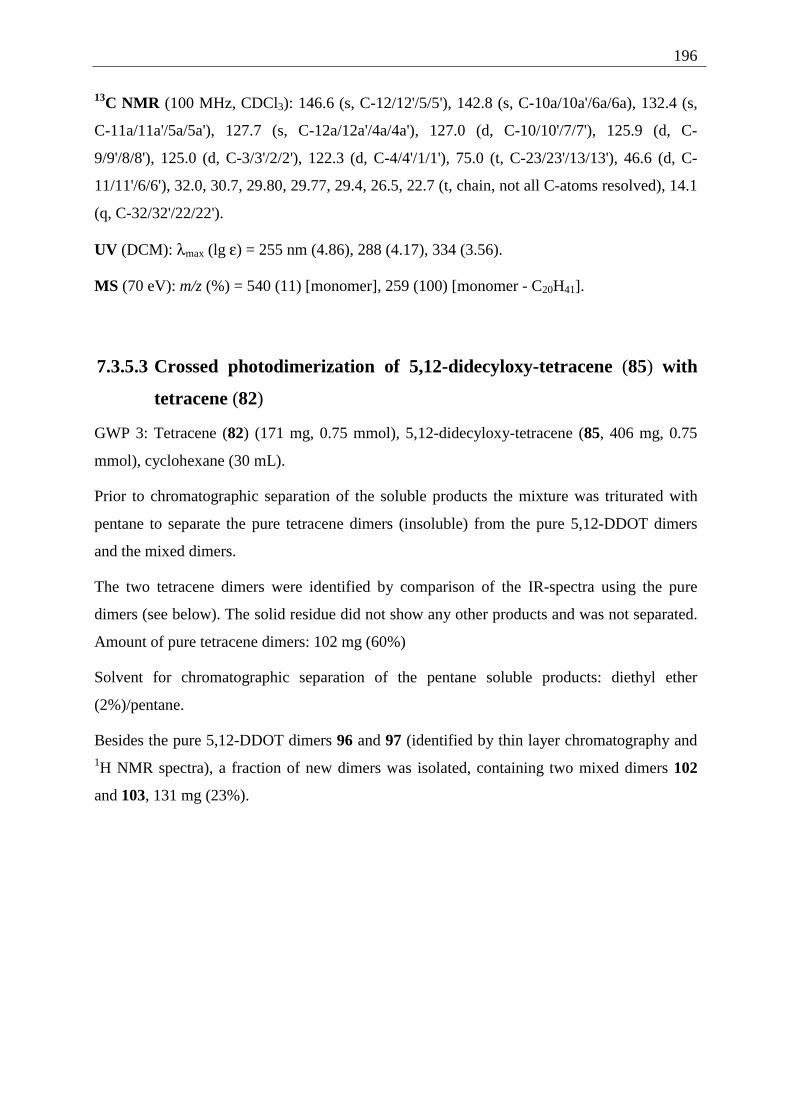
196
13C NMR (100 MHz, CDCl3): 146.6 (s, C-12/12'/5/5'), 142.8 (s, C-10a/10a'/6a/6a), 132.4 (s,
C-11a/11a'/5a/5a'), 127.7 (s, C-12a/12a'/4a/4a'), 127.0 (d, C-10/10'/7/7'), 125.9 (d, C-
9/9'/8/8'), 125.0 (d, C-3/3'/2/2'), 122.3 (d, C-4/4'/1/1'), 75.0 (t, C-23/23'/13/13'), 46.6 (d, C-
11/11'/6/6'), 32.0, 30.7, 29.80, 29.77, 29.4, 26.5, 22.7 (t, chain, not all C-atoms resolved), 14.1
(q, C-32/32'/22/22').
UV (DCM): λmax (lg ε) = 255 nm (4.86), 288 (4.17), 334 (3.56).
MS (70 eV): m/z (%) = 540 (11) [monomer], 259 (100) [monomer - C20H41].
7.3.5.3 Crossed photodimerization of 5,12-didecyloxy-tetracene (85) with
tetracene (82)
GWP 3: Tetracene (82) (171 mg, 0.75 mmol), 5,12-didecyloxy-tetracene (85, 406 mg, 0.75
mmol), cyclohexane (30 mL).
Prior to chromatographic separation of the soluble products the mixture was triturated with
pentane to separate the pure tetracene dimers (insoluble) from the pure 5,12-DDOT dimers
and the mixed dimers.
The two tetracene dimers were identified by comparison of the IR-spectra using the pure
dimers (see below). The solid residue did not show any other products and was not separated.
Amount of pure tetracene dimers: 102 mg (60%)
Solvent for chromatographic separation of the pentane soluble products: diethyl ether
(2%)/pentane.
Besides the pure 5,12-DDOT dimers 96 and 97 (identified by thin layer chromatography and 1H NMR spectra), a fraction of new dimers was isolated, containing two mixed dimers 102
and 103, 131 mg (23%).
197
5,12-DDOT-T-D1 (102)
O
O
H HCH2
8CH3
C10H21
1
2
34
567
8
910
1112
23
4a5a6a
10a 11a 12a
16a 17a 18a
1322a23a24a14
15
1617
1819
20
21
2224
25-34
35-44
102
IR (ATR): max
~ν = 3067 cm-1 (w), 3019 (w), 2922 (s), 2853 (s), 1600 (w), 1502 (w), 1484 (w),
1456 (m), 1347 (s), 1320 (m), 1293 (w), 1266 (w), 1229 (w), 1171 (w), 1093 (m).
1H NMR (400 MHz, CDCl3): δ = 7.76 (mc, N = 9.7 Hz, 2 H, 4/1-H), 7.49 (mc, N = 9.4 Hz, 2
H, 22/19-H), 7.45 (s, 2 H, 23/18-H), 7.21 (mc, N = 9.6 Hz, 2 H, 3/2-H), 7.18 (mc, N = 9.4 Hz,
2 H, 21/20-H), 7.00-6.97 (m, 4 H, 16/13-H or 10/7-H), 6.86-6.83 (m, 4 H, 15/14-H or 9/8-H),
5.33 (d, 3J = 11.2 Hz, 2 H, 11/6-H), 4.86 (d, 3J = 11.2 Hz, 2 H, 24/17-H), 4.17-4.11 (m, 2 H,
35/25-Hα or 35/25-Hβ), 3.86-3.82 (m, 2 H, 35/25-Hα or 35/25-Hβ), 2.08-2.00 (m, 4 H, chain),
1.72-1.57 (m, 4 H, chain), 1.55-1.34 (m, 24 H, chain), 0.92 (t, 3J = 6.9 Hz, 6 H, 44/34-H).
13C NMR (100 MHz, CDCl3): δ = 146.8 (s, C-12/5), 143.1 (s, C-24a/16a or C-10a/6a), 143.07
(s, C-24a/16a or C-10a/6a), 141.0 (s, C-23a/17a), 132.2 (s, C-11a/5a), 131.9 (s, C-22a/18a),
127.7 (s, C-12a/4a), 127.2 (d, C-16/13 or C-10/7), 127.13 (d, C-16/13 or C-10/7), 127.10 (d,
C-22/19), 125.89 (d, C-15/14 or C-9/8), 125.85 (d, C-15/14 or C-9/8), 125.4 (d, C-23/18),
125.15 (d, C-21/20), 125.06 (d, C-3/2), 122.3 (d, C-4/1), 74.7 (t, C-35/25), 53.6 (d, C-24/17),
47.0 (d, C-11/6), 32.0, 30.6, 29.8, 29.73, 29.71, 29.43, 26.3, 22.7 (t, chain), 14.2 (q, C-44/34).
UV (DCM): λmax (lg ε) = 230 nm (5.02), 281 (4.19, sh).
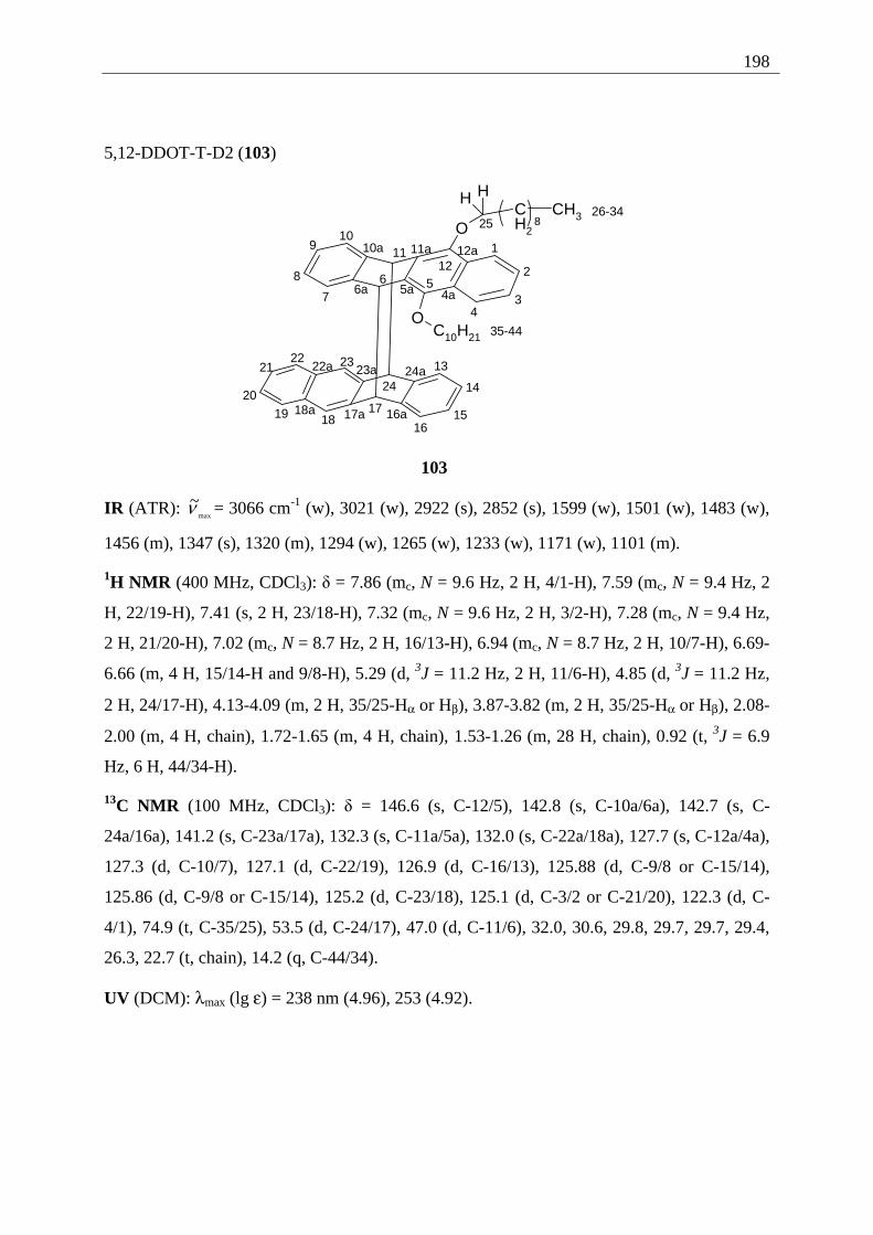
198
5,12-DDOT-T-D2 (103)
O
O
H HCH2
8CH3
C10H21
1
2
34
567
8
910
1112
23
4a5a6a
10a 11a 12a
16a17a18a
1322a 23a 24a14
1516
171819
20
2122
24
2526-34
35-44
103
IR (ATR): max
~ν = 3066 cm-1 (w), 3021 (w), 2922 (s), 2852 (s), 1599 (w), 1501 (w), 1483 (w),
1456 (m), 1347 (s), 1320 (m), 1294 (w), 1265 (w), 1233 (w), 1171 (w), 1101 (m).
1H NMR (400 MHz, CDCl3): δ = 7.86 (mc, N = 9.6 Hz, 2 H, 4/1-H), 7.59 (mc, N = 9.4 Hz, 2
H, 22/19-H), 7.41 (s, 2 H, 23/18-H), 7.32 (mc, N = 9.6 Hz, 2 H, 3/2-H), 7.28 (mc, N = 9.4 Hz,
2 H, 21/20-H), 7.02 (mc, N = 8.7 Hz, 2 H, 16/13-H), 6.94 (mc, N = 8.7 Hz, 2 H, 10/7-H), 6.69-
6.66 (m, 4 H, 15/14-H and 9/8-H), 5.29 (d, 3J = 11.2 Hz, 2 H, 11/6-H), 4.85 (d, 3J = 11.2 Hz,
2 H, 24/17-H), 4.13-4.09 (m, 2 H, 35/25-Hα or Hβ), 3.87-3.82 (m, 2 H, 35/25-Hα or Hβ), 2.08-
2.00 (m, 4 H, chain), 1.72-1.65 (m, 4 H, chain), 1.53-1.26 (m, 28 H, chain), 0.92 (t, 3J = 6.9
Hz, 6 H, 44/34-H).
13C NMR (100 MHz, CDCl3): δ = 146.6 (s, C-12/5), 142.8 (s, C-10a/6a), 142.7 (s, C-
24a/16a), 141.2 (s, C-23a/17a), 132.3 (s, C-11a/5a), 132.0 (s, C-22a/18a), 127.7 (s, C-12a/4a),
127.3 (d, C-10/7), 127.1 (d, C-22/19), 126.9 (d, C-16/13), 125.88 (d, C-9/8 or C-15/14),
125.86 (d, C-9/8 or C-15/14), 125.2 (d, C-23/18), 125.1 (d, C-3/2 or C-21/20), 122.3 (d, C-
4/1), 74.9 (t, C-35/25), 53.5 (d, C-24/17), 47.0 (d, C-11/6), 32.0, 30.6, 29.8, 29.7, 29.7, 29.4,
26.3, 22.7 (t, chain), 14.2 (q, C-44/34).
UV (DCM): λmax (lg ε) = 238 nm (4.96), 253 (4.92).
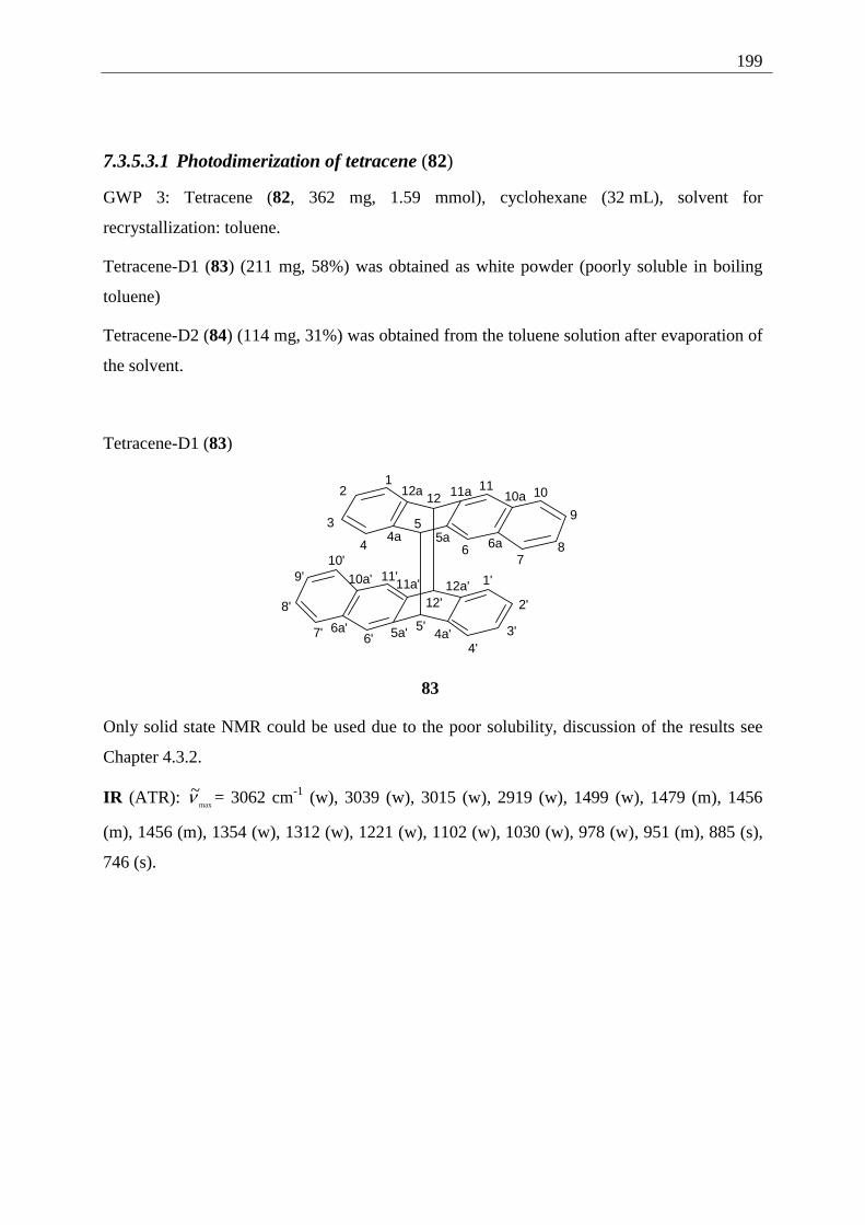
199
7.3.5.3.1 Photodimerization of tetracene (82)
GWP 3: Tetracene (82, 362 mg, 1.59 mmol), cyclohexane (32 mL), solvent for
recrystallization: toluene.
Tetracene-D1 (83) (211 mg, 58%) was obtained as white powder (poorly soluble in boiling
toluene)
Tetracene-D2 (84) (114 mg, 31%) was obtained from the toluene solution after evaporation of
the solvent.
Tetracene-D1 (83)
12
3
4
5
67
8
9
101112
4a 5a 6a
10a11a12a
1'
2'
3'4'
5'6'7'
8'
9'10'
11'
12'
4a'5a'6a'
10a' 11a' 12a'
83
Only solid state NMR could be used due to the poor solubility, discussion of the results see
Chapter 4.3.2.
IR (ATR): max
~ν = 3062 cm-1 (w), 3039 (w), 3015 (w), 2919 (w), 1499 (w), 1479 (m), 1456
(m), 1456 (m), 1354 (w), 1312 (w), 1221 (w), 1102 (w), 1030 (w), 978 (w), 951 (m), 885 (s),
746 (s).
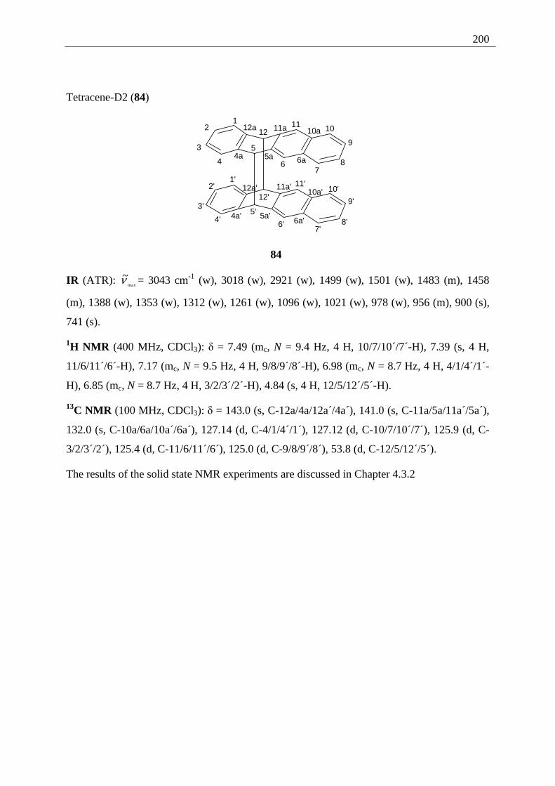
200
Tetracene-D2 (84)
12
3
4
5
67
8
9
101112
4a 5a 6a
10a11a12a
1'2'
3'
4'5'
6'7'
8'
9'
10'11'
12'
4a' 5a'6a'
10a'11a'12a'
84
IR (ATR): max
~ν = 3043 cm-1 (w), 3018 (w), 2921 (w), 1499 (w), 1501 (w), 1483 (m), 1458
(m), 1388 (w), 1353 (w), 1312 (w), 1261 (w), 1096 (w), 1021 (w), 978 (w), 956 (m), 900 (s),
741 (s).
1H NMR (400 MHz, CDCl3): δ = 7.49 (mc, N = 9.4 Hz, 4 H, 10/7/10´/7´-H), 7.39 (s, 4 H,
11/6/11´/6´-H), 7.17 (mc, N = 9.5 Hz, 4 H, 9/8/9´/8´-H), 6.98 (mc, N = 8.7 Hz, 4 H, 4/1/4´/1´-
H), 6.85 (mc, N = 8.7 Hz, 4 H, 3/2/3´/2´-H), 4.84 (s, 4 H, 12/5/12´/5´-H).
13C NMR (100 MHz, CDCl3): δ = 143.0 (s, C-12a/4a/12a´/4a´), 141.0 (s, C-11a/5a/11a´/5a´),
132.0 (s, C-10a/6a/10a´/6a´), 127.14 (d, C-4/1/4´/1´), 127.12 (d, C-10/7/10´/7´), 125.9 (d, C-
3/2/3´/2´), 125.4 (d, C-11/6/11´/6´), 125.0 (d, C-9/8/9´/8´), 53.8 (d, C-12/5/12´/5´).
The results of the solid state NMR experiments are discussed in Chapter 4.3.2
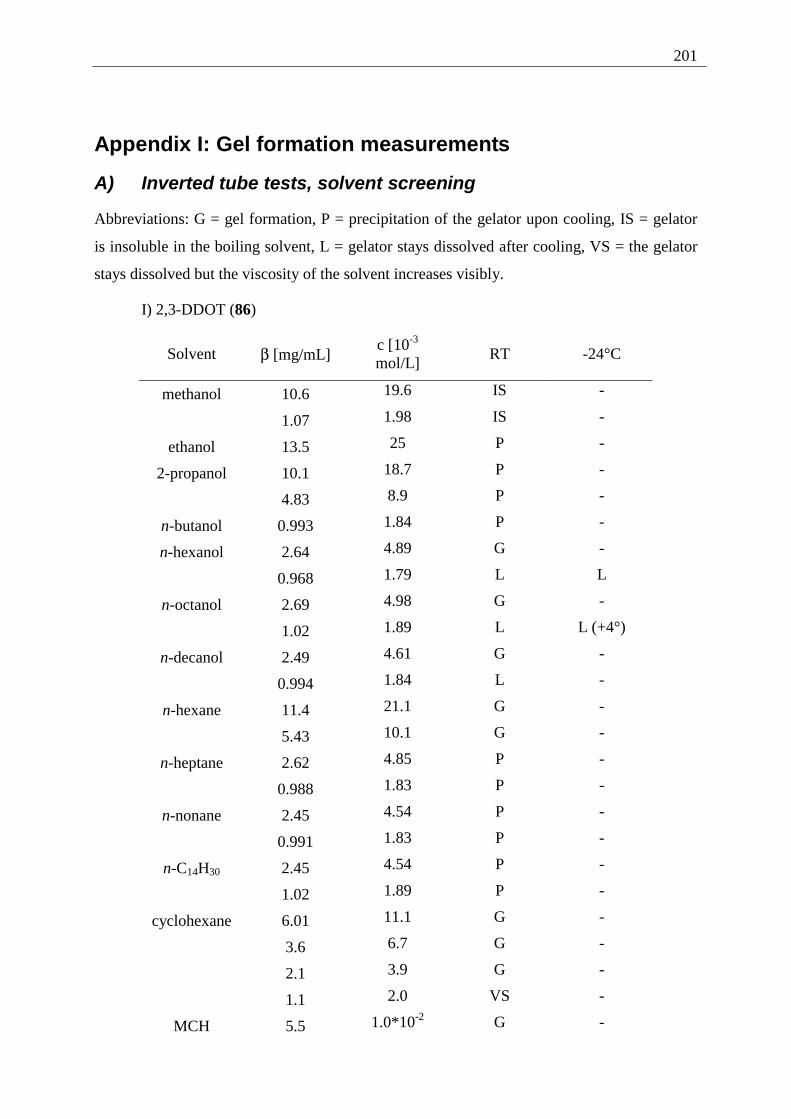
201
Appendix I: Gel formation measurements
A) Inverted tube tests, solvent screening
Abbreviations: G = gel formation, P = precipitation of the gelator upon cooling, IS = gelator
is insoluble in the boiling solvent, L = gelator stays dissolved after cooling, VS = the gelator
stays dissolved but the viscosity of the solvent increases visibly.
I) 2,3-DDOT (86)
Solvent β [mg/mL] c [10-3 mol/L]
RT -24°C
methanol 10.6 19.6 IS -
1.07 1.98 IS -
ethanol 13.5 25 P -
2-propanol 10.1 18.7 P -
4.83 8.9 P -
n-butanol 0.993 1.84 P -
n-hexanol 2.64 4.89 G -
0.968 1.79 L L
n-octanol 2.69 4.98 G -
1.02 1.89 L L (+4°)
n-decanol 2.49 4.61 G -
0.994 1.84 L -
n-hexane 11.4 21.1 G -
5.43 10.1 G -
n-heptane 2.62 4.85 P -
0.988 1.83 P -
n-nonane 2.45 4.54 P -
0.991 1.83 P -
n-C14H30 2.45 4.54 P -
1.02 1.89 P -
cyclohexane 6.01 11.1 G -
3.6 6.7 G -
2.1 3.9 G -
1.1 2.0 VS -
MCH 5.5 1.0*10-2 G -
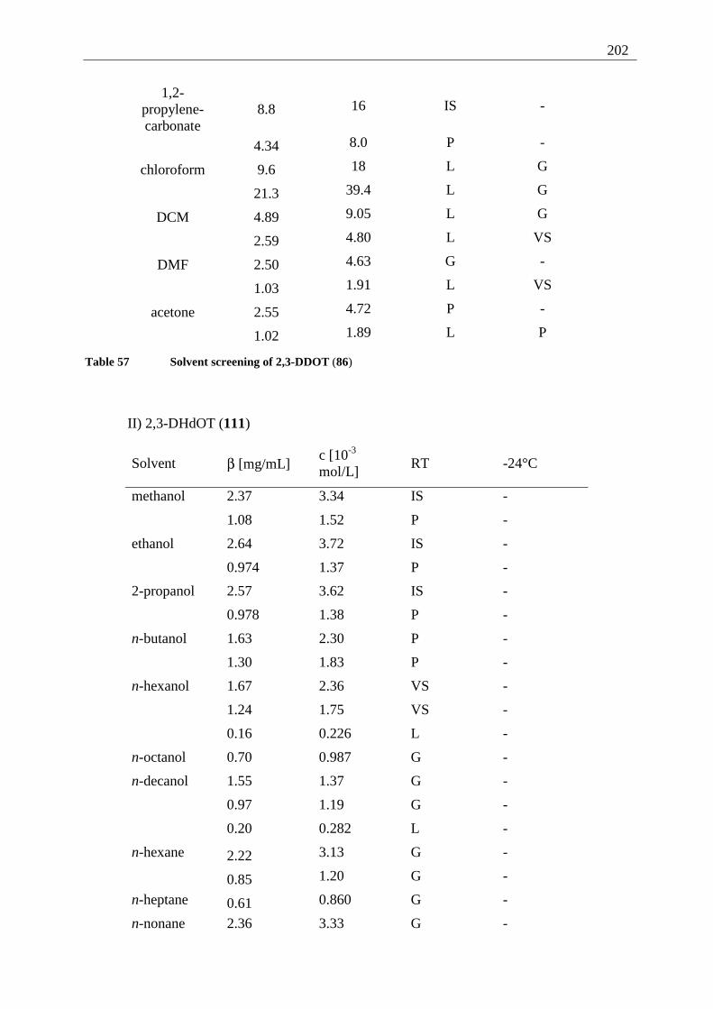
202
1,2-propylene- carbonate
8.8 16 IS -
4.34 8.0 P -
chloroform 9.6 18 L G
21.3 39.4 L G
DCM 4.89 9.05 L G
2.59 4.80 L VS
DMF 2.50 4.63 G -
1.03 1.91 L VS
acetone 2.55 4.72 P -
1.02 1.89 L P
Table 57 Solvent screening of 2,3-DDOT (86)
II) 2,3-DHdOT (111)
Solvent β [mg/mL] c [10-3 mol/L]
RT -24°C
methanol 2.37 3.34 IS -
1.08 1.52 P -
ethanol 2.64 3.72 IS -
0.974 1.37 P -
2-propanol 2.57 3.62 IS -
0.978 1.38 P -
n-butanol 1.63 2.30 P -
1.30 1.83 P -
n-hexanol 1.67 2.36 VS -
1.24 1.75 VS -
0.16 0.226 L -
n-octanol 0.70 0.987 G -
n-decanol 1.55 1.37 G -
0.97 1.19 G -
0.20 0.282 L -
n-hexane 2.22 3.13 G -
0.85 1.20 G -
n-heptane 0.61 0.860 G -
n-nonane 2.36 3.33 G -
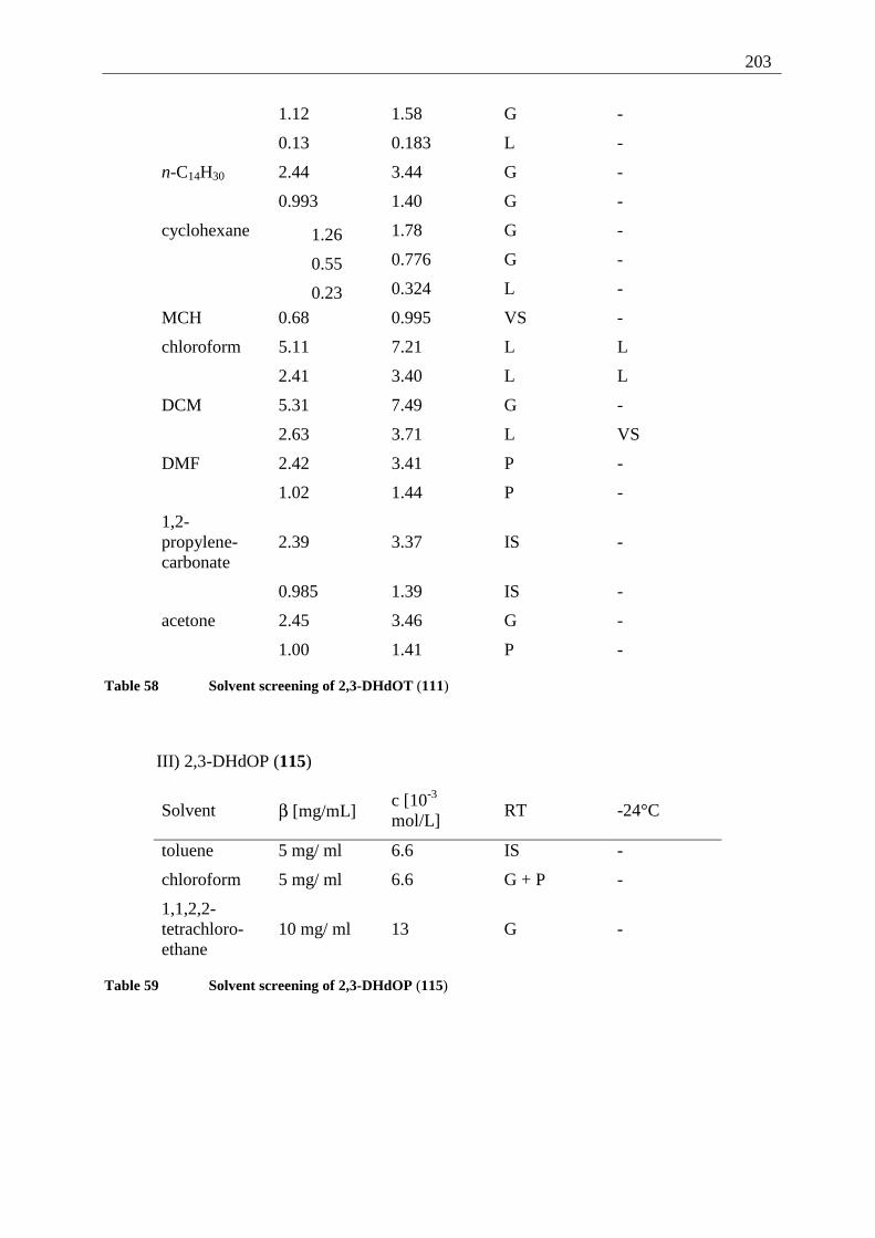
203
1.12 1.58 G -
0.13 0.183 L -
n-C14H30 2.44 3.44 G -
0.993 1.40 G -
cyclohexane 1.26 1.78 G -
0.55 0.776 G -
0.23 0.324 L -
MCH 0.68 0.995 VS -
chloroform 5.11 7.21 L L
2.41 3.40 L L
DCM 5.31 7.49 G -
2.63 3.71 L VS
DMF 2.42 3.41 P -
1.02 1.44 P -
1,2-propylene-carbonate
2.39 3.37 IS -
0.985 1.39 IS -
acetone 2.45 3.46 G -
1.00 1.41 P -
Table 58 Solvent screening of 2,3-DHdOT (111)
III) 2,3-DHdOP (115)
Solvent β [mg/mL] c [10-3 mol/L]
RT -24°C
toluene 5 mg/ ml 6.6 IS -
chloroform 5 mg/ ml 6.6 G + P -
1,1,2,2- tetrachloro- ethane
10 mg/ ml 13 G -
Table 59 Solvent screening of 2,3-DHdOP (115)
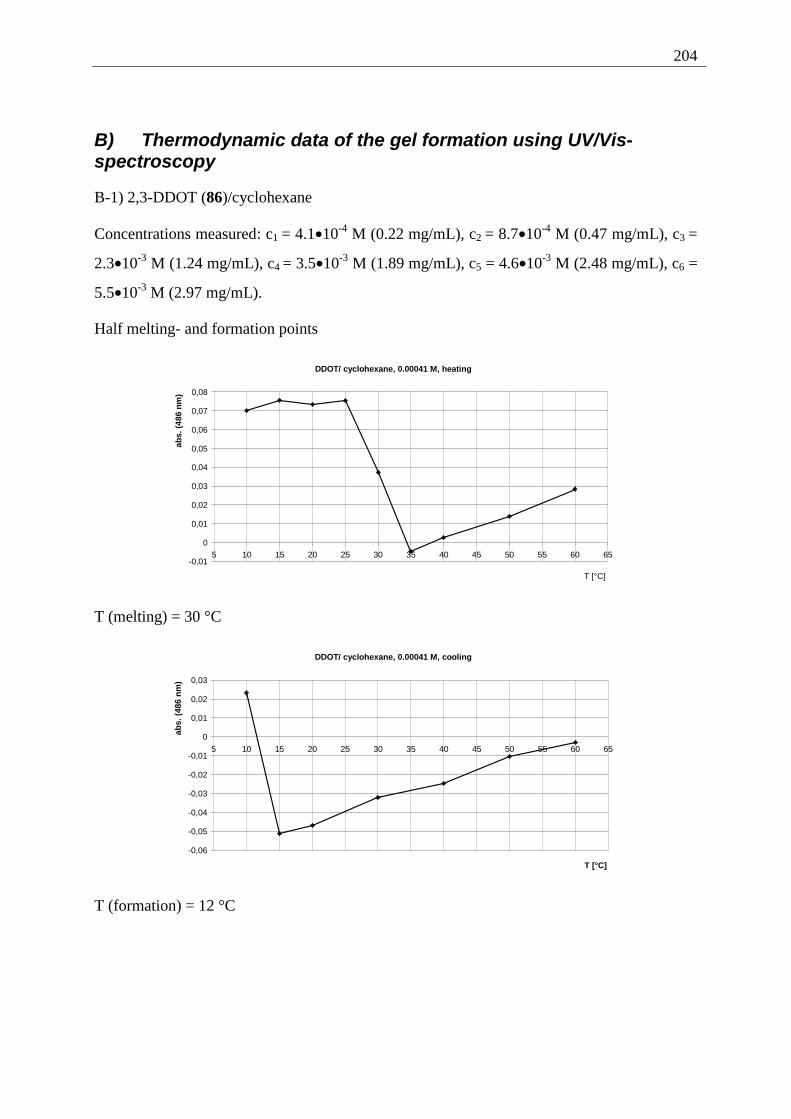
204
B) Thermodynamic data of the gel formation using UV/Vis-spectroscopy
B-1) 2,3-DDOT (86)/cyclohexane
Concentrations measured: c1 = 4.1•10-4 M (0.22 mg/mL), c2 = 8.7•10-4 M (0.47 mg/mL), c3 =
2.3•10-3 M (1.24 mg/mL), c4 = 3.5•10-3 M (1.89 mg/mL), c5 = 4.6•10-3 M (2.48 mg/mL), c6 =
5.5•10-3 M (2.97 mg/mL).
Half melting- and formation points
DDOT/ cyclohexane, 0.00041 M, heating
-0,01
0
0,01
0,02
0,03
0,04
0,05
0,06
0,07
0,08
5 10 15 20 25 30 35 40 45 50 55 60 65
T [°C]
abs.
(48
6 n
m)
T (melting) = 30 °C
DDOT/ cyclohexane, 0.00041 M, cooling
-0,06
-0,05
-0,04
-0,03
-0,02
-0,01
0
0,01
0,02
0,03
5 10 15 20 25 30 35 40 45 50 55 60 65
T [°C]
abs.
(48
6 n
m)
T (formation) = 12 °C
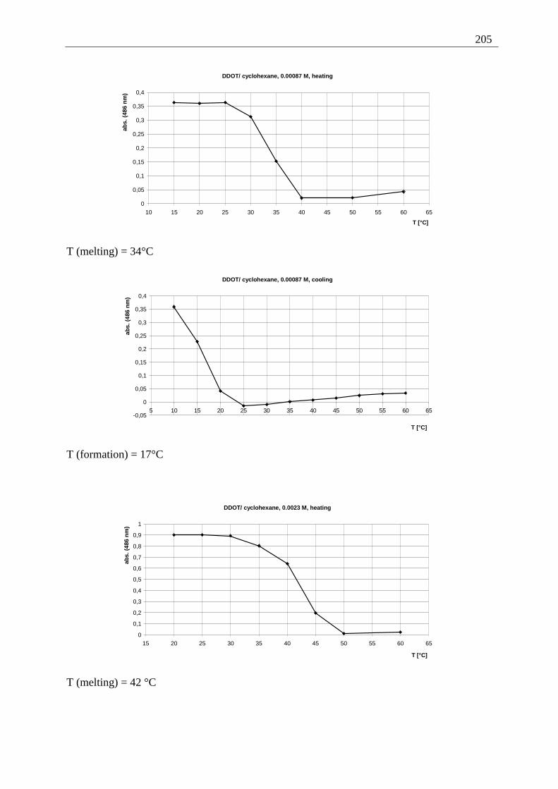
205
DDOT/ cyclohexane, 0.00087 M, heating
0
0,05
0,1
0,15
0,2
0,25
0,3
0,35
0,4
10 15 20 25 30 35 40 45 50 55 60 65
T [°C]
abs.
(48
6 n
m)
T (melting) = 34°C
DDOT/ cyclohexane, 0.00087 M, cooling
-0,05
0
0,05
0,1
0,15
0,2
0,25
0,3
0,35
0,4
5 10 15 20 25 30 35 40 45 50 55 60 65
T [°C]
abs.
(48
6 n
m)
T (formation) = 17°C
DDOT/ cyclohexane, 0.0023 M, heating
0
0,1
0,2
0,3
0,4
0,5
0,6
0,7
0,8
0,9
1
15 20 25 30 35 40 45 50 55 60 65
T [°C]
abs.
(48
6 n
m)
T (melting) = 42 °C
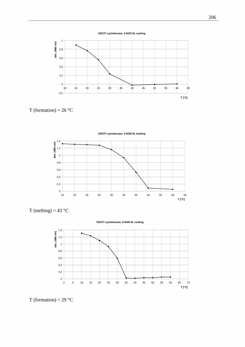
206
DDOT/ cyclohexane, 0.0023 M, cooling
-0,2
0
0,2
0,4
0,6
0,8
1
10 15 20 25 30 35 40 45 50 55 60 65
T [°C]
abs.
(48
6 n
m)
T (formation) = 26 °C
DDOT/ cyclohexane, 0.0035 M, heating
0
0,2
0,4
0,6
0,8
1
1,2
1,4
15 20 25 30 35 40 45 50 55 60 65
T [°C]
abs.
(48
6 nm
)
T (melting) = 43 °C
DDOT/ cyclohexane, 0.0035 M, cooling
0
0,2
0,4
0,6
0,8
1
1,2
1,4
0 5 10 15 20 25 30 35 40 45 50 55 60 65 70
T [°C]
abs.
(48
6 nm
)
T (formation) = 29 °C
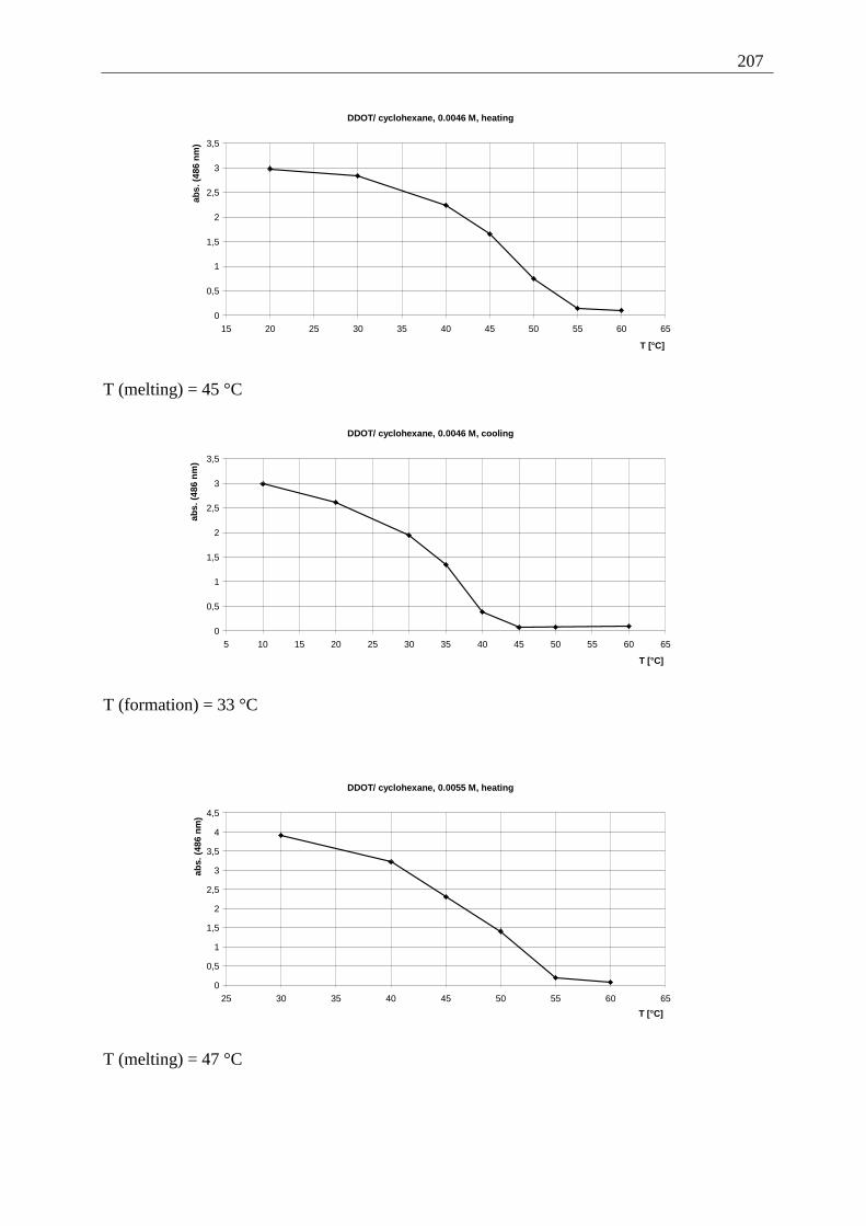
207
DDOT/ cyclohexane, 0.0046 M, heating
0
0,5
1
1,5
2
2,5
3
3,5
15 20 25 30 35 40 45 50 55 60 65
T [°C]
abs.
(48
6 n
m)
T (melting) = 45 °C
DDOT/ cyclohexane, 0.0046 M, cooling
0
0,5
1
1,5
2
2,5
3
3,5
5 10 15 20 25 30 35 40 45 50 55 60 65
T [°C]
abs.
(48
6 n
m)
T (formation) = 33 °C
DDOT/ cyclohexane, 0.0055 M, heating
0
0,5
1
1,5
2
2,5
3
3,5
4
4,5
25 30 35 40 45 50 55 60 65
T [°C]
abs.
(48
6 n
m)
T (melting) = 47 °C

208
DDOT/ cyclohexane, 0.0055 M, cooling
0
0,5
1
1,5
2
2,5
3
3,5
15 20 25 30 35 40 45 50 55 60 65
T [°C]
abs.
(48
6 nm
)
T (formation) = 37 °C
Phase transition diagram (2,3-DDOT (86)/cyclohexane)
Phase transition diagram (DDOT/cyclohexane)
0
5
10
15
20
25
30
35
40
45
50
0,00E+00 1,00E-03 2,00E-03 3,00E-03 4,00E-03 5,00E-03 6,00E-03c (DDOT) [mol/L]
T [
°C]
melting
formation
Determination of the thermodynamic properties (∆H, ∆S, 2,3-DDOT (86)/cyclohexane)
Calculation of thermodynamic properties (DDOT/cyclohexane)
y = -14788x + 38,821R2 = 0,9897
y = -9392,2x + 23,056R2 = 0,9838
-10,5
-10
-9,5
-9
-8,5
-8
-7,5
-7
-6,5
-6
3,00E-03 3,20E-03 3,40E-03 3,60E-031/T [1/K]
ln (
φ(D
DO
T))
melting
formation
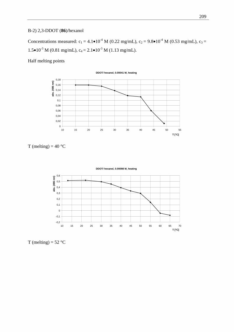
209
B-2) 2,3-DDOT (86)/hexanol
Concentrations measured: c1 = 4.1•10-4 M (0.22 mg/mL), c2 = 9.8•10-4 M (0.53 mg/mL), c3 =
1.5•10-3 M (0.81 mg/mL), c4 = 2.1•10-3 M (1.13 mg/mL).
Half melting points
DDOT/ hexanol, 0.00041 M, heating
0
0,02
0,04
0,06
0,08
0,1
0,12
0,14
0,16
0,18
10 15 20 25 30 35 40 45 50 55
T [°C]
abs.
(48
6 n
m)
T (melting) = 40 °C
DDOT/ hexanol, 0.00098 M, heating
-0,2
-0,1
0
0,1
0,2
0,3
0,4
0,5
0,6
10 15 20 25 30 35 40 45 50 55 60 65 70
T [°C]
abs.
(48
6 n
m)
T (melting) = 52 °C
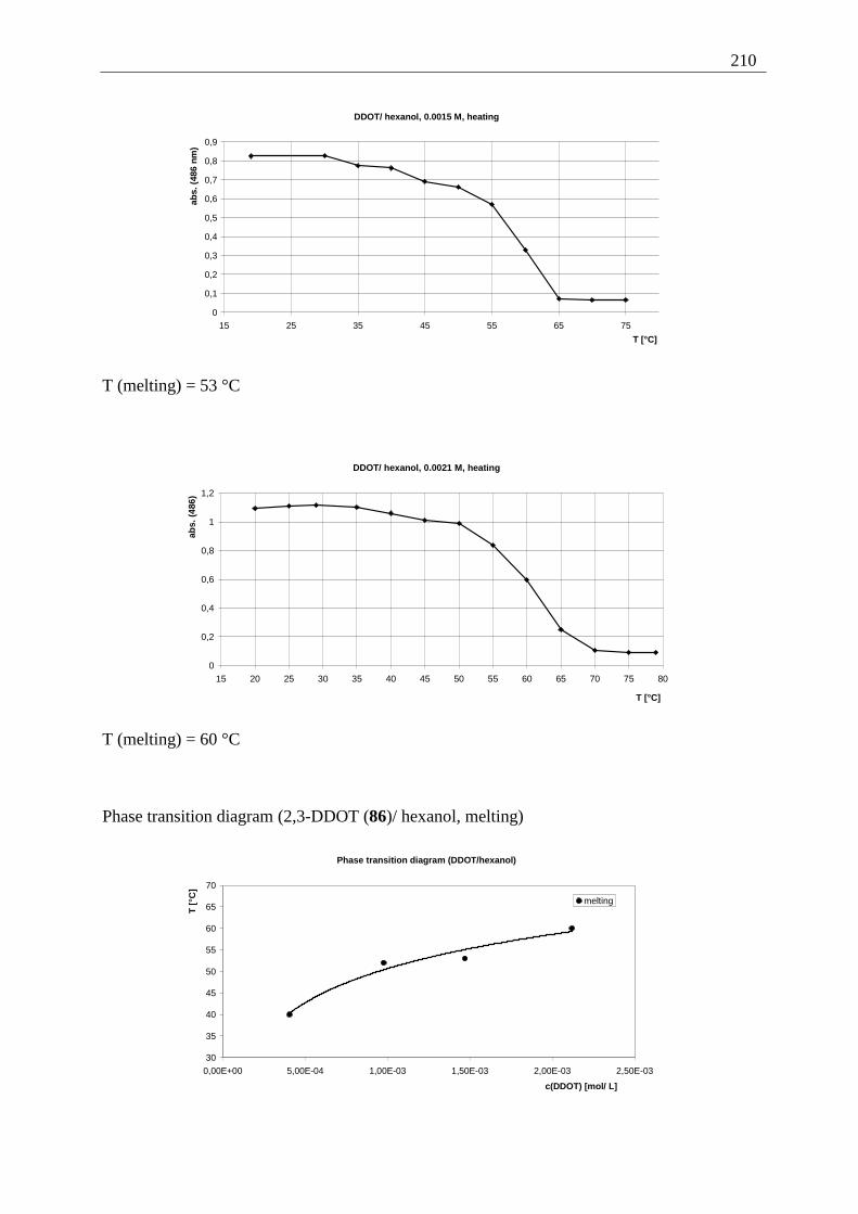
210
DDOT/ hexanol, 0.0015 M, heating
0
0,1
0,2
0,3
0,4
0,5
0,6
0,7
0,8
0,9
15 25 35 45 55 65 75
T [°C]
abs.
(48
6 nm
)
T (melting) = 53 °C
DDOT/ hexanol, 0.0021 M, heating
0
0,2
0,4
0,6
0,8
1
1,2
15 20 25 30 35 40 45 50 55 60 65 70 75 80
T [°C]
abs.
(48
6)
T (melting) = 60 °C
Phase transition diagram (2,3-DDOT (86)/ hexanol, melting)
Phase transition diagram (DDOT/hexanol)
30
35
40
45
50
55
60
65
70
0,00E+00 5,00E-04 1,00E-03 1,50E-03 2,00E-03 2,50E-03
c(DDOT) [mol/ L]
T [
°C]
melting
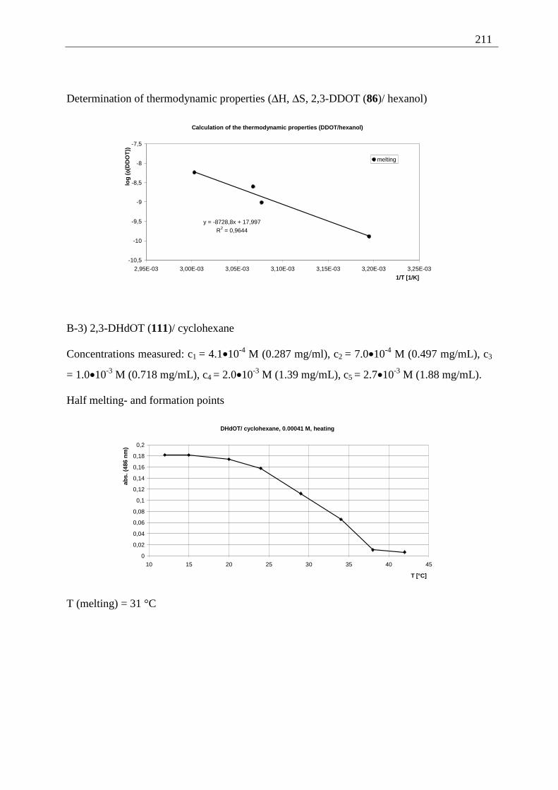
211
Determination of thermodynamic properties (∆H, ∆S, 2,3-DDOT (86)/ hexanol)
Calculation of the thermodynamic properties (DDOT/hexanol)
y = -8728,8x + 17,997R2 = 0,9644
-10,5
-10
-9,5
-9
-8,5
-8
-7,5
2,95E-03 3,00E-03 3,05E-03 3,10E-03 3,15E-03 3,20E-03 3,25E-03
1/T [1/K]
log
( φ(D
DO
T))
melting
B-3) 2,3-DHdOT (111)/ cyclohexane
Concentrations measured: c1 = 4.1•10-4 M (0.287 mg/ml), c2 = 7.0•10-4 M (0.497 mg/mL), c3
= 1.0•10-3 M (0.718 mg/mL), c4 = 2.0•10-3 M (1.39 mg/mL), c5 = 2.7•10-3 M (1.88 mg/mL).
Half melting- and formation points
DHdOT/ cyclohexane, 0.00041 M, heating
0
0,02
0,04
0,06
0,08
0,1
0,12
0,14
0,16
0,18
0,2
10 15 20 25 30 35 40 45
T [°C]
abs.
(48
6 n
m)
T (melting) = 31 °C
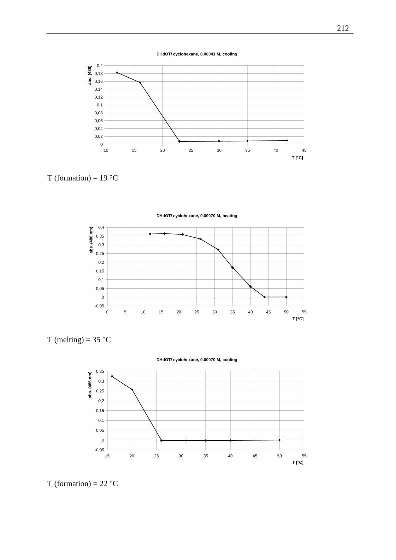
212
DHdOT/ cyclohexane, 0.00041 M, cooling
0
0,02
0,04
0,06
0,08
0,1
0,12
0,14
0,16
0,18
0,2
10 15 20 25 30 35 40 45
T [°C]
abs.
(48
6)
T (formation) = 19 °C
DHdOT/ cyclohexane, 0.00070 M, heating
-0,05
0
0,05
0,1
0,15
0,2
0,25
0,3
0,35
0,4
0 5 10 15 20 25 30 35 40 45 50 55
T [°C]
abs.
(48
6 n
m)
T (melting) = 35 °C
DHdOT/ cyclohexane, 0.00070 M, cooling
-0,05
0
0,05
0,1
0,15
0,2
0,25
0,3
0,35
15 20 25 30 35 40 45 50 55
T [°C]
abs.
(48
6 n
m)
T (formation) = 22 °C
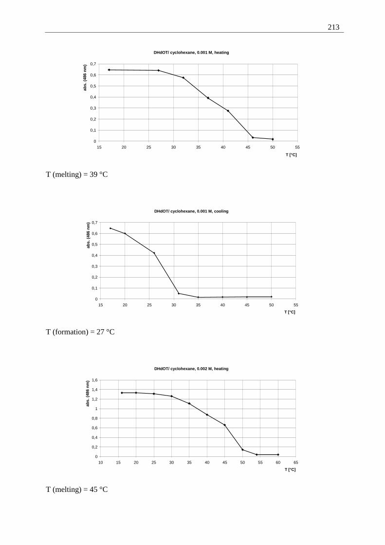
213
DHdOT/ cyclohexane, 0.001 M, heating
0
0,1
0,2
0,3
0,4
0,5
0,6
0,7
15 20 25 30 35 40 45 50 55
T [°C]
abs.
(48
6 n
m)
T (melting) = 39 °C
DHdOT/ cyclohexane, 0.001 M, cooling
0
0,1
0,2
0,3
0,4
0,5
0,6
0,7
15 20 25 30 35 40 45 50 55
T [°C]
abs.
(48
6 n
m)
T (formation) = 27 °C
DHdOT/ cyclohexane, 0.002 M, heating
0
0,2
0,4
0,6
0,8
1
1,2
1,4
1,6
10 15 20 25 30 35 40 45 50 55 60 65
T [°C]
abs.
(48
6 n
m)
T (melting) = 45 °C
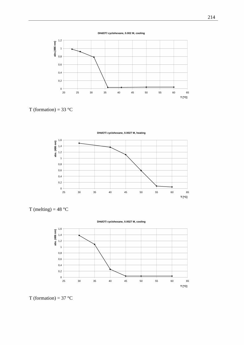
214
DHdOT/ cyclohexane, 0.002 M, cooling
0
0,2
0,4
0,6
0,8
1
1,2
20 25 30 35 40 45 50 55 60 65
T [°C]
abs.
(486
nm
)
T (formation) = 33 °C
DHdOT/ cyclohexane, 0.0027 M, heating
0
0,2
0,4
0,6
0,8
1
1,2
1,4
1,6
25 30 35 40 45 50 55 60 65
T [°C]
abs.
(48
6 n
m)
T (melting) = 48 °C
DHdOT/ cyclohexane, 0.0027 M, cooling
0
0,2
0,4
0,6
0,8
1
1,2
1,4
1,6
25 30 35 40 45 50 55 60 65
T [°C]
abs.
(48
6 n
m)
T (formation) = 37 °C
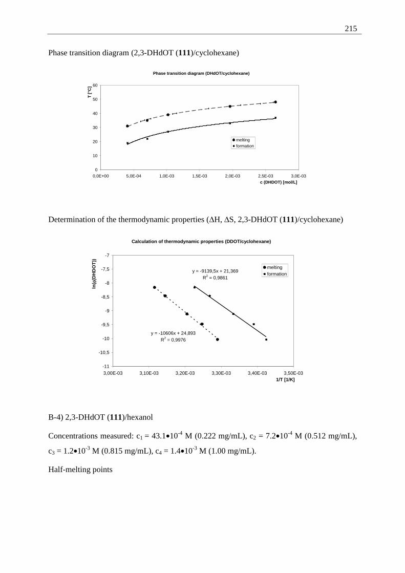
215
Phase transition diagram (2,3-DHdOT (111)/cyclohexane)
Phase transition diagram (DHdOT/cyclohexane)
0
10
20
30
40
50
60
0,0E+00 5,0E-04 1,0E-03 1,5E-03 2,0E-03 2,5E-03 3,0E-03
c (DHDOT) [mol/L]
T [°
C]
meltingformation
Determination of the thermodynamic properties (∆H, ∆S, 2,3-DHdOT (111)/cyclohexane)
Calculation of thermodynamic properties (DDOT/cyclohexane)
y = -10606x + 24,893R2 = 0,9976
y = -9139,5x + 21,369R2 = 0,9861
-11
-10,5
-10
-9,5
-9
-8,5
-8
-7,5
-7
3,00E-03 3,10E-03 3,20E-03 3,30E-03 3,40E-03 3,50E-031/T [1/K]
ln( φ
(DH
DO
T))
meltingformation
B-4) 2,3-DHdOT (111)/hexanol
Concentrations measured: c1 = 43.1•10-4 M (0.222 mg/mL), c2 = 7.2•10-4 M (0.512 mg/mL),
c3 = 1.2•10-3 M (0.815 mg/mL), c4 = 1.4•10-3 M (1.00 mg/mL).
Half-melting points
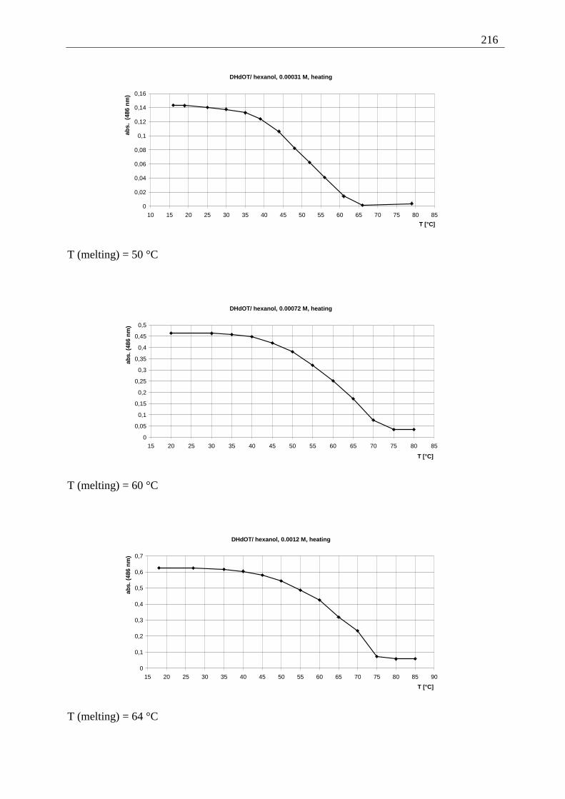
216
DHdOT/ hexanol, 0.00031 M, heating
0
0,02
0,04
0,06
0,08
0,1
0,12
0,14
0,16
10 15 20 25 30 35 40 45 50 55 60 65 70 75 80 85
T [°C]
abs.
(48
6 n
m)
T (melting) = 50 °C
DHdOT/ hexanol, 0.00072 M, heating
0
0,05
0,1
0,15
0,2
0,25
0,3
0,35
0,4
0,45
0,5
15 20 25 30 35 40 45 50 55 60 65 70 75 80 85
T [°C]
abs.
(48
6 n
m)
T (melting) = 60 °C
DHdOT/ hexanol, 0.0012 M, heating
0
0,1
0,2
0,3
0,4
0,5
0,6
0,7
15 20 25 30 35 40 45 50 55 60 65 70 75 80 85 90
T [°C]
abs.
(48
6 n
m)
T (melting) = 64 °C
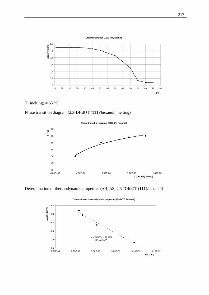
217
DHdOT/ hexanol, 0.0014 M, heating
0
0,2
0,4
0,6
0,8
1
1,2
20 25 30 35 40 45 50 55 60 65 70 75 80 85 90
T [°C]
abs.
(48
6 n
m)
T (melting) = 65 °C
Phase transition diagram (2,3-DHdOT (111)/hexanol, melting)
Phase transition diagram (DHDOT/ hexanol)
40
45
50
55
60
65
70
0,00E+00 4,00E-04 8,00E-04 1,20E-03 1,60E-03
c (DHDOT) [mol/L]
T [
°C]
Determination of thermodynamic properties (∆H, ∆S, 2,3-DHdOT (111)/hexanol)
Calculation of thermodynamic properties (DHDOT/ hexanol)
y = -10555x + 22,493R2 = 0,9825
-10,4
-10
-9,6
-9,2
-8,8
-8,4
2,90E-03 2,95E-03 3,00E-03 3,05E-03 3,10E-03 3,15E-03
1/T [1/K]
ln (
φ(D
HD
OT
))
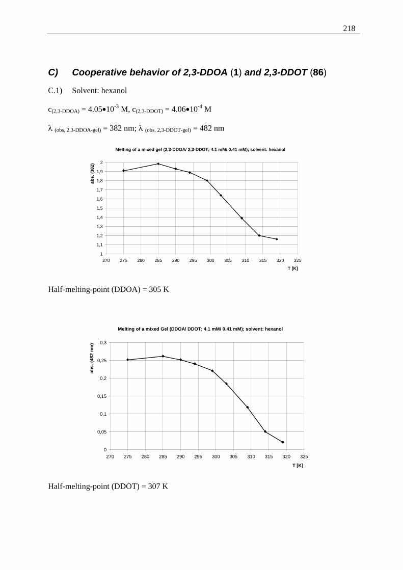
218
C) Cooperative behavior of 2,3-DDOA (1) and 2,3-DDOT (86)
C.1) Solvent: hexanol
c(2,3-DDOA) = 4.05•10-3 M, c(2,3-DDOT) = 4.06•10-4 M
λ (obs, 2,3-DDOA-gel) = 382 nm; λ (obs, 2,3-DDOT-gel) = 482 nm
Melting of a mixed gel (2,3-DDOA/ 2,3-DDOT; 4.1 mM/ 0.41 mM); solvent: hexanol
1
1,1
1,2
1,3
1,4
1,5
1,6
1,7
1,8
1,9
2
270 275 280 285 290 295 300 305 310 315 320 325
T [K]
abs.
(38
2)
Half-melting-point (DDOA) = 305 K
Melting of a mixed Gel (DDOA/ DDOT; 4.1 mM/ 0.41 mM); solvent: hexanol
0
0,05
0,1
0,15
0,2
0,25
0,3
270 275 280 285 290 295 300 305 310 315 320 325
T [K]
abs.
(48
2 n
m)
Half-melting-point (DDOT) = 307 K
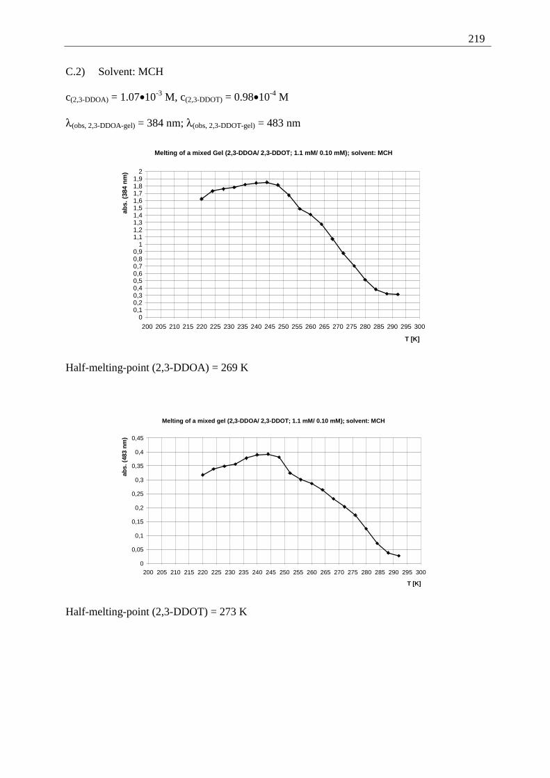
219
C.2) Solvent: MCH
c(2,3-DDOA) = 1.07•10-3 M, c(2,3-DDOT) = 0.98•10-4 M
λ(obs, 2,3-DDOA-gel) = 384 nm; λ(obs, 2,3-DDOT-gel) = 483 nm
Melting of a mixed Gel (2,3-DDOA/ 2,3-DDOT; 1.1 mM/ 0.10 mM); solvent: MCH
00,10,20,30,40,50,60,70,80,9
11,11,21,31,41,51,61,71,81,9
2
200 205 210 215 220 225 230 235 240 245 250 255 260 265 270 275 280 285 290 295 300
T [K]
abs.
(38
4 n
m)
Half-melting-point (2,3-DDOA) = 269 K
Melting of a mixed gel (2,3-DDOA/ 2,3-DDOT; 1.1 mM/ 0.10 mM); solvent: MCH
0
0,05
0,1
0,15
0,2
0,25
0,3
0,35
0,4
0,45
200 205 210 215 220 225 230 235 240 245 250 255 260 265 270 275 280 285 290 295 300
T [K]
abs.
(48
3 n
m)
Half-melting-point (2,3-DDOT) = 273 K
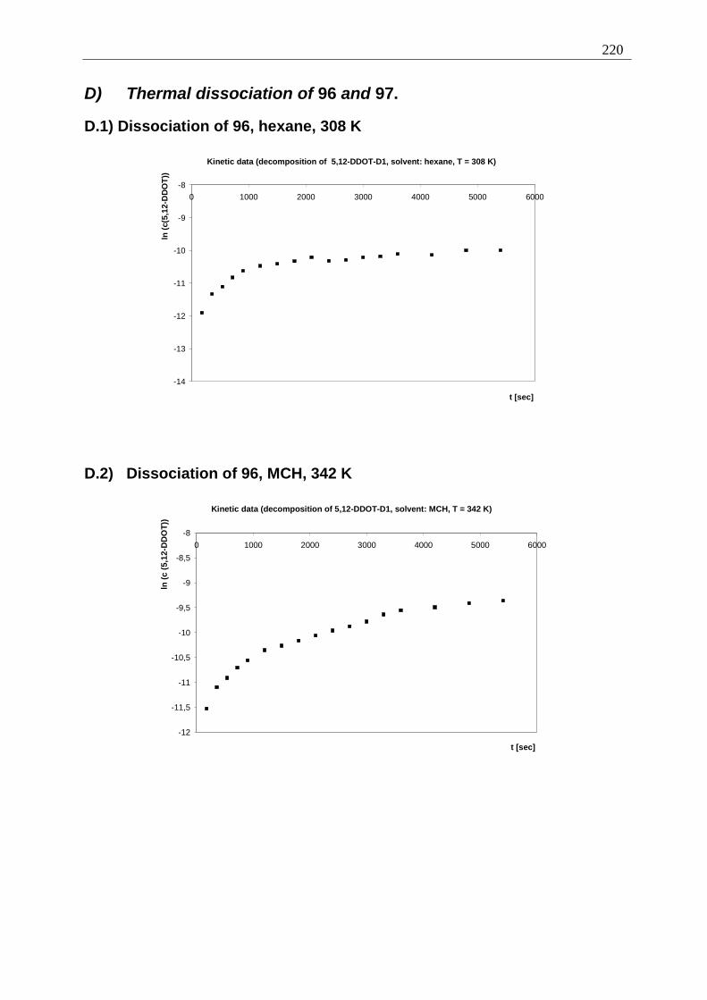
220
D) Thermal dissociation of 96 and 97.
D.1) Dissociation of 96, hexane, 308 K
Kinetic data (decomposition of 5,12-DDOT-D1, solvent: hexane, T = 308 K)
-14
-13
-12
-11
-10
-9
-80 1000 2000 3000 4000 5000 6000
t [sec]
ln (
c(5,
12-D
DO
T))
D.2) Dissociation of 96, MCH, 342 K
Kinetic data (decomposition of 5,12-DDOT-D1, solvent: MCH, T = 342 K)
-12
-11,5
-11
-10,5
-10
-9,5
-9
-8,5
-8
0 1000 2000 3000 4000 5000 6000
t [sec]
ln (
c (5
,12-
DD
OT
))
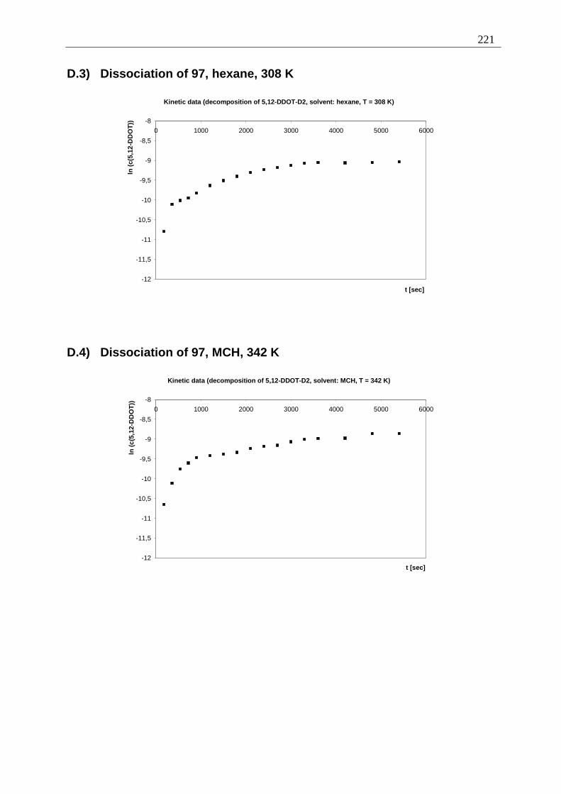
221
D.3) Dissociation of 97, hexane, 308 K
Kinetic data (decomposition of 5,12-DDOT-D2, solvent: hexane, T = 308 K)
-12
-11,5
-11
-10,5
-10
-9,5
-9
-8,5
-8
0 1000 2000 3000 4000 5000 6000
t [sec]
ln (
c(5,
12-D
DO
T))
D.4) Dissociation of 97, MCH, 342 K
Kinetic data (decomposition of 5,12-DDOT-D2, solvent: MCH, T = 342 K)
-12
-11,5
-11
-10,5
-10
-9,5
-9
-8,5
-8
0 1000 2000 3000 4000 5000 6000
t [sec]
ln (
c(5,
12-D
DO
T))
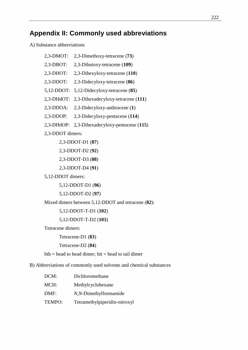
222
Appendix II: Commonly used abbreviations
A) Substance abbreviations
2,3-DMOT: 2,3-Dimethoxy-tetracene (73)
2,3-DBOT: 2,3-Dibutoxy-tetracene (109)
2,3-DHOT: 2,3-Dihexyloxy-tetracene (110)
2,3-DDOT: 2,3-Didecyloxy-tetracene (86)
5,12-DDOT: 5,12-Didecyloxy-tetracene (85)
2,3-DHdOT: 2,3-Dihexadecyloxy-tetracene (111)
2,3-DDOA: 2,3-Didecyloxy-anthracene (1)
2,3-DDOP: 2,3-Didecyloxy-pentacene (114)
2,3-DHdOP: 2,3-Dihexadecyloxy-pentacene (115)
2,3-DDOT dimers:
2,3-DDOT-D1 (87)
2,3-DDOT-D2 (92)
2,3-DDOT-D3 (88)
2,3-DDOT-D4 (91)
5,12-DDOT dimers:
5,12-DDOT-D1 (96)
5,12-DDOT-D2 (97)
Mixed dimers between 5,12-DDOT and tetracene (82):
5,12-DDOT-T-D1 (102)
5,12-DDOT-T-D2 (103)
Tetracene dimers:
Tetracene-D1 (83)
Tetracene-D2 (84)
hth = head to head dimer; htt = head to tail dimer
B) Abbreviations of commonly used solvents and chemical substances
DCM: Dichloromethane
MCH: Methylcyclohexane
DMF: N,N-Dimethylformamide
TEMPO: Tetramethylpiperidin-nitroxyl
223
Appendix III: Literature
[1] a) P. Terech, R. G. Weiss, Chem Rev., 1997, 97, 3133-3159; b) A. T. Polishuk,
J. Am. Soc. Lubn. Eng., 1977, 33, 133-138; c) N. Pilpel, Chem. Rev., 1963, 63,
221-234; d) O. Martin-Borret, R. Ramasseul, R. Rassat, Bull. Soc. Chim. Fr.,
1979, 401-408; e) T. Itoh, D. E. Katsoulis, I. Mita, J. Mater. Chem., 1993, 3,
1303-1305; f) K. Murata, M. Aoki, T. Susuki, T. Harada, H. Kawabata, T.
Komori, F. Ohseto, K. Ueda, S. Shinkai, J. Am. Chem. Soc., 1994, 116, 6664-
66676; g) J. Campbell, M. Kuzma, M. Labes, Mol. Cryst. Liq. Cryst., 1983, 95,
45-50; h) P. Terech, C. Chachaty, J. Gaillard, A. M. Godquin-Giroud, J. Phys.
Fr., 1987, 48, 663-671; R. Ramasseul, P. Maldivi, J. C. Marchon, Liquid
Crystals, 1993, 13, 729-733; j) R. J. Twieg, T. P. Russel, R. Siemens, J. F.
Rabolt, Macromolecules, 1985, 18, 1361-1362; k) J.-M. Lehn, M. Mascal, A.
DeCian, J. Fisher, J. Chem. Soc., Chem. Commun., 1990, 479-481.
[2] T. Brotin, R. Utermöhlen, F. Fages, H. Bouas-Laurent, J.-P. Desvergne, J.
Chem. Soc., Chem. Commun., 1991, 416-418.
[3] T. Brotin, J. Waluk, J.-P. Desvergne, H. Bouas-Laurent, J. Michl, Photochem.
Photobiol., 1992, 335-347.
[4] H. Bouas-Laurent, A. Castellan, J.-P. Desvergne, R. Lapouyade, Chem. Soc.
Rev., 2000, 29, 43-55.
[5] Photochromism: Molecules and Systems, H. Dürr, H. Bouas-Laurent (eds.),
Elsevier, Amsterdam, 2003, chapter 14, 583.
[6] H. Bouas-Laurent, A. Castellan, J. Chem. Soc., Chem. Comm., 1970, 1648-
1649.
[7] R. Lapouyade, A. Nourmamode, H. Bouas-Laurent, Tetrahedron, 1980, 2311-
2316.
[8] H. G. Bungenberg de Jong, Colloid Science II, 1949 ed. H. R. Kruyt, (Elsevier
Publ. Co., Inc., Amsterdam).
[9] P. J. Flory, Faraday Discuss. Chem. Soc., 1974, 57, 7-18.
[10] P. Terech, I. Furman, H. Bouas-Laurent, J. P. Desvergne, R. Ramasseul, R. G.
Weiss; J. Chem Soc., Faraday Discuss., 1995, 101, 345-358.

224
[11] J.-L. Pozzo, G. M. Clavier, M. Colomes, H. Bouas-Laurent, Tetrahedron,
1997, 53 6377-6390.
[12] D. N. Gupta, P. Hodge, N. Khan; J. Chem. Soc., Perkin Trans. 1, 1981, 689-
696.
[13] D. M. Bowles, J. E. Anthony, Org. Lett., 2000, 2, 85-87.
[14] A. Mallouli, Y. Lepage, Synthesis, 1980, 9, 689.
[15] R. W. Franck, P. B. Gupta; J. Org. Chem., 1985, 50, 4632-4635.
[16] J. Reichwagen, Diploma Thesis, Braunschweig 2001.
[17] B. Serpaud, Y. Lepage, Bull. Soc. Chim. Fr., 1977, 5, 539-542.
[18] R. Adams, T. A. Geissmann, R. B. Baker, H. M. Teeter, J. Am. Chem. Soc., 63,
1941, 528-534.
[19] J. W. Grissom, G. U. Gunawardena, Tetrahedron Lett., 36, 1995, 4951-4954.
[20] X. Zhang, B. W. Fox, J. A. Hadfield, Synth. Comm., 1996, 26, 49-62.
[21] R. T. Plimpton, J. Chem. Soc., 1880, 37, 633-645.
[22] H. Laatsch, Justus Liebigs Ann. Chem., 1985, 251-274.
[23] V. P. Papageorgiou, E. A. Couladouras, Z. F. Plyta, S. A. Haroutouniau, J.
Org. Chem., 1997, 62, 6-10.
[24] A. P. Marchand, G. M. Reddy, Synthesis, 1991, 3, 198-200.
[25] E. Barranco, N. Martín, J. L. Seguro, C. Seoane, Tetrahedron, 1993, 49 , 4881-
4892.
[26] M. Albrecht, M. Schneider, Synthesis, 2000, 11, 1557-1560.
[27] M. V. Bhatt, S. S. El-Morey, Synthesis, 1982, 12, 1048-1050.
[28] S. Andersson, Synthesis, 1985, 4, 437-439.
[29] A. Robertson, R. B. Waters, J. Chem. Soc., 1933, 83-86.
[30] G. Grethe, V. Toome, H. L. Lee, M. Uskakovic, A. Brossi, J. Org. Chem.,
1968, 504-508.
[31] N. P. Buu-Hoi, D. Lavit, J. Org. Chem., 1956, 21-23.
[32] A. Williamson, Justus Liebigs Ann. Chem., 1851, 77, 37-49.
225
[33] K. Lagodzinski, Ber. Dtsch. Chem. Ges., 1895, 28, 1533-1535.
[34] R. Broene, F. Diederich, Tetrahedron Lett., 1991, 32, 5227-5230.
[35] T. R. Criswell, B. H. Klanderman, J. Org. Chem., 1974, 39, 770-774.
[36] S. Coffey, V. Boyd, J. Chem. Soc., 1954, 2468-2470.
[37] H. Meerwein, R. Schmidt, Justus Liebigs Ann. Chem., 1926, 444, 221-238.
[38] W. Ponndorf, Angew. Chem., 1926, 39, 138-143.
[39] U. Müller, M. Adam, K. Müllen, Chem. Ber., 1994, 127, 437-444.
[40] D. Perepichka, M. Bendikov, H. Meng, F. Wudl, J. Am. Chem. Soc., 2003, 125,
10190-10191.
[41] G. S. Tulevski, Q. Miao, M. Fukuto, R. Abram, B. Ocko, R. Pindak, M. L.
Steigerwald, C. R. Kagan, C. Nuckolls, J. Am. Chem. Soc., 2004, 126, 15048-
15050.
[42] N. Vets, M. Smet, W. Dehaen, Tetrahedron Lett., 2004, 7287-7289.
[43] A. Afzali, C. D. Dimitrakopoulos, T. L. Breen, J. Am. Chem. Soc., 2002, 8812-
8813.
[44] M. M. Payne, J. H. Delcamp, S. R. Parkin, J. E. Anthony, Org. Lett., 2004,
1609-1612.
[45] Y. Sakamoto, T. Suzuki, M. Kobayashi, Y. Gao, Y. Fukai, Y. Inoue, F. Sato, S.
Tokito, J. Am. Chem. Soc., 2004, 8138-8140.
[46] J. W. Cook, L. Hunter, R. Schoental, J. Chem. Soc., 1949, Suppl. Issue, 228.
[47] F. Weygand, K. G. Kinkel, D. Tietjen, Chem. Ber., 1950, 83, 394-399.
[48] W. Ried, H. Bodem, Chem. Ber., 1956, 89, 708-712.
[49] R. G. Carlson, K. Srinivasachar, R. S. Givens, B. K. Matuszewski, J. Org.
Chem., 1986, 51, 3978-3983.
[50] Photochromism: Molecules and Systems, H. Dürr and H. Bouas-Laurent (eds.),
revised edition, Elsevier, Amsterdam 2003, chapter 14, p. 584.
[51] K. Lagodzinski, Ber. Dtsch. Chem. Ges., 1895, 28, 116-119.
[52] J. R. Johnson, W. H. Lobling, G. W. Bodamer, J. Am. Chem. Soc., 1941, 63,
131-135.
226
[53] J.-P. Desvergne, T. Brotin, D. Meerschaut, G. Clavier, F. Placin, J.-L. Pozzo
and H. Bouas-Laurent, unpublished data.
[54] P. Terech, D. Meerschaut, J.-P. Desvergne, M. Colomes and H. Bouas-Laurent,
J. Colloid Interface Sci., 2003, 441-445.
[55] A. Del Guerzo, J. Reichwagen, H. Hopf, C. Belin, H. Bouas-Laurent, J.-P.
Desvergne: Self-Assembled Fibers with Tunable Optical Properties:
Organogels of Anthracene and Tetracene Derivatives (Poster), HRSMC & EPA
Summer School "New Perspectives in Photochemistry" Egmond aan Zee,
2003.
[56] Analytical chemistry: the authentic text to the FECS curriculum analytical
chemistry/ ed. by R. Kellner, J.-M. Mermet, M. Otto, H.M. Widmer; Weinheim
1998.
[57] R. B. Woodward, R. Hoffmann, Angew. Chem., Int. Ed., 1969, 781-853,
Angew. Chem., 1969, 797-870.
[58] B. A. Hess, jr., L. S. Schaad, J. Am. Chem. Soc., 1971, 305-310.
[59] J.-P. Desvergne, F. Fages, I. Frisch, H. Bouas-Laurent, J. Chem. Soc., Chem.
Commun., 1988, 1413-1415.
[60] R. Calas, R. Lalande, Bull. Soc. Chim. Fr., 1959, 770-772.
[61] N. C. Yang, H. Shou, T. Wang, J. Masnovi, J. Am. Chem. Soc., 1980, 102,
6652-6654.
[62] T. Brotin, J.-P. Desvergne, F. Fages, R. Utermöhlen, R. Bonneau, H. Bouas-
Laurent, Photochem. Photobiol., 1992, 349-358.
[63] L. B. Birks, Photochem. Photobiol., 1963, 493 [quoted in 7].
[64] L. F. Fieser, J. Am. Chem. Soc., 1931, 53, 2329-2338.
[65] M. Zander, Polycyclische Aromaten: Kohlenwasserstoffe und Fullerene,
Stuttgart: Teubner, 1995.
[66] P. Labandibar, R. Lapouyade, H. Bouas-Laurent, C. R. Hebd. Seances Acad.
Sci. Ser. C, 1971, 272, 1257-1260.
[67] S. Gabriel, E. Leupold, Ber. Dtsch. Chem. Ges., 1898, , 31 1272-1286.
[68] H. Waldmann, H. Mathiowetz, Ber. Dtsch. Chem. Ges., 1931, 64, 1713-1721.
227
[69] M. P. Cava, A. A. Deana, K. Muth, J. Am Chem. Soc., 1959, 81, 6458-6460.
[70] J. G. Smith, P. W. Dibble, J. Org. Chem., 1983, 48, 5361-5362.
[71] D. Marquis, J.-P. Desvergne, H. Bouas-Laurent, J. Org. Chem., 1995, 60,
7984-7996.
[72] F. Fages, J.-P. Desvergne, H. Bouas-Laurent, Bull. Soc. Chim. Fr., 1985, 959-
964.
[73] C. G. Hatchard, C. A. Parker, Proc. R. Soc. Lond. A, 1956, 518-535.
[74] F. Fages, J.-P. Desvergne, I. Frisch, H. Bouas-Laurent, J. Chem. Soc., Chem.
Commun., 1988, 1413-1415.
[75] H. Bouas-Laurent, H. Dürr, Pure Appl. Chem., 2001, 73, 639-665.
[76] Y. S. Liu, P. de Mayo, W. R. Ware, J. Phys. Chem., 1993, 5995-6001.
[77] J. Olmstedt, J. Phys. Chem., 1979, 83, 2581-2584.
[78] H. Langhals, Nachr. Chem. Techn. Lab., 1980, 28, 716-718.
[79] T. Salthammer, Fluoreszensspektroskopische Untersuchungen von Energie-
übertragungsprozessen an Perylen und 3,9-Dibromperylen, Dissertation,
Braunschweig, 1990, 24-40.
228
Lebenslauf
Jens Reichwagen
geboren am 17. Januar 1974
in Hamburg
ledig
Promotion
04/ 2005 Disputation
02/ 2005 Einreichen der Disserttation
seit 10/ 2001 Promotion am Institut für Organische Chemie,
TU Braunschweig; Mentor: Professor Dr. Henning Hopf
Thema der Dissertation:
Synthesis of acene based organic gelators. Examination of the
spectroscopical, photochemical and rheological properties
seit 05/ 2004 Wissenschaftliche Hilfskraft am Institut für Anorganische
und Analytische Chemie, TU Braunschweig
04/ 2002 - 03/ 2004 Promotionsstipendium des Fonds der Chemischen
Industrie
10/ 2001 - 03/ 2002 Wissenschaftliche Hilfskraft am Institut für Organische
Chemie, TU Braunschweig
07/ 2003 - 08/ 2003 Forschungen an der Université Bordeaux I bei
und Professor Dr.Henri Bouas-Laurent
06/ 2002 - 07/ 2002
Hochschulbildung
09/ 2001 Abschluss des Diplomstudiums Chemie an der
TU Braunschweig
Thema der Diplomarbeit: 2,3-Didecyloxytetracen als neuer
Gel-Bildner.
08/ 1998 - 05/ 2001 Hauptstudium mit Vertiefungsrichtung Ökologische
Chemie und Abfallanalytik
229
05/ 1999 - 04/ 2001 Studienbegleitendes Stipendium der Hoechst Stiftung
10/ 1996 - 08/ 1998 Grundstudium.
Berufsausbildung
08/ 1993 - 06/ 1996 Ausbildung zum Chemielaboranten im Staatlichen
Veterinäruntersuchungsamt für Fische und Fischwaren,
Cuxhaven.
Schulbildung
08/ 1986 - 05/ 1993 Gymnasium Cuxhaven.
08/ 1980 - 06/ 1986 Grundschule und Orientierungsstufe Franzenburg
Katastrophenschutzdienst
10/ 1996 - 11/ 1999 Deutsches Rotes Kreuz, Braunschweig
11/ 1992 - 10/ 1996 Fernmeldezug der Stadt Cuxhaven


![COURSE AND EXAMINATION REGULATIONS [STUDIEN- UND ... · 1 course and examination regulations [studien- und prÜfungsordnung (spo)] of karlshochschule international university karlsruhe](https://img.pdfslide.org/doc/110x75/605d22ed61909d79217cc0a6/course-and-examination-regulations-studien-und-1-course-and-examination-regulations.jpg)