Embed Size (px)
Citation preview
TECHNISCHE UNIVERSITÄT MÜNCHEN
Institut für Organische Chemie und Biochemie
Lehrstuhl für Biotechnologie
Regulation of SRF via Rac-Actin-MAL
Signalling in Epithelial Cells
Stephan Busche
Vollständiger Abdruck der von der Fakultät für Chemie
der Technischen Universität München zur Erlangung des akademischen Grades eines
Doktors der Naturwissenschaften
genehmigten Dissertation.
Vorsitzender: Univ.-Prof. Dr. St. J. Glaser
Prüfer der Dissertation:
1. Priv.-Doz. Dr. N. Budisa
2. Univ.-Prof. Dr. J. Buchner
Die Dissertation wurde am 29.10.2008 bei der Technischen Universität München
eingereicht und durch die Fakultät für Chemie am 03.12.2008 angenommen.

for my parents
Index i
Index
I. Summary ....................................................................................................................................... 1 I. Zusammenfassung ........................................................................................................................ 2 II. Introduction .................................................................................................................................. 3
1. The Epithelium ............................................................................................................................ 3 2. Molecular composition of the apical junctional complex ........................................................... 4
2.1. Tight junctions ..................................................................................................................... 4 2.2. Adherens junctions .............................................................................................................. 4
3. The actin cytoskeleton ................................................................................................................. 6 3.1. Actin filament assambly ...................................................................................................... 6 3.2. Actin filament disassembly ................................................................................................. 8 3.3. Actin contractility ................................................................................................................ 8 3.4. Actin modifying compounds ............................................................................................... 9
4. Small guanosine triphosphatases ................................................................................................. 9 4.1. Clostridial toxins as inhibitors of RhoGTPases ................................................................. 10
5. Regulation of epithelial cell-cell adhesion ................................................................................ 10 5.1. Epithelial junction formation ............................................................................................. 11
5.1.1. Contact formation induced activation of GTPases .................................................... 11 5.1.2. Actin binding proteins as effectors of GTPases ........................................................ 12 5.1.3. Myosin involvement in junction formation ............................................................... 12 5.1.4. Additional mechanisms inducing junction formation and stabilization .................... 13
5.2. Epithelial junction dissociation ......................................................................................... 13 5.2.1. Epithelial-Mesenchymal Transition .......................................................................... 13 5.2.2. Scattering ................................................................................................................... 14 5.2.3. Calcium as a tool to manipulate epithelial junction dynamics .................................. 15
5.3. Discrimination between adherens and tight junction mediated signalling ........................ 15 6. Rho-actin-SRF pathway ............................................................................................................ 16
6.1. Serum Response Factor ..................................................................................................... 16 6.2. Regulation of SRF via the Rho-actin pathway in fibroblasts ............................................ 17 6.3. Endogenous SRF target genes ........................................................................................... 18
7. Cellular model systems ............................................................................................................. 19 III. Aims of this PhD thesis ............................................................................................................... 20 IV. Results .......................................................................................................................................... 21
1. Serum stimulation does not activate SRF in confluent epithelial cells ..................................... 21 2. Calcium as a tool to manipulate the epithelial integrity of MDCK cells................................... 22 3. SRF activity upon epithelial cell-cell contact formation ........................................................... 23 4. SRF activation induced by the dissociation of epithelial cell-cell contacts ............................... 24 5. E-cadherin deficient cell lines do not activate SRF ................................................................... 27 6. Time course of cell-cell contact disintegration and SRF induction ........................................... 28
Index ii
7. Epithelial cell-cell contact dissociation activates the small GTPases Rac1 and RhoA ............. 30 8. Active Rac1, but not Rho, induces SRF .................................................................................... 32 9. Dissociating epithelial cell-cell contacts activate SRF via monomeric actin ............................ 34 10. Actomyosin contractility is not sufficient to activate SRF .................................................... 37 11. SRF activation upon epithelial cell-cell contact dissociation is dependent on MAL ............ 39 12. Adherens junctions seem to be essential for SRF activation ................................................. 41 13. Induction of endogenous SRF target genes ........................................................................... 43 14. SRF activation upon junction dissociation independent of calcium ...................................... 44
V. Discussion .................................................................................................................................... 45 VI. Materials and Methods............................................................................................................... 53
1. Materials .................................................................................................................................... 53 1.1. Laboratory hardware ......................................................................................................... 53 1.2. Chemicals and reagents ..................................................................................................... 54 1.3. Drugs and inhibitors used in cellular assays ...................................................................... 55 1.4. Kits and miscellaneous materials ...................................................................................... 56 1.5. Media, buffers and solutions ............................................................................................. 56
1.5.1. Bacterial media .......................................................................................................... 56 1.5.2. Cell culture media...................................................................................................... 57
1.6. Buffers and solutions ......................................................................................................... 57 1.7. Oligonucleotides ................................................................................................................ 59
1.7.1. Sequencing primers ................................................................................................... 59 1.7.2. Cloning primers ......................................................................................................... 60 1.7.3. Mutagenesis primer ................................................................................................... 61 1.7.4. Small hairpin RNA encoding oligonucleotides ......................................................... 61 1.7.5. Quantitative real-time RT-PCR primer ..................................................................... 62
1.8. Plasmids............................................................................................................................. 62 1.8.1. Basic vectors .............................................................................................................. 62 1.8.2. Modified vectors ........................................................................................................ 63
1.9. Peptides ............................................................................................................................. 66 1.10. Antibodies ..................................................................................................................... 66
1.10.1. Primary antibodies ..................................................................................................... 66 1.10.2. Secondary antibodies ................................................................................................. 68
1.11. Enzymes ........................................................................................................................ 68 1.12. Cells ............................................................................................................................... 69
1.12.1. Bacterial strains ......................................................................................................... 69 1.12.2. Mammalian cell lines ................................................................................................ 69
1.13. Scientific software ......................................................................................................... 70 2. Molecular biology methods ....................................................................................................... 70
2.1. Microbiological techniques ............................................................................................... 70 2.1.1. Cultivation and maintenance of bacterial strains ....................................................... 70
Index iii
2.1.2. Generation of competent bacteria .............................................................................. 70 2.1.3. Transformation of competent bacteria ....................................................................... 71 2.1.4. TAT-C3 purification .................................................................................................. 71
2.2. DNA modification ............................................................................................................. 71 2.2.1. Plasmid preparation ................................................................................................... 71 2.2.2. Restriction digestion of DNA .................................................................................... 71 2.2.3. Blunt end creation ..................................................................................................... 71 2.2.4. Dephosphorylation of DNA 5’-termini ..................................................................... 71 2.2.5. Ligation of DNA fragments ....................................................................................... 72 2.2.6. Generation of shRNA expressing plasmids ............................................................... 72 2.2.7. Sequencing ................................................................................................................ 72 2.2.8. Agarose gel electrophoresis ....................................................................................... 72 2.2.9. Isolation of DNA fragments and plasmids from agarose gels ................................... 72 2.2.10. DNA amplification by Polymerase Chain Reaction .................................................. 72 2.2.11. Mutagenesis by PCR ................................................................................................. 73 2.2.12. Quantitative real-time RT-PCR ................................................................................. 73
3. Methods in mammalian cell culture .......................................................................................... 73 3.1. General cell culture methods ............................................................................................. 73 3.2. Generation of monoclonal cell lines .................................................................................. 73 3.3. Calcium switch .................................................................................................................. 74 3.4. Serum stimulation .............................................................................................................. 74 3.5. Methods to introduce DNA in mammalian cells ............................................................... 74
3.5.1. Calcium phosphate mediated transfection ................................................................. 74 3.5.2. Lipofection ................................................................................................................ 74 3.5.3. Electoporation ........................................................................................................... 75 3.5.4. Nucleofection ............................................................................................................ 75 3.5.5. Retroviral infection .................................................................................................... 75
4. Gene reporter assays .................................................................................................................. 75 4.1. Luciferase reporter assay ................................................................................................... 75 4.2. β-Galactosidase reporter assay .......................................................................................... 76
5. Protein analytical methods ........................................................................................................ 76 5.1. Lysis of cells with Triton X-100 ....................................................................................... 76 5.2. Determination of protein concentration ............................................................................. 76
5.2.1. Bradford protein assay ............................................................................................... 76 5.2.2. BCA protein assay ..................................................................................................... 76
5.3. SDS-Polyacrylamid Gel Electrophoresis .......................................................................... 76 5.4. Coomassie staining of SDS-PAGE gels ............................................................................ 77 5.5. Western blotting ................................................................................................................ 77 5.6. Immunoblot detection ........................................................................................................ 77 5.7. Stripping ............................................................................................................................ 77
Index iv
5.8. Densitometric analysis of western blots ............................................................................ 77 5.9. Small G-protein pull-down assays ..................................................................................... 77 5.10. Immunoprecipitation ..................................................................................................... 78 5.11. Determination of the G- to F-actin ratio by ultracentrifugation .................................... 78
6. Immonufluorescence techniques ............................................................................................... 78 6.1. Immonofluorescence staining ............................................................................................ 78 6.2. Microscopy ........................................................................................................................ 79
6.2.1. Conventional immunofluorescence microscopy ....................................................... 79 6.2.2. Confocal microscopy ................................................................................................. 79 6.2.3. Life imaging .............................................................................................................. 79
VII. References .................................................................................................................................... 80 VIII. Abbreviations .............................................................................................................................. 93 IX. Publications ................................................................................................................................. 96 X. Acknowledgements ..................................................................................................................... 97
I. Summary 1
I. Summary
Compact layers of epithelial cells cover all external and internal surfaces throughout the body. These
cells are interconnected via several specialised major adhesive contacts, some of which are in turn
dynamically and functionally linked to the actin cytoskeleton. The actin remodelling-dependent
regulation of contact formation and dissociation is an essential process for morphogenetic processes
during the development of new tissues and the controlled growth and turnover of adult tissues.
Contact-dissociation is a hallmark of Epithelial-Mesenchymal Transition (EMT), a highly conserved
process indispensable during morphogenesis and implicated in promoting carcinoma invasion and
metastasis.
Within this study I investigated whether epithelial cell-cell contact dissociation has an impact on
MAL-dependent Serum Response Factor (SRF)-mediated gene expression. I show that the calcium
dependent dissociation of epithelial contacts strictly correlates with SRF-mediated transcription, with a
specific threshold of 0.05 mM. By constrast, normal and cancer cells lacking E-cadherin-dependent
contacts fail to activate SRF. AGS cells, which are deficient for Adherens junctions (AJs) but still
form Tight junctions (TJs), also fail to activate SRF; an introduction of E-cadherin reconstitutes SRF
inducibility. This suggests that AJ rather than TJ components are essential for signal transmission.
Along with contact dissociation Rac1 and RhoA are fast and transiently GTP-loaded. Constitutive
active mutants identified both GTPases as sufficient for SRF activation. However, the utilization of
clostridial toxins identified Rac, but not Rho, as required for SRF reporter activation and expression of
the known endogenous MAL-dependent SRF targets vinculin and smooth muscle α-actin. Upon
calcium withdrawal the actin cytoskeleton is remodelled and the cellular G-actin pool slightly
depleted. An elevation of the G-actin level blocks SRF activation, whereas its depletion induces SRF.
This demonstrates that changes in the actin treadmilling process, which reduce the cellular G-actin
level, are sufficient and required for SRF activation. Rho/ROCK-signalling induced actomyosin
contraction is not sufficient to activate SRF, yet a prerequisite. Furtermore, direct evidence for a
MAL-dependency in signal transmission is provided: on the one hand calcium withdrawal induces the
dissociation of the actin/MAL complex, and on the other hand dominant negative or constitutive active
MAL constructs block or activate SRF, respectively. Overall, I conclude that E-cadherin dependent
cell-cell contacts regulate SRF through the cellular G-actin pool. Furthermore, contact dissociation
activates, presumably via Adherens junctions, a signalling cascade including Rac, G-actin and MAL to
mediate SRF-induced target gene expression.
I. Zusammenfassung 2
I. Zusammenfassung
Epitheliale Zellen kleiden alle äußeren und inneren Oberflächen des Körpers aus. Sie sind
untereinander durch spezialisierte adhäsive Zellkontakte verbunden, von denen manche funktionell an
das Aktin-Zytoskelett gekoppelt sind. Die Regulation der Ausbildung und Auflösung von epithelialen
Zellkontakten ist abhängig von Umlagerungen des Aktin Zytoskeletts. Dies wiederum ist ein
essentieller Prozess während der Entwicklung neuer Gewebe und kontrolliertem Wachstum sowie der
Regeneration von bestehenden Geweben. Die Auflösung von Zellkontakten ist ein Kennzeichen der
Epithelialen-Mesenchymalen Transition (EMT). EMT ist ein hochkonservierter Prozess, der
unabdingbar während der Embryonalentwicklung ist, aber auch die Invasivität von Karzinomen und
Metastasierung fördert.
In dieser Arbeit untersuche ich, ob die Auflösung epithelialer Zellkontakte einen Einfluss auf die
MAL-abhängige SRF-vermittelte Genexpression hat. Ich zeige, dass die kalziumabhängige Auflösung
epithelialer Kontakte exakt mit Serum Response Factor (SRF)-vermittelter Transkription korreliert,
mit einem spezifischen Grenzwert von 0.05 mM. Normale Zellen oder Krebszellen, die keine E-
Cadherin-abhängigen Zellkontakte ausbilden, zeigen im Gegensatz dazu keine SRF Aktivierung. AGS
Zellen, die keine Adherens junctions (AJs), jedoch Tight junctions (TJs) ausbilden, aktivieren SRF
ebenfalls nicht; eine Einbringung von E-Cadherin in die Zellen stellt die SRF Aktivierbarkeit her.
Daher scheinen Komponenten der AJ essentiell für eine Signalweiterleitung zu sein. Einhergehend mit
der Auflösung der Zellkontakte werden Rac1 und RhoA schnell und transient mit GTP beladen.
Konstitutive aktive Mutanten beider GTPasen sind hinreichend für die SRF Aktivierung. Durch
Verwendung von clostridialen Toxinen konnte Rac, nicht jedoch Rho, als notwendig für die
Aktivierung des SRF Reporterkonstrukts und der Expression der bereits beschriebenen endogenen
MAL-abhängigen SRF Zielgene Vinculin und Smooth Muscle α-Actin identifiziert werden. Nach
Kalziumentzug wird das Aktin Zytoskelett umgelagert und der zelluläre G-Aktin Vorrat vermindert.
Eine Erhöhung des G-Aktin Levels verhindert die SRF Aktivierung, während eine Verminderung SRF
aktiviert. Dies beweist, dass Veränderungen der Aktin-Dynamik, welche den zellulären G-Aktin Level
vermindern, hinreichend und notwendig für die SRF Aktivierung sind. Rho-ROCK vermittelte
Aktomyosin Kontraktilität ist nicht hinreichend für die SRF Aktivierung, wohl aber eine
Voraussetzung. Desweitern wurden direkte Beweise für die MAL-abhängige Signalweiterleitung
erbracht: so bewirkt der Kalziumentzug die Trennung des Aktin/MAL Komplexes, und dominant
negative oder konstitutiv aktive MAL-Konstrukte blockieren beziehungsweise aktivieren SRF.
Unabhängig davon zeige ich, dass die E-Cadherin-vermittelte Ausbildung von epithelialen Kontakten
zu einer SRF Aktivierung führt. Für eine Signalweiterleitung ist die Verminderung des zellulären G-
Aktin Levels notwendig. Zusammenfassend schlussfolgere ich, dass E-Cadherin-bedingte Zellkontakte
SRF über das zelluläre G-Aktin regulieren. Dabei aktiviert die Auflösung der Kontakte,
wahrscheinlich der Adherens junctions, eine Signalkaskade über Rac, G-Aktin und MAL und
vermittelt so die SRF Zielgenexpression.
II. Introduction 3
II. Introduction
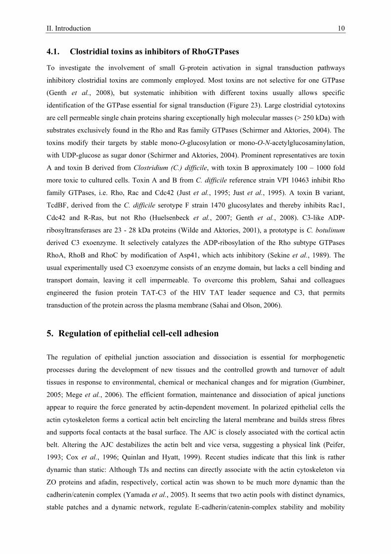
1. The Epithelium
The epithelium is a sheetlike layer of tissue covering an external surface or lining a cavity within the
body. Several different types of epithelia are characterized dependent on morphology and function.
Simple, single-layered epithelia often selectively transport small molecules and ions through the tissue
layer whereas stratified, multi-layered epithelia commonly serve as protective surfaces and barriers.
Multilayered transitional epithelia allow expansion and contraction. Simple and stratified epithelia are
formed by either squamous, cuobidal or columnar shaped cells, transitional epithelia by a mixture of
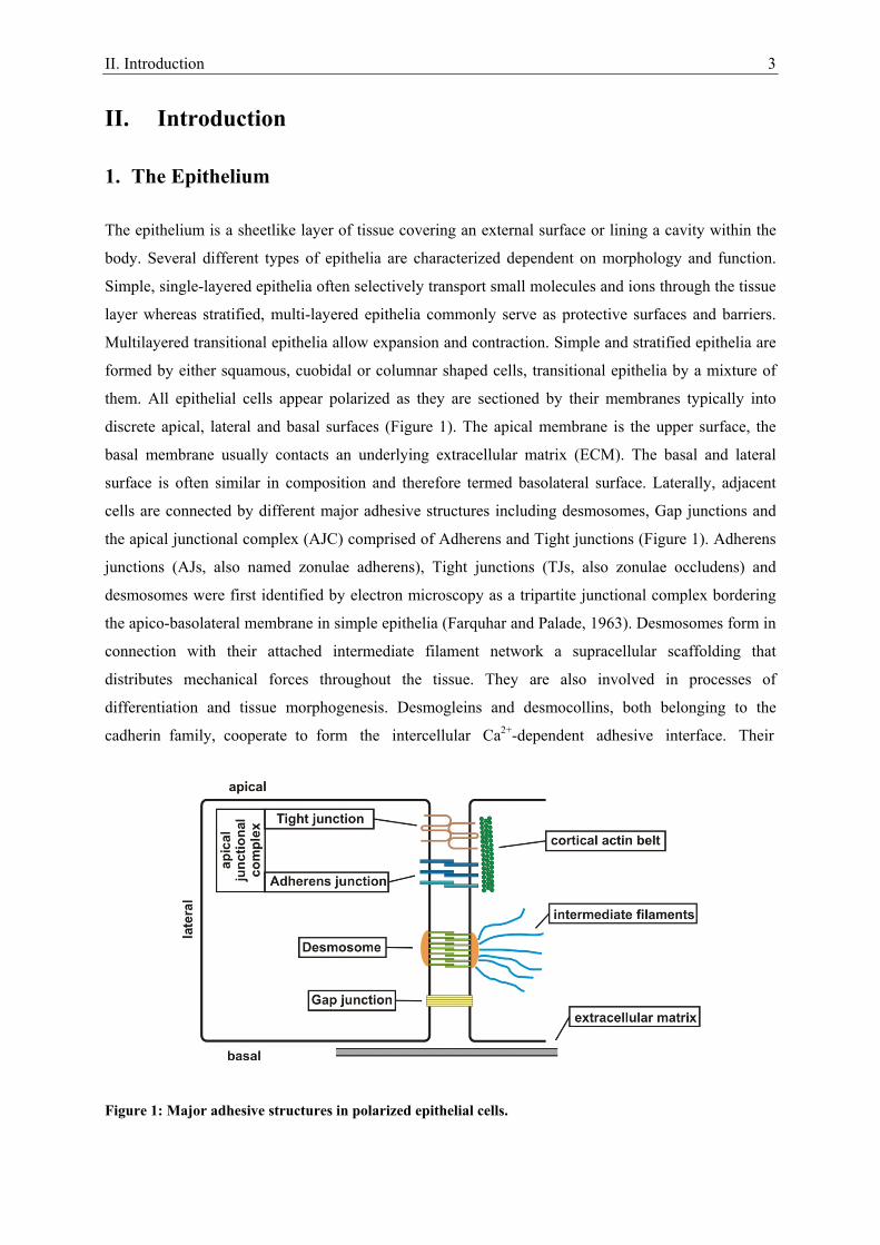
them. All epithelial cells appear polarized as they are sectioned by their membranes typically into
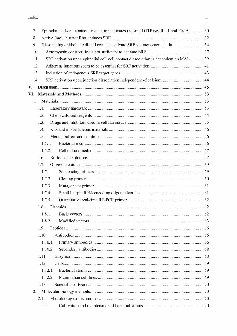
discrete apical, lateral and basal surfaces (Figure 1). The apical membrane is the upper surface, the
basal membrane usually contacts an underlying extracellular matrix (ECM). The basal and lateral
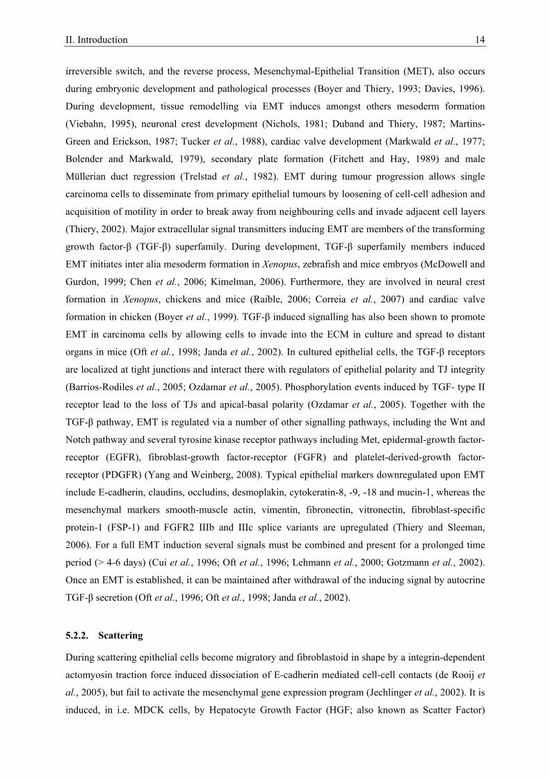
surface is often similar in composition and therefore termed basolateral surface. Laterally, adjacent
cells are connected by different major adhesive structures including desmosomes, Gap junctions and
the apical junctional complex (AJC) comprised of Adherens and Tight junctions (Figure 1). Adherens
junctions (AJs, also named zonulae adherens), Tight junctions (TJs, also zonulae occludens) and
desmosomes were first identified by electron microscopy as a tripartite junctional complex bordering
the apico-basolateral membrane in simple epithelia (Farquhar and Palade, 1963). Desmosomes form in
connection with their attached intermediate filament network a supracellular scaffolding that
distributes mechanical forces throughout the tissue. They are also involved in processes of
differentiation and tissue morphogenesis. Desmogleins and desmocollins, both belonging to the
cadherin family, cooperate to form the intercellular Ca2+-dependent adhesive interface. Their
Figure 1: Major adhesive structures in polarized epithelial cells.
II. Introduction 4
cytoplasmic tails bind to armadillo family member proteins which are, in turn, linked to the
intermediate filaments via the plakin family member desmoplakin (Green and Simpson, 2007). Gap
junctions were also first identified by electron microscopy (Robertson, 1963). They connect the
cytoplasms of adjacent cells via protein-lined channels allowing ion and small molecule exchange.
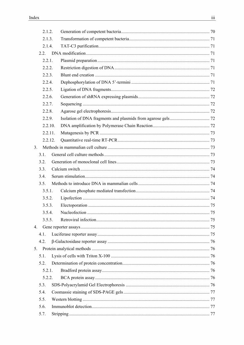
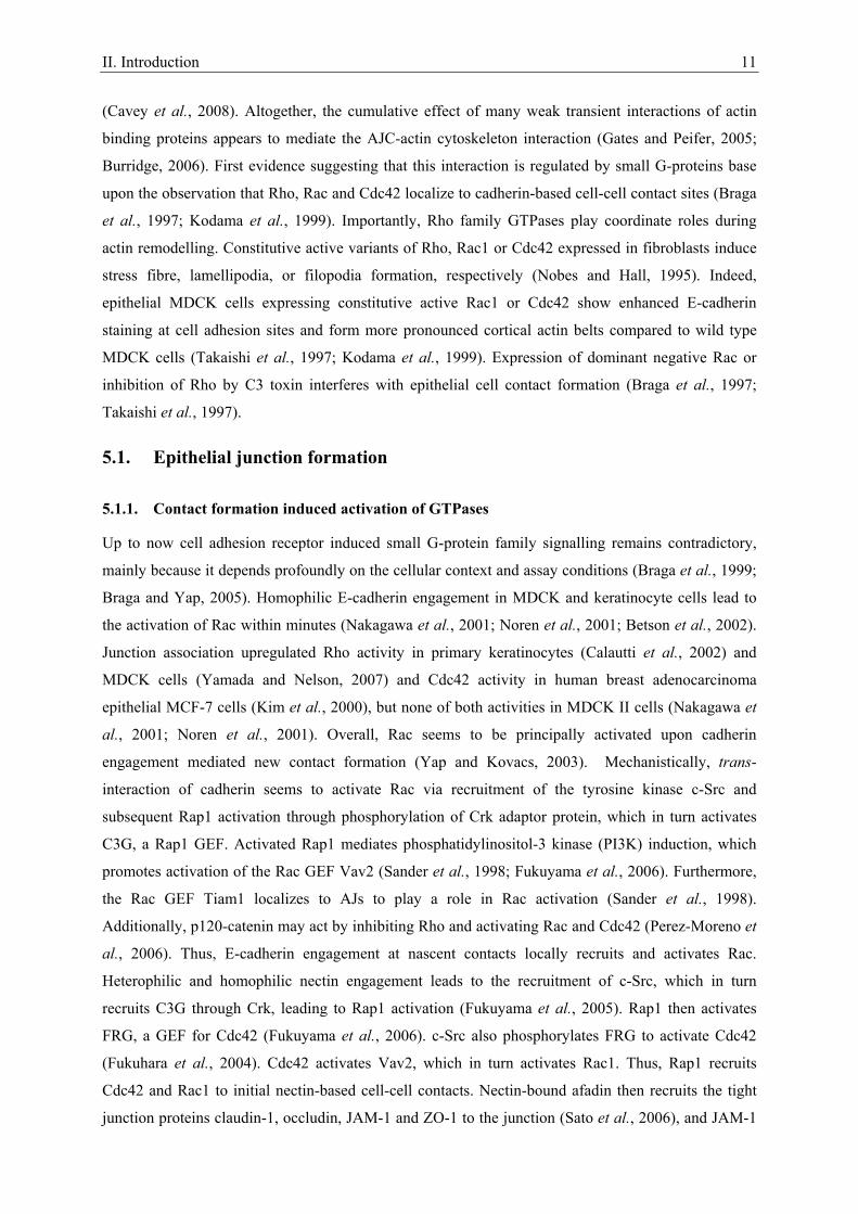
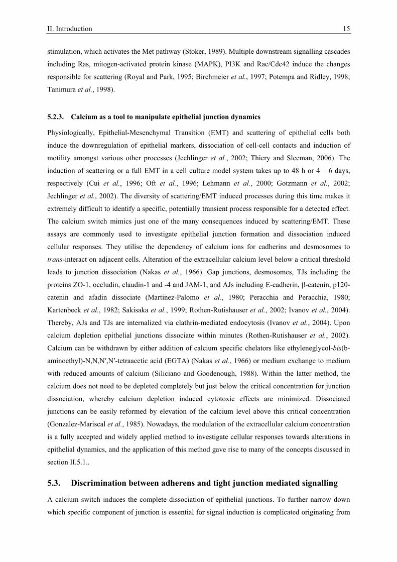
2. Molecular composition of the apical junctional complex
Per definition the apical junctional complex is comprised of AJs and TJs (Figure 1).
2.1. Tight junctions
TJs are located most apical providing an ion- and size-specific barrier (Anderson et al., 2004). They
mark the border between the apical and basolateral membrane domains and impede in a “fence” like
fashion the diffusion of their respective components (Miyoshi and Takai, 2008). Major
tramsmembrane components of the TJ are the IgG-like family of junctional adhesion molecules
(JAMs) and the four transmembrane spanning protein families of occludins and claudins
(Schneeberger and Lynch, 2004). JAMs are not exclusively found on TJ containing cells and forced
expression in fibroblasts does not induce the formation of TJs (Ebnet et al., 2004). Occludin deficient
TJs are fully functional, leaving the physiological role of occludins unclear (Saitou et al., 2000). The
claudin family consists of at least 24 organ and tissue specific members, which induce Ca2+-
independent cell-cell adhesion upon expression in fibroblasts. They are responsible for strength, size
and ion specificity of TJ barriers (Furuse and Tsukita, 2006). JAMs, occludin and claudins are locally
clustered in tight junction strands by interaction with cytoplasmic scaffolding partners, most
importantly the zonula occludens proteins ZO-1, ZO-2 and ZO-3, which belong to the guanylate
kinase-like homologs family (Schneeberger and Lynch, 2004). Epithelial cells depleted for ZO-1 and
ZO-2 are well polarized including functional AJs, but completely lack TJs. In this system ZO-1 and
ZO-2, but not ZO-3, were shown to be essential for claudin clustering, TJ strand formation and barrier
function (Umeda et al., 2006). The ZO-proteins directly interact with claudins and occludin via their
N-terminal PDZ domain, whereas their C-terminus can associate with actin, providing a direct link to
the cytoskeleton (Schneeberger and Lynch, 2004).
2.2. Adherens junctions
AJs are located immediately below the TJs (Figure 1). They perform multiple functions including the
initiation and stabilization of cell-cell adhesion, regulation of the actin cytoskeleton and intracellular
signalling (Gumbiner, 2005). The core of the AJ is formed by classical cadherin superfamily
glycoproteins, such as E-, P-, N- and R-cadherin (Gooding et al., 2004), and catenin family members
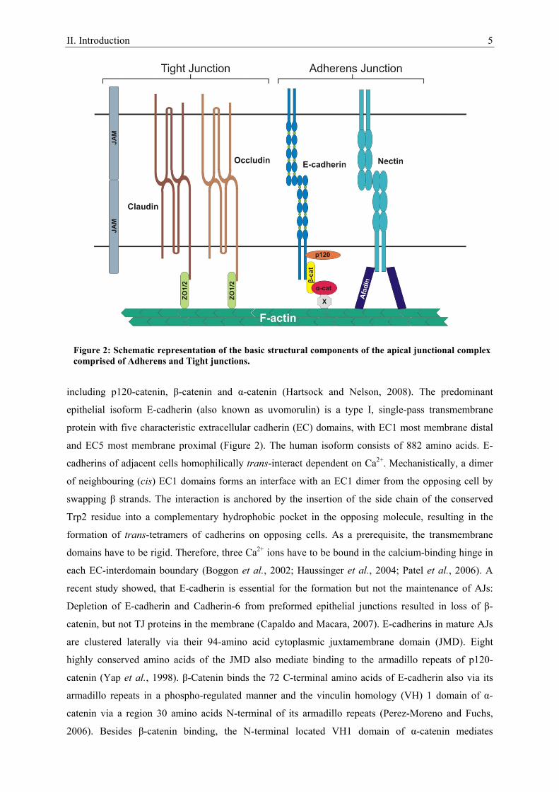
II. Introduction 5
including p120-catenin, β-catenin and α-catenin (Hartsock and Nelson, 2008). The predominant
epithelial isoform E-cadherin (also known as uvomorulin) is a type I, single-pass transmembrane
protein with five characteristic extracellular cadherin (EC) domains, with EC1 most membrane distal
and EC5 most membrane proximal (Figure 2). The human isoform consists of 882 amino acids. E-
cadherins of adjacent cells homophilically trans-interact dependent on Ca2+. Mechanistically, a dimer
of neighbouring (cis) EC1 domains forms an interface with an EC1 dimer from the opposing cell by
swapping β strands. The interaction is anchored by the insertion of the side chain of the conserved
Trp2 residue into a complementary hydrophobic pocket in the opposing molecule, resulting in the
formation of trans-tetramers of cadherins on opposing cells. As a prerequisite, the transmembrane
domains have to be rigid. Therefore, three Ca2+ ions have to be bound in the calcium-binding hinge in
each EC-interdomain boundary (Boggon et al., 2002; Haussinger et al., 2004; Patel et al., 2006). A
recent study showed, that E-cadherin is essential for the formation but not the maintenance of AJs:
Depletion of E-cadherin and Cadherin-6 from preformed epithelial junctions resulted in loss of β-
catenin, but not TJ proteins in the membrane (Capaldo and Macara, 2007). E-cadherins in mature AJs
are clustered laterally via their 94-amino acid cytoplasmic juxtamembrane domain (JMD). Eight
highly conserved amino acids of the JMD also mediate binding to the armadillo repeats of p120-
catenin (Yap et al., 1998). β-Catenin binds the 72 C-terminal amino acids of E-cadherin also via its
armadillo repeats in a phospho-regulated manner and the vinculin homology (VH) 1 domain of α-
catenin via a region 30 amino acids N-terminal of its armadillo repeats (Perez-Moreno and Fuchs,
2006). Besides β-catenin binding, the N-terminal located VH1 domain of α-catenin mediates
Figure 2: Schematic representation of the basic structural components of the apical junctional complex comprised of Adherens and Tight junctions.
II. Introduction 6
homodimerization, making both events mutually exclusive. Thus, α-catenin subsists either as
monomer capable of β-catenin binding or a homodimer competent for actin filament binding, but not
vice-versa, disproving its function as direct linker between β-catenin and the actin cytoskeleton.
Nevertheless, numerous studies indicate that AJs are somehow linked to the actin cytoskeleton, likely
in a rather dynamic than static interaction (Gates and Peifer, 2005; Burridge, 2006). The linkage might
be at least partially dependent on α-catenin by its interaction with actin binding proteins like vinculin,
spectrin, afadin, ZO-1, α-actinin, formin-1 or EPLIN via its VH2 or VH3 domain (Gates and Peifer,
2005; Burridge, 2006; Abe and Takeichi, 2008). Furthermore, the nectin/afadin complex, the second
basic adhesive unit of the AJ, might mediate linkage (Gates and Peifer, 2005). The nectin family of
IgG-like adhesion receptors, consisting of the four members Nectin-1 to -4, forms lateral homodimers
capable of Ca2+-independent homo- and heterophilic engagement with other nectins or nectin-like
receptors. Their extracellular domain consists of three IgG-like loops, a single transmembrane region
and a cytoplasmic tail with a PDZ domain. Nectins seem to provide a first scaffold for AJ and TJ
formation, as cadherin mediated cell-cell adhesion and TJ formation are dependent on nectins. All
nectin family members are directly linked to the actin cytoskeleton by binding the PDZ domain of the
actin-binding protein afadin, also known as AF-6, via a conserved C-terminal motif (Irie et al., 2004).
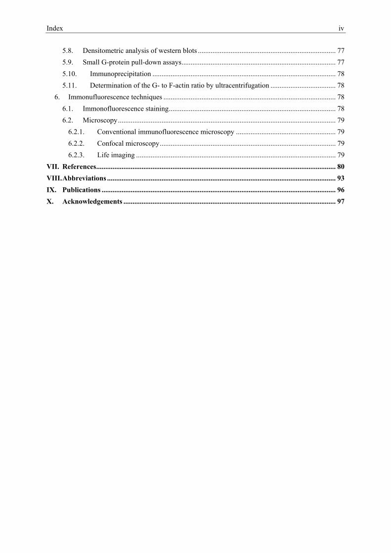
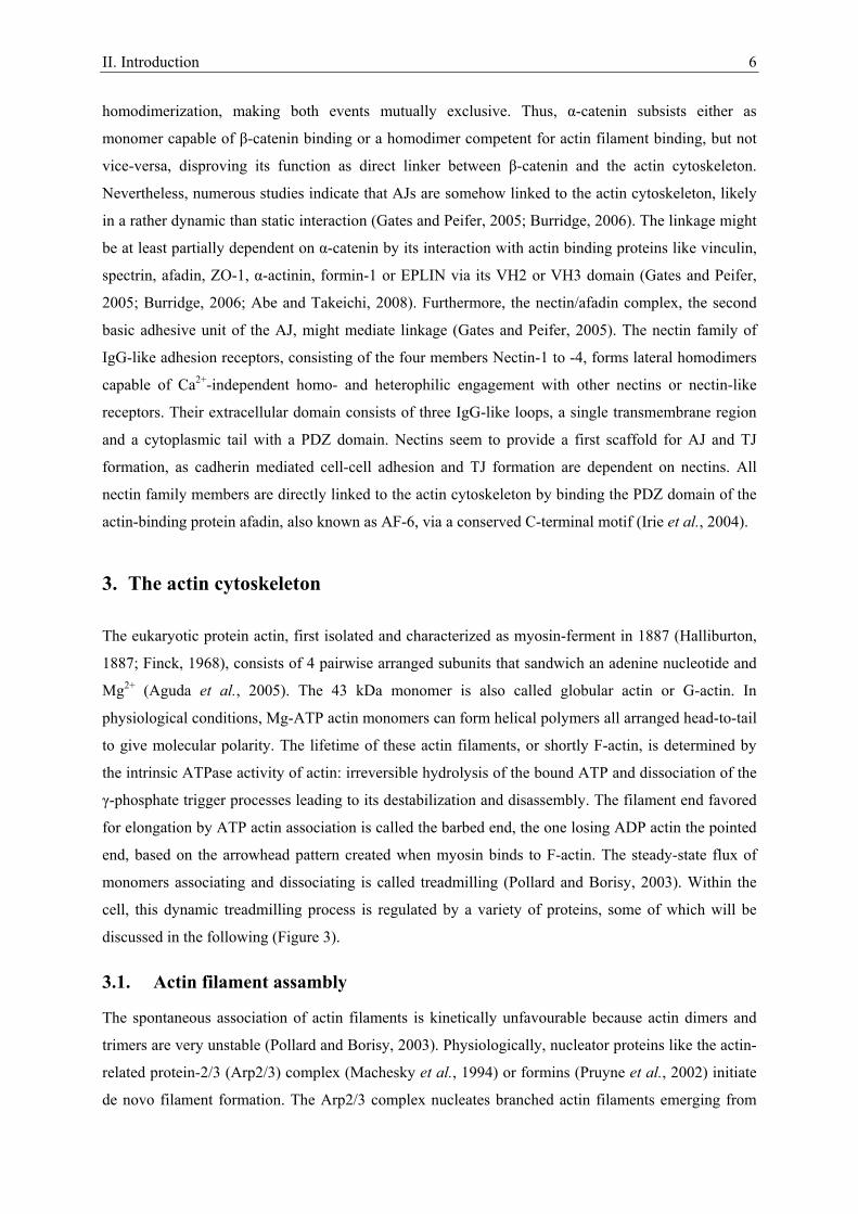
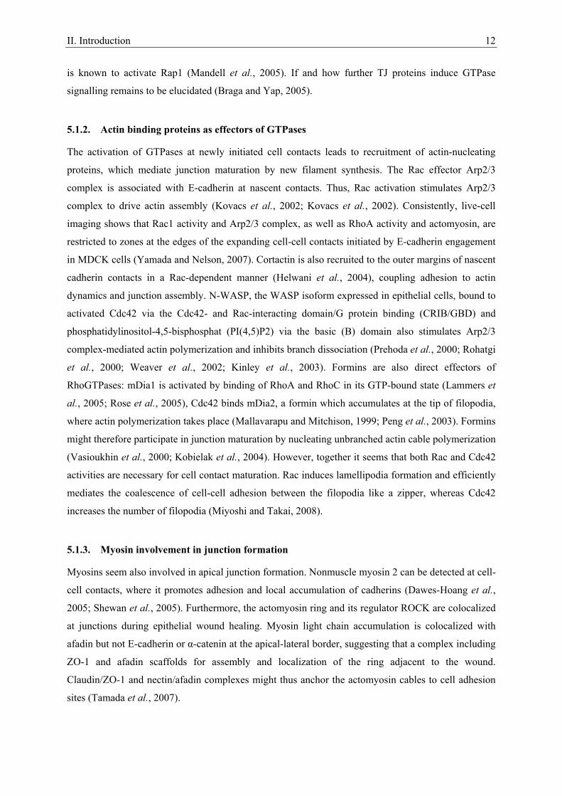
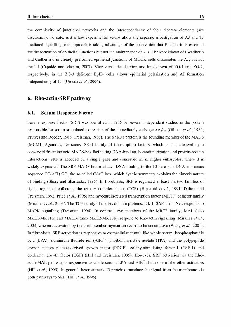
3. The actin cytoskeleton
The eukaryotic protein actin, first isolated and characterized as myosin-ferment in 1887 (Halliburton,
1887; Finck, 1968), consists of 4 pairwise arranged subunits that sandwich an adenine nucleotide and
Mg2+ (Aguda et al., 2005). The 43 kDa monomer is also called globular actin or G-actin. In
physiological conditions, Mg-ATP actin monomers can form helical polymers all arranged head-to-tail
to give molecular polarity. The lifetime of these actin filaments, or shortly F-actin, is determined by
the intrinsic ATPase activity of actin: irreversible hydrolysis of the bound ATP and dissociation of the
γ-phosphate trigger processes leading to its destabilization and disassembly. The filament end favored
for elongation by ATP actin association is called the barbed end, the one losing ADP actin the pointed
end, based on the arrowhead pattern created when myosin binds to F-actin. The steady-state flux of
monomers associating and dissociating is called treadmilling (Pollard and Borisy, 2003). Within the
cell, this dynamic treadmilling process is regulated by a variety of proteins, some of which will be
discussed in the following (Figure 3).
3.1. Actin filament assambly
The spontaneous association of actin filaments is kinetically unfavourable because actin dimers and
trimers are very unstable (Pollard and Borisy, 2003). Physiologically, nucleator proteins like the actin-
related protein-2/3 (Arp2/3) complex (Machesky et al., 1994) or formins (Pruyne et al., 2002) initiate
de novo filament formation. The Arp2/3 complex nucleates branched actin filaments emerging from
II. Introduction 7
existing ones upon recruitment of the nucleation promoting factors (NPFs) class I and II. Class I is
comprised of the Wiskott-Aldrich syndrome proteins (WASP) along with the isoforms of the
suppressor of cyclic AMP repressor (SCAR, also called WASP-family verprolin-homologous proteins
(WAVE)) (Machesky and Insall, 1998), class II of i.e. cortactin (Weed et al., 2000). WASP or
SCAR/WAVE binding induces conformational changes, which bring the Arp2 and Arp3 subunits in
close vicinity, possibly mimicking the barbed end of a filament. Furthermore, they deliver the first
actin monomer (Chereau et al., 2005). Cortactin seems to stabilize the y-branched organization of
newly generated actin networks (Goley and Welch, 2006). There are 15 formin isoforms in mammals,
i.e. murine mDia 1 and 2. All share as defining feature a FH1 and FH2 domain, which cooperatively
mediate de novo nucleation of actin filaments (Pruyne et al., 2002; Higashida et al., 2004). Thereby,
formin homodimers encircle the filament end like a donut, and recruit profilin-bound ATP actin for
elongation. Once bound, formins remain continuously associated with the barbed end (Kovar, 2006).
The enabled/vasodilator stimulated phosphoprotein (Ena/VASP) protein family elongates pre-existing
actin filaments by interacting with the barbed ends, shielding them from capping proteins (Bear et al.,
2002) and mediating filament bundling (Schirenbeck et al., 2006). Furthermore, Ena/VASP appears to
antagonize the formation of Arp2/3 complex induced actin branching (Skoble et al., 2001; Bear et al.,
2002) via an up to now unknown mechanism (Trichet et al., 2008). The surface-attached sheetlike
membrane protrusions formed by an Arp2/3 complex nucleated debranched actin meshwork is called
lamellipodium. Emanating from the lamellipodium at the leading edge of the cell are thin, formin
nucleated finger-like protrusions containing parallel bundles of 10 – 30 actin filaments oriented with
their barbed ends toward the membrane, the so-called filopodia (Chhabra and Higgs, 2007).
Figure 3: Scheme of selected actin binding proteins involved in actin treadmilling.
II. Introduction 8
3.2. Actin filament disassembly
Debranching, severing and depolymerization of actin filaments from their pointed ends is induced by
actin-depolymerizing factor (ADF)/Cofilin proteins, which bind ADP F-actin and stimulate γ-
phosphate dissociation from ADP-Pi actin (Carlier et al., 1997; Blanchoin et al., 2000).
Mechanistically, the binding induces filaments to twist by approximately 5° per subunit, which
changes the thermodynamic stability of the filament leading to depolymerization (McGough et al.,
1997). ADF/Cofilin activity is inter alia regulated via phosphorylation by LIM kinase (LIMK) and
testicular protein kinase (TESK) (Bamburg, 1999). Phosphorylation downregulates the actin
interactions of ADF/cofilin (Bamburg, 1999). The small actin binding protein profilin compedes with
ADF/Cofilin for ADP actin binding and promotes the dissociation of ADP (Mockrin and Korn, 1980;
Vinson et al., 1998). Due to the higher concentration of ATP in living cells and its higher affinity to
ATP than ADP, nucleotide-free actin binds preferentially ATP. Profilin has a higher affinity for ATP
actin than ADF/Cofilin, thereby renewing the ATP actin monomer pool ready for polymerization
(Rosenblatt et al., 1995; Pollard and Borisy, 2003).
3.3. Actin contractility
Non-muscle cells contain prominent transitory or permanent contractile bundles of actin filaments
crosslinked mainly by α-actinin (Lazarides and Burridge, 1975), non-muscle myosin (Weber and
Groeschel-Stewart, 1974) and tropomyosin (Lazarides, 1975). In the rigor state, myosin connected to
an actin filament is tightly bound via its head to a second, parallel filament. On contraction, this
interaction is weakened upon ATP binding to the myosin head. The intrinsic ATPase activity of
myosin hydrolyzes the ATP, which causes a conformational change in the head that moves it to a new
position where it rebinds the second filament. The phosphate dissociation of ADP-Pi induces another
conformational change exerting a force to move the tightly bound myosin on the second filament
leading to contraction, followed by the release of ADP to complete the cycle. Essential for this process
is the phosphorylation of the regulatory light chain of myosin 2 at Ser 19, which leads to an increase in
its intrinsic ATPase activity (Somlyo and Somlyo, 2003). One of the central regulators of contractility
is RhoA activated coiled-coil kinase (ROCK) (Leung et al., 1995; Ishizaki et al., 1996; Leung et al.,
1996). ROCK stimulates actomyosin contractility by acting on myosin light chain 2 (MLC) in at least
four ways (Figure 34): (i) direct phosphorylation of myosin light chain 2 at Ser19 (Amano et al., 1996;
Katoh et al., 2001); (ii) inhibition of MLC phosphatase (MLCPP) (Kimura et al., 1996); (iii)
phosphorylation of ZIP kinase (ZIPK) which activates MLC and inhibits MLCPP (Murata-Hori et al.,
1999; MacDonald et al., 2001; MacDonald et al., 2001); (iv) activation of CPI-17, a MLCPP inhibitor
(Kitazawa et al., 2000; Koyama et al., 2000).
II. Introduction 9
3.4. Actin modifying compounds
Numerous actin modifying compounds are available to study the role of the actin cytoskeleton in
signal transduction processes. These compounds modulate actin dynamics by accelerating either
polymerization or depolymerization. The underlying mechanisms are sometimes quite complex. The
basic effects of compounds employed in this study are shortly summarized in the following.
Jasplakinolide is a cell membrane permeable actin filament stabilizer isolated from the marine sponge
Jaspis johnstoni (Scott et al., 1988; Zampella et al., 1999). It also acts as inducer of actin
polymerization and thereby deplets the monomeric actin pool (Bubb et al., 1994). Latrunculins and
cytochalasins are F-actin destabilizing compounds. Eight forms of latrunculin have been isolated from
different murine sponges and nudibranchs (Spector et al., 1983; Vilozny et al., 2004). Latrunculin B is
isolated from the sponge Latrunculia magnifica (Spector et al., 1983) and prevents assembly and
polymerization of actin monomers via specific binding in the ATP-binding cleft of monomeric actin
(Spector et al., 1999). Numerous cytochalasins, natural compounds derived from the marine fungus
Zygosporium masonii (Minato and Katayama, 1970) and derivates, exist. They have various effects on
actin, the most efficient is to act by capping the barbed end of actin filaments (Cooper, 1987). At
higher concentrations they can also sever F-actin and sequester monomers or dimers and stimulate the
ATPase activity of actin monomers (Cooper, 1987; Sampath and Pollard, 1991). Cytochalasin D is one
of the best characterized cytochalasins and, in contrast to cytochalasins A and B, does not inhibit
monosaccharide transport across the plasma membrane (Rampal et al., 1980).
4. Small guanosine triphosphatases
Small guanosine triphosphatases (also known as small G-proteins or GTPases) are molecular switches
that cycle between an inactive GDP-bound and an active GTP-bound confirmation. They possess low
intrinsic GTP hydrolysis and GDP/GTP exchange activities. When active, they interact with effector
proteins, which induce downstream signalling events. The GDP-GTP cycle is highly regulated by
guanine nucleotide exchange factors (GEFs) and GTPase-activating proteins (GAPs). GEFs induce the
release of bound GDP to be replaced by GTP and thereby turn on signalling, GAPs provide an
essential catalytic group for GTP hydrolysis, which terminates signalling (Wennerberg et al., 2005;
Bos et al., 2007). The human Ras superfamily of GTPases consists of at least 154 members divided
into five principal families: Ras, Rho, Rab, Arf and Ran (Wennerberg et al., 2005).
To date, there are 22 mammalian Rho family GTPases known (Jaffe, Hall 2005). The most prominent
and investigated members are RhoA, Rac1 and Cdc42. To investigate the specific function of single
GTPases, constitutive active and dominant mutants have been engineered. Constitutive activation for
Rac1 is achieved by replacement of glycine at position 12 with valine, for RhoA by substitution of
glutamine at position 63 with leucine. Both mutations reduce the intrinsic GTPase activity (Diekmann
et al., 1991; Khosravi-Far et al., 1995).
II. Introduction 10
4.1. Clostridial toxins as inhibitors of RhoGTPases
To investigate the involvement of small G-protein activation in signal transduction pathways
inhibitory clostridial toxins are commonly employed. Most toxins are not selective for one GTPase
(Genth et al., 2008), but systematic inhibition with different toxins usually allows specific
identification of the GTPase essential for signal transduction (Figure 23). Large clostridial cytotoxins
are cell permeable single chain proteins sharing exceptionally high molecular masses (> 250 kDa) with
substrates exclusively found in the Rho and Ras family GTPases (Schirmer and Aktories, 2004). The
toxins modify their targets by stable mono-O-glucosylation or mono-O-N-acetylglucosaminylation,
with UDP-glucose as sugar donor (Schirmer and Aktories, 2004). Prominent representatives are toxin
A and toxin B derived from Clostridium (C.) difficile, with toxin B approximately 100 – 1000 fold
more toxic to cultured cells. Toxin A and B from C. difficile reference strain VPI 10463 inhibit Rho
family GTPases, i.e. Rho, Rac and Cdc42 (Just et al., 1995; Just et al., 1995). A toxin B variant,
TcdBF, derived from the C. difficile serotype F strain 1470 glucosylates and thereby inhibits Rac1,
Cdc42 and R-Ras, but not Rho (Huelsenbeck et al., 2007; Genth et al., 2008). C3-like ADP-
ribosyltransferases are 23 - 28 kDa proteins (Wilde and Aktories, 2001), a prototype is C. botulinum
derived C3 exoenzyme. It selectively catalyzes the ADP-ribosylation of the Rho subtype GTPases
RhoA, RhoB and RhoC by modification of Asp41, which acts inhibitory (Sekine et al., 1989). The
usual experimentally used C3 exoenzyme consists of an enzyme domain, but lacks a cell binding and
transport domain, leaving it cell impermeable. To overcome this problem, Sahai and colleagues
engineered the fusion protein TAT-C3 of the HIV TAT leader sequence and C3, that permits
transduction of the protein across the plasma membrane (Sahai and Olson, 2006).
5. Regulation of epithelial cell-cell adhesion
The regulation of epithelial junction association and dissociation is essential for morphogenetic
processes during the development of new tissues and the controlled growth and turnover of adult
tissues in response to environmental, chemical or mechanical changes and for migration (Gumbiner,
2005; Mege et al., 2006). The efficient formation, maintenance and dissociation of apical junctions
appear to require the force generated by actin-dependent movement. In polarized epithelial cells the
actin cytoskeleton forms a cortical actin belt encircling the lateral membrane and builds stress fibres
and supports focal contacts at the basal surface. The AJC is closely associated with the cortical actin
belt. Altering the AJC destabilizes the actin belt and vice versa, suggesting a physical link (Peifer,
1993; Cox et al., 1996; Quinlan and Hyatt, 1999). Recent studies indicate that this link is rather
dynamic than static: Although TJs and nectins can directly associate with the actin cytoskeleton via
ZO proteins and afadin, respectively, cortical actin was shown to be much more dynamic than the
cadherin/catenin complex (Yamada et al., 2005). It seems that two actin pools with distinct dynamics,
stable patches and a dynamic network, regulate E-cadherin/catenin-complex stability and mobility
II. Introduction 11
(Cavey et al., 2008). Altogether, the cumulative effect of many weak transient interactions of actin
binding proteins appears to mediate the AJC-actin cytoskeleton interaction (Gates and Peifer, 2005;
Burridge, 2006). First evidence suggesting that this interaction is regulated by small G-proteins base
upon the observation that Rho, Rac and Cdc42 localize to cadherin-based cell-cell contact sites (Braga
et al., 1997; Kodama et al., 1999). Importantly, Rho family GTPases play coordinate roles during
actin remodelling. Constitutive active variants of Rho, Rac1 or Cdc42 expressed in fibroblasts induce
stress fibre, lamellipodia, or filopodia formation, respectively (Nobes and Hall, 1995). Indeed,
epithelial MDCK cells expressing constitutive active Rac1 or Cdc42 show enhanced E-cadherin
staining at cell adhesion sites and form more pronounced cortical actin belts compared to wild type
MDCK cells (Takaishi et al., 1997; Kodama et al., 1999). Expression of dominant negative Rac or
inhibition of Rho by C3 toxin interferes with epithelial cell contact formation (Braga et al., 1997;
Takaishi et al., 1997).
5.1. Epithelial junction formation
5.1.1. Contact formation induced activation of GTPases
Up to now cell adhesion receptor induced small G-protein family signalling remains contradictory,
mainly because it depends profoundly on the cellular context and assay conditions (Braga et al., 1999;
Braga and Yap, 2005). Homophilic E-cadherin engagement in MDCK and keratinocyte cells lead to
the activation of Rac within minutes (Nakagawa et al., 2001; Noren et al., 2001; Betson et al., 2002).
Junction association upregulated Rho activity in primary keratinocytes (Calautti et al., 2002) and
MDCK cells (Yamada and Nelson, 2007) and Cdc42 activity in human breast adenocarcinoma
epithelial MCF-7 cells (Kim et al., 2000), but none of both activities in MDCK II cells (Nakagawa et
al., 2001; Noren et al., 2001). Overall, Rac seems to be principally activated upon cadherin
engagement mediated new contact formation (Yap and Kovacs, 2003). Mechanistically, trans-
interaction of cadherin seems to activate Rac via recruitment of the tyrosine kinase c-Src and
subsequent Rap1 activation through phosphorylation of Crk adaptor protein, which in turn activates
C3G, a Rap1 GEF. Activated Rap1 mediates phosphatidylinositol-3 kinase (PI3K) induction, which
promotes activation of the Rac GEF Vav2 (Sander et al., 1998; Fukuyama et al., 2006). Furthermore,
the Rac GEF Tiam1 localizes to AJs to play a role in Rac activation (Sander et al., 1998).
Additionally, p120-catenin may act by inhibiting Rho and activating Rac and Cdc42 (Perez-Moreno et
al., 2006). Thus, E-cadherin engagement at nascent contacts locally recruits and activates Rac.
Heterophilic and homophilic nectin engagement leads to the recruitment of c-Src, which in turn
recruits C3G through Crk, leading to Rap1 activation (Fukuyama et al., 2005). Rap1 then activates
FRG, a GEF for Cdc42 (Fukuyama et al., 2006). c-Src also phosphorylates FRG to activate Cdc42
(Fukuhara et al., 2004). Cdc42 activates Vav2, which in turn activates Rac1. Thus, Rap1 recruits
Cdc42 and Rac1 to initial nectin-based cell-cell contacts. Nectin-bound afadin then recruits the tight
junction proteins claudin-1, occludin, JAM-1 and ZO-1 to the junction (Sato et al., 2006), and JAM-1
II. Introduction 12
is known to activate Rap1 (Mandell et al., 2005). If and how further TJ proteins induce GTPase
signalling remains to be elucidated (Braga and Yap, 2005).
5.1.2. Actin binding proteins as effectors of GTPases
The activation of GTPases at newly initiated cell contacts leads to recruitment of actin-nucleating
proteins, which mediate junction maturation by new filament synthesis. The Rac effector Arp2/3
complex is associated with E-cadherin at nascent contacts. Thus, Rac activation stimulates Arp2/3
complex to drive actin assembly (Kovacs et al., 2002; Kovacs et al., 2002). Consistently, live-cell
imaging shows that Rac1 activity and Arp2/3 complex, as well as RhoA activity and actomyosin, are
restricted to zones at the edges of the expanding cell-cell contacts initiated by E-cadherin engagement
in MDCK cells (Yamada and Nelson, 2007). Cortactin is also recruited to the outer margins of nascent
cadherin contacts in a Rac-dependent manner (Helwani et al., 2004), coupling adhesion to actin
dynamics and junction assembly. N-WASP, the WASP isoform expressed in epithelial cells, bound to
activated Cdc42 via the Cdc42- and Rac-interacting domain/G protein binding (CRIB/GBD) and
phosphatidylinositol-4,5-bisphosphat (PI(4,5)P2) via the basic (B) domain also stimulates Arp2/3
complex-mediated actin polymerization and inhibits branch dissociation (Prehoda et al., 2000; Rohatgi
et al., 2000; Weaver et al., 2002; Kinley et al., 2003). Formins are also direct effectors of
RhoGTPases: mDia1 is activated by binding of RhoA and RhoC in its GTP-bound state (Lammers et
al., 2005; Rose et al., 2005), Cdc42 binds mDia2, a formin which accumulates at the tip of filopodia,
where actin polymerization takes place (Mallavarapu and Mitchison, 1999; Peng et al., 2003). Formins
might therefore participate in junction maturation by nucleating unbranched actin cable polymerization
(Vasioukhin et al., 2000; Kobielak et al., 2004). However, together it seems that both Rac and Cdc42
activities are necessary for cell contact maturation. Rac induces lamellipodia formation and efficiently
mediates the coalescence of cell-cell adhesion between the filopodia like a zipper, whereas Cdc42
increases the number of filopodia (Miyoshi and Takai, 2008).
5.1.3. Myosin involvement in junction formation
Myosins seem also involved in apical junction formation. Nonmuscle myosin 2 can be detected at cell-
cell contacts, where it promotes adhesion and local accumulation of cadherins (Dawes-Hoang et al.,
2005; Shewan et al., 2005). Furthermore, the actomyosin ring and its regulator ROCK are colocalized
at junctions during epithelial wound healing. Myosin light chain accumulation is colocalized with
afadin but not E-cadherin or α-catenin at the apical-lateral border, suggesting that a complex including
ZO-1 and afadin scaffolds for assembly and localization of the ring adjacent to the wound.
Claudin/ZO-1 and nectin/afadin complexes might thus anchor the actomyosin cables to cell adhesion
sites (Tamada et al., 2007).
II. Introduction 13
5.1.4. Additional mechanisms inducing junction formation and stabilization
Independent of GTPases, E-cadherin engagement recruits Ena/VASP to the cell surface, which in turn
can polymerize actin cables providing the force necessary to bring epithelial cells actively together
(Vasioukhin et al., 2000; Schirenbeck et al., 2006; Scott et al., 2006). α-Catenin recruits and regulates
formins to and at AJs, respectively (Kobielak et al., 2004). Furthermore, α-catenin binds ZO-1 (Itoh et
al., 1997) during the junction formation process supporting the formation of linear actin cables
(Ikenouchi et al., 2007). Initial cell-cell contacts are approached by actin filaments perpendicular
through local Arp2/3 dependent polymerization, while mature contacts are characterized by a parallel
orientation to the membrane (Adams et al., 1998; Vasioukhin et al., 2000). Essential for this switch
might be α-catenin. As a monomer it binds the E-cadherin/β-catenin complex, as homodimer it
competes with the Arp2/3 complex for F-actin binding. Thus, cadherin-catenin clustering at maturing
junctions would generate a high local concentration of cadherin-free α-catenin, which could
antagonize branching and promote linear organization of parallel bundles (Drees et al., 2005).
Nevertheless, the fusion protein of a C-terminal truncated E-cadherin (tEC) lacking the β-catenin
binding site and α-catenin is able to link adherens junctions to the actin cytoskeleton (Nagafuchi et al.,
1994). Further analysis revealed that a fusion protein of tEC and amino acids 509 – 643 of the 906
amino acid protein α-catenin, the so-called adhesion modulation domain located in and downstream of
the VH2 domain, is sufficient and essential for adhesion (Nagafuchi et al., 1994; Imamura et al.,
1999). However, fusion protein expressing cells are not committed to rapid remodelling (Nagafuchi et
al., 1994), further reinforcing the role of α-catenin as allosteric regulator of the actin cytoskeleton. E-
cadherin and nectin trans-interactions stabilize newly formed cell-cell adhesion: activation of Rac and
Cdc42 promotes retention of E-cadherin at the plama membrane by inhibition of clathrin-dependent
endocytosis (Izumi et al., 2004).
5.2. Epithelial junction dissociation
5.2.1. Epithelial-Mesenchymal Transition
Epithelial-Mesenchymal Transition (EMT) is a process allowing polarized, immotile epithelial cells to
convert into motile, mesenchymal cells. This process is highly conserved and an indispensable
mechanism during morphogenesis. Furthermore, it is implicated in promoting carcinoma invasion and
metastasis (Thiery and Sleeman, 2006). Overall, EMT can be loosely defined by three major changes
in cellular phenotype: (i) Cobblestone-like polarized epithelial cells in monolayers change to spindle-
shaped mesenchymal cells with migratory protrusions; (ii) epithelial differentiation markers change
from cell-cell junction proteins and cytokeratin intermediate filaments to vimentin filaments and
fibronectin; (iii) functional changes leading to the acquisition of the ability to migrate and invade the
ECM. Thereby, not all three changes are always observed during EMT, and (iii) alone is considered as
a functional hallmark of EMT (Boyer and Thiery, 1993; Hay, 1995). Vive versa, EMT is not an
II. Introduction 14
irreversible switch, and the reverse process, Mesenchymal-Epithelial Transition (MET), also occurs
during embryonic development and pathological processes (Boyer and Thiery, 1993; Davies, 1996).
During development, tissue remodelling via EMT induces amongst others mesoderm formation
(Viebahn, 1995), neuronal crest development (Nichols, 1981; Duband and Thiery, 1987; Martins-
Green and Erickson, 1987; Tucker et al., 1988), cardiac valve development (Markwald et al., 1977;
Bolender and Markwald, 1979), secondary plate formation (Fitchett and Hay, 1989) and male
Müllerian duct regression (Trelstad et al., 1982). EMT during tumour progression allows single
carcinoma cells to disseminate from primary epithelial tumours by loosening of cell-cell adhesion and
acquisition of motility in order to break away from neighbouring cells and invade adjacent cell layers
(Thiery, 2002). Major extracellular signal transmitters inducing EMT are members of the transforming
growth factor-β (TGF-β) superfamily. During development, TGF-β superfamily members induced
EMT initiates inter alia mesoderm formation in Xenopus, zebrafish and mice embryos (McDowell and
Gurdon, 1999; Chen et al., 2006; Kimelman, 2006). Furthermore, they are involved in neural crest
formation in Xenopus, chickens and mice (Raible, 2006; Correia et al., 2007) and cardiac valve
formation in chicken (Boyer et al., 1999). TGF-β induced signalling has also been shown to promote
EMT in carcinoma cells by allowing cells to invade into the ECM in culture and spread to distant
organs in mice (Oft et al., 1998; Janda et al., 2002). In cultured epithelial cells, the TGF-β receptors
are localized at tight junctions and interact there with regulators of epithelial polarity and TJ integrity
(Barrios-Rodiles et al., 2005; Ozdamar et al., 2005). Phosphorylation events induced by TGF- type II
receptor lead to the loss of TJs and apical-basal polarity (Ozdamar et al., 2005). Together with the
TGF-β pathway, EMT is regulated via a number of other signalling pathways, including the Wnt and
Notch pathway and several tyrosine kinase receptor pathways including Met, epidermal-growth factor-
receptor (EGFR), fibroblast-growth factor-receptor (FGFR) and platelet-derived-growth factor-
receptor (PDGFR) (Yang and Weinberg, 2008). Typical epithelial markers downregulated upon EMT
include E-cadherin, claudins, occludins, desmoplakin, cytokeratin-8, -9, -18 and mucin-1, whereas the
mesenchymal markers smooth-muscle actin, vimentin, fibronectin, vitronectin, fibroblast-specific
protein-1 (FSP-1) and FGFR2 IIIb and IIIc splice variants are upregulated (Thiery and Sleeman,
2006). For a full EMT induction several signals must be combined and present for a prolonged time
period (> 4-6 days) (Cui et al., 1996; Oft et al., 1996; Lehmann et al., 2000; Gotzmann et al., 2002).
Once an EMT is established, it can be maintained after withdrawal of the inducing signal by autocrine
TGF-β secretion (Oft et al., 1996; Oft et al., 1998; Janda et al., 2002).
5.2.2. Scattering
During scattering epithelial cells become migratory and fibroblastoid in shape by a integrin-dependent
actomyosin traction force induced dissociation of E-cadherin mediated cell-cell contacts (de Rooij et
al., 2005), but fail to activate the mesenchymal gene expression program (Jechlinger et al., 2002). It is
induced, in i.e. MDCK cells, by Hepatocyte Growth Factor (HGF; also known as Scatter Factor)
II. Introduction 15
stimulation, which activates the Met pathway (Stoker, 1989). Multiple downstream signalling cascades
including Ras, mitogen-activated protein kinase (MAPK), PI3K and Rac/Cdc42 induce the changes
responsible for scattering (Royal and Park, 1995; Birchmeier et al., 1997; Potempa and Ridley, 1998;
Tanimura et al., 1998).
5.2.3. Calcium as a tool to manipulate epithelial junction dynamics
Physiologically, Epithelial-Mesenchymal Transition (EMT) and scattering of epithelial cells both
induce the downregulation of epithelial markers, dissociation of cell-cell contacts and induction of
motility amongst various other processes (Jechlinger et al., 2002; Thiery and Sleeman, 2006). The
induction of scattering or a full EMT in a cell culture model system takes up to 48 h or 4 – 6 days,
respectively (Cui et al., 1996; Oft et al., 1996; Lehmann et al., 2000; Gotzmann et al., 2002;
Jechlinger et al., 2002). The diversity of scattering/EMT induced processes during this time makes it
extremely difficult to identify a specific, potentially transient process responsible for a detected effect.
The calcium switch mimics just one of the many consequences induced by scattering/EMT. These
assays are commonly used to investigate epithelial junction formation and dissociation induced
cellular responses. They utilise the dependency of calcium ions for cadherins and desmosomes to
trans-interact on adjacent cells. Alteration of the extracellular calcium level below a critical threshold
leads to junction dissociation (Nakas et al., 1966). Gap junctions, desmosomes, TJs including the
proteins ZO-1, occludin, claudin-1 and -4 and JAM-1, and AJs including E-cadherin, β-catenin, p120-
catenin and afadin dissociate (Martinez-Palomo et al., 1980; Peracchia and Peracchia, 1980;
Kartenbeck et al., 1982; Sakisaka et al., 1999; Rothen-Rutishauser et al., 2002; Ivanov et al., 2004).
Thereby, AJs and TJs are internalized via clathrin-mediated endocytosis (Ivanov et al., 2004). Upon
calcium depletion epithelial junctions dissociate within minutes (Rothen-Rutishauser et al., 2002).
Calcium can be withdrawn by either addition of calcium specific chelators like ethyleneglycol-bis(b-
aminoethyl)-N,N,N′,N′-tetraacetic acid (EGTA) (Nakas et al., 1966) or medium exchange to medium
with reduced amounts of calcium (Siliciano and Goodenough, 1988). Within the latter method, the
calcium does not need to be depleted completely but just below the critical concentration for junction
dissociation, whereby calcium depletion induced cytotoxic effects are minimized. Dissociated
junctions can be easily reformed by elevation of the calcium level above this critical concentration
(Gonzalez-Mariscal et al., 1985). Nowadays, the modulation of the extracellular calcium concentration
is a fully accepted and widely applied method to investigate cellular responses towards alterations in
epithelial dynamics, and the application of this method gave rise to many of the concepts discussed in
section II.5.1..
5.3. Discrimination between adherens and tight junction mediated signalling
A calcium switch induces the complete dissociation of epithelial junctions. To further narrow down
which specific component of junction is essential for signal induction is complicated originating from
II. Introduction 16
the complexity of junctional networks and the interdependency of their discrete elements (see
discussion). To date, just a few experimental setups allow the separate investigation of AJ and TJ
mediated signalling: one approach is taking advantage of the observation that E-cadherin is essential
for the formation of epithelial junctions but not the maintenance of AJs. The knockdown of E-cadherin
and Cadherin-6 in already preformed epithelial junctions of MDCK cells dissociates the AJ, but not
the TJ (Capaldo and Macara, 2007). Vice versa, the deletion and knockdown of ZO-1 and ZO-2,
respectively, in the ZO-3 deficient EpH4 cells allows epithelial polarization and AJ formation
independently of TJs (Umeda et al., 2006).
6. Rho-actin-SRF pathway
6.1. Serum Response Factor
Serum response Factor (SRF) was identified in 1986 by several independent studies as the protein
responsible for serum-stimulated expression of the immediately early gene c-fos (Gilman et al., 1986;
Prywes and Roeder, 1986; Treisman, 1986). The 67 kDa protein is the founding member of the MADS
(MCM1, Agamous, Deficiens, SRF) family of transcription factors, which is characterized by a
conserved 56 amino acid MADS-box facilitating DNA-binding, homodimerization and protein-protein
interactions. SRF is encoded on a single gene and conserved in all higher eukaryotes, where it is
widely expressed. The SRF MADS-box mediates DNA binding to the 10 base pair DNA consensus
sequence CC(A/T)6GG, the so-called CArG box, which dyadic symmetry explains the dimeric nature
of binding (Shore and Sharrocks, 1995). In fibroblasts, SRF is regulated at least via two families of
signal regulated cofactors, the ternary complex factor (TCF) (Hipskind et al., 1991; Dalton and
Treisman, 1992; Price et al., 1995) and myocardin-related transcription factor (MRTF) cofactor family
(Miralles et al., 2003). The TCF family of the Ets domain proteins, Elk-1, SAP-1 and Net, responds to
MAPK signalling (Treisman, 1994). In contrast, two members of the MRTF family, MAL (also
MKL1/MRTFa) and MAL16 (also MKL2/MRTFb), respond to Rho-actin signalling (Miralles et al.,
2003) whereas activation by the third member myocardin seems to be constitutive (Wang et al., 2001).
In fibroblasts, SRF activation is responsive to extracellular stimuli like whole serum, lysophosphatidic
acid (LPA), aluminium fluoride ion (AlF4־ ), phorbol myristate acetate (TPA) and the polypeptide
growth factors platelet-derived growth factor (PDGF), colony-stimulating factor-1 (CSF-1) and
epidermal growth factor (EGF) (Hill and Treisman, 1995). However, SRF activation via the Rho-
actin-MAL pathway is responsive to whole serum, LPA and AlF4־ , but none of the other activators
(Hill et al., 1995). In general, heterotrimeric G proteins transduce the signal from the membrane via
both pathways to SRF (Hill et al., 1995).
II. Introduction 17
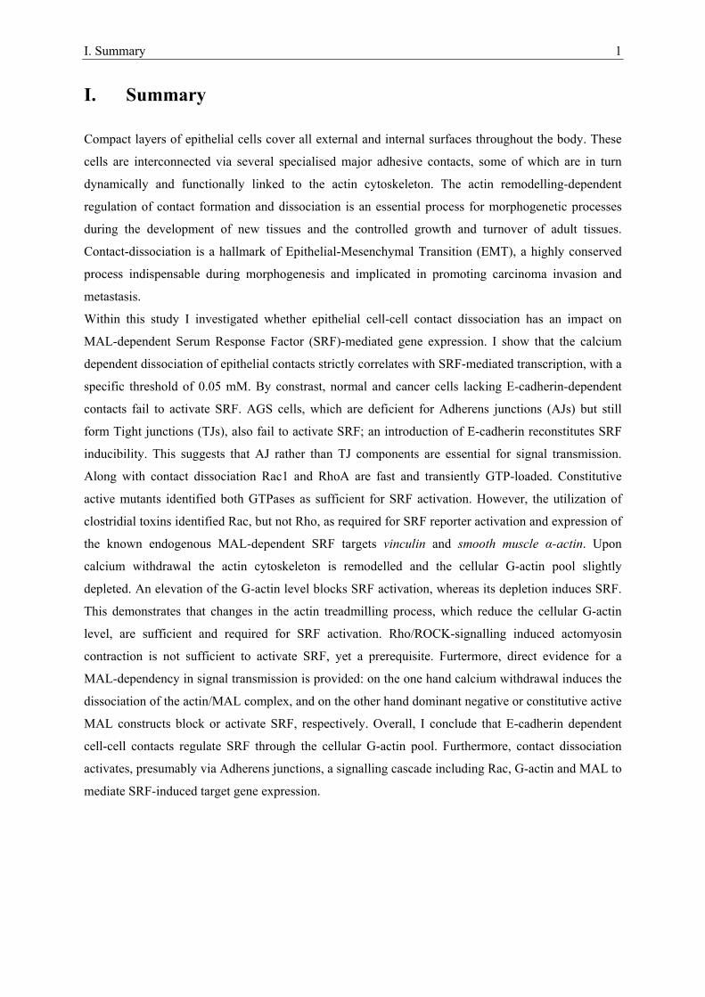
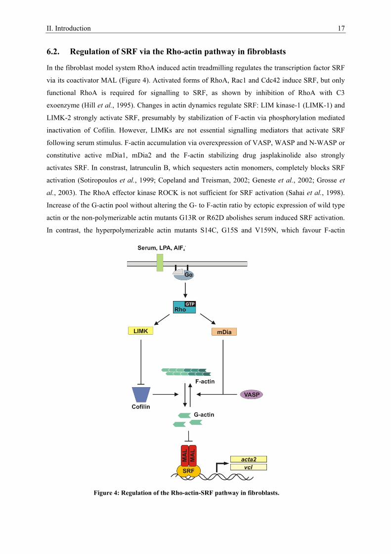
6.2. Regulation of SRF via the Rho-actin pathway in fibroblasts
In the fibroblast model system RhoA induced actin treadmilling regulates the transcription factor SRF
via its coactivator MAL (Figure 4). Activated forms of RhoA, Rac1 and Cdc42 induce SRF, but only
functional RhoA is required for signalling to SRF, as shown by inhibition of RhoA with C3
exoenzyme (Hill et al., 1995). Changes in actin dynamics regulate SRF: LIM kinase-1 (LIMK-1) and
LIMK-2 strongly activate SRF, presumably by stabilization of F-actin via phosphorylation mediated
inactivation of Cofilin. However, LIMKs are not essential signalling mediators that activate SRF
following serum stimulus. F-actin accumulation via overexpression of VASP, WASP and N-WASP or
constitutive active mDia1, mDia2 and the F-actin stabilizing drug jasplakinolide also strongly
activates SRF. In constrast, latrunculin B, which sequesters actin monomers, completely blocks SRF
activation (Sotiropoulos et al., 1999; Copeland and Treisman, 2002; Geneste et al., 2002; Grosse et
al., 2003). The RhoA effector kinase ROCK is not sufficient for SRF activation (Sahai et al., 1998).
Increase of the G-actin pool without altering the G- to F-actin ratio by ectopic expression of wild type
actin or the non-polymerizable actin mutants G13R or R62D abolishes serum induced SRF activation.
In contrast, the hyperpolymerizable actin mutants S14C, G15S and V159N, which favour F-actin
Figure 4: Regulation of the Rho-actin-SRF pathway in fibroblasts.
II. Introduction 18
formation, strongly activate SRF. Together, these results indicate that a change in the G- to F-actin
ratio mediating a depletion of the cellular G-actin pool is essential for signalling to SRF (Posern et al.,
2002; Posern et al., 2004). G-actin can directly bind to the N-terminal RPEL motifs of MAL (compare
Figure 40) and thereby inhibit SRF activation (Miralles et al., 2003). In unstimulated cells, the
actin/MAL complex rapidly shuttles between the cytoplasm and the nucleus. Upon serum stimulation
induced actin polymerization it translocates to the nucleus. There it accumulates because serum
stimulation efficiently blocks its export. Importantly, MAL binds to SRF but remains inactive until
actin dissociates (Miralles et al., 2003; Vartiainen et al., 2007).
6.3. Endogenous SRF target genes
SRF induced transcription is essential for life, as homozygous SRF-deficient embryos die during
gastrulation by not developing mesodermal cells (Arsenian et al., 1998). SRF-/- embryonic stem cells
grow, but do not display serum-induced immediate early growth response gene expression (Schratt et
al., 2001). Furthermore, these cells exhibit morphological perturbations dependent on the defective
formation of cytoskeletal structures as well as defects in cell spreading, cell adhesion and migration
(Schratt et al., 2002). Besides the prototypical SRF target gene involved in cell growth, c-fos, in
mammals approximately 160 genes harbouring a conserved CArG box element (the CArGome) have
been identified as direct transcriptional targets of SRF in a computational genome wide screen, half of
which have been validated experimentally (Sun et al., 2006). Amongst the SRF target genes are many
immediately early growth response genes encoding cytoskeletal components, signalling molecules and
transcription factors (Sun et al., 2006). A second major class of SRF target genes is involved in
myogenesis, whereof some are expressed either in cardiac, smooth or skeletal muscle, others in
multiple muscle types (Pipes et al., 2006). The fact that the widely expressed SRF is required for the
expression of muscle genes suggests that cofactors to initiate specific transcription exist. During
myogenesis this cofactor is myocardin, which expression is largely confined to cardiac and smooth
muscles of i.e. the cardiovascular system (Wang et al., 2001). In general, SRF toggles between
disparate programs of gene expression dependent on the signal transduction pathway (Sotiropoulos et
al., 1999; Gineitis and Treisman, 2001) and resulting cofactor binding (Wang et al., 2001;
Zaromytidou et al., 2006). Activation of the MAPK pathway leads to the phosphorylation of TCF
family proteins, which in turn initiate transcription by association with both, SRF and adjacent Ets-
binding sites (Hipskind et al., 1991). Induction of the RhoA-actin pathway promotes MAL/SRF
complex formation. Thereby, MAL adds a β-strand to the SRF DNA-binding domain β-sheet region,
and SRF induced DNA bending facilitates MAL-DNA contact (Zaromytidou et al., 2006).
Mechanistically, the cofactors compete for interaction with a common surface in the MADS-box of
SRF (Miralles et al., 2003; Wang et al., 2004; Zaromytidou et al., 2006). Amongst others, known SRF
targets induced by TCF binding are the growth related c-fos and egr-1 genes, the latter one encoding
for early growth response protein-1. To the RhoA-actin activated MAL dependent SRF targets belong
II. Introduction 19
the cytoskeletal components encoding smooth muscle α-actin and vinculin genes and srf itself
(Sotiropoulos et al., 1999).
7. Cellular model systems
The in this study mainly used Madin-Darby Canine Kidney (MDCK) cell line was derived from
normal female adult Cocker Spaniel by S.H. Madin and N.B. Darby in September 1958 (Gaush et al.,
1966). Single cells lack apical-basal polarity, but E-cadherin mediated cluster fomation and collagen
deposition at the basal surfaces of the cells leads to full polarization including the tight junction
mediated intersection of the apical and lateral plasma membrane (Wang et al., 1990). Furthermore,
cell-cell adhesion between adjacent MDCK cells is mediated via desmosomes and gap junctions
(Whitesell et al., 1981; Cereijido et al., 1984). MDCK cells are a widely used model system to
investigate the formation and dissociation of epithelial cell-cell adhesion (Hinck et al., 1994; Nathke et
al., 1994; Balda et al., 1996; Sander et al., 1998; Sakisaka et al., 1999). HGF stimulation of MDCK
cells in monolayer culture induces scattering (Stoker, 1989). As EMT model system EpRas cells were
employed. These are oncogenic v-Ha-Ras expressing mouse mammary epithelial EpH4 cells (Oft et
al., 1996). Once a full EMT is established it persists in the absence of inducing signals due to an
autocrine TGF-β secretion loop.
III. Specific Aims 20
III. Aims of this PhD thesis
Epithelial cell-cell contacts are functionally and dynamically linked to the actin cytoskeleton. The
dissociation of these contacts is a hallmark of Epithelial-Mesenchymal Transition, which is an
essential process during morphogenesis and tumour progression. The aim of this study was to
investigate whether epithelial cell-cell contacts regulate cellular gene expression. Specifically, I
wanted to analyse if and how transcription controlled by serum response factor and its coactivator
MAL is affected by epithelial cell-cell contact dissociation. Differential regulation of endogenous
target genes, as well as promoter-reporter constructs, should be analysed for this purpose. Subsequent
characterisation of the signal transduction pathway should be performed. I wanted to analyse a
potential dependency on actin treadmilling, more precisely the depletion of the cellular G-actin level,
and on the MAL protein. The participation of crucial small guanosine triphosphatases of the Rho
family in signal transduction should be clarified, and the sufficient or required small GTPases should
be identified. Effects of modifiers of F-actin function should also be tested. Moreover, the junction
component within the epithelial contact critical for signal induction to SRF should be identified.
IV. Results 21
IV. Results
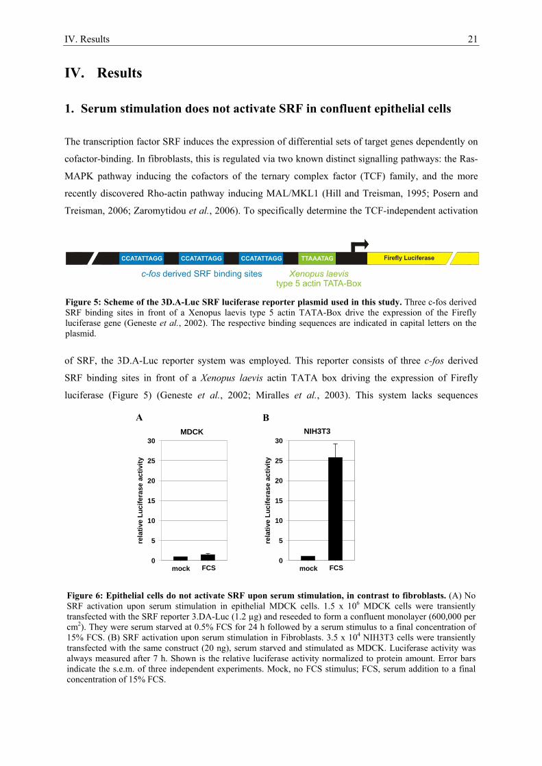
1. Serum stimulation does not activate SRF in confluent epithelial cells
The transcription factor SRF induces the expression of differential sets of target genes dependently on
cofactor-binding. In fibroblasts, this is regulated via two known distinct signalling pathways: the Ras-
MAPK pathway inducing the cofactors of the ternary complex factor (TCF) family, and the more
recently discovered Rho-actin pathway inducing MAL/MKL1 (Hill and Treisman, 1995; Posern and
Treisman, 2006; Zaromytidou et al., 2006). To specifically determine the TCF-independent activation
of SRF, the 3D.A-Luc reporter system was employed. This reporter consists of three c-fos derived
SRF binding sites in front of a Xenopus laevis actin TATA box driving the expression of Firefly
luciferase (Figure 5) (Geneste et al., 2002; Miralles et al., 2003). This system lacks sequences
rela
tive
Luci
fera
se a
ctiv
ity
0
5
10
15
20
25
30
mock FCS
MDCK
rela
tive
Luci
fera
se a
ctiv
ity
0
5
10
15
20
25
30
mock FCS
NIH3T3
Figure 5: Scheme of the 3D.A-Luc SRF luciferase reporter plasmid used in this study. Three c-fos derived SRF binding sites in front of a Xenopus laevis type 5 actin TATA-Box drive the expression of the Firefly luciferase gene (Geneste et al., 2002). The respective binding sequences are indicated in capital letters on the plasmid.
Figure 6: Epithelial cells do not activate SRF upon serum stimulation, in contrast to fibroblasts. (A) No SRF activation upon serum stimulation in epithelial MDCK cells. 1.5 x 10⁶ MDCK cells were transiently transfected with the SRF reporter 3.DA-Luc (1.2 µg) and reseeded to form a confluent monolayer (600,000 per cm2). They were serum starved at 0.5% FCS for 24 h followed by a serum stimulus to a final concentration of 15% FCS. (B) SRF activation upon serum stimulation in Fibroblasts. 3.5 x 10⁴ NIH3T3 cells were transiently transfected with the same construct (20 ng), serum starved and stimulated as MDCK. Luciferase activity was always measured after 7 h. Shown is the relative luciferase activity normalized to protein amount. Error bars indicate the s.e.m. of three independent experiments. Mock, no FCS stimulus; FCS, serum addition to a final concentration of 15% FCS.
A B
IV. Results 22
essential for TCF binding and is therefore unresponsive to the Ras-MAPK pathway (Hill and
Treisman, 1995). Transiently 3D.A-Luc transfected NIH3T3 fibroblasts activated the SRF reporter
upon serum stimulation (Figure 6 B), consistent with previous reports (Miralles et al., 2003). By
contrast, confluent epithelial MDCK cells did not significantly activate the reporter upon serum
stimulation (Figure 6 A). Serum unresponsiveness was also observed in other epithelial cell lines like
EpH4 mouse mammary gland or NBTII rat bladder carcinoma cells (not shown). These findings
indicate that contrary to fibroblasts, epithelial cells regulate SRF-controlled genes independendly of
serum. Other extracellular stimuli must mediate its regulation. Further investigations focused on how
SRF is regulated in epithelial cells.
2. Calcium as a tool to manipulate the epithelial integrity of MDCK cells
Vertebrate epithelial cells form contacts to neighbouring cells via tight junctions, adherens junctions,
desmosomes and gap junctions. Major components of adherens junctions and desmosomes are
transmembrane proteins belonging to the cadherin familiy, which homophilically interact with their
extracellular domains dependent on calcium. Reduction of the extracellular calcium level below a
specific threshold leads to a dissociation of the contacts (Nakas et al., 1966). Junction integrity
regulated via calcium concentration can be monitored via the localization of the adherens junction
component E-cadherin. During this study, I established specific protocols to dissociate and reassociate
epithelial junctions by switching the extracellular calcium concentration. Epithelial canine kidney
MDCK cells, maintained in normal culture medium, clustered via E-cadherin containing junctions
(Figure 7, left). Calcium removal led to E-cadherin internalization and cellular detachment (Figure 7,
middle) without apparently harming the cells, even if applied for several days (not shown). Readdition
of calcium to the physiological concentration of 1.8 mM induced the reformation of the junctions
within 15 minutes (Figure 7, right). Taken together, variation of the extracellular calcium
concentration is a useful tool to regulate epithelial junction dynamics and analyse cellular responses.
Figure 7: Calcium dependent epithelial junction dissociation and reassociation. MDCK cells (5 x 10⁴ per cm2) were maintained in normal culture medium (left) followed by a 24 h cultivation in calcium reduced medium (middle) and the readdition of calcium to physiological concentration for 15 minutes (right), fixed, and stained for E-cadherin (DECMA-1, green), F-actin (rhodamin-phalloidin, red) and DNA (blue). Bar: 25 µm.
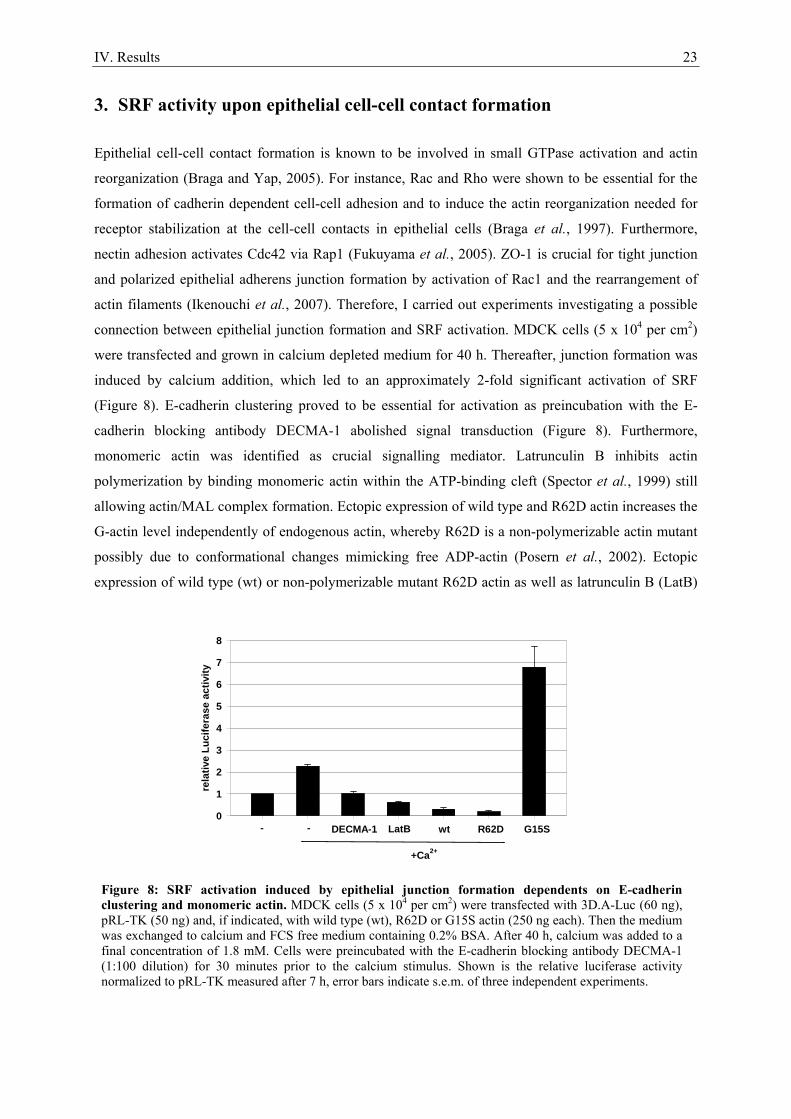
IV. Results 23
3. SRF activity upon epithelial cell-cell contact formation
Epithelial cell-cell contact formation is known to be involved in small GTPase activation and actin
reorganization (Braga and Yap, 2005). For instance, Rac and Rho were shown to be essential for the
formation of cadherin dependent cell-cell adhesion and to induce the actin reorganization needed for
receptor stabilization at the cell-cell contacts in epithelial cells (Braga et al., 1997). Furthermore,
nectin adhesion activates Cdc42 via Rap1 (Fukuyama et al., 2005). ZO-1 is crucial for tight junction
and polarized epithelial adherens junction formation by activation of Rac1 and the rearrangement of
actin filaments (Ikenouchi et al., 2007). Therefore, I carried out experiments investigating a possible
connection between epithelial junction formation and SRF activation. MDCK cells (5 x 10⁴ per cm2)
were transfected and grown in calcium depleted medium for 40 h. Thereafter, junction formation was
induced by calcium addition, which led to an approximately 2-fold significant activation of SRF
(Figure 8). E-cadherin clustering proved to be essential for activation as preincubation with the E-
cadherin blocking antibody DECMA-1 abolished signal transduction (Figure 8). Furthermore,
monomeric actin was identified as crucial signalling mediator. Latrunculin B inhibits actin
polymerization by binding monomeric actin within the ATP-binding cleft (Spector et al., 1999) still
allowing actin/MAL complex formation. Ectopic expression of wild type and R62D actin increases the
G-actin level independently of endogenous actin, whereby R62D is a non-polymerizable actin mutant
possibly due to conformational changes mimicking free ADP-actin (Posern et al., 2002). Ectopic
expression of wild type (wt) or non-polymerizable mutant R62D actin as well as latrunculin B (LatB)
Figure 8: SRF activation induced by epithelial junction formation dependents on E-cadherin clustering and monomeric actin. MDCK cells (5 x 10⁴ per cm2) were transfected with 3D.A-Luc (60 ng), pRL-TK (50 ng) and, if indicated, with wild type (wt), R62D or G15S actin (250 ng each). Then the medium was exchanged to calcium and FCS free medium containing 0.2% BSA. After 40 h, calcium was added to a final concentration of 1.8 mM. Cells were preincubated with the E-cadherin blocking antibody DECMA-1 (1:100 dilution) for 30 minutes prior to the calcium stimulus. Shown is the relative luciferase activity normalized to pRL-TK measured after 7 h, error bars indicate s.e.m. of three independent experiments.
rela
tive
Luci
fera
se a
ctiv
ity
0
1
2
3
4
5
6
7
8
+Ca2+
DECMA-1- - LatB wt G15SR62D
IV. Results 24
treatment repressed SRF induction. Conversely, the F-actin stabilizing mutant G15S (Posern et al.,
2004) led to a strong activation (Figure 8). Importantly, the ectopic expression of the different actins
did not interfere with calcium dependent junction dissociation or formation, as visualized by E-
cadherin staining (Figure 29 and Figure 31). Further investigations focused on triggering the relatively
weak 2-fold SRF induction revealed no influence of cell density on inducibility (not shown). In
summary, epithelial junction formation led to a small but significant SRF activation reliant on E-
cadherin clustering and monomeric actin.
4. SRF activation induced by the dissociation of epithelial cell-cell contacts
Given that SRF activation during epithelial junction formation was very modest, I investigated other
aspects of junction remodelling and their effects on transcription. Dissociation of epithelial cell-cell
contacts is an essential process underlying the conversion of epithelial to mesenchymal cells, a process
called epithelial-mesenchymal transition (EMT), and the disintegration of the epithelium. EMT is an
indispensable mechanism during morphogenesis and pathologically a hallmark of malignant cancer
progression initiating the dissemination of metastasis (Thiery and Sleeman, 2006). Only little is known
about how epithelial cell-cell contact dissociation regulates transcription. To be able to address this
question, I carried out experiments analysing epithelial junction integrity in dependence of the
extracellular calcium level. The critical calcium concentration for MDCK cells grown in a confluent
monolayer was between 0.04 and 0.05 mM, as visualized by immunostaining for E-cadherin (Figure 9,
upper panel) and by the formation of bright circles around uncoupled epithelial cells due to their
altered light scattering properties, the so-called halo effect, in phase contrast microscopy (Figure 9,
lower panel). To test whether the dissociation of epithelial cell-cell contacts influences transcriptional
regulation MDCK cells were transiently transfected with the 3D.A-Luc SRF luciferase reporter and
seeded to form a confluent monolayer. To ensure proper junction formation cells remained then
untouched for 24 – 36 h. After this, the culture medium was exchanged to medium containing the
physiological (1.8 mM) or reduced amounts of calcium. SRF reporter activation, measured after 7 h,
was significantly induced upon calcium withdrawal with a concentration dependent threshold of about
0.04 mM calcium. The maximum activation was detected at extracellular calcium concentrations of
0.02 mM and lower (Figure 10). The dissociation of the epithelial cell-cell contacts strictly correlated
with SRF activation (Figure 9 and Figure 10). This denotes that epithelial sheet disintegration
regulates transcription via SRF signalling.To ensure that the induction of the reporter by calcium
withdrawal was strictly dependent on SRF, a mutant reporter lacking functional SRF binding sites was
generated by replacing the sequence CCATATTAGG to CCCAATCGGG (compare Figure 5) (Hill
and Treisman, 1995). The mutated reporter 2M.A-Luc was transfected into MDCK cells. The cells
were seeded to form a confluent epithelial monolayer and 24h later medium was exchanged to medium
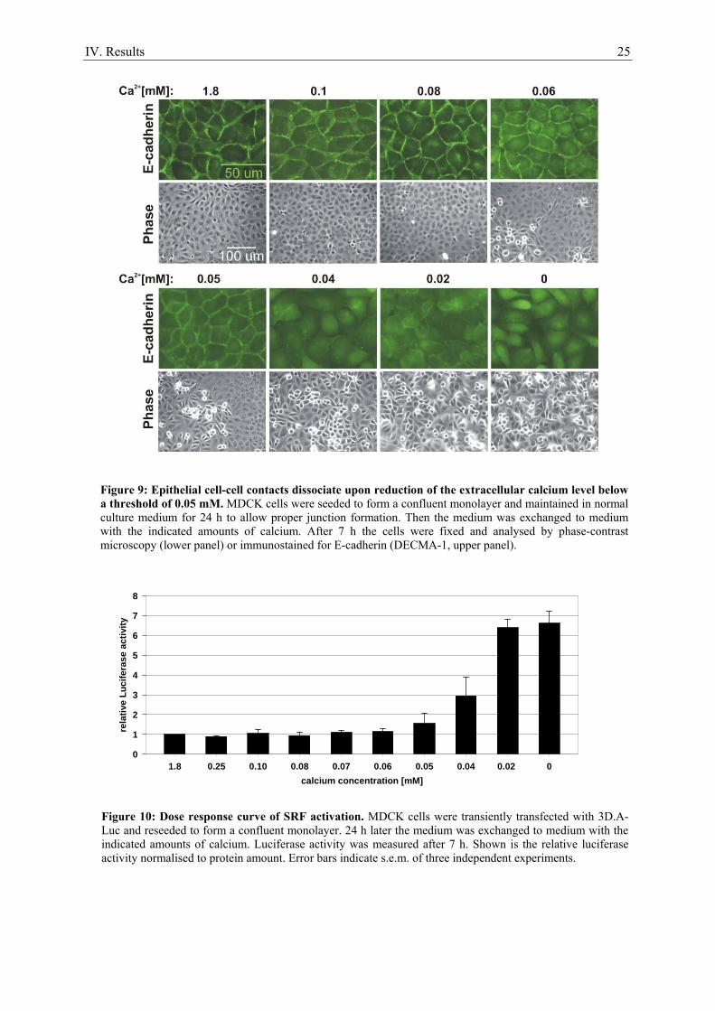
IV. Results 25
calcium concentration [mM]1.8 0.25 0.10 0.08 0.07 0.06 0.05 0.04 0.02 0
rela
tive
Luci
fera
se a
ctiv
ity
0
1
2
3
4
5
6
7
8
Figure 9: Epithelial cell-cell contacts dissociate upon reduction of the extracellular calcium level below a threshold of 0.05 mM. MDCK cells were seeded to form a confluent monolayer and maintained in normal culture medium for 24 h to allow proper junction formation. Then the medium was exchanged to medium with the indicated amounts of calcium. After 7 h the cells were fixed and analysed by phase-contrast microscopy (lower panel) or immunostained for E-cadherin (DECMA-1, upper panel).
Figure 10: Dose response curve of SRF activation. MDCK cells were transiently transfected with 3D.A-Luc and reseeded to form a confluent monolayer. 24 h later the medium was exchanged to medium with the indicated amounts of calcium. Luciferase activity was measured after 7 h. Shown is the relative luciferase activity normalised to protein amount. Error bars indicate s.e.m. of three independent experiments.
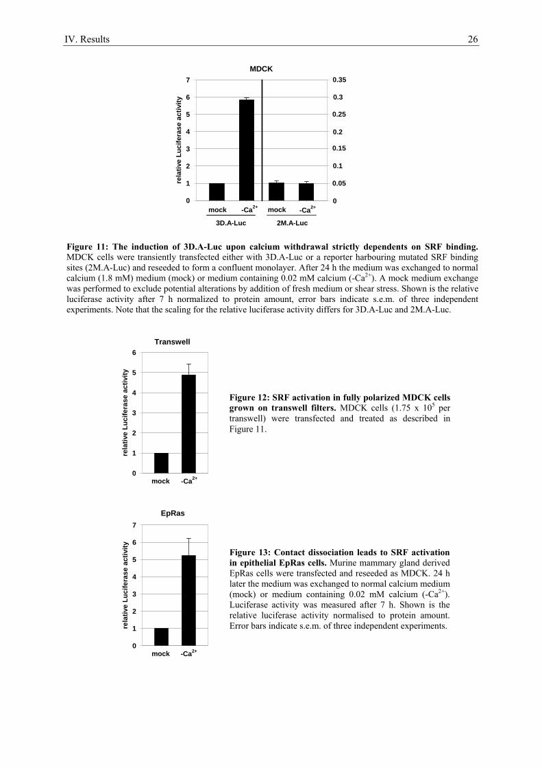
IV. Results 26
rela
tive
Luci
fera
se a
ctiv
ity
0
1
2
3
4
5
6
7
mock -Ca2+
EpRas
rela
tive
Luci
fera
se a
ctiv
ity
0
1
2
3
4
5
6
7
mock -Ca2+ -Ca2+mock
0.05
0.1
0.15
0.2
0.25
0
0.35
0.3
3D.A-Luc 2M.A-Luc
MDCK
rela
tive
Luci
fera
se a
ctiv
ity
0
1
2
3
4
5
6
mock
Transwell
-Ca2+
Figure 11: The induction of 3D.A-Luc upon calcium withdrawal strictly dependents on SRF binding. MDCK cells were transiently transfected either with 3D.A-Luc or a reporter harbouring mutated SRF binding sites (2M.A-Luc) and reseeded to form a confluent monolayer. After 24 h the medium was exchanged to normal calcium (1.8 mM) medium (mock) or medium containing 0.02 mM calcium (-Ca2+). A mock medium exchange was performed to exclude potential alterations by addition of fresh medium or shear stress. Shown is the relative luciferase activity after 7 h normalized to protein amount, error bars indicate s.e.m. of three independent experiments. Note that the scaling for the relative luciferase activity differs for 3D.A-Luc and 2M.A-Luc.
Figure 12: SRF activation in fully polarized MDCK cells grown on transwell filters. MDCK cells (1.75 x 10⁵ per transwell) were transfected and treated as described in Figure 11.
Figure 13: Contact dissociation leads to SRF activation in epithelial EpRas cells. Murine mammary gland derived EpRas cells were transfected and reseeded as MDCK. 24 h later the medium was exchanged to normal calcium medium (mock) or medium containing 0.02 mM calcium (-Ca2+). Luciferase activity was measured after 7 h. Shown is the relative luciferase activity normalised to protein amount. Error bars indicate s.e.m. of three independent experiments.
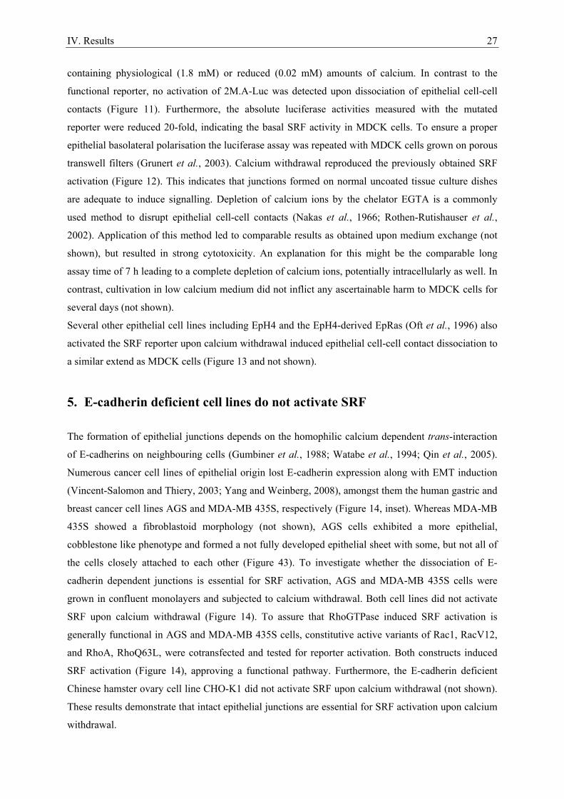
IV. Results 27
containing physiological (1.8 mM) or reduced (0.02 mM) amounts of calcium. In contrast to the
functional reporter, no activation of 2M.A-Luc was detected upon dissociation of epithelial cell-cell
contacts (Figure 11). Furthermore, the absolute luciferase activities measured with the mutated
reporter were reduced 20-fold, indicating the basal SRF activity in MDCK cells. To ensure a proper
epithelial basolateral polarisation the luciferase assay was repeated with MDCK cells grown on porous
transwell filters (Grunert et al., 2003). Calcium withdrawal reproduced the previously obtained SRF
activation (Figure 12). This indicates that junctions formed on normal uncoated tissue culture dishes
are adequate to induce signalling. Depletion of calcium ions by the chelator EGTA is a commonly
used method to disrupt epithelial cell-cell contacts (Nakas et al., 1966; Rothen-Rutishauser et al.,
2002). Application of this method led to comparable results as obtained upon medium exchange (not
shown), but resulted in strong cytotoxicity. An explanation for this might be the comparable long
assay time of 7 h leading to a complete depletion of calcium ions, potentially intracellularly as well. In
contrast, cultivation in low calcium medium did not inflict any ascertainable harm to MDCK cells for
several days (not shown).
Several other epithelial cell lines including EpH4 and the EpH4-derived EpRas (Oft et al., 1996) also
activated the SRF reporter upon calcium withdrawal induced epithelial cell-cell contact dissociation to
a similar extend as MDCK cells (Figure 13 and not shown).
5. E-cadherin deficient cell lines do not activate SRF
The formation of epithelial junctions depends on the homophilic calcium dependent trans-interaction
of E-cadherins on neighbouring cells (Gumbiner et al., 1988; Watabe et al., 1994; Qin et al., 2005).
Numerous cancer cell lines of epithelial origin lost E-cadherin expression along with EMT induction
(Vincent-Salomon and Thiery, 2003; Yang and Weinberg, 2008), amongst them the human gastric and
breast cancer cell lines AGS and MDA-MB 435S, respectively (Figure 14, inset). Whereas MDA-MB
435S showed a fibroblastoid morphology (not shown), AGS cells exhibited a more epithelial,
cobblestone like phenotype and formed a not fully developed epithelial sheet with some, but not all of
the cells closely attached to each other (Figure 43). To investigate whether the dissociation of E-
cadherin dependent junctions is essential for SRF activation, AGS and MDA-MB 435S cells were
grown in confluent monolayers and subjected to calcium withdrawal. Both cell lines did not activate
SRF upon calcium withdrawal (Figure 14). To assure that RhoGTPase induced SRF activation is
generally functional in AGS and MDA-MB 435S cells, constitutive active variants of Rac1, RacV12,
and RhoA, RhoQ63L, were cotransfected and tested for reporter activation. Both constructs induced
SRF activation (Figure 14), approving a functional pathway. Furthermore, the E-cadherin deficient
Chinese hamster ovary cell line CHO-K1 did not activate SRF upon calcium withdrawal (not shown).
These results demonstrate that intact epithelial junctions are essential for SRF activation upon calcium
withdrawal.
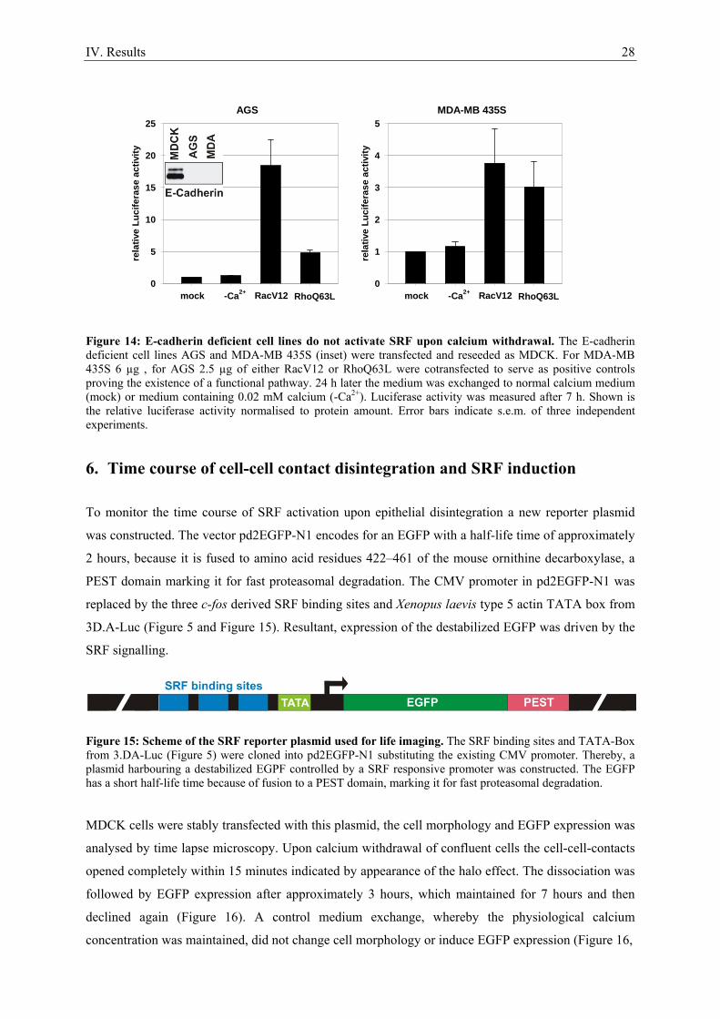
IV. Results 28
rela
tive
Luci
fera
se a
ctiv
ity
0
5
10
15
20
25
mock -Ca2+ RhoQ63LRacV12
AGS
rela
tive
Luci
fera
se a
ctiv
ity
0
1
2
3
4
5
mock -Ca2+ RhoQ63LRacV12
MDA-MB 435S
Figure 14: E-cadherin deficient cell lines do not activate SRF upon calcium withdrawal. The E-cadherin deficient cell lines AGS and MDA-MB 435S (inset) were transfected and reseeded as MDCK. For MDA-MB 435S 6 µg , for AGS 2.5 µg of either RacV12 or RhoQ63L were cotransfected to serve as positive controls proving the existence of a functional pathway. 24 h later the medium was exchanged to normal calcium medium (mock) or medium containing 0.02 mM calcium (-Ca2+). Luciferase activity was measured after 7 h. Shown is the relative luciferase activity normalised to protein amount. Error bars indicate s.e.m. of three independent experiments.
6. Time course of cell-cell contact disintegration and SRF induction
To monitor the time course of SRF activation upon epithelial disintegration a new reporter plasmid
was constructed. The vector pd2EGFP-N1 encodes for an EGFP with a half-life time of approximately
2 hours, because it is fused to amino acid residues 422–461 of the mouse ornithine decarboxylase, a
PEST domain marking it for fast proteasomal degradation. The CMV promoter in pd2EGFP-N1 was
replaced by the three c-fos derived SRF binding sites and Xenopus laevis type 5 actin TATA box from
3D.A-Luc (Figure 5 and Figure 15). Resultant, expression of the destabilized EGFP was driven by the
SRF signalling.
Figure 15: Scheme of the SRF reporter plasmid used for life imaging. The SRF binding sites and TATA-Box from 3.DA-Luc (Figure 5) were cloned into pd2EGFP-N1 substituting the existing CMV promoter. Thereby, a plasmid harbouring a destabilized EGPF controlled by a SRF responsive promoter was constructed. The EGFP has a short half-life time because of fusion to a PEST domain, marking it for fast proteasomal degradation.
MDCK cells were stably transfected with this plasmid, the cell morphology and EGFP expression was
analysed by time lapse microscopy. Upon calcium withdrawal of confluent cells the cell-cell-contacts
opened completely within 15 minutes indicated by appearance of the halo effect. The dissociation was
followed by EGFP expression after approximately 3 hours, which maintained for 7 hours and then
declined again (Figure 16). A control medium exchange, whereby the physiological calcium
concentration was maintained, did not change cell morphology or induce EGFP expression (Figure 16,
IV. Results 29
Figure 16: Calcium withdrawal induces full dissociation of epithelial junctions after 15 minutes followed by SRF activation. MDCK cells stably transfected with the life imaging SRF reporter (Figure 15) were seeded to form a confluent monolayer. 24h later living cells were analysed by microscopy monitoring cell morphology (upper panel) or EGFP expression (lower panel) after calcium withdrawal to 0.02 mM at the indicated times. Mock shows cell morphology and EGFP expression after control medium exchange to normal calcium for 7 h to exclude potential alterations by addition of fresh medium, shear stress or photocytotoxicity.
Time [hours]
0 2 4 6 8 10 12 14 16 18 20
arbi
trar
y flu
ores
cenc
e un
its
0
5
10
15
20
25
30
35
40
45
Figure 17: SRF activation upon calcium withdrawal peaks at about 8 hours. The induction profile shows the arbitrary EGFP fluorescence intensity monitored in Figure 16. It was calculated by subtraction of the measured fluorescence intensity in the mock experiment from the corresponding value upon calcium withdrawal for each time point.
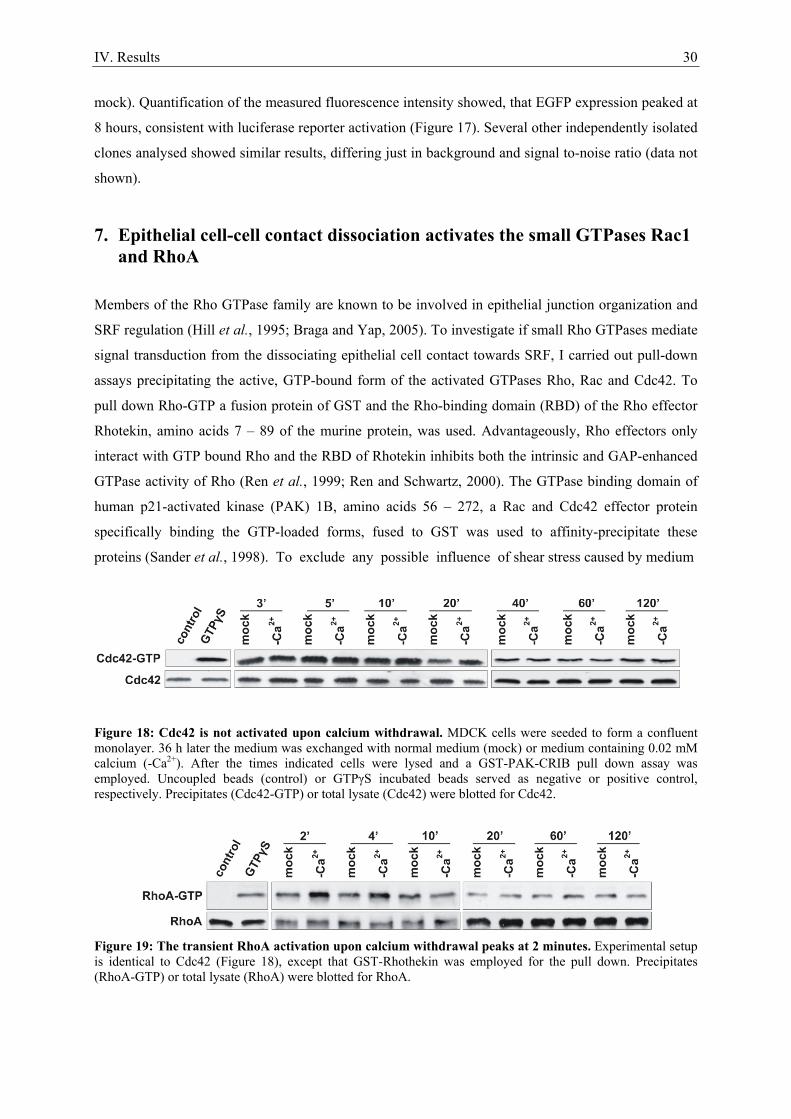
IV. Results 30
mock). Quantification of the measured fluorescence intensity showed, that EGFP expression peaked at
8 hours, consistent with luciferase reporter activation (Figure 17). Several other independently isolated
clones analysed showed similar results, differing just in background and signal to-noise ratio (data not
shown).
7. Epithelial cell-cell contact dissociation activates the small GTPases Rac1 and RhoA
Members of the Rho GTPase family are known to be involved in epithelial junction organization and
SRF regulation (Hill et al., 1995; Braga and Yap, 2005). To investigate if small Rho GTPases mediate
signal transduction from the dissociating epithelial cell contact towards SRF, I carried out pull-down
assays precipitating the active, GTP-bound form of the activated GTPases Rho, Rac and Cdc42. To
pull down Rho-GTP a fusion protein of GST and the Rho-binding domain (RBD) of the Rho effector
Rhotekin, amino acids 7 – 89 of the murine protein, was used. Advantageously, Rho effectors only
interact with GTP bound Rho and the RBD of Rhotekin inhibits both the intrinsic and GAP-enhanced
GTPase activity of Rho (Ren et al., 1999; Ren and Schwartz, 2000). The GTPase binding domain of
human p21-activated kinase (PAK) 1B, amino acids 56 – 272, a Rac and Cdc42 effector protein
specifically binding the GTP-loaded forms, fused to GST was used to affinity-precipitate these
proteins (Sander et al., 1998). To exclude any possible influence of shear stress caused by medium
Figure 18: Cdc42 is not activated upon calcium withdrawal. MDCK cells were seeded to form a confluent monolayer. 36 h later the medium was exchanged with normal medium (mock) or medium containing 0.02 mM calcium (-Ca2+). After the times indicated cells were lysed and a GST-PAK-CRIB pull down assay was employed. Uncoupled beads (control) or GTPγS incubated beads served as negative or positive control, respectively. Precipitates (Cdc42-GTP) or total lysate (Cdc42) were blotted for Cdc42.
Figure 19: The transient RhoA activation upon calcium withdrawal peaks at 2 minutes. Experimental setup is identical to Cdc42 (Figure 18), except that GST-Rhothekin was employed for the pull down. Precipitates (RhoA-GTP) or total lysate (RhoA) were blotted for RhoA.
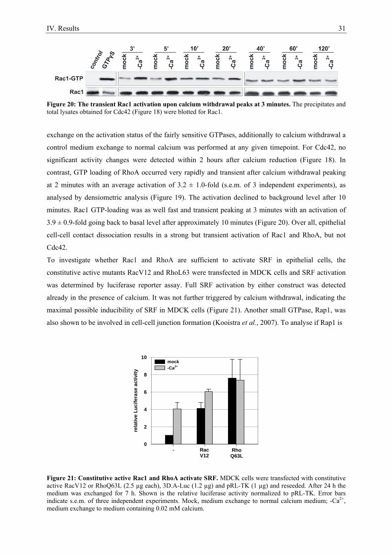
IV. Results 31
Figure 20: The transient Rac1 activation upon calcium withdrawal peaks at 3 minutes. The precipitates and total lysates obtained for Cdc42 (Figure 18) were blotted for Rac1.
exchange on the activation status of the fairly sensitive GTPases, additionally to calcium withdrawal a
control medium exchange to normal calcium was performed at any given timepoint. For Cdc42, no
significant activity changes were detected within 2 hours after calcium reduction (Figure 18). In
contrast, GTP loading of RhoA occurred very rapidly and transient after calcium withdrawal peaking
at 2 minutes with an average activation of 3.2 ± 1.0-fold (s.e.m. of 3 independent experiments), as
analysed by densiometric analysis (Figure 19). The activation declined to background level after 10
minutes. Rac1 GTP-loading was as well fast and transient peaking at 3 minutes with an activation of
3.9 ± 0.9-fold going back to basal level after approximately 10 minutes (Figure 20). Over all, epithelial
cell-cell contact dissociation results in a strong but transient activation of Rac1 and RhoA, but not
Cdc42.
To investigate whether Rac1 and RhoA are sufficient to activate SRF in epithelial cells, the
constitutive active mutants RacV12 and RhoL63 were transfected in MDCK cells and SRF activation
was determined by luciferase reporter assay. Full SRF activation by either construct was detected
already in the presence of calcium. It was not further triggered by calcium withdrawal, indicating the
maximal possible inducibility of SRF in MDCK cells (Figure 21). Another small GTPase, Rap1, was
also shown to be involved in cell-cell junction formation (Kooistra et al., 2007). To analyse if Rap1 is
Figure 21: Constitutive active Rac1 and RhoA activate SRF. MDCK cells were transfected with constitutive active RacV12 or RhoQ63L (2.5 µg each), 3D.A-Luc (1.2 µg) and pRL-TK (1 µg) and reseeded. After 24 h the medium was exchanged for 7 h. Shown is the relative luciferase activity normalized to pRL-TK. Error bars indicate s.e.m. of three independent experiments. Mock, medium exchange to normal calcium medium; -Ca2+, medium exchange to medium containing 0.02 mM calcium.
rela
tive
Luci
fera
se a
ctiv
ity
0
2
4
6
8
10mock-Ca2+
- RacV12
RhoQ63L
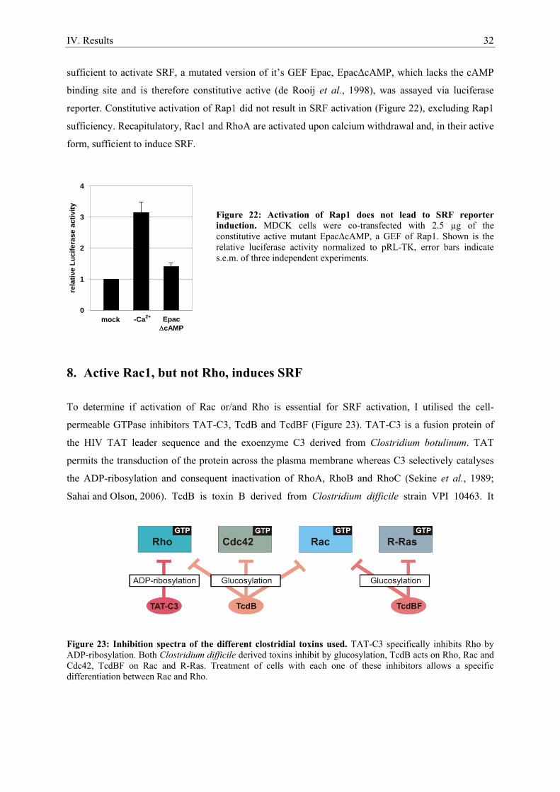
IV. Results 32
sufficient to activate SRF, a mutated version of it’s GEF Epac, EpacΔcAMP, which lacks the cAMP
binding site and is therefore constitutive active (de Rooij et al., 1998), was assayed via luciferase
reporter. Constitutive activation of Rap1 did not result in SRF activation (Figure 22), excluding Rap1
sufficiency. Recapitulatory, Rac1 and RhoA are activated upon calcium withdrawal and, in their active
form, sufficient to induce SRF.
Figure 22: Activation of Rap1 does not lead to SRF reporter induction. MDCK cells were co-transfected with 2.5 µg of the constitutive active mutant EpacΔcAMP, a GEF of Rap1. Shown is the relative luciferase activity normalized to pRL-TK, error bars indicate s.e.m. of three independent experiments.
8. Active Rac1, but not Rho, induces SRF
To determine if activation of Rac or/and Rho is essential for SRF activation, I utilised the cell-
permeable GTPase inhibitors TAT-C3, TcdB and TcdBF (Figure 23). TAT-C3 is a fusion protein of
the HIV TAT leader sequence and the exoenzyme C3 derived from Clostridium botulinum. TAT
permits the transduction of the protein across the plasma membrane whereas C3 selectively catalyses
the ADP-ribosylation and consequent inactivation of RhoA, RhoB and RhoC (Sekine et al., 1989;
Sahai and Olson, 2006). TcdB is toxin B derived from Clostridium difficile strain VPI 10463. It
Figure 23: Inhibition spectra of the different clostridial toxins used. TAT-C3 specifically inhibits Rho by ADP-ribosylation. Both Clostridium difficile derived toxins inhibit by glucosylation, TcdB acts on Rho, Rac and Cdc42, TcdBF on Rac and R-Ras. Treatment of cells with each one of these inhibitors allows a specific differentiation between Rac and Rho.
rela
tive
Luci
fera
se a
ctiv
ity
0
1
2
3
4
mock -Ca2+ EpacΔcAMP
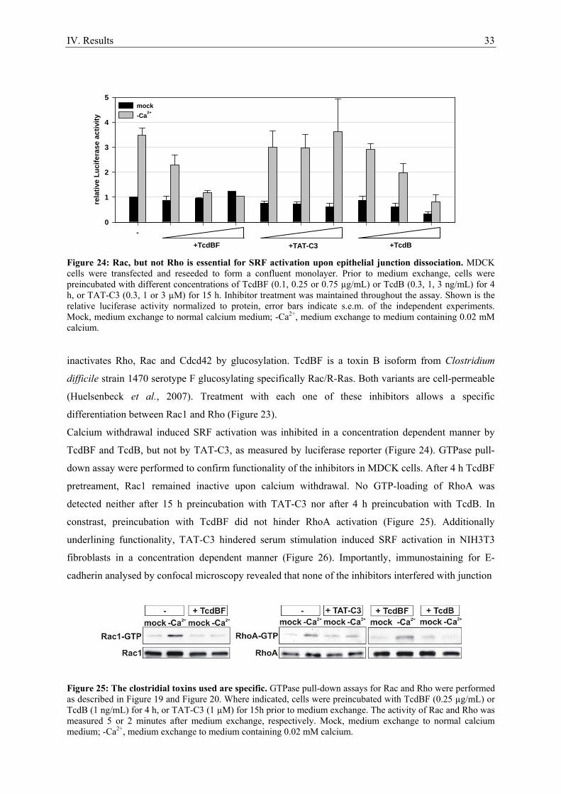
IV. Results 33
rela
tive
Luci
fera
se a
ctiv
ity
0
1
2
3
4
5mock-Ca2+
-
+TcdBF +TcdB+TAT-C3 Figure 24: Rac, but not Rho is essential for SRF activation upon epithelial junction dissociation. MDCK cells were transfected and reseeded to form a confluent monolayer. Prior to medium exchange, cells were preincubated with different concentrations of TcdBF (0.1, 0.25 or 0.75 µg/mL) or TcdB (0.3, 1, 3 ng/mL) for 4 h, or TAT-C3 (0.3, 1 or 3 µM) for 15 h. Inhibitor treatment was maintained throughout the assay. Shown is the relative luciferase activity normalized to protein, error bars indicate s.e.m. of the independent experiments. Mock, medium exchange to normal calcium medium; -Ca2+, medium exchange to medium containing 0.02 mM calcium.
inactivates Rho, Rac and Cdcd42 by glucosylation. TcdBF is a toxin B isoform from Clostridium
difficile strain 1470 serotype F glucosylating specifically Rac/R-Ras. Both variants are cell-permeable
(Huelsenbeck et al., 2007). Treatment with each one of these inhibitors allows a specific
differentiation between Rac1 and Rho (Figure 23).
Calcium withdrawal induced SRF activation was inhibited in a concentration dependent manner by
TcdBF and TcdB, but not by TAT-C3, as measured by luciferase reporter (Figure 24). GTPase pull-
down assay were performed to confirm functionality of the inhibitors in MDCK cells. After 4 h TcdBF
pretreament, Rac1 remained inactive upon calcium withdrawal. No GTP-loading of RhoA was
detected neither after 15 h preincubation with TAT-C3 nor after 4 h preincubation with TcdB. In
constrast, preincubation with TcdBF did not hinder RhoA activation (Figure 25). Additionally
underlining functionality, TAT-C3 hindered serum stimulation induced SRF activation in NIH3T3
fibroblasts in a concentration dependent manner (Figure 26). Importantly, immunostaining for E-
cadherin analysed by confocal microscopy revealed that none of the inhibitors interfered with junction
Figure 25: The clostridial toxins used are specific. GTPase pull-down assays for Rac and Rho were performed as described in Figure 19 and Figure 20. Where indicated, cells were preincubated with TcdBF (0.25 µg/mL) or TcdB (1 ng/mL) for 4 h, or TAT-C3 (1 µM) for 15h prior to medium exchange. The activity of Rac and Rho was measured 5 or 2 minutes after medium exchange, respectively. Mock, medium exchange to normal calcium medium; -Ca2+, medium exchange to medium containing 0.02 mM calcium.
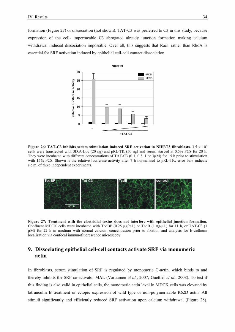
IV. Results 34
formation (Figure 27) or dissociation (not shown). TAT-C3 was preferred to C3 in this study, because
expression of the cell- impermeable C3 abrogated already junction formation making calcium
withdrawal induced dissociation impossible. Over all, this suggests that Rac1 rather than RhoA is
essential for SRF activation induced by epithelial cell-cell contact dissociation.
Figure 26: TAT-C3 inhibits serum stimulation induced SRF activation in NIH3T3 fibroblasts. 3.5 x 10⁴ cells were transfected with 3D.A-Luc (20 ng) and pRL-TK (50 ng) and serum starved at 0.5% FCS for 20 h. They were incubated with different concentrations of TAT-C3 (0.1, 0.3, 1 or 3µM) for 15 h prior to stimulation with 15% FCS. Shown is the relative luciferase activity after 7 h normalized to pRL-TK, error bars indicate s.e.m. of three independent experiments.
Figure 27: Treatment with the clostridial toxins does not interfere with epithelial junction formation. Confluent MDCK cells were incubated with TcdBF (0.25 µg/mL) or TcdB (1 ng/µL) for 11 h, or TAT-C3 (1 µM) for 22 h in medium with normal calcium concentration prior to fixation and analysis for E-cadherin localization via confocal immunofluorescence microscopy.
9. Dissociating epithelial cell-cell contacts activate SRF via monomeric actin
In fibroblasts, serum stimulation of SRF is regulated by monomeric G-actin, which binds to and
thereby inhibits the SRF co-activator MAL (Vartiainen et al., 2007; Guettler et al., 2008). To test if
this finding is also valid in epithelial cells, the monomeric actin level in MDCK cells was elevated by
latrunculin B treatment or ectopic expression of wild type or non-polymerizable R62D actin. All
stimuli significantly and efficiently reduced SRF activation upon calcium withdrawal (Figure 28).
rela
tive
Luci
fera
se a
ctiv
ity
0
5
10
15
20
25
30-FCS+FCS
-
+TAT-C3
NIH3T3
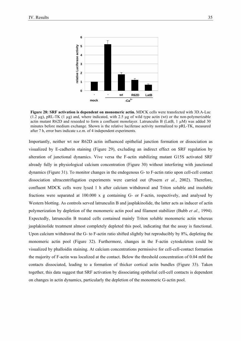
IV. Results 35
Importantly, neither wt nor R62D actin influenced epithelial junction formation or dissociation as
visualized by E-cadherin staining (Figure 29), excluding an indirect effect on SRF regulation by
alteration of junctional dynamics. Vive versa the F-actin stabilizing mutant G15S activated SRF
already fully in physiological calcium concentration (Figure 30) without interfering with junctional
dynamics (Figure 31). To monitor changes in the endogenous G- to F-actin ratio upon cell-cell contact
dissociation ultracentrifugation experiments were carried out (Posern et al., 2002). Therefore,
confluent MDCK cells were lysed 1 h after calcium withdrawal and Triton soluble and insoluble
fractions were separated at 100.000 x g containing G- or F-actin, respectively, and analysed by
Western blotting. As controls served latrunculin B and jasplakinolide, the latter acts as inducer of actin
polymerization by depletion of the monomeric actin pool and filament stabilizer (Bubb et al., 1994).
Expectedly, latrunculin B treated cells contained mainly Triton soluble monomeric actin whereas
jasplakinolide treatment almost completely depleted this pool, indicating that the assay is functional.
Upon calcium withdrawal the G- to F-actin ratio shifted slightly but reproducibly by 8%, depleting the
monomeric actin pool (Figure 32). Furthermore, changes in the F-actin cytoskeleton could be
visualized by phalloidin staining. At calcium concentrations permissive for cell-cell-contact formation
the majority of F-actin was localized at the contact. Below the threshold concentration of 0.04 mM the
contacts dissociated, leading to a formation of thicker cortical actin bundles (Figure 33). Taken
together, this data suggest that SRF activation by dissociating epithelial cell-cell contacts is dependent
on changes in actin dynamics, particularly the depletion of the monomeric G-actin pool.
rela
tive
Luci
fera
se a
ctiv
ity
0
1
2
3
4
5
6
mock -Ca2+
R62Dwt- - LatB
Figure 28: SRF activation is dependent on monomeric actin. MDCK cells were transfected with 3D.A-Luc (1.2 µg), pRL-TK (1 µg) and, where indicated, with 2.5 µg of wild type actin (wt) or the non-polymerizable actin mutant R62D and reseeded to form a confluent monolayer. Latrunculin B (LatB, 1 µM) was added 30 minutes before medium exchange. Shown is the relative luciferase activity normalized to pRL-TK, measured after 7 h, error bars indicate s.e.m. of 4 independent experiments.
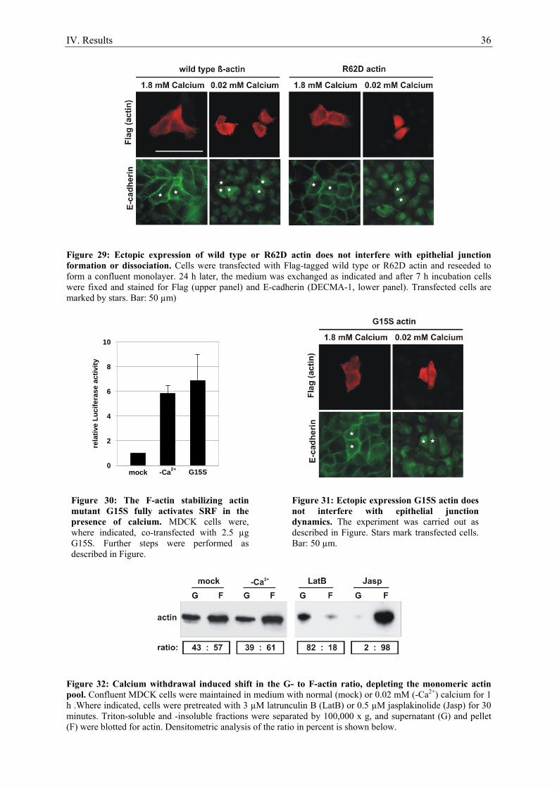
IV. Results 36
Figure 29: Ectopic expression of wild type or R62D actin does not interfere with epithelial junction formation or dissociation. Cells were transfected with Flag-tagged wild type or R62D actin and reseeded to form a confluent monolayer. 24 h later, the medium was exchanged as indicated and after 7 h incubation cells were fixed and stained for Flag (upper panel) and E-cadherin (DECMA-1, lower panel). Transfected cells are marked by stars. Bar: 50 µm)
rela
tive
Luci
fera
se a
ctiv
ity
0
2
4
6
8
10
mock -Ca2+ G15S
Figure 32: Calcium withdrawal induced shift in the G- to F-actin ratio, depleting the monomeric actin pool. Confluent MDCK cells were maintained in medium with normal (mock) or 0.02 mM (-Ca2+) calcium for 1 h .Where indicated, cells were pretreated with 3 µM latrunculin B (LatB) or 0.5 µM jasplakinolide (Jasp) for 30 minutes. Triton-soluble and -insoluble fractions were separated by 100,000 x g, and supernatant (G) and pellet (F) were blotted for actin. Densitometric analysis of the ratio in percent is shown below.
Figure 30: The F-actin stabilizing actin mutant G15S fully activates SRF in the presence of calcium. MDCK cells were, where indicated, co-transfected with 2.5 µg G15S. Further steps were performed as described in Figure.
Figure 31: Ectopic expression G15S actin does not interfere with epithelial junction dynamics. The experiment was carried out as described in Figure. Stars mark transfected cells. Bar: 50 µm.
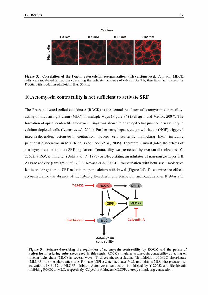
IV. Results 37
Figure 33: Correlation of the F-actin cytoskeleton reorganization with calcium level. Confluent MDCK cells were incubated in medium containing the indicated amounts of calcium for 7 h, then fixed and stained for F-actin with rhodamin-phalloidin. Bar: 50 µm.
10. Actomyosin contractility is not sufficient to activate SRF
The RhoA activated coiled-coil kinase (ROCK) is the central regulator of actomyosin contractility,
acting on myosin light chain (MLC) in multiple ways (Figure 34) (Pellegrin and Mellor, 2007). The
formation of apical contractile actomyosin rings was shown to drive epithelial junction disassembly in
calcium depleted cells (Ivanov et al., 2004). Furthermore, hepatocyte growth factor (HGF)-triggered
integrin-dependent actomyosin contraction induces cell scattering mimicking EMT including
junctional dissociation in MDCK cells (de Rooij et al., 2005). Therefore, I investigated the effects of
actomyosin contraction on SRF regulation. Contractility was repressed by two small molecules: Y-
27632, a ROCK inhibitor (Uehata et al., 1997) or Blebbistatin, an inhibitor of non-muscle myosin II
ATPase activity (Straight et al., 2003; Kovacs et al., 2004). Preincubation with both small molecules
led to an abrogation of SRF activation upon calcium withdrawal (Figure 35). To examine the effects
accountable for the absence of inducibility E-cadherin and phalloidin micrographs after Blebbistatin
Figure 34: Scheme describing the regulation of actomyosin contractility by ROCK and the points of action for interfering substances used in this study. ROCK stimulates actomyosin contractility by acting on myosin light chain (MLC) in several ways: (i) direct phosphorylation; (ii) inhibition of MLC phosphatase (MLCPP) (iii) phosphorylation of ZIP kinase (ZIPK) which activates MLC and inhibits MLC phosphatase; (iv) activation of CPI-17, a MLCPP inhibitor. Actomyosin contraction is inhibited by Y-27632 and Blebbistatin inhibiting ROCK or MLC, respectively. Calyculin A hinders MLCPP, thereby stimulating contraction.
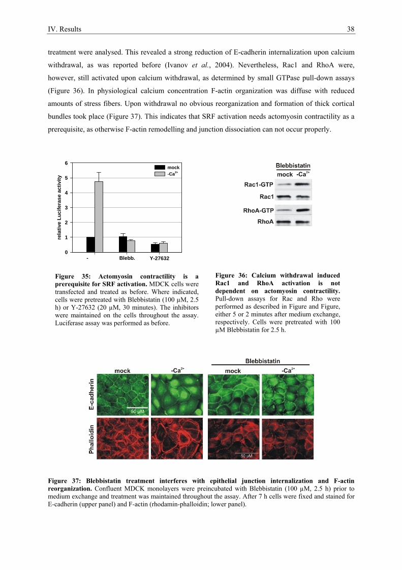
IV. Results 38
treatment were analysed. This revealed a strong reduction of E-cadherin internalization upon calcium
withdrawal, as was reported before (Ivanov et al., 2004). Nevertheless, Rac1 and RhoA were,
however, still activated upon calcium withdrawal, as determined by small GTPase pull-down assays
(Figure 36). In physiological calcium concentration F-actin organization was diffuse with reduced
amounts of stress fibers. Upon withdrawal no obvious reorganization and formation of thick cortical
bundles took place (Figure 37). This indicates that SRF activation needs actomyosin contractility as a
prerequisite, as otherwise F-actin remodelling and junction dissociation can not occur properly.
Figure 37: Blebbistatin treatment interferes with epithelial junction internalization and F-actin reorganization. Confluent MDCK monolayers were preincubated with Blebbistatin (100 µM, 2.5 h) prior to medium exchange and treatment was maintained throughout the assay. After 7 h cells were fixed and stained for E-cadherin (upper panel) and F-actin (rhodamin-phalloidin; lower panel).
Figure 36: Calcium withdrawal induced Rac1 and RhoA activation is not dependent on actomyosin contractility. Pull-down assays for Rac and Rho were performed as described in Figure and Figure, either 5 or 2 minutes after medium exchange, respectively. Cells were pretreated with 100 µM Blebbistatin for 2.5 h.
rela
tive
Luci
fera
se a
ctiv
ity
0
1
2
3
4
5
6mock-Ca2+
- Blebb. Y-27632
Figure 35: Actomyosin contractility is a prerequisite for SRF activation. MDCK cells were transfected and treated as before. Where indicated, cells were pretreated with Blebbistatin (100 µM, 2.5 h) or Y-27632 (20 µM, 30 minutes). The inhibitors were maintained on the cells throughout the assay. Luciferase assay was performed as before.
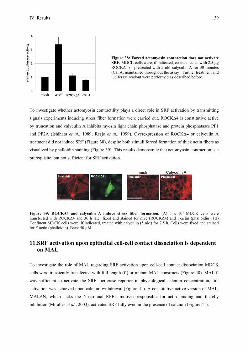
IV. Results 39
Figure 38: Forced actomyosin contraction does not activate SRF. MDCK cells were, if indicated, co-transfected with 2.5 µg ROCKΔ4 or pretreated with 5 nM calyculin A for 30 minutes (Cal.A; maintained throughout the assay). Further treatment and luciferase readout were performed as described before.
To investigate whether actomyosin contractility plays a direct role in SRF activation by transmitting
signals experiments inducing stress fiber formation were carried out. ROCKΔ4 is constitutive active
by truncation and calyculin A inhibits myosin light chain phosphatase and protein phosphatases PP1
and PP2A (Ishihara et al., 1989; Resjo et al., 1999). Overexpression of ROCKΔ4 or calyculin A
treatment did not induce SRF (Figure 38), despite both stimuli forced formation of thick actin fibers as
visualized by phalloidin staining (Figure 39). This results demonstrate that actomyosin contraction is a
prerequisite, but not sufficient for SRF activation.
Figure 39: ROCKΔ4 and calyculin A induce stress fiber formation. (A) 5 x 10⁴ MDCK cells were transfected with ROCKΔ4 and 36 h later fixed and stained for myc (ROCKΔ4) and F-actin (phalloidin). (B) Confluent MDCK cells were, if indicated, treated with calyculin (5 nM) for 7.5 h. Cells were fixed and stained for F-actin (phalloidin). Bars: 50 µM.
11. SRF activation upon epithelial cell-cell contact dissociation is dependent on MAL
To investigate the role of MAL regarding SRF activation upon cell-cell contact dissociation MDCK
cells were transiently transfected with full length (fl) or mutant MAL constructs (Figure 40). MAL fl
was sufficient to activate the SRF luciferase reporter in physiological calcium concentration, full
activation was achieved upon calcium withdrawal (Figure 41). A constitutive active version of MAL,
MALΔN, which lacks the N-terminal RPEL motives responsible for actin binding and thereby
inhibition (Miralles et al., 2003), activated SRF fully even in the presence of calcium (Figure 41).
rela
tive
Luci
fera
se a
ctiv
ity
0
1
2
3
4
mock -Ca2+ Cal.AROCKΔ4
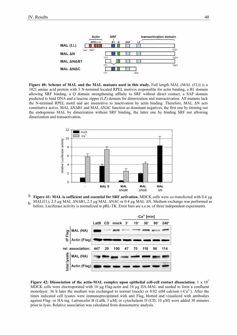
IV. Results 40
In contrast, two dominant negative constructs of MAL were tested: MALΔNΔB1 lacks the SRF
binding site and acts by titrating out the endogenous MAL by dimerization via the leucine zipper (LZ) Figure 41: MAL is sufficient and essential for SRF activation. MDCK cells were co-transfected with 0.4 µg MAL(f.l.), 2.5 µg MAL ΔNΔB1, 2.5 µg MAL ΔNΔC or 0.4 µg MAL ΔN. Medium exchange was performed as before. Luciferase activity is normalized to pRL-TK. Error bars are s.e.m. of three independent experiments.
Figure 42: Dissociation of the actin-MAL complex upon epithelial cell-cell contact dissociation. 1 x 10⁷ MDCK cells were electroporated with 10 µg Flag-actin and 10 µg HA-MAL and seeded to form a confluent monolayer. 36 h later the medium was exchanged to normal (mock) or 0.02 mM calcium (-Ca2+). After the times indicated cell lysates were immunoprecipitated with anti Flag, blotted and visualized with antibodies against Flag- or HA-tag. Latrunculin B (LatB, 3 µM) or cytochalasin D (CD, 10 µM) were added 30 minutes prior to lysis. Relative association was calculated from densiometric analysis.
rela
tive
Luci
fera
se a
ctiv
ity
0
2
4
6
8
10
12mock-Ca2+
- MAL fl MALΔΝΔΒ1
MALΔΝΔC
MALΔΝ
Figure 40: Scheme of MAL and the MAL mutants used in this study. Full length MAL (MAL (f.l.)) is a 1021 amino acid protein with 3 N-terminal located RPEL motives responsible for actin binding, a B1 domain allowing SRF binding, a Q domain strengthening affinity to SRF without direct contact, a SAP domain predicted to bind DNA and a leucine zipper (LZ) domain for dimerization and transactivation. All mutants lack the N-terminal RPEL motif and are insensitive to inactivation by actin binding. Therefore, MAL ΔN acts constitutive active. MAL ΔNΔB1 and MAL ΔNΔC function as dominant negatives, the first one by titrating out the endogenous MAL by dimerization without SRF binding, the latter one by binding SRF not allowing dimerization and transactivation.
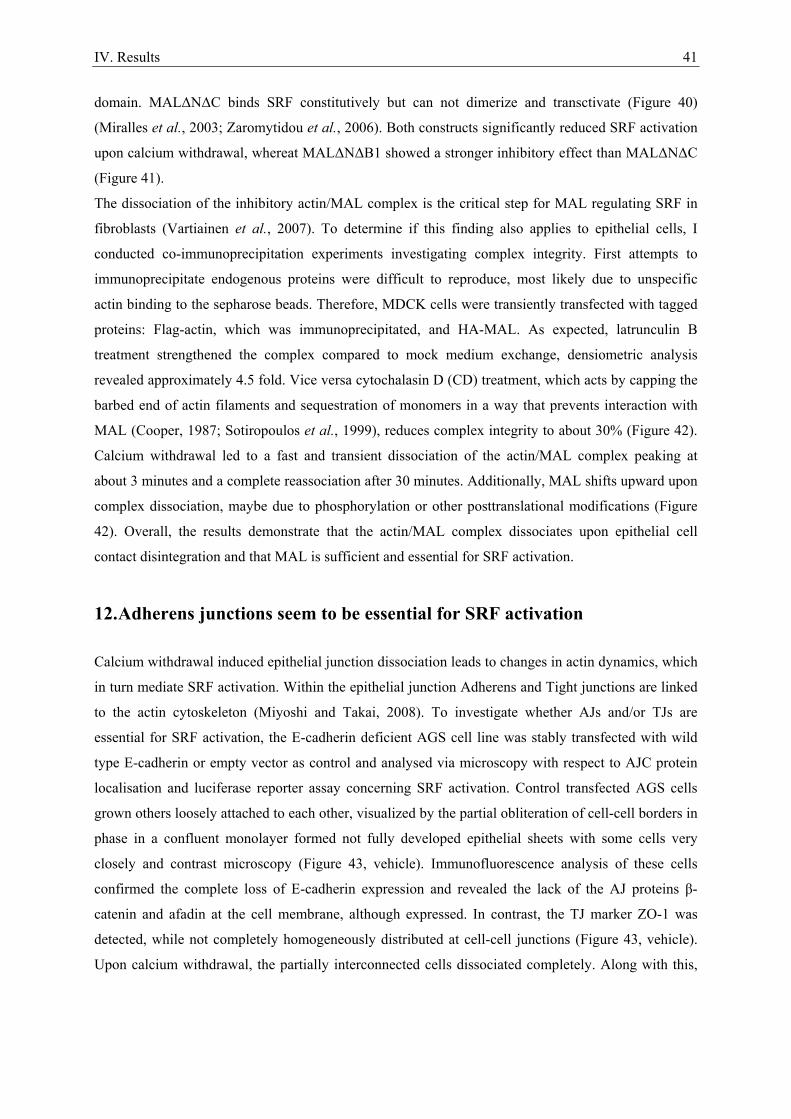
IV. Results 41
domain. MALΔNΔC binds SRF constitutively but can not dimerize and transctivate (Figure 40)
(Miralles et al., 2003; Zaromytidou et al., 2006). Both constructs significantly reduced SRF activation
upon calcium withdrawal, whereat MALΔNΔB1 showed a stronger inhibitory effect than MALΔNΔC
(Figure 41).
The dissociation of the inhibitory actin/MAL complex is the critical step for MAL regulating SRF in
fibroblasts (Vartiainen et al., 2007). To determine if this finding also applies to epithelial cells, I
conducted co-immunoprecipitation experiments investigating complex integrity. First attempts to
immunoprecipitate endogenous proteins were difficult to reproduce, most likely due to unspecific
actin binding to the sepharose beads. Therefore, MDCK cells were transiently transfected with tagged
proteins: Flag-actin, which was immunoprecipitated, and HA-MAL. As expected, latrunculin B
treatment strengthened the complex compared to mock medium exchange, densiometric analysis
revealed approximately 4.5 fold. Vice versa cytochalasin D (CD) treatment, which acts by capping the
barbed end of actin filaments and sequestration of monomers in a way that prevents interaction with
MAL (Cooper, 1987; Sotiropoulos et al., 1999), reduces complex integrity to about 30% (Figure 42).
Calcium withdrawal led to a fast and transient dissociation of the actin/MAL complex peaking at
about 3 minutes and a complete reassociation after 30 minutes. Additionally, MAL shifts upward upon
complex dissociation, maybe due to phosphorylation or other posttranslational modifications (Figure
42). Overall, the results demonstrate that the actin/MAL complex dissociates upon epithelial cell
contact disintegration and that MAL is sufficient and essential for SRF activation.
12. Adherens junctions seem to be essential for SRF activation
Calcium withdrawal induced epithelial junction dissociation leads to changes in actin dynamics, which
in turn mediate SRF activation. Within the epithelial junction Adherens and Tight junctions are linked
to the actin cytoskeleton (Miyoshi and Takai, 2008). To investigate whether AJs and/or TJs are
essential for SRF activation, the E-cadherin deficient AGS cell line was stably transfected with wild
type E-cadherin or empty vector as control and analysed via microscopy with respect to AJC protein
localisation and luciferase reporter assay concerning SRF activation. Control transfected AGS cells
grown others loosely attached to each other, visualized by the partial obliteration of cell-cell borders in
phase in a confluent monolayer formed not fully developed epithelial sheets with some cells very
closely and contrast microscopy (Figure 43, vehicle). Immunofluorescence analysis of these cells
confirmed the complete loss of E-cadherin expression and revealed the lack of the AJ proteins β-
catenin and afadin at the cell membrane, although expressed. In contrast, the TJ marker ZO-1 was
detected, while not completely homogeneously distributed at cell-cell junctions (Figure 43, vehicle).
Upon calcium withdrawal, the partially interconnected cells dissociated completely. Along with this,
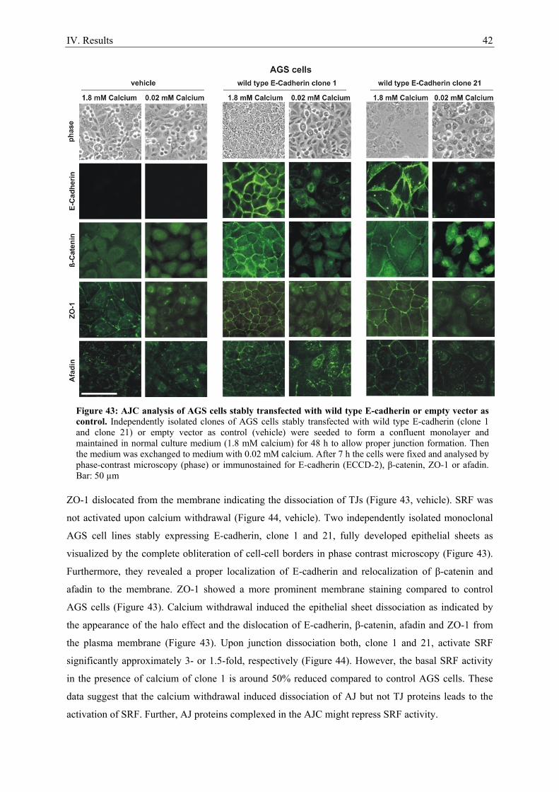
IV. Results 42
ZO-1 dislocated from the membrane indicating the dissociation of TJs (Figure 43, vehicle). SRF was
not activated upon calcium withdrawal (Figure 44, vehicle). Two independently isolated monoclonal
AGS cell lines stably expressing E-cadherin, clone 1 and 21, fully developed epithelial sheets as
visualized by the complete obliteration of cell-cell borders in phase contrast microscopy (Figure 43).
Furthermore, they revealed a proper localization of E-cadherin and relocalization of β-catenin and
afadin to the membrane. ZO-1 showed a more prominent membrane staining compared to control
AGS cells (Figure 43). Calcium withdrawal induced the epithelial sheet dissociation as indicated by
the appearance of the halo effect and the dislocation of E-cadherin, β-catenin, afadin and ZO-1 from
the plasma membrane (Figure 43). Upon junction dissociation both, clone 1 and 21, activate SRF
significantly approximately 3- or 1.5-fold, respectively (Figure 44). However, the basal SRF activity
in the presence of calcium of clone 1 is around 50% reduced compared to control AGS cells. These
data suggest that the calcium withdrawal induced dissociation of AJ but not TJ proteins leads to the
activation of SRF. Further, AJ proteins complexed in the AJC might repress SRF activity.
Figure 43: AJC analysis of AGS cells stably transfected with wild type E-cadherin or empty vector as control. Independently isolated clones of AGS cells stably transfected with wild type E-cadherin (clone 1 and clone 21) or empty vector as control (vehicle) were seeded to form a confluent monolayer and maintained in normal culture medium (1.8 mM calcium) for 48 h to allow proper junction formation. Then the medium was exchanged to medium with 0.02 mM calcium. After 7 h the cells were fixed and analysed by phase-contrast microscopy (phase) or immunostained for E-cadherin (ECCD-2), β-catenin, ZO-1 or afadin. Bar: 50 µm
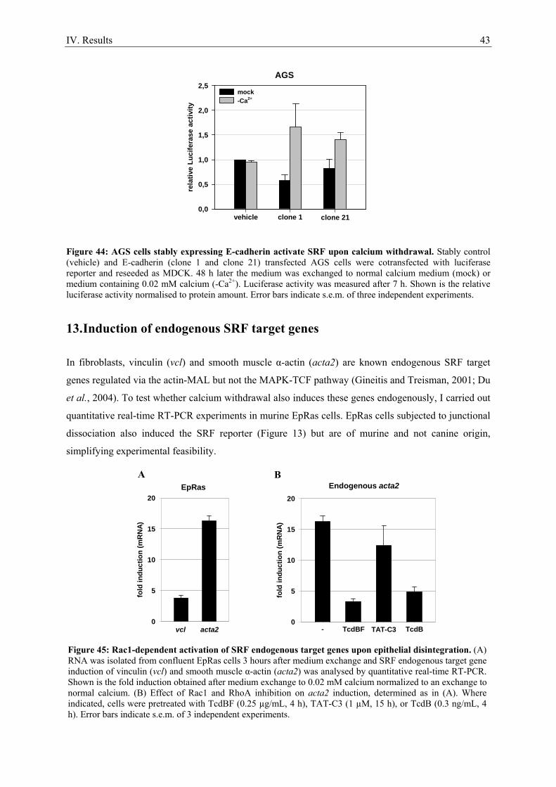
IV. Results 43
Figure 44: AGS cells stably expressing E-cadherin activate SRF upon calcium withdrawal. Stably control (vehicle) and E-cadherin (clone 1 and clone 21) transfected AGS cells were cotransfected with luciferase reporter and reseeded as MDCK. 48 h later the medium was exchanged to normal calcium medium (mock) or medium containing 0.02 mM calcium (-Ca2+). Luciferase activity was measured after 7 h. Shown is the relative luciferase activity normalised to protein amount. Error bars indicate s.e.m. of three independent experiments.
13. Induction of endogenous SRF target genes
In fibroblasts, vinculin (vcl) and smooth muscle α-actin (acta2) are known endogenous SRF target
genes regulated via the actin-MAL but not the MAPK-TCF pathway (Gineitis and Treisman, 2001; Du
et al., 2004). To test whether calcium withdrawal also induces these genes endogenously, I carried out
quantitative real-time RT-PCR experiments in murine EpRas cells. EpRas cells subjected to junctional
dissociation also induced the SRF reporter (Figure 13) but are of murine and not canine origin,
simplifying experimental feasibility.
fold
indu
ctio
n (m
RN
A)
0
5
10
15
20
- TcdBF TcdBTAT-C3
Endogenous acta2
fold
indu
ctio
n (m
RN
A)
0
5
10
15
20
vcl acta2
EpRas
rela
tive
Luci
fera
se a
ctiv
ity
0,0
0,5
1,0
1,5
2,0
2,5mock-Ca2+
vehicle clone 1 clone 21
AGS
A B
Figure 45: Rac1-dependent activation of SRF endogenous target genes upon epithelial disintegration. (A) RNA was isolated from confluent EpRas cells 3 hours after medium exchange and SRF endogenous target gene induction of vinculin (vcl) and smooth muscle α-actin (acta2) was analysed by quantitative real-time RT-PCR. Shown is the fold induction obtained after medium exchange to 0.02 mM calcium normalized to an exchange to normal calcium. (B) Effect of Rac1 and RhoA inhibition on acta2 induction, determined as in (A). Where indicated, cells were pretreated with TcdBF (0.25 µg/mL, 4 h), TAT-C3 (1 µM, 15 h), or TcdB (0.3 ng/mL, 4 h). Error bars indicate s.e.m. of 3 independent experiments.
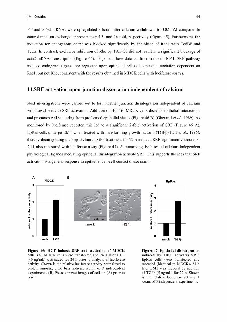
IV. Results 44
Vcl and acta2 mRNAs were upregulated 3 hours after calcium withdrawal to 0.02 mM compared to
control medium exchange approximately 4.5- and 16-fold, respectively (Figure 45). Furthermore, the
induction for endogenous acta2 was blocked significantly by inhibition of Rac1 with TcdBF and
TcdB. In contrast, exclusive inhibition of Rho by TAT-C3 did not result in a significant blockage of
acta2 mRNA transcription (Figure 45). Together, these data confirm that actin-MAL-SRF pathway
induced endogenous genes are regulated upon epithelial cell-cell contact dissociation dependent on
Rac1, but not Rho, consistent with the results obtained in MDCK cells with luciferase assays.
14. SRF activation upon junction dissociation independent of calcium
Next investigations were carried out to test whether junction disintegration independent of calcium
withdrawal leads to SRF activation. Addition of HGF to MDCK cells disrupts epithelial interactions
and promotes cell scattering from preformed epithelial sheets (Figure 46 B) (Gherardi et al., 1989). As
monitored by luciferase reporter, this led to a significant 2-fold activation of SRF (Figure 46 A).
EpRas cells undergo EMT when treated with transforming growth factor β (TGFβ) (Oft et al., 1996),
thereby disintegrating their epithelium. TGFβ treatment for 72 h induced SRF significantly around 3-
fold, also measured with luciferase assay (Figure 47). Summarizing, both tested calcium-independent
physiological ligands mediating epithelial disintegration activate SRF. This supports the idea that SRF
activation is a general response to epithelial cell-cell contact dissociation.
rela
tive
Luci
fera
se a
ctiv
ity
0
1
2
3
mock HGF
MDCK
rela
tive
Luci
fera
se a
ctiv
ity
0
1
2
3
4
mock TGFβ
EpRas
Figure 46: HGF induces SRF and scattering of MDCK cells. (A) MDCK cells were transfected and 24 h later HGF (40 ng/mL) was added for 24 h prior to analysis of luciferase activity. Shown is the relative luciferase activity normalized to protein amount, error bars indicate s.e.m. of 3 independent experiments. (B) Phase contrast images of cells in (A) prior to lysis.
Figure 47: Epithelial disintegration induced by EMT activates SRF. EpRas cells were transfected and reseeded (identical to MDCK). 24 h later EMT was induced by addition of TGFβ (5 ng/mL) for 72 h. Shown is the relative luciferase activity ± s.e.m. of 3 independent experiments.
A B
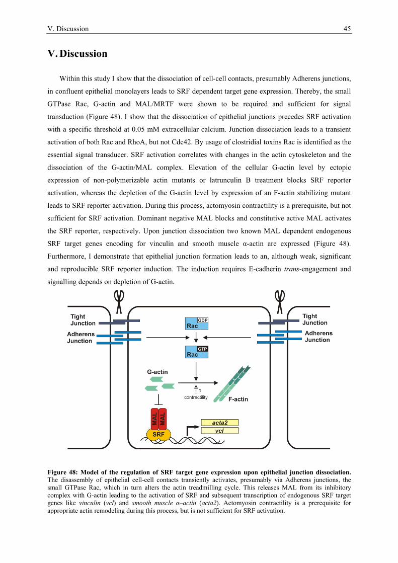
V. Discussion 45
V. Discussion
Within this study I show that the dissociation of cell-cell contacts, presumably Adherens junctions,
in confluent epithelial monolayers leads to SRF dependent target gene expression. Thereby, the small
GTPase Rac, G-actin and MAL/MRTF were shown to be required and sufficient for signal
transduction (Figure 48). I show that the dissociation of epithelial junctions precedes SRF activation
with a specific threshold at 0.05 mM extracellular calcium. Junction dissociation leads to a transient
activation of both Rac and RhoA, but not Cdc42. By usage of clostridial toxins Rac is identified as the
essential signal transducer. SRF activation correlates with changes in the actin cytoskeleton and the
dissociation of the G-actin/MAL complex. Elevation of the cellular G-actin level by ectopic
expression of non-polymerizable actin mutants or latrunculin B treatment blocks SRF reporter
activation, whereas the depletion of the G-actin level by expression of an F-actin stabilizing mutant
leads to SRF reporter activation. During this process, actomyosin contractility is a prerequisite, but not
sufficient for SRF activation. Dominant negative MAL blocks and constitutive active MAL activates
the SRF reporter, respectively. Upon junction dissociation two known MAL dependent endogenous
SRF target genes encoding for vinculin and smooth muscle α-actin are expressed (Figure 48).
Furthermore, I demonstrate that epithelial junction formation leads to an, although weak, significant
and reproducible SRF reporter induction. The induction requires E-cadherin trans-engagement and
signalling depends on depletion of G-actin.
Figure 48: Model of the regulation of SRF target gene expression upon epithelial junction dissociation. The disassembly of epithelial cell-cell contacts transiently activates, presumably via Adherens junctions, the small GTPase Rac, which in turn alters the actin treadmilling cycle. This releases MAL from its inhibitory complex with G-actin leading to the activation of SRF and subsequent transcription of endogenous SRF target genes like vinculin (vcl) and smooth muscle α–actin (acta2). Actomyosin contractility is a prerequisite for appropriate actin remodeling during this process, but is not sufficient for SRF activation.
V. Discussion 46
Serum stimulation of confluent epithelial cell monolayers does not trigger the activation of
SRF (Figure 6), raising the question how SRF-controlled genes are regulated in epithelia. Both,
epithelial junction formation and dissociation are known to cause changes in the actin cytoskeleton,
which is an essential signalling event in serum-stimulated fibroblasts (Volberg et al., 1986; Gumbiner
et al., 1988; Sotiropoulos et al., 1999). I therefore investigated whether junction remodelling has an
impact on SRF regulation. Epithelial junction formation induces the SRF reporter 2-fold (Figure 8).
This activation is hindered by preincubation with the E-cadherin blocking antibody DECMA-1 (Figure
8), clearly identifying junction formation rather than other potential calcium induced side-effects as
the essential signal transmitter. Regardless, the specific junctional component essential for induction
can not be identified by this experiment, since DECMA-1 hinders besides AJ also TJ and desmosomal
junction formation (Gumbiner et al., 1988; Watabe et al., 1994). In addition, actin mutant expression
and latrunculin B treatment showed that changes in the G- to F-actin ratio, leading to the depletion of
the cellular monomeric actin pool, are essential for signal transmission to SRF (Figure 8). The F-actin
stabilizing mutant G15S is thought to maximally induce MAL-dependent SRF transcription (G.P. and
S.B.). This maximal inducibility is in confluent epithelial MDCK cells around 6 to 7-fold (Figure 8
and Figure 30). However, upon epithelial junction formation the induction is, although reproducible
and significant, just around 2-fold. In parallel I made the observation that epithelial junction
dissociation activates SRF to the same extend as G15S (Figure 10). Further, I focused my
investigations on this exciting finding.
By employment of the calcium switch method I dissociate epithelial junctions, thereby
mimicking one aspect of a full EMT. To assure that the calcium withdrawal induced junction
dissociation and not secondary effects induce SRF activation several experiments were carried out.
The titration of extracellular calcium showed that epithelial junction disintegration strictly correlates
with SRF activation (Figure 9 and Figure 10), indicating a direct interdependence. Normal and cancer
cell lines lacking E-cadherin expression fail to activate SRF upon calcium withdrawal despite the
possession of a functional pathway (Figure 14 and not shown). Upon stable expression of E-cadherin
in the E-cadherin-deficient gastric cancer cell line AGS AJs are reformed and activate SRF in response
to calcium withdrawal (Figure 43 and Figure 44). Finally, a HGF or TGF-β mediated calcium
withdrawal independent junction dissociation activates SRF. These observations prove that SRF
activation is a direct consequence of junction dissociation induced signalling.
The AJC is known to regulate GTPases of the Rho family and is dynamically connected to the
cortical actin belt of confluent epithelial cells (Braga, 2002). Calcium withdrawal as well as HGF or
TGF-β treatment leads to the dissociation of the complete AJC including AJs and TJs. To clarify
whether AJs or TJs are essential for signalling to SRF, I utilised the E-cadherin deficient human
gastric cancer cell line AGS. Adjacent AGS cells grown in a confluent monolayer lack E-cadherin/β-
catenin and nectin/afadin complex dependent AJs. Although the detected ZO-1 membrane staining
appears to be slightly reduced compared to E-cadherin expressing AGS cells, they do possess fully
V. Discussion 47
developed TJs between most cells (Figure 43). Calcium withdrawal of these cells induces TJ
dissociation, probably by disassembly of the calcium dependent desmosomal cadherins. Importantly,
this is not associated with SRF activation (Figure 44), suggesting that neither desmosome nor TJ
disassembly regulates SRF. Conversely, stable expression of E-cadherin in AGS cells leads to the
formation of fully developed AJs (Figure 43), whose dissociation induces SRF (Figure 44). These
results suggest that components of the AJ rather than the TJ mediate SRF activation.
Several additional experiments can further support the idea of AJs as essential signal
transducers to SRF. ZO-1 deleted and ZO-2 depleted epithelial EpH4 cells are well polarized and form
fully functional AJs, but completely lack TJs (Umeda et al., 2006). If these cells fail to activate SRF
upon calcium withdrawal an involvement of TJs in SRF activation can be ruled out. The knockdown
of E-cadherin and Cadherin-6 in preformed epithelial junctions was shown to disrupt AJs, but not TJs
(Capaldo and Macara, 2007). A calcium switch in depleted cells can narrow down the potential signal
transducers to the AJ components excluding α-catenin or even E-cadherin alone (Capaldo and Macara,
2007). Hence, I constructed shRNA expression vectors for transfection or amphotrophic infection
targeting both mRNAs (Table 12 and not shown). The plasmid delivery efficiency for both methods
turned out to be around 5 – 10% in MDCK cells and the resulting knockdown was not complete. Thus,
knockdown cells were forming partial junctions with adjacent untransfected/uninfected cells mediated
by their residual E-cadherin and Cadherin-6. Corollary, SRF was still activated in these cells upon
calcium withdrawal (not shown). Currently, I am engineering a stable cell line carrying an inducible
shRNA expression vector targeting E-cadherin and Cadherin-6 to overcome this problem.
To further differentiate between E-cadherin/catenin and nectin/afadin complex mediated
signalling to SRF, calcium switch experiments in afadin depleted AGS cells, which stably express E-
cadherin, can be carried out. However, afadin-independent E-cadherin mediated junction formation
would be a prerequisite for this experiment.
A further dissection to identify the specific AJ component crucial for signalling proves
difficult, as targeting of either β-catenin, α-catenin, p120-catenin or afadin induces severe defects in
AJs and TJs, which usually lead to the complete loss of polarization and cell-cell contacts (Behrens et
al., 1993; Ikeda et al., 1999; Ireton et al., 2002; Capaldo and Macara, 2007). However, the fusion
protein of a C-terminal truncated E-cadherin lacking the β-catenin binding site and specific parts of α-
catenin is able mediate adhesion and connects to the actin cytoskeleton (Nagafuchi et al., 1994;
Imamura et al., 1999). Calcium switch experiments with AGS cells stably expressing these fusion
proteins would allow the identification or exclusion of β-catenin, formin-1, vinculin, α-actinin or
EPLIN as essential signal transducer (Kobielak and Fuchs, 2004; Abe and Takeichi, 2008). During
this study I engineered the cDNA constructs encoding for several different E-cadherin/α-catenin fusion
proteins (Table 12 and not shown). AGS cell lines stably transfected with the cDNAs expressed the
fusion proteins but, however, failed to correctly localize them to the membrane (not shown). Further
transfections will be carried out to isolate clones with correctly localized fusion proteins. Overall, the
V. Discussion 48
suggested experiments will allow the identification of the specific AJC component essential for signal
transduction to SRF.
Ezrin, belonging to the ezrin-radixin-moesin (ERM) protein family, and merlin, which is
encoded by the neurofibromatosis type-2 (NF2) gene and is a close relative to the ERM family, are
both known to localize to the AJC and regulate cell-cell adhesion (Gould et al., 1989; Rouleau et al.,
1993; Trofatter et al., 1993; Lallemand et al., 2003; Pujuguet et al., 2003; Van Furden et al., 2004).
Both, ezrin and merlin, are also connected to the actin cytoskeleton (Gould et al., 1989; Gronholm et
al., 1999; Brault et al., 2001; James et al., 2001), regulate the actin polymerization function of N-
WASP (Manchanda et al., 2005) and can signal via Rho familiy GTPases (Maeda et al., 1999; Kissil
et al., 2003; Pujuguet et al., 2003). These observations prompted me to investigate a potential
involvement of these two proteins in the regulation of SRF. Unexpectedly, overexpression of none of
the therefore constructed constitutive active and dominant negative ezrin or merlin constructs (Table
12) has an impact on SRF regulation in epithelial MDCK cells (not shown). Further, the serum
inducibility of SRF in subconfluent or confluent NIH3T3 cells is not influenced by any overexpressed
construct (not shown). Together, these results give as yet no indication for a regulatory function of
ezrin or merlin towards SRF.
The relationship between epithelial junctions and small Rho family GTPases has been studied
extensively, but mainly during junction formation (for details refer to Introduction). Here, I provide
evidence that the small GTPases Rac1 and RhoA are quickly and transiently GTP loaded upon
epithelial junction dissociation mediated by medium exchange to 0.02 mM calcium. By contrast,
EGTA induced calcium depletion for 30 minutes has previously been shown to inhibit Rac activation
in MDCKII or Fisher rat thyroid cells, whereas RhoA remained unchanged in MDCKII cells
(Nakagawa et al., 2001; Balzac et al., 2005). This apparent discrepancy is probably explained by the
very transient activation of Rac1 and RhoA, which previously might have been missed, junction-
independent effects of EGTA, consistent with the in my study observed toxicity upon extended
treatment, or cell type specific differences. Already within the two strains of MDCK cells major
junctional differences exist: strain I cells are derived from an early passage and form tight epithelia
with a transepithelial resistance > 3000 ohm/cm2, whereas strain II cells predominate in later stages
and formed cell layers are “leaky” with a resistance of around 100 ohm/cm2 (Richardson et al., 1981;
Balcarova-Stander et al., 1984; Hansson et al., 1986).
Constitutive active variants of both Rac1 and RhoA are both sufficient to activate SRF in epithelial
cells (Figure 21). However, by using clostridial toxins I show that Rac but not Rho is the essential
signal transducer to SRF. Dominant negative constructs of both Rho and Rac were not employed in
this study, because expression of both interferes already with junction formation (not shown) (Braga et
al., 1997; Jou and Nelson, 1998). In addition, overexpression of RacN17 or RhoN19 may not be as
specific as previously anticipated (Wells et al., 2004). Within this study, the clostridial toxins were
used in concentrations and times to efficiently block their specific GTPase target (Figure 25) but to not
V. Discussion 49
interfere with existing junctions (Figure 27) or their calcium withdrawal induced dissociation (not
shown). The finding that dissociating epithelial junctions specifically require Rac for SRF activation
contrasts the situation in fibroblasts, which induce SRF upon serum stimulation specifically dependent
on Rho (Hill et al., 1995). Confluent epithelial cells are serum stimulation insensitive (Figure 6). Rac
inhibition in NIH3T3 fibroblasts by TcdBF treatment leads to quick cell rounding and detachment.
Yet, the detached cells are viable and do not show a concentration-dependent reduction of serum-
induced SRF activation (not shown). These observations suggest that serum stimulation and epithelial
disintegration utilise distinct pathways, which merge at the level of G-actin to induce SRF dependent
gene expression.
Due to their very rapid activation, Rac1 and RhoA seem to be GTP-loaded as a direct
consequence of junction dissociation. Cell culture medium containing less than 0.05 mM calcium
withdraws the calcium ions required for E-cadherin-rigidity. The thereby induced conformational
changes in E-cadherin might directly, or indirectly via connected AJC proteins, mediate GTPase
activation. Regardless of the inducing AJC component, the increase in Rac1-GTP is likely achieved by
either activation of a GEF or inactivation of a GAP. Several Rac GEFs colocalize with the AJC and
might potentially activate Rac upon AJC dissociation. In nonmotile MDCK cells the Rac1-specific
GEF Tiam1 is localized to the sites of cell-cell contact, whereas it localizes to lamellae of migrating
cells (Sander et al., 1998). It is required for TJ formation (Chen and Macara, 2005; Nishimura et al.,
2005), and endogenous levels of Tiam1 are essential for the maintenance of epithelial integrity (Malliri
et al., 2004). Overall it seems contradictory that the same Rac1-specific GEF that mediates junction
formation and promotes their maintenance could potentially get activated upon junction dissociation.
Nevertheless, a complex interplay with other GTPases like Cdc42 with respect to junction formation
or RhoA concerning junction dissociation, respectively, does not allow a complete exclusion of Tiam1
as potential signal transducer from dissociating junctions to Rac1.
The GEF Vav2 is known to target four Rho family GTPases: Rac1, RhoA, RhoG and Cdc42
(Schuebel et al., 1998; Abe et al., 2000). It is bound and activated by cytoplasmic p120-catenin,
whereby p120-catenin shuttles between a cytoplasmic pool and an E-cadherin-bound state. Activated
Vav2 in turn induces Rac1-GTP loading. Factors that perturb cell-cell junctions lead to an increase in
cytoplasmic p120-catenin (Noren et al., 2000). Therefore, the dissociation of the AJC might induce
Rac1-GTP loading via Vav2 by increasing the cytoplasmic p120-catenin pool and thereby induce SRF
activation. To clarify a potential involvement of Tiam1 or Vav2 in epithelial junction dissociation
mediated Rac1 activation detailed experimental analyses have to be carried out.
The multidomain protein Trio contains two functional GEF domains, one for Rac and the
other one for Rho (Debant et al., 1996). It was shown that on the onset of myoblast fusion Trio is
complexed with M-cadherin and Rac1 (Charrasse et al., 2007). Potentially Trio is associated with E-
cadherin in epithelial cells, where it gets activated upon junction dissociation and in turn promotes
Rac1- and RhoA-GTP loading. Preliminary studies carried out within this lab show that Rac1 and Trio
V. Discussion 50
colocalize in the cytoplasm and translocate to the membrane upon calcium withdrawal. Furthermore, a
transient interaction between Trio and E-cadherin was detected upon calcium withdrawal (Schächterle,
2008). Whether this is connected to SRF activation remains to be elucidated.
Additionally, a multitude of GAPs and other GEFs exist, whereof each individual GAP and
GEF has a certain specificity profile for individual members of a G-protein family (Bos et al., 2007).
Whether these are linked to the AJC and might be the essential signal transmitter remains to be
elucidated. Furthermore, contact dissociation might translocate Rac1 or deactivate a guanosine
nucleotide dissociation inhibitor (GDI) to allow GTP-loading.
Accompanying Rac1, RhoA is also fast and transiently GTP-loaded after junctional
dissociation (Figure 19), but conversely not required for SRF activation (Figure 24 and Figure 45). An
explanation for this observation might be connected with the finding that RhoA mediates the
disassembly of epithelial apical junctions by inducing the ROCK signalling pathway (Samarin et al.,
2007). The disassembly and subsequent endocytotic internalization of the AJC following calcium
withdrawal as well as HGF induced scattering of MDCK cells depends on ROCK-dependent
actomyosin contractility (Sahai and Marshall, 2002; Ivanov et al., 2004; de Rooij et al., 2005; Samarin
et al., 2007). Consistent with this, inhibition of actomyosin contractility by Blebbistatin hinders
junction internalization (Figure 37), although the trans-interacting junctional proteins still disengage
upon calcium withdrawal to induce both Rac1- and RhoA-GTP loading (Figure 36). Hence, RhoA
might regulate the internalization and degradation of the AJC via ROCK induced actomyosin
contractility. Since only the increase of RhoA-GTP loading is completely blocked upon TAT-C3
treatment (Figure 25), the remaining basal RhoA activity seems to be sufficient to induce this process.
Upon AJC disintegration, Rac gets activated and signals to SRF by depleting the monomeric
actin pool. This might be facilitated by the regulation of actin treadmilling towards induction of actin
polymerization. Actin treadmiling is regulated during lamellipodial extension by Rac1-GTP via
binding and activation of PAK1 (Manser et al., 1994), which in turn activates LIMK. Active LIMK
then phosphorylates and thereby inactivates Cofilin, which, when active, catalyzes the debranching,
severing and depolymerization of actin filaments (Carlier et al., 1997; Edwards et al., 1999; Blanchoin
et al., 2000). Rac is also known to induce Arp2/3 dependent actin polymerization via WAVE family
proteins, which are complexed amongst others to NCK-associated protein (Nap1) or Abl interactor 1
(Abi1) (Eden et al., 2002; Innocenti et al., 2004). However, if these Rac effectors are also activated
upon AJC disassembly to induce actin remodelling remains to be determined.
In line with my findings, a very recently published study by Fan and colleagues shows that the
smooth muscle α-actin promoter, which is regulated by MAL-SRF signalling, is induced upon
disruption of epithelial junctions (Fan et al., 2007). Both, inhibition of non-muscle myosin II by
Blebbistatin or ROCK by the specific inhibitor Y-27632 blocks SRF activation in my or smooth
muscle α-actin promoter in their study, respectively (Figure 35) (Fan et al., 2007). I show, however,
that Blebbistatin treatment induces the disassembly of cellular F-actin and abrogates cytoskeletal
V. Discussion 51
changes provoked by calcium withdrawal (Figure 37). Thus, a basal level of ROCK induced
actomyosin contractility is required for maintaining the integrity and treadmilling of the actin
cytoskeleton as prerequisite for signal transduction from Rac to MAL. Fan and colleagues conclude
from their observations that cell contacts regulate MAL-SRF-dependent smooth muscle α-actin
promoter activation via Rho-ROCK-induced actomyosin contraction. Contradictory to that, I show
that forced actomyosin contraction by inhibition of myosin light chain phosphatase with Calyculin A
or overexpression of constitutive active ROCK is not sufficient to activate SRF in epithelial cells
(Figure 38 and Figure 39), precluding a direct role for actomyosin in SRF regulation. Further
supporting this, ROCK also fails to induce SRF in fibroblasts (Sahai et al., 1998). By contrast, ectopic
expression of the F-actin stabilizing mutant G15S or activated mDia1, which do not facilitate
contraction but ROCK-independent actin polymerization are sufficient to induce SRF even in the
presence of calcium (Figure 30 and not shown). Additionally, more general effects of Blebbistatin on
gene expression cannot be ruled out. These results clearly indicate that the depletion of the cellular G-
actin level mediated by changes in the actin treadmilling cycle rather than Rho-ROCK induced
actomyosin contraction provides the essential signal which induces SRF.
The induction of SRF upon both junction formation and dissociation might seem unexpected.
However, both processes are dependent on actin remodelling. The highly dynamic process of
epithelial junction formation is an orchestrated interplay of filopodia formation and lamellipodia
extension (Mege et al., 2006). Hypothetically, the associated actin polymerization might deplete the
cellular G-actin pool, which releases MAL from its inhibition. In a positive feedback loop this might
lead to the SRF dependent expression of cytoskeletal components to refill the cellular pool.
Furthermore, proteins required for junction formation might be induced. In line with this, a very recent
study demonstrates that SRF deleted endothelial cells show defects in actin polymerization and
intercellular junctions (Franco et al., 2008). Moreover, SRF is shown to be essential for the expression
of VE-cadherin in endothelial cells (Franco et al., 2008). The just 2-fold SRF activation upon junction
formation might be sufficient to refill the cellular pools and allocate the needed junctional proteins.
However, it remains unclear whether VE-cadherin expression depends on MAL and if epithelial and
endothelial systems are comparable.
Calcium withdrawal leads to the dissociation of epithelial junctions, which induces a slight
shift of the G- to F-actin ratio towards F-actin (Figure 32) and actin remodelling (Figure 33).
Mechanistically, these effects lead to the SRF activation. During EMT the dissociation of junctions is
a key aspect and prerequisite for its most important hallmark, the conversion of a predominantly
stationary epithelial cell into a motile one (Boyer and Thiery, 1993; Hay, 1995). In this study, two
endogenous MAL-SRF target genes encoding for smooth muscle α-actin and vinculin were shown to
be upregulated upon calcium withdrawal induced epithelial junction dissociation (Figure 45). Smooth
muscle α-actin is expressed in mobile mesenchymal cells after EMT and is a prototypical EMT marker
(Thiery and Sleeman, 2006). The ubiquitously expressed actin-binding protein vinculin seems to
V. Discussion 52
stabilize focal adhesions and thereby suppress cell migration (Ziegler et al., 2006). However, along the
lamella of motile cells short lived small matrix adhesions, the so-called focal complexes, are formed
(Rottner et al., 1999). Some of the focal complexes mature to focal adhesions, which anchor the cell in
the ECM, allowing the contractile actomyosin system to pull the cell body, trailing the edge forward
and restrain the migration process (Huttenlocher et al., 1996; Lauffenburger and Horwitz, 1996; Small
et al., 1996). Initial focal complexes contain no or few vinculin. Upon development to stable focal
adhesions, vinculin is recruited, and seems to be substantial to maintain migration (Lee and Jacobson,
1997; Zaidel-Bar et al., 2003; Munevar et al., 2004). Overall, both induced target genes seem to be
required for migration. Therefore, the junction dissociation induced MAL and subsequent SRF
activation seems to enhance EMT, pointing towards a feed-forward loop. Along this line, MAL/SRF-
mediated transcription might promote the pathological EMT observed during carcinoma invasion and
metastasis.
The hypothesis of MAL/SRF in supporting EMT is futher strengthened by the phenotype of
SRF deleted mice. Mouse embryos lacking SRF expression develop normally until day 6.5 of
embryogenesis, but die afterwards by not developing mesodermal cells (Arsenian et al., 1998). In wild
type animals, during days 5.5 to 6.5 ectodermal cells are converted into migrating mesodermal cells
(Beddington and Smith, 1993). Thus, epithelial remodelling might activate MAL and SRF during
gastrulation to promote developmental EMT and mesoderm formation. To experimentally challenge
this hypothesis i.e. Xenopus laevis embryos can be injected with the 3D.A-Luc SRF luciferase reporter
plasmid. An increase of luciferase activity upon the onset of mesoderm formation – at embryonic stage
10 to 11 – would clearly identify MAL/SRF signalling as essential for embryonic development.
However, calcium withdrawal does not induce the full genetic program required for migration,
as calcium depleted MDCK cells remain stationary for several days (not shown). Nevertheless, it
might activate a SRF dependent subset of migratory genes, which just in concert with other EMT
induced signalling cascades are sufficient for migration. Supporting this idea, the TGF-β mediated full
EMT in EpRas cells leads along with junction dissociation to SRF activation (Figure 47). Furthermore,
Morita and colleagues show in a recently published paper that MAL cooperates with Smad3 and SRF
to regulate TGF-β mediated EMT (Morita et al., 2007). However, the deeper analysis of the potential
dualistic nature of MAL/SRF regulated target genes, which point on the one hand towards junction
formation and immobility, on the other hand towards the induction of migration, is an exiting task
which remains to be elucidated.
VI. Materials and Methods 53
VI. Materials and Methods
1. Materials
1.1. Laboratory hardware
ABI 3730 sequencer Applied Biosystems (Darmstadt) Agarose gel electrophoresis system Workshop MPI of Biochemistry (Martinsried) Horizontal-Elpho Amaxa Nucleofector device Amaxa (Cologne) Balances Kern 572, Kern & Sohn GmbH (Balingen) Mettler AE200, Mettler Toledo (Giessen) Celloshaker Renner GmbH (Dannstadt) Centrifuges Eppendorf 5417R (Wesseling-Berzdorf) Eppendorf 5417C (Wesseling-Berzdorf) Beckman Coulter Allegra 6KR (Krefeld) Sorvall Evolution RC, Thermo Fisher Scientific (Schwerte) Hettich Universal 16 (Kirchlengern) Beckman TL-100 Ultracentrifuge (Krefeld) Dark Reader® Clare Chemical Research (Dolores, USA) Duomax 1030 Heidolph (Kelheim) Polyacrylamide gel electrophoresis Bio-Rad (Munich) casting stand, casting frames, glass plates, combs & sample loading guide Polyacrylamide gel electrophoresis Workshop MPI of Biochemistry (Martinsried) system Vertical Elpho “B“ Elisareader BioTek (Winooski, USA) Fireboy plus Integra Biosciences (Fernwald) GelAir dryer Bio-Rad (Munich) Gel chambers Workshop MPI for Biochemistry (Martinsried) Genepulser XCellTM BioRad (Munich) Icemachine Ziegra (Isernhagen) IDA gel documentation system Raytest (Straubenhardt) ImmersolTM 518F Zeiss (Jena) Incubator HERAcell® 150i Thermo Fisher Scientific (Schwerte) LightCycler instrument Roche (Penzberg) Microplate Luminometer LB 96V EG and G Berthold (Schwerzenbach, CH) Microscopes Zeiss Axio Observer.A1 (Jena)
Zeiss Axiovert 200M (Jena) Leica TCS SP2 Confocal Laser Scanning Microscope (Wetzlar)
Microwave Siemens (Munich) Power supply Consort EV 261 (Turnhout, B) Sonicator Sonoplus HD70 Bandelin (Berlin) Spectrophotometer BioPhotometer, Eppendorf (Wesseling-Berzdorf) Nanodrop ND 1000, Thermo Fisher Scientific (Schwerte) Sterile Lamin Air Hood Lamin Air Model 1.2, Heraeus (Hanau) Lamin Air HA 2472 GS, Holten (Allerod, DK) Thermocycler Biometra T3000 (Göttingen) Thermomixer Comfort Eppendorf (Wesseling-Berzdorf) Vortex Genie 2TM Bender and Hobein (Zurich, CH) Western Blotting Chamber Bio-Rad (Munich) Mini Trans-Blot Cell Table 1: List of laboratory hardware used in this study.
VI. Materials and Methods 54
1.2. Chemicals and reagents
Acrylamide Serva (Heidelberg) Agar (Difco™) BD Biosciences (Heidelberg) Agarose Eurogentec (Cologne) Alexa Fluor® 546 phalloidin Molecular Probes
(Eugene, USA) Ampicillin Roche (Mannheim) Antipain Fluka (Buchs, SUI) Aprotinin Sigma-Aldrich (Steinheim) Ammonium peroxodisulfate (APS) Bio-Rad (Munich) Bisacrylamide Serva (Heidelberg) Blasticidine S hydrochloride Sigma-Aldrich (Steinheim) Bovine serum albumin (BSA) Sigma-Aldrich (Steinheim) Bradford reagent Sigma-Aldrich (Steinheim) Bromphenol blue Sigma-Aldrich (Steinheim) Chelex® 100 Chelating Ion Exchange Resin Bio-Rad (Munich) Chloramphenicol Merck (Darmstadt) Chlorophenolred-β-D-galactopyranosid Merck (Darmstadt) Chloroquine Sigma-Aldrich (Steinheim) CompleteTM Protease Inhibitor Cocktail Tablets Roche (Mannheim) Coomassie Brilliant Blue G-250 Serva (Heidelberg) 4',6-diamidino-2-phenylindole hydrochloride (DAPI) Roche (Mannheim) 1,4-Diazabicyclo[2.2.2]octane (DABCO®) Sigma-Aldrich (Steinheim) Dimethyl sulfoxide (DMSO) Riedel-de Haën (Seelze) Dithiothreitol (DTT) Sigma-Aldrich (Steinheim) Doxycycline, Hyclate Calbiochem (Nottingham,GB) Ethanol p.a. Riedel-de Haën (Seelze) Ethidium bromide Roth (Karlsruhe) Gelatine from cold water fish skin Sigma-Aldrich (Steinheim) G418 Invitrogen (Karsruhe) 4-(2-Hydroxyethyl)piperazine-1-ethanesulfonic acid (HEPES) Biomol (Hamburg) Hoechst 33258 Sigma-Aldrich (Steinheim) Isopropanol Fluka (Buchs, SUI) Isopropyl thiogalactopyranoside (IPTG) Fermentas (St. Leon-Rot) Kanamycin Invitrogen (Karlsruhe) Leupeptin Serva (Heidelberg) Lipofectamine® (GibCo) Invitrogen (Karsruhe) Lipofectamine 2000® (GibCo) Invitrogen (Karsruhe) Methanol Fisher Scientific (Schwerte) Mowiol 4-88 Sigma-Aldrich (Steinheim) N,N,N’,N’-Tetramethylethylenediamine (TEMED) Serva (Heidelberg) Nonidet® P-40 Substitute (NP-40) Fluka (Buchs, CH) Paraformaldehyde (PFA) Sigma-Aldrich (Steinheim) Pepstatin Roche (Mannheim) Phenylmethanesulfonyl fluoride (PMSF) Sigma-Aldrich (Steinheim) Puromycin Dichloride Calbiochem (Nottingham,GB) Rhodamine pha lloidin Molecular Probes (Eugene,USA) Sodium azide Serva (Heidelberg) Sodium dodecyl sulfate (SDS) Serva (Heidelberg) Sodium fluoride Sigma-Aldrich (Steinheim) Sodium orthovanadate Sigma-Aldrich (Steinheim) TransFastTM Promega (Madison, USA) Tris Sigma-Aldrich (Steinheim) Triton X-100 Roth (Karlsruhe)
VI. Materials and Methods 55
Tryptone (Bacto™) BD Biosciences (Heidelberg) Tween 20 Sigma-Aldrich (Steinheim) Yeast extract (Bacto™) BD Biosciences (Heidelberg)
Table 2: List of chemicals and reagents used in this study.
All other chemicals were purchased in analytical grade from Merck (Darmstadt).
1.3. Drugs and inhibitors used in cellular assays
The drugs and inhibitors listed in Table 3 were preincubated in the concentration indicated in the corresponding figure legends. Drug/Inhibitor Description Source/Reference (-)-Blebbistatin specific small molecule inhibitor of skeletal muscle Merck (Darmstadt)
and nonmuscle myosin II adenosine triphosphate (ATPase) activity
Calyculin A derived from the marine sponge Discodermia calyx, Merck (Darmstadt)
inhibits myosin light chain phosphatase and protein phosphatases PP1 and PP2A
Cytochalasin D isolated from the marine fungus Zygosporium masonii, Merck (Darmstadt)
acts by capping the barbed end of actin filaments, severs F-actin and sequester monomers or dimers and stimulate the ATPase activity of actin monomers
Jasplakinolide derived from the marine sponge Jaspis johnstoni, Merck (Darmstadt) actin filament stabilizer inducer of actin polymerization and thereby deplets the monomeric actin pool
Latrunculin B derived from marine sponge Latrunculia magnifica, Merck (Darmstadt)
prevents assembly and polymerization of actin monomers via specific binding in the ATP-binding cleft of monomeric actin
TAT-C3 C3 exoenzyme derived from Clostridium botulinum, E.Sahai, CRUK
selectively inhibits the Rho subtype GTPases RhoA, (London, GB) RhoB and RhoC by ADP-ribosylation of Asp41, fused to the HIV TAT leader sequence to permit plasma membrane trasnduction
TcdB Toxin B derived from Clostridium difficile reference H. Genth, MHH
strain YPI10463, inhibits Rho family GTPases, i.e. (Hannover) Rho, Rac and Cdc42 by stable glucosylation, with UDP-glucose as sugar donor
TcdBF Toxin B variant derived from Clostridium difficile H. Genth, MHH
strain 1470 serotype F, glucosylates and thereby (Hannover) inhibits Rac1, Cdc42 and R-Ras, but not Rho.
Y-27632 small molecule inhibitor of ROCK Sigma-Aldrich (Steinheim) Table 3: List of drugs and inhibitors used in this study.
VI. Materials and Methods 56
1.4. Kits and miscellaneous materials
Amaxa Nucleofection Kit L Amaxa (Cologne) Cellophane Pütz Folien (Taunusstein) Coverslips Hartenstein (Würzburg) Chromatography paper 3MM Whatman (Dassel) CryoTube™ vials Nunc (Roskilde, DK) Dual-GloTM Luciferase Assay Kit Promega (Madison, USA) ECL PlusTM GE Healthcare (Munich) Electroporation cuvettes (0.2 cm) Bio-Rad (Munich) Electroporation cuvettes (0.4 cm) Bio-Rad (Munich) Flag M2 agarose beads Sigma-Aldrich (Steinheim) Glutathione Sepharose beads GE Healthcare (Munich) Hyperfilm MP GE Healthcare (Munich) LightCycler® FastStart DNA Master SYBR Roche (Penzberg) Green I mix Micro BCA Protein Assay Kit Pierce (Sankt Augustin) Parafilm Pechiney Plastic Packaging (Chicago, USA) Plastic ware BD Falcon (Heidelberg)
Eppendorf (Wesseling-Berzdorf) Greiner bio-one (Frickenhausen) Nunc (Roskilde, DK)
Precision Plus ProteinTM Dual Color Standard Bio-Rad (Munich) Protein A-Sepharose GE Healthcare (Munich) Protein G-Sepharose GE Healthcare (Munich) PVDF membrane Immobilion-P Transfer 0.45 µm Millipore (Billerica, USA) QIAquick Gel Extraction Kit Qiagen (Hilden) QIAquick MinElute Gel Extraction Kit Qiagen (Hilden) QIAquick PCR Purification Kit Qiagen (Hilden) QIAquick MinElute PCR Purification Kit Qiagen (Hilden) QIAGEN Plasmid Mini Kit Qiagen (Hilden) QIAGEN Plasmid Maxi Kit Qiagen (Hilden) QIAGEN RNeasy Mini Kit Qiagen (Hilden) Reverse-iT-1st-Strand-Synthesis-Kit ABgene (Hamburg) SmartLadder DNA marker Eurogentec (Cologne) Sterile filter 0.22 μm, cellulose acetate Thermo Fisher Scientific (Schwerte) Sterile filter 0.45 μm, cellulose acetate Thermo Fisher Scientific (Schwerte) Thrombin CleanCleaveTM Kit Sigma-Aldrich (Steinheim) Vivaspin 20 column Satorius Stedim Biotech (Aubagne Cedex, F) Western LightningTM Chemoluminescence PerkinElmer (Boston, USA) Reagent Plus
Table 4: List of kits and miscellaneous materials used in this study.
1.5. Media, buffers and solutions
1.5.1. Bacterial media
Luria-Bertani (LB) broth (1% Tryptone, 0.5% yeast extract, 1% NaCl. pH 7.2) was used for cultivation of Escherichia (E.) coli bacteria. If required, 100 µg/mL Ampecillin, 70 µg/mL Kanamycin or 34 µg/mL Chloramphenicol were added to the medium after autoclavation. For plate preparation, 1.5% Agar was added.
VI. Materials and Methods 57
1.5.2. Cell culture media
All cell lines used in this study were cultured in Dulbecco’s Modified Eagle Medium (DMEM) supplemented with 4.5 mg/mL glucose (high glucose) and L-glutamine (all obtained from Invitrogen (GibcoTM)), and were further supplemented with 4 – 10% fetal calf serum (FCS), 2 mM L-glutamine, 1 mM sodium-pyruvate, 50 U/mL penicillin and 50 µg/mL streptomycin (all obtained from Invitrogen (GibcoTM)). Freeze medium consisted of 90% FCS and 10% DMSO. For calcium switch experiments (3.3) the culture medium was exchanged to medium with reduced calcium. Calcium reduced medium was prepared as normal medium, but with calcium free DMEM and FCS and supplemented with the desired amount of calcium (usually 0.02 mM). Calcium free DMEM was obtained from Invitrogen (GibcoTM), calcium free FCS was generated using Chelex® 100 chelating ion exchange resin according to Brennan and colleagues (Brennan et al., 1975).
1.6. Buffers and solutions
All buffers and solutions were prepared in bi-distilled water if not indicated otherwise. Acrylamide solution (30/0,8) 30 % (w/v) acrylamide
0.8 % (w/v) bisacrylamide Annealing buffer 20 mM Tris-HCl pH 7.5
100 mM NaCl 2 mM EDTA
Blocking solution 10% (v/v) FCS
1% (v/v) gelatin 0.05% (v/v) Triton X-100 in PBS
5x Bradford solution 20% (v/v) Bradford solution (prepared
according to manufacturer’s instructions) 10% (v/v) H2O 20% (v/v) ethanol 50% (v/v) H3PO4 after mixing filter through paper filter
2 x BBS solution 50 mM BES
280 mM NaCl 1.5 mM Na2HPO4 pH 6.96
Coomassie R-250 solution 0.25 % (w/v) Serva Blue R
45 % (v/v) methanol 10 % (v/v) acetic acid
Coomassie destaining solution 10 % (v/v) methanol 10 % (v/v) acetic acid DNA loading buffer (6x) 30 % (v/v) glycerol
0.3 % (w/v) bromophenole blue 0.3 % (w/v) xylene cyanol 100 mM EDTA
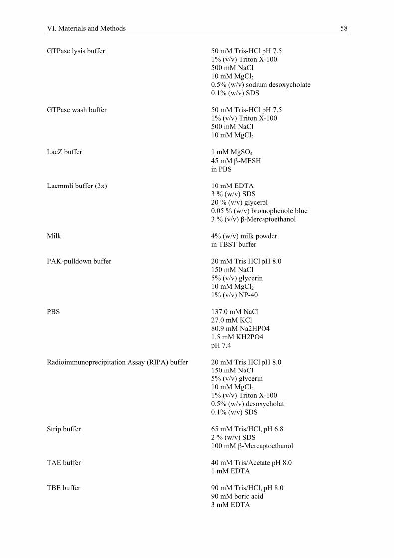
VI. Materials and Methods 58
GTPase lysis buffer 50 mM Tris-HCl pH 7.5 1% (v/v) Triton X-100 500 mM NaCl 10 mM MgCl2 0.5% (w/v) sodium desoxycholate 0.1% (w/v) SDS
GTPase wash buffer 50 mM Tris-HCl pH 7.5
1% (v/v) Triton X-100 500 mM NaCl 10 mM MgCl2
LacZ buffer 1 mM MgSO4
45 mM β-MESH in PBS
Laemmli buffer (3x) 10 mM EDTA
3 % (w/v) SDS 20 % (v/v) glycerol 0.05 % (w/v) bromophenole blue 3 % (v/v) β-Mercaptoethanol
Milk 4% (w/v) milk powder in TBST buffer PAK-pulldown buffer 20 mM Tris HCl pH 8.0
150 mM NaCl 5% (v/v) glycerin 10 mM MgCl2 1% (v/v) NP-40
PBS 137.0 mM NaCl
27.0 mM KCl 80.9 mM Na2HPO4 1.5 mM KH2PO4 pH 7.4
Radioimmunoprecipitation Assay (RIPA) buffer 20 mM Tris HCl pH 8.0
150 mM NaCl 5% (v/v) glycerin 10 mM MgCl2 1% (v/v) Triton X-100 0.5% (w/v) desoxycholat 0.1% (v/v) SDS
Strip buffer 65 mM Tris/HCl, pH 6.8
2 % (w/v) SDS 100 mM β-Mercaptoethanol
TAE buffer 40 mM Tris/Acetate pH 8.0
1 mM EDTA TBE buffer 90 mM Tris/HCl, pH 8.0
90 mM boric acid 3 mM EDTA
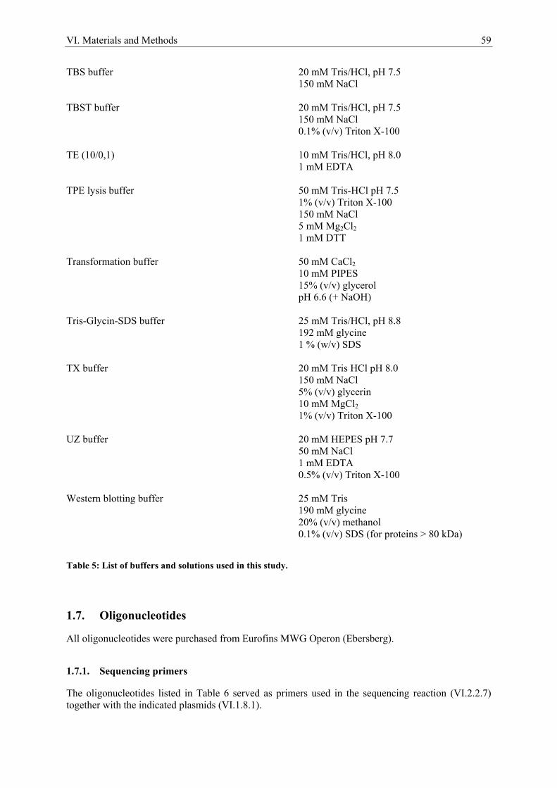
VI. Materials and Methods 59
TBS buffer 20 mM Tris/HCl, pH 7.5 150 mM NaCl TBST buffer 20 mM Tris/HCl, pH 7.5 150 mM NaCl 0.1% (v/v) Triton X-100 TE (10/0,1) 10 mM Tris/HCl, pH 8.0
1 mM EDTA TPE lysis buffer 50 mM Tris-HCl pH 7.5
1% (v/v) Triton X-100 150 mM NaCl 5 mM Mg2Cl2 1 mM DTT
Transformation buffer 50 mM CaCl2 10 mM PIPES 15% (v/v) glycerol pH 6.6 (+ NaOH) Tris-Glycin-SDS buffer 25 mM Tris/HCl, pH 8.8
192 mM glycine 1 % (w/v) SDS
TX buffer 20 mM Tris HCl pH 8.0
150 mM NaCl 5% (v/v) glycerin 10 mM MgCl2 1% (v/v) Triton X-100
UZ buffer 20 mM HEPES pH 7.7
50 mM NaCl 1 mM EDTA 0.5% (v/v) Triton X-100
Western blotting buffer 25 mM Tris
190 mM glycine 20% (v/v) methanol
0.1% (v/v) SDS (for proteins > 80 kDa)
Table 5: List of buffers and solutions used in this study.
1.7. Oligonucleotides
All oligonucleotides were purchased from Eurofins MWG Operon (Ebersberg).
1.7.1. Sequencing primers
The oligonucleotides listed in Table 6 served as primers used in the sequencing reaction (VI.2.2.7) together with the indicated plasmids (VI.1.8.1).
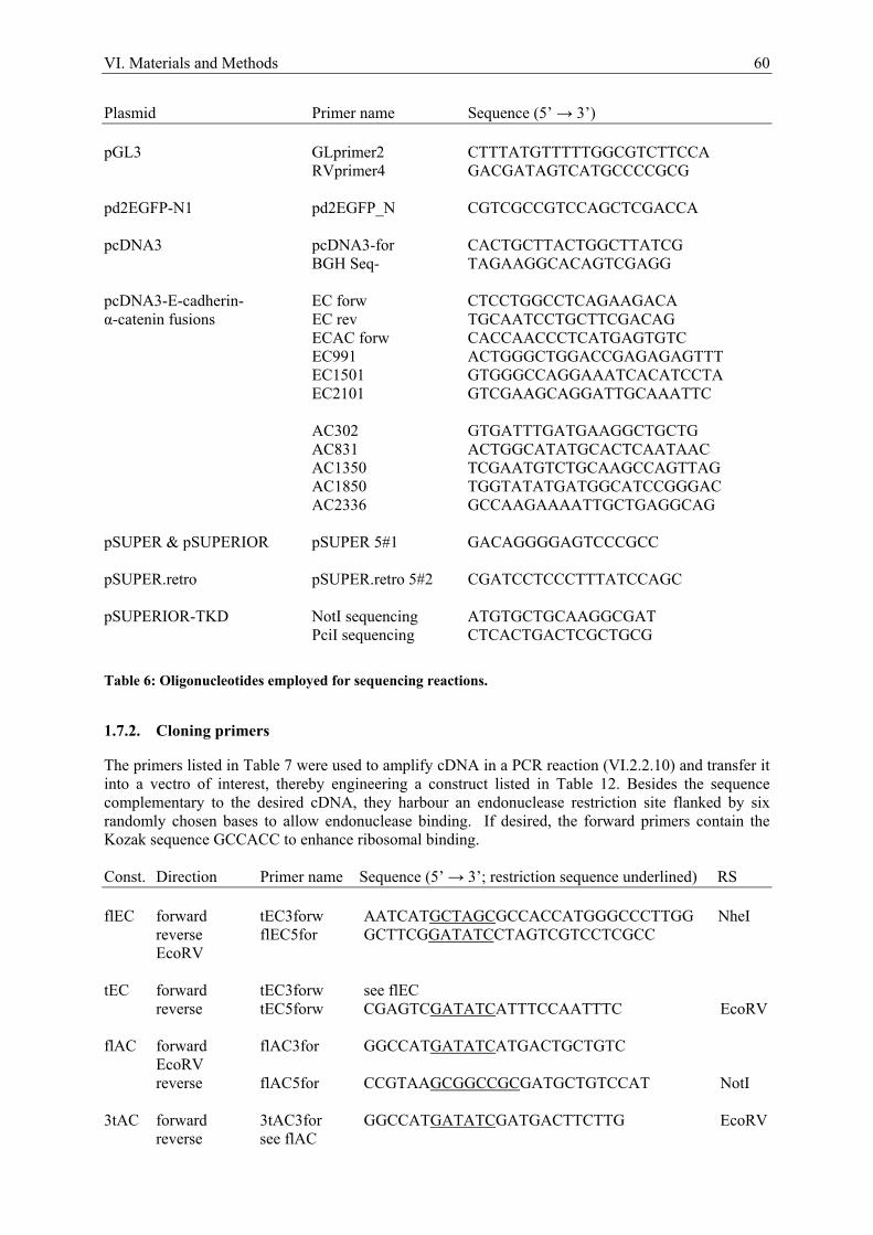
VI. Materials and Methods 60
Plasmid Primer name Sequence (5’ → 3’) pGL3 GLprimer2 CTTTATGTTTTTGGCGTCTTCCA
RVprimer4 GACGATAGTCATGCCCCGCG pd2EGFP-N1 pd2EGFP_N CGTCGCCGTCCAGCTCGACCA pcDNA3 pcDNA3-for CACTGCTTACTGGCTTATCG
BGH Seq- TAGAAGGCACAGTCGAGG pcDNA3-E-cadherin- EC forw CTCCTGGCCTCAGAAGACA α-catenin fusions EC rev TGCAATCCTGCTTCGACAG ECAC forw CACCAACCCTCATGAGTGTC EC991 ACTGGGCTGGACCGAGAGAGTTT EC1501 GTGGGCCAGGAAATCACATCCTA EC2101 GTCGAAGCAGGATTGCAAATTC AC302 GTGATTTGATGAAGGCTGCTG AC831 ACTGGCATATGCACTCAATAAC AC1350 TCGAATGTCTGCAAGCCAGTTAG AC1850 TGGTATATGATGGCATCCGGGAC AC2336 GCCAAGAAAATTGCTGAGGCAG pSUPER & pSUPERIOR pSUPER 5#1 GACAGGGGAGTCCCGCC pSUPER.retro pSUPER.retro 5#2 CGATCCTCCCTTTATCCAGC pSUPERIOR-TKD NotI sequencing ATGTGCTGCAAGGCGAT PciI sequencing CTCACTGACTCGCTGCG
Table 6: Oligonucleotides employed for sequencing reactions.
1.7.2. Cloning primers
The primers listed in Table 7 were used to amplify cDNA in a PCR reaction (VI.2.2.10) and transfer it into a vectro of interest, thereby engineering a construct listed in Table 12. Besides the sequence complementary to the desired cDNA, they harbour an endonuclease restriction site flanked by six randomly chosen bases to allow endonuclease binding. If desired, the forward primers contain the Kozak sequence GCCACC to enhance ribosomal binding. Const. Direction Primer name Sequence (5’ → 3’; restriction sequence underlined) RS flEC forward tEC3forw AATCATGCTAGCGCCACCATGGGCCCTTGG NheI reverse flEC5for GCTTCGGATATCCTAGTCGTCCTCGCC EcoRV tEC forward tEC3forw see flEC
reverse tEC5forw CGAGTCGATATCATTTCCAATTTC EcoRV flAC forward flAC3for GGCCATGATATCATGACTGCTGTC EcoRV reverse flAC5for CCGTAAGCGGCCGCGATGCTGTCCAT NotI 3tAC forward 3tAC3for GGCCATGATATCGATGACTTCTTG EcoRV reverse see flAC
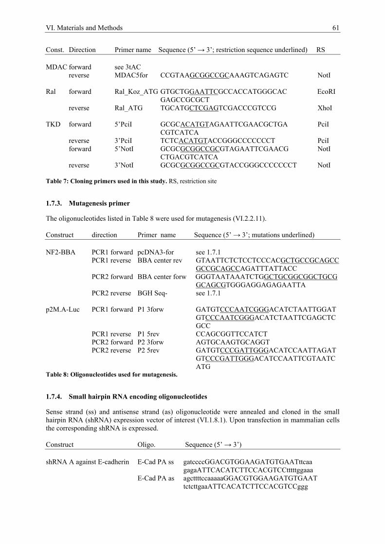
VI. Materials and Methods 61
Const. Direction Primer name Sequence (5’ → 3’; restriction sequence underlined) RS MDAC forward see 3tAC
reverse MDAC5for CCGTAAGCGGCCGCAAAGTCAGAGTC NotI Ral forward Ral_Koz_ATG GTGCTGGAATTCGCCACCATGGGCAC EcoRI
GAGCCGCGCT reverse Ral_ATG TGCATGCTCGAGTCGACCCGTCCG XhoI TKD forward 5’PciI GCGCACATGTAGAATTCGAACGCTGA PciI
CGTCATCA reverse 3’PciI TCTCACATGTACCGGGCCCCCCCT PciI forward 5’NotI GCGCGCGGCCGCGTAGAATTCGAACG NotI
CTGACGTCATCA reverse 3’NotI GCGCGCGGCCGCGTACCGGGCCCCCCCT NotI
Table 7: Cloning primers used in this study. RS, restriction site
1.7.3. Mutagenesis primer
The oligonucleotides listed in Table 8 were used for mutagenesis (VI.2.2.11). Construct direction Primer name Sequence (5’ → 3’; mutations underlined) NF2-BBA PCR1 forward pcDNA3-for see 1.7.1
PCR1 reverse BBA center rev GTAATTCTCTCCTCCCACGCTGCCGCAGCC GCCGCAGCCAGATTTATTACC
PCR2 forward BBA center forw GGGTAATAAATCTGGCTGCGGCGGCTGCG GCAGCGTGGGAGGAGAGAATTA
PCR2 reverse BGH Seq- see 1.7.1 p2M.A-Luc PCR1 forward P1 3forw GATGTCCCAATCGGGACATCTAATTGGAT
GTCCCAATCGGGACATCTAATTCGAGCTC GCC
PCR1 reverse P1 5rev CCAGCGGTTCCATCT PCR2 forward P2 3forw AGTGCAAGTGCAGGT PCR2 reverse P2 5rev GATGTCCCGATTGGGACATCCAATTAGAT
GTCCCGATTGGGACATCCAATTCGTAATC ATG
Table 8: Oligonucleotides used for mutagenesis.
1.7.4. Small hairpin RNA encoding oligonucleotides
Sense strand (ss) and antisense strand (as) oligonucleotide were annealed and cloned in the small hairpin RNA (shRNA) expression vector of interest (VI.1.8.1). Upon transfection in mammalian cells the corresponding shRNA is expressed. Construct Oligo. Sequence (5’ → 3’) shRNA A against E-cadherin E-Cad PA ss gatccccGGACGTGGAAGATGTGAATttcaa
gagaATTCACATCTTCCACGTCCtttttggaaa E-Cad PA as agcttttccaaaaaGGACGTGGAAGATGTGAAT
tctcttgaaATTCACATCTTCCACGTCCggg
VI. Materials and Methods 62
Construct Oligo. Sequence (5’ → 3’) shRNA B against E-cadherin E-Cad PB ss gatccccGTCTAACAGGGACAAAGAAttcaa
gagaTTCTTTGTCCCTGTTAGACtttttggaaa E-Cad PB as agcttttccaaaaaGTCTAACAGGGACAAAGAA
tctcttgaaTTCTTTGTCCCTGTTAGACggg shRNA against Cadherin-6 Cad6 ss gatccccGCGGCTACAGTCAGAATTAttcaa
gagaTAATTCTGACTGTAGCCGCtttttggaaa Cad6 as agcttttccaaaaaGCGGCTACAGTCAGAATTA
tctcttgaaTAATTCTGACTGTAGCCGCggg
Table 9: Oligonucleotides annealed to give shRNAs. Capital letters indicate the silencing sequence, small letters the stem. ss, sense strand; as, antisense strand
1.7.5. Quantitative real-time RT-PCR primer
The oligonucleotides listed in 1.7.5 were used as primers in quantitative real-time RT-PCR (VI.2.2.12) for expression analysis in mouse. Gene direction Sequence (5’ → 3’) hprt forward TCAGTCAACGGGGGACATAAA reverse GGGGCTGTACTGCTTAACCAG acta2 forward TGACGCTGAAGTATCCGATAGA reverse GTACGTCCAGAGGCATAGAGG vinculin forward GGCCGGACCAACATCAGTG reverse ATGTACCAGCCAGATTTGACG
Table 10: Oligonucleotides used as primers in quantitative real-time RT-PCR analysis.
1.8. Plasmids
1.8.1. Basic vectors
Plasmid Description Source pcDNA3 mammalian expression vector, Ampr, Invitrogen (Karlsruhe)
Neor, CMV promotor, BGH poly A, high copy number plasmid, F1+ origin
pcDNA6-TR tetracycline repressor protein expression Invitrogen (Karlsruhe)
vector, Ampr, Blastr, CMV promoter for high expression level
pEF plink mammalian expression vector, pUC12 G. Posern
backbone, EF1α enhancer/promoter, Ampr (Sotiropoulos et al., 1999) pEF-Flag plink like pEF plink, with 5’ Flag-tag G. Posern (Sotiropoulos et al., 1999)
VI. Materials and Methods 63
Plasmid Description Source pGL3-Basic reporter vector encoding for firefly Promega (Madison, USA)
(Photinus pyralis) luciferase, Ampr pRL-TK internal control reporter for pGL3, herpes Promega (Madison, USA)
simplex virus thymidine kinase promoter driving Renilla reniformis luciferase expression, Ampr
pSUPER shRNA expression vector, Ampr, Puror, OligoEngine (Seattle, USA)
H1 promoter pSUPER.retro shRNA expression vector for retroviral OligoEngine (Seattle, USA)
infection, Ampr, Puror, H1 promoter pSUPERIOR doxycycline inducible shRNA expression OligoEngine (Seattle, USA)
vector, Ampr, Puror, modified H1 promoter pd2EGFP-N1 cDNA of EGFP, destabilized by C-terminally BD Biosciences (Heidelberg) fused mouse ornithine decarboxylase PEST
domain
Table 11: List of basic vectors used in this study.
1.8.2. Modified vectors
The vectors engineered in this study were generated by cloning, mutagenesis or shRNA encoding oligonucletide insertion into the basic vectors (VI.1.8.1). Vectors are mammalian expression vectors, if not indicated otherwise. Plasmid Description Source/Reference p2M.A-Luc control SRF reporter derived from p3D.A-Luc, this study
in which two SRF binding sites were made insensitive (CCATATTAGG mutated to CCCAATCGGG) (Hill and Treisman, 1995) and the third one deleted
p3D.A-EGPF-N1 SRF promoter from p3D.A-Luc replacing the this study
CMV promoter in pd2EGFP-N1, thereby driving the expression of the destabilized EGFP
p3D.A-Luc three c-fos derived SRF binding sites in front G. Posern
of a Xenopus laevis type 5 actin TATA-Box (Geneste et al., 2002) in pGL3-basic driving the expression of the Firefly luciferase gene
pcDNA3-flEC cDNA of full length E-cadherin this study pcDNA3-tEC-flAC cDNA of truncated E-cadherin (amino acids this study 1 – 809) lacking the β-catenin binding site
fused to full length α-catenin
VI. Materials and Methods 64
Plasmid Description Source/Reference pcDNA3-tEC-3tAC cDNA of truncated E-cadherin (amino acids this study 1 – 809) lacking the β-catenin binding site
fused to the C-terminal part of α-catenin (amino acids 509 – 906)
pcDNA3-tEC-MDAC cDNA of truncated E-cadherin (amino acids this study 1 – 809) lacking the β-catenin binding site
fused to the adhesion modulation domain of α-catenin (amino acids 509 – 643)
pcDNA3-mDia1 cDNA of murine mDia1, constitutive active R. Fässler, MPI FH1FH2 truncation, myc-tagged Biochemistry (Martinsried) pcDNA3-NF2 cDNA of human wild type NF2 H. Morrison, FLI (Jena) pcDNA3-NF2-S518A cDNA of human NF2, constitutive active this study because not phosphorylatable, subcloned from pUHD10-3-NF2-S518A pcDNA3-NF2-S518D cDNA of human NF2, dominant negative by this study mimicking constitutive phosphorylation, subcloned from pUHD10-3-NF2-S518D pcDNA3-NF2-BBA cDNA of NF2 with amino acids 177 – 183 this study mutated to alanine (within the FERM domain),
dominant negative, derived by mutagenesis from pcDNA3-NF2
pcDNA3-RhoQ63L cDNA of RhoA, constitutive active by R. Fässler, MPI replacement of Q63 with L Biochemistry
(Martinsried) pcDNA3-ROCKΔ4 cDNA of murine ROCK, constitutive active R. Fässler, MPI
truncation, myc-tagged Biochemistry (Martinsried) pcDNA3.1- cDNA of human Ezrin, dominant negative, H. Morrison, FLI EzrinR579A by replacement of R579 with A myc-tagged, (Jena) Ampr, Neor pcDNA3.1- cDNA of human Ezrin, constitutive active by H. Morrison, FLI EzrinT567D mimicking constitutive phosphorylation by (Jena)
replacement of T567 with D myc-tagged, Ampr, Neor
pEF-Flag-G15S cDNA of β-actin, hyperpolymerizable by G. Posern
replacement of G15 with S, Flag-tagged (Posern et al., 2004) pEF-Flag-R62D cDNA of β-actin, nonpolymerizable by G. Posern replacement of R62 with D, Flag-tagged (Posern et al., 2002)
VI. Materials and Methods 65
Plasmid Description Source/Reference pEF-Flag-wt-actin cDNA of wild type β-actin, Flag-tagged G. Posern
(Posern et al., 2002)
pEF-MAL-HA (f.l.) cDNA of murine full length MAL, G. Posern HA-tagged (Miralles et al., 2003)
pEF-MAL-ΔN cDNA of murine MAL, constitutive active G. Posern
N-terminal truncation (amino acids 1 – 171), (Miralles et al., 2003) HA-tagged
pEF-MAL-ΔNΔB1 cDNA of murine MAL, dominant negative G. Posern
truncation (amino acids 1 – 171 and 316 – (Miralles et al., 2003) 341), HA-tagged
pEF-MAL-ΔNΔC cDNA of murine MAL, dominant negative G. Posern
truncation (amino acids 1 – 171 and 563 – (Miralles et al., 2003) 1021), HA-tagged
pEF-RacV12 cDNA of Rac1, constitutive active by G. Posern
replacement of G12 with V (Hill et al., 1995) pEGFP-N2- cDNA of human wild type E-cadherin Birgit Luber, TUM wtE-cadherin N-terminally fused to EGFP, Kanr, Neor (Munich) pGEX-2T-PAK CRIB cDNA of amino acids 68 – 151 of human A. Ullrich, MPI
PAK1, bacterial expression plasmid, Ampr Biochemistry (Martinsried)
pGEX-2T-Rhotekin cDNA of amino acids 7 - 90 of murine A. Ullrich, MPI RBD Rhotekin RBD, bacterial expression plasmid, Biochemistry
Ampr (Martinsried) pGEX-KG TAT-C3 cDNA of Clostridium botulinum C3 E. Sahai, CRUK
N-terminally fused to the HIV-TAT (London, GB) sequence, bacterial expression plasmid, Ampr
pMT2-HA- cDNA of human Epac, constitutive active G. Posern EpacΔcAMP truncation, HA-tagged, SV40 promoter, Ampr (Posern et al., 2000) pOTB7-CTNNA1 cDNA of human α-catenin 1, cloning RZPD (Berlin)
vector, Chloramphenicolr
pSUPER-EcadA shRNA A against E-cadherin this study pSUPER-EcadB shRNA B against E-cadherin this study pSUPER-Cad6 shRNA against Cadherin-6 this study pSUPER.retro-EcadA shRNA A against E-cadherin this study pSUPER.retro-EcadB shRNA B against E-cadherin this study
VI. Materials and Methods 66
Plasmid Description Source/Reference pSUPER.retro-Cad6 shRNA against Cadherin-6 this study pSUPERIOR-EcadA shRNA A against E-cadherin this study pSUPERIOR-EcadB shRNA B against E-cadherin this study pSUPERIOR-Cad6 shRNA against Cadherin-6 this study pSUPERIOR-TKD shRNA A against E-cadherin, insertion of this study
H1 promoter and shRNA B against E-cadherin via NotI, insertion of H1 promoter and shRNA against Cadherin-6 via PciI
pUHD10-3-NF2- cDNA of human NF2, constitutive active because H. Morrison, FLI S518A not phosphorylatable, in the reverse tetracycline- (Jena)
dependent transactivator cloning vector pUHD10-3, Ampr
pUHD10-3-NF2- cDNA of human NF2, dominant negative by H. Morrison, FLI S518D mimicking constitutive phosphorylation, compare (Jena)
pUHD10-3-NF2-S518A Table 12: List of modified vectors used in this study.
1.9. Peptides
The HAV peptide (H-IAKYILYSHAVSSNGNAVED-NH2) was synthesized and purified using standard Fmoc chemistry by the core facility of the Max Planck Institute of Biochemistry (Martinsried).
1.10. Antibodies
1.10.1. Primary antibodies
The antibodies listed in Table 13 were used as primary antibodies in immunoblot or immunofluorescence (IF) analysis. The IF-row specifies which fixation method was used for immunofluorescence staining with M for Methanol, P for 4% PFA, - indicated that neither M nor P worked and n/a for not tested. If a costaining with actin wanted to be obtained, cells had to be fixated with the PFA method as organic solvents erase the actin signal. Antibody Origin/Description Source IF Actin (clone C4) mouse, monoclonal MP Biomedicals (Aurora, USA) n/a AF-6 mouse, monoclonal, raised against BD Biosciences (Heidelberg) M
human AF-6 aa 1091-1223

VI. Materials and Methods 67
Antibody Origin/Description Source IF β-Catenin mouse, monoclonal, binds C- BD Biosciences (Heidelberg) M&P
terminal part of β-catenin
Cdc42 mouse, monoclonal, raised against BD Biosciences (Heidelberg) n/a human Cdc42 aa 1-191 E-cadherin rat, monoclonal, binds extracellular Merck (Darmstadt) M (ECCD-2) domain E-cadherin mouse, monoclonal, raised against BD Biosciences (Heidelberg) M&P (clone 36) human E-cadherin aa 735-883 E-cadherin mouse, monoclonal, blocking Sigma-Aldrich (Steinheim) M&P (DECMA-1) antibody,does not recognize human
E-cadherin Flag M2 mouse, monoclonal Sigma-Aldrich (Steinheim) P Flag M2 p.c. see Flag M2, peroxidase conjugate Sigma-Aldrich (Steinheim) n/a HA mouse, monoclonal, recognizes the BAbCO (Richmond, USA) P
influenza hemagglutinin epitope HA p.c. (3F10) see HA, peroxidase conjugated BAbCO (Richmond, USA) n/a MRTF-A rabbit, polyclonal, raised against Santa Cruz (Santa Cruz, USA) - (H-140) human MRTF-Aaa 761-900 MRTF rabbit, polyclonal, recognizes homemade (Posern Lab) P
MRTF-A and MRTF-B c-Myc (9E10) mouse, monoclonal Cancer Research UK P Rac1 mouse, monoclonal, raised against BD Biosciences (Heidelberg) n/a human Rac1 aa 1-192 RhoA (26C4) mouse, monoclonal, raised against Santa Cruz (Santa Cruz, USA) n/a human RhoA aa 120-150 ZO-1 rabbit, polyclonal, raist against the Zymed (San Francisco, USA) M N-terminal region of human ZO-1 Table 13: Primary antibodies used in immunoblot or immunofluorescence analysis. The IF-row specifies which fixation method was used for immunofluorescence staining: M, Methanol; P, 4% PFA; -, neither M nor P worked; n/a, not tested.
VI. Materials and Methods 68
1.10.2. Secondary antibodies
For immunoblot analysis the horseradish peroxidase (HRP) conjugated anti-IgG antibodies listed in Table 14 were used in this study. Antibody Dilution Source Polyclonal goat anti-mouse-HRP 1:7500 DakoCytomation (Glostrup, DK) Polyclonal swine anti-rabbit-HRP 1:3000 DakoCytomation (Glostrup, DK) Polyclonal goat anti-rat-HRP 1:5000 Jackson ImmonuResearch (Newmarket, GB) Table 14: Secondary horseradish peroxidase (HRP) conjugated anti-IgG antibodies used in immunoblot analysis.
For immunofluorescence analysis the fluorescent dye conjugated anti-IgG antibodies listed in Table 15 were used in this study. Antibody Dilution Source Alexa Fluor® 350 goat anti-rabbit 1:1000 Molecular Probes (Eugene, USA) Alexa Fluor® 488 goat anti-mouse 1:1000 Molecular Probes (Eugene, USA) Alexa Fluor® 546 goat anti-mouse 1:1000 Molecular Probes (Eugene, USA) Alexa Fluor® 546 goat anti-rabbit 1:1000 Molecular Probes (Eugene, USA) Polyclonal swine anti-rabbit-FITC 1:30 DakoCytomation (Glostrup, Denmark) Polyclonal swine anti-rabbit-TRITC 1:40 DakoCytomation (Glostrup, Denmark) Polyclonal goat anti-rat-Cy3 1:100 Jackson ImmonuResearch (Newmarket, GB)
Table 15: Secondary fluorescent dye conjugated anti-IgG antibody used for immunofluorescence analysis. FITC, Fluorescein Isothiocyanate; TRITC, Tetramethylrhodamine Isothiocyanate; Cy3, Carbocyanin 3.
1.11. Enzymes
Enzyme Source Antarctic Phosphatase NEB (Frankfurt/Main) DNA Polymerase I, Large (Klenow) Fragment NEB (Frankfurt/Main) DNAse I, RNAse free Roche (Mannheim) PhusionTM High-Fidelity DNA-Polymerase Finnzymes (Espoo, FI) Restriction Endonucleases NEB (Frankfurt/Main)
MBI (Fermentas, St. Leon-Rot) T4-DNA Ligase NEB (Frankfurt/Main) Trypsin (Gibco) Invitrogen (Karsruhe) Table 16: Enzymes used in this study.
VI. Materials and Methods 69
1.12. Cells
1.12.1. Bacterial strains
The following Escherichia coli (E.coli) strains were used for transformations to amplify plasmids (DH5α) and for protein expression (RosettaTM). Strain Genotype/Properties Origin DH5α F- φ80dlacZM15 (lacZYA-argF)U169 deoR recA1 endA1 Invitrogen (Karlsruhe) endA1 hsdR17(r k- m k+) phoA supE44 thi-1 gyrA96 relA1 λ- RosettaTM BL21 derivatives, express 6 additional tRNAs rarely occuring Novagen (DE3) in E. coli; F- ompT hsdSB (rB- mB-) gal dcm (DE3) pRARE (Madison, USA) CamR Table 17: Bacterial strains used in this study.
1.12.2. Mammalian cell lines
The following eukaryotic cell lines were used in this study. Cell line Description Origin/Reference AGS human gastric adenocarcinoma cells, E-cadherin ATCC (Manassas, USA) deficient CHO-K1 chinese hamster ovary cells, E-cadherin deficient ATCC (Manassas, USA) EpH4 normal mouse mammary epithelia cells H.Beug, IMP (Vienna, A) EpRas EpH4 cells stably expressing v-Ha-Ras. Cells H.Beug, IMP (Vienna, A)
undergo EMT upon TGF-β treatment (Oft et al., 1996).
MDA-MB metastatic human breast cancer cells, E-cadherin ATCC (Manassas, USA) 435S deficient MDCK canine kidney normal epithelial cells ATCC (Manassas, USA) NIH3T3 mouse embryonic fibroblasts R.Treisman, CRUK
(London, GB) PhoenixA amphotropic retrovirus producing cell line derived A.Ullrich, MPI Biochemistry
from the Human Embryonic Kidney (HEK) 293T (Martinsried) cell line
Table 18: Mammalian cell lines used in this study. ATCC, American Type Culture Collection.
VI. Materials and Methods 70
1.13. Scientific software
Software Source AIDA Raytest (Staubenhardt) KC4 BioTek (Winooski, USA) MetaVueTM Imaging Software Molecular Devices (Downingtown, USA) MetaMorphTM Imaging Software Molecular Devices (Downingtown, USA) SigmaPlot SPSS Science Software (Erkrath) SoftWoRx Applied Precision (Issaquah, USA) WinGlow EG and G Berthold (Schwerzenbach, CH)
Table 19: Scientific software used in this study.
2. Molecular biology methods
2.1. Microbiological techniques
2.1.1. Cultivation and maintenance of bacterial strains
E. coli strains were grown at 37 °C for 12 – 16 h either in LB medium or on LB agar plates supplemented with the for selection appropriate antibiotics. For short-term storage, E. coli cultures were kept on LB agar plates at 4 °C. For long-term storage 1 mL glycerol stocks consisting of overnight culture in 50 % (v/v) glycerol were stored in CryoTubeTM vials at -80 °C.
2.1.2. Generation of competent bacteria
To generate electrocompetent bacteria a well isolated bacterial colony grown on a fresh LB agar plate was inoculated in 5 mL LB medium and incubated shaking (180 rpm) at 37°C over night. The next day the overnight culture was diluted 1:100 in fresh LB medium and incubated as before until the cell density reached the optical density of 0.5 at 600 nm. At this point the bacteria were harvested by centrifugation at 1200 x g and 4°C for 10 minutes. Then the supernatant was decanted and the bacteria resuspended in sterile ice-cold 10% glycerol (same amount than supernatant decanted) and incubated for 20 minutes on ice. Afterwards the bacteria were pelleted as before and resuspended in 10% glycerol (1/10th of the amount of decanted supernatant) followed by another incubation on ice for 20 minutes. Thereafter the bacteria were pelleted again as before and resuspended in 10% glycerol (1/5th of the amount of decanted supernatant). The now electrocompetent bacteria were shock frozen in 40 µL aliquots in liquid nitrogen and stored at -80°C. Newly generated competent bacteria were tested for efficiency by transformation of 0.1 ng of a known plasmid, which should result in 108 – 109 colonies per µg DNA. To generate chemically competent bacteria a 2 mL over night culture of bacteria in LB medium was diluted 1/100 in fresh LB medium and incubated while shaking (180 rpm) at 37°C until the OD reached 0.2. The bacteria solution was incubated for 10 minutes on ice, centrifuged at 400 x g and 4°C for 10 minutes and the resulting pellet resuspendend in 25 mL cold transformation buffer. The suspension was inbcubated for 20 minutes on ice and centrifuged as before, the pellet this time resuspended in 2.5 mL cold transformation buffer. The now chemically competent bacteria were snap-frozen in 50 µL aliquots and stored at -80°C. Newly generated competent bacteria were tested for efficiency by transformation of 0.1 ng of a known plasmid, which should result in 107 – 108 colonies per µg DNA.
VI. Materials and Methods 71
2.1.3. Transformation of competent bacteria
Competent bacteria were either transformed by electroporation or the “heat shock” method. For each electroporation 50 µL of electrocompetent bacteria were thawed on ice and supplemented with 1 – 2 µL ligation reaction. The mixture was then transferred into an ice-cold 0.2 cm electroporation cuvette and electoporated with the Genepulser XCellTM at 2.5 kV, 25 µF and 200 Ohm. Immediately afterwards 500 µL of SOC medium were added, followed by an incubated on a rotating wheel at 37°C for 1 h. Thereafter 200 µL of the mixture were plated on a LB agar plate with the appropriate antibiotics. For each heat shock induced transformation 50 µL of chemically competent bacteria were thawed on ice and incubated with the whole ligation reaction for 10 minutes on ice. For the following heat shock, the cells were incubated at 42°C for 90 seconds pursued by another incubation on ice for 5 minutes. Then 200 µL LB medium were added and the cells were incubated on a rotating wheel at 37°C for 1 h. Afterwards the mixture was plated on a LB agar plate with the appropriate antibiotics.
2.1.4. TAT-C3 purification
TAT-C3 was purified according to the large scale protein production described by E. Sahai and colleagues (Sahai and Olson, 2006) with a modified, calcium-free thrombin cleavage buffer (1 mM MgCl2, 1 mM DTT in TBS).
2.2. DNA modification
2.2.1. Plasmid preparation
Plasmids were prepared using the QIAGEN Plasmid Mini Kit for small amount of DNA and the QIAGEN Plasmid Maxi Kit for larger amounts of DNA according to manufacturer’s instructions.
2.2.2. Restriction digestion of DNA
For restriction digestion approximately 0.5 – 2 µg of DNA were digested with the endonuclease of interest at the concentration/temperature and with the buffer recommended by the manufacturer. For analytical digestions, the reaction volume was 20 µL incubated for 1 h, for preparative digestions 50 µL incubated for 3 h – over night.
2.2.3. Blunt end creation
DNA polymerase I, large (Klenow) fragment fills-in 5’ overhangs and removes 3’ overhangs to create blunt ends. The DNA fragment or plasmid of choice was incubated with DNA polymerase I, large (Klenow) fragment according to manufacturer’s instructions. Aferwards, the DNA was purified by agarose gel electrophoresis or directly via the QIAquick PCR Purification Kit or QIAquick MinElute PCR Purification Kit.
2.2.4. Dephosphorylation of DNA 5’-termini
To prevent self-ligation of vector termini generated by restriction digestion, the 5’- termini of vectors were dephosphorylated with Antarctic Phosphatase according to manufacturer’s protocols. The reaction was stopped by either heat inactivation at 65°C for 5 minutes or direct purification of the DNA via the QIAquick PCR Purification Kit or QIAquick MinElute PCR Purification Kit.
VI. Materials and Methods 72
2.2.5. Ligation of DNA fragments
Before ligation, the fragments were purified by agarose gel electrophoresis followed by gel extraction with the QIAquick Gel Extraction Kit. Dephosphorylated and QIAquick PCR Purification Kit purified vector fragments were directly used for ligation. For ligation, 20 - 200 ng of vector and the 3 – 6 fold molar amount of insert were incubated together with 400 U of T4 DNA Ligase and T4 Ligase buffer in a total of 20 µL at 14°C over night or at RT for 1 – 2 h. As control, the ligation was performed as described above with the difference that the insert fragment was missing. Immediately after incubation the cells were transformed into competent E. coli DH5α.
2.2.6. Generation of shRNA expressing plasmids
For annealing 3 µg of sense and the corresponding antisense shRNA oligonucleotide strand were mixed in a total of 50 µL Annealing buffer. The mixture was heated to 95°C for 10 minutes and then cooled down with 0.01°C per second to 4°C in a Biometra T3000 Thermocycler. The annealed shRNAs habouring a BglII and HindIII overhang were purified via agarose gel electrophoreses and extracted. The insertion into the desired shRNA expression vector was done according to manufacturer’s instructions.
2.2.7. Sequencing
DNA samples were sequenced on an ABI 3730 sequencer by the core facility of the Max Planck Institute of Biochemistry (Martinsried).
2.2.8. Agarose gel electrophoresis
DNA fragments and plasmids were separated by agarose gel electrophoresis (Sambrook, 1989). For gel preparation 0.7 – 2 % (w/v) agarose in TAE or TBE buffer was boiled and afterwards supplemented with 0.01% (v/v) ethidium bromide. After gel polymerization, DNA samples were mixed with the corresponding amount of DNA loading buffer and loaded on the agarose gel together with the SmartLadder DNA marker for size determination. Electrophoresis was performed in TAE or TBE buffer, according to gel preparation, at 70 – 120 V for 20 – 60 minutes in a agarose gel electrophoresis sytem Horizontal-Elpho. After electrophoresis, the DNA was visualized at 302 nm and photographs were taken with the IDA gel documentation system. If the separated DNA fragments or plasmids were used for further experiments, they were visualized with a Dark Reader® at 460 nm to avoid the introduction of mutations.
2.2.9. Isolation of DNA fragments and plasmids from agarose gels
The DNA fragments or plasmids of interest were cut out of the gel with a sterile blade and purified using the QIAquick Gel Extraction Kit according to manufacturer’s instructions.
2.2.10. DNA amplification by Polymerase Chain Reaction
DNA fragments of interest were exponentially amplified via Polymerase Chain Reaction (PCR) by a repeated cycle of denaturation, primer annealing and polymerase-driven primer elongation (Mullis and Faloona, 1987). The PCR reaction was carried out employing the PhusionTM High-Fidelity DNA-Polymerase according to manufacturer’s instructions with the primers denoted in Table 7 for cloning and Table 8 for mutagenesis in a Biometra T3000 Thermocycler. PCR products were analysed via agarose gel electrophoreses and positive products were extracted from the gel (VI.2.2.8 and VI.2.2.9).
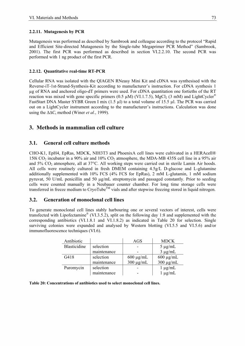
VI. Materials and Methods 73
2.2.11. Mutagenesis by PCR
Mutagenesis was performed as described by Sambrook and colleague according to the protocol “Rapid and Efficient Site-directed Mutagenesis by the Single-tube Megaprimer PCR Method” (Sambrook, 2001). The first PCR was performed as described in section VI.2.2.10. The second PCR was performed with 1 ng product of the first PCR.
2.2.12. Quantitative real-time RT-PCR
Cellular RNA was isolated with the QIAGEN RNeasy Mini Kit and cDNA was synthesised with the Reverse-iT-1st-Strand-Synthesis-Kit according to manufacturer’s instruction. For cDNA synthesis 1 µg of RNA and anchored oligo-dT primers were used. For cDNA quantitation one fortieths of the RT reaction was mixed with gene specific primers (0.5 µM) (VI.1.7.5), MgCl2 (3 mM) and LightCycler® FastStart DNA Master SYBR Green I mix (1.5 µl) to a total volume of 15.5 µl. The PCR was carried out on a LightCycler instrument according to the manufacturer’s instructions. Calculation was done using the ΔΔCt method (Winer et al., 1999).
3. Methods in mammalian cell culture
3.1. General cell culture methods
CHO-K1, EpH4, EpRas, MDCK, NIH3T3 and PhoenixA cell lines were cultivated in a HERAcell® 150i CO2 incubator in a 90% air and 10% CO2 atmosphere, the MDA-MB 435S cell line in a 95% air and 5% CO2 atmosphere, all at 37°C. All working steps were carried out in sterile Lamin Air hoods. All cells were routinely cultured in fresh DMEM containing 4.5g/L D-glucose and L-glutamine additionally supplemented with 10% FCS (4% FCS for EpRas), 2 mM L-glutamin, 1 mM sodium pyruvat, 50 U/mL penicillin and 50 µg/mL streptomycin and passaged constantly. Prior to seeding cells were counted manually in a Neubauer counter chamber. For long time storage cells were transferred in freeze medium to CryoTubeTM vials and after stepwise freezing stored in liquid nitrogen.
3.2. Generation of monoclonal cell lines
To generate monoclonal cell lines stably harbouring one or several vectors of interest, cells were transfected with Lipofectamine® (VI.3.5.2), split on the following day 1:8 and supplemented with the corresponding antibiotics (VI.1.8.1 and VI.1.8.2) as indicated in Table 20 for selection. Single surviving colonies were expanded and analysed by Western blotting (VI.5.5 and VI.5.6) and/or immunofluorescence techniques (VI.6).
Antibiotic AGS MDCK Blasticidine selection - 5 µg/mL maintenance - 3 µg/mL G418 selection 600 µg/mL 600 µg/mL maintenance 300 µg/mL 300 µg/mL Puromycin selection - 1 µg/mL maintenance - 1 µg/mL
Table 20: Concentrations of antibiotics used to select monoclonal cell lines.
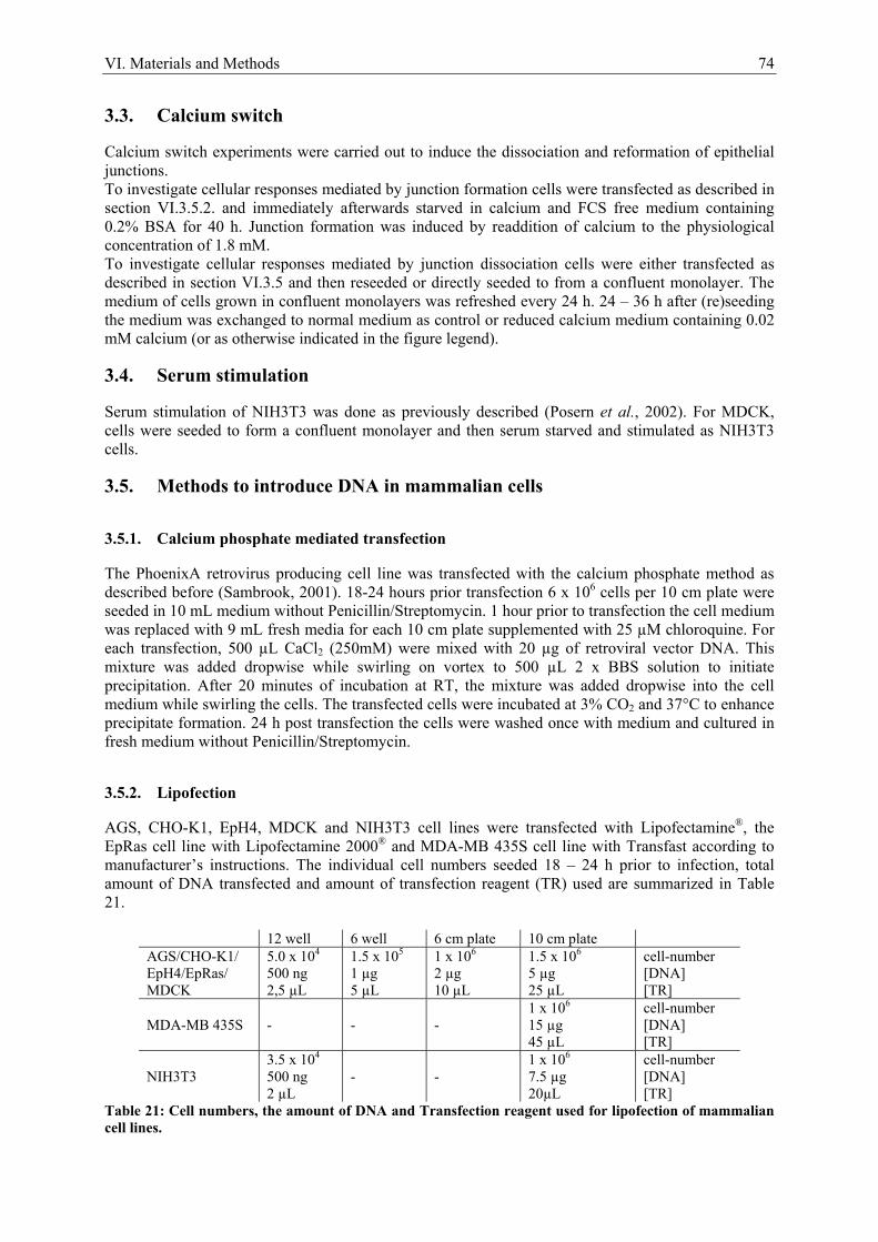
VI. Materials and Methods 74
3.3. Calcium switch
Calcium switch experiments were carried out to induce the dissociation and reformation of epithelial junctions. To investigate cellular responses mediated by junction formation cells were transfected as described in section VI.3.5.2. and immediately afterwards starved in calcium and FCS free medium containing 0.2% BSA for 40 h. Junction formation was induced by readdition of calcium to the physiological concentration of 1.8 mM. To investigate cellular responses mediated by junction dissociation cells were either transfected as described in section VI.3.5 and then reseeded or directly seeded to from a confluent monolayer. The medium of cells grown in confluent monolayers was refreshed every 24 h. 24 – 36 h after (re)seeding the medium was exchanged to normal medium as control or reduced calcium medium containing 0.02 mM calcium (or as otherwise indicated in the figure legend).
3.4. Serum stimulation
Serum stimulation of NIH3T3 was done as previously described (Posern et al., 2002). For MDCK, cells were seeded to form a confluent monolayer and then serum starved and stimulated as NIH3T3 cells.
3.5. Methods to introduce DNA in mammalian cells
3.5.1. Calcium phosphate mediated transfection
The PhoenixA retrovirus producing cell line was transfected with the calcium phosphate method as described before (Sambrook, 2001). 18-24 hours prior transfection 6 x 10⁶ cells per 10 cm plate were seeded in 10 mL medium without Penicillin/Streptomycin. 1 hour prior to transfection the cell medium was replaced with 9 mL fresh media for each 10 cm plate supplemented with 25 µM chloroquine. For each transfection, 500 µL CaCl2 (250mM) were mixed with 20 µg of retroviral vector DNA. This mixture was added dropwise while swirling on vortex to 500 µL 2 x BBS solution to initiate precipitation. After 20 minutes of incubation at RT, the mixture was added dropwise into the cell medium while swirling the cells. The transfected cells were incubated at 3% CO2 and 37°C to enhance precipitate formation. 24 h post transfection the cells were washed once with medium and cultured in fresh medium without Penicillin/Streptomycin.
3.5.2. Lipofection
AGS, CHO-K1, EpH4, MDCK and NIH3T3 cell lines were transfected with Lipofectamine®, the EpRas cell line with Lipofectamine 2000® and MDA-MB 435S cell line with Transfast according to manufacturer’s instructions. The individual cell numbers seeded 18 – 24 h prior to infection, total amount of DNA transfected and amount of transfection reagent (TR) used are summarized in Table 21.
12 well 6 well 6 cm plate 10 cm plate AGS/CHO-K1/ EpH4/EpRas/ MDCK
5.0 x 10⁴ 500 ng 2,5 µL
1.5 x 10⁵ 1 µg 5 µL
1 x 10⁶ 2 µg 10 µL
1.5 x 10⁶ 5 µg 25 µL
cell-number [DNA] [TR]
MDA-MB 435S
-
-
-
1 x 10⁶ 15 µg 45 µL
cell-number [DNA] [TR]
NIH3T3
3.5 x 10⁴ 500 ng 2 µL
-
-
1 x 10⁶ 7.5 µg 20µL
cell-number [DNA] [TR]
Table 21: Cell numbers, the amount of DNA and Transfection reagent used for lipofection of mammalian cell lines.
VI. Materials and Methods 75
3.5.3. Electoporation
For each electroporation 5 x 10⁶ MDCK cells in 500 µL normal medium were transferred together with 20 µg DNA in a 0.4 cm wide electroporation cuvette. Cells were electroporated in the square wave modus 3 times at 250 V for 20 ms, with a 100 ms break between the pulses, in a Genepulser XCellTM. Afterwards, 1 mL of fresh prewarmed medium was added immediately, before the cells were reseeded in a 6 cm plate in a total of 3 mL medium.
3.5.4. Nucleofection
MDCK cells were nucleofected in an Amaxa Nucleofector device according to manufacturer’s instructions. Briefly, for each nucleofection 2.5 x 10⁶ MDCK cells were trypsinized, centrifuged at 90 x g for 10 minutes, resuspended in 100 µL Nucleofector solution L and mixed with a total of 4.1 µg DNA. Then the cells were transferred in the nucleofector cuvette and nucleofected with program A-024. Immediately afterward 500 µL prewarmed medium was added and the cells were reseeded.
3.5.5. Retroviral infection
One 10 cm plate of PhoenixA cells per well of target cells in a 6 cm plate was transfected with the calcium phosphate method. 24h post transfection the cells were washed once with medium and then cultured in 6.5 mL DMEM without Penicillin/Streptomycin at 7% CO2 and 32°C to enhance virus stability. 48 h post Phoenix cell transfection the target cells, either 1 x 10⁵ NIH3T3 cells or 1.5 x 10⁶ MDCK cells per 6 cm plate seeded 24h prior to infection in normal medium, were infected. Therefore, the supernatant from the 10 cm plate of transfected Phoenix cells was filtered through a 0.45 µm PVDF-filter to remove cells and concentrated to 1 mL by centrifugation at 2000 x g in a PBS-equilibrated vivaspin 20 column. The 10 cm plate of Phoenix cells was supplemented with 6.5 mL of fresh medium and further incubated at 7% CO2 and 32°C for a second round of infection. During the centrifugation the medium from the target cells was removed and 1 mL of medium supplemented with 8 µg/mL polybrene was added. When the supernatant was concentrated to 1 mL, it was after membrane rinsing removed from the vivaspin column and added to the target cells, which were after gentle swirling incubated at 7% CO2 and 32°C. 9 h later the target cells were infected a second time identically, except that the old medium from the target cells was just removed half, and after readdition of 4 µg/mL polybrene the 1 ml concentrate was added. 24 h post infection the medium of the target cells was replaced by normal medium. 48 h post infection the infection efficiency was monitored by EGFP expression of a control infection.
4. Gene reporter assays
4.1. Luciferase reporter assay
For luciferase reporter assays AGS, CHO-K1, EpH4, MDCK, MDA-MB 435S and NIH3T3 cells were transfected as described in section VI.3.5 with p3D.A-Luc encoding for the Firefly luciferase gene, pRL-TK encoding for the Renilla luciferase gene and additional plasmids of interest as indicated in the figure legends. The luciferase reporter assay was carried out with reagents from the Dual-GloTM Luciferase Assay Kit. After the treatment of interest the cells were washed twice with ice-cold PBS and lysed by incubation in 50 µL Passive Lysis Buffer (PLB) per 12 well or transwell filter on ice for 10 minutes. Thereafter, the cells were scraped of the plate, the lysate collected in an Eppendorf tube and centrifuged at 20000 x g and 4°C for 10 minutes. 20 µL of the supernatant were transferred into a well in a white 96 well microtiter plate and mixed with 45 µL LARII solution. The emitted Firefly luminescence was detected in a Microplate Luminometer LB 96V. After detection 45 µL of Stop & Glow reagent supplemented with Stop & Glow substrate were added to the well to stop the Firefly reaction and start Renilla luminescence emission, which was detected as above. The protein content of each lysate was
VI. Materials and Methods 76
determined by Bradford assay (VI.5.2.1). Firefly luciferase activity was normalized to either protein content or Renilla luciferase activity, as indicated in the figure legend. Figures show mean ± SEM of at least three independent experiments.
4.2. β-Galactosidase reporter assay
20 µL of LacZ substrate (10 mg/ml Chlorophenolred-β-D-galactopyranosid in PBS) were mixed with 160 µL of LacZ buffer and added to 20 µL lysate in a transparent mikrotiter plate and incubated at 37°C for 1 – 4 h. Afterwards, absorption was detected at 570 nm (630 nm reference) in the BioTek Elisaeader.
5. Protein analytical methods
5.1. Lysis of cells with Triton X-100
Prior to lysis the cells were washed with twice cold PBS and then lysed for 3 minutes on ice in RIPA-buffer containing protease (CompleteTM Protease Inhibitor Cocktail Tablets) and phosphatase (2 mM sodium orthovanadate, 10 mM sodium fluoride) inhibitors. After scraping the lysates were precleared by centrifugation at 20000 x g and 4°C for 10 min.
5.2. Determination of protein concentration
5.2.1. Bradford protein assay
Protein concentration measurement using the Bradford method (Bradford, 1976) is based on the absorbance shift of Coomassie Brilliant Blue G-250 from red to blue in proportion with protein binding (absorption maximum: 595 nm). The protein lysate of interest was mixed with 100 µL of Bradford solution and incubated for 5 minutes at room temperature. Then the absorption was measured at 595 nm in a BioTek Elisareader. The protein concentration was determined by comparison with a standard curve of BSA (1 - 10 µg).
5.2.2. BCA protein assay
Protein concentration measurement using the BCA protein (Smith et al., 1985) assay is based on the reduction of copper ions from Cu2+ to Cu+ by peptides in a temperature-dependent reaction. Cu+ gets chelated by bicinchoninic acid (BCA), which causes a color change from light green to purple in proportion to the protein concentration (absorption maximum: 562 nm). The BCA assay was carried out with the Micro BCA Protein Assay Kit according to manufacturer’s instructions. The absorption was measured at 570 nm in a BioTek Elisareader. The protein concentration was determined by comparison with a standard curve of BSA.
5.3. SDS-Polyacrylamid Gel Electrophoresis
SDS-Polyacrylamid Gel Electrophoresis (SDS-PAGE) was mainly carried out as described before (Laemmli, 1970). Samples were run on two-layered gels consisting of stacking and separating gel in a polyacrylamide gel electrophoresis system Vertical Elpho “B“. The stacking gel contained 5% acrylamide solution in 127 mM Tris-HCl pH 6.8, 0.1% (w/v) SDS, 4.5% (v/v) glycerol and 0.1% (w/v) APS. The separating gel contained 7 – 15 % acrylamid solution (according to protein size) in 377 mM Tris-HCl pH 8.8, 0.1% (w/v) SDS and 0.1% (w/v) APS. To start the polymerization reaction 0.1 % (v/v) TEMED was added. Before SDS-PAGE all samples were mixed with the appropriate amount of Laemmli buffer and boiled at 95 °C for 2 minutes to remove all secondary, tertiary and quaternary structures. The Precision Plus ProteinTM Dual Color Standard was used as molecular weight
VI. Materials and Methods 77
standard. SDS-PAGE was performed at 80 – 140 V. After electrophoresis gels were either Coomassie stained (VI.5.4) or transferred to a PVDF membrane for western blotting (VI.5.5).
5.4. Coomassie staining of SDS-PAGE gels
For Coomassie staining gels were washed for 10 minutes in Coomassie R-250 solution followed by removal of unbound colour by incubation in Coomassie destaining solution for several hours in. For drying, gels were inserted air bubble-free between two wet layers of cellophane and left for 1- 2 h in a GelAir dryer.
5.5. Western blotting
For Western blotting SDS-PAGE was performed (VI.5.3) and afterwards proteins were transferred to a PVDF membrane. Therefore, the polyacrylamide gel was blotted air-bubble free in Tris-Glycin-SDS buffer in the Mini Trans-Blot Cell at 100 V for 1 h in the following order: anode, pad, 2 layers of Whatman paper, PVDF membrane, polyacrylamide gel, 2 layers of Whatman paper, pad and cathode.
5.6. Immunoblot detection
After blotting the proteins were transferred to the PVDF membrane and providing access for reaction with immunodetection reagents. To block unspecific binding sites the membrane was incubated in milk for at least 1 h at room temperature or 4°C over night. The membrane was then incubated with the first antibody in milk (VI.1.10.1) for at least 1 h at room temperature or 4°C over night, afterwards washed three times in TBST buffer for 5 minutes and incubated with the secondary horseradish peroxidase conjugated anti-IgG antibody diluted in milk (VI.1.10.2) for 1 – 2 h. Thereafter the membrane was washed again three times in TBST buffer for 5 minutes and luminescent bands were detected on Hyperfilm MP using the Western LightningTM Chemoluminescence Reagent Plus or ECL Plus Reagent according to manufacturer’s instructions.
5.7. Stripping
To reincubate with a different set of antibodies the membrane was stripped to remove all bound antibodies. Therefore, the membrane was washed two times for 10 minutes in TBST buffer and then incubated at 50°C in a closed container in stripping buffer. Afterwards the membrane was washed three more times in TBST buffer for 5 minutes before it was ready to be incubated with another set of antibodies.
5.8. Densitometric analysis of western blots
Densiomatric analysis of western blots was done with AIDA software and normalized to total lysates or ratios.
5.9. Small G-protein pull-down assays
Preparation of GST-Rhotekin-RBD and GST-PAK-CRIB fusion protein was done using E. coli RosettaTM (DE). An overnight culture in LB medium supplemented with the corresponding antibiotics and 1% glucose was diluted 1/200 in fresh LB medium and incubated at 37°C while shaking (180 rpm) until the OD reached 0.6. Protein expression was induced by addition of 1 mM IPTG and cells were incubated at 30° for 90 min followed centrifugation at 1200 x g and 4°C for 30 minutes. The pelleted bacteria were lysed in ice-cold TPE lysis buffer containing protease inhibitors (1/20th of the original volume), tranferred to prechilled 50 mL Falcon tubes and sonicated on ice 6 times for 10 seconds at the strongest level in a sonicator Sonoplus HD70. Afterwards the lysate was centrifuged at 25000 x g and 4°C or 30 minutes and the supernatant snap-frozen in liquid nitrogen and stored at -80°C. Optimized amounts of lysate were used for coupling to Glutathione Sepharose beads directly prior to GTPase pull-down assays. The pull-down assays were carried out as described before (Sander et al.,
VI. Materials and Methods 78
1998; Ren and Schwartz, 2000). 1 x 10⁷ MDCK cells were seeded in 10 cm dishes to form a confluent monolayer and cultured for 36 h. After medium exchange cells were lysed in 800 µL GTPase lysis buffer containing protease inhibitors and the lysates cleared by centrifugation. 50 µL of the supernatant were mixed with an appropriate amount of Laemmli buffer and kept as total lysate, the remaining 750 µL of supernatant were incubated with immobilized GST-Rhotekin-RBD or GST-PAK-CRIB on the rotating wheel at 4°C for 35 minutes. Thereafter the beads were washed three times in GTPase wash buffer, carefully aspirated and finally mixed with 20 µL Laemmli buffer. As controls, cell lysates were incubated either with uncoupled beads alone or preincubated with GTPγS (1 mM for 10 minutes) prior to precipitation. Bound proteins were detected by Western blotting.
5.10. Immunoprecipitation
For each immunoprecipitation 1 x 10⁷ MDCK cells were electroporated with 10 µg of each Flag-actin and HA-MAL and seeded in a 10 cm ∅ plate to form a confluent monolayer. A medium exchange to fresh medium was performed 12 h later and another 24 h later the medium was replaced with either calcium reduced or normal medium as control. After the times desired the cells were lysed in 500 µL RIPA buffer containing protease and phosphatase inhibitors and the lysate was cleared by centrifugation at maximum speed at 4°C for 10 minutes. The resulting supernatant was combined with 20 µL TX-buffer washed Flag M2 agarose beads in 500 µL TX buffer and incubated on a rotating wheel at 4°C for 2 h. Afterwards the beads were pelleted at 3000 x g and 4°C for 5 minutes. To prepare the total lysate 50 µL of the supernatant were supplemented with 3x Laemmli buffer and frozen at -20°C. The rest of the supernatant was frozen at -80°C or discarded. The beads were washed three times in ice-cold TX-buffer and after the final aspiration resuspended in 20 µL 3 x Laemmli buffer. Afterwards they were, together with the total lysate, incubated at 95°C for 2 minutes, cooled on ice, spun down at 3000 x g for 5 minutes and analyzed by SDS-PAGE followed by western blotting.
5.11. Determination of the G- to F-actin ratio by ultracentrifugation
MDCK cell were seeded in a 6 cm plate to form a confluent monolayer. 24 h later the medium was exchanged to normal medium as control or reduced calcium medium. After the desired time cells were lysed in 750 µL UZ buffer on ice for 10 minutes. Thereafter the cells were scraped off, transferred into suitable polycarbonate tubes and centrifuged at 100,000 x g in a Beckman TL-100 Ultracentrifuge at 4°C for 1 h. 75 µL of the supernatant were transferred into Laemmli buffer giving the G-actin fraction. The rest of the supernatant was removed and the pellet resuspended in 750 µL UZ buffer, mixed with Laemmli buffer, sonicated for 10 seconds at maximum level in the sonicator Sonoplus HD70, giving the F-actin fraction. The G- to F-actin ratio was determined by Western blotting.
6. Immonufluorescence techniques
6.1. Immonofluorescence staining
Cells were seeded on uncoated glass coverslips (∅ = 12 mm) and the assay of interest was performed. Prior to fixation, the cells were washed twice in PBS on ice. This step was omitted when cells were stimulated and the wash would have influenced the result, e.g by washing out a drug or reversing a short lasting effect. Then, cells were fixed in 4% PFA for 10 – 15 minutes at RT and permeabilized for 10 minutes in 0.2% Triton X-100 in PBS. Alternatively, the cells were fixed and permeabilized by 2 minutes incubation in ice-cold methanol at -20°C followed by 3 rehydration washes in PBS. Unspecific epitopes were blocked by incubation for 30 minutes – 2 h in blocking solution. Then cells were incubated with the first antibody (VI.1.10.1) diluted as indicated in the Results section or according to manufacturer’s instructions in blocking solution for 1 – 2 h. Afterwards cells were washed three times in PBS and incubated with the second antibody diluted as indicated in section VI.1.10.2 in blocking solution for 30 minutes – 1 h. If desired, labelled phalloidin or/and DAPI (2 µg/mL) or Hoechst 33258 (1 µg/mL) was/were incubated together with the second antibody. Thereafter cells were again washed 3 times in PBS followed by 1 short was in deionised water to
VI. Materials and Methods 79
reduce salt effects and mounted with Mowiol (prepared according to Harlow and colleague (Harlow, 1999)) on a microscopic slide. Mowiol was allowed to harden prior to microscopic analysis for at least 1 hour.
6.2. Microscopy
6.2.1. Conventional immunofluorescence microscopy
Standard micrographs were taken using a Zeiss Axioplan 2 with MetaVueTM software. To allow direct comparability of different micrographs, all specimens within one experimental setup were stained in parallel and pictures were taken with the same exposure time without further computational calculations.
6.2.2. Confocal microscopy
Confocal microscopy was carried out on a Leica TCS SP2 AOBS confocal laser scanning microscope with SoftWoRx software. All specimens within one experimental setup were stained in parallel and pictures were taken with identical settings.
6.2.3. Life imaging
Life imaging was performed using a Zeiss Axiovert 200M microscope with MetaMorphTM software. The induction profile showing the time course of EGFP expression was calculated by subtracting the arbitrary fluorescence intensity at each time point in the mock experiment from the equivalent arbitrary fluorescence intensity upon calcium withdrawal.
VII. References 80
VII. References
Abe, K., Rossman, K.L., Liu, B., Ritola, K.D., Chiang, D., Campbell, S.L., Burridge, K., and Der, C.J. (2000). Vav2 is an activator of Cdc42, Rac1, and RhoA. J Biol Chem 275, 10141-10149. Abe, K., and Takeichi, M. (2008). EPLIN mediates linkage of the cadherin catenin complex to F-actin and stabilizes the circumferential actin belt. Proc Natl Acad Sci U S A 105, 13-19. Adams, C.L., Chen, Y.T., Smith, S.J., and Nelson, W.J. (1998). Mechanisms of epithelial cell-cell adhesion and cell compaction revealed by high-resolution tracking of E-cadherin-green fluorescent protein. J Cell Biol 142, 1105-1119. Aguda, A.H., Burtnick, L.D., and Robinson, R.C. (2005). The state of the filament. EMBO Rep 6, 220-226. Amano, M., Ito, M., Kimura, K., Fukata, Y., Chihara, K., Nakano, T., Matsuura, Y., and Kaibuchi, K. (1996). Phosphorylation and activation of myosin by Rho-associated kinase (Rho-kinase). J Biol Chem 271, 20246-20249. Anderson, J.M., Van Itallie, C.M., and Fanning, A.S. (2004). Setting up a selective barrier at the apical junction complex. Curr Opin Cell Biol 16, 140-145. Arsenian, S., Weinhold, B., Oelgeschlager, M., Ruther, U., and Nordheim, A. (1998). Serum response factor is essential for mesoderm formation during mouse embryogenesis. Embo J 17, 6289-6299. Balcarova-Stander, J., Pfeiffer, S.E., Fuller, S.D., and Simons, K. (1984). Development of cell surface polarity in the epithelial Madin-Darby canine kidney (MDCK) cell line. Embo J 3, 2687-2694. Balda, M.S., Whitney, J.A., Flores, C., Gonzalez, S., Cereijido, M., and Matter, K. (1996). Functional dissociation of paracellular permeability and transepithelial electrical resistance and disruption of the apical-basolateral intramembrane diffusion barrier by expression of a mutant tight junction membrane protein. J Cell Biol 134, 1031-1049. Balzac, F., Avolio, M., Degani, S., Kaverina, I., Torti, M., Silengo, L., Small, J.V., and Retta, S.F. (2005). E-cadherin endocytosis regulates the activity of Rap1: a traffic light GTPase at the crossroads between cadherin and integrin function. J Cell Sci 118, 4765-4783. Bamburg, J.R. (1999). Proteins of the ADF/cofilin family: essential regulators of actin dynamics. Annu Rev Cell Dev Biol 15, 185-230. Barrios-Rodiles, M., Brown, K.R., Ozdamar, B., Bose, R., Liu, Z., Donovan, R.S., Shinjo, F., Liu, Y., Dembowy, J., Taylor, I.W., Luga, V., Przulj, N., Robinson, M., Suzuki, H., Hayashizaki, Y., Jurisica, I., and Wrana, J.L. (2005). High-throughput mapping of a dynamic signaling network in mammalian cells. Science 307, 1621-1625. Bear, J.E., Svitkina, T.M., Krause, M., Schafer, D.A., Loureiro, J.J., Strasser, G.A., Maly, I.V., Chaga, O.Y., Cooper, J.A., Borisy, G.G., and Gertler, F.B. (2002). Antagonism between Ena/VASP proteins and actin filament capping regulates fibroblast motility. Cell 109, 509-521. Beddington, R.S., and Smith, J.C. (1993). Control of vertebrate gastrulation: inducing signals and responding genes. Curr Opin Genet Dev 3, 655-661. Behrens, J., Vakaet, L., Friis, R., Winterhager, E., Van Roy, F., Mareel, M.M., and Birchmeier, W. (1993). Loss of epithelial differentiation and gain of invasiveness correlates with tyrosine phosphorylation of the E-cadherin/beta-catenin complex in cells transformed with a temperature-sensitive v-SRC gene. J Cell Biol 120, 757-766. Betson, M., Lozano, E., Zhang, J., and Braga, V.M. (2002). Rac activation upon cell-cell contact formation is dependent on signaling from the epidermal growth factor receptor. J Biol Chem 277, 36962-36969. Birchmeier, W., Brinkmann, V., Niemann, C., Meiners, S., DiCesare, S., Naundorf, H., and Sachs, M. (1997). Role of HGF/SF and c-Met in morphogenesis and metastasis of epithelial cells. Ciba Found Symp 212, 230-240; discussion 240-236. Blanchoin, L., Pollard, T.D., and Mullins, R.D. (2000). Interactions of ADF/cofilin, Arp2/3 complex, capping protein and profilin in remodeling of branched actin filament networks. Curr Biol 10, 1273-1282. Boggon, T.J., Murray, J., Chappuis-Flament, S., Wong, E., Gumbiner, B.M., and Shapiro, L. (2002). C-cadherin ectodomain structure and implications for cell adhesion mechanisms. Science 296, 1308-1313.
VII. References 81
Bolender, D.L., and Markwald, R.R. (1979). Epithelial-mesenchymal transformation in chick atrioventricular cushion morphogenesis. Scan Electron Microsc, 313-321. Bos, J.L., Rehmann, H., and Wittinghofer, A. (2007). GEFs and GAPs: critical elements in the control of small G proteins. Cell 129, 865-877. Boyer, A.S., Ayerinskas, II, Vincent, E.B., McKinney, L.A., Weeks, D.L., and Runyan, R.B. (1999). TGFbeta2 and TGFbeta3 have separate and sequential activities during epithelial-mesenchymal cell transformation in the embryonic heart. Dev Biol 208, 530-545. Boyer, B., and Thiery, J.P. (1993). Epithelium-mesenchyme interconversion as example of epithelial plasticity. Apmis 101, 257-268. Bradford, M.M. (1976). A rapid and sensitive method for the quantitation of microgram quantities of protein utilizing the principle of protein-dye binding. Anal Biochem 72, 248-254. Braga, V.M. (2002). Cell-cell adhesion and signalling. Curr Opin Cell Biol 14, 546-556. Braga, V.M., Del Maschio, A., Machesky, L., and Dejana, E. (1999). Regulation of cadherin function by Rho and Rac: modulation by junction maturation and cellular context. Mol Biol Cell 10, 9-22. Braga, V.M., Machesky, L.M., Hall, A., and Hotchin, N.A. (1997). The small GTPases Rho and Rac are required for the establishment of cadherin-dependent cell-cell contacts. J Cell Biol 137, 1421-1431. Braga, V.M., and Yap, A.S. (2005). The challenges of abundance: epithelial junctions and small GTPase signalling. Curr Opin Cell Biol 17, 466-474. Brault, E., Gautreau, A., Lamarine, M., Callebaut, I., Thomas, G., and Goutebroze, L. (2001). Normal membrane localization and actin association of the NF2 tumor suppressor protein are dependent on folding of its N-terminal domain. J Cell Sci 114, 1901-1912. Brennan, J.K., Mansky, J., Roberts, G., and Lichtman, M.A. (1975). Improved methods for reducing calcium and magnesium concentrations in tissue culture medium: application to studies of lymphoblast proliferation in vitro. In Vitro 11, 354-360. Bubb, M.R., Senderowicz, A.M., Sausville, E.A., Duncan, K.L., and Korn, E.D. (1994). Jasplakinolide, a cytotoxic natural product, induces actin polymerization and competitively inhibits the binding of phalloidin to F-actin. J Biol Chem 269, 14869-14871. Burridge, K. (2006). Cell biology: a break in the chain? Nature 440, 38-39. Calautti, E., Grossi, M., Mammucari, C., Aoyama, Y., Pirro, M., Ono, Y., Li, J., and Dotto, G.P. (2002). Fyn tyrosine kinase is a downstream mediator of Rho/PRK2 function in keratinocyte cell-cell adhesion. J Cell Biol 156, 137-148. Capaldo, C.T., and Macara, I.G. (2007). Depletion of E-cadherin disrupts establishment but not maintenance of cell junctions in Madin-Darby canine kidney epithelial cells. Mol Biol Cell 18, 189-200. Carlier, M.F., Laurent, V., Santolini, J., Melki, R., Didry, D., Xia, G.X., Hong, Y., Chua, N.H., and Pantaloni, D. (1997). Actin depolymerizing factor (ADF/cofilin) enhances the rate of filament turnover: implication in actin-based motility. J Cell Biol 136, 1307-1322. Cavey, M., Rauzi, M., Lenne, P.F., and Lecuit, T. (2008). A two-tiered mechanism for stabilization and immobilization of E-cadherin. Nature 453, 751-756. Cereijido, M., Robbins, E., Sabatini, D.D., and Stefani, E. (1984). Cell-to-cell communication in monolayers of epithelioid cells (MDCK) as a function of the age of the monolayer. J Membr Biol 81, 41-48. Charrasse, S., Comunale, F., Fortier, M., Portales-Casamar, E., Debant, A., and Gauthier-Rouviere, C. (2007). M-cadherin activates Rac1 GTPase through the Rho-GEF trio during myoblast fusion. Mol Biol Cell 18, 1734-1743. Chen, C., Ware, S.M., Sato, A., Houston-Hawkins, D.E., Habas, R., Matzuk, M.M., Shen, M.M., and Brown, C.W. (2006). The Vg1-related protein Gdf3 acts in a Nodal signaling pathway in the pre-gastrulation mouse embryo. Development 133, 319-329. Chen, X., and Macara, I.G. (2005). Par-3 controls tight junction assembly through the Rac exchange factor Tiam1. Nat Cell Biol 7, 262-269. Chereau, D., Kerff, F., Graceffa, P., Grabarek, Z., Langsetmo, K., and Dominguez, R. (2005). Actin-bound structures of Wiskott-Aldrich syndrome protein (WASP)-homology domain 2 and the implications for filament assembly. Proc Natl Acad Sci U S A 102, 16644-16649. Chhabra, E.S., and Higgs, H.N. (2007). The many faces of actin: matching assembly factors with cellular structures. Nat Cell Biol 9, 1110-1121.
VII. References 82
Cooper, J.A. (1987). Effects of cytochalasin and phalloidin on actin. J Cell Biol 105, 1473-1478. Copeland, J.W., and Treisman, R. (2002). The diaphanous-related formin mDia1 controls serum response factor activity through its effects on actin polymerization. Mol Biol Cell 13, 4088-4099. Correia, A.C., Costa, M., Moraes, F., Bom, J., Novoa, A., and Mallo, M. (2007). Bmp2 is required for migration but not for induction of neural crest cells in the mouse. Dev Dyn 236, 2493-2501. Cox, R.T., Kirkpatrick, C., and Peifer, M. (1996). Armadillo is required for adherens junction assembly, cell polarity, and morphogenesis during Drosophila embryogenesis. J Cell Biol 134, 133-148. Cui, W., Fowlis, D.J., Bryson, S., Duffie, E., Ireland, H., Balmain, A., and Akhurst, R.J. (1996). TGFbeta1 inhibits the formation of benign skin tumors, but enhances progression to invasive spindle carcinomas in transgenic mice. Cell 86, 531-542. Dalton, S., and Treisman, R. (1992). Characterization of SAP-1, a protein recruited by serum response factor to the c-fos serum response element. Cell 68, 597-612. Davies, J.A. (1996). Mesenchyme to epithelium transition during development of the mammalian kidney tubule. Acta Anat (Basel) 156, 187-201. Dawes-Hoang, R.E., Parmar, K.M., Christiansen, A.E., Phelps, C.B., Brand, A.H., and Wieschaus, E.F. (2005). folded gastrulation, cell shape change and the control of myosin localization. Development 132, 4165-4178. de Rooij, J., Kerstens, A., Danuser, G., Schwartz, M.A., and Waterman-Storer, C.M. (2005). Integrin-dependent actomyosin contraction regulates epithelial cell scattering. J Cell Biol 171, 153-164. de Rooij, J., Zwartkruis, F.J., Verheijen, M.H., Cool, R.H., Nijman, S.M., Wittinghofer, A., and Bos, J.L. (1998). Epac is a Rap1 guanine-nucleotide-exchange factor directly activated by cyclic AMP. Nature 396, 474-477. Debant, A., Serra-Pages, C., Seipel, K., O'Brien, S., Tang, M., Park, S.H., and Streuli, M. (1996). The multidomain protein Trio binds the LAR transmembrane tyrosine phosphatase, contains a protein kinase domain, and has separate rac-specific and rho-specific guanine nucleotide exchange factor domains. Proc Natl Acad Sci U S A 93, 5466-5471. Diekmann, D., Brill, S., Garrett, M.D., Totty, N., Hsuan, J., Monfries, C., Hall, C., Lim, L., and Hall, A. (1991). Bcr encodes a GTPase-activating protein for p21rac. Nature 351, 400-402. Drees, F., Pokutta, S., Yamada, S., Nelson, W.J., and Weis, W.I. (2005). Alpha-catenin is a molecular switch that binds E-cadherin-beta-catenin and regulates actin-filament assembly. Cell 123, 903-915. Du, K.L., Chen, M., Li, J., Lepore, J.J., Mericko, P., and Parmacek, M.S. (2004). Megakaryoblastic leukemia factor-1 transduces cytoskeletal signals and induces smooth muscle cell differentiation from undifferentiated embryonic stem cells. J Biol Chem 279, 17578-17586. Duband, J.L., and Thiery, J.P. (1987). Distribution of laminin and collagens during avian neural crest development. Development 101, 461-478. Ebnet, K., Suzuki, A., Ohno, S., and Vestweber, D. (2004). Junctional adhesion molecules (JAMs): more molecules with dual functions? J Cell Sci 117, 19-29. Eden, S., Rohatgi, R., Podtelejnikov, A.V., Mann, M., and Kirschner, M.W. (2002). Mechanism of regulation of WAVE1-induced actin nucleation by Rac1 and Nck. Nature 418, 790-793. Edwards, D.C., Sanders, L.C., Bokoch, G.M., and Gill, G.N. (1999). Activation of LIM-kinase by Pak1 couples Rac/Cdc42 GTPase signalling to actin cytoskeletal dynamics. Nat Cell Biol 1, 253-259. Fan, L., Sebe, A., Peterfi, Z., Masszi, A., Thirone, A.C., Rotstein, O.D., Nakano, H., McCulloch, C.A., Szaszi, K., Mucsi, I., and Kapus, A. (2007). Cell contact-dependent regulation of epithelial-myofibroblast transition via the rho-rho kinase-phospho-myosin pathway. Mol Biol Cell 18, 1083-1097. Farquhar, M.G., and Palade, G.E. (1963). Junctional complexes in various epithelia. J Cell Biol 17, 375-412. Finck, H. (1968). On the discovery of actin. Science 160, 332. Fitchett, J.E., and Hay, E.D. (1989). Medial edge epithelium transforms to mesenchyme after embryonic palatal shelves fuse. Dev Biol 131, 455-474. Franco, C.A., Mericskay, M., Parlakian, A., Gary-Bobo, G., Gao-Li, J., Paulin, D., Gustafsson, E., and Li, Z. (2008). Serum response factor is required for sprouting angiogenesis and vascular integrity. Dev Cell 15, 448-461.
VII. References 83
Fukuhara, T., Shimizu, K., Kawakatsu, T., Fukuyama, T., Minami, Y., Honda, T., Hoshino, T., Yamada, T., Ogita, H., Okada, M., and Takai, Y. (2004). Activation of Cdc42 by trans interactions of the cell adhesion molecules nectins through c-Src and Cdc42-GEF FRG. J Cell Biol 166, 393-405. Fukuyama, T., Ogita, H., Kawakatsu, T., Fukuhara, T., Yamada, T., Sato, T., Shimizu, K., Nakamura, T., Matsuda, M., and Takai, Y. (2005). Involvement of the c-Src-Crk-C3G-Rap1 signaling in the nectin-induced activation of Cdc42 and formation of adherens junctions. J Biol Chem 280, 815-825. Fukuyama, T., Ogita, H., Kawakatsu, T., Inagaki, M., and Takai, Y. (2006). Activation of Rac by cadherin through the c-Src-Rap1-phosphatidylinositol 3-kinase-Vav2 pathway. Oncogene 25, 8-19. Furuse, M., and Tsukita, S. (2006). Claudins in occluding junctions of humans and flies. Trends Cell Biol 16, 181-188. Gates, J., and Peifer, M. (2005). Can 1000 reviews be wrong? Actin, alpha-Catenin, and adherens junctions. Cell 123, 769-772. Gaush, C.R., Hard, W.L., and Smith, T.F. (1966). Characterization of an established line of canine kidney cells (MDCK). Proc Soc Exp Biol Med 122, 931-935. Geneste, O., Copeland, J.W., and Treisman, R. (2002). LIM kinase and Diaphanous cooperate to regulate serum response factor and actin dynamics. J Cell Biol 157, 831-838. Genth, H., Dreger, S.C., Huelsenbeck, J., and Just, I. (2008). Clostridium difficile toxins: more than mere inhibitors of Rho proteins. Int J Biochem Cell Biol 40, 592-597. Gherardi, E., Gray, J., Stoker, M., Perryman, M., and Furlong, R. (1989). Purification of scatter factor, a fibroblast-derived basic protein that modulates epithelial interactions and movement. Proc Natl Acad Sci U S A 86, 5844-5848. Gilman, M.Z., Wilson, R.N., and Weinberg, R.A. (1986). Multiple protein-binding sites in the 5'-flanking region regulate c-fos expression. Mol Cell Biol 6, 4305-4316. Gineitis, D., and Treisman, R. (2001). Differential usage of signal transduction pathways defines two types of serum response factor target gene. J Biol Chem 276, 24531-24539. Goley, E.D., and Welch, M.D. (2006). The ARP2/3 complex: an actin nucleator comes of age. Nat Rev Mol Cell Biol 7, 713-726. Gonzalez-Mariscal, L., Chavez de Ramirez, B., and Cereijido, M. (1985). Tight junction formation in cultured epithelial cells (MDCK). J Membr Biol 86, 113-125. Gooding, J.M., Yap, K.L., and Ikura, M. (2004). The cadherin-catenin complex as a focal point of cell adhesion and signalling: new insights from three-dimensional structures. Bioessays 26, 497-511. Gotzmann, J., Huber, H., Thallinger, C., Wolschek, M., Jansen, B., Schulte-Hermann, R., Beug, H., and Mikulits, W. (2002). Hepatocytes convert to a fibroblastoid phenotype through the cooperation of TGF-beta1 and Ha-Ras: steps towards invasiveness. J Cell Sci 115, 1189-1202. Gould, K.L., Bretscher, A., Esch, F.S., and Hunter, T. (1989). cDNA cloning and sequencing of the protein-tyrosine kinase substrate, ezrin, reveals homology to band 4.1. Embo J 8, 4133-4142. Green, K.J., and Simpson, C.L. (2007). Desmosomes: new perspectives on a classic. J Invest Dermatol 127, 2499-2515. Gronholm, M., Sainio, M., Zhao, F., Heiska, L., Vaheri, A., and Carpen, O. (1999). Homotypic and heterotypic interaction of the neurofibromatosis 2 tumor suppressor protein merlin and the ERM protein ezrin. J Cell Sci 112 (Pt 6), 895-904. Grosse, R., Copeland, J.W., Newsome, T.P., Way, M., and Treisman, R. (2003). A role for VASP in RhoA-Diaphanous signalling to actin dynamics and SRF activity. Embo J 22, 3050-3061. Grunert, S., Jechlinger, M., and Beug, H. (2003). Diverse cellular and molecular mechanisms contribute to epithelial plasticity and metastasis. Nat Rev Mol Cell Biol 4, 657-665. Guettler, S., Vartiainen, M.K., Miralles, F., Larijani, B., and Treisman, R. (2008). RPEL motifs link the serum response factor cofactor MAL but not myocardin to Rho signaling via actin binding. Mol Cell Biol 28, 732-742. Gumbiner, B., Stevenson, B., and Grimaldi, A. (1988). The role of the cell adhesion molecule uvomorulin in the formation and maintenance of the epithelial junctional complex. J Cell Biol 107, 1575-1587. Gumbiner, B.M. (2005). Regulation of cadherin-mediated adhesion in morphogenesis. Nat Rev Mol Cell Biol 6, 622-634. Halliburton, W.D. (1887). On Muscle-Plasma. J Physiol 8, 133-202. Hansson, G.C., Simons, K., and van Meer, G. (1986). Two strains of the Madin-Darby canine kidney (MDCK) cell line have distinct glycosphingolipid compositions. Embo J 5, 483-489.
VII. References 84
Harlow, E.L., D. (1999). Using Antibodies - A Laboratory Manual. Hartsock, A., and Nelson, W.J. (2008). Adherens and tight junctions: structure, function and connections to the actin cytoskeleton. Biochim Biophys Acta 1778, 660-669. Haussinger, D., Ahrens, T., Aberle, T., Engel, J., Stetefeld, J., and Grzesiek, S. (2004). Proteolytic E-cadherin activation followed by solution NMR and X-ray crystallography. Embo J 23, 1699-1708. Hay, E.D. (1995). An overview of epithelio-mesenchymal transformation. Acta Anat (Basel) 154, 8-20. Helwani, F.M., Kovacs, E.M., Paterson, A.D., Verma, S., Ali, R.G., Fanning, A.S., Weed, S.A., and Yap, A.S. (2004). Cortactin is necessary for E-cadherin-mediated contact formation and actin reorganization. J Cell Biol 164, 899-910. Higashida, C., Miyoshi, T., Fujita, A., Oceguera-Yanez, F., Monypenny, J., Andou, Y., Narumiya, S., and Watanabe, N. (2004). Actin polymerization-driven molecular movement of mDia1 in living cells. Science 303, 2007-2010. Hill, C.S., and Treisman, R. (1995). Differential activation of c-fos promoter elements by serum, lysophosphatidic acid, G proteins and polypeptide growth factors. Embo J 14, 5037-5047. Hill, C.S., Wynne, J., and Treisman, R. (1995). The Rho family GTPases RhoA, Rac1, and CDC42Hs regulate transcriptional activation by SRF. Cell 81, 1159-1170. Hinck, L., Nathke, I.S., Papkoff, J., and Nelson, W.J. (1994). Dynamics of cadherin/catenin complex formation: novel protein interactions and pathways of complex assembly. J Cell Biol 125, 1327-1340. Hipskind, R.A., Rao, V.N., Mueller, C.G., Reddy, E.S., and Nordheim, A. (1991). Ets-related protein Elk-1 is homologous to the c-fos regulatory factor p62TCF. Nature 354, 531-534. Huelsenbeck, J., Dreger, S., Gerhard, R., Barth, H., Just, I., and Genth, H. (2007). Difference in the cytotoxic effects of toxin B from Clostridium difficile strain VPI 10463 and toxin B from variant Clostridium difficile strain 1470. Infect Immun 75, 801-809. Huttenlocher, A., Ginsberg, M.H., and Horwitz, A.F. (1996). Modulation of cell migration by integrin-mediated cytoskeletal linkages and ligand-binding affinity. J Cell Biol 134, 1551-1562. Ikeda, W., Nakanishi, H., Miyoshi, J., Mandai, K., Ishizaki, H., Tanaka, M., Togawa, A., Takahashi, K., Nishioka, H., Yoshida, H., Mizoguchi, A., Nishikawa, S., and Takai, Y. (1999). Afadin: A key molecule essential for structural organization of cell-cell junctions of polarized epithelia during embryogenesis. J Cell Biol 146, 1117-1132. Ikenouchi, J., Umeda, K., Tsukita, S., Furuse, M., and Tsukita, S. (2007). Requirement of ZO-1 for the formation of belt-like adherens junctions during epithelial cell polarization. J Cell Biol 176, 779-786. Imamura, Y., Itoh, M., Maeno, Y., Tsukita, S., and Nagafuchi, A. (1999). Functional domains of alpha-catenin required for the strong state of cadherin-based cell adhesion. J Cell Biol 144, 1311-1322. Innocenti, M., Zucconi, A., Disanza, A., Frittoli, E., Areces, L.B., Steffen, A., Stradal, T.E., Di Fiore, P.P., Carlier, M.F., and Scita, G. (2004). Abi1 is essential for the formation and activation of a WAVE2 signalling complex. Nat Cell Biol 6, 319-327. Ireton, R.C., Davis, M.A., van Hengel, J., Mariner, D.J., Barnes, K., Thoreson, M.A., Anastasiadis, P.Z., Matrisian, L., Bundy, L.M., Sealy, L., Gilbert, B., van Roy, F., and Reynolds, A.B. (2002). A novel role for p120 catenin in E-cadherin function. J Cell Biol 159, 465-476. Irie, K., Shimizu, K., Sakisaka, T., Ikeda, W., and Takai, Y. (2004). Roles and modes of action of nectins in cell-cell adhesion. Semin Cell Dev Biol 15, 643-656. Ishihara, H., Martin, B.L., Brautigan, D.L., Karaki, H., Ozaki, H., Kato, Y., Fusetani, N., Watabe, S., Hashimoto, K., Uemura, D., and et al. (1989). Calyculin A and okadaic acid: inhibitors of protein phosphatase activity. Biochem Biophys Res Commun 159, 871-877. Ishizaki, T., Maekawa, M., Fujisawa, K., Okawa, K., Iwamatsu, A., Fujita, A., Watanabe, N., Saito, Y., Kakizuka, A., Morii, N., and Narumiya, S. (1996). The small GTP-binding protein Rho binds to and activates a 160 kDa Ser/Thr protein kinase homologous to myotonic dystrophy kinase. Embo J 15, 1885-1893. Itoh, M., Nagafuchi, A., Moroi, S., and Tsukita, S. (1997). Involvement of ZO-1 in cadherin-based cell adhesion through its direct binding to alpha catenin and actin filaments. J Cell Biol 138, 181-192. Ivanov, A.I., McCall, I.C., Parkos, C.A., and Nusrat, A. (2004). Role for actin filament turnover and a myosin II motor in cytoskeleton-driven disassembly of the epithelial apical junctional complex. Mol Biol Cell 15, 2639-2651. Ivanov, A.I., Nusrat, A., and Parkos, C.A. (2004). Endocytosis of epithelial apical junctional proteins by a clathrin-mediated pathway into a unique storage compartment. Mol Biol Cell 15, 176-188.
VII. References 85
Izumi, G., Sakisaka, T., Baba, T., Tanaka, S., Morimoto, K., and Takai, Y. (2004). Endocytosis of E-cadherin regulated by Rac and Cdc42 small G proteins through IQGAP1 and actin filaments. J Cell Biol 166, 237-248. James, M.F., Manchanda, N., Gonzalez-Agosti, C., Hartwig, J.H., and Ramesh, V. (2001). The neurofibromatosis 2 protein product merlin selectively binds F-actin but not G-actin, and stabilizes the filaments through a lateral association. Biochem J 356, 377-386. Janda, E., Lehmann, K., Killisch, I., Jechlinger, M., Herzig, M., Downward, J., Beug, H., and Grunert, S. (2002). Ras and TGF[beta] cooperatively regulate epithelial cell plasticity and metastasis: dissection of Ras signaling pathways. J Cell Biol 156, 299-313. Jechlinger, M., Grunert, S., and Beug, H. (2002). Mechanisms in epithelial plasticity and metastasis: insights from 3D cultures and expression profiling. J Mammary Gland Biol Neoplasia 7, 415-432. Jou, T.S., and Nelson, W.J. (1998). Effects of regulated expression of mutant RhoA and Rac1 small GTPases on the development of epithelial (MDCK) cell polarity. J Cell Biol 142, 85-100. Just, I., Selzer, J., Wilm, M., von Eichel-Streiber, C., Mann, M., and Aktories, K. (1995). Glucosylation of Rho proteins by Clostridium difficile toxin B. Nature 375, 500-503. Just, I., Wilm, M., Selzer, J., Rex, G., von Eichel-Streiber, C., Mann, M., and Aktories, K. (1995). The enterotoxin from Clostridium difficile (ToxA) monoglucosylates the Rho proteins. J Biol Chem 270, 13932-13936. Kartenbeck, J., Schmid, E., Franke, W.W., and Geiger, B. (1982). Different modes of internalization of proteins associated with adhaerens junctions and desmosomes: experimental separation of lateral contacts induces endocytosis of desmosomal plaque material. Embo J 1, 725-732. Katoh, K., Kano, Y., Amano, M., Onishi, H., Kaibuchi, K., and Fujiwara, K. (2001). Rho-kinase--mediated contraction of isolated stress fibers. J Cell Biol 153, 569-584. Khosravi-Far, R., Solski, P.A., Clark, G.J., Kinch, M.S., and Der, C.J. (1995). Activation of Rac1, RhoA, and mitogen-activated protein kinases is required for Ras transformation. Mol Cell Biol 15, 6443-6453. Kim, S.H., Li, Z., and Sacks, D.B. (2000). E-cadherin-mediated cell-cell attachment activates Cdc42. J Biol Chem 275, 36999-37005. Kimelman, D. (2006). Mesoderm induction: from caps to chips. Nat Rev Genet 7, 360-372. Kimura, K., Ito, M., Amano, M., Chihara, K., Fukata, Y., Nakafuku, M., Yamamori, B., Feng, J., Nakano, T., Okawa, K., Iwamatsu, A., and Kaibuchi, K. (1996). Regulation of myosin phosphatase by Rho and Rho-associated kinase (Rho-kinase). Science 273, 245-248. Kinley, A.W., Weed, S.A., Weaver, A.M., Karginov, A.V., Bissonette, E., Cooper, J.A., and Parsons, J.T. (2003). Cortactin interacts with WIP in regulating Arp2/3 activation and membrane protrusion. Curr Biol 13, 384-393. Kissil, J.L., Wilker, E.W., Johnson, K.C., Eckman, M.S., Yaffe, M.B., and Jacks, T. (2003). Merlin, the product of the Nf2 tumor suppressor gene, is an inhibitor of the p21-activated kinase, Pak1. Mol Cell 12, 841-849. Kitazawa, T., Eto, M., Woodsome, T.P., and Brautigan, D.L. (2000). Agonists trigger G protein-mediated activation of the CPI-17 inhibitor phosphoprotein of myosin light chain phosphatase to enhance vascular smooth muscle contractility. J Biol Chem 275, 9897-9900. Kobielak, A., and Fuchs, E. (2004). Alpha-catenin: at the junction of intercellular adhesion and actin dynamics. Nat Rev Mol Cell Biol 5, 614-625. Kobielak, A., Pasolli, H.A., and Fuchs, E. (2004). Mammalian formin-1 participates in adherens junctions and polymerization of linear actin cables. Nat Cell Biol 6, 21-30. Kodama, A., Takaishi, K., Nakano, K., Nishioka, H., and Takai, Y. (1999). Involvement of Cdc42 small G protein in cell-cell adhesion, migration and morphology of MDCK cells. Oncogene 18, 3996-4006. Kooistra, M.R., Dube, N., and Bos, J.L. (2007). Rap1: a key regulator in cell-cell junction formation. J Cell Sci 120, 17-22. Kovacs, E.M., Ali, R.G., McCormack, A.J., and Yap, A.S. (2002). E-cadherin homophilic ligation directly signals through Rac and phosphatidylinositol 3-kinase to regulate adhesive contacts. J Biol Chem 277, 6708-6718. Kovacs, E.M., Goodwin, M., Ali, R.G., Paterson, A.D., and Yap, A.S. (2002). Cadherin-directed actin assembly: E-cadherin physically associates with the Arp2/3 complex to direct actin assembly in nascent adhesive contacts. Curr Biol 12, 379-382.
VII. References 86
Kovacs, M., Toth, J., Hetenyi, C., Malnasi-Csizmadia, A., and Sellers, J.R. (2004). Mechanism of blebbistatin inhibition of myosin II. J Biol Chem 279, 35557-35563. Kovar, D.R. (2006). Molecular details of formin-mediated actin assembly. Curr Opin Cell Biol 18, 11-17. Koyama, M., Ito, M., Feng, J., Seko, T., Shiraki, K., Takase, K., Hartshorne, D.J., and Nakano, T. (2000). Phosphorylation of CPI-17, an inhibitory phosphoprotein of smooth muscle myosin phosphatase, by Rho-kinase. FEBS Lett 475, 197-200. Laemmli, U.K. (1970). Cleavage of structural proteins during the assembly of the head of bacteriophage T4. Nature 227, 680-685. Lallemand, D., Curto, M., Saotome, I., Giovannini, M., and McClatchey, A.I. (2003). NF2 deficiency promotes tumorigenesis and metastasis by destabilizing adherens junctions. Genes Dev 17, 1090-1100. Lammers, M., Rose, R., Scrima, A., and Wittinghofer, A. (2005). The regulation of mDia1 by autoinhibition and its release by Rho*GTP. Embo J 24, 4176-4187. Lauffenburger, D.A., and Horwitz, A.F. (1996). Cell migration: a physically integrated molecular process. Cell 84, 359-369. Lazarides, E. (1975). Tropomyosin antibody: the specific localization of tropomyosin in nonmuscle cells. J Cell Biol 65, 549-561. Lazarides, E., and Burridge, K. (1975). Alpha-actinin: immunofluorescent localization of a muscle structural protein in nonmuscle cells. Cell 6, 289-298. Lee, J., and Jacobson, K. (1997). The composition and dynamics of cell-substratum adhesions in locomoting fish keratocytes. J Cell Sci 110 (Pt 22), 2833-2844. Lehmann, K., Janda, E., Pierreux, C.E., Rytomaa, M., Schulze, A., McMahon, M., Hill, C.S., Beug, H., and Downward, J. (2000). Raf induces TGFbeta production while blocking its apoptotic but not invasive responses: a mechanism leading to increased malignancy in epithelial cells. Genes Dev 14, 2610-2622. Leung, T., Chen, X.Q., Manser, E., and Lim, L. (1996). The p160 RhoA-binding kinase ROK alpha is a member of a kinase family and is involved in the reorganization of the cytoskeleton. Mol Cell Biol 16, 5313-5327. Leung, T., Manser, E., Tan, L., and Lim, L. (1995). A novel serine/threonine kinase binding the Ras-related RhoA GTPase which translocates the kinase to peripheral membranes. J Biol Chem 270, 29051-29054. MacDonald, J.A., Borman, M.A., Muranyi, A., Somlyo, A.V., Hartshorne, D.J., and Haystead, T.A. (2001). Identification of the endogenous smooth muscle myosin phosphatase-associated kinase. Proc Natl Acad Sci U S A 98, 2419-2424. MacDonald, J.A., Eto, M., Borman, M.A., Brautigan, D.L., and Haystead, T.A. (2001). Dual Ser and Thr phosphorylation of CPI-17, an inhibitor of myosin phosphatase, by MYPT-associated kinase. FEBS Lett 493, 91-94. Machesky, L.M., Atkinson, S.J., Ampe, C., Vandekerckhove, J., and Pollard, T.D. (1994). Purification of a cortical complex containing two unconventional actins from Acanthamoeba by affinity chromatography on profilin-agarose. J Cell Biol 127, 107-115. Machesky, L.M., and Insall, R.H. (1998). Scar1 and the related Wiskott-Aldrich syndrome protein, WASP, regulate the actin cytoskeleton through the Arp2/3 complex. Curr Biol 8, 1347-1356. Maeda, M., Matsui, T., Imamura, M., Tsukita, S., and Tsukita, S. (1999). Expression level, subcellular distribution and rho-GDI binding affinity of merlin in comparison with Ezrin/Radixin/Moesin proteins. Oncogene 18, 4788-4797. Mallavarapu, A., and Mitchison, T. (1999). Regulated actin cytoskeleton assembly at filopodium tips controls their extension and retraction. J Cell Biol 146, 1097-1106. Malliri, A., van Es, S., Huveneers, S., and Collard, J.G. (2004). The Rac exchange factor Tiam1 is required for the establishment and maintenance of cadherin-based adhesions. J Biol Chem 279, 30092-30098. Manchanda, N., Lyubimova, A., Ho, H.Y., James, M.F., Gusella, J.F., Ramesh, N., Snapper, S.B., and Ramesh, V. (2005). The NF2 tumor suppressor Merlin and the ERM proteins interact with N-WASP and regulate its actin polymerization function. J Biol Chem 280, 12517-12522. Mandell, K.J., Babbin, B.A., Nusrat, A., and Parkos, C.A. (2005). Junctional adhesion molecule 1 regulates epithelial cell morphology through effects on beta1 integrins and Rap1 activity. J Biol Chem 280, 11665-11674.
VII. References 87
Manser, E., Leung, T., Salihuddin, H., Zhao, Z.S., and Lim, L. (1994). A brain serine/threonine protein kinase activated by Cdc42 and Rac1. Nature 367, 40-46. Markwald, R.R., Fitzharris, T.P., and Manasek, F.J. (1977). Structural development of endocardial cushions. Am J Anat 148, 85-119. Martinez-Palomo, A., Meza, I., Beaty, G., and Cereijido, M. (1980). Experimental modulation of occluding junctions in a cultured transporting epithelium. J Cell Biol 87, 736-745. Martins-Green, M., and Erickson, C.A. (1987). Basal lamina is not a barrier to neural crest cell emigration: documentation by TEM and by immunofluorescent and immunogold labelling. Development 101, 517-533. McDowell, N., and Gurdon, J.B. (1999). Activin as a morphogen in Xenopus mesoderm induction. Semin Cell Dev Biol 10, 311-317. McGough, A., Pope, B., Chiu, W., and Weeds, A. (1997). Cofilin changes the twist of F-actin: implications for actin filament dynamics and cellular function. J Cell Biol 138, 771-781. Mege, R.M., Gavard, J., and Lambert, M. (2006). Regulation of cell-cell junctions by the cytoskeleton. Curr Opin Cell Biol 18, 541-548. Minato, H., and Katayama, T. (1970). SStudies on the metabolites of Zygosporium masonii. II. Structures of zygosporins D, E, F, and G. J Chem Soc [Perkin 1] 1, 45-47. Miralles, F., Posern, G., Zaromytidou, A.I., and Treisman, R. (2003). Actin dynamics control SRF activity by regulation of its coactivator MAL. Cell 113, 329-342. Miyoshi, J., and Takai, Y. (2008). Structural and functional associations of apical junctions with cytoskeleton. Biochim Biophys Acta 1778, 670-691. Mockrin, S.C., and Korn, E.D. (1980). Acanthamoeba profilin interacts with G-actin to increase the rate of exchange of actin-bound adenosine 5'-triphosphate. Biochemistry 19, 5359-5362. Morita, T., Mayanagi, T., and Sobue, K. (2007). Dual roles of myocardin-related transcription factors in epithelial mesenchymal transition via slug induction and actin remodeling. J Cell Biol 179, 1027-1042. Mullis, K.B., and Faloona, F.A. (1987). Specific synthesis of DNA in vitro via a polymerase-catalyzed chain reaction. Methods Enzymol 155, 335-350. Munevar, S., Wang, Y.L., and Dembo, M. (2004). Regulation of mechanical interactions between fibroblasts and the substratum by stretch-activated Ca2+ entry. J Cell Sci 117, 85-92. Murata-Hori, M., Suizu, F., Iwasaki, T., Kikuchi, A., and Hosoya, H. (1999). ZIP kinase identified as a novel myosin regulatory light chain kinase in HeLa cells. FEBS Lett 451, 81-84. Nagafuchi, A., Ishihara, S., and Tsukita, S. (1994). The roles of catenins in the cadherin-mediated cell adhesion: functional analysis of E-cadherin-alpha catenin fusion molecules. J Cell Biol 127, 235-245. Nakagawa, M., Fukata, M., Yamaga, M., Itoh, N., and Kaibuchi, K. (2001). Recruitment and activation of Rac1 by the formation of E-cadherin-mediated cell-cell adhesion sites. J Cell Sci 114, 1829-1838. Nakas, M., Higashino, S., and Loewenstein, W.R. (1966). Uncoupling of an epithelial cell membrane junction by calcium-ion removal. Science 151, 89-91. Nathke, I.S., Hinck, L., Swedlow, J.R., Papkoff, J., and Nelson, W.J. (1994). Defining interactions and distributions of cadherin and catenin complexes in polarized epithelial cells. J Cell Biol 125, 1341-1352. Nichols, D.H. (1981). Neural crest formation in the head of the mouse embryo as observed using a new histological technique. J Embryol Exp Morphol 64, 105-120. Nishimura, T., Yamaguchi, T., Kato, K., Yoshizawa, M., Nabeshima, Y., Ohno, S., Hoshino, M., and Kaibuchi, K. (2005). PAR-6-PAR-3 mediates Cdc42-induced Rac activation through the Rac GEFs STEF/Tiam1. Nat Cell Biol 7, 270-277. Nobes, C.D., and Hall, A. (1995). Rho, rac, and cdc42 GTPases regulate the assembly of multimolecular focal complexes associated with actin stress fibers, lamellipodia, and filopodia. Cell 81, 53-62. Noren, N.K., Liu, B.P., Burridge, K., and Kreft, B. (2000). p120 catenin regulates the actin cytoskeleton via Rho family GTPases. J Cell Biol 150, 567-580. Noren, N.K., Niessen, C.M., Gumbiner, B.M., and Burridge, K. (2001). Cadherin engagement regulates Rho family GTPases. J Biol Chem 276, 33305-33308. Oft, M., Heider, K.H., and Beug, H. (1998). TGFbeta signaling is necessary for carcinoma cell invasiveness and metastasis. Curr Biol 8, 1243-1252.
VII. References 88
Oft, M., Peli, J., Rudaz, C., Schwarz, H., Beug, H., and Reichmann, E. (1996). TGF-beta1 and Ha-Ras collaborate in modulating the phenotypic plasticity and invasiveness of epithelial tumor cells. Genes Dev 10, 2462-2477. Ozdamar, B., Bose, R., Barrios-Rodiles, M., Wang, H.R., Zhang, Y., and Wrana, J.L. (2005). Regulation of the polarity protein Par6 by TGFbeta receptors controls epithelial cell plasticity. Science 307, 1603-1609. Patel, S.D., Ciatto, C., Chen, C.P., Bahna, F., Rajebhosale, M., Arkus, N., Schieren, I., Jessell, T.M., Honig, B., Price, S.R., and Shapiro, L. (2006). Type II cadherin ectodomain structures: implications for classical cadherin specificity. Cell 124, 1255-1268. Peifer, M. (1993). The product of the Drosophila segment polarity gene armadillo is part of a multi-protein complex resembling the vertebrate adherens junction. J Cell Sci 105 (Pt 4), 993-1000. Pellegrin, S., and Mellor, H. (2007). Actin stress fibres. J Cell Sci 120, 3491-3499. Peng, J., Wallar, B.J., Flanders, A., Swiatek, P.J., and Alberts, A.S. (2003). Disruption of the Diaphanous-related formin Drf1 gene encoding mDia1 reveals a role for Drf3 as an effector for Cdc42. Curr Biol 13, 534-545. Peracchia, C., and Peracchia, L.L. (1980). Gap junction dynamics: reversible effects of divalent cations. J Cell Biol 87, 708-718. Perez-Moreno, M., Davis, M.A., Wong, E., Pasolli, H.A., Reynolds, A.B., and Fuchs, E. (2006). p120-catenin mediates inflammatory responses in the skin. Cell 124, 631-644. Perez-Moreno, M., and Fuchs, E. (2006). Catenins: keeping cells from getting their signals crossed. Dev Cell 11, 601-612. Pipes, G.C., Creemers, E.E., and Olson, E.N. (2006). The myocardin family of transcriptional coactivators: versatile regulators of cell growth, migration, and myogenesis. Genes Dev 20, 1545-1556. Pollard, T.D., and Borisy, G.G. (2003). Cellular motility driven by assembly and disassembly of actin filaments. Cell 112, 453-465. Posern, G., Miralles, F., Guettler, S., and Treisman, R. (2004). Mutant actins that stabilise F-actin use distinct mechanisms to activate the SRF coactivator MAL. Embo J 23, 3973-3983. Posern, G., Rapp, U.R., and Feller, S.M. (2000). The Crk signaling pathway contributes to the bombesin-induced activation of the small GTPase Rap1 in Swiss 3T3 cells. Oncogene 19, 6361-6368. Posern, G., Sotiropoulos, A., and Treisman, R. (2002). Mutant actins demonstrate a role for unpolymerized actin in control of transcription by serum response factor. Mol Biol Cell 13, 4167-4178. Posern, G., and Treisman, R. (2006). Actin' together: serum response factor, its cofactors and the link to signal transduction. Trends Cell Biol 16, 588-596. Potempa, S., and Ridley, A.J. (1998). Activation of both MAP kinase and phosphatidylinositide 3-kinase by Ras is required for hepatocyte growth factor/scatter factor-induced adherens junction disassembly. Mol Biol Cell 9, 2185-2200. Prehoda, K.E., Scott, J.A., Mullins, R.D., and Lim, W.A. (2000). Integration of multiple signals through cooperative regulation of the N-WASP-Arp2/3 complex. Science 290, 801-806. Price, M.A., Rogers, A.E., and Treisman, R. (1995). Comparative analysis of the ternary complex factors Elk-1, SAP-1a and SAP-2 (ERP/NET). Embo J 14, 2589-2601. Pruyne, D., Evangelista, M., Yang, C., Bi, E., Zigmond, S., Bretscher, A., and Boone, C. (2002). Role of formins in actin assembly: nucleation and barbed-end association. Science 297, 612-615. Prywes, R., and Roeder, R.G. (1986). Inducible binding of a factor to the c-fos enhancer. Cell 47, 777-784. Pujuguet, P., Del Maestro, L., Gautreau, A., Louvard, D., and Arpin, M. (2003). Ezrin regulates E-cadherin-dependent adherens junction assembly through Rac1 activation. Mol Biol Cell 14, 2181-2191. Qin, Y., Capaldo, C., Gumbiner, B.M., and Macara, I.G. (2005). The mammalian Scribble polarity protein regulates epithelial cell adhesion and migration through E-cadherin. J Cell Biol 171, 1061-1071. Quinlan, M.P., and Hyatt, J.L. (1999). Establishment of the circumferential actin filament network is a prerequisite for localization of the cadherin-catenin complex in epithelial cells. Cell Growth Differ 10, 839-854.
VII. References 89
Raible, D.W. (2006). Development of the neural crest: achieving specificity in regulatory pathways. Curr Opin Cell Biol 18, 698-703. Rampal, A.L., Pinkofsky, H.B., and Jung, C.Y. (1980). Structure of cytochalasins and cytochalasin B binding sites in human erythrocyte membranes. Biochemistry 19, 679-683. Ren, X.D., Kiosses, W.B., and Schwartz, M.A. (1999). Regulation of the small GTP-binding protein Rho by cell adhesion and the cytoskeleton. Embo J 18, 578-585. Ren, X.D., and Schwartz, M.A. (2000). Determination of GTP loading on Rho. Methods Enzymol 325, 264-272. Resjo, S., Oknianska, A., Zolnierowicz, S., Manganiello, V., and Degerman, E. (1999). Phosphorylation and activation of phosphodiesterase type 3B (PDE3B) in adipocytes in response to serine/threonine phosphatase inhibitors: deactivation of PDE3B in vitro by protein phosphatase type 2A. Biochem J 341 (Pt 3), 839-845. Richardson, J.C., Scalera, V., and Simmons, N.L. (1981). Identification of two strains of MDCK cells which resemble separate nephron tubule segments. Biochim Biophys Acta 673, 26-36. Robertson, J.D. (1963). The Occurrence Of A Subunit Pattern In The Unit Membranes Of Club Endings In Mauthner Cell Synapses In Goldfish Brains. J Cell Biol 19, 201-221. Rohatgi, R., Ho, H.Y., and Kirschner, M.W. (2000). Mechanism of N-WASP activation by CDC42 and phosphatidylinositol 4, 5-bisphosphate. J Cell Biol 150, 1299-1310. Rose, R., Weyand, M., Lammers, M., Ishizaki, T., Ahmadian, M.R., and Wittinghofer, A. (2005). Structural and mechanistic insights into the interaction between Rho and mammalian Dia. Nature 435, 513-518. Rosenblatt, J., Peluso, P., and Mitchison, T.J. (1995). The bulk of unpolymerized actin in Xenopus egg extracts is ATP-bound. Mol Biol Cell 6, 227-236. Rothen-Rutishauser, B., Riesen, F.K., Braun, A., Gunthert, M., and Wunderli-Allenspach, H. (2002). Dynamics of tight and adherens junctions under EGTA treatment. J Membr Biol 188, 151-162. Rottner, K., Hall, A., and Small, J.V. (1999). Interplay between Rac and Rho in the control of substrate contact dynamics. Curr Biol 9, 640-648. Rouleau, G.A., Merel, P., Lutchman, M., Sanson, M., Zucman, J., Marineau, C., Hoang-Xuan, K., Demczuk, S., Desmaze, C., Plougastel, B., and et al. (1993). Alteration in a new gene encoding a putative membrane-organizing protein causes neuro-fibromatosis type 2. Nature 363, 515-521. Royal, I., and Park, M. (1995). Hepatocyte growth factor-induced scatter of Madin-Darby canine kidney cells requires phosphatidylinositol 3-kinase. J Biol Chem 270, 27780-27787. Sahai, E., Alberts, A.S., and Treisman, R. (1998). RhoA effector mutants reveal distinct effector pathways for cytoskeletal reorganization, SRF activation and transformation. Embo J 17, 1350-1361. Sahai, E., and Marshall, C.J. (2002). ROCK and Dia have opposing effects on adherens junctions downstream of Rho. Nat Cell Biol 4, 408-415. Sahai, E., and Olson, M.F. (2006). Purification of TAT-C3 exoenzyme. Methods Enzymol 406, 128-140. Saitou, M., Furuse, M., Sasaki, H., Schulzke, J.D., Fromm, M., Takano, H., Noda, T., and Tsukita, S. (2000). Complex phenotype of mice lacking occludin, a component of tight junction strands. Mol Biol Cell 11, 4131-4142. Sakisaka, T., Nakanishi, H., Takahashi, K., Mandai, K., Miyahara, M., Satoh, A., Takaishi, K., and Takai, Y. (1999). Different behavior of l-afadin and neurabin-II during the formation and destruction of cell-cell adherens junction. Oncogene 18, 1609-1617. Samarin, S.N., Ivanov, A.I., Flatau, G., Parkos, C.A., and Nusrat, A. (2007). Rho/Rho-associated kinase-II signaling mediates disassembly of epithelial apical junctions. Mol Biol Cell 18, 3429-3439. Sambrook, J., Fritsch, E. F., and Maniatis, M. (1989). Molecular Cloning: A laboratory manual. Cold Spring Habor Laboratory Press: Cold Spring Habor. Sambrook, J., Russel, DW. (2001). Molecular Cloning: A laboratory manual. Third Edition. Cold Spring Habor Laboratory Press: Cold Spring Habor. Sampath, P., and Pollard, T.D. (1991). Effects of cytochalasin, phalloidin, and pH on the elongation of actin filaments. Biochemistry 30, 1973-1980. Sander, E.E., van Delft, S., ten Klooster, J.P., Reid, T., van der Kammen, R.A., Michiels, F., and Collard, J.G. (1998). Matrix-dependent Tiam1/Rac signaling in epithelial cells promotes either cell-cell adhesion or cell migration and is regulated by phosphatidylinositol 3-kinase. J Cell Biol 143, 1385-1398.
VII. References 90
Sato, T., Fujita, N., Yamada, A., Ooshio, T., Okamoto, R., Irie, K., and Takai, Y. (2006). Regulation of the assembly and adhesion activity of E-cadherin by nectin and afadin for the formation of adherens junctions in Madin-Darby canine kidney cells. J Biol Chem 281, 5288-5299. Schächterle, C. (2008). Lokalisation, Interaktionspartner und Beteiligung des GTP-Austauschfaktors Trio im Serum Response Factor Signalweg in Epithelzellen. Diploma Thesis. Schirenbeck, A., Arasada, R., Bretschneider, T., Stradal, T.E., Schleicher, M., and Faix, J. (2006). The bundling activity of vasodilator-stimulated phosphoprotein is required for filopodium formation. Proc Natl Acad Sci U S A 103, 7694-7699. Schirmer, J., and Aktories, K. (2004). Large clostridial cytotoxins: cellular biology of Rho/Ras-glucosylating toxins. Biochim Biophys Acta 1673, 66-74. Schneeberger, E.E., and Lynch, R.D. (2004). The tight junction: a multifunctional complex. Am J Physiol Cell Physiol 286, C1213-1228. Schratt, G., Philippar, U., Berger, J., Schwarz, H., Heidenreich, O., and Nordheim, A. (2002). Serum response factor is crucial for actin cytoskeletal organization and focal adhesion assembly in embryonic stem cells. J Cell Biol 156, 737-750. Schratt, G., Weinhold, B., Lundberg, A.S., Schuck, S., Berger, J., Schwarz, H., Weinberg, R.A., Ruther, U., and Nordheim, A. (2001). Serum response factor is required for immediate-early gene activation yet is dispensable for proliferation of embryonic stem cells. Mol Cell Biol 21, 2933-2943. Schuebel, K.E., Movilla, N., Rosa, J.L., and Bustelo, X.R. (1998). Phosphorylation-dependent and constitutive activation of Rho proteins by wild-type and oncogenic Vav-2. Embo J 17, 6608-6621. Scott, J.A., Shewan, A.M., den Elzen, N.R., Loureiro, J.J., Gertler, F.B., and Yap, A.S. (2006). Ena/VASP proteins can regulate distinct modes of actin organization at cadherin-adhesive contacts. Mol Biol Cell 17, 1085-1095. Scott, V.R., Boehme, R., and Matthews, T.R. (1988). New class of antifungal agents: jasplakinolide, a cyclodepsipeptide from the marine sponge, Jaspis species. Antimicrob Agents Chemother 32, 1154-1157. Sekine, A., Fujiwara, M., and Narumiya, S. (1989). Asparagine residue in the rho gene product is the modification site for botulinum ADP-ribosyltransferase. J Biol Chem 264, 8602-8605. Shewan, A.M., Maddugoda, M., Kraemer, A., Stehbens, S.J., Verma, S., Kovacs, E.M., and Yap, A.S. (2005). Myosin 2 is a key Rho kinase target necessary for the local concentration of E-cadherin at cell-cell contacts. Mol Biol Cell 16, 4531-4542. Shore, P., and Sharrocks, A.D. (1995). The MADS-box family of transcription factors. Eur J Biochem 229, 1-13. Siliciano, J.D., and Goodenough, D.A. (1988). Localization of the tight junction protein, ZO-1, is modulated by extracellular calcium and cell-cell contact in Madin-Darby canine kidney epithelial cells. J Cell Biol 107, 2389-2399. Skoble, J., Auerbuch, V., Goley, E.D., Welch, M.D., and Portnoy, D.A. (2001). Pivotal role of VASP in Arp2/3 complex-mediated actin nucleation, actin branch-formation, and Listeria monocytogenes motility. J Cell Biol 155, 89-100. Small, J.V., Anderson, K., and Rottner, K. (1996). Actin and the coordination of protrusion, attachment and retraction in cell crawling. Biosci Rep 16, 351-368. Smith, P.K., Krohn, R.I., Hermanson, G.T., Mallia, A.K., Gartner, F.H., Provenzano, M.D., Fujimoto, E.K., Goeke, N.M., Olson, B.J., and Klenk, D.C. (1985). Measurement of protein using bicinchoninic acid. Anal Biochem 150, 76-85. Somlyo, A.P., and Somlyo, A.V. (2003). Ca2+ sensitivity of smooth muscle and nonmuscle myosin II: modulated by G proteins, kinases, and myosin phosphatase. Physiol Rev 83, 1325-1358. Sotiropoulos, A., Gineitis, D., Copeland, J., and Treisman, R. (1999). Signal-regulated activation of serum response factor is mediated by changes in actin dynamics. Cell 98, 159-169. Spector, I., Braet, F., Shochet, N.R., and Bubb, M.R. (1999). New anti-actin drugs in the study of the organization and function of the actin cytoskeleton. Microsc Res Tech 47, 18-37. Spector, I., Shochet, N.R., Kashman, Y., and Groweiss, A. (1983). Latrunculins: novel marine toxins that disrupt microfilament organization in cultured cells. Science 219, 493-495. Stoker, M. (1989). Effect of scatter factor on motility of epithelial cells and fibroblasts. J Cell Physiol 139, 565-569.
VII. References 91
Straight, A.F., Cheung, A., Limouze, J., Chen, I., Westwood, N.J., Sellers, J.R., and Mitchison, T.J. (2003). Dissecting temporal and spatial control of cytokinesis with a myosin II Inhibitor. Science 299, 1743-1747. Sun, Q., Chen, G., Streb, J.W., Long, X., Yang, Y., Stoeckert, C.J., Jr., and Miano, J.M. (2006). Defining the mammalian CArGome. Genome Res 16, 197-207. Takaishi, K., Sasaki, T., Kotani, H., Nishioka, H., and Takai, Y. (1997). Regulation of cell-cell adhesion by rac and rho small G proteins in MDCK cells. J Cell Biol 139, 1047-1059. Tamada, M., Perez, T.D., Nelson, W.J., and Sheetz, M.P. (2007). Two distinct modes of myosin assembly and dynamics during epithelial wound closure. J Cell Biol 176, 27-33. Tanimura, S., Chatani, Y., Hoshino, R., Sato, M., Watanabe, S., Kataoka, T., Nakamura, T., and Kohno, M. (1998). Activation of the 41/43 kDa mitogen-activated protein kinase signaling pathway is required for hepatocyte growth factor-induced cell scattering. Oncogene 17, 57-65. Thiery, J.P. (2002). Epithelial-mesenchymal transitions in tumour progression. Nat Rev Cancer 2, 442-454. Thiery, J.P., and Sleeman, J.P. (2006). Complex networks orchestrate epithelial-mesenchymal transitions. Nat Rev Mol Cell Biol 7, 131-142. Treisman, R. (1986). Identification of a protein-binding site that mediates transcriptional response of the c-fos gene to serum factors. Cell 46, 567-574. Treisman, R. (1994). Ternary complex factors: growth factor regulated transcriptional activators. Curr Opin Genet Dev 4, 96-101. Trelstad, R.L., Hayashi, A., Hayashi, K., and Donahoe, P.K. (1982). The epithelial-mesenchymal interface of the male rate Mullerian duct: loss of basement membrane integrity and ductal regression. Dev Biol 92, 27-40. Trichet, L., Sykes, C., and Plastino, J. (2008). Relaxing the actin cytoskeleton for adhesion and movement with Ena/VASP. J Cell Biol 181, 19-25. Trofatter, J.A., MacCollin, M.M., Rutter, J.L., Murrell, J.R., Duyao, M.P., Parry, D.M., Eldridge, R., Kley, N., Menon, A.G., Pulaski, K., and et al. (1993). A novel moesin-, ezrin-, radixin-like gene is a candidate for the neurofibromatosis 2 tumor suppressor. Cell 72, 791-800. Tucker, G.C., Duband, J.L., Dufour, S., and Thiery, J.P. (1988). Cell-adhesion and substrate-adhesion molecules: their instructive roles in neural crest cell migration. Development 103 Suppl, 81-94. Uehata, M., Ishizaki, T., Satoh, H., Ono, T., Kawahara, T., Morishita, T., Tamakawa, H., Yamagami, K., Inui, J., Maekawa, M., and Narumiya, S. (1997). Calcium sensitization of smooth muscle mediated by a Rho-associated protein kinase in hypertension. Nature 389, 990-994. Umeda, K., Ikenouchi, J., Katahira-Tayama, S., Furuse, K., Sasaki, H., Nakayama, M., Matsui, T., Tsukita, S., Furuse, M., and Tsukita, S. (2006). ZO-1 and ZO-2 independently determine where claudins are polymerized in tight-junction strand formation. Cell 126, 741-754. Van Furden, D., Johnson, K., Segbert, C., and Bossinger, O. (2004). The C. elegans ezrin-radixin-moesin protein ERM-1 is necessary for apical junction remodelling and tubulogenesis in the intestine. Dev Biol 272, 262-276. Vartiainen, M.K., Guettler, S., Larijani, B., and Treisman, R. (2007). Nuclear actin regulates dynamic subcellular localization and activity of the SRF cofactor MAL. Science 316, 1749-1752. Vasioukhin, V., Bauer, C., Yin, M., and Fuchs, E. (2000). Directed actin polymerization is the driving force for epithelial cell-cell adhesion. Cell 100, 209-219. Viebahn, C. (1995). Epithelio-mesenchymal transformation during formation of the mesoderm in the mammalian embryo. Acta Anat (Basel) 154, 79-97. Vilozny, B., Amagata, T., Mooberry, S.L., and Crews, P. (2004). A new dimension to the biosynthetic products isolated from the sponge Negombata magnifica. J Nat Prod 67, 1055-1057. Vincent-Salomon, A., and Thiery, J.P. (2003). Host microenvironment in breast cancer development: epithelial-mesenchymal transition in breast cancer development. Breast Cancer Res 5, 101-106. Vinson, V.K., De La Cruz, E.M., Higgs, H.N., and Pollard, T.D. (1998). Interactions of Acanthamoeba profilin with actin and nucleotides bound to actin. Biochemistry 37, 10871-10880. Volberg, T., Geiger, B., Kartenbeck, J., and Franke, W.W. (1986). Changes in membrane-microfilament interaction in intercellular adherens junctions upon removal of extracellular Ca2+ ions. J Cell Biol 102, 1832-1842.
VII. References 92
Wang, A.Z., Ojakian, G.K., and Nelson, W.J. (1990). Steps in the morphogenesis of a polarized epithelium. I. Uncoupling the roles of cell-cell and cell-substratum contact in establishing plasma membrane polarity in multicellular epithelial (MDCK) cysts. J Cell Sci 95 (Pt 1), 137-151. Wang, D., Chang, P.S., Wang, Z., Sutherland, L., Richardson, J.A., Small, E., Krieg, P.A., and Olson, E.N. (2001). Activation of cardiac gene expression by myocardin, a transcriptional cofactor for serum response factor. Cell 105, 851-862. Wang, Z., Wang, D.Z., Hockemeyer, D., McAnally, J., Nordheim, A., and Olson, E.N. (2004). Myocardin and ternary complex factors compete for SRF to control smooth muscle gene expression. Nature 428, 185-189. Watabe, M., Nagafuchi, A., Tsukita, S., and Takeichi, M. (1994). Induction of polarized cell-cell association and retardation of growth by activation of the E-cadherin-catenin adhesion system in a dispersed carcinoma line. J Cell Biol 127, 247-256. Weaver, A.M., Heuser, J.E., Karginov, A.V., Lee, W.L., Parsons, J.T., and Cooper, J.A. (2002). Interaction of cortactin and N-WASp with Arp2/3 complex. Curr Biol 12, 1270-1278. Weber, K., and Groeschel-Stewart, U. (1974). Antibody to myosin: the specific visualization of myosin-containing filaments in nonmuscle cells. Proc Natl Acad Sci U S A 71, 4561-4564. Weed, S.A., Karginov, A.V., Schafer, D.A., Weaver, A.M., Kinley, A.W., Cooper, J.A., and Parsons, J.T. (2000). Cortactin localization to sites of actin assembly in lamellipodia requires interactions with F-actin and the Arp2/3 complex. J Cell Biol 151, 29-40. Wells, C.M., Walmsley, M., Ooi, S., Tybulewicz, V., and Ridley, A.J. (2004). Rac1-deficient macrophages exhibit defects in cell spreading and membrane ruffling but not migration. J Cell Sci 117, 1259-1268. Wennerberg, K., Rossman, K.L., and Der, C.J. (2005). The Ras superfamily at a glance. J Cell Sci 118, 843-846. Whitesell, L., Edwardson, J.M., and Cuthbert, A.W. (1981). Adhesive properties of MDCK cell membranes possible role in epithelial histogenesis. J Cell Sci 51, 255-271. Wilde, C., and Aktories, K. (2001). The Rho-ADP-ribosylating C3 exoenzyme from Clostridium botulinum and related C3-like transferases. Toxicon 39, 1647-1660. Winer, J., Jung, C.K., Shackel, I., and Williams, P.M. (1999). Development and validation of real-time quantitative reverse transcriptase-polymerase chain reaction for monitoring gene expression in cardiac myocytes in vitro. Anal Biochem 270, 41-49. Yamada, S., and Nelson, W.J. (2007). Localized zones of Rho and Rac activities drive initiation and expansion of epithelial cell-cell adhesion. J Cell Biol 178, 517-527. Yamada, S., Pokutta, S., Drees, F., Weis, W.I., and Nelson, W.J. (2005). Deconstructing the cadherin-catenin-actin complex. Cell 123, 889-901. Yang, J., and Weinberg, R.A. (2008). Epithelial-mesenchymal transition: at the crossroads of development and tumor metastasis. Dev Cell 14, 818-829. Yap, A.S., and Kovacs, E.M. (2003). Direct cadherin-activated cell signaling: a view from the plasma membrane. J Cell Biol 160, 11-16. Yap, A.S., Niessen, C.M., and Gumbiner, B.M. (1998). The juxtamembrane region of the cadherin cytoplasmic tail supports lateral clustering, adhesive strengthening, and interaction with p120ctn. J Cell Biol 141, 779-789. Zaidel-Bar, R., Ballestrem, C., Kam, Z., and Geiger, B. (2003). Early molecular events in the assembly of matrix adhesions at the leading edge of migrating cells. J Cell Sci 116, 4605-4613. Zampella, A., Giannini, C., Debitus, C., Roussakis, C., and D'Auria, M.V. (1999). New jaspamide derivatives from the marine sponge Jaspis splendans collected in Vanuatu. J Nat Prod 62, 332-334. Zaromytidou, A.I., Miralles, F., and Treisman, R. (2006). MAL and ternary complex factor use different mechanisms to contact a common surface on the serum response factor DNA-binding domain. Mol Cell Biol 26, 4134-4148. Ziegler, W.H., Liddington, R.C., and Critchley, D.R. (2006). The structure and regulation of vinculin. Trends Cell Biol 16, 453-460.
VIII. Abbreviations 93
VIII. Abbreviations
Within this thesis the abbreviations showed in the following list were used. For additional abbreviations of chemical substances and reagents see section VI.1.2. Abi1 abl interactor 1 AC α-catenin acta-2 smooth-muscle α-actin gene ADF actin-depolymerizing factor ADP adenosine diphosphate AJ Adherens junction AJC apical junctional complex AlF4
- aluminium fluoride ion Amp ampecilline AMP adenosine monophosphate Arp2/3 complex actin-related protein-2/3 complex as antisense strand ATP adenosine triphosphate ATPase adenosine triphosphate phosphatase B domain basic domain Blebb. blebbistatin C3 C3 exoenzyme derived from C. botulinum Cal.A calyculin A Cam chloramphenicol cAMP cyclic adenosine monophosphate C. botulinum Clostridium botulinum CD cytochalasin D cDNA complementary DNA C. difficile Clostridium difficile CHO chinese hamster ovary CMV cytomegalovirus CPI-17 protein kinase C-potentiated protein phosphatase 1 inhibitory protein of 17
kDa CRIP Cdc42- and Rac-interacting domain CRUK Cancer Research United Kingdom CSF-1 colony-stimulating factor-1 Cy3 carbocyanin 3 DMEM Dulbecco’s Modified Eagle Medium DNA deoxyribonucleic acid E.coli Escherichia coli EC domain extracellular cadherin domain ECM extracellular matrix EGF epidermal-growth factor EGFP enhanced green fluorescent protein EGFR epidermal-growth factor-receptor EGR-1 early growth response protein-1 EMT epithelail-mesenchymal transition Ena/VASP enabled/vasodilator stimulated phosphoprotein EPLIN epithelial protein lost in neoplasm ERM ezrin-radixin-moesin EtOH ethanol F-actin filamentous actin FCS fetal calf serum FERM four-point one, ERM FGFR fibroblast-growth factor-receptor
VIII. Abbreviations 94
FH domain forkhead domain FITC fluorescein isothiocyanate fl full length FSP-1 fibroblast-specific protein 1 G-actin globular actin GAP GTPase-activating protein GBD G protein binding GDP guanosine diphosphate GEF guanine nucleotide exchange factor GST glutathione S-transferase GTP guanosine triphosphate GTPase small guanosine triphosphatase HA hemagglutinin HGF hepatocyte growth factor HIV human immunodeficiency virus HRP horseraddish peroxidase IF immunofluorescence IgG immunoglobulin G JAM junctional adhesion molecules Jasp jasplakinolide JMD juxtamembrane domain Kan kanamycine kDa kilo Dalton LatB latrunculin B LB broth Luria-Bertani broth LIMK LIM kinase LPA lysophosphatidic acid LZ leucine zipper MADS MCM1, Agamous, Deficiens, SRF MAL Megakaryoblastic leukemia protein MAPK mitogen-activated protein kinase MD adhesion modulation domain MDCK Madin-Darby Canine Kidney mDia murine diaphanous MeOH methanol MET mesenchymal-epithelial transition MLC myosin light chain MLCPP myosin light chain phosphatase MPI Max Planck Institute MRTF myocardin related transcription factor N-WASP neuronal Wiskott-Aldrich syndrome protein Nap1 NCK-associated protein 1 NF-2 neurofibromatosis type-2 gene NPF nucleation promoting factor Pi γ-phosphate PAGE polyacrylamid gel electrophoresis PAK p21-activated kinase PCR polymerase chain reaction PDGF platelet-derived growth factor PDGFR platelet-derived growth factor-receptor PDZ domain post synaptic density protein, Drosophila disc large tumor suppressor, and zonula occludens-1 protein domain PI3K phosphatidylinositol-3 kinase PI(4,5)P2 phosphatidylinosditol-4,5-bisphosphat PP protein phosphatase RBD Rho-binding domain
VIII. Abbreviations 95
RNA ribonucleic acid RIPA radioimmunoprecipitation assay ROCK RhoA activated coiled-coil kinase RS restriction site RT reverse transcription s.e.m. standard error of the mean SAP-1 sphingolipid activator protein-1 SAP domain SAF-A/B, Acinus and PIAS domain SCAR suppressor of cyclic AMP repressor shRNA small hairpin RNA SRF serum response factor ss sense strand TAT trans-activator of transcription TcdB toxin B derived from C. difficile reference strain VPI 10463 TcdBF toxin B derived from C. difficile serotype F strain 1470 TCF ternary complex factor tEC truncated E-cadherin TESK testicular protein kinase TGF-β transforming growth factor-β TJ Tight junction TPA phorbol myristate acetate TRITC tetramethylrhodamine isothiocyanate TUM Technische Universität München UDP uracil diphosphate VASP vasodilator stimulated phosphoprotein vcl vinculin gene VH domain vinculin homology domain WASP Wiskott-Aldrich syndrome protein WAVE WASP-family verprolin-homolougous protein wt wild type ZIPK zipper-interacting protein kinase ZO zonula occludens protein
Amino acids were described by their one or three letter code.

IX. Publications 96
IX. Publications
Parts of this thesis are published in: Busche, S., Descot, A., Julien, S., Genth, H., and Posern, G. (2008). Epithelial cell-cell contacts
regulate SRF-mediated transcription via Rac-actin-MAL signalling. J Cell Sci 121 (Pt 7), 1025-1035.

X. Acknowledgements 97
X. Acknowledgements
Many people have contributed to the accomplishment of this thesis. It was instructive, exciting and fun
to work with you all in such a great environment. Thanks!!!
In particular, I am very grateful to
Prof. Dr. Johannes Buchner for supervising and promoting my thesis at the Technical University of
Munich.
Dr. Guido Posern for giving me the chance to work on this exciting project, the supervision, valuable
discussions and suggestions, and for supporting my ideas.
Prof. Dr. Axel Ullrich for supporting me with the excellent infrastructure in the Department of
Molecular Biology at the Max Planck Institute of Biochemistry.
my PhD advisory committee members Dr. Hendrik Daub, Dr. Cord Brakebusch and especially Dr.
Michael Sixt for productive discussions, guidance and advice.
my current and former lab members, particularly Laura Leitner, Monika Rex-Haffner and Arnaud
Descot for all their help, discussions, the relaxed lab atmosphere, the distraction in stressful times…,
and my student Carolin, it was fun to supervise your diploma thesis.
my current and former office colleagues, especially my neighbour Dr. Wolfgang Reindl, but not to
forget Martin Gräber, Dr. Robert Torka, Dr. Hüseyin Cavga, Dr. Anke Kiessling and all the others for
interesting discussions and distraction when needed.
all members of the Department of Molecular Biology for their help and the great time in and beyond
the lab. Thanks Michaela, Nina S., Simone, Susanne, Ute, Jacqueline, Andreas R., Claus, Christoph,
Felix, Markus, Martin, Mathias B., Matthias S., Philipp M., Phillip V. and Nils H., my most favourite
Hermeking. Further I want to thank the departmental soccer team for all the fun we had playing and
for giving me the opportunity to win the MPI tournament twice. And Christopher, my former fellow
student, for a great time in SF.
Abschließend möchte ich ganz besonders meiner Familie und Vladia für die andauernde Unterstützung
und die erholsamen Aufenthalte zu Hause während der Zeit dieser Doktorarbeit danken.